US9730590B2 - System or method for assessing a subject's peripheral blood circulation - Google Patents
System or method for assessing a subject's peripheral blood circulation Download PDFInfo
- Publication number
- US9730590B2 US9730590B2 US14/049,366 US201314049366A US9730590B2 US 9730590 B2 US9730590 B2 US 9730590B2 US 201314049366 A US201314049366 A US 201314049366A US 9730590 B2 US9730590 B2 US 9730590B2
- Authority
- US
- United States
- Prior art keywords
- posture
- subject
- dependent
- parameter
- limb
- Prior art date
- Legal status (The legal status is an assumption and is not a legal conclusion. Google has not performed a legal analysis and makes no representation as to the accuracy of the status listed.)
- Expired - Fee Related
Links
- 230000017531 blood circulation Effects 0.000 title claims abstract description 26
- 238000000034 method Methods 0.000 title claims description 19
- 210000005259 peripheral blood Anatomy 0.000 title description 8
- 239000011886 peripheral blood Substances 0.000 title description 8
- 210000004369 blood Anatomy 0.000 claims abstract description 48
- 239000008280 blood Substances 0.000 claims abstract description 48
- 230000001419 dependent effect Effects 0.000 claims abstract description 44
- 238000012545 processing Methods 0.000 claims abstract description 10
- 230000008859 change Effects 0.000 claims description 50
- 230000001144 postural effect Effects 0.000 claims description 29
- 230000004044 response Effects 0.000 claims description 14
- 238000004364 calculation method Methods 0.000 claims description 6
- 230000003287 optical effect Effects 0.000 claims description 5
- 206010012601 diabetes mellitus Diseases 0.000 claims description 4
- 208000037265 diseases, disorders, signs and symptoms Diseases 0.000 claims description 4
- 238000007781 pre-processing Methods 0.000 claims description 4
- 230000002792 vascular Effects 0.000 claims description 4
- 201000010099 disease Diseases 0.000 claims description 3
- 206010003210 Arteriosclerosis Diseases 0.000 claims description 2
- 201000001320 Atherosclerosis Diseases 0.000 claims description 2
- 208000011775 arteriosclerosis disease Diseases 0.000 claims description 2
- 208000011580 syndromic disease Diseases 0.000 claims description 2
- 238000001514 detection method Methods 0.000 abstract description 3
- 230000036544 posture Effects 0.000 description 60
- 210000003414 extremity Anatomy 0.000 description 31
- 210000003141 lower extremity Anatomy 0.000 description 31
- 230000010412 perfusion Effects 0.000 description 26
- 210000002414 leg Anatomy 0.000 description 18
- 210000002683 foot Anatomy 0.000 description 16
- 230000000541 pulsatile effect Effects 0.000 description 14
- 238000005259 measurement Methods 0.000 description 12
- 230000007246 mechanism Effects 0.000 description 7
- 230000008081 blood perfusion Effects 0.000 description 6
- 230000004087 circulation Effects 0.000 description 6
- 210000001142 back Anatomy 0.000 description 5
- 230000031700 light absorption Effects 0.000 description 4
- 230000008569 process Effects 0.000 description 3
- 238000004458 analytical method Methods 0.000 description 2
- 230000008321 arterial blood flow Effects 0.000 description 2
- 230000003028 elevating effect Effects 0.000 description 2
- 238000005516 engineering process Methods 0.000 description 2
- 230000002706 hydrostatic effect Effects 0.000 description 2
- 230000007170 pathology Effects 0.000 description 2
- 239000000523 sample Substances 0.000 description 2
- 230000037384 skin absorption Effects 0.000 description 2
- 231100000274 skin absorption Toxicity 0.000 description 2
- 238000012360 testing method Methods 0.000 description 2
- 210000001519 tissue Anatomy 0.000 description 2
- 206010011409 Cross infection Diseases 0.000 description 1
- 241000321034 Gyrodinium dorsum Species 0.000 description 1
- 206010029803 Nosocomial infection Diseases 0.000 description 1
- 206010033546 Pallor Diseases 0.000 description 1
- 210000003484 anatomy Anatomy 0.000 description 1
- 210000003423 ankle Anatomy 0.000 description 1
- 210000001367 artery Anatomy 0.000 description 1
- 230000004888 barrier function Effects 0.000 description 1
- 238000005452 bending Methods 0.000 description 1
- 230000036772 blood pressure Effects 0.000 description 1
- 238000011109 contamination Methods 0.000 description 1
- 230000008878 coupling Effects 0.000 description 1
- 238000010168 coupling process Methods 0.000 description 1
- 238000005859 coupling reaction Methods 0.000 description 1
- 238000003745 diagnosis Methods 0.000 description 1
- 208000035475 disorder Diseases 0.000 description 1
- 230000000694 effects Effects 0.000 description 1
- 230000005484 gravity Effects 0.000 description 1
- 238000005286 illumination Methods 0.000 description 1
- 230000003993 interaction Effects 0.000 description 1
- 239000004816 latex Substances 0.000 description 1
- 229920000126 latex Polymers 0.000 description 1
- 210000001699 lower leg Anatomy 0.000 description 1
- 238000012986 modification Methods 0.000 description 1
- 230000004048 modification Effects 0.000 description 1
- 230000003836 peripheral circulation Effects 0.000 description 1
- 230000002093 peripheral effect Effects 0.000 description 1
- 239000002861 polymer material Substances 0.000 description 1
- 230000010349 pulsation Effects 0.000 description 1
- 231100000401 skin blanching Toxicity 0.000 description 1
- 230000036555 skin type Effects 0.000 description 1
- 210000002303 tibia Anatomy 0.000 description 1
- 208000019553 vascular disease Diseases 0.000 description 1
- 210000005166 vasculature Anatomy 0.000 description 1
Images


Classifications
-
- A—HUMAN NECESSITIES
- A61—MEDICAL OR VETERINARY SCIENCE; HYGIENE
- A61B—DIAGNOSIS; SURGERY; IDENTIFICATION
- A61B5/00—Measuring for diagnostic purposes; Identification of persons
- A61B5/02—Detecting, measuring or recording pulse, heart rate, blood pressure or blood flow; Combined pulse/heart-rate/blood pressure determination; Evaluating a cardiovascular condition not otherwise provided for, e.g. using combinations of techniques provided for in this group with electrocardiography or electroauscultation; Heart catheters for measuring blood pressure
- A61B5/026—Measuring blood flow
- A61B5/0261—Measuring blood flow using optical means, e.g. infrared light
-
- A—HUMAN NECESSITIES
- A61—MEDICAL OR VETERINARY SCIENCE; HYGIENE
- A61B—DIAGNOSIS; SURGERY; IDENTIFICATION
- A61B5/00—Measuring for diagnostic purposes; Identification of persons
- A61B5/02—Detecting, measuring or recording pulse, heart rate, blood pressure or blood flow; Combined pulse/heart-rate/blood pressure determination; Evaluating a cardiovascular condition not otherwise provided for, e.g. using combinations of techniques provided for in this group with electrocardiography or electroauscultation; Heart catheters for measuring blood pressure
- A61B5/026—Measuring blood flow
- A61B5/0295—Measuring blood flow using plethysmography, i.e. measuring the variations in the volume of a body part as modified by the circulation of blood therethrough, e.g. impedance plethysmography
-
- A—HUMAN NECESSITIES
- A61—MEDICAL OR VETERINARY SCIENCE; HYGIENE
- A61B—DIAGNOSIS; SURGERY; IDENTIFICATION
- A61B5/00—Measuring for diagnostic purposes; Identification of persons
- A61B5/68—Arrangements of detecting, measuring or recording means, e.g. sensors, in relation to patient
- A61B5/6801—Arrangements of detecting, measuring or recording means, e.g. sensors, in relation to patient specially adapted to be attached to or worn on the body surface
- A61B5/6813—Specially adapted to be attached to a specific body part
- A61B5/6829—Foot or ankle
Definitions
- Embodiments of the invention relate to assessing a subject's peripheral blood circulation, in particular, but not exclusively, arterial blood flow to the foot.
- Healthy peripheral circulation is an important factor in quality of life, independent living and personal freedom.
- Disorders of the vascular system can arise from a number of diseases such as, for example, diabetes, arteriosclerosis, Reynard's syndrome, atherosclerosis.
- a system for assessing blood circulation in a subject's limb comprising: detection means for detecting a signal dependent upon the arterial blood volume in a limb of the subject when the subject is in a first posture and also when the subject is in a second posture, different to the first posture; and processing means for calculating a quantitative indicator that is dependent upon the ratio of the signal for the first posture to the signal for the second posture.
- the signal may be a pulsating (arterial) component of a measured parameter, the measured parameter being dependent upon the blood volume in the subject's limb.
- the signal may be dependent upon the volume of arterial blood in the limb and other subject specific factors.
- the ratio of signals at different postures eliminates the subject specific factors and provides an objective indicator.
- the measured parameter may, for example, be the intensity of light reflected from the limb.
- the intensity of the reflected light may be modeled as an attenuation of the intensity of incident light by a first exponential factor and a second exponential factor.
- the first exponential factor has a first exponent that represents the light absorption by arterial blood of the subject.
- the second exponential factor has a second exponent that represents the light absorption by other factors including venous blood for the subject. According to this model, rapid variations in the measured parameter arise from the first exponent.
- the first exponent is very small and the first exponential factor can therefore be represented mathematically as a first order polynomial (i.e. 1+first exponent).
- a good approximation of the first exponent may be obtained, for example, by taking the ratio of the ac component of the measured parameter to the dc component of the measured parameter.
- the first exponent comprises a factor that represents the light absorbency of the subject's arterial blood and a factor that represents the volume of the subject's arterial blood.
- the ratio of the first exponent for a first posture to the first exponent for a second posture gives a ‘pure’ ratio of arterial volumes without other factors. This ratio can therefore be used as an objective quantitative indicator.
- a system for assessing a subject's peripheral blood circulation comprising: measurement means for measuring a parameter dependent upon the blood volume in a limb of the subject when the subject is in a first posture and also when the subject is in a second posture, different to the first posture; means for separating the parameter into a first component and a second component; and processing means for calculating a quantitative indicator wherein the calculation takes as inputs the first component of the parameter for the first posture and the first component of the parameter for the second posture.
- a method for assessing a subject's peripheral blood circulation comprising: measuring a parameter dependent upon the blood volume in a limb of the subject when the subject is in a first posture and also when the subject is in a second posture, different to the first posture; separating the parameter into a first component and a second component; and processing means for calculating a quantitative indicator wherein the calculation takes as inputs the first component of the parameter for the first posture and the first component of the parameter for the second posture.
- the first component is a pulsating component and the second component is non-pulsating component.
- the indicator may be dependent upon the ratio of the first component of the parameter for the first posture to the first component of the parameter for the second posture.
- the first component of the parameter may be dependent upon the volume of arterial blood in the limb and other subject specific factors.
- the use of first components at different postures may be used to eliminate the subject specific factors and provide an objective indicator.
- a system for assessing a subject's peripheral blood circulation comprising: measurement means for measuring a parameter dependent upon the blood volume in a limb of the subject when the subject is in a first posture; means for separating the parameter into a first component and a second component; and processing means for calculating a quantitative indicator wherein the calculation takes as inputs the first component of the parameter for the first posture and the second component of the parameter for the first posture.
- a method for assessing a subject's peripheral blood circulation comprising: measuring a parameter dependent upon the blood volume in a limb of the subject when the subject is in a first posture; separating the parameter into a first component and a second component; and calculating a quantitative indicator wherein the calculation takes as inputs the first component of the parameter for the first posture and the second component of the parameter for the first posture.
- the measured parameter may, for example, be the intensity of light reflected from the limb.
- the intensity of the reflected light may be modeled as an attenuation of the intensity of incident light by a first exponential factor and a second exponential factor.
- the first exponential factor has a first exponent that represents the light absorption by arterial blood of the subject.
- the second exponential factor has a second exponent that represents the light absorption by other factors including venous blood for the subject. According to this model, rapid variations in the measured parameter arise from the first exponent.
- the first exponent is very small and the first exponential factor can therefore be represented mathematically as a first order polynomial (i.e. 1+first exponent).
- a good approximation of the first exponent may be obtained by, for example, taking the ratio of the ac component of the measured parameter to the dc component of the measured parameter.
- the first exponent comprises a factor that represents the light absorbency of the subject's arterial blood and a factor that represents the volume of the subject's arterial blood.
- a system for assessing blood circulation in a subject's limb comprising: measurement means operable to measure a parameter indicative of the blood volume of the subject's limb when the subject is in a first posture and to measure the parameter when the subject is in a second posture and comprising means for isolating a variable value, of the measured parameter; processing means for determining a quantitative indictor that is dependent upon the ratio of the variable value of the parameter measured for the first posture to the variable value of the parameter measured for the second posture.
- a method for assessing blood circulation in a subject's limb comprising: measuring a parameter indicative of the blood volume of the subject's limb when the subject is in a first posture; isolating a time-variable value of the parameter measured for the first posture; measuring the parameter indicative of the blood volume of the subject's limb when the subject is in a second posture; isolating a time-variable value of the parameter measured for the second posture; and determining a quantitative indictor that is dependent upon the ratio of the variable value of the parameter measured for the first posture to the variable value of the parameter measured for the second posture.
- Embodiments of the invention therefore provide a quantitative indicator in a robust and quick manner at modest cost without discomfort to the user. Some embodiments may be automated,
- FIG. 1 schematically illustrates a system 10 for the objective assessment of blood perfusion in a lower limb 12 of a subject
- FIG. 2 illustrates a system 10 for the objective assessment of blood perfusion in a lower limb 12 of a subject using an optical sensor 4 ;
- FIG. 3 schematically illustrates the components of the system illustrated in FIG. 2 .
- FIGS. 4A and 4B illustrate the change in pulsatile perfusion for a healthy subject's leg when raised to 30 degrees
- FIGS. 5A and 5B illustrate the change in pulsatile perfusion for an at risk subject's leg when raised to 30 degrees.
- the Figures illustrate a system 10 for assessing blood circulation in a subjects limb, comprising: detection means for detecting a signal dependent upon the arterial blood volume in a limb of the subject when the subject is in a first posture and also when the subject is in a second posture, different to the first posture; and processing means 5 for calculating a quantitative indicator that is dependent upon the ratio of the signal for the first posture to the signal for the second posture.
- the blood volume in a lower limb includes a variable volume, and a fixed volume.
- the variable blood volume arises from pulsating blood flow within the arteries of the lower limb and varies with a periodicity in the range 0.5-3 Hz.
- the fixed volume includes the venous volume of blood and varies, if at all, over a time scale of several seconds.
- the blood circulation system is governed in part by forces exerted by gravity.
- the vascular system can adjust to any local pressure changes resulting from postural changes.
- the resistance of the peripheral vascular system in the feet is high when a subject is standing compared with when they are supine.
- certain patterns can be identified that characterise the response of the circulation system to specific changes (e.g. postural changes). These patterns can be disrupted when a pathology is present such as an arterial blockage or when the vasculature has a reduced capability to respond to changes imposed upon it. This is the principle of response testing which is exploited in this invention.
- the circulation system When a lower limb is raised above the heart, the circulation system will respond to the change in localised blood pressure in a manner characteristic of any pathology. This characteristic change is detected by measuring changes in the blood volume in the limb.
- FIG. 1 schematically illustrates a system 10 for the objective assessment of blood perfusion in a lower limb 12 of a subject.
- the system 10 automatically determines quantitative indicators of lower limb blood perfusion and provides these perfusion indicators and/or an assessment of the subject's risk of vascular disorders.
- One perfusion indicator is obtained by measuring the change in pedal arterial circulation volume in a lower limb 12 in response to its elevation H above the subject's heart.
- Another indicator is obtained by measuring a change in the lower limb 12 flesh tone (colour) in response to its elevation H above the subject's heart.
- the subject lies flat on their back, while a measurement is taken with the subject's lower limb 12 in a non-elevated position and a measurement is taken with the subject's lower limb 12 in an elevated position. Elevation is relative to the subject's heart reference level 3 .
- the system 10 comprises a lifting mechanism for raising a subject's leg 2 from the non-elevated position to the elevated position.
- the leg 2 pivots about the subject's hip 1 .
- the mechanism may be a lifting pulley 7 or alternatively a lifting platform 6 .
- the system additionally comprises a blood volume sensor 4 and a control unit 5 .
- the sensor 4 includes a support that fits around the lower limb 12 , specifically around the ankle and dorsum of the foot.
- the sensor 4 may include a strain gauge wrapped around the dorsum or alternatively it may include a light source and light sensors to detect blood volume changes.
- the control unit 5 receives a first input from the sensor when the subject is in a first posture e.g. the foot has a first zero elevation above the subject's heart and a second input from the sensor when the subject is in a second posture e.g. the foot has a second non-zero elevation above the subject's heart.
- the control unit 5 processes the first and second inputs to quantify the change in arterial blood volume with postural change. This may be used as a quantitative perfusion indicator that indicates the status of arterial blood circulation in the foot 12 .
- the lifting mechanism may be calibrated to enable the foot to be manually elevated to the correct height.
- the lifting mechanism may provide a signal to the control unit 5 , which automatically controls the lifting mechanism to stop elevation at a desired height or provides an alert to an operator to stop elevating the foot at the required height.
- an electronic inclinometer would be attached to the subject's lower limb with a correct orientation. It may, for example, be integrated into the sensor 4 or control unit 5 .
- the leg length L may be input into the control unit 5 after direct measurement or may be estimated by the control unit 5 from a value of the subject's height input to the control unit 5 .
- the control unit 4 may additionally either control the rate of change of elevation by controlling the lifting mechanism or may monitor the rate of change of elevation and provide an audio alert if the rate of elevation is too fast or too slow.
- control unit 5 forms part of the sensor support 4 , but in other embodiments it may be mounted on the sensor support and directly connected to it or it may be positioned remote from the sensor support 4 and indirectly connected to it e.g. using radio transceivers.
- the control unit 5 may include a user interface including a user input device such as a keyboard and a user output device such as a display.
- the display may, for example, display the elevation of the lower limb 12 , the equivalent hydrostatic pressure for that elevation, a first perfusion indicator dependent upon the pedal arterial circulation volume in the elevated lower limb 12 and a second perfusion indicator dependent upon the flesh tone (colour) of the elevated lower limb.
- FIG. 2 illustrates a system 10 for the objective assessment of blood perfusion in a lower limb 12 of a subject using an optical sensor 4 .
- the Figure illustrates a non-contact embodiment.
- the system 10 comprises a photo-plethysmographic (PPG) 4 sensor, an electronic inclinometer (INC) 14 , an optically diffusive skin (ODS) 16 and an electronic control unit (ECU) 5 that includes pre-processing circuitry 60 and an analysis unit (ANU) 56 , which may be a microprocessor.
- PPG photo-plethysmographic
- IOC electronic inclinometer
- ODS optically diffusive skin
- ECU electronic control unit
- pre-processing circuitry 60 pre-processing circuitry 60
- ANU analysis unit
- the PPG sensor 4 illuminates the dorsum of the foot from a range of a few cm.
- the PPG sensor 4 uses an array of light emitting diodes to provide a diffuse illumination pattern that extends over a significant fraction of the dorsum (approx. 20 sq cm).
- the optical receiver is located adjacent to the array of photodiodes or is mounted within the array.
- Light collection optics can be used to shape the beam pattern and collection aperture.
- the optically diffusive skin (ODS) 16 covers the area of tissue to be illuminated (e.g. dorsum of the foot).
- the skin may, for example, be made from a polymer material such as latex.
- the skin 16 functions to reduce in-homogeneity in the optical interaction with the tissue by creating a smooth but diffusive interface between the illuminating light field and the actual skin surface.
- the inclinometer 14 is aligned with the shinbone.
- the inclinometer 14 registers the angle ⁇ at which the leg 2 is inclined. This value is converted to an elevation and hydrostatic pressure change by the electronic control unit 5 .
- the leg length may be entered directly to the control unit 4 via or it may be derived from a look up table based on the subject's height.
- the PPG sensor 4 produces an output voltage that is dependent upon the intensity of the light detected by the sensor. This output voltage is provided to the pre-processing circuitry 60 of the electronic control unit 5 as illustrated in FIG. 3 .
- the pre-processing circuitry 60 includes an input node 50 , a low pass filter 54 , a high pass filter 52 , a first analogue to digital converter (ADC) 55 , a second analogue to digital converter (ADC) 53 and an amplifier 52 .
- ADC analogue to digital converter
- ADC analogue to digital converter
- the low pass filter 54 and high pass filter 51 are connected in parallel to the input node 50 .
- the low pass filter 54 is connected in series to the first ADC 55 and the high pass filter is connected in series to the amplifier 52 which is connected to the second ADC.
- the low pass filter 54 converts the sensor output 41 into a signal (Idc) that represents the steady-state or slowly varying intensity of the light detected by the PPG sensor 4 .
- This signal is sampled and digitised by the second ADC and then provided to the processor 56 .
- the high pass filter 51 converts the sensor output 41 into a signal (Iac) that represents the varying intensity of the light detected by the PPG sensor 4 .
- This signal is amplified by amplifier 52 and it is then sampled and digitised by the second ADC and then provided to the processor 56 .
- the low pass filter passes signals with a frequency less than ⁇ 1 Hz i.e. non-pulsating signals, whereas the high pass filter passes signals with a frequency of greater than ⁇ 1 Hz i.e. pulsating signals.
- the high pass signal Iac is therefore representative of the change of intensity caused by arterial pulses in the lower limb 12 of the subject. These cut-off frequencies may have to adapt, in practice, to individual heart rates.
- the processor 56 processes the digitised signals Idc and Iac. It calculates a ratio of ratios R,
- the value R represents a first perfusion indicator and it may be displayed on display 58 .
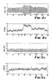
- FIG. 4A illustrates the change in pulsatile perfusion for a healthy subject's leg when raised to 30 degrees.
- the subject was a 35 year old female.
- the Fig plots a trace of pulsatile perfusion i.e. I AC /I DC on the Y-axis against time in seconds on the X-axis.
- the plot of FIG. 4A may be displayed contemporaneously on the display 58 .
- FIG. 4B illustrates the envelope of the trace in FIG. 4A .
- the plot of FIG. 4B may be displayed contemporaneously on the display 58 .
- the ratio R for this healthy subject is in the region of 2. This indicates increased perfusion when the leg is elevated.
- FIG. 5A illustrates the change in pulsatile perfusion for an at-risk subject's leg when raised to 30 degrees.
- the subject was a 79 year old diabetic male.
- the Fig plots a trace of pulsatile perfusion i.e. I AC /I DC on the Y-axis against time in seconds on the X-axis.
- the plot of FIG. 5A may be displayed contemporaneously on the display 58 .
- FIG. 5B illustrates the envelope of the trace in FIG. 5A .
- the plot of FIG. 5B may be displayed contemporaneously on the display 58 .
- the ratio R for this at-risk subject is in the region of 1. This indicates no change in perfusion when the leg is elevated.
- the value of R indicates that the subject is at-risk of developing circulatory complications in the foot in extreme at-risk cases, the value of R may be less than 1.
- a clinically determined threshold value T may be determined, such that when R for a particular subject is below the threshold, they are deemed at risk. This may be detected and displayed on the display 58 .
- the extent of risk may also be calibrated against the difference between the threshold and calculated value of R and a quantitative or qualitative indication of the extent of risk may be displayed on the display 58 .
- the process of objective assessment of blood perfusion in a lower limb 12 of a subject involves the following steps.
- the subject rests in a supine position and an optically diffusive skin (ODS) 16 is placed over at least the subject's dorsum.
- ODS optically diffusive skin
- the system 10 is then attached to the lower limb 12 using a strap.
- the approximate height of the subject is entered into the control unit 5 using the keyboard 57 .
- the control unit 5 via the inclinometer 14 registers the base position (i.e. horizontal) and gives green light to proceed via display 58 .
- the lower limb 12 of the subject is slowly raised and then held for a few seconds.
- the perfusion indicators are calculated and displayed.
- the PPG sensor 4 is illustrated in FIG. 2 as a non-contact sensor, in alternative embodiments the sensor 4 may be attached to the lower limb, for example, by using an elasticated strap to hold the sensor 4 and control unit 5 in position during the test.
- the skin 16 also serves as a disposable hygiene barrier, reducing contamination of the sensors and cross infection between subjects.
- the system 10 illustrated in FIGS. 2 and 3 may be simply adapted to determined a second perfusion indicator dependent upon the blanching of the skin tone of the lower limb 12 when it is elevated.
- This second perfusion indicator may be calculated in addition to or as an alternative to the first perfusion indicator R.
- the foot may be illuminated using IR light and also red light.
- An IR sensor's output may be pre-processed as described with reference to FIG. 3 to produce I dc [IR], I ac [IR].
- a red light sensor's output may be pre-processed as described with reference to FIG. 3 to produce I dc [red], I ac [red].
- embodiments of the invention do not provide a diagnosis but provide an interim clinical indicator that will, for example, help in the assessment of the risks associated with a condition such as diabetes.
- the indicator indicates that something is wrong with the subject's circulation but not necessarily what is specifically wrong or what disease is the cause.
- the limb at which measurement is made as a foot the hand, lower arm or lower leg may also be suitable.
Abstract
A system for assessing blood circulation in a subject's limb, including detection means for detecting a signal dependent upon the arterial blood volume in a limb of the subject when the subject is in a first posture and also when the subject is in a second posture, different to the first posture; and processing means for calculating a quantitative indicator that is dependent upon the ratio of the signal for the first posture to the signal for the second posture.
Description
This is a continuation patent application of co-pending application No. 10/584,179 filed Apr. 17, 2007, which is a National Stage Application of International Application No. PCT/GB2005/000051 filed Jan. 10, 2005, which is hereby incorporated by reference in its entirety.
Embodiments of the invention relate to assessing a subject's peripheral blood circulation, in particular, but not exclusively, arterial blood flow to the foot.
Healthy peripheral circulation is an important factor in quality of life, independent living and personal freedom. Disorders of the vascular system can arise from a number of diseases such as, for example, diabetes, arteriosclerosis, Reynard's syndrome, atherosclerosis.
There are a few clinic/GP based technologies that are used currently to assess peripheral blood circulation. However, these technologies are generally interpretive and must be practised by a correctly trained person.
It would be desirable to provide for the objective assessment of peripheral blood circulation.
According to one aspect of the invention, there is provided a system for assessing blood circulation in a subject's limb, comprising: detection means for detecting a signal dependent upon the arterial blood volume in a limb of the subject when the subject is in a first posture and also when the subject is in a second posture, different to the first posture; and processing means for calculating a quantitative indicator that is dependent upon the ratio of the signal for the first posture to the signal for the second posture.
According to this aspect of the invention, there is provided a method for assessing blood circulation in a subject's limb, comprising: detecting a signal dependent upon the arterial blood volume in a limb of the subject when the subject is in a first posture; detecting the signal dependent upon the arterial blood volume in the limb of the subject when the subject is in a second posture, different to the first posture; and calculating a quantitative indicator that is dependent upon the ratio of the signal for the first posture to the signal for the second posture.
The signal may be a pulsating (arterial) component of a measured parameter, the measured parameter being dependent upon the blood volume in the subject's limb.
The signal may be dependent upon the volume of arterial blood in the limb and other subject specific factors. The ratio of signals at different postures eliminates the subject specific factors and provides an objective indicator.
The measured parameter may, for example, be the intensity of light reflected from the limb. The intensity of the reflected light may be modeled as an attenuation of the intensity of incident light by a first exponential factor and a second exponential factor. The first exponential factor has a first exponent that represents the light absorption by arterial blood of the subject. The second exponential factor has a second exponent that represents the light absorption by other factors including venous blood for the subject. According to this model, rapid variations in the measured parameter arise from the first exponent. The first exponent is very small and the first exponential factor can therefore be represented mathematically as a first order polynomial (i.e. 1+first exponent). A good approximation of the first exponent may be obtained, for example, by taking the ratio of the ac component of the measured parameter to the dc component of the measured parameter. Also according to this model, the first exponent comprises a factor that represents the light absorbency of the subject's arterial blood and a factor that represents the volume of the subject's arterial blood. According to this model, as the light absorbency of a subject's arterial blood remains constant between postural changes, then the ratio of the first exponent for a first posture to the first exponent for a second posture gives a ‘pure’ ratio of arterial volumes without other factors. This ratio can therefore be used as an objective quantitative indicator.
According to another aspect of the Invention, there is provided a system for assessing a subject's peripheral blood circulation, comprising: measurement means for measuring a parameter dependent upon the blood volume in a limb of the subject when the subject is in a first posture and also when the subject is in a second posture, different to the first posture; means for separating the parameter into a first component and a second component; and processing means for calculating a quantitative indicator wherein the calculation takes as inputs the first component of the parameter for the first posture and the first component of the parameter for the second posture.
According to this aspect of the invention, there is provided a method for assessing a subject's peripheral blood circulation, comprising: measuring a parameter dependent upon the blood volume in a limb of the subject when the subject is in a first posture and also when the subject is in a second posture, different to the first posture; separating the parameter into a first component and a second component; and processing means for calculating a quantitative indicator wherein the calculation takes as inputs the first component of the parameter for the first posture and the first component of the parameter for the second posture.
Typically, the first component is a pulsating component and the second component is non-pulsating component. The indicator may be dependent upon the ratio of the first component of the parameter for the first posture to the first component of the parameter for the second posture.
The first component of the parameter may be dependent upon the volume of arterial blood in the limb and other subject specific factors. The use of first components at different postures may be used to eliminate the subject specific factors and provide an objective indicator.
According to another aspect of the invention there is provided a system for assessing a subject's peripheral blood circulation, comprising: measurement means for measuring a parameter dependent upon the blood volume in a limb of the subject when the subject is in a first posture; means for separating the parameter into a first component and a second component; and processing means for calculating a quantitative indicator wherein the calculation takes as inputs the first component of the parameter for the first posture and the second component of the parameter for the first posture.
According to this aspect of the invention there is provided a method for assessing a subject's peripheral blood circulation, comprising: measuring a parameter dependent upon the blood volume in a limb of the subject when the subject is in a first posture; separating the parameter into a first component and a second component; and calculating a quantitative indicator wherein the calculation takes as inputs the first component of the parameter for the first posture and the second component of the parameter for the first posture.
The measured parameter may, for example, be the intensity of light reflected from the limb. The intensity of the reflected light may be modeled as an attenuation of the intensity of incident light by a first exponential factor and a second exponential factor. The first exponential factor has a first exponent that represents the light absorption by arterial blood of the subject. The second exponential factor has a second exponent that represents the light absorption by other factors including venous blood for the subject. According to this model, rapid variations in the measured parameter arise from the first exponent. The first exponent is very small and the first exponential factor can therefore be represented mathematically as a first order polynomial (i.e. 1+first exponent). A good approximation of the first exponent may be obtained by, for example, taking the ratio of the ac component of the measured parameter to the dc component of the measured parameter. Also according to this model, the first exponent comprises a factor that represents the light absorbency of the subject's arterial blood and a factor that represents the volume of the subject's arterial blood.
According to a further aspect of the invention there is provided a system for assessing blood circulation in a subject's limb, for example a foot, comprising: measurement means operable to measure a parameter indicative of the blood volume of the subject's limb when the subject is in a first posture and to measure the parameter when the subject is in a second posture and comprising means for isolating a variable value, of the measured parameter; processing means for determining a quantitative indictor that is dependent upon the ratio of the variable value of the parameter measured for the first posture to the variable value of the parameter measured for the second posture.
According to this further aspect of the invention there is provided a method for assessing blood circulation in a subject's limb, comprising: measuring a parameter indicative of the blood volume of the subject's limb when the subject is in a first posture; isolating a time-variable value of the parameter measured for the first posture; measuring the parameter indicative of the blood volume of the subject's limb when the subject is in a second posture; isolating a time-variable value of the parameter measured for the second posture; and determining a quantitative indictor that is dependent upon the ratio of the variable value of the parameter measured for the first posture to the variable value of the parameter measured for the second posture.
Embodiments of the invention therefore provide a quantitative indicator in a robust and quick manner at modest cost without discomfort to the user. Some embodiments may be automated,
For a better understanding of the present invention reference will now be made by way of example only to the accompanying drawings in which:
The Figures illustrate a system 10 for assessing blood circulation in a subjects limb, comprising: detection means for detecting a signal dependent upon the arterial blood volume in a limb of the subject when the subject is in a first posture and also when the subject is in a second posture, different to the first posture; and processing means 5 for calculating a quantitative indicator that is dependent upon the ratio of the signal for the first posture to the signal for the second posture.
The blood volume in a lower limb includes a variable volume, and a fixed volume. The variable blood volume arises from pulsating blood flow within the arteries of the lower limb and varies with a periodicity in the range 0.5-3 Hz. The fixed volume includes the venous volume of blood and varies, if at all, over a time scale of several seconds.
The blood circulation system is governed in part by forces exerted by gravity. In order that a suitable blood flow is maintained throughout the body, the vascular system can adjust to any local pressure changes resulting from postural changes. Thus the resistance of the peripheral vascular system in the feet is high when a subject is standing compared with when they are supine. Although the relationships between pressure, flow and blood volume are complex, certain patterns can be identified that characterise the response of the circulation system to specific changes (e.g. postural changes). These patterns can be disrupted when a pathology is present such as an arterial blockage or when the vasculature has a reduced capability to respond to changes imposed upon it. This is the principle of response testing which is exploited in this invention.
When a lower limb is raised above the heart, the circulation system will respond to the change in localised blood pressure in a manner characteristic of any pathology. This characteristic change is detected by measuring changes in the blood volume in the limb.
The subject lies flat on their back, while a measurement is taken with the subject's lower limb 12 in a non-elevated position and a measurement is taken with the subject's lower limb 12 in an elevated position. Elevation is relative to the subject's heart reference level 3.
The system 10 comprises a lifting mechanism for raising a subject's leg 2 from the non-elevated position to the elevated position. The leg 2 pivots about the subject's hip 1. The mechanism may be a lifting pulley 7 or alternatively a lifting platform 6.
The system additionally comprises a blood volume sensor 4 and a control unit 5. The sensor 4 includes a support that fits around the lower limb 12, specifically around the ankle and dorsum of the foot. The sensor 4 may include a strain gauge wrapped around the dorsum or alternatively it may include a light source and light sensors to detect blood volume changes.
The control unit 5 receives a first input from the sensor when the subject is in a first posture e.g. the foot has a first zero elevation above the subject's heart and a second input from the sensor when the subject is in a second posture e.g. the foot has a second non-zero elevation above the subject's heart. The control unit 5 processes the first and second inputs to quantify the change in arterial blood volume with postural change. This may be used as a quantitative perfusion indicator that indicates the status of arterial blood circulation in the foot 12.
It may be desirable for the second input to be taken when the subject's foot 12 is at a particular elevation. The lifting mechanism may be calibrated to enable the foot to be manually elevated to the correct height. Alternatively, the lifting mechanism may provide a signal to the control unit 5, which automatically controls the lifting mechanism to stop elevation at a desired height or provides an alert to an operator to stop elevating the foot at the required height.
Instead of using the lifting mechanism to measure the elevation, it is also possible to use the control unit 5 to estimate the elevation. In this case, an electronic inclinometer would be attached to the subject's lower limb with a correct orientation. It may, for example, be integrated into the sensor 4 or control unit 5. The inclinometer provides an incline input θ to the control unit 5 which uses a value of the subject's leg length L to estimate the elevation H of the lower limb 12 using trigonometry (H=L*tan θ). The leg length L may be input into the control unit 5 after direct measurement or may be estimated by the control unit 5 from a value of the subject's height input to the control unit 5. The control unit 4 may additionally either control the rate of change of elevation by controlling the lifting mechanism or may monitor the rate of change of elevation and provide an audio alert if the rate of elevation is too fast or too slow.
In the illustrated embodiment, the control unit 5 forms part of the sensor support 4, but in other embodiments it may be mounted on the sensor support and directly connected to it or it may be positioned remote from the sensor support 4 and indirectly connected to it e.g. using radio transceivers.
The control unit 5 may include a user interface including a user input device such as a keyboard and a user output device such as a display. The display may, for example, display the elevation of the lower limb 12, the equivalent hydrostatic pressure for that elevation, a first perfusion indicator dependent upon the pedal arterial circulation volume in the elevated lower limb 12 and a second perfusion indicator dependent upon the flesh tone (colour) of the elevated lower limb.
The system 10 comprises a photo-plethysmographic (PPG) 4 sensor, an electronic inclinometer (INC) 14, an optically diffusive skin (ODS) 16 and an electronic control unit (ECU) 5 that includes pre-processing circuitry 60 and an analysis unit (ANU) 56, which may be a microprocessor.
The PPG sensor 4 illuminates the dorsum of the foot from a range of a few cm. The PPG sensor 4 uses an array of light emitting diodes to provide a diffuse illumination pattern that extends over a significant fraction of the dorsum (approx. 20 sq cm). The optical receiver is located adjacent to the array of photodiodes or is mounted within the array. Light collection optics (reflectors or lenses) can be used to shape the beam pattern and collection aperture.
The optically diffusive skin (ODS) 16 covers the area of tissue to be illuminated (e.g. dorsum of the foot). The skin may, for example, be made from a polymer material such as latex. The skin 16 functions to reduce in-homogeneity in the optical interaction with the tissue by creating a smooth but diffusive interface between the illuminating light field and the actual skin surface.
The inclinometer 14 is aligned with the shinbone. The inclinometer 14 registers the angle θ at which the leg 2 is inclined. This value is converted to an elevation and hydrostatic pressure change by the electronic control unit 5. The leg length may be entered directly to the control unit 4 via or it may be derived from a look up table based on the subject's height.
The PPG sensor 4 produces an output voltage that is dependent upon the intensity of the light detected by the sensor. This output voltage is provided to the pre-processing circuitry 60 of the electronic control unit 5 as illustrated in FIG. 3 .
The pre-processing circuitry 60 includes an input node 50, a low pass filter 54, a high pass filter 52, a first analogue to digital converter (ADC) 55, a second analogue to digital converter (ADC) 53 and an amplifier 52.
The low pass filter 54 and high pass filter 51 are connected in parallel to the input node 50. The low pass filter 54 is connected in series to the first ADC 55 and the high pass filter is connected in series to the amplifier 52 which is connected to the second ADC.
The low pass filter 54 converts the sensor output 41 into a signal (Idc) that represents the steady-state or slowly varying intensity of the light detected by the PPG sensor 4. This signal is sampled and digitised by the second ADC and then provided to the processor 56.
The high pass filter 51 converts the sensor output 41 into a signal (Iac) that represents the varying intensity of the light detected by the PPG sensor 4. This signal is amplified by amplifier 52 and it is then sampled and digitised by the second ADC and then provided to the processor 56.
The low pass filter passes signals with a frequency less than ˜1 Hz i.e. non-pulsating signals, whereas the high pass filter passes signals with a frequency of greater than ˜1 Hz i.e. pulsating signals. The high pass signal Iac is therefore representative of the change of intensity caused by arterial pulses in the lower limb 12 of the subject. These cut-off frequencies may have to adapt, in practice, to individual heart rates.
The processor 56 processes the digitised signals Idc and Iac. It calculates a ratio of ratios R,
where
- I(θ)ac represents the varying intensity of the light detected by the PPG sensor 4 when the subject is in a first posture i.e. the lower limb is elevated with an incline of θ.
- I(θ)dc represents the steady state intensity of the light detected by the PPG sensor 4 d when the subject is in a first posture i.e. the lower limb is elevated with an incline of θ.
- I(0)ac represents the varying intensity of the light detected by the PPG sensor 4 when the subject is in a second posture i.e. the lower limb is elevated with an incline of 0 (zero).
- I(0)dc represents the steady state intensity of the light detected by the PPG sensor 4 when the subject is in a second posture i.e. the lower limb is elevated with an incline of 0 (zero)
- R will be unity if the blood volume pulsations remain unchanged as a proportion of the total blood volume when the limb is elevated. The ratio R can be used to categorise the postural response of the circulation system in the lower limb and foot.
For example, it might be the case that as the lower limb 12 is raised in a healthy arterial system Iac remains approximately constant but changes significantly for an unhealthy arterial system.
The value R represents a first perfusion indicator and it may be displayed on display 58.
The ratio R may be calculated from the ratio of pulsatile perfusion (I(θ)ac/I(θ)dc) when t=60 and pulsatile perfusion (I(0)ac/I(0)dc) when t=45. That is the ratio of the pulsatile perfusion soon after, but not immediately after, the leg has been raised to the pulsatile perfusion just before the leg has been raised. The ratio R for this healthy subject is in the region of 2. This indicates increased perfusion when the leg is elevated.
The ratio R may be calculated from the ratio of pulsatile perfusion (I(θ)ac/(θ)dc) when t=60 and pulsatile perfusion (I(0)ac/I(0)dc) when t=45. That is the ratio of the pulsatile perfusion soon after, but not immediately after, the leg has been raised to the pulsatile perfusion just before the leg has been raised. The ratio R for this at-risk subject is in the region of 1. This indicates no change in perfusion when the leg is elevated.
The value of R indicates that the subject is at-risk of developing circulatory complications in the foot in extreme at-risk cases, the value of R may be less than 1.
A clinically determined threshold value T may be determined, such that when R for a particular subject is below the threshold, they are deemed at risk. This may be detected and displayed on the display 58. The extent of risk may also be calibrated against the difference between the threshold and calculated value of R and a quantitative or qualitative indication of the extent of risk may be displayed on the display 58.
The total output I of the PPG sensor 4 can be represented as Idc+Iac. The value of I is governed to a major extent by the blood volume illuminated and the skin absorption. The effect of skin absorption is removed by the ratio of ratios R. Because R is calculated, the actual light intensity used for a particular subject can vary according to what is required to obtain a well-resolved signal. This will be a function of many factors including skin type, thickness, anatomy, probe placement and probe coupling. Adjustment of the light intensity can be performed to optimise the signal acquisition with respect to the dynamic range of measurement.
The process of objective assessment of blood perfusion in a lower limb 12 of a subject involves the following steps. The subject rests in a supine position and an optically diffusive skin (ODS) 16 is placed over at least the subject's dorsum. The system 10 is then attached to the lower limb 12 using a strap. The approximate height of the subject is entered into the control unit 5 using the keyboard 57. The control unit 5 via the inclinometer 14 registers the base position (i.e. horizontal) and gives green light to proceed via display 58. The lower limb 12 of the subject is slowly raised and then held for a few seconds. The perfusion indicators are calculated and displayed.
Although the PPG sensor 4 is illustrated in FIG. 2 as a non-contact sensor, in alternative embodiments the sensor 4 may be attached to the lower limb, for example, by using an elasticated strap to hold the sensor 4 and control unit 5 in position during the test. The skin 16 also serves as a disposable hygiene barrier, reducing contamination of the sensors and cross infection between subjects.
The system 10 illustrated in FIGS. 2 and 3 , may be simply adapted to determined a second perfusion indicator dependent upon the blanching of the skin tone of the lower limb 12 when it is elevated. This second perfusion indicator may be calculated in addition to or as an alternative to the first perfusion indicator R.
For example, a discrete spectrometer can be used to analyse the light reflected from the lower limb 12, when it is in the non-elevated position and when it is in the elevated position. The spectrometer can give a quantitative value for the blanching that occurs on elevating the limb.
As another example, the foot may be illuminated using IR light and also red light. An IR sensor's output may be pre-processed as described with reference to FIG. 3 to produce Idc[IR], Iac[IR]. A red light sensor's output may be pre-processed as described with reference to FIG. 3 to produce Idc[red], Iac[red]. The processor 56 may calculate a ratio R′ac or R′ac
R′ dc=(I dc[red](θ)/(I dc [IR](θ)+I dc[red](θ)))/(I dc[red](0)/(I dc [IR](0)+I dc[red](0)))
R′ ac=(I ac[red](θ)/(I ac [IR](θ)+I ac[red](θ)))/(I ac[red](0)/(I ac [IR](0)+I ac[red](0)))
R′ dc=(I dc[red](θ)/(I dc [IR](θ)+I dc[red](θ)))/(I dc[red](0)/(I dc [IR](0)+I dc[red](0)))
R′ ac=(I ac[red](θ)/(I ac [IR](θ)+I ac[red](θ)))/(I ac[red](0)/(I ac [IR](0)+I ac[red](0)))
It should be appreciated that embodiments of the invention do not provide a diagnosis but provide an interim clinical indicator that will, for example, help in the assessment of the risks associated with a condition such as diabetes. The indicator indicates that something is wrong with the subject's circulation but not necessarily what is specifically wrong or what disease is the cause.
The previously described embodiments relate to a change in posture in which the limb at which measurement is made is elevated above the level of the subject's heart. However, more general postural changes are possible. For example, the position of the limb at which measurement is made may be simply changed between the first and second postures. Such a change in position may be by raising the limb or lowering the limb and the limb need not be elevated above the heart level of the subject. Also, a postural change may arise from changing the position of a part of the subject's body at which measurement is not made. For example, the subject may change posture by bending from the torso while the limb at which measurement is made remains stationary.
Consequently a more general expression of the ratio of ratios R calculated by the processor 56 from the digitised signals Idc and Iac is:
where
- I(1)ac represents the varying intensity of the light detected by the PPG sensor 4 when the subject is in a first posture
- I(1)dc represents the steady state intensity of the light detected by the PPG sensor 4 when the subject is in the first posture
- I(2)ac represents the varying intensity of the light detected by the PPG sensor 4 when the subject is in a second posture
- I(2)dc represents the steady state intensity of the light detected by the PPG sensor 4 when the subject is in a second posture
Although the preceding embodiments describe the limb at which measurement is made as a foot, the hand, lower arm or lower leg may also be suitable.
Although embodiments of the present invention have been described in the preceding paragraphs with reference to various examples, it should be appreciated that modifications to the examples given can be made without departing from the scope of the invention as claimed.
Whilst endeavoring in the foregoing specification to draw attention to those features of the invention believed to be of particular importance it should be understood that the Applicant claims protection in respect of any patentable feature or combination of features hereinbefore referred to and/or shown in the drawings whether or not particular emphasis has been placed thereon.
Claims (12)
1. A method of assessing a subject by producing a subject-dependent indicator that measures a response of a subject's blood circulation system to postural change, comprising:
providing a signal for a first posture dependent upon a blood volume in a limb of the subject when the subject is in a first posture by measuring a parameter indicative of the blood volume of the subject's limb when the subject is in the first posture prior to a postural change;
providing a signal for a second posture dependent upon a blood volume in a limb of a subject after a change in subject posture from the first posture to a second posture, different to the first posture by measuring the parameter when the subject is in a second posture after the postural change;
isolating at least a pulsating component of the measured parameter; and
using a processor to calculate a quantitative subject-dependent indicator that measures a response of the subject's blood circulation system to the postural change and is dependent upon at least the pulsating component of the measured parameter for the first posture and the pulsating component of the measured parameter for the second posture.
2. A method as claimed in claim 1 , further comprising:
isolating a non-pulsating component of the measured parameter, wherein the quantitative indicator is dependent upon the non-pulsating component of the measured parameter of the signal for the second posture and the non-pulsating component of the measured parameter of the signal for the first posture.
3. A method as claimed in claim 2 , wherein the limb is a foot.
4. A method as claimed in claim 1 , wherein the position of the limb is changed between the first posture and the second posture.
5. A method as claimed in claim 1 wherein, in the first posture the limb is at a first elevation relative to the heart and in the second posture the limb is at a second different elevation relative to the heart.
6. A method as claimed in claim 1 , wherein providing a signal for a second posture dependent upon a blood volume in a limb of a subject after a change in subject posture from the first posture to a second posture, different to the first posture comprises one of:
providing a signal dependent upon a blood volume in the limb of the subject during an immediate response of the subject's blood circulation to the posture change;
providing a non-steady state signal dependent upon a non-steady state blood volume in the limb of the subject after the posture change; and
providing a signal dependent upon a blood volume in the limb of the subject less than 30 seconds after the posture change.
7. A method as claimed in claim 1 , further comprising using the quantitative indicator to assess risk associated with a subject-dependent disease affecting the vascular system selected from the group comprising: diabetes, arteriosclerosis, Reynard's syndrome, and atherosclerosis.
8. A system that is configured to produce an indicator that measures a response of a subject's blood circulation to postural change, comprising:
pre-processing circuitry configured to detect a signal for a first posture dependent upon a blood volume in a limb of the subject when the subject is in a first posture and is configured to detect the signal for a second posture dependent upon the blood volume in the limb of the subject after a change in the subject's posture from the first posture to a second posture, different to the first posture; and
processing circuitry configured to calculate a quantitative indicator that quantifies a response of the subject's blood circulation to postural change and is dependent upon the signal for the first posture and the signal for the second posture, wherein the processing circuitry is configured to determine automatically when a posture change occurs.
9. A system as claimed in claim 8 , further comprising a detector configured to detect postural change and provide a signal to the processing circuitry indicative of postural change.
10. A method of assessing a subject by producing a subject-dependent indicator that measures a response of a subject's blood circulation system to postural change, comprising:
providing a signal for a first posture dependent upon a blood volume in a limb of the subject when the subject is in a first posture;
providing a signal for a second posture dependent upon a blood volume in a limb of a subject after a change in subject posture from the first posture to a second posture, different to the first posture; and
using a processor to calculate a quantitative subject-dependent indicator that measures a response of the subject's blood circulation system to the postural change and is dependent upon the signal for the first posture and the signal for the second posture, the method further comprising: determining automatically when a posture change occurs.
11. A method of assessing a subject by producing a subject-dependent indicator that measures a response of a subject's blood circulation system to postural change, comprising:
measuring a parameter indicative of the blood volume of the subject's limb when the subject is in a first posture prior to the postural change;
measuring the parameter when the subject is in a second posture after the postural change; and
separating the parameter into a first component and a second component;
using a processor to calculate a quantitative subject-dependent indicator that measures a response of the subject's blood circulation system to the postural change,
wherein the calculation takes as a first input value the first component of the parameter for the first posture, measured prior to the postural change, takes as a second input value, the first component of the measured parameter for the second posture, measured after the postural change takes as a third input value the second component of the parameter for the first posture, measured prior to the postural change, and that takes as a fourth input value, the second component of the measured parameter for the second posture, measured after the postural change.
12. A method as claimed in claim 11 , comprising
measuring a further parameter indicative of the blood volume of the subject's limb when the subject is in a first posture prior to the postural change;
measuring the further parameter when the subject is in a second posture after the postural change;
separating the further parameter into a first component and a second component;
wherein the calculation that takes as input values the first and second components of the measured parameter for the first posture, the first and second components of the measured parameter for the second posture, measured after the postural change; the first and second components of the further parameter for the first posture, and the first and second components of the measured further parameter for the second posture, measured after the postural change, wherein the parameter and further parameter are different optical parameters associated with different wavelengths of light.
Priority Applications (1)
| Application Number | Priority Date | Filing Date | Title |
|---|---|---|---|
| US14/049,366 US9730590B2 (en) | 2004-01-08 | 2013-10-09 | System or method for assessing a subject's peripheral blood circulation |
Applications Claiming Priority (5)
| Application Number | Priority Date | Filing Date | Title |
|---|---|---|---|
| GB0400281A GB2413078C (en) | 2004-01-08 | 2004-01-08 | A system or method for assessing a subject's pedalblood circulation. |
| GB0400281.2 | 2004-01-08 | ||
| PCT/GB2005/000051 WO2005065533A1 (en) | 2004-01-08 | 2005-01-10 | A system or method for assessing a subject’s peripheral blood circulation |
| US10/584,179 US8585602B2 (en) | 2004-01-08 | 2005-01-10 | System or method for assessing a subject's peripheral blood circulation |
| US14/049,366 US9730590B2 (en) | 2004-01-08 | 2013-10-09 | System or method for assessing a subject's peripheral blood circulation |
Related Parent Applications (2)
| Application Number | Title | Priority Date | Filing Date |
|---|---|---|---|
| US10/584,179 Continuation US8585602B2 (en) | 2004-01-08 | 2005-01-10 | System or method for assessing a subject's peripheral blood circulation |
| PCT/GB2005/000051 Continuation WO2005065533A1 (en) | 2004-01-08 | 2005-01-10 | A system or method for assessing a subject’s peripheral blood circulation |
Publications (2)
| Publication Number | Publication Date |
|---|---|
| US20140039326A1 US20140039326A1 (en) | 2014-02-06 |
| US9730590B2 true US9730590B2 (en) | 2017-08-15 |
Family
ID=31503543
Family Applications (2)
| Application Number | Title | Priority Date | Filing Date |
|---|---|---|---|
| US10/584,179 Expired - Fee Related US8585602B2 (en) | 2004-01-08 | 2005-01-10 | System or method for assessing a subject's peripheral blood circulation |
| US14/049,366 Expired - Fee Related US9730590B2 (en) | 2004-01-08 | 2013-10-09 | System or method for assessing a subject's peripheral blood circulation |
Family Applications Before (1)
| Application Number | Title | Priority Date | Filing Date |
|---|---|---|---|
| US10/584,179 Expired - Fee Related US8585602B2 (en) | 2004-01-08 | 2005-01-10 | System or method for assessing a subject's peripheral blood circulation |
Country Status (17)
| Country | Link |
|---|---|
| US (2) | US8585602B2 (en) |
| EP (3) | EP1694203B1 (en) |
| JP (1) | JP5057783B2 (en) |
| CN (2) | CN101732045A (en) |
| AT (1) | ATE440538T1 (en) |
| AU (2) | AU2005203912B2 (en) |
| BR (1) | BRPI0506752A (en) |
| CA (1) | CA2552924C (en) |
| CY (1) | CY1109638T1 (en) |
| DE (1) | DE602005016212D1 (en) |
| DK (1) | DK1694203T3 (en) |
| ES (1) | ES2331617T3 (en) |
| GB (1) | GB2413078C (en) |
| PL (1) | PL1694203T3 (en) |
| PT (1) | PT1694203E (en) |
| SI (1) | SI1694203T1 (en) |
| WO (1) | WO2005065533A1 (en) |
Families Citing this family (30)
| Publication number | Priority date | Publication date | Assignee | Title |
|---|---|---|---|---|
| CN2802441Y (en) * | 2005-06-17 | 2006-08-02 | 南京德朔实业有限公司 | Portable inclination measuring device |
| GB0603006D0 (en) * | 2006-02-15 | 2006-03-29 | Dialog Devices Ltd | Assessing blood supply to a peripheral portion of an animal |
| KR100818669B1 (en) * | 2007-03-09 | 2008-04-02 | 한국과학기술원 | Apparatus for measuring the perfusion rate of legs |
| GB2465230B (en) * | 2008-11-17 | 2013-08-21 | Dialog Devices Ltd | Assessing a subject's circulatory system |
| US8057400B2 (en) * | 2009-05-12 | 2011-11-15 | Angiologix, Inc. | System and method of measuring changes in arterial volume of a limb segment |
| US9066660B2 (en) | 2009-09-29 | 2015-06-30 | Nellcor Puritan Bennett Ireland | Systems and methods for high-pass filtering a photoplethysmograph signal |
| JP2012152493A (en) * | 2011-01-28 | 2012-08-16 | Seiko Epson Corp | Pulse wave signal measuring device and program |
| US10206802B2 (en) | 2011-03-07 | 2019-02-19 | Theranova, Llc | Wearable apparatus for the treatment or prevention of osteopenia and osteoporosis, stimulating bone growth, preserving or improving bone mineral density, and inhibiting adipogenesis |
| US9655555B2 (en) * | 2011-03-07 | 2017-05-23 | Potrero Medical, Inc. | Sensing foley catheter |
| US20140316292A1 (en) * | 2013-04-19 | 2014-10-23 | Semler Scientific, Inc. | Circulation Monitoring System |
| MX2016001866A (en) * | 2013-08-14 | 2016-06-14 | Univ Nanyang Tech | Systems and methods for revascularization assessment. |
| US10420490B2 (en) * | 2013-09-06 | 2019-09-24 | Xhale Assurance, Inc. | Systems and methods for physiological monitoring using multiple signal processing devices |
| US10039455B2 (en) | 2014-05-19 | 2018-08-07 | Qualcomm Incorporated | Continuous calibration of a blood pressure measurement device |
| WO2016073945A1 (en) | 2014-11-07 | 2016-05-12 | Respirix, Inc. | Devices and methods for monitoring physiologic parameters |
| EP3219252A4 (en) * | 2014-11-10 | 2018-06-27 | Terumo Kabushiki Kaisha | Skin perfusion pressure measurement device |
| CN107205673A (en) | 2015-01-30 | 2017-09-26 | 皇家飞利浦有限公司 | Photo-plethysmographic device |
| US10264984B2 (en) | 2015-08-28 | 2019-04-23 | Maged Choucair | Non-invasive cardiovascular monitoring device |
| JP6854612B2 (en) | 2015-10-06 | 2021-04-07 | 三星電子株式会社Samsung Electronics Co.,Ltd. | Biological information measuring device, biometric information measuring method, and computer-readable recording medium |
| US9786148B2 (en) | 2016-01-21 | 2017-10-10 | Plethy, Inc. | Devices, systems, and methods for health monitoring using circumferential changes of a body portion |
| EP3386388A4 (en) | 2016-01-21 | 2019-08-14 | Plethy, Inc. | Devices, systems and methods for health monitoring using circumferential changes of a body portion |
| WO2018003221A1 (en) * | 2016-07-01 | 2018-01-04 | ソニー株式会社 | Information processing device, information processing method, and program |
| WO2019075185A1 (en) | 2017-10-11 | 2019-04-18 | Plethy, Inc. | Devices, systems, and methods for adaptive health monitoring using behavioral, psychological, and physiological changes of a body portion |
| GB201813125D0 (en) * | 2018-08-10 | 2018-09-26 | Univ Oxford Innovation Ltd | Apparatus and method for determining an indication of blood flow |
| CN109394200B (en) * | 2018-12-16 | 2021-10-19 | 冯兴怀 | Microcirculation pulse blood flow monitoring system and method for liquid treatment and volume management |
| US11324422B2 (en) | 2018-12-21 | 2022-05-10 | Starkey Laboratories, Inc. | Fall detection using photoplethysmography detectors in ear-wearable devices |
| CN109846464A (en) * | 2018-12-29 | 2019-06-07 | 曹乃钊 | The system for non-invasive measurement and data processing system of blood flow mechanics parameter |
| RU195388U1 (en) * | 2019-05-24 | 2020-01-24 | Федеральное государственное бюджетное учреждение науки Государственный научный центр Российской Федерации - Институт медико-биологических проблем Российской академии наук (ГНЦ РФ - ИМБП РАН) | Sensor mounting device for microcirculation research in the fingers of human limbs |
| US10842415B1 (en) | 2019-10-25 | 2020-11-24 | Plethy, Inc. | Devices, systems, and methods for monitoring and assessing gait, stability, and/or balance of a user |
| KR102348184B1 (en) * | 2019-12-16 | 2022-01-05 | 서울대학교 산학협력단 | Blood oxygen saturation sensing device for non-pulsatile extracorporeal blood circulation circuit and a control method of the same |
| EP4074251A1 (en) * | 2021-04-12 | 2022-10-19 | Koninklijke Philips N.V. | Tissue perfusion analysis |
Citations (46)
| Publication number | Priority date | Publication date | Assignee | Title |
|---|---|---|---|---|
| GB1185443A (en) | 1967-03-30 | 1970-03-25 | Jarnhs Elek Ska Aktiebolag | Apparatus for Quantitative Indicating of Small and Rapid Volume Changes in a Part of an Extremity. |
| US4036215A (en) | 1975-02-06 | 1977-07-19 | Doll Research, Inc. | Apparatus and method for eliminating perturbations of a kinetic origin in the blood flow waveform |
| US4569355A (en) | 1982-05-28 | 1986-02-11 | Hemodynamics Technology, Inc. | Method and apparatus for monitoring and diagnosing peripheral blood flow |
| EP0282210A1 (en) | 1987-03-03 | 1988-09-14 | Hitoshi Fujii | Apparatus for monitoring a bloodstream |
| EP0293504A1 (en) | 1987-06-03 | 1988-12-07 | Hewlett-Packard GmbH | Method for determining the perfusion |
| US4836212A (en) | 1986-03-18 | 1989-06-06 | Fa Nattermann Arneimittel Gmbh | Apparatus for the noninvasive determination and acoustical representation of the dynamic behavior of peripheral venous hemodynamic |
| DE3744538A1 (en) | 1987-12-30 | 1989-07-13 | Nattermann A & Cie | DEVICE FOR NON-INVASIVE MEASUREMENT OF THE BLOOD VOLUME IN HUMAN EXTREMITIES |
| EP0335356A2 (en) | 1988-03-30 | 1989-10-04 | Nellcor Incorporated | Method and apparatus for compensating for distortion in a pulse oximetry device |
| US5040540A (en) | 1988-08-24 | 1991-08-20 | Nims, Inc. | Method and apparatus for non-invasive monitoring of central venous pressure, and improved transducer therefor |
| EP0481612A1 (en) | 1990-10-19 | 1992-04-22 | Nellcor Incorporated | Adhesive pulse oximeter sensor with reusable portion |
| US5137023A (en) | 1990-04-19 | 1992-08-11 | Worcester Polytechnic Institute | Method and apparatus for monitoring blood analytes noninvasively by pulsatile photoplethysmography |
| JPH04259447A (en) | 1991-02-13 | 1992-09-16 | Fukuda Denshi Co Ltd | Method and stand for measuring blood pressure |
| US5237994A (en) | 1991-03-12 | 1993-08-24 | Square One Technology | Integrated lead frame pulse oximetry sensor |
| EP0572684A1 (en) | 1992-05-15 | 1993-12-08 | Hewlett-Packard GmbH | Medical sensor |
| US5291895A (en) | 1985-06-03 | 1994-03-08 | Mcintyre Kevin M | Evaluation of heart mechanical performance |
| US5361769A (en) | 1991-08-22 | 1994-11-08 | Gert Nilsson | Method and a system for measuring fluid flow movements by a laser-doppler technique |
| US5365924A (en) | 1992-07-31 | 1994-11-22 | Frederick Erdman Association | Method and apparatus for non-invasive cardiovascular diagnosis |
| WO1995002358A1 (en) | 1993-07-14 | 1995-01-26 | Masimo Corporation | Finger cot oximetric probe |
| US5437275A (en) | 1994-02-02 | 1995-08-01 | Biochem International Inc. | Pulse oximetry sensor |
| US5542421A (en) | 1992-07-31 | 1996-08-06 | Frederick Erdman Association | Method and apparatus for cardiovascular diagnosis |
| US5638593A (en) | 1993-09-07 | 1997-06-17 | Ohmeda Inc. | Method for manufacturing a heat-sealed neo-natal medical monitoring probe |
| US5730136A (en) | 1995-03-14 | 1998-03-24 | Vnus Medical Technologies, Inc. | Venous pump efficiency test system and method |
| WO1998017174A1 (en) | 1996-10-23 | 1998-04-30 | Cardiac Crc Nominees Pty. Limited | Non-invasive determination of oxygen saturation in blood in deep tissue |
| US5778879A (en) | 1995-02-16 | 1998-07-14 | Omron Corporation | Electronic blood pressure meter with posture detector |
| DE19707442A1 (en) | 1997-02-25 | 1998-08-27 | Medis Medizinische Mestechnik | Hydrostatic determination of peripheral arterial blood pressure, preferably of foot |
| DE19741982A1 (en) | 1996-09-23 | 1998-10-08 | Blazek Vladimir Prof Dr Ing | System for local noninvasive functional indicating of dermal blood perfusion |
| US5842982A (en) | 1996-08-07 | 1998-12-01 | Nellcor Puritan Bennett Incorporated | Infant neonatal pulse oximeter sensor |
| US5991654A (en) | 1997-06-06 | 1999-11-23 | Kci New Technologies, Inc. | Apparatus and method for detecting deep vein thrombosis |
| US5991648A (en) | 1998-03-30 | 1999-11-23 | Palco Labs, Inc. | Adjustable pulse oximetry sensor for pediatric use |
| US6047201A (en) | 1998-04-02 | 2000-04-04 | Jackson, Iii; William H. | Infant blood oxygen monitor and SIDS warning device |
| WO2000021433A1 (en) | 1998-10-15 | 2000-04-20 | Sensidyne | Reusable pulse oximeter probe and disposable bandage apparatus |
| WO2000074562A1 (en) | 1999-06-03 | 2000-12-14 | Hutchinson Technology Incorporated | Disposable tissue probe tip |
| WO2001024845A2 (en) | 1999-10-07 | 2001-04-12 | Mills Alexander K | Method and apparatus for non-invasive continuous determination of physiological parameters of a patient's blood |
| WO2001041634A2 (en) | 1999-12-09 | 2001-06-14 | Masimo Corporation | Resposable pulse oximetry sensor |
| US20010037058A1 (en) | 2000-04-28 | 2001-11-01 | Stone Robert T. | Method for determining blood constituents |
| US6334065B1 (en) | 1998-06-03 | 2001-12-25 | Masimo Corporation | Stereo pulse oximeter |
| WO2002028274A1 (en) | 2000-10-05 | 2002-04-11 | Cybro Medical Ltd. | A pulse oximeter and a method of its operation |
| US6470199B1 (en) | 2000-06-21 | 2002-10-22 | Masimo Corporation | Elastic sock for positioning an optical probe |
| US20020161291A1 (en) * | 1999-01-25 | 2002-10-31 | Kianl Massi E. | Pulse oximeter user interface |
| US6589171B2 (en) | 2001-06-27 | 2003-07-08 | Eastman Kodak Company | Sensor glove for physiological parameter measurement |
| US20030163033A1 (en) * | 2002-02-22 | 2003-08-28 | Dekker Andreas Lubbertus Aloysius Johannes | Apparatus and method for monitoring respiration with a pulse oximeter |
| WO2003080152A2 (en) | 2002-03-21 | 2003-10-02 | Datex-Ohmeda, Inc. | Neonatal bootie wrap |
| WO2003094729A1 (en) | 2002-05-07 | 2003-11-20 | Stefan Nilsson | Method and apparatus for diagnosing venous disease in the lower limbs |
| GB2394178A (en) | 2002-10-09 | 2004-04-21 | Pulse Time Products Ltd | Method of calibrating a blood pressure monitoring apparatus |
| WO2004041079A1 (en) | 2002-11-06 | 2004-05-21 | Itamar Medical Ltd. | Detecting medical conditions with noninvasive body probes |
| GB2402472A (en) | 2003-06-03 | 2004-12-08 | Orsense Ltd | Non-invasive optical measurement of blood parameters |
Family Cites Families (4)
| Publication number | Priority date | Publication date | Assignee | Title |
|---|---|---|---|---|
| DE3827501A1 (en) * | 1988-08-12 | 1990-04-12 | Schmitt Hans J Prof | DEVICE FOR OPTOELECTRONIC NON-INVASIVE DETECTION OF THE FLOW PARAMETERS IN HUMAN EXTREMITIES |
| JP3143190B2 (en) * | 1992-01-28 | 2001-03-07 | 日本コーリン株式会社 | Autonomic ataxia measuring device |
| JP2001070251A (en) * | 1999-09-03 | 2001-03-21 | Kao Corp | Colorimetric probe |
| CN1646055A (en) * | 2002-02-22 | 2005-07-27 | 德特克斯-奥米达公司 | Monitoring physiological parameters based on variations in a photoplethysmographic signal |
-
2004
- 2004-01-08 GB GB0400281A patent/GB2413078C/en not_active Expired - Fee Related
-
2005
- 2005-01-10 EP EP05701821A patent/EP1694203B1/en not_active Not-in-force
- 2005-01-10 PL PL05701821T patent/PL1694203T3/en unknown
- 2005-01-10 DE DE602005016212T patent/DE602005016212D1/en active Active
- 2005-01-10 PT PT05701821T patent/PT1694203E/en unknown
- 2005-01-10 WO PCT/GB2005/000051 patent/WO2005065533A1/en active Application Filing
- 2005-01-10 SI SI200530836T patent/SI1694203T1/en unknown
- 2005-01-10 AU AU2005203912A patent/AU2005203912B2/en not_active Ceased
- 2005-01-10 EP EP09166048.0A patent/EP2108309B1/en active Active
- 2005-01-10 CN CN200910220841A patent/CN101732045A/en active Pending
- 2005-01-10 CA CA2552924A patent/CA2552924C/en not_active Expired - Fee Related
- 2005-01-10 CN CN200580002144A patent/CN100577090C/en not_active Expired - Fee Related
- 2005-01-10 ES ES05701821T patent/ES2331617T3/en active Active
- 2005-01-10 EP EP09166058.9A patent/EP2108310B1/en active Active
- 2005-01-10 DK DK05701821T patent/DK1694203T3/en active
- 2005-01-10 BR BRPI0506752-9A patent/BRPI0506752A/en not_active Application Discontinuation
- 2005-01-10 AT AT05701821T patent/ATE440538T1/en active
- 2005-01-10 US US10/584,179 patent/US8585602B2/en not_active Expired - Fee Related
- 2005-01-10 JP JP2006548387A patent/JP5057783B2/en not_active Expired - Fee Related
-
2009
- 2009-11-25 CY CY20091101215T patent/CY1109638T1/en unknown
-
2011
- 2011-03-08 AU AU2011201021A patent/AU2011201021B2/en not_active Ceased
-
2013
- 2013-10-09 US US14/049,366 patent/US9730590B2/en not_active Expired - Fee Related
Patent Citations (51)
| Publication number | Priority date | Publication date | Assignee | Title |
|---|---|---|---|---|
| GB1185443A (en) | 1967-03-30 | 1970-03-25 | Jarnhs Elek Ska Aktiebolag | Apparatus for Quantitative Indicating of Small and Rapid Volume Changes in a Part of an Extremity. |
| US4036215A (en) | 1975-02-06 | 1977-07-19 | Doll Research, Inc. | Apparatus and method for eliminating perturbations of a kinetic origin in the blood flow waveform |
| US4569355A (en) | 1982-05-28 | 1986-02-11 | Hemodynamics Technology, Inc. | Method and apparatus for monitoring and diagnosing peripheral blood flow |
| US5291895A (en) | 1985-06-03 | 1994-03-08 | Mcintyre Kevin M | Evaluation of heart mechanical performance |
| US4836212A (en) | 1986-03-18 | 1989-06-06 | Fa Nattermann Arneimittel Gmbh | Apparatus for the noninvasive determination and acoustical representation of the dynamic behavior of peripheral venous hemodynamic |
| EP0282210A1 (en) | 1987-03-03 | 1988-09-14 | Hitoshi Fujii | Apparatus for monitoring a bloodstream |
| EP0293504A1 (en) | 1987-06-03 | 1988-12-07 | Hewlett-Packard GmbH | Method for determining the perfusion |
| DE3744538A1 (en) | 1987-12-30 | 1989-07-13 | Nattermann A & Cie | DEVICE FOR NON-INVASIVE MEASUREMENT OF THE BLOOD VOLUME IN HUMAN EXTREMITIES |
| EP0335356A2 (en) | 1988-03-30 | 1989-10-04 | Nellcor Incorporated | Method and apparatus for compensating for distortion in a pulse oximetry device |
| US5040540A (en) | 1988-08-24 | 1991-08-20 | Nims, Inc. | Method and apparatus for non-invasive monitoring of central venous pressure, and improved transducer therefor |
| US5137023A (en) | 1990-04-19 | 1992-08-11 | Worcester Polytechnic Institute | Method and apparatus for monitoring blood analytes noninvasively by pulsatile photoplethysmography |
| EP0481612A1 (en) | 1990-10-19 | 1992-04-22 | Nellcor Incorporated | Adhesive pulse oximeter sensor with reusable portion |
| JPH04259447A (en) | 1991-02-13 | 1992-09-16 | Fukuda Denshi Co Ltd | Method and stand for measuring blood pressure |
| US5237994A (en) | 1991-03-12 | 1993-08-24 | Square One Technology | Integrated lead frame pulse oximetry sensor |
| US5361769A (en) | 1991-08-22 | 1994-11-08 | Gert Nilsson | Method and a system for measuring fluid flow movements by a laser-doppler technique |
| EP0572684A1 (en) | 1992-05-15 | 1993-12-08 | Hewlett-Packard GmbH | Medical sensor |
| US5365924A (en) | 1992-07-31 | 1994-11-22 | Frederick Erdman Association | Method and apparatus for non-invasive cardiovascular diagnosis |
| US5542421A (en) | 1992-07-31 | 1996-08-06 | Frederick Erdman Association | Method and apparatus for cardiovascular diagnosis |
| WO1995002358A1 (en) | 1993-07-14 | 1995-01-26 | Masimo Corporation | Finger cot oximetric probe |
| US5638593A (en) | 1993-09-07 | 1997-06-17 | Ohmeda Inc. | Method for manufacturing a heat-sealed neo-natal medical monitoring probe |
| US5437275A (en) | 1994-02-02 | 1995-08-01 | Biochem International Inc. | Pulse oximetry sensor |
| US5778879A (en) | 1995-02-16 | 1998-07-14 | Omron Corporation | Electronic blood pressure meter with posture detector |
| US5730136A (en) | 1995-03-14 | 1998-03-24 | Vnus Medical Technologies, Inc. | Venous pump efficiency test system and method |
| EP0959757B1 (en) | 1996-08-07 | 2004-05-06 | Nellcor Puritan Bennett Incorporated | Infant/neonatal pulse oximeter sensor |
| US5842982A (en) | 1996-08-07 | 1998-12-01 | Nellcor Puritan Bennett Incorporated | Infant neonatal pulse oximeter sensor |
| DE19741982A1 (en) | 1996-09-23 | 1998-10-08 | Blazek Vladimir Prof Dr Ing | System for local noninvasive functional indicating of dermal blood perfusion |
| WO1998017174A1 (en) | 1996-10-23 | 1998-04-30 | Cardiac Crc Nominees Pty. Limited | Non-invasive determination of oxygen saturation in blood in deep tissue |
| DE19707442A1 (en) | 1997-02-25 | 1998-08-27 | Medis Medizinische Mestechnik | Hydrostatic determination of peripheral arterial blood pressure, preferably of foot |
| US5991654A (en) | 1997-06-06 | 1999-11-23 | Kci New Technologies, Inc. | Apparatus and method for detecting deep vein thrombosis |
| US5991648A (en) | 1998-03-30 | 1999-11-23 | Palco Labs, Inc. | Adjustable pulse oximetry sensor for pediatric use |
| US6047201A (en) | 1998-04-02 | 2000-04-04 | Jackson, Iii; William H. | Infant blood oxygen monitor and SIDS warning device |
| US6334065B1 (en) | 1998-06-03 | 2001-12-25 | Masimo Corporation | Stereo pulse oximeter |
| WO2000021433A1 (en) | 1998-10-15 | 2000-04-20 | Sensidyne | Reusable pulse oximeter probe and disposable bandage apparatus |
| EP1121049A1 (en) | 1998-10-15 | 2001-08-08 | Sensidyne | Reusable pulse oximeter probe and disposable bandage apparatus |
| US20020161291A1 (en) * | 1999-01-25 | 2002-10-31 | Kianl Massi E. | Pulse oximeter user interface |
| WO2000074562A1 (en) | 1999-06-03 | 2000-12-14 | Hutchinson Technology Incorporated | Disposable tissue probe tip |
| EP1233697A2 (en) | 1999-10-07 | 2002-08-28 | Alexander K. Mills | Device and method for noninvasive continuous determination of physiologic characteristics |
| WO2001024845A2 (en) | 1999-10-07 | 2001-04-12 | Mills Alexander K | Method and apparatus for non-invasive continuous determination of physiological parameters of a patient's blood |
| WO2001041634A2 (en) | 1999-12-09 | 2001-06-14 | Masimo Corporation | Resposable pulse oximetry sensor |
| US20010037058A1 (en) | 2000-04-28 | 2001-11-01 | Stone Robert T. | Method for determining blood constituents |
| US6470199B1 (en) | 2000-06-21 | 2002-10-22 | Masimo Corporation | Elastic sock for positioning an optical probe |
| EP1322216A1 (en) | 2000-10-05 | 2003-07-02 | Cybro Medical Ltd. | A pulse oximeter and a method of its operation |
| WO2002028274A1 (en) | 2000-10-05 | 2002-04-11 | Cybro Medical Ltd. | A pulse oximeter and a method of its operation |
| US6589171B2 (en) | 2001-06-27 | 2003-07-08 | Eastman Kodak Company | Sensor glove for physiological parameter measurement |
| US20030163033A1 (en) * | 2002-02-22 | 2003-08-28 | Dekker Andreas Lubbertus Aloysius Johannes | Apparatus and method for monitoring respiration with a pulse oximeter |
| WO2003080152A2 (en) | 2002-03-21 | 2003-10-02 | Datex-Ohmeda, Inc. | Neonatal bootie wrap |
| WO2003094729A1 (en) | 2002-05-07 | 2003-11-20 | Stefan Nilsson | Method and apparatus for diagnosing venous disease in the lower limbs |
| GB2394178A (en) | 2002-10-09 | 2004-04-21 | Pulse Time Products Ltd | Method of calibrating a blood pressure monitoring apparatus |
| WO2004041079A1 (en) | 2002-11-06 | 2004-05-21 | Itamar Medical Ltd. | Detecting medical conditions with noninvasive body probes |
| EP1558134A1 (en) | 2002-11-06 | 2005-08-03 | Itamar Medical Ltd | Detecting medical conditions with noninvasive body probes |
| GB2402472A (en) | 2003-06-03 | 2004-12-08 | Orsense Ltd | Non-invasive optical measurement of blood parameters |
Non-Patent Citations (7)
| Title |
|---|
| Coffman et al: "Intermittent Claudication and rest pain: Physiologic concepts and therapeutic approaches", vol. 22, No. 1, Jul. 1, 1979, XP023061850. |
| COFFMAN, J.D.: "Intermittent claudication and rest pain: Physiologic concepts and therapeutic approaches", PROGRESS IN CARDIOVASCULAR DISEASES., SAUNDERS, PHILADELPHIA, PA., US, vol. 22, no. 1, 1 July 1979 (1979-07-01), US, pages 53 - 72, XP023061850, ISSN: 0033-0620, DOI: 10.1016/0033-0620(79)90003-3 |
| Kim, et al. "Pulse Oximetry and Circulatory Kinetics Associated with Pulse Volume Amplitude Measured by Photoelectric," 1986, Anesth Analg, 65: 1333-1339. |
| Nasimi, et al., "Periodic posture stimulation of the baroreceptors and the local vasomotor reflexes," Journal of Biomedical Engineering, 1994 14(4): 307-312. |
| Rathgeber et al. Influence of different types of recovery positions on perfusion indices of the forearm. Resuscitation. 1996.32: 13-17. * |
| Vegfors, et al. "The influence of changes in blood flow on the accuracy of pulse oximetry in humans." May 1992, Acta Anaesthesiol Scand. 36(4): 346-349. |
| Vegfors, et al. "The influence of changes in blood flow on the accuracy of pulse oximetry in humans." May 1992. Acta Anaesthesiol Scand. 36(4): 346-9. * |
Also Published As
Similar Documents
| Publication | Publication Date | Title |
|---|---|---|
| US9730590B2 (en) | System or method for assessing a subject's peripheral blood circulation | |
| US7976472B2 (en) | Noninvasive hypovolemia monitor | |
| KR101068116B1 (en) | Apparatus and method for sensing radial arterial pulses for noninvasive and continuous measurement of blood pressure | |
| US20060058690A1 (en) | Method and instrument for automated measurement of skin perfusion pressure | |
| US6968221B2 (en) | Low-cost method and apparatus for non-invasively measuring blood glucose levels | |
| Hashem et al. | Design and development of a heart rate measuring device using fingertip | |
| US4819646A (en) | Feedback-controlled method and apparatus for processing signals used in oximetry | |
| KR102033914B1 (en) | method for measuring blood glucose and wearable type apparatus for the same | |
| JP5039123B2 (en) | Finger artery elasticity measurement program, finger artery elasticity measurement device, and finger artery elasticity measurement method | |
| US20150238134A1 (en) | Lymphatic Pressure-Measuring System and Method for Controlling Same | |
| KR20120021098A (en) | Method for evaluting vascular aging using frequency domain analysis of pulse waves | |
| JP6356246B2 (en) | Biological tissue diagnostic apparatus and method of operating biological tissue diagnostic apparatus | |
| EP3777667A1 (en) | A method of detection of the presence of a pathology that impairs blood microcirculation oscillations in a woman and device therefor | |
| Perloff et al. | Edward D. Frohlich, MD; Martha Hill, PhD, RN; Mary McDonald, MSPH, RN; and Bruce Z. Morgenstern, MD, Writing Group |
Legal Events
| Date | Code | Title | Description |
|---|---|---|---|
| STCF | Information on status: patent grant |
Free format text: PATENTED CASE |
|
| CC | Certificate of correction | ||
| FEPP | Fee payment procedure |
Free format text: MAINTENANCE FEE REMINDER MAILED (ORIGINAL EVENT CODE: REM.); ENTITY STATUS OF PATENT OWNER: SMALL ENTITY |
|
| LAPS | Lapse for failure to pay maintenance fees |
Free format text: PATENT EXPIRED FOR FAILURE TO PAY MAINTENANCE FEES (ORIGINAL EVENT CODE: EXP.); ENTITY STATUS OF PATENT OWNER: SMALL ENTITY |
|
| STCH | Information on status: patent discontinuation |
Free format text: PATENT EXPIRED DUE TO NONPAYMENT OF MAINTENANCE FEES UNDER 37 CFR 1.362 |
|
| FP | Lapsed due to failure to pay maintenance fee |
Effective date: 20210815 |