WO1994004563A1 - PEPTIDES CONTAINING RESPECTIVE AMINO ACID SEQUENCES SELECTED FROM AMONG THOSE OF LIPOPROTEIN(a) AND APOLIPOPROTEIN(a), ANTIBODIES RESPECTIVELY RECOGNIZING THESE AMINO ACID SEQUENCES, AND METHOD OF ASSAYING WITH THESE ANTIBODIES - Google Patents
PEPTIDES CONTAINING RESPECTIVE AMINO ACID SEQUENCES SELECTED FROM AMONG THOSE OF LIPOPROTEIN(a) AND APOLIPOPROTEIN(a), ANTIBODIES RESPECTIVELY RECOGNIZING THESE AMINO ACID SEQUENCES, AND METHOD OF ASSAYING WITH THESE ANTIBODIES Download PDFInfo
- Publication number
- WO1994004563A1 WO1994004563A1 PCT/JP1993/001142 JP9301142W WO9404563A1 WO 1994004563 A1 WO1994004563 A1 WO 1994004563A1 JP 9301142 W JP9301142 W JP 9301142W WO 9404563 A1 WO9404563 A1 WO 9404563A1
- Authority
- WO
- WIPO (PCT)
- Prior art keywords
- amino acid
- lipoprotein
- apolipoprotein
- seq
- antibody
- Prior art date
Links
Classifications
-
- C—CHEMISTRY; METALLURGY
- C07—ORGANIC CHEMISTRY
- C07K—PEPTIDES
- C07K16/00—Immunoglobulins [IGs], e.g. monoclonal or polyclonal antibodies
- C07K16/18—Immunoglobulins [IGs], e.g. monoclonal or polyclonal antibodies against material from animals or humans
-
- C—CHEMISTRY; METALLURGY
- C07—ORGANIC CHEMISTRY
- C07K—PEPTIDES
- C07K14/00—Peptides having more than 20 amino acids; Gastrins; Somatostatins; Melanotropins; Derivatives thereof
- C07K14/435—Peptides having more than 20 amino acids; Gastrins; Somatostatins; Melanotropins; Derivatives thereof from animals; from humans
- C07K14/46—Peptides having more than 20 amino acids; Gastrins; Somatostatins; Melanotropins; Derivatives thereof from animals; from humans from vertebrates
- C07K14/47—Peptides having more than 20 amino acids; Gastrins; Somatostatins; Melanotropins; Derivatives thereof from animals; from humans from vertebrates from mammals
-
- C—CHEMISTRY; METALLURGY
- C07—ORGANIC CHEMISTRY
- C07K—PEPTIDES
- C07K14/00—Peptides having more than 20 amino acids; Gastrins; Somatostatins; Melanotropins; Derivatives thereof
- C07K14/435—Peptides having more than 20 amino acids; Gastrins; Somatostatins; Melanotropins; Derivatives thereof from animals; from humans
- C07K14/775—Apolipopeptides
-
- G—PHYSICS
- G01—MEASURING; TESTING
- G01N—INVESTIGATING OR ANALYSING MATERIALS BY DETERMINING THEIR CHEMICAL OR PHYSICAL PROPERTIES
- G01N33/00—Investigating or analysing materials by specific methods not covered by groups G01N1/00 - G01N31/00
- G01N33/48—Biological material, e.g. blood, urine; Haemocytometers
- G01N33/50—Chemical analysis of biological material, e.g. blood, urine; Testing involving biospecific ligand binding methods; Immunological testing
- G01N33/92—Chemical analysis of biological material, e.g. blood, urine; Testing involving biospecific ligand binding methods; Immunological testing involving lipids, e.g. cholesterol, lipoproteins, or their receptors
Definitions
- the present invention relates to a peptide selected from apolipoprotein (a) which is a risk factor for arteriosclerosis and a component of riboprotein (a) which is clinically significant, as a lipoprotein ( a ) or as an apolipoprotein ( a ).
- the present invention relates to antibodies and a method for measuring lipoprotein (a) or apolipoprotein (a) using these antibodies.
- Lipoprotein (a) (lipoprotein (a), L p (a)) was first reported as a variant of i3-lipoprotein by Berg in 1963. L. Ber, Acta Patho l. Mi cr ob iol. i> cand., 59, 369-382 (1963)]. After various studies, it was found that lipoprotein (a) is a low-density lipoprotein (LDL) that normally plays a major role in the transport of cholesterol esters in vivo and apolipoprotein, a protein unique to lipoprotein (a).
- LDL low-density lipoprotein
- Disulfide consists of protein (a) (apo 1 ipoprotein (a), apo (a), apo (a)), and adisulfide between apolipoprotein B-100, which is the protein part of LDL, and apolipoprotein (a) The substance was found to be bound by bonding (see Figure 36).
- apolipoprotein (a) is called kringle 4 of plasminogen It has a maximum of 37 highly homologous parts to the structure, followed by a part that is highly homologous to the plasminogen Kring 5 structure, and a serine protease structural part that has a highly homologous site to plasminogen. I have. Of these 37 kringles, 28 have a completely repeating structure.
- lipoprotein (a) has an isoform (Pheno eve) [G. Utermann eta 1., J. Clin. Invest., 80, 458-465. (1 987), HG Kr afteta 1., Arteriosclerosis, 8., 21 22-2 16 (1 988), G. Ut ermann eta 1., Hum. Genet., 78, 41-46 (1988)], which is thought to be due to the difference in the number of repeats of the kringle 4 equivalent part of the apolipoprotein (a) [VN Trieuetal., J. Biol. Chem., 266, 5480- 5485
- FDS, B, S1, S2, S3, S4, and 0 types were recognized by fractionation using SDS electrophoresis and immunoplotting. [G. Ut Ermann et al., J. Clin. Invest., 80, 458-465 (1987)].
- lipoprotein (a) and apolipoprotein (a) are important keys in considering arteriosclerosis, lipoprotein, and blood coagulation / fibrinolysis system from the same viewpoint.
- lipoprotein (a) is not only a risk factor for atherosclerosis, but is also high in people who have restenosis after glycemic nephropathy or PTCA (percutaneous coronary lumen dilatation). It has been reported that it has a tendency to show lipoprotein (a) and apolipoprotein (a) in the living body such as blood, etc. Has become clinically important, and a method that can accurately measure lipoprotein (a) and apolipoprotein (a) is desired.
- Lipoprotein (a) is measured by simple immunodiffusion, rocket immunoelectrophoresis, immunoturbidimetry, latex nephelometry, radioimmunoassay (RIA) [JJ A 1 berseta 1., J.L. iid R e s., 1 8, 33 1-
- lipoprotein (a) contains LDL in its molecule and has a high homology to brassminogen. As a result, it reacts with the LD-brassminogen contained in the biological sample and also measures LDL and brasminogen, so that accurate lipoprotein (a) measurement values cannot be obtained. Therefore, apolipoprotein (L) was removed from lipoprotein (a).
- polyclonal antibody does not cross-react with LDL
- this antiserum polyclonal antibody
- apolipoprotein (a) has a high homology with plasminogen. Has cross-reactivity.
- this antiserum (polyclonal antibody) cannot be used without a complicated operation of absorbing human plasminogen and removing the antibody reacting with plasminogen.
- apolipoprotein (a) When apolipoprotein (a) is specifically measured, that is, when apolipoprotein (a) in which lipoprotein (a) has disulfide bond broken is measured, apolipoprotein (a) is used.
- a method of applying an antiserum (polyclonal antibody) prepared as an immunogen to the above-described immunoassay can be considered.
- antiserum (polyclonal antibody) prepared using apolipoprotein (a) as an immunogen also reacts with lipoprotein (a), so that in addition to the absorption operation using human plasminogen, the absorption operation using lipoprotein (a) is performed.
- Do lipo It is necessary to remove the antibody that reacts with the protein (a). However, this is complicated and the yield is low, so that it is difficult to obtain a large amount of antiserum (polyclonal antibody).
- the antiserum and the polyclonal antibody against the lipoprotein (a) or apolipoprotein (a) prepared by such an operation have a very large difference in the lot, and the lipoprotein (a) or the apolipoprotein (a) under the same conditions.
- the measurement value will differ depending on the lot of antiserum or polyclonal antibody, so it is necessary to reset the measurement conditions for each mouth.
- lipoprotein (a) or apolipoprotein (a) as an immunogen for obtaining these antisera, polyclonal antibodies, or monoclonal antibodies cannot be obtained unless purified from a biological sample. The operation requires skill and is complicated.
- the lipoproteins (a) and apolipoproteins (a), which are immunogens, are not very stable and can be stored for a long time even after purification through complicated procedures.
- the difficulty is that it is difficult.
- apolipoprotein (a) when apolipoprotein (a) is prepared by removing the LDL portion from lipoprotein (a) to be used as an immunogen for antibody production, apolipoprotein (a) may be denatured during the process. It is likely that it is no longer native apolipoprotein (a).
- the present inventors have found that the obtained antibody itself does not cause a cross-reaction with LDL and plasminogen, so that the absorption operation on LDL or plasminogen, LDL and plasminogen, and There is no need for complicated procedures such as selection of antibody-producing cell strains without cross-reaction, lot-to-lot difference correction, and immunogen purification, which is more labor-intensive, time-consuming, and cost-effective than conventional methods.
- Antibodies specifically recognizing lipoprotein (a), immunogens for producing these antibodies, methods for measuring lipoprotein (a) using these antibodies, and lipoproteins With the task of developing a peptide selected from the amino acid sequence of (a), we conducted intensive research.
- the present inventors have found that the obtained antibody itself does not cause a cross-reaction with lipoprotein (a) and plasminogen, and therefore, the absorption operation for lipoprotein (a) or plasminogen, lipoprotein (a) and It does not require complicated operations such as selection of antibody-producing cell lines that do not cross-react with plasminogen, lot-to-lot difference correction, and immunogen purification.
- the present invention includes the following inventions.
- a peptide comprising 50 or less amino acids, including part or all of the amino acid sequence represented by SEQ ID NO: 1 or SEQ ID NO: 2 in the sequence listing selected from the amino acid sequence of lipoprotein (a) .
- an amino acid sequence selected from the amino acid sequence of lipoprotein (a) is characterized in that it has specificity as lipoprotein (a) and has low homology to LDL and brassinogen.
- the amino acid sequence was selected from the amino acid sequence of lipoprotein (a).
- the amino acid sequence having such a characteristic and a peptide containing a part or the whole of the amino acid sequence thereof show the same antigenicity as the specific antigenicity of lipoprotein (a), and the antigen of LDL and plasminogen. It has no immunogenicity, that is, it has immunogenicity capable of producing an antibody that specifically recognizes lipoprotein (a), and cannot specifically bind to an antibody against lipoprotein (a). can, therefore, help determine the antibody that specifically recognizes lipoprotein (a), as immunogens to produce an antibody specifically recognizing lipoic evening protein of (a), Matari lipoproteins (a ) Is used as a standard substance for immunoassay, and is used for purification of antibodies that specifically recognize lipoprotein (a) by affinity chromatography. It is useful, for example, as a gand.
- an amino acid sequence selected from the amino acid sequence of apolipoprotein (a) has specificity as apolipoprotein (a) and has low homology to brassinogen.
- an amino acid sequence characterized by having no antigenicity as lipoprotein (a) is selected from the amino acid sequence of apolipoprotein (a).
- An amino acid sequence having such characteristics and a part or all of the amino acid sequence may be an antigen similar to the specific antigenicity of apolipoprotein (a). And has no antigenicity of lipoprotein (a) or plasminogen, that is, immunogenicity capable of producing an antibody that specifically recognizes apolipoprotein (a). And specifically binds to an antibody against apolipoprotein (a), thus helping to determine antibodies that specifically recognize apolipoprotein (a) and specifically recognize apolipoprotein (a)
- An antibody that specifically recognizes apolipoprotein (a) is used as an immunogen for producing antibodies, as a standard substance when apolipoprotein (a) is measured by immunoassay, and as an antibody. It is useful, for example, as a ligand for purification by T-chromatography.
- an amino acid sequence satisfying the two conditions of (1) having specificity as lipoprotein (a) and (2) having low homology to LDL and plasminogen is defined as lipoprotein (a).
- lipoprotein (a) an amino acid sequence satisfying the two conditions of (1) having specificity as lipoprotein (a) and (2) having low homology to LDL and plasminogen is defined as lipoprotein (a).
- amino acid sequence of the present invention that satisfies the two conditions of (1) having specificity as apolipoprotein (a) and (2) having no antigenicity as lipoprotein (a) and plasminogen. Is also selected from the amino acid sequence of apolipoprotein (a).
- apolipoprotein apolipoprotein
- lipoprotein (a) or apolipoprotein (a) is advantageous for the measurement of apo lipoprotein (a), and because potential force 5 'high accommodate various isoforms of lipoprotein (a) or Aporipotanpa click protein (a).
- Such an amino acid sequence is used for the 4,529 amino acids of apolipoprotein (a). Select from sequences consisting of acids.
- the selected amino acid sequence is divided into several segments for examination.
- the amino acid sequence that specifically expresses the antigenicity of the substance in an immunogen for antibody production is highly hydrophilic and likely to be present on the surface of the protein molecule.
- the amino acid sequence of a portion that is not included in a special three-dimensional structure and belongs to a flexible structure having large spatial fluctuations is suitable. Therefore, the properties of each segment are estimated from such a viewpoint.
- a segment having an amino acid sequence satisfying the above conditions is selected.
- the amino acid sequence selected from the amino acid sequence of the lipoprotein (a) in the present invention the amino acid sequence of the selected segment and the amino acid sequence of plasminogen are compared in detail, and amino acids having low homology are identified. The sequence is selected, and an amino acid sequence that is suitable as an immunogen for producing an antibody against lipoprotein (a) is used.
- amino acid sequence represented by SEQ ID NO: 1 or SEQ ID NO: 2 in the sequence listing of the present invention can be obtained by selecting as described above.
- amino acid sequence selected from the amino acid sequence of apolipoprotein (a) in the present invention has a low homology by comparing the amino acid sequence of the selected segment and the amino acid sequence of plasminogen in detail. Select the amino acid sequence.
- lipoprotein (a) an antibody was prepared using a peptide containing these amino acid sequences as an immunogen for antibody production, and the obtained antibody was obtained. Confirm the reactivity between lipoprotein (a) and lipoprotein (a) by Western blotting. In addition, the reactivity of this antibody with apolipoprotein (a) is confirmed by Western blot method or the like.
- amino acid sequence contained in the immunogen of the antibody that does not react with the lipoprotein (a) is selected as the amino acid sequence having no antigenicity as the lipoprotein (a).
- amino acid sequence selected in this manner is adopted as the amino acid sequence selected from the amino acid sequence of apoliboprotein (a) in the present invention.
- amino acid sequence represented by SEQ ID NO: 3 in the sequence listing in the present invention can be obtained by selecting as described above.
- a peptide comprising 50 or less amino acids containing a part or the whole of the amino acid sequence represented by SEQ ID NO: 1 or SEQ ID NO: 2 in the sequence listing selected from the amino acid sequence of lipoprotein (a) is , (1) lipoprotein (a) and (2) has low homology to LDL and plasminogen, and exhibits specific antigenicity to lipoprotein (a). Determination of antibodies that do not show the antigenicity of LDL or plasminogen and that specifically recognize lipoprotein (a), immunogens for producing such antibodies, and standard substances for lipoprotein (a) measurement It is useful as a ligand when purifying an antibody that specifically recognizes lipoprotein (a).
- a peptide comprising 50 or less amino acids, including a part or the whole of the amino acid sequence represented by SEQ ID NO: 3 in the sequence listing selected from the amino acid sequence of apolipoprotein (a), (1) it has the specificity of apolipoprotein (a), and (2) it has no antigenicity as lipoprotein (a) and plasminogen.
- an antibody that specifically recognizes apolipoprotein (a) without showing the antigenicity of lipoprotein (a) or plasminogen while showing antigenicity specific to quality (a)
- an immunogen for production a standard substance for measuring apolipoprotein (a), and a ligand for purifying antibodies that specifically recognize apolipoprotein (a) It is those of use.
- the amino acid sequence also includes those in which the amino acid or the peptide is further bound to the N-terminal or C-terminal or both the N-terminal and the C-terminal of the peptide Means that The amino acid or peptide to be bound here is not particularly limited as long as it does not contain an amino acid sequence highly homologous to LDL or plasminogen.
- the peptides of the invention are preferably composed of up to 50 amino acids, more preferably up to 30 amino acids.
- a peptide composed of amino acids within 5 ⁇ including a part or the whole of the amino acid sequence represented by SEQ ID NO: 3 in the sequence listing is defined as a part of or a whole of the amino acid sequence represented by SEQ ID NO: 3.
- the amino acid or the peptide further binds to the N-terminal or C-terminal or both the N-terminal and the C-terminal of the peptide.
- the amino acid or peptide to be bound here is not particularly limited as long as it does not contain an amino acid sequence highly homologous to plasminogen or an amino acid sequence having antigenicity as lipoprotein (a).
- the peptide of the present invention is preferably composed of 50 amino acids or less, more preferably 30 amino acids or less, because of the possibility of taking amino acids.
- the present invention comprises a part or the whole of the amino acid sequence represented by SEQ ID NO: 1 or SEQ ID NO: 2 in the sequence listing selected from the amino acid sequence of lipoprotein (a).
- a peptide composed of amino acids within 50, and amino acids within 50, including a part or all of the amino acid sequence represented by SEQ ID NO: 3 in the sequence listing selected from the amino acid sequence of apolipoprotein (a) Can be synthesized by a peptide synthesis method such as a liquid phase method and a solid phase method, or an automatic peptide synthesizer may be used.
- Protein chemistry such as a liquid phase method and a solid phase method, or an automatic peptide synthesizer may be used.
- peptides may be prepared from the DNA having the corresponding sequence using recombinant DNA technology. See “Sequence Chemistry Experiment Course 1 Genetic Research Method I” edited by The Biochemical Society of Japan '' 6., The Biochemical Society of Japan, “Sequence Chemistry Experiment Lecture, 1 Genetic Research Method II", Tokyo Kagaku Dojin, 1986. The preparation may be performed with reference to Tokyo Chemical Dojin, 1987. -[3] Immunogens for antibody production
- an antibody against the lipoprotein (a) comprising a peptide comprising 50 or less amino acids, including a part or the whole of the amino acid sequence represented by SEQ ID NO: 1 or SEQ ID NO: 2 in the Sequence Listing of the present invention.
- the peptide comprising 50 or less amino acids refers to the peptide of the present invention described above.
- a peptide comprising up to 50 amino acids including a part or all of the amino acid sequence shown in SEQ ID NO: 1 or SEQ ID NO: 2 in the sequence listing selected from the amino acid sequence of lipoprotein (a) Consisting of
- the peptide of the present invention comprising a peptide comprising 50 or less amino acids, including a part or the whole of the amino acid sequence represented by SEQ ID NO: 1 or SEQ ID NO: 2 in the sequence listing.
- An immunogen for producing an antibody against the poprotein (a) is used to (1) specifically recognize the lipoprotein (a) and (2) does not cross-react with LDL and plasminogen. An antibody satisfying the conditions can be obtained.
- apolipoprotein (a) consisting of a peptide comprising 50 or less amino acids, including a part or the whole of the amino acid sequence represented by SEQ ID NO: 3 in the sequence listing.
- a peptide comprising 50 or less amino acids, including a part or all of the amino acid sequence represented by SEQ ID NO: 3 in the sequence listing is the apolipoprotein (a) of the present invention described above. It comprises a peptide comprising 50 or less amino acids, including a part or the whole of the amino acid sequence represented by SEQ ID NO: 3 in the sequence listing selected from the following.
- the present invention provides a method for producing an antibody against apolipoprotein (a), comprising a peptide comprising 50 or less amino acids, including a part or all of the amino acid sequence represented by SEQ ID NO: 3 in the sequence listing.
- the immunogen can be used to obtain an antibody that satisfies the two conditions of (1) specifically recognizing the apolipoprotein (a) and (2) not cross-reacting with the lipoprotein (a) and plasminogen. You.
- an antibody against lipoprotein (a) comprising a peptide comprising 50 or less amino acids, including a part or the whole of the amino acid sequence represented by SEQ ID NO: 1 or SEQ ID NO: 2 in the sequence listing
- An antibody against apolipoprotein (a) comprising an immunogen for producing, and a peptide comprising 50 or less amino acids, including a part or all of the amino acid sequence shown by SEQ ID NO: 3 in the sequence listing.
- an immunogen for production an animal may be immunized with these peptides themselves as an immunogen for antibody production, or an immunogen for antibody production may be obtained by combining these peptides with a carrier (carrier). The animal may be immunized as such.
- the immunogen When the immunogen is a low-molecular substance, it is common to immunize a substance bound to a carrier, but a peptide having 5 amino acids is used as an immunogen to specifically react with the immunogen. There is also a report that the body has been produced [Kiyama et al. “Summary of the 11th Annual Meeting of the Pharmaceutical Society of Japan 3”, 1992, p. 122.], so the use of a carrier is not essential.
- KLH keyhole limpet hemocyanin
- BSA sea urchin serum albumin
- human serum albumin human serum albumin
- nitrile serum albumin poly (L-lysine)
- poly (alanyl lysine) poly (alanyl lysine)
- Known carriers such as mytilysine, tetanus toxoid or polysaccharide can be used.
- the method of binding the peptide to the carrier of the present invention includes the glutaraldehyde method, the 1-ethyl-3- (3-dimethylaminobutyral pill) carpoimide method, and the maleimidbenzoyl-N-hydroxysuccinimide ester method.
- a known binding method such as a N-succimidyl-3- (2-pyridyldithio) propionic acid method, a bisdiazonated benzylidine method, or a dipalmityl lysine method can be used.
- a carrier such as nitrocellulose particles, polyvinylpyrrolidone, or ribosome to which the above-mentioned peptide is adsorbed can be used as an immunogen for antibody production.
- the antibody of the present invention for lipoprotein (a) comprising a peptide comprising 50 or less amino acids, including a part or the whole of the amino acid sequence represented by SEQ ID NO: 1 or SEQ ID NO: 2 in the sequence listing.
- An antibody against apolipoprotein (a) comprising an immunogen to be produced, and a peptide comprising up to 50 amino acids, including a part or all of the amino acid sequence represented by SEQ ID NO: 3 in the sequence listing.
- Immunogens are stable due to their low molecular weight and can be stored for long periods of time.
- a polyclonal antibody against lipoprotein (a) that specifically recognizes part or all of the amino acid sequence represented by SEQ ID NO: 1 in the sequence listing, and part of the amino acid sequence represented by SEQ ID NO: 2 in the sequence listing
- an antibody against lipoprotein (a) that specifically recognizes the whole does not cause cross-reaction with LDL and plasminogen, and can be used as an antibody that specifically recognizes lipoprotein (a). Wear.
- the polyclonal antibody and the antibody against the lipoprotein (a) have specific affinity for a part or the whole of these amino acid sequences in the lipoprotein (a), in other words, the specific antibody. It has a binding property.
- the polyclonal antibody and the antibody against the lipoprotein (a) according to the present invention have the ability to specifically bind to the lipoprotein (a) and the amino acid sequence represented by SEQ ID NO: 1 or SEQ ID NO: 2 in the sequence listing. It can specifically bind to a peptide or a protein containing a part or the whole, respectively.
- amino acid sequence here means that the antibody recognizes the antigenic determinant (epitope) of the antigen in a three-dimensional structure. It is not limited to amino acid sequences but should mean any three or more amino acids in these amino acid sequences.
- the amino acid sequence obtained by narrowing down these amino acid sequences from the viewpoint of low homology with plasminogen includes:
- the amino acid sequence of SEQ ID NO: 1 in the sequence listing includes the amino acid sequence of SEQ ID NO: 4, and the amino acid sequence of SEQ ID NO: 2 includes the amino acid sequence of SEQ ID NO: 5.
- the antibody against apolipoprotein (a) specifically recognizing a part or the whole of the amino acid sequence represented by SEQ ID NO: 3 in the sequence listing crosses the lipoprotein (a) and plasminogen. It can be used as an antibody that does not react and specifically recognizes apolipoprotein (a).
- the antibody against apolipoprotein (a) that specifically recognizes a part or the whole of the amino acid sequence represented by SEQ ID NO: 3 in the sequence listing of the present invention is a lipoprotein (a) containing apolipoprotein (a) in its molecule.
- the reason for specifically recognizing and binding to apolipoprotein (a) in which lipoprotein (a) does not bind to disulfide bond but does not bind to (a) is currently unclear. But three-dimensional structure It is speculated that this is due to the difference.
- An antibody against apolipoprotein (a) that specifically recognizes a part or all of the amino acid sequence represented by SEQ ID NO: 3 in this sequence listing may be a part or a part of these amino acid sequences in apolipoprotein (a). It has specific affinity for the whole, in other words, it has specific binding.
- An antibody against apolipoprotein ( a ) that specifically recognizes a part or all of the amino acid sequence represented by SEQ ID NO: 3 in the sequence listing of the present invention can specifically bind to apolipoprotein (a) Can specifically bind to a peptide or a protein containing a part or the whole of the amino acid sequence represented by SEQ ID NO: 3 in the sequence listing, respectively.
- amino acid sequence here means that the antibody is three-dimensionally recognizing the antigenic determinant (epitope) of the antigen. It is not limited to an amino acid sequence but should mean any three or more amino acids in these amino acid sequences.
- this amino acid sequence has homology to plasminogen.
- the amino acid sequence obtained by narrowing down from the aspect of low amino acid sequence includes the amino acid sequence of SEQ ID NO: 6.
- the antibody against ribonucleoprotein (a) that specifically recognizes the entirety is composed of amino acids within 50, including a part or the whole of the amino acid sequence represented by SEQ ID NO: 1 or SEQ ID NO: 2 in the sequence listing, respectively.
- the antibody can be obtained by immunizing an animal with an immunogen for producing an antibody against the lipoprotein (a) comprising the peptide to be prepared.
- an antibody against apolipoprotein ( a ) which specifically recognizes apolipoprotein ( a ) comprises a peptide comprising 50 amino acids or less, including a part or the whole of the amino acid sequence represented by SEQ ID NO: 3 in the sequence listing. It can be obtained by immunizing an animal with an immunogen for producing an antibody against the protein (a).
- the polyclonal antibody against the lipoprotein (a) that specifically recognizes a part or the whole of the amino acid sequence represented by SEQ ID NO: 1 in the sequence listing of the present invention comprises the polyclonal antibody itself or the polyclonal antibody It may be of any type of antiserum, and also includes fragments of these antibodies (Fab, F (ab ') 2 , Fab', etc.).
- an antibody against the lipoprotein ( a ) specifically recognizing a part or the whole of the amino acid sequence represented by SEQ ID NO: 2 in the sequence listing, and the amino acid sequence represented by SEQ ID NO: 3 in the sequence listing of the present invention.
- the antibody against apolipoprotein (a) that specifically recognizes part or all may be any type of polyclonal antibody, antiserum comprising polyclonal antibody, or monoclonal antibody.
- Antibody fragments Fab, F (ab ') 2 , Fab', etc. are also included.
- Polyclonal antibodies and antisera can be prepared by the following procedures. First, an antibody-producing immunity comprising a peptide comprising 50 or less amino acids containing a part or the whole of the amino acid sequence represented by SEQ ID NO: 1, SEQ ID NO: 2, or SEQ ID NO: 3 in the above sequence listing. Immunize a mammal (mouse, egret, rat, sheep, goat, magma, etc.) or bird (eg, chicken) with the original (carriers may or may not be used).
- the amount of immunization of the antibody-producing immunogen is appropriately determined depending on the type of the immunized animal, the site of the immunization injection, and the like.
- An immunogen for antibody production is injected by an amount containing 1 ag to 5 mg, preferably 50 g to 1 mg of the peptide.
- an amount of the antibody for antibody production containing 10 ⁇ g to several tens of mg of the peptide per heron
- the immunogen is immunized.
- the immunogen for antibody production is preferably mixed with an adjuvant and injected for immunization.
- an adjuvant known ones such as Freund's complete adjuvant, Freund's incomplete adjuvant, aluminum hydroxide adjuvant, or B. pertussis adjuvant can be used.
- the immunization injection may be performed at a site such as subcutaneous, intravenous, intraperitoneal or back.
- booster injections of the immunogen for antibody production are given to the site such as subcutaneous, intravenous, intraperitoneal or back at intervals of 2 to 3 weeks.
- an adjuvant to the immunogen for antibody production and to perform a booster injection.
- the antibody titer in the serum of the immunized animal is repeatedly measured by the ELISA method or the like.
- the antibody titer reaches a plateau, whole blood is collected, and the serum is separated to obtain an antiserum.
- the antiserum is purified by salting out with ammonium sulfate, sodium sulfate, etc., ion exchange chromatography, gel filtration, affinity chromatography, etc., or by combining these methods to purify the antibody.
- the polyclonal antibody of the present invention can be obtained.
- the resulting antibody or antiserum may contain an antibody that cross-reacts with human serum albumin. It is preferable to perform such an antibody removal treatment.
- the removal method includes the ability to remove the aggregates formed by adding human serum albumin or BSA used as a carrier to the obtained antibody or antiserum solution, and the human serum albumin used as a carrier.
- a method in which BSA is solid-phased on an insolubilized carrier and removed by affinity chromatography can be used.
- Monoclonal antibodies are based on the cell fusion method of Keller et al. [G. Koehleret a 1., Naure, 256, 495-497 (1975)], or antibody-producing cells such as tumorigenic cells caused by a virus such as Ebstan-bar virus.
- Preparation of the monoclonal antibody by the cell fusion method can be performed by the following operation.
- an immunogen for antibody production consisting of a peptide comprising 50 or less amino acids, including a part or all of the amino acid sequence represented by SEQ ID NO: 2 or SEQ ID NO: 3 in the above-mentioned sequence listing, was prepared using a mammal ( Immunize mice, nude mice, rats, etc., such as BALBZc) of inbred mice or birds (eg, chickens).
- the immunological amount of the immunogen for antibody production is appropriately determined depending on the type of the immunized animal, the site of the immunization injection, and the like.For example, in the case of a mouse, 0.1 g to 5 mg per mouse is used. It is preferable to carry out immunoinjection of an immunogen for antibody production in an amount containing the peptide.
- the immunogen for antibody production is preferably mixed with an adjuvant and injected for immunization.
- adjuvants such as Freund's complete adjuvant, Freund's incomplete adjuvant, aluminum hydroxide adjuvant, or B. pertussis adjuvant can be used.
- the immunization injection may be performed at a site such as subcutaneous, intravenous, intraperitoneal or back.
- booster injections of the immunogen for antibody production are given at intervals of 1 to 2 weeks, such as subcutaneously, intravenously, intraperitoneally or on the back.
- the frequency of this booster injection is generally 2 to 6 times.
- the antibody titer in the serum of the immunized animal is repeatedly measured by ELISA or the like, and when the antibody titer reaches a plateau, the antibody-producing immunogen is added to physiological saline (0.9% chloride). Sodium solution) is injected intravenously or intraperitoneally for final immunization.
- physiological saline (0.9% chloride).
- Sodium solution is injected intravenously or intraperitoneally for final immunization.
- cells having antibody-producing ability such as spleen cells, lymph node cells, or peripheral lymphocytes of the immunized animal are obtained.
- a cell line deficient in enzymes such as xanthine, guanine, phosphoribosyl, transferase (HGPRT) or thymidine kinase (TK) is preferable.
- HGPRT-deficient cell line derived from BALBZc mouse, Ag8 strain (ATCC TI B9), P3-X63—Ag8—U1 strain (Cancer Research Research Source Bank (JCRB) 9085), P3-NS1-1-1 Ag4-1 strain (JCRB0009), P3-X63 -Ag 8 ⁇ 653 strains (JCRB 0028) or SP2 / 0-Ag- 14 strains (JCRB
- Cell fusion can be carried out by force using a fusion promoter such as polyethylene glycol (PEG) of various molecular weights, ribosomes or Sendai virus (HVJ), or by a gas fusion method.
- a fusion promoter such as polyethylene glycol (PEG) of various molecular weights, ribosomes or Sendai virus (HVJ), or by a gas fusion method.
- PEG polyethylene glycol
- HVJ Sendai virus
- the Mie cell is an HGPRT-deficient strain or a TK-deficient strain
- cells that have antibody-producing ability can be obtained by using a selection medium (HAT medium) containing hypoxanthine, aminopterin, and thymidine.
- HAT medium a selection medium containing hypoxanthine, aminopterin, and thymidine. Only the fused cells (hybridomas) of Escherichia coli and myeloma cells can be selectively cultured and proliferated.
- the culture supernatant of the hybridoma obtained in this manner was measured by an immunoassay such as an ELISA method or a Western blotting method to obtain a part of the amino acid sequence represented by SEQ ID NO: 2 in the sequence listing.
- Hybridomas can be selected, and by combining this method with a known cloning method such as a limiting dilution method, the cell line producing the monoclonal antibody of the present invention can be isolated and obtained. .
- This monoclonal antibody-producing cell line is cultured in an appropriate medium, and the culture supernatant is used.
- the monoclonal antibody of the present invention can be obtained from the above, a serum-free medium or a low-concentration serum medium may be used as the medium. In this case, it is preferable in that the antibody can be easily purified.
- a medium such as RPMI 1640 medium or ASF medium 103 can be used.
- the monoclonal antibody-producing cell line may be injected into the abdominal cavity of a mammal that has been stimulated with pristane, etc., which is compatible therewith, and after a certain period of time, the monoclonal antibody of the present invention may be obtained from ascites accumulated in the abdominal cavity. it can.
- the monoclonal antibody thus obtained can be obtained by a method such as salting out with ammonium sulfate or sodium sulfate, ion exchange chromatography, gel filtration, or affinity chromatography, or a combination of these methods. As a result, a purified monoclonal antibody of the present invention can be obtained.
- a polyclonal antibody against lipoprotein which specifically recognizes a part or the whole of the amino acid sequence represented by SEQ ID NO: 1 in the sequence listing, (2) SEQ ID NO: 1 in the sequence listing A part of the amino acid sequence represented by SEQ ID NO: 4 in the sequence listing, wherein the polyclonal antibody according to (1), (3) the amino acid sequence represented by SEQ ID NO: 1 in the sequence listing (1) characterized in that it is obtained from an immunogen for producing an antibody against lipoprotein (a), comprising a peptide comprising up to 50 amino acids, including a part or the whole of an amino acid sequence.
- an antibody against lipoprotein (a) that specifically recognizes a part or all of the amino acid sequence represented by SEQ ID NO: 2 in the sequence listing; (5) an antibody against lipoprotein (a) represented by SEQ ID NO: 2 in the sequence listing Amino acid sequence A part thereof is the amino acid sequence represented by SEQ ID NO: 5 in the sequence listing; (6) comprising a part or the whole of the amino acid sequence represented by SEQ ID NO: 2 in the sequence listing; An antibody against lipoprotein (a) consisting of a peptide composed of 50 amino acids (4) the antibody of (4), which is obtained from an immunogen for production; (7) the antibody of (4), (5) or (6), which is a monoclonal antibody;
- the method for measuring lipoprotein (a), which is characterized by using at least one of the antibodies in 7) above, does not measure LDL or plasminogen in the sample. This is a method that can accurately measure the concentration of lipoprotein (a).
- an antibody against apolipoprotein which specifically recognizes a part or all of the amino acid sequence represented by SEQ ID NO: 3 in the sequence listing,
- the antibody of (1) wherein a part of the amino acid sequence represented by SEQ ID NO: 3 in the sequence listing is the amino acid sequence represented by SEQ ID NO: 6 in the sequence listing; Obtained from an immunogen for producing an antibody against apolipoprotein (a), comprising a peptide comprising 50 or less amino acids, including a part or the whole of the amino acid sequence represented by SEQ ID NO: 3.
- apolipoprotein a
- the antibody of the above (1) which is characterized by comprising:
- the method for measuring apolipoprotein (a), characterized in that at least one of the antibodies (1) to (4) is used, comprises the steps of: measuring lipoprotein (a) and plasminogen in a sample; This is a method that can accurately measure the concentration of apolipoprotein (a) without measuring the concentration of apolipoprotein (a).
- the assay is an antibody, that is, an immunological assay
- the desired effect can be obtained by using the aforementioned antibody as the antibody used in the assay in any method.
- ELISA EIA
- fluorescence immunoassay fluorescence immunoassay
- radioimmunoassay RIA
- luminescence immunoassay enzyme-linked immunosorbent assay
- fluorescent antibody assay immunoturbidimetry, latex agglutination, latex turbidimetry, Hemagglutination reaction, particle agglutination reaction, Western blot method, etc. This measurement method is performed.
- Samples used in this assay include blood, serum, plasma, urine, cerebrospinal fluid, saliva, sweat, ascites, amniotic fluid, cell or organ extract, lipoprotein (a) or apolipoprotein (a) Or, if it is a biological sample that may contain those components, it is a target.
- this assay is performed by an immunoassay using a labeled antibody, such as an enzyme immunoassay, a fluorescence immunoassay, a radioimmunoassay, or a luminescence immunoassay, it can also be performed by a sandwich method or a competition method.
- a sandwich method at least one of the antibodies directly binding to the lipoprotein (a) or the apolipoprotein (a) such as the immobilized antibody and the labeling antibody may be the above-mentioned antibodies.
- Solid carriers include polystyrene, polycarbonate, polyvinyltoluene, polypropylene, polyethylene, polyvinyl chloride, nylon, polymethacrylate, latex, gelatin, agarose, cellulose, sepharose, glass, metal, ceramics, magnetic materials, etc. Solid supports in the form of beads, microplates, test tubes, sticks, or test pieces made of the following materials can be used.
- the immobilized antibody can be prepared by a known method such as a physical adsorption method, a chemical bonding method, or a combination thereof, using a solid phase carrier and an antibody.
- labeling substance in the case of an enzyme immunoassay, peroxidase (POD), alkaline phosphatase (ALP), J3-galactosidase, perease, force codase, glucose oxidase are used. , Lactate dehydrogenase or amylase, etc., in the case of fluorescence immunoassay, fluorescein isothiocyanate, tetramethyl rhodamine isotiosinate, substituted rhodamine isotiosinate or dichlorotria.
- the light emitting immune measurement method is, NADH-FMNH 2 - Rushifuweraze system, luminol one hydrogen peroxide one
- a known method such as a glutaraldehyde method, a maleimide method, a pyridyl disulfide method, or a periodic acid method can be used as a method for binding the labeling substance to the antibody.
- the sample For example, reacting the sample with the immobilized antibody and reacting the labeled antibody at the same time, or reacting the labeled antibody after washing, and then immobilizing the antibody-lipoprotein (a) —the labeled antibody or the immobilized antibody Form a complex of antibody-apolipoprotein (a) -labeled antibody. Then, the unbound labeled antibody is washed and separated, and the amount of lipoprotein (a) or apolipoprotein (a) in the sample can be measured from the amount of bound labeled antibody or the amount of unbound labeled antibody.
- a substrate is reacted with a labeled enzyme under the optimal conditions, and the amount of the reaction product is measured by an optical method or the like.
- fluorescent immunoassay the fluorescence intensity by fluorescent substance labeling is measured
- radioimmunoassay the radiation dose by radioactive substance labeling is measured.
- luminescence immunoassay the amount of luminescence by a luminescence reaction system is measured.
- a phosphate buffer, a glycine buffer, a Tris buffer, a good buffer, or the like can be used as a solvent, and a reaction accelerator such as polyethylene glycol can be used. Or a non-specific reaction inhibitor.
- polystyrene When using antibodies sensitized to a solid support, use polystyrene as the solid support.
- Styrene-butadiene copolymer, (meth) acrylate polymers, latex, gelatin, ribosomes, microcapsules, erythrocytes, silica, alumina, carbon black, metal compounds, metals, ceramics or magnetic materials Can be used.
- a known method such as a physical adsorption method, a chemical bonding method, or a combination of these methods can be used.
- the measurement can be performed by a known method.
- the sample and the antibody, or the sample and the antibody sensitized to the solid phase carrier are reacted, and the The transmitted light and scattered light are measured by the point method or the rate method.
- the measurement is performed visually, the sample and the antibody sensitized to the solid phase carrier are reacted in a container such as a plate or a microtiter plate, and the state of aggregation is visually determined.
- the measurement may be performed using a device such as a microplate reader instead of performing the measurement visually.
- FIG. 1 is a graph showing the estimation of the hydrophilicity of each amino acid residue by the method of Hop et al.
- FIG. 2 is a graph of estimation of the hydrophilicity of each amino acid residue by the method of Parker et al.
- FIG. 3 is a graph of estimation of whether each amino acid residue belongs to a special steric structure by the method of Garnier et al.
- FIG. 4 is a graph of estimation of whether each amino acid residue belongs to a flexible structure having a large spatial fluctuation according to the method of Riichi Brass et al.
- FIG. 5 shows the results of analysis of the synthetic peptide obtained in Example 3 by high performance liquid chromatography (HPLC).
- FIG. 6 is a mass spectrum of the synthetic peptide obtained in Example 3.
- FIG. 7 shows the results of HPLC analysis of the synthetic peptide obtained in Example 4.
- FIG. 8 is a mass spectrum of the synthetic peptide obtained in Example 4.
- FIG. 9 shows the results of HPLC analysis of the synthetic peptide obtained in Example 5.
- FIG. 10 is a mass spectrum of the synthetic peptide obtained in Example 5.
- FIG. 11 shows the results of the ELISA method in which the reactivity of the polyclonal antibody against the lipoprotein (a) obtained in Example 9 with the peptide represented by SEQ ID NO: 8 in the sequence listing was determined.
- FIG. 12 shows the results of the ELISA method in which the reactivity of the polyclonal antibody against the lipoprotein (a) obtained in Example 10 with the peptide represented by SEQ ID NO: 9 in the sequence listing was determined.
- FIG. 13 shows the electrophoresis pattern in the Western blot method, which shows the reactivity of the polyclonal antibody against lipoprotein (a) obtained in Examples 9 and 10 with lipoprotein (a) and LDL. It is a photograph.
- Fig. 14 is a photograph showing the electrophoresis pattern of the western blotting method showing the reactivity of a polyclonal antibody against lipoprotein (a) obtained in Examples 9 and 10 with plasminogen. It is.
- FIG. 15 shows the results of the ELISA method in which the reactivity of the polyclonal antibody against apolipoprotein (a) obtained in Example 11 with the peptide represented by SEQ ID NO: 10 in the sequence listing was determined.
- FIG. 16 is a photograph showing an electrophoresis pattern by Western blotting which shows the reactivity of a polyclonal antibody against apolipoprotein (a) obtained in Example 11 with apolipoprotein (a).
- FIG. 17 shows the electrophoresis pattern of a negative control by Western blotting, which shows the reactivity of the polyclonal antibody against apolipoprotein (a) to apolipoprotein (a) obtained in Example 11 It is a photograph.
- FIG. 18 is a photograph showing the electrophoresis pattern of a control by the Western blotting method, which shows the reactivity of the polyclonal antibody against apolipoprotein (a) obtained in Example 11 to apolipoprotein (a). It is.
- FIG. 19 is a photograph showing the electrophoresis pattern of the Western blot method, which shows the reactivity of the polyclonal antibody against apolipoprotein (a) obtained in Example 11 with lipoprotein (a). is there.
- FIG. 20 is a graph of a calibration curve for lipoprotein (a) measurement by immunoturbidimetry using a polyclonal antibody against lipoprotein (a) obtained in Example 9.
- FIG. 21 is a graph of a calibration curve for lipoprotein (a) measurement by immunoturbidimetry using a polyclonal antibody against lipoprotein (a) obtained in Example 10.
- FIG. 22 is a graph of a calibration curve for lipoprotein (a) measurement by ELISA using a polyclonal antibody against lipoprotein (a) obtained in Example 9.
- FIG. 23 is a graph of a calibration curve for lipoprotein (a) measurement by ELISA using a polyclonal antibody against lipoprotein (a) obtained in Example 10.
- FIG. 24 is a graph showing the effect of plasminogen on the lipoprotein (a) measurement method of the present invention.
- FIG. 25 is a graph of a calibration curve for apolipoprotein (a) measurement by the ELISA method using a polyclonal antibody against apolipoprotein (a) obtained in Example 11;
- FIG. 26 shows an ELISA method in which the reactivity of the monoclonal antibody against the lipoprotein (a) obtained in Example 22 to the peptides shown in SEQ ID NO: 8, SEQ ID NO: 9, and SEQ ID NO: 10 in the sequence listing was determined.
- FIG. 27 shows ELISA showing the reactivity of the monoclonal antibody against apolipoprotein (a) obtained in Example 23 to the peptides shown in SEQ ID NO: 8, SEQ ID NO: 9 and SEQ ID NO: 10 in the sequence listing. The result of the law.
- FIG. 28 is a photograph showing an electrophoresis pattern by Western blotting which shows the reactivity of a monoclonal antibody against apolipoprotein (a) obtained in Example 23 with apolipoprotein (a).
- FIG. 29 is a photograph showing the electrophoresis pattern of a negative control by Western blotting which shows the reactivity of the monoclonal antibody against apolipoprotein (a) obtained in Example 23 to apolipoprotein (a). It is.
- FIG. 30 is a photograph showing an electrophoresis pattern of a control by Western blotting which shows the reactivity of a monoclonal antibody against apolipoprotein (a) obtained in Example 23 to apolipoprotein (a). . ⁇
- FIG. 31 shows the reactivity of the monoclonal antibody against lipoprotein (a) obtained in Example 22 and the monoclonal antibody against apolipoprotein (a) obtained in Example 23 with lipoprotein (a).
- 4 is a photograph showing an electrophoresis pattern in a Western blotting method in which the sample was viewed.
- FIG. 32 shows Western blots showing the reactivity of the monoclonal antibody against lipoprotein (a) obtained in Example 22 and the monoclonal antibody against apolipoprotein (a) obtained in Example 23 to LDL.
- 4 is a photograph showing an electrophoresis pattern in a proto method.
- FIG. 33 shows the reactivity of the monoclonal antibody against lipoprotein (a) obtained in Example 22 and the monoclonal antibody against apolipoprotein (a) obtained in Example 23 with plasminogen.
- 4 is a photograph showing an electrophoresis pattern in a Western plot method.
- FIG. 34 is a graph of a calibration curve for lipoprotein (a) measurement by ELISA using a monoclonal antibody against lipoprotein (a) obtained in Example 22. It is.
- FIG. 35 is a graph showing a calibration curve for apolipoprotein (a) measurement by ELISA using a monoclonal antibody against apolipoprotein (a) obtained in Example 23.
- FIG. 36 is a schematic diagram of the structure of lipoprotein (a). BEST MODE FOR CARRYING OUT THE INVENTION
- segment 3 The amino acid sequence represented by SEQ ID NO: 7 from segment 3 alanine to the 14th cysteine from the N-terminus is segment 1, segment 15 from tyrosine 35 Segment 2 from the cysteine of the eye, Segment 3 from the glutamine at the 36th to the cystine at the 63rd, Segment 4 from the arginine at the 64th to the 74th cysteine, and Segmentation from the tyrosine at the 75th to the 86th cysteine.
- segment 5 was divided into segments 6 from asparagine 87 to cystein 91, segment 7 from serine 92 to glutamine 116, and 7 segments based on cysteine residues. .
- the horizontal axis represents the order of amino acid residues from the N-terminal side
- the vertical axis represents that the larger the value, the higher the hydrophilicity.
- the horizontal axis represents the order of the amino acid residues from the N-terminal side
- the vertical axis represents that the larger the value, the harder the amino acid residue belongs to a special steric structure.
- the horizontal axis indicates the order of amino acid residues from the N-terminal side
- the vertical axis indicates that the larger the numerical value, the more easily the amino acid residue belongs to a flexible structure with large spatial fluctuation.
- segment 2 has a special three-dimensional structure.
- segment 5 is likely to be buried inside the protein molecule due to its high hydrophobicity.
- the amino acid sequences of these two segments were considered to be inappropriate as amino acid sequences that specifically indicate antigenicity of immunogens for producing antibodies.
- segment 6 since sugar is bound to segment 6, it is not suitable as an amino acid sequence that specifically indicates antigenicity.
- the amino acid sequence of segment 3 is highly homologous to the amino acid sequence of plasminogene: Cannot be used.
- the antigenicity of lipoprotein (a), which has specificity as lipoprotein (a) and has a feature of being small in homology with LDL and plasminogen is specified. It is preferable to select the amino acid sequence to be represented from segment 1, segment 4 and segment 7, and to study in detail so that the homology with the plasminogen amino acid sequence is as small as possible. As a result of examining the suitability of the antibody against protein (a) as an immunogen for antibody production, the amino acid sequences shown in SEQ ID NO: 1 and SEQ ID NO: 2 in the sequence listing were selected. Example 2 Selection of amino acid sequence from apolipoprotein (a)
- the amino acid sequence represented by SEQ ID NO: 7 has a segment 1 from the alanine at the third end to the 14th cysteine from the N-terminus and a segment 2 from the tyrosine at the 15th position to the cysteine at the 35th position.
- Glutamine to 63rd cystine in segment 3; 64th arginine to 74th cysteine in segment 4; 75th tyrosine to 86th cysteine in segment 5; 87th asparagine to 9 Segment 6 was divided into the first cysteine, segment 7 from the 92nd serine to 11th glutamine, and 7 segments based on cysteine residues as the main criteria.
- segment 2 is likely to have a special three-dimensional structure, and segment 5 is likely to be buried inside the protein molecule due to its high hydrophobicity.
- segment 5 is likely to be buried inside the protein molecule due to its high hydrophobicity.
- amino acid sequences of these two segments are not appropriate as amino acid sequences that specifically express antigenicity of immunogens for producing antibodies. It was thought.
- segment 6 is not suitable as an amino acid sequence that specifically expresses antigenicity because it has sugar s bonds, and the amino acid sequence of segment 3 is highly homologous to the amino acid sequence of plasminogen. Cannot be used.
- the amino acid sequence specifically representing the antigenicity of apolipoprotein (a) is preferably selected from segment 1, segment 4 and segment 7, and has as little homology as possible to the plasminogen amino acid sequence.
- the amino acid sequences shown in SEQ ID NO: 1, SEQ ID NO: 2 and SEQ ID NO: 3 in the sequence listing were selected as candidates.
- the antigen of apolipoprotein (a) according to the present invention which has specificity as apolipoprotein (a) and has no antigenicity as lipoprotein (a) or plasminogen
- the amino acid sequence represented by SEQ ID NO: 3 in the sequence listing was selected as the amino acid sequence that specifically indicates the sex.
- t-butoxycarbonyl amino acid was fixed using a model 43 OA peptide automatic synthesizer (Applied Biosystems) (Applied Biosystems) according to the instruction manual.
- the peptide was synthesized by a phase method.
- elimination of the synthesized peptide from the resin by the hydrogen fluoride method in the presence of dimethyl sulfide, P-thiocresol, m-cresol, and anisol as scavengers was used.
- the scavenger was extracted with dimethyl ether, and the peptide synthesized with 2N acetic acid was extracted.
- the purified and fractionated synthetic peptide was freeze-dried with an evaporator and concentrated.
- the purity of the obtained synthetic peptide was analyzed by HPLC.
- the equipment and conditions were determined using a reverse phase 0D S column YMC-R-0DS-5 (4.9 mm x 300 mm) from Yamamura Chemical Research Laboratories, and a TWI NCLE pump from JASCO and a GP from JASCO.
- UV manufactured by JASCO Corporation I DEC—Detection was performed with a 100 V detector (210 nm, 1.28 AUFS).
- PKNO represents the number of the peak in the chart
- T IME represents the elution time
- AREA represents the peak area
- CONC represents the ratio of the peak area in the total peak area (that is, percent concentration).
- the amino acid composition of the obtained synthetic peptide was analyzed using a Waters (Pico-Tag) amino acid analyzer of Millipore (Mi11 ipore) according to the instruction manual.
- the peptide samples were hydrolyzed in 6N hydrochloric acid containing 1% phenol at 150 ° C for 1 hour.
- Table 1 shows the results of the amino acid analysis. (Since the cysteine cannot be quantified by the hydrochloric acid hydrolysis method, the analytical values are omitted.) Number of amino acid residues in synthesized peptides
- the obtained synthetic peptide had the same composition as the amino acid sequence represented by SEQ ID NO: 8 in the sequence listing, and was the peptide represented by SEQ ID NO: 8 in the sequence listing.
- the isoelectric point of the obtained synthetic peptide was 2.9.
- Fig. 6 shows the mass spectrum.
- a peptide comprising the amino acid sequence represented by SEQ ID NO: 2 in the sequence listing was synthesized as the peptide represented by SEQ ID NO: 9 in the sequence listing.
- FIG. 7 shows the results obtained by analyzing the purity of the obtained synthetic peptides by HPLC.
- PKN0 is the peak number in the chart
- T IME is the elution time
- ARE A indicates the peak area
- CONC indicates the ratio of the peak area to the total peak area (that is, the percentage concentration).
- G 1X represents glutamine or glutamic acid.
- the synthetic peptide had the same composition as the amino acid sequence represented by SEQ ID NO: 9 in the sequence listing, and was the peptide represented by SEQ ID NO: 9 in the sequence listing.
- the isoelectric point of the obtained synthetic peptide was 4.
- Fig. 8 shows the mass spectrum.
- FIG. 9 shows the results obtained by analyzing the purity of the obtained synthetic peptide by HPLC.
- PKN0 is the peak number in the chart
- T IME is the elution time
- ARE A indicates the peak area
- CNC indicates the ratio of the peak area to the total peak area (that is, the percent concentration).
- Table 3 shows the results of amino acid composition analysis of the obtained synthetic peptides. .. Table 3
- the obtained synthetic peptide had the same composition as the amino acid sequence represented by SEQ ID NO: 10 in the sequence listing, and was a peptide represented by SEQ ID NO: 10 in the sequence listing.
- the isoelectric point of the obtained synthetic peptide was 5.
- Fig. 10 shows the mass spectrum.
- KLH Keyhole limpet hemocyanin
- BSA Serum albumin
- 1 Omg is 1 OmM potassium dihydrogen phosphate-dipotassium hydrogen phosphate buffer ( PH 7.0) and 150 ul of 2.5% maleimidobenzoyl N-hydroxysuccinimide ester (MBS) [manufactured by Pierce] dissolved in N, N-dimethylformamide. The mixture was reacted at room temperature with stirring for 30 minutes.
- the MBS-carrier-bound component was adjusted to pH 7.0 with trisodium phosphate, and the peptide represented by SEQ ID NO: 8 in the sequence listing synthesized in Example 3 was added thereto and mixed, followed by reaction for 150 minutes. .
- the mixture was dialyzed three times against water, and then freeze-dried to obtain an immunogen for antibody production comprising a carrier bound to peptide shown in SEQ ID NO: 8 in the sequence listing.
- the harmful lj ratio (weight ratio) of the peptide represented by SEQ ID NO: 8 in the immunogen for antibody production was 33% when the carrier was KLH and 27% when the carrier was BSA. .
- Example 7 Preparation of an immunogen for antibody production comprising a carrier bound to a peptide represented by SEQ ID NO: 9 in the sequence listing
- Example 4 Preparation was carried out in the same manner as in Example 6. From the peptide synthesized in Example 4, an immunogen for antibody production comprising a carrier bound to the peptide represented by SEQ ID NO: 9 in the sequence listing was obtained.
- the ratio (weight ratio) of the peptide represented by SEQ ID NO: 9 in the immunogen for antibody production is 23% when the carrier is KLH and 25% when the carrier is BSA. 7
- Example 5 Preparation was carried out in the same manner as in Example 6. From the peptide synthesized in Example 5, an immunogen for antibody production consisting of a carrier bound to the peptide represented by SEQ ID NO: 10 in the sequence listing was obtained. The yield was 75% when the carrier was KLH, and 52% when the carrier was BSA.
- the ratio (weight ratio) of the peptide represented by SEQ ID NO: 10 in the immunogen for antibody production was 30% when the carrier was KLH and 21% when the carrier was BSA.
- the antibody-producing immunogen (of KLH carrier) obtained in Example 6 was dissolved in physiological saline (0.9% aqueous sodium chloride solution) to a concentration of 360/1111, and this was dissolved in Freund's complete adjuvant. Emulsions were mixed by volume, and 0.5 ml of the abdominal subcutaneous injection was injected into 8-week-old female BALBc mice (Charles River Japan). ⁇
- the above-mentioned immunogen for antibody production was dissolved in physiological saline to a concentration of 180 g / m1 and mixed with an equal amount of Freund's incomplete adjuvant to form an emulsion.
- a booster injection was given by 1. This booster injection was given every two weeks.
- the antibody titer in the serum of this immunized mouse was measured every week by enzyme immunoassay (ELISA, EIA).
- the antibody-producing immunogen obtained in Example 6 (the carrier is BSA) is immobilized on a microplate, the serum of the immunized animal is added thereto, and the mixture is washed.
- the reaction is carried out by adding a POD-labeled anti-mouse IgG antibody, and after washing, hydrogen peroxide and 2,2'-azinobis (3-ethylbenzthiazoline-6-sulfonic acid) [ [ABTS] was added to develop a color, and the antibody titer was determined by measuring the absorbance at 415 nm using an EIA plate reader (manufactured by BioRad).
- This antiserum was centrifuged at 10, OOO r. P.m. for 30 minutes to remove insolubles, and then salted out at 0.10 g / ml of antiserum at a temperature of 20 ° C with 0.18 g of sodium sulfate.
- the immunoglobulin precipitate fraction obtained here was dissolved in a minimum amount of 17.5 mM sodium dihydrogen phosphate-disodium hydrogen phosphate buffer (pH 6.3). Dialysis was sufficiently performed with a sodium dihydrogen acid-disodium hydrogen phosphate buffer solution (pH 6.3).
- the amount of the obtained antibody was 1.0 mg in terms of protein.
- the polyclonal antibody against the lipoprotein (a) obtained in Example 9 was converted into a peptide containing the amino acid sequence represented by SEQ ID NO: 1 in the Sequence Listing to the peptide represented by SEQ ID NO: 8 in the Sequence Listing. Reactivity was confirmed by ELISA.
- Example 6 The antibody-producing immunogen (of BSA carrier) obtained in Example 6 was dissolved in physiological saline (0.9% aqueous sodium chloride solution) to a concentration of 5 wgm1. 100 ⁇ l / well was added to a well-microplate (manufactured by Nunc), and the mixture was allowed to stand at 37 ° C for 2 hours to immobilize the peptide.
- a polyclonal antibody against lipoprotein (a) obtained in Example 9 dissolved in phosphate buffered saline containing 3% BSA to 60 gZm1, and 3% BSA make a series of dilutions by doubling from 2 to 2,048-fold with phosphate buffered saline solution, add 100 1 per well, and allow to stand at 37 ° C for 2 hours for reaction. And then washed with a washing solution.
- the microplate of (2) was added in an amount of 1 O 2 per 1 ⁇ l, and allowed to stand at 37 ° C. for 2 hours to cause a reaction, followed by washing with a washing solution.
- peroxidase reaction solution (2 w 1 per 1 ml of 5 OmM disodium hydrogen phosphate containing 3 mM o-phenylenediamine-24 mM citrate buffer) (7% hydrogen peroxide added immediately before use) was added in an amount of 100 ⁇ l / well and allowed to react at room temperature. After 15 minutes, the reaction was stopped by adding 50 u1 of 6 N sulfuric acid per 1 ⁇ l.
- Example 9 the polyclonal antibody against lipoprotein (a) obtained in Example 9 is a peptide containing the amino acid sequence shown in SEQ ID NO: 1 in the sequence listing, and is shown in SEQ ID NO: 8 in the sequence listing. It was confirmed that the peptide specifically recognized and bound.
- the immunogen for antibody production (with KLH as the carrier) obtained in Example 7 was dissolved in physiological saline so as to be 500 ug / m1 at the time of the first immunization, and 250 ag / Example 1 except that the antibody titer was dissolved in physiological saline so as to obtain an m1 and that the immunogen for antibody production (of BSA carrier) obtained in Example 7 was immobilized on a solid phase to measure the antibody titer.
- An antibody was prepared in the same manner as in 9, to obtain a mouse polyclonal antibody against lipoprotein (a) that specifically recognizes the amino acid sequence represented by SEQ ID NO: 2 in the sequence listing.
- the amount of the obtained antibody was 1.1 lm in terms of protein.
- Example 10 Instead of using the polyclonal antibody against lipoprotein (a) obtained in Example 10 at the same concentration in place of the polyclonal antibody against lipoprotein (a) obtained in Example 9, it was obtained in Example 6. In the same manner as in Reference Example 1, except that the same concentration of the immunogen for antibody production (of BSA carrier) obtained in Example 7 was immobilized on a microplate instead of the immunogen for antibody production. A measurement was made.
- polyclonal lipoprotein (a) obtained in Example 10 was -It was confirmed that the null antibody specifically recognizes and binds to the peptide represented by SEQ ID NO: 9 in the sequence listing, which is a peptide containing the amino acid sequence represented by SEQ ID NO: 2 in the sequence listing.
- Lipoprotein (a) Ultracentrifugation of human serum with high concentration and fractionation of the specific gravity of 1.05 or more and 1.12 or less, and further lysine-cepharose 4B affinity chromatography ( And purified lipoprotein (a). ⁇
- Example 9 Each of the polyclonal antibodies against lipoprotein (a) obtained in Example 9 and Example 10 was dissolved in 20 ⁇ l of phosphate buffered saline (80 ⁇ g), and the two types were dissolved. Each of the nitrocellulose membranes subjected to the operation of (7) was immersed in the solution of (2) at room temperature for 2 hours to react.
- nitrocellulose membrane obtained in (7) was applied to any of the polyclonal antibody, the anti-lipoprotein (a) antibody and the goat anti-a polypoprotein B antibody obtained in Examples 9 and 10. Those that did not act were also provided as negative controls.
- Figure 13 shows the results of this Western blot method.
- P is a control
- N is a negative control
- 1 and 2 are the results of the action of the polyclonal antibody against lipoprotein (a) obtained in Example 9 and Example 10, respectively.
- LDL is transcribed on the right side of the lipoprotein (a).
- the polyclonal antibody against lipoprotein (a) obtained in Example 9 and Example 10 showed color development at the same position as that of the commercially available anti-lipoprotein (a) antibody, and showed that No color development was observed at the position where color development was observed with the antibody B, confirming that it specifically binds to lipoprotein (a) and does not bind to LDL.
- Human plasma with a high plasminogen concentration is subjected to ultracentrifugation to separate a portion having a specific gravity of 1.21 or more.
- affinity chromatography in which an anti-lipoprotein (a) antibody (manufactured by Imno) was bound as a ligand to obtain a purified fraction, thereby obtaining purified plasminogen.
- Electrophoresis was performed using 101 of plasminogen dissolved in physiological saline (0.9% aqueous sodium chloride solution) so as to have a concentration of 1.0 mg Zm1.
- the support is a 3- to 12% SDS polyacrylamide gel, and the electrophoresis buffer is 25 mM Tris containing 0.1% SDS and 0.19 M glycine buffer. I went.
- a 9 cm x 9 cm nitrocellulose membrane (manufactured by Bio-Rad) is overlaid on the 3-12% SDS polyacrylamide gel of (2) placed on the transfer device, and 48 mM Tris, 39 mM Using a transfer buffer consisting of glycine, 0.0375% (W / V) sodium dodecyl sulfate (SDS), and 20% (V / V) methanol, transfer was performed at a current of 65 mA for 2 hours.
- Figure 14 shows the results of this Western blot method.
- P is a control
- N is a negative control
- 1 and 2 are the results obtained by the action of the polyclonal antibody against the lipoprotein (a) obtained in Examples 9 and 10, respectively.
- Example 9 the polyclonal antibody against lipoprotein (a) obtained in Example 9 and Example 10 did not bind to brassminogen because no color was observed at the position where color was developed with a commercially available anti-plasminogen antibody. Was confirmed.
- non-specific color development did not occur. Indicated.
- Example 11 Preparation of mouse polyclonal antibody against apolipoprotein (a) that specifically recognizes the amino acid sequence represented by SEQ ID NO: 3 in the sequence listing
- the antibody-producing immunogen obtained in Example 8 (with KLH as the carrier) was dissolved in physiological saline at the time of the first immunization to 400 wg / m1, and 200 ii g / ml at the time of booster immunization.
- Example 9 except that the antibody titer was dissolved in physiological saline as described above, and the antibody titer was measured by immobilizing the antibody-producing immunogen obtained in Example 8 (with BSA as the carrier). Then, an antibody was prepared to obtain a mouse polyclonal antibody against apolipoprotein (a) which specifically recognizes the amino acid sequence represented by SEQ ID NO: 3 in the sequence listing.
- the amount of the obtained antibody was 1.0 mg in terms of protein.
- polyclonal antibody against lipoprotein (a) obtained in Example 9 polyclonal antibody against apolipoprotein (a) obtained in Example 11 was used at the same concentration.
- a null antibody and using the same concentration of the immunogen for antibody production (of BSA carrier) obtained in Example 8 on a microplate instead of the immunogen for antibody production obtained in Example 6 The measurement was performed in the same manner as in Reference Example 1 except that the conversion was performed.
- Example 11 the polyclonal antibody against apolipoprotein (a) obtained in Example 11 is represented by SEQ ID NO: 10 in the sequence listing, which is a peptide containing the amino acid sequence represented by SEQ ID NO: 3 in the sequence listing. It was confirmed that the peptide specifically recognized and bound.
- Lipoprotein (a) Ultracentrifugation was performed on the four types of human sera with high concentration to separate fractions with specific gravities of 1.05 or more and 1.12 or less, and then lysine-sepharose 4B Four types of purified lipoproteins (a) were obtained by affinity chromatography (Pharmacia-Elke-Bi).
- SDS polyacrylamide gel electrophoresis was performed using a 4% SDS polyacrylamide gel (manufactured by Tefco).
- 40 mM Tris, 40 mM boric acid Using a buffer consisting of 0.1% sodium dodecyl sulfate (SDS) (pH 8.64) and 0.43M Tris buffer (pH 9.18) as the anode buffer, applying a current of 20 mA for 75 minutes I went.
- Example 1 80 W and g dissolved apolipoprotein obtained in 1 (a) for Poriku ⁇ - Anal antibody in phosphate buffered saline 2 Om l, the solution operations (6) The nitrocellulose membrane thus immersed was reacted at room temperature for 2 hours.
- a hidge anti-lipoprotein (a) antibody reacting with the same concentration of apolipoprotein (a) (7) was performed by using (made by Imno Corporation).
- the nitrocellulose membrane obtained in (6) was coated with a polyclonal antibody against apolipoprotein (a) obtained in Example 11 and an anti-lipid anti-lipoprotein.
- Protein (a) A protein in which none of the antibodies acted was prepared as a negative control.
- FIGS. 16, 17 and 18 show the results obtained when a polyclonal antibody against apolipoprotein (a) obtained in Example 11 was allowed to act.
- FIG. 16 1 to 4 show the results of electrophoresis of purified apolipoprotein (a) derived from four kinds of human sera, and B shows the position of apolipoprotein B-100 as a guide for the migration position. is there.
- FIG. 17 shows the results of the negative control, wherein 1 to 4 show the results of electrophoresis of purified apolipoprotein (a) derived from four types of human sera.
- FIG. 18 shows the results obtained by reacting an anti-lipoprotein (a) antibody reacting with apolipoprotein (a) as a control, and the above-mentioned serum-derived purified apolipoprotein (a) of one band was used as a sample.
- B indicates the position of apolipoprotein B-100 as a guide.
- the polyclonal antibody against apolipoprotein (a) obtained in Example 11 shows color development at the same position as the anti-riboprotein (a) antibody that reacts with apolipoprotein (a). This confirmed that it specifically binds to apolipoprotein (a).
- FIG. 16 it was confirmed that the polyclonal antibody to apolipoprotein (a) obtained in Example 11 reacted with various isoforms of apolipoprotein (a).
- FIG. 17 non-specificity was observed because no color was observed in the negative control in which the polyclonal antibody against the apolipoprotein (a) and the anti-lipoprotein (a) antibody obtained in Example 11 were not acted on. No color development has occurred.
- Lipoprotein (a) High-concentration human serum is subjected to ultracentrifugation to separate a portion having a specific gravity of not less than 1.05 and not more than 1.12, and further lysine-sepharose 4B affinity chromatography ( (Pharmacia-Elke-B) Thus, purified lipoprotein (a) was obtained.
- This purified lipoprotein (a) is dissolved in physiological saline (0.9% aqueous sodium chloride solution) to a concentration of 0.5 mgZm1, and this 21 is used as a sample for titan-jule-lipoprotein electrophoresis. Electrophoresis was performed using a kit (Helena Research Laboratories). The support was an agarose gel, and the electrophoresis was performed using a balpital buffer solution ( ⁇ 8.8) at a voltage of 90 V for 75 minutes.
- a 9 cm x 9 cm nitrocellulose membrane (manufactured by Bio-Rad) was placed on the agarose gel of (2) placed on the transfer device, and 48 mM Tris, 39 mM glycine, 0.0375% (W / V ) Using a transfer buffer consisting of SDS and 20% (V / V) methanol, transfer was performed at a current of 65 mA for 2 hours.
- Fig. 19 shows the results of the Western blot method.
- P represents a control
- N represents a negative control
- S represents the result of the action of a polyclonal antibody against the apolipoprotein (a) obtained in Example 11.
- the reactivity of the polyclonal antibody against apolipoprotein (a) obtained in Example 11 to plasminogen was confirmed by Western blotting.
- the operation was performed in the same manner as in Reference Example 4.
- the polyclonal antibody against apolipoprotein (a) obtained in Example 11 did not bind to plasminogen because no color was observed at the position where color was developed with a commercially available anti-plasminogen antibody. It was confirmed that it would not.
- Example 1 2 Preparation of a Perian Heron polyclonal antibody against lipoprotein (a) that specifically recognizes the amino acid sequence represented by SEQ ID NO: 1 in the sequence listing
- Example 6 The antibody-producing immunogen obtained in Example 6 (with KLH as the carrier) was dissolved in physiological saline (0.9% aqueous sodium chloride solution) to a concentration of 1.6 mg Zml, and this was dissolved in a complete oral adjuvant. Emulsion was mixed with the same amount as above, and 1 ml was immunized subcutaneously into the back of 20-month-old egos (Japanese white) at more than 20 sites.
- physiological saline 0.9% aqueous sodium chloride solution
- the above-mentioned immunogen for antibody production was dissolved in physiological saline to a concentration of 0.8 mg Zml, mixed with an equal amount of Freund's incomplete adjuvant to form an emulsion, and added in 1 ml. Immunization injections were given. This booster injection was given every two weeks.
- ELISA enzyme immunoassay
- the immunogen for antibody production (of which the carrier is BSA) obtained in Example 6 was immobilized on a microplate, the serum of the immunized animal was added thereto, reacted, washed, and further purified.
- Oxidase (POD) -labeled anti-Egret IgG antibody is added to allow the reaction to proceed, and after washing, hydrogen peroxide and 2,2'-monoazinobis (3-ethylbenzthiazoline-6-sulfonic acid)
- a color developing solution containing [ABTS] was added to develop a color, and the antibody titer was determined by measuring the absorbance at 415 nm using an EIA plate reader (manufactured by Biorad).
- This antiserum was centrifuged at 10, OO r. P.m. for 30 minutes to remove insolubles, and then salted out at 0.10 g / ml of antiserum at a temperature of 20 ° C. with 0.18 g of sodium sulfate.
- the immunoglobulin precipitated fraction obtained here was dissolved in a minimum amount of 17.5 mM sodium dihydrogen phosphate-ninadium phosphate buffer (PH6.3), and the 17.5 mM dihydrogen phosphate was added.
- the dialysis was sufficiently performed with a sodium disodium hydrogen phosphate buffer (pH 6.3).
- the amount of the obtained antibody was 0.74 g in terms of protein.
- the immunogen for antibody production (with KLH as the carrier) obtained in Example 7 was dissolved in physiological saline so as to be 2.2 mg / m 1 at the time of the first immunization and 1. lm gZm l at the time of the booster immunization.
- Example 12 except that the antibody titer was dissolved in physiological saline to obtain an antibody titer, and the immunogen for antibody production (with BSA as the carrier) obtained in Example 7 was immobilized on a solid phase.
- An antibody was prepared in the same manner as described above, to obtain a polyclonal antibody to lipoprotein (a) specifically recognizing the amino acid sequence represented by SEQ ID NO: 2 in the sequence listing. The amount of the obtained antibody was 0.78 in terms of protein.
- the immunogen for antibody production (with KLH as the carrier) obtained in Example 8 was dissolved in physiological saline at the time of the first immunization to 1.7 mg / ml, and 0.9 mg / ml at the time of booster immunization.
- Example 12 except that the antibody was dissolved in physiological saline so that the antibody-producing immunogen obtained in Example 8 (with BSA as the carrier) was immobilized and the antibody titer was measured.
- an antibody was prepared to obtain a rabbit polyclonal antibody against apolipoprotein (a) that specifically recognizes the amino acid sequence represented by SEQ ID NO: 3 in the sequence listing.
- the amount of the obtained antibody was 0.71 in terms of protein.
- the reagents used for the measurement were 4 mM Tris-HCl buffer (pH 7.0) containing 300 mM sodium chloride, 5 (W / W) polyethylene glycol 6000 as reagent 1, and 6 mg / m 1 as reagent 2.
- Phosphate buffered saline containing a polyclonal antibody against lipoprotein (a) obtained in Example 9 (5.59 mM disodium hydrogen phosphate, 1.47 mM potassium dihydrogen phosphate, 137 mM sodium chloride sodium salt, 2. 68 mM potassium chloride (pH 7.2)) was prepared.
- Fig. 20 shows the calibration curves obtained by measuring three types of samples.
- Example 15 Instead of a polyclonal antibody against lipoprotein (a) obtained in Example 9 as a component of reagent 2 of (1), lipoprotein (a) obtained in Example 10 at the same concentration was used. The measurement was carried out in the same manner as in Example 15 except that a polyclonal antibody against was used.
- microplate of (5) was added at 100 1 / well, and allowed to stand at 37 ° C for 2 hours to react (secondary antibody).
- a peroxidase reaction solution (3 mM; 5 mM Oxygen Phosphate Hydrogen Phosphate Containing One Dienylene Diamine) 2 u1 per 1 ml of 24 mM citrate buffer 1.7% of 2 u1 Hydrogen peroxide added immediately before use) was added at 100 u1 per i-well and allowed to react at room temperature. 15 minutes later 1 ⁇ L The reaction was stopped by adding 50 ⁇ l of 6 ⁇ sulfuric acid.
- a lipoprotein (a) measurement system was established by the ELISA method.
- Example 17 Instead of the polyclonal antibody against lipoprotein (a) obtained in Example 9 of (5), a polyclonal antibody against lipoprotein (a) obtained in Example 10 at the same concentration is used. Except for the above, the measurement was performed in the same manner as in Example 17.
- Fig. 23 shows the calibration curves obtained by measuring five types of samples.
- Lipoprotein (a) assay using a polyclonal antibody against lipoprotein (a) obtained in Example 10 of the present invention ELISA method
- lipoprotein (a) assay reagent ELI manufactured by Company A
- sample 1 For sample 1, sample 2 and sample 3 consisting of three kinds of sera, lipoprotein (a) assay using polyclonal antibody against lipoprotein (a) obtained in Example 10 of the present invention (ELISA method) was measured. The operation of the ELISA method was performed in the same manner as in Example 17.
- a passoxidase-labeled anti-mouse IgG antibody (Amersham) was prepared as a secondary antibody.
- FIG. 24 shows the results obtained by measuring five kinds of plasminogen samples.
- the measurement system using the antibody (B) that binds only to plasminogen as the primary antibody and the measurement system using the antibody (C) obtained by using lipoprotein (a) as an immunogen detect plasminogen. You can see that it has been measured.
- the lipoprotein (a) of the present invention using a polyclonal antibody (A-1, A-2) against the lipoprotein (a) obtained in Example 9 or Example 10 as a primary antibody ) It turns out that the measurement system does not measure plasminogen at all.
- the method for measuring lipoprotein (a) of the present invention was a method capable of correctly quantifying the concentration of lipoprotein (a) without being affected by plasminogen in the sample.
- Example 20 Apolipoprotein (a) assay by ELISA method Using a polyclonal antibody against apolipoprotein (a) obtained in Example 11, an apolipoprotein (a) assay system by ELISA was established.
- An anti-lipoprotein (a) antibody (available from Internatinal National Enzyme) which also reacts with apolipoprotein (a) was treated with pepsin to obtain F (ab ') 2 Thereafter, 15 wg / m 1 of this antibody fragment was added to a 96-well microplate (manufactured by Nunc) at 100 1 / well, and allowed to stand at 37 ° C for 2 hours.
- the anti-lipoprotein (a) antibody fragment was immobilized (immobilized antibody).
- This microplate is washed with a washing solution (0.05% Tween 20 (Tween)
- Lipoprotein (a) Ultracentrifugation is performed on human serum with a high concentration to separate a portion with a specific gravity of 1.05 or more and 1.12 or less, and then lysine-cepharose 4B affinity chromatography (Pharmacia-ELKB) to obtain purified lipoprotein (a).
- purified lipoprotein (a) 1 mM dithiothreitol was added to the purified lipoprotein (a) for processing, followed by ultracentrifugation again to separate out a portion having a specific gravity of 1.21 or more to remove the LDL portion. This was designated as purified apolipoprotein (a).
- the purified apolipoprotein (a) was diluted with physiological saline (0.9% aqueous sodium chloride solution) to 4 Omg / d1, and then diluted with physiological saline in four steps. a) Four samples having concentrations of 1 Omg // d1, 2 Omg / d1, 3 Omg / dl, and 4 Omg "d1 were prepared.
- Fig. 25 shows the calibration curves obtained by measuring four types of samples.
- the apolipoprotein (a) can be quantitatively measured by the apolipoprotein (a) measurement method of the present invention.
- Example 21 Influence of serum sample on apolipoprotein (a) assay Apolipoprotein (a) assay using polyclonal antibody against apolipoprotein (a) obtained in Example 11 (ELISA method) In), a spike recovery test confirmed that the sample was not affected by the serum sample.
- the purified apolipoprotein (a) 0.1 ml obtained in Example 20 was prepared by using 0.9 ml of the three types of serum (A, B, C) in 100 mg Zd 1 with physiological saline. By adding and mixing, the three types of serum samples prepared in 1 increased the concentration of apolipoprotein (a) by 1 OmgZd1.
- the measurement method of the apolipoprotein (a) of the present invention can obtain a measurement value almost in accordance with the theoretical value even in the measurement of one serum sample.
- the apolipoprotein (a) measurement method of the present invention is a method that can accurately measure apolipoprotein (a) in a serum sample without being affected by a nonspecific reaction or the like caused by the serum sample. However, it was confirmed that there was no practical problem in clinical examination.
- the immunogen for antibody production (carrier is KLH) obtained in Example 7 was dissolved in physiological saline (0.9% sodium chloride aqueous solution) to 500 g Zm1 and the solution was dissolved. Emulsion was mixed with an equal volume of a complete adjuvant in the mouth and used as an emulsion, and 0.5 subcutaneously in the abdomen of an 8-week-old female B ALBZc mouse (Charles River Japan). ml was immunized.
- the antibody titer in the serum of the immunized mouse was measured every week from the 6th week after the first immunization by enzyme immunoassay (ELISA, EIA).
- ELISA enzyme immunoassay
- the antibody-producing immunogen (in which the carrier is BSA) obtained in Example 7 was immobilized on a microplate, the serum of an immunized animal was added thereto, reacted, washed, and further purified.
- the reaction is carried out by adding an anti-mouse IgG antibody labeled with oxidase (POD), and after washing, hydrogen peroxide and 2,2'-azinobis (3-ethylbenzthiazoline-6-sulfonic acid) [ABTS was developed by adding a color-developing solution containing the above], and measuring the absorbance at 415 nm with an EIA plate reader (manufactured by BioRad) to determine the antibody titer.
- POD oxidase
- ABTS 2,2'-azinobis (3-ethylbenzthiazoline-6-sulfonic acid)
- mice of the immunized animals were obtained from mice of the immunized animals.
- BAL BZc Mouse derived hypoxanthine / guanine / phosphoribosyl / transferase-deficient myeloma cell line P3—X63—Ag8-U1 (Cancer Research Source Bank 9085) and 10% embryonic serum
- the cells were grown in RPMI 1640 tissue culture medium (BioCell) supplemented with glutamin, penicillin and streptomycin.
- myeloma cells are placed in a medium-sized cell culture bottle (Nunc, 200 ml).
- the spleen obtained from the mouse of the immunized animal was sufficiently loosened using a stainless steel mesh # 200, and filtered while washing with a serum-free RPMI 1640 medium solution.
- centrifugation was performed at 200 g to separate spleen cells.
- spleen cells were again washed three times with RPMI 1640 medium containing no serum.
- the mixed cells were slowly suspended in an RPMI 1640 medium containing 50% polyethylene glycol 1500 (PEG 1500, manufactured by Principle Mannheim).
- each well was supplemented with 100 1 HAT medium (the above-mentioned growth medium was supplemented with 0.01 ⁇ M hypoxanthine, 1.6 ⁇ M thymidine and 0.04 ⁇ M aminobuterin, respectively). (Both manufactured by Tokyo Chemical Industry Co., Ltd.). For the next three days, about half of the HAT medium was replaced with a new HAT medium every day, and thereafter, the same replacement was performed every two to three days.
- hybridomas were cultured and maintained in HT medium (HAT medium without aminopterin and hybridoma cloning factor).
- the cell number of these hybridomas was counted by trypan blue exclusion and a hemocytometer.
- hybridomas are suspended in two ratios of 0.5 viable cell ratio and 1 viable cell ratio per 100 HT medium. 100 1 was dispensed per 1 ⁇ l.
- Example 7 The medium was exchanged every two to three days, and the hybridoma was grown. (2) Two weeks later, the number of colonies in each well was examined under a microscope, and the immunogen for antibody production obtained in Example 7, which was a peptide containing the amino acid sequence represented by SEQ ID NO: 2 in the sequence listing ( Pells that produced antibodies that bound to the carrier and those that did not bind to BSA were examined by ELISA in the same manner as described above.
- one hybridoma was a cell line that produced an antibody against lipoprotein (a) that specifically recognizes the amino acid sequence represented by SEQ ID NO: 2 in the sequence listing.
- a hybridoma cell line producing a monoclonal antibody against lipoprotein (a) specifically recognizing a part or the whole of the amino acid sequence represented by SEQ ID NO: 2 in the sequence listing of the present invention [243G7E7F 10 strain ]
- This hybridoma cell line [243 G7E7F 10 strain] has been deposited on August 4, 1993 with the Ministry of International Trade and Industry at the National Institute of Bioscience and Human-Technology, National Institute of Industrial Science and Technology as FERM BP-4379.
- the obtained monoclonal antibody-producing cell line against lipoprotein (a) was cultured in a medium-sized bottle (manufactured by Nunc) in HT medium until cells occupy about 80% of the bottom surface.
- the hybridoma suspension (lml) obtained in (2) was injected.
- immunoglobulin G (IgG) was contained in the flow-through fraction of the eluate, and this was collected and concentrated to 2 ml.
- the amount of the obtained monoclonal antibody was 15 mg in terms of protein.
- Example 6 The immunogen for antibody production obtained in Example 6 (the carrier is BSA), the immunogen for antibody production obtained in Example 7 (the carrier is BSA), and the immunogen obtained in Example 8
- the antibody-producing immunogens were dissolved in physiological saline (0.9% aqueous sodium chloride solution) to a concentration of 5 gZm1, and these were dissolved in a 96- ⁇ l microplate (manufactured by Nunc). ) was added at 100 ⁇ l / well, and allowed to stand at 37 ° C for 2 hours to immobilize the peptide.
- Example 22 The monoclonal antibody against lipoprotein (a) obtained in Example 22 was diluted with phosphate-buffered saline containing 3% BSA to give 0.1 l / g / ml and 0.5 gZm Samples were prepared at four different antibody concentrations: l, 1.O gZml and 5.0 g / ml. Each of these was added at 1 O O per well, and allowed to stand at 37 ° C for 2 hours to react, followed by washing with a washing solution.
- a peroxidase reaction solution (1.7% excess of 21% per 1 ml of 5 OmM dihydrogen phosphate containing 3 mM o-phenylenediamine, 24 mM citrate buffer) (Hydrogen oxide added immediately before use) was added at a rate of 100 w / l and reacted at room temperature. After 15 minutes, the reaction was stopped by adding 50 1 of 6 N sulfuric acid per 1 ⁇ l.
- Example 26 1, 2, and 3 are the immunogen for antibody production obtained in Example 6, the immunogen for antibody production obtained in Example 7, and the immunogen for antibody production obtained in Example 8, respectively.
- the measured value (absorbance) for is shown.
- the monoclonal antibody against lipoprotein (a) obtained in Example 22 was a peptide having the amino acid sequences represented by SEQ ID NO: 1 and SEQ ID NO: 3 in the sequence listing, respectively.
- a peptide containing an amino acid sequence represented by SEQ ID NO: 2 which does not bind to the peptides represented by SEQ ID NO: 8 and SEQ ID NO: 10 of the Sequence Listing. It was confirmed that it specifically recognized and bound.
- Example 8 The immunogen for antibody production (with KLH carrier) obtained in Example 8 was dissolved in physiological saline (0.9% sodium chloride aqueous solution) to 400 jg1. This was mixed with an equal volume of Freund's complete adjuvant in an equal volume to give an emulsion, and 0.5 ml of an 8-week-old female B ALBZc mouse (Charles River Japan) was subcutaneously injected subcutaneously in the abdomen.
- the antibody titer in the serum of the immunized mouse was measured every week from the 6th week after the first immunization by enzyme immunoassay (ELISA, EIA).
- ELISA enzyme immunoassay
- the antibody-producing immunogen (in which the carrier is BSA) obtained in Example 8 was immobilized on a microplate, the serum of an immunized animal was added thereto, reacted, washed, and further purified.
- the reaction was carried out by adding a oxidase (POD) -labeled anti-mouse IgG antibody, and after washing, hydrogen peroxide and 2,2′-azinobis (3-ethylbenzthiazoline-6-sulfonic acid) [ABTS] were added.
- Color development was performed by adding the contained color developing solution, and the antibody titer was determined by measuring absorbance at 415 nm using an EIA plate reader (manufactured by BioRad).
- the antibody titer was found to have reached a plateau 18 weeks after the first immunization. Therefore, the immunized animal was subcutaneously abdominalized to 800 / LL gZml with saline in Example 8. 0.5 ml of the antibody-producing immunogen (of which the carrier is KLH) obtained in (1) was injected.
- mice of the immunized animals were obtained from mice of the immunized animals.
- B P3 X63-Ag8-U1, a myeloma cell line deficient in hypoxanthine / guanine / phosphoribosyl / transferase derived from ALB / c mice (Cancer Research Source Bank 9085), RPMI 1640 tissue culture medium supplemented with glutamine, penicillin and streptomycin (BioCell).
- the myeloma cells were grown in a medium-sized cell culture bottle (Nunc, 200 ml) until about 80% of the bottom of the bottle was occupied by cells.
- the number of cells was counted by a tripan blue dye exclusion method and a hemocytometer.
- the spleen obtained from the mouse of the immunized animal was sufficiently loosened using a stainless steel mesh # 200, and filtered while washing with a serum-free RPMI 1640 medium solution.
- centrifugation was performed at 200 g to separate spleen cells.
- spleen cells were washed again three times with RPMI 1640 medium without serum.
- the mixed cells were slowly suspended in an RPM 1640 medium containing 50% of polyethylene glycol 1500 (PEG 1500, manufactured by Hopkins Mannheim).
- PEG 1500 polyethylene glycol 1500, manufactured by Hopkins Mannheim.
- the clone of the hybridoma appears after 10 days, and after 14 days, the supernatant of the well is subjected to ELISA to test for the production of an antibody recognizing the amino acid sequence represented by SEQ ID NO: 3 in the sequence listing. Screened.
- the operation of the ELISA method was performed in the same manner as in Reference Example 5.
- hybridomas were cultured and maintained in HT medium (HAT medium containing neither aminobuterin nor hybrid-macro-Jung factor).
- the cell number of these hybridomas was counted by trypan blue exclusion and a hemocytometer.
- hybridomas were suspended at two ratios, 0.5 viable cell ratio and 1 viable cell ratio, per 100 HT medium and 96 wells. 100 u1 was dispensed per 1 ⁇ l of the flat bottom microplate.
- the medium was changed every two to three days, and the hybridoma was grown.
- Example 8 Two weeks later, the number of colonies in each well was examined under a microscope, and the immunogen (carrier) for antibody production obtained in Example 8, which was a peptide containing the amino acid sequence represented by SEQ ID NO: 3 in the sequence listing, was used.
- the antibody that binds to BSA and produces an antibody that does not bind to BSA was examined by the ELISA method in the same manner as described above.
- the procedure was as follows: only one type of purified apolipoprotein (a) was swim in (2) of Reference Example 6, and instead of the polyclonal antibody against apolipoprotein (a) obtained in Example 11 The procedure was performed in the same manner as in Reference Example 6, except that the culture supernatant of the hybridoma was used.
- hybridoma was a cell line that produced an antibody against apolipoprotein (a) that specifically recognizes the amino acid sequence represented by SEQ ID NO: 3 in the sequence listing.
- the cells produced an antibody against apolipoprotein (a) which specifically recognizes the amino acid sequence represented by SEQ ID NO: 3 in the clone sequence listing of all of these hybridomas.
- a hybridoma cell line that produces a monoclonal antibody against apolipoprotein (a) that specifically recognizes a part or the whole of the amino acid sequence represented by SEQ ID NO: 3 in the sequence listing of the present invention [161 E2H6 strain ]
- This hybridoma cell line ⁇ 161 E2H6 strain has been deposited with the Ministry of International Trade and Industry, National Institute of Advanced Industrial Science and Technology, as a FERM BP-4378 on August 4, 1993.
- the obtained monoclonal antibody-producing cell strain against apolipoprotein (a) was cultured in a medium-sized bottle (manufactured by Nunc) in HT medium until cells occupy about 80% of the bottom surface.
- the hybridoma suspension (lml) obtained in (2) was injected.
- immunoglobulin G (IgG) was contained in the flow-through fraction of the eluate, and this was collected and concentrated to 2 ml.
- the amount of the obtained monoclonal antibody was 1 Omg in terms of protein.
- the antibody class and subtype of the monoclonal antibody against apolipoprotein (a) obtained here were determined by IgG-! Using the commercially available specific anti-mouse immunoglobulin antiserum (manufactured by Dako) for IgG! The chain was determined.
- Reference Example 11 1 Reactivity of the monoclonal antibody against the apolipoprotein (a) obtained in Example 23 to the peptide represented by SEQ ID NO: 8, SEQ ID NO: 9 and SEQ ID NO: 10 in the sequence listing
- SEQ ID NO: 8 which is a peptide containing the amino acid sequence represented by SEQ ID NO: 1, SEQ ID NO: 2 and SEQ ID NO: 3 in the monoclonal antibody against apolipoprotein (a) obtained in Example 23,
- the reactivity to the peptides represented by SEQ ID NO: 9 and SEQ ID NO: 10 was confirmed by ELISA.
- Example 22 instead of using the monoclonal antibody against lipoprotein (a) obtained in Example 22 in place of the monoclonal antibody against apolipoprotein (a) obtained in Example 23, the sample concentration of this antibody was changed. The measurement was carried out in the same manner as in Reference Example 10 except that the values were 5.0 g / m 1, 10 jug / m 1, 50 ug / m 1 and 100 / m 1.
- 1, 2, and 3 represent the immunogen for antibody production obtained in Example 6, the immunogen for antibody production obtained in Example ⁇ ⁇ , and the immunogen for antibody production obtained in Example 8, respectively. Indicates the measured value (absorbance) for the original.
- the monoclonal antibody against apolipoprotein (a) obtained in Example 23 was a peptide containing the amino acid sequence shown in SEQ ID NO: 1 and SEQ ID NO: 2 in the sequence listing, respectively.
- a peptide that does not bind to the peptides represented by SEQ ID NO: 8 and SEQ ID NO: 9 in the sequence listing and that contains the amino acid sequence represented by SEQ ID NO: 3 in the sequence listing, represented by SEQ ID NO: 10 in the sequence listing It was confirmed that specifically recognized and bound.
- Monoclonal antibody against apolipoprotein (a) obtained in Example 23 The reactivity of A with the apolipoprotein (a) was confirmed by Western blotting.
- the procedure was based on the fact that the number of purified apolipoproteins (a) derived from human serum to be electrophoresed was five, and that the polyclonal antibody to apolipoprotein (a) obtained in Example 11 was used instead.
- the procedure was performed in the same manner as in Reference Example 6, except that a monoclonal antibody against the apolipoprotein (a) obtained in Example 23 was used.
- FIGS. 28, 29 and 30 The results of this Western blotting method are shown in FIGS. 28, 29 and 30.
- FIG. 28 shows the results obtained when a monoclonal antibody against the apolipoprotein (a) obtained in Example 23 was allowed to act.
- FIG. 28 1 to 5 show the results of electrophoresis of purified apolipoproteins (a) derived from five kinds of human sera, and B shows the position of apolipoprotein B-100 as a guide for the migration position. is there. ⁇
- FIG. 29 shows a negative control, where 1 to 5 are the results of electrophoresis of purified apolipoprotein (a) derived from five types of human serum. Further, R indicates, as a guide, the migration position of the purified apolipoprotein (a) derived from the serum of the aforementioned one band.
- FIG. 30 shows the results obtained by reacting an anti-lipoprotein (a) antibody reacting with apolipoprotein (a) as a control, and using the serum-derived purified apolipoprotein (a) of one band as the sample.
- B indicates the electrophoretic position of apolipoprotein B-100 as a guide.
- apolipoprotein (s) obtained in Example 23 was obtained.
- the monoclonal antibody against is colored at the same position as the anti-lipoprotein (a) antibody that reacts with apolipoprotein (a), confirming that it specifically binds to apolipoprotein (a). .
- the reactivity of the monoclonal antibody against lipoprotein (a) obtained in Example 22 and the monoclonal antibody against apolipoprotein (a) obtained in Example 23 with lipoprotein (a) was confirmed by Western blotting.
- the procedure was as follows. Instead of the polyclonal antibody against apolipoprotein (a) obtained in Example 11, the monoclonal antibody against lipoprotein (a) obtained in Example 22 and the monoclonal antibody obtained in Example 23 were used. The procedure was performed in the same manner as in Reference Example 7, except that a monoclonal antibody against apolipoprotein (a) was used.
- Figure 31 shows the results of this Western blot method.
- Fig. 31 P is a control, N is a negative control, 2 is a monoclonal antibody against lipoprotein (a) obtained in Example 22, and 3 is apolipoprotein obtained in Example 23. A monoclonal antibody against (a) was reacted.
- Example 23 shows no color development at the position where color is developed with a commercially available anti-lipoprotein (a) antibody. Did not bind.
- a 9 cm x 9 cm nitrocellulose membrane (manufactured by Bio-Rad) was placed on the agarose gel of (2) placed on the transfer device, and 48 mM Tris, 39 mM glycine, 0.0375% (W / V ) Using a transfer buffer consisting of sodium dodecyl sulfate (SDS) and 20% (V / V) methanol, transfer was performed at a current of 65 mA for 2 hours.
- SDS sodium dodecyl sulfate
- V / V 20%
- Figure 32 shows the results of this Western blot method.
- P is a control
- N is a negative control
- 2 is a monoclonal antibody against the lipoprotein ( a ) obtained in Example 22
- 3 is a fruit
- ⁇ apolipoprotein obtained in Example 23 This is the result of the action of a monoclonal antibody against protein (a).
- the monoclonal antibody against the lipoprotein (a) obtained in Example 22 and the monoclonal antibody against the apolipoprotein (a) obtained in Example 23 were each a commercially available anti-apolipoprotein. No color development was observed at the position where color development was observed with the Poprotein B antibody, confirming that it did not bind to LDL.
- Example 22 The reactivity of the monoclonal antibody against lipoprotein (a) obtained in Example 22 and the monoclonal antibody against apolipoprotein (a) obtained in Example 23 with plasminogen was confirmed by Western blotting.
- the procedure was similar to that of the monoclonal antibody against lipoprotein (a) obtained in Example 22 except that the polyclonal antibody against lipoprotein (a) obtained in Example 9 and Example 10 was used.
- the procedure was performed in the same manner as in Reference Example 4, except that a monoclonal antibody against the apolipoprotein (a) obtained in 23 was used.
- Figure 33 shows the results of this Western blot method.
- P is a control
- N is a negative control
- 2 is a monoclonal antibody against lipoprotein (a) obtained in Example 22
- 3 is an apolipoprotein obtained in Example 23 ( a) A monoclonal antibody against a) was reacted.
- the monoclonal antibody against lipoprotein (a) obtained in Example 22 and the monoclonal antibody against apolipoprotein (a) obtained in Example 23 were colored with a commercially available anti-plasminogen antibody. Since no color was formed at the position indicating, it was confirmed that it did not bind to brassinogen.
- Example 17 Polypeptide against lipoprotein (a) obtained in Example 9 of (5) The measurement was carried out in the same manner as in Example 17, except that the monoclonal antibody against the lipoprotein (a) obtained in Example 22 was used at the same concentration instead of the oral antibody.
- Fig. 34 shows the calibration curves obtained by measuring five types of samples.
- the lipoprotein (a) assay using a monoclonal antibody against the lipoprotein (a) obtained in Example 22 of the present invention was carried out by the assay method (ELISA method). A measurement was made. The operation of the ELISA method was performed in the same manner as in Example 17.
- the lipoprotein (a) measurement value obtained by the lipoprotein (a) measurement method of the present invention shows the same value as the measurement value obtained by the existing measurement method. It was confirmed that there was no problem in clinical laboratory tests.
- Example 26 Apolipoprotein (a) assay by ELISA method Using a monoclonal antibody against apolipoprotein (a) obtained in Example 23, an apolipoprotein (a) assay system by ELISA was established. The procedure used was to use a monoclonal antibody against apolipoprotein (a) obtained in Example 23 in place of the polyclonal antibody against apolipoprotein (a) obtained in Example 11; (A) The measurement was performed in the same manner as in Example 20 except that five types of samples having a concentration of 1 OmgZdl, 2 Omg / d1, 30 mg / dl, 40 mg / dl, and 50 mg / dl were used.
- 1 OmgZdl 2 Omg / d1
- 30 mg / dl 30 mg / dl
- 40 mg / dl 40 mg / dl
- 50 mg / dl were used.
- the calibration curve obtained by measuring five types of samples is shown in FIG. Thus, it was confirmed that the apolipoprotein (a) can be quantitatively measured by the apolipoprotein (a) measurement method of the present invention.
- Example 27 Effect of Serum Sample on Apolipoprotein (a) Assay Method
- apolipoprotein (a) assay method using monoclonal antibody against apolipoprotein (a) obtained in Example 23 ELISA method
- the measurement method of the apolipoprotein (a) of the present invention can obtain a measured value almost in accordance with the theoretical value even in the measurement of a serum sample.
- the apolipoprotein (a) measurement method of the present invention is a method that can accurately measure apolipoprotein (a) in a serum sample without being affected by a nonspecific reaction or the like caused by the serum sample.
- apolipoprotein (a) measurement method of the present invention is a method that can accurately measure apolipoprotein (a) in a serum sample without being affected by a nonspecific reaction or the like caused by the serum sample.
- the antibody against lipoprotein (a) of the present invention is an antibody that specifically recognizes lipoprotein (a) without causing a cross reaction with LDL and plasminogen. This eliminates the need for complicated operations such as absorption of LDL or plasminogen and selection of a cell line that produces an antibody that does not cross-react with LDL and plasminogen. Labor, time and cost This has the advantage that it can be obtained without taking any action.
- the peptide of the present invention selected from the amino acid sequence of the lipoprotein (a) and the immunogen for antibody production of the antibody against the lipoprotein (a) do not require a complicated and skillful purification operation from a biological sample. It has the advantage that it can be stored stably for a long time.
- the lipoprotein (a) measurement method of the present invention is a measurement method capable of accurately measuring the lipoprotein (a) concentration without measuring LDL and plasminogen in a sample.
- the antibody against apolipoprotein (a) of the present invention is an antibody that specifically recognizes apolipoprotein (a) without causing a cross-reaction with lipoprotein (a) and plasminogen. Therefore, there is no need for complicated operations such as an operation for absorbing lipoprotein (a) or plasminogen and an operation for selecting a cell line that produces an antibody that does not cross-react with lipoprotein (a) and plasminogen. Compared to antibodies against apolipoprotein (a), it has the advantage that it can be obtained with less labor, time and cost.
- the peptide of the present invention selected from the amino acid sequence of the apolipoprotein (a) and the immunogen for antibody production of the antibody against the apolipoprotein (a) do not require a complicated and skillful purification operation from a biological sample, It has the advantage that it can be stored stably for a long time.
- the method for measuring apolipoprotein (a) of the present invention is a measurement method capable of accurately quantifying apolipoprotein (a) concentration without measuring lipoprotein (a) and plasminogen in a sample. is there. Sequence listing
Description


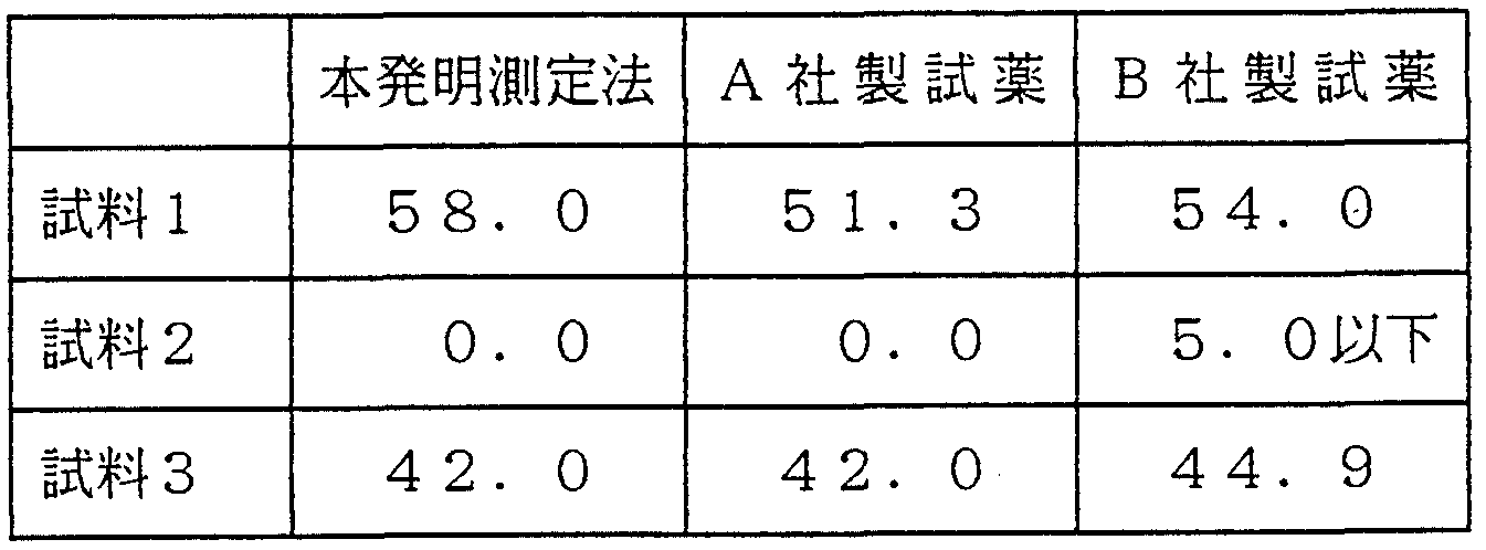


Claims
Priority Applications (3)
| Application Number | Priority Date | Filing Date | Title |
|---|---|---|---|
| US08/211,747 US5733549A (en) | 1992-08-14 | 1993-08-12 | Peptides including amino acid sequences selected from lipoprotein (a) and apolipoprotein (a), antibodies recognizing these amino acid sequences, and methods of determination using these antibodies |
| EP94908101A EP0621284A4 (en) | 1992-08-14 | 1993-08-12 | PEPTIDES CONTAINING SELECTED RESPECTIVE AMINO ACID SELECTED FROM LIPOPROTEIN (a) AND APOLIPOPROTEIN (a), ANTIBODIES RECOGNIZING THESE AMINO ACID SEQUENCES AND ANALYSIS METHOD USING THE SAME. |
| AU47614/93A AU672028B2 (en) | 1992-08-14 | 1993-08-12 | Peptides containing respective amino acid sequences selected from among those of lipoprotein(a) and apolipoprotein(a), antibodies respectively recognizing these amino acid sequences, and method of assaying with these antibodies |
Applications Claiming Priority (4)
| Application Number | Priority Date | Filing Date | Title |
|---|---|---|---|
| JP4/237621 | 1992-08-14 | ||
| JP23762192 | 1992-08-14 | ||
| JP4/322237 | 1992-11-09 | ||
| JP32223792 | 1992-11-09 |
Publications (1)
| Publication Number | Publication Date |
|---|---|
| WO1994004563A1 true WO1994004563A1 (en) | 1994-03-03 |
Family
ID=26533285
Family Applications (1)
| Application Number | Title | Priority Date | Filing Date |
|---|---|---|---|
| PCT/JP1993/001142 WO1994004563A1 (en) | 1992-08-14 | 1993-08-12 | PEPTIDES CONTAINING RESPECTIVE AMINO ACID SEQUENCES SELECTED FROM AMONG THOSE OF LIPOPROTEIN(a) AND APOLIPOPROTEIN(a), ANTIBODIES RESPECTIVELY RECOGNIZING THESE AMINO ACID SEQUENCES, AND METHOD OF ASSAYING WITH THESE ANTIBODIES |
Country Status (4)
| Country | Link |
|---|---|
| US (1) | US5733549A (ja) |
| EP (1) | EP0621284A4 (ja) |
| AU (1) | AU672028B2 (ja) |
| WO (1) | WO1994004563A1 (ja) |
Cited By (5)
| Publication number | Priority date | Publication date | Assignee | Title |
|---|---|---|---|---|
| WO1995009363A1 (fr) * | 1993-09-29 | 1995-04-06 | Yamasa Corporation | Procede de titrage de lipoproteine oxydee et applications de ce procede |
| US5486476A (en) * | 1993-02-09 | 1996-01-23 | Boehringer Mannheim Gmbh | Lipoprotein (a) peptides and their use |
| AU683508B2 (en) * | 1993-12-21 | 1997-11-13 | Akzo Nobel N.V. | Immunoreactive peptides of APO(a) |
| JP2002539458A (ja) * | 1999-03-16 | 2002-11-19 | セレックス, インコーポレイテッド | 唾液中のapoaおよびapob、ならびにそれらの比を検出するための方法およびデバイス |
| JP2014060968A (ja) * | 2012-09-21 | 2014-04-10 | Anges Mg Inc | アポリポプロテイン(a)の特異的エピトープを含むDNAワクチン |
Families Citing this family (26)
| Publication number | Priority date | Publication date | Assignee | Title |
|---|---|---|---|---|
| US5783400A (en) * | 1990-04-27 | 1998-07-21 | Genzyme Corporation | Method for the isolation of lipoprotein allowing for the subsequent quantification of its mass and cholesterol content |
| AU672028B2 (en) * | 1992-08-14 | 1996-09-19 | Shino-Test Corporation | Peptides containing respective amino acid sequences selected from among those of lipoprotein(a) and apolipoprotein(a), antibodies respectively recognizing these amino acid sequences, and method of assaying with these antibodies |
| AU7728596A (en) * | 1995-11-09 | 1997-05-29 | Arch Development Corporation | Isolation of apo(a), compositions, and methods of use |
| WO1997021099A1 (en) * | 1995-12-05 | 1997-06-12 | Entremed, Inc. | Method of diagnosis and treatment of atherosclerosis using anti-cholesterol antibodies |
| US6210906B1 (en) | 1998-01-20 | 2001-04-03 | Abbott Laboratories | Specific antibodies to kringle 5 of apo(a) and methods of use therefor |
| CA2319129A1 (en) * | 1998-01-30 | 1999-08-05 | Human Genome Sciences, Inc. | 67 human secreted proteins |
| DE19960500A1 (de) * | 1999-12-15 | 2001-07-12 | Giesing Michael | Antikörper und Verfahren zu ihrer Herstellung, deren Verwendung sowie Immunisierungscocktails, Immunoassay-Sets und Peptide |
| US7144862B2 (en) | 2000-08-24 | 2006-12-05 | The Regents Of The University Of California | Orally administered peptides to ameliorate atherosclerosis |
| US7199102B2 (en) * | 2000-08-24 | 2007-04-03 | The Regents Of The University Of California | Orally administered peptides synergize statin activity |
| US7166578B2 (en) | 2000-08-24 | 2007-01-23 | The Regents Of The University Of California | Orally administered peptides synergize statin activity |
| US8568766B2 (en) * | 2000-08-24 | 2013-10-29 | Gattadahalli M. Anantharamaiah | Peptides and peptide mimetics to treat pathologies associated with eye disease |
| US7723303B2 (en) * | 2000-08-24 | 2010-05-25 | The Regents Of The University Of California | Peptides and peptide mimetics to treat pathologies characterized by an inflammatory response |
| US6664230B1 (en) | 2000-08-24 | 2003-12-16 | The Regents Of The University Of California | Orally administered peptides to ameliorate atherosclerosis |
| US7148197B2 (en) * | 2000-08-24 | 2006-12-12 | The Regents Of The University Of California | Orally administered small peptides synergize statin activity |
| BR0115257A (pt) * | 2000-11-10 | 2003-08-12 | Proteopharma Aps | Análogos de apolipoproteìna |
| US7217785B2 (en) * | 2001-05-09 | 2007-05-15 | The Regents Of The University Of California | Cysteine-containing peptides having antioxidant properties |
| US6930085B2 (en) * | 2002-04-05 | 2005-08-16 | The Regents Of The University Of California | G-type peptides to ameliorate atherosclerosis |
| GR1004477B (el) * | 2002-04-23 | 2004-03-08 | Ευσταθιος Γκονος | Μεθοδος και κιτ ποσοτικης μετρησης της απολιποπρωτεινης j/clusterin στον ορο του αιματος |
| CA2549529A1 (en) * | 2003-12-15 | 2005-06-30 | The Regents Of The University Of California | Helical synthetic peptides that stimulate cellular cholesterol efflux |
| JP2008513479A (ja) * | 2004-09-16 | 2008-05-01 | ザ リージェンツ オブ ザ ユニバーシティ オブ カリフォルニア | アテローム性動脈硬化症および他の病理を改善するためのg型ペプチドおよび他の薬剤 |
| CA2595067A1 (en) | 2004-12-06 | 2006-06-15 | The Regents Of The University Of California | Methods for improving the structure and function of arterioles |
| AU2006242651B2 (en) * | 2005-04-29 | 2013-05-16 | The Regents Of The University Of California | Peptides and peptide mimetics to treat pathologies characterized by an inflammatory response |
| US20080293639A1 (en) * | 2005-04-29 | 2008-11-27 | The Regents Of The University Of California | Peptides and peptide mimetics to treat pathologies characterized by an inflammatory response |
| JP2010538005A (ja) * | 2007-08-28 | 2010-12-09 | ユーエービー リサーチ ファウンデーション | 合成アポリポ蛋白質e模倣ポリペプチドおよび使用方法 |
| JP2010537638A (ja) | 2007-08-28 | 2010-12-09 | ユーエービー リサーチ ファウンデーション | 合成アポリポ蛋白質e模倣ポリペプチドおよび使用方法 |
| JP7081923B2 (ja) | 2014-07-31 | 2022-06-07 | ユーエイビー リサーチ ファンデーション | アポe模倣ペプチド及び血漿コレステロールを取り除くためのより高い効果 |
Family Cites Families (19)
| Publication number | Priority date | Publication date | Assignee | Title |
|---|---|---|---|---|
| US4970144A (en) * | 1984-12-31 | 1990-11-13 | International Genetic Engineering | Peptide fragments of human apolipoprotein, type-specific antibodies and methods of use |
| WO1986004144A1 (en) * | 1984-12-31 | 1986-07-17 | International Genetic Engineering, Inc. | Peptide fragments of human apolipoprotein, type-specific antibodies and methods of use |
| SE448324B (sv) * | 1985-09-27 | 1987-02-09 | Pharmacia Ab | Sett vid immunkemisk bestemning av lipoproteiner och apolipoproteiner |
| US4839343A (en) * | 1987-03-13 | 1989-06-13 | Debiopharm, S.A. | Preparation containing hexatriacontapeptides and methods of use |
| ATE152244T1 (de) * | 1987-10-13 | 1997-05-15 | Terrapin Tech Inc | Verfahren zur herstellung von imunodiagnose- mitteln |
| SE8705139D0 (sv) * | 1987-12-23 | 1987-12-23 | Trion Forskning & Utveckling | Forfarande for framstellning av ett stort antal peptidanaloger och nya peptidanaloger |
| ATE121197T1 (de) * | 1988-01-19 | 1995-04-15 | Pasteur Institut | Immunometrisches verfahren zum nachweis von lipoprotein-teilchen, anti-lp(a) monoklonale antikörper und ihre anwendung zur diagnose von atherosklerose. |
| EP0330506A3 (en) * | 1988-02-26 | 1990-06-20 | Dana Farber Cancer Institute | Vla proteins |
| US4945040A (en) * | 1988-02-29 | 1990-07-31 | Arch Development Corporation | Immunoassay for lipoprotein(a) |
| AU3149289A (en) * | 1988-03-18 | 1989-09-21 | Rockefeller University, The | Method and agent for inhibiting the binding of human polymorphonuclear leukocytes to endothelium and compositions therefor |
| DE68926427T2 (de) * | 1988-03-25 | 1996-12-05 | Univ Rockefeller | Vom streptococcus m-protein abgeleitete synthetische peptide und damit hergestellte impfstoffe |
| CA2003316A1 (en) * | 1988-11-18 | 1990-05-18 | John F. Oram | Hdl-binding proteins |
| JPH05507276A (ja) * | 1990-05-03 | 1993-10-21 | ニュー・イングランド・ディーコネス・ホスピタル・コーポレイション | 動脈造影用合成ペプチド類 |
| AU9088891A (en) * | 1990-11-29 | 1992-06-25 | John E. Carbaugh | Diagnostic and therapeutic compositions and methods for lipoprotein(a) |
| JP3018110B2 (ja) * | 1991-03-25 | 2000-03-13 | 第一化学薬品株式会社 | モノクローナル抗体 |
| AU672028B2 (en) * | 1992-08-14 | 1996-09-19 | Shino-Test Corporation | Peptides containing respective amino acid sequences selected from among those of lipoprotein(a) and apolipoprotein(a), antibodies respectively recognizing these amino acid sequences, and method of assaying with these antibodies |
| JPH06113880A (ja) * | 1992-09-30 | 1994-04-26 | Chemo Sero Therapeut Res Inst | モノクローナル抗体及びこれを用いたリポプロティンスモールaの測定方法 |
| DE59409345D1 (de) * | 1993-02-09 | 2000-06-21 | Roche Diagnostics Gmbh | Lipoprotein (a)-Peptide und deren Verwendung |
| ATE193021T1 (de) * | 1993-02-09 | 2000-06-15 | Roche Diagnostics Gmbh | Lipoprotein (a)-peptide und deren verwendung |
-
1993
- 1993-08-12 AU AU47614/93A patent/AU672028B2/en not_active Ceased
- 1993-08-12 EP EP94908101A patent/EP0621284A4/en not_active Withdrawn
- 1993-08-12 WO PCT/JP1993/001142 patent/WO1994004563A1/ja not_active Application Discontinuation
- 1993-08-12 US US08/211,747 patent/US5733549A/en not_active Expired - Fee Related
Non-Patent Citations (3)
| Title |
|---|
| Biochem. J., Vol. 288, No. 1 (1992), GI CHEN et al., "Studies on epitopes on low-density lipoprotein modified by 4-hydroxynonenal. Biochemical characterization and determination", pp. 249-254. * |
| Nature (London), Vol. 330, No. 6144 (1987), JOHN W. McLEAN et al., "cDNA sequence of human apolipoprotein (a) is homologous to plasminogen", pp. 132-137. * |
| See also references of EP0621284A4 * |
Cited By (5)
| Publication number | Priority date | Publication date | Assignee | Title |
|---|---|---|---|---|
| US5486476A (en) * | 1993-02-09 | 1996-01-23 | Boehringer Mannheim Gmbh | Lipoprotein (a) peptides and their use |
| WO1995009363A1 (fr) * | 1993-09-29 | 1995-04-06 | Yamasa Corporation | Procede de titrage de lipoproteine oxydee et applications de ce procede |
| AU683508B2 (en) * | 1993-12-21 | 1997-11-13 | Akzo Nobel N.V. | Immunoreactive peptides of APO(a) |
| JP2002539458A (ja) * | 1999-03-16 | 2002-11-19 | セレックス, インコーポレイテッド | 唾液中のapoaおよびapob、ならびにそれらの比を検出するための方法およびデバイス |
| JP2014060968A (ja) * | 2012-09-21 | 2014-04-10 | Anges Mg Inc | アポリポプロテイン(a)の特異的エピトープを含むDNAワクチン |
Also Published As
| Publication number | Publication date |
|---|---|
| EP0621284A4 (en) | 1996-06-12 |
| US5733549A (en) | 1998-03-31 |
| AU672028B2 (en) | 1996-09-19 |
| EP0621284A1 (en) | 1994-10-26 |
| AU4761493A (en) | 1994-03-15 |
Similar Documents
| Publication | Publication Date | Title |
|---|---|---|
| WO1994004563A1 (en) | PEPTIDES CONTAINING RESPECTIVE AMINO ACID SEQUENCES SELECTED FROM AMONG THOSE OF LIPOPROTEIN(a) AND APOLIPOPROTEIN(a), ANTIBODIES RESPECTIVELY RECOGNIZING THESE AMINO ACID SEQUENCES, AND METHOD OF ASSAYING WITH THESE ANTIBODIES | |
| JP5382570B2 (ja) | ヒトhmgb1と特異的に結合する鳥類由来の抗体、ヒトhmgb1の免疫学的測定方法及びヒトhmgb1の免疫学的測定試薬 | |
| US9347954B2 (en) | Antibody capable of binding to specific region of periostin, and method of measuring periostin using the same | |
| JP4823465B2 (ja) | ヒトhmg−1に特異的に結合する抗体並びにこの抗体を用いるヒトhmg−1の免疫学的測定方法及び免疫学的測定試薬 | |
| JPH0967398A (ja) | 血清アミロイドaを認識するモノクローナル抗体 | |
| JP2959837B2 (ja) | 癌関連ハプトグロビン | |
| JPH04503006A (ja) | 血液凝固XIIa因子βモノクローナル抗体およびイムノアッセイ | |
| JPWO2020067396A1 (ja) | 糖化ヘモグロビン(%)の測定方法 | |
| WO1999018435A1 (fr) | Procede d'analyse de l'annexine v dans l'urine et utilisation | |
| JP5055598B2 (ja) | ヒトhmg−1に特異的に結合する抗体を用いるヒトhmg−1の免疫学的測定方法及び免疫学的測定試薬 | |
| JP2012082210A (ja) | Adamts13活性検定用抗体及び活性検定方法 | |
| EP0812859B1 (en) | ANTIFACTOR Xa-TISSUE FACTOR PATHWAY INHIBITOR COMPLEX MONOCLONAL ANTIBODY AND USE OF THE SAME | |
| KR960008672B1 (ko) | 심방 나트륨이뇨성 폴리펩티드를 인지하는 단일클론성 항체 | |
| WO2014147873A1 (ja) | Hmgb1の分解産物と特異的に結合する抗体、並びにhmgb1の分解産物の測定方法及び測定試薬 | |
| JPH10226700A (ja) | Miaの検出のためのイムノアッセイ | |
| JP5852433B2 (ja) | ソルチリンによる動脈硬化の判定方法 | |
| JP3713585B2 (ja) | 変性又は修飾リポタンパク質(a)に結合する抗体及びこの抗体を用いる測定法 | |
| JP5280916B2 (ja) | 抗ラットオステオカルシンモノクローナル抗体 | |
| JPH0667319B2 (ja) | Anpのc端側を認識するモノクローナル抗体 | |
| JP4028925B2 (ja) | モノクローナル抗体 | |
| JP2000069963A (ja) | アポリポプロテインe4特異モノクローナル抗体 | |
| Nunomura et al. | Characterization of mouse monoclonal antibodies to human protein 4.1 R FERM domain: epitope mapping and application to FERM domain binding to red blood cell inside-out vesicles | |
| JP2021063020A (ja) | Hmgb1の分解産物に結合する抗体、hmgb1分解産物の測定方法及びhmgb1分解産物の測定試薬 | |
| JP2005502711A (ja) | Aa4rpアッセイのための組成物および方法 | |
| JPH0346116B2 (ja) |
Legal Events
| Date | Code | Title | Description |
|---|---|---|---|
| AK | Designated states |
Kind code of ref document: A1 Designated state(s): AU JP US |
|
| AL | Designated countries for regional patents |
Kind code of ref document: A1 Designated state(s): AT BE CH DE DK ES FR GB GR IE IT LU MC NL PT SE |
|
| WWE | Wipo information: entry into national phase |
Ref document number: 1994908101 Country of ref document: EP Ref document number: 08211747 Country of ref document: US |
|
| 121 | Ep: the epo has been informed by wipo that ep was designated in this application | ||
| WWP | Wipo information: published in national office |
Ref document number: 1994908101 Country of ref document: EP |
|
| ENP | Entry into the national phase |
Ref document number: 1996 730525 Country of ref document: US Date of ref document: 19961011 Kind code of ref document: A |
|
| WWW | Wipo information: withdrawn in national office |
Ref document number: 1994908101 Country of ref document: EP |