WO1999000424A2 - Antibodies and other binding molecules specific for hepatitis b viral antigens - Google Patents
Antibodies and other binding molecules specific for hepatitis b viral antigens Download PDFInfo
- Publication number
- WO1999000424A2 WO1999000424A2 PCT/EP1998/004018 EP9804018W WO9900424A2 WO 1999000424 A2 WO1999000424 A2 WO 1999000424A2 EP 9804018 W EP9804018 W EP 9804018W WO 9900424 A2 WO9900424 A2 WO 9900424A2
- Authority
- WO
- WIPO (PCT)
- Prior art keywords
- hepatitis
- antibodies
- specific binding
- binding molecule
- peptide
- Prior art date
Links
- 239000000427 antigen Substances 0.000 title claims abstract description 40
- 102000036639 antigens Human genes 0.000 title claims abstract description 40
- 108091007433 antigens Proteins 0.000 title claims abstract description 40
- 230000027455 binding Effects 0.000 title claims abstract description 23
- 230000003612 virological effect Effects 0.000 title abstract description 8
- 208000006454 hepatitis Diseases 0.000 title description 6
- 231100000283 hepatitis Toxicity 0.000 title description 3
- 108090000765 processed proteins & peptides Proteins 0.000 claims abstract description 82
- 208000002672 hepatitis B Diseases 0.000 claims abstract description 52
- 238000000034 method Methods 0.000 claims abstract description 44
- 230000009870 specific binding Effects 0.000 claims abstract description 40
- 239000002245 particle Substances 0.000 claims abstract description 28
- 238000001514 detection method Methods 0.000 claims abstract description 17
- 238000003745 diagnosis Methods 0.000 claims abstract description 14
- 238000003149 assay kit Methods 0.000 claims abstract description 7
- 125000003275 alpha amino acid group Chemical group 0.000 claims description 20
- 238000004113 cell culture Methods 0.000 claims description 8
- 101000874347 Streptococcus agalactiae IgA FC receptor Proteins 0.000 claims description 5
- 241000700721 Hepatitis B virus Species 0.000 abstract description 41
- 102000004196 processed proteins & peptides Human genes 0.000 abstract description 34
- 238000003556 assay Methods 0.000 description 43
- 235000001014 amino acid Nutrition 0.000 description 42
- 150000001413 amino acids Chemical class 0.000 description 35
- 210000004027 cell Anatomy 0.000 description 34
- 230000009257 reactivity Effects 0.000 description 34
- 108090000623 proteins and genes Proteins 0.000 description 21
- 239000012634 fragment Substances 0.000 description 15
- 235000018102 proteins Nutrition 0.000 description 15
- 102000004169 proteins and genes Human genes 0.000 description 15
- 238000006467 substitution reaction Methods 0.000 description 15
- 238000012360 testing method Methods 0.000 description 13
- DHMQDGOQFOQNFH-UHFFFAOYSA-N Glycine Chemical compound NCC(O)=O DHMQDGOQFOQNFH-UHFFFAOYSA-N 0.000 description 11
- 241000282414 Homo sapiens Species 0.000 description 11
- 208000015181 infectious disease Diseases 0.000 description 11
- 210000002966 serum Anatomy 0.000 description 10
- 230000035899 viability Effects 0.000 description 10
- 108020004414 DNA Proteins 0.000 description 8
- QNAYBMKLOCPYGJ-REOHCLBHSA-N L-alanine Chemical compound C[C@H](N)C(O)=O QNAYBMKLOCPYGJ-REOHCLBHSA-N 0.000 description 8
- 235000004279 alanine Nutrition 0.000 description 8
- COLNVLDHVKWLRT-QMMMGPOBSA-N L-phenylalanine Chemical compound OC(=O)[C@@H](N)CC1=CC=CC=C1 COLNVLDHVKWLRT-QMMMGPOBSA-N 0.000 description 7
- 238000005516 engineering process Methods 0.000 description 7
- 238000011160 research Methods 0.000 description 7
- 206010008909 Chronic Hepatitis Diseases 0.000 description 6
- MTCFGRXMJLQNBG-UHFFFAOYSA-N Serine Natural products OCC(N)C(O)=O MTCFGRXMJLQNBG-UHFFFAOYSA-N 0.000 description 6
- QAOWNCQODCNURD-UHFFFAOYSA-N Sulfuric acid Chemical compound OS(O)(=O)=O QAOWNCQODCNURD-UHFFFAOYSA-N 0.000 description 6
- 210000004369 blood Anatomy 0.000 description 6
- 239000008280 blood Substances 0.000 description 6
- 238000006243 chemical reaction Methods 0.000 description 6
- 238000013507 mapping Methods 0.000 description 6
- 239000000463 material Substances 0.000 description 6
- 239000002609 medium Substances 0.000 description 6
- 244000005700 microbiome Species 0.000 description 6
- 239000008279 sol Substances 0.000 description 6
- 101710132601 Capsid protein Proteins 0.000 description 5
- 108010047041 Complementarity Determining Regions Proteins 0.000 description 5
- 241001529936 Murinae Species 0.000 description 5
- 241000700605 Viruses Species 0.000 description 5
- 210000004408 hybridoma Anatomy 0.000 description 5
- 230000035772 mutation Effects 0.000 description 5
- 238000003752 polymerase chain reaction Methods 0.000 description 5
- 229920001184 polypeptide Polymers 0.000 description 5
- 238000012216 screening Methods 0.000 description 5
- 238000012546 transfer Methods 0.000 description 5
- 108090000790 Enzymes Proteins 0.000 description 4
- 102000004190 Enzymes Human genes 0.000 description 4
- 239000004471 Glycine Substances 0.000 description 4
- ROHFNLRQFUQHCH-YFKPBYRVSA-N L-leucine Chemical compound CC(C)C[C@H](N)C(O)=O ROHFNLRQFUQHCH-YFKPBYRVSA-N 0.000 description 4
- ROHFNLRQFUQHCH-UHFFFAOYSA-N Leucine Natural products CC(C)CC(N)C(O)=O ROHFNLRQFUQHCH-UHFFFAOYSA-N 0.000 description 4
- 238000002835 absorbance Methods 0.000 description 4
- 230000001154 acute effect Effects 0.000 description 4
- 210000004899 c-terminal region Anatomy 0.000 description 4
- 230000001684 chronic effect Effects 0.000 description 4
- 230000036541 health Effects 0.000 description 4
- 238000003018 immunoassay Methods 0.000 description 4
- 238000000338 in vitro Methods 0.000 description 4
- COLNVLDHVKWLRT-UHFFFAOYSA-N phenylalanine Natural products OC(=O)C(N)CC1=CC=CC=C1 COLNVLDHVKWLRT-UHFFFAOYSA-N 0.000 description 4
- 239000007787 solid Substances 0.000 description 4
- UAIUNKRWKOVEES-UHFFFAOYSA-N 3,3',5,5'-tetramethylbenzidine Chemical compound CC1=C(N)C(C)=CC(C=2C=C(C)C(N)=C(C)C=2)=C1 UAIUNKRWKOVEES-UHFFFAOYSA-N 0.000 description 3
- 206010059193 Acute hepatitis B Diseases 0.000 description 3
- FSHURBQASBLAPO-WDSKDSINSA-N Ala-Met Chemical compound CSCC[C@@H](C(O)=O)NC(=O)[C@H](C)N FSHURBQASBLAPO-WDSKDSINSA-N 0.000 description 3
- 239000004475 Arginine Substances 0.000 description 3
- 238000002965 ELISA Methods 0.000 description 3
- 206010019755 Hepatitis chronic active Diseases 0.000 description 3
- 206010019759 Hepatitis chronic persistent Diseases 0.000 description 3
- 108010001336 Horseradish Peroxidase Proteins 0.000 description 3
- 241000699666 Mus <mouse, genus> Species 0.000 description 3
- 108020004511 Recombinant DNA Proteins 0.000 description 3
- 241000283984 Rodentia Species 0.000 description 3
- UKINEYBQXPMOJO-UBHSHLNASA-N Trp-Asn-Ser Chemical compound C1=CC=C2C(=C1)C(=CN2)C[C@@H](C(=O)N[C@@H](CC(=O)N)C(=O)N[C@@H](CO)C(=O)O)N UKINEYBQXPMOJO-UBHSHLNASA-N 0.000 description 3
- 208000037628 acute hepatitis B virus infection Diseases 0.000 description 3
- 238000013459 approach Methods 0.000 description 3
- ODKSFYDXXFIFQN-UHFFFAOYSA-N arginine Natural products OC(=O)C(N)CCCNC(N)=N ODKSFYDXXFIFQN-UHFFFAOYSA-N 0.000 description 3
- 230000005540 biological transmission Effects 0.000 description 3
- 230000007423 decrease Effects 0.000 description 3
- 238000012217 deletion Methods 0.000 description 3
- 230000037430 deletion Effects 0.000 description 3
- 230000000694 effects Effects 0.000 description 3
- 238000002474 experimental method Methods 0.000 description 3
- 230000001900 immune effect Effects 0.000 description 3
- 238000000099 in vitro assay Methods 0.000 description 3
- 238000001727 in vivo Methods 0.000 description 3
- 238000011534 incubation Methods 0.000 description 3
- 238000002372 labelling Methods 0.000 description 3
- 238000004519 manufacturing process Methods 0.000 description 3
- 108010024654 phenylalanyl-prolyl-alanine Proteins 0.000 description 3
- 230000002285 radioactive effect Effects 0.000 description 3
- 230000035945 sensitivity Effects 0.000 description 3
- 238000012163 sequencing technique Methods 0.000 description 3
- 239000000243 solution Substances 0.000 description 3
- 239000000126 substance Substances 0.000 description 3
- 229960005486 vaccine Drugs 0.000 description 3
- 102100039358 3-hydroxyacyl-CoA dehydrogenase type-2 Human genes 0.000 description 2
- FWMNVWWHGCHHJJ-SKKKGAJSSA-N 4-amino-1-[(2r)-6-amino-2-[[(2r)-2-[[(2r)-2-[[(2r)-2-amino-3-phenylpropanoyl]amino]-3-phenylpropanoyl]amino]-4-methylpentanoyl]amino]hexanoyl]piperidine-4-carboxylic acid Chemical compound C([C@H](C(=O)N[C@H](CC(C)C)C(=O)N[C@H](CCCCN)C(=O)N1CCC(N)(CC1)C(O)=O)NC(=O)[C@H](N)CC=1C=CC=CC=1)C1=CC=CC=C1 FWMNVWWHGCHHJJ-SKKKGAJSSA-N 0.000 description 2
- HKRXJBBCQBAGIM-FXQIFTODSA-N Arg-Asp-Ser Chemical compound C(C[C@@H](C(=O)N[C@@H](CC(=O)O)C(=O)N[C@@H](CO)C(=O)O)N)CN=C(N)N HKRXJBBCQBAGIM-FXQIFTODSA-N 0.000 description 2
- 208000000419 Chronic Hepatitis B Diseases 0.000 description 2
- 108020004705 Codon Proteins 0.000 description 2
- YWAQATDNEKZFFK-BYPYZUCNSA-N Gly-Gly-Ser Chemical compound NCC(=O)NCC(=O)N[C@@H](CO)C(O)=O YWAQATDNEKZFFK-BYPYZUCNSA-N 0.000 description 2
- 206010019799 Hepatitis viral Diseases 0.000 description 2
- 101001035740 Homo sapiens 3-hydroxyacyl-CoA dehydrogenase type-2 Proteins 0.000 description 2
- FFEARJCKVFRZRR-BYPYZUCNSA-N L-methionine Chemical compound CSCC[C@H](N)C(O)=O FFEARJCKVFRZRR-BYPYZUCNSA-N 0.000 description 2
- OUYCCCASQSFEME-QMMMGPOBSA-N L-tyrosine Chemical compound OC(=O)[C@@H](N)CC1=CC=C(O)C=C1 OUYCCCASQSFEME-QMMMGPOBSA-N 0.000 description 2
- 241001465754 Metazoa Species 0.000 description 2
- 241000699670 Mus sp. Species 0.000 description 2
- 108090001074 Nucleocapsid Proteins Proteins 0.000 description 2
- 108700026244 Open Reading Frames Proteins 0.000 description 2
- JLLJTMHNXQTMCK-UBHSHLNASA-N Phe-Pro-Ala Chemical compound OC(=O)[C@H](C)NC(=O)[C@@H]1CCCN1C(=O)[C@@H](N)CC1=CC=CC=C1 JLLJTMHNXQTMCK-UBHSHLNASA-N 0.000 description 2
- 206010035226 Plasma cell myeloma Diseases 0.000 description 2
- SRSPTFBENMJHMR-WHFBIAKZSA-N Ser-Ser-Gly Chemical compound OC[C@H](N)C(=O)N[C@@H](CO)C(=O)NCC(O)=O SRSPTFBENMJHMR-WHFBIAKZSA-N 0.000 description 2
- 108010003723 Single-Domain Antibodies Proteins 0.000 description 2
- AYFVYJQAPQTCCC-UHFFFAOYSA-N Threonine Natural products CC(O)C(N)C(O)=O AYFVYJQAPQTCCC-UHFFFAOYSA-N 0.000 description 2
- 239000004473 Threonine Substances 0.000 description 2
- COYSIHFOCOMGCF-UHFFFAOYSA-N Val-Arg-Gly Natural products CC(C)C(N)C(=O)NC(C(=O)NCC(O)=O)CCCN=C(N)N COYSIHFOCOMGCF-UHFFFAOYSA-N 0.000 description 2
- 239000002671 adjuvant Substances 0.000 description 2
- 238000004458 analytical method Methods 0.000 description 2
- 210000004102 animal cell Anatomy 0.000 description 2
- 230000008901 benefit Effects 0.000 description 2
- 150000001875 compounds Chemical class 0.000 description 2
- 239000012228 culture supernatant Substances 0.000 description 2
- 230000003247 decreasing effect Effects 0.000 description 2
- 238000011161 development Methods 0.000 description 2
- LOKCTEFSRHRXRJ-UHFFFAOYSA-I dipotassium trisodium dihydrogen phosphate hydrogen phosphate dichloride Chemical compound P(=O)(O)(O)[O-].[K+].P(=O)(O)([O-])[O-].[Na+].[Na+].[Cl-].[K+].[Cl-].[Na+] LOKCTEFSRHRXRJ-UHFFFAOYSA-I 0.000 description 2
- 239000000975 dye Substances 0.000 description 2
- 150000002148 esters Chemical class 0.000 description 2
- 230000017188 evasion or tolerance of host immune response Effects 0.000 description 2
- 239000013604 expression vector Substances 0.000 description 2
- 125000002485 formyl group Chemical class [H]C(*)=O 0.000 description 2
- 108020001507 fusion proteins Proteins 0.000 description 2
- 102000037865 fusion proteins Human genes 0.000 description 2
- 108010050475 glycyl-leucyl-tyrosine Proteins 0.000 description 2
- 108010089804 glycyl-threonine Proteins 0.000 description 2
- 108010092114 histidylphenylalanine Proteins 0.000 description 2
- 238000002955 isolation Methods 0.000 description 2
- 208000019423 liver disease Diseases 0.000 description 2
- 239000002184 metal Substances 0.000 description 2
- 150000002736 metal compounds Chemical class 0.000 description 2
- 229930182817 methionine Natural products 0.000 description 2
- 239000000203 mixture Substances 0.000 description 2
- 201000000050 myeloid neoplasm Diseases 0.000 description 2
- 239000013642 negative control Substances 0.000 description 2
- 239000012071 phase Substances 0.000 description 2
- 239000008363 phosphate buffer Substances 0.000 description 2
- 239000002953 phosphate buffered saline Substances 0.000 description 2
- 239000000047 product Substances 0.000 description 2
- 108010048818 seryl-histidine Proteins 0.000 description 2
- 210000000952 spleen Anatomy 0.000 description 2
- 239000000758 substrate Substances 0.000 description 2
- 239000006228 supernatant Substances 0.000 description 2
- 238000003786 synthesis reaction Methods 0.000 description 2
- 238000002560 therapeutic procedure Methods 0.000 description 2
- 201000001862 viral hepatitis Diseases 0.000 description 2
- YRNWIFYIFSBPAU-UHFFFAOYSA-N 4-[4-(dimethylamino)phenyl]-n,n-dimethylaniline Chemical compound C1=CC(N(C)C)=CC=C1C1=CC=C(N(C)C)C=C1 YRNWIFYIFSBPAU-UHFFFAOYSA-N 0.000 description 1
- ISVACHFCVRKIDG-SRVKXCTJSA-N Arg-Val-Arg Chemical compound NC(N)=NCCC[C@H](N)C(=O)N[C@@H](C(C)C)C(=O)N[C@@H](CCCN=C(N)N)C(O)=O ISVACHFCVRKIDG-SRVKXCTJSA-N 0.000 description 1
- 102000014914 Carrier Proteins Human genes 0.000 description 1
- 108010078791 Carrier Proteins Proteins 0.000 description 1
- 108020004635 Complementary DNA Proteins 0.000 description 1
- 241000252095 Congridae Species 0.000 description 1
- 102100031673 Corneodesmosin Human genes 0.000 description 1
- 102000053602 DNA Human genes 0.000 description 1
- 230000004544 DNA amplification Effects 0.000 description 1
- 102100027723 Endogenous retrovirus group K member 6 Rec protein Human genes 0.000 description 1
- 101710091045 Envelope protein Proteins 0.000 description 1
- 241000588724 Escherichia coli Species 0.000 description 1
- 101001065501 Escherichia phage MS2 Lysis protein Proteins 0.000 description 1
- AFWYPMDMDYCKMD-KBPBESRZSA-N Gly-Leu-Tyr Chemical compound NCC(=O)N[C@@H](CC(C)C)C(=O)N[C@H](C(O)=O)CC1=CC=C(O)C=C1 AFWYPMDMDYCKMD-KBPBESRZSA-N 0.000 description 1
- 241000282412 Homo Species 0.000 description 1
- 108060003951 Immunoglobulin Proteins 0.000 description 1
- CKLJMWTZIZZHCS-REOHCLBHSA-N L-aspartic acid Chemical compound OC(=O)[C@@H](N)CC(O)=O CKLJMWTZIZZHCS-REOHCLBHSA-N 0.000 description 1
- QIVBCDIJIAJPQS-VIFPVBQESA-N L-tryptophane Chemical compound C1=CC=C2C(C[C@H](N)C(O)=O)=CNC2=C1 QIVBCDIJIAJPQS-VIFPVBQESA-N 0.000 description 1
- KZSNJWFQEVHDMF-BYPYZUCNSA-N L-valine Chemical compound CC(C)[C@H](N)C(O)=O KZSNJWFQEVHDMF-BYPYZUCNSA-N 0.000 description 1
- MMEDVBWCMGRKKC-GARJFASQSA-N Leu-Asp-Pro Chemical compound CC(C)C[C@@H](C(=O)N[C@@H](CC(=O)O)C(=O)N1CCC[C@@H]1C(=O)O)N MMEDVBWCMGRKKC-GARJFASQSA-N 0.000 description 1
- RDFIVFHPOSOXMW-ACRUOGEOSA-N Leu-Tyr-Phe Chemical compound [H]N[C@@H](CC(C)C)C(=O)N[C@@H](CC1=CC=C(O)C=C1)C(=O)N[C@@H](CC1=CC=CC=C1)C(O)=O RDFIVFHPOSOXMW-ACRUOGEOSA-N 0.000 description 1
- 206010067125 Liver injury Diseases 0.000 description 1
- 101710085938 Matrix protein Proteins 0.000 description 1
- 101710127721 Membrane protein Proteins 0.000 description 1
- 102100032965 Myomesin-2 Human genes 0.000 description 1
- 108091034117 Oligonucleotide Proteins 0.000 description 1
- 108010038807 Oligopeptides Proteins 0.000 description 1
- 102000015636 Oligopeptides Human genes 0.000 description 1
- 241000282579 Pan Species 0.000 description 1
- 108091005804 Peptidases Proteins 0.000 description 1
- 102000035195 Peptidases Human genes 0.000 description 1
- 108010033276 Peptide Fragments Proteins 0.000 description 1
- 102000007079 Peptide Fragments Human genes 0.000 description 1
- 208000037581 Persistent Infection Diseases 0.000 description 1
- ONIBWKKTOPOVIA-UHFFFAOYSA-N Proline Natural products OC(=O)C1CCCN1 ONIBWKKTOPOVIA-UHFFFAOYSA-N 0.000 description 1
- 239000004365 Protease Substances 0.000 description 1
- 229940096437 Protein S Drugs 0.000 description 1
- 101710188315 Protein X Proteins 0.000 description 1
- 101150010882 S gene Proteins 0.000 description 1
- 240000004808 Saccharomyces cerevisiae Species 0.000 description 1
- MLSQXWSRHURDMF-GARJFASQSA-N Ser-His-Pro Chemical compound C1C[C@@H](N(C1)C(=O)[C@H](CC2=CN=CN2)NC(=O)[C@H](CO)N)C(=O)O MLSQXWSRHURDMF-GARJFASQSA-N 0.000 description 1
- VLMIUSLQONKLDV-HEIBUPTGSA-N Ser-Thr-Thr Chemical compound [H]N[C@@H](CO)C(=O)N[C@@H]([C@@H](C)O)C(=O)N[C@@H]([C@@H](C)O)C(O)=O VLMIUSLQONKLDV-HEIBUPTGSA-N 0.000 description 1
- QJIODPFLAASXJC-JHYOHUSXSA-N Thr-Thr-Phe Chemical compound C[C@H]([C@@H](C(=O)N[C@@H]([C@@H](C)O)C(=O)N[C@@H](CC1=CC=CC=C1)C(=O)O)N)O QJIODPFLAASXJC-JHYOHUSXSA-N 0.000 description 1
- GRQCSEWEPIHLBI-JQWIXIFHSA-N Trp-Asn Chemical compound C1=CC=C2C(C[C@H](N)C(=O)N[C@@H](CC(N)=O)C(O)=O)=CNC2=C1 GRQCSEWEPIHLBI-JQWIXIFHSA-N 0.000 description 1
- QIVBCDIJIAJPQS-UHFFFAOYSA-N Tryptophan Natural products C1=CC=C2C(CC(N)C(O)=O)=CNC2=C1 QIVBCDIJIAJPQS-UHFFFAOYSA-N 0.000 description 1
- COYSIHFOCOMGCF-WPRPVWTQSA-N Val-Arg-Gly Chemical compound CC(C)[C@H](N)C(=O)N[C@H](C(=O)NCC(O)=O)CCCN=C(N)N COYSIHFOCOMGCF-WPRPVWTQSA-N 0.000 description 1
- KZSNJWFQEVHDMF-UHFFFAOYSA-N Valine Natural products CC(C)C(N)C(O)=O KZSNJWFQEVHDMF-UHFFFAOYSA-N 0.000 description 1
- 108010003533 Viral Envelope Proteins Proteins 0.000 description 1
- 108010031318 Vitronectin Proteins 0.000 description 1
- JLCPHMBAVCMARE-UHFFFAOYSA-N [3-[[3-[[3-[[3-[[3-[[3-[[3-[[3-[[3-[[3-[[3-[[5-(2-amino-6-oxo-1H-purin-9-yl)-3-[[3-[[3-[[3-[[3-[[3-[[5-(2-amino-6-oxo-1H-purin-9-yl)-3-[[5-(2-amino-6-oxo-1H-purin-9-yl)-3-hydroxyoxolan-2-yl]methoxy-hydroxyphosphoryl]oxyoxolan-2-yl]methoxy-hydroxyphosphoryl]oxy-5-(5-methyl-2,4-dioxopyrimidin-1-yl)oxolan-2-yl]methoxy-hydroxyphosphoryl]oxy-5-(6-aminopurin-9-yl)oxolan-2-yl]methoxy-hydroxyphosphoryl]oxy-5-(6-aminopurin-9-yl)oxolan-2-yl]methoxy-hydroxyphosphoryl]oxy-5-(6-aminopurin-9-yl)oxolan-2-yl]methoxy-hydroxyphosphoryl]oxy-5-(6-aminopurin-9-yl)oxolan-2-yl]methoxy-hydroxyphosphoryl]oxyoxolan-2-yl]methoxy-hydroxyphosphoryl]oxy-5-(5-methyl-2,4-dioxopyrimidin-1-yl)oxolan-2-yl]methoxy-hydroxyphosphoryl]oxy-5-(4-amino-2-oxopyrimidin-1-yl)oxolan-2-yl]methoxy-hydroxyphosphoryl]oxy-5-(5-methyl-2,4-dioxopyrimidin-1-yl)oxolan-2-yl]methoxy-hydroxyphosphoryl]oxy-5-(5-methyl-2,4-dioxopyrimidin-1-yl)oxolan-2-yl]methoxy-hydroxyphosphoryl]oxy-5-(6-aminopurin-9-yl)oxolan-2-yl]methoxy-hydroxyphosphoryl]oxy-5-(6-aminopurin-9-yl)oxolan-2-yl]methoxy-hydroxyphosphoryl]oxy-5-(4-amino-2-oxopyrimidin-1-yl)oxolan-2-yl]methoxy-hydroxyphosphoryl]oxy-5-(4-amino-2-oxopyrimidin-1-yl)oxolan-2-yl]methoxy-hydroxyphosphoryl]oxy-5-(4-amino-2-oxopyrimidin-1-yl)oxolan-2-yl]methoxy-hydroxyphosphoryl]oxy-5-(6-aminopurin-9-yl)oxolan-2-yl]methoxy-hydroxyphosphoryl]oxy-5-(4-amino-2-oxopyrimidin-1-yl)oxolan-2-yl]methyl [5-(6-aminopurin-9-yl)-2-(hydroxymethyl)oxolan-3-yl] hydrogen phosphate Polymers Cc1cn(C2CC(OP(O)(=O)OCC3OC(CC3OP(O)(=O)OCC3OC(CC3O)n3cnc4c3nc(N)[nH]c4=O)n3cnc4c3nc(N)[nH]c4=O)C(COP(O)(=O)OC3CC(OC3COP(O)(=O)OC3CC(OC3COP(O)(=O)OC3CC(OC3COP(O)(=O)OC3CC(OC3COP(O)(=O)OC3CC(OC3COP(O)(=O)OC3CC(OC3COP(O)(=O)OC3CC(OC3COP(O)(=O)OC3CC(OC3COP(O)(=O)OC3CC(OC3COP(O)(=O)OC3CC(OC3COP(O)(=O)OC3CC(OC3COP(O)(=O)OC3CC(OC3COP(O)(=O)OC3CC(OC3COP(O)(=O)OC3CC(OC3COP(O)(=O)OC3CC(OC3COP(O)(=O)OC3CC(OC3COP(O)(=O)OC3CC(OC3CO)n3cnc4c(N)ncnc34)n3ccc(N)nc3=O)n3cnc4c(N)ncnc34)n3ccc(N)nc3=O)n3ccc(N)nc3=O)n3ccc(N)nc3=O)n3cnc4c(N)ncnc34)n3cnc4c(N)ncnc34)n3cc(C)c(=O)[nH]c3=O)n3cc(C)c(=O)[nH]c3=O)n3ccc(N)nc3=O)n3cc(C)c(=O)[nH]c3=O)n3cnc4c3nc(N)[nH]c4=O)n3cnc4c(N)ncnc34)n3cnc4c(N)ncnc34)n3cnc4c(N)ncnc34)n3cnc4c(N)ncnc34)O2)c(=O)[nH]c1=O JLCPHMBAVCMARE-UHFFFAOYSA-N 0.000 description 1
- 239000002253 acid Substances 0.000 description 1
- 230000009798 acute exacerbation Effects 0.000 description 1
- 230000009435 amidation Effects 0.000 description 1
- 238000007112 amidation reaction Methods 0.000 description 1
- 150000001408 amides Chemical class 0.000 description 1
- 125000000539 amino acid group Chemical group 0.000 description 1
- 238000010171 animal model Methods 0.000 description 1
- 230000009830 antibody antigen interaction Effects 0.000 description 1
- 230000000890 antigenic effect Effects 0.000 description 1
- 150000001483 arginine derivatives Chemical class 0.000 description 1
- 235000003704 aspartic acid Nutrition 0.000 description 1
- 102000005936 beta-Galactosidase Human genes 0.000 description 1
- 108010005774 beta-Galactosidase Proteins 0.000 description 1
- OQFSQFPPLPISGP-UHFFFAOYSA-N beta-carboxyaspartic acid Natural products OC(=O)C(N)C(C(O)=O)C(O)=O OQFSQFPPLPISGP-UHFFFAOYSA-N 0.000 description 1
- 238000002306 biochemical method Methods 0.000 description 1
- 230000004071 biological effect Effects 0.000 description 1
- 230000015572 biosynthetic process Effects 0.000 description 1
- 239000010836 blood and blood product Substances 0.000 description 1
- 229940125691 blood product Drugs 0.000 description 1
- 210000001124 body fluid Anatomy 0.000 description 1
- 239000010839 body fluid Substances 0.000 description 1
- 238000010804 cDNA synthesis Methods 0.000 description 1
- 244000309466 calf Species 0.000 description 1
- 230000021523 carboxylation Effects 0.000 description 1
- 238000006473 carboxylation reaction Methods 0.000 description 1
- 239000000969 carrier Substances 0.000 description 1
- 239000006285 cell suspension Substances 0.000 description 1
- 239000000919 ceramic Substances 0.000 description 1
- 238000007385 chemical modification Methods 0.000 description 1
- 208000016350 chronic hepatitis B virus infection Diseases 0.000 description 1
- 238000003776 cleavage reaction Methods 0.000 description 1
- 238000010367 cloning Methods 0.000 description 1
- 230000002860 competitive effect Effects 0.000 description 1
- 230000000295 complement effect Effects 0.000 description 1
- 239000002299 complementary DNA Substances 0.000 description 1
- 239000000470 constituent Substances 0.000 description 1
- 238000012258 culturing Methods 0.000 description 1
- 125000004122 cyclic group Chemical group 0.000 description 1
- 238000002405 diagnostic procedure Methods 0.000 description 1
- 238000010790 dilution Methods 0.000 description 1
- 239000012895 dilution Substances 0.000 description 1
- 230000008034 disappearance Effects 0.000 description 1
- 201000010099 disease Diseases 0.000 description 1
- 208000037265 diseases, disorders, signs and symptoms Diseases 0.000 description 1
- 238000004520 electroporation Methods 0.000 description 1
- 230000008030 elimination Effects 0.000 description 1
- 238000003379 elimination reaction Methods 0.000 description 1
- 230000002255 enzymatic effect Effects 0.000 description 1
- 210000003743 erythrocyte Anatomy 0.000 description 1
- 239000000284 extract Substances 0.000 description 1
- 239000012530 fluid Substances 0.000 description 1
- 239000007850 fluorescent dye Substances 0.000 description 1
- 230000006870 function Effects 0.000 description 1
- 230000004927 fusion Effects 0.000 description 1
- 230000002068 genetic effect Effects 0.000 description 1
- ZDXPYRJPNDTMRX-UHFFFAOYSA-N glutamine Natural products OC(=O)C(N)CCC(N)=O ZDXPYRJPNDTMRX-UHFFFAOYSA-N 0.000 description 1
- 230000013595 glycosylation Effects 0.000 description 1
- 238000006206 glycosylation reaction Methods 0.000 description 1
- 231100000753 hepatic injury Toxicity 0.000 description 1
- HNDVDQJCIGZPNO-UHFFFAOYSA-N histidine Natural products OC(=O)C(N)CC1=CN=CN1 HNDVDQJCIGZPNO-UHFFFAOYSA-N 0.000 description 1
- 238000003119 immunoblot Methods 0.000 description 1
- 102000018358 immunoglobulin Human genes 0.000 description 1
- 230000016784 immunoglobulin production Effects 0.000 description 1
- 238000013198 immunometric assay Methods 0.000 description 1
- 230000001771 impaired effect Effects 0.000 description 1
- 230000006872 improvement Effects 0.000 description 1
- 230000002458 infectious effect Effects 0.000 description 1
- 238000011081 inoculation Methods 0.000 description 1
- 230000003993 interaction Effects 0.000 description 1
- 238000001038 ionspray mass spectrometry Methods 0.000 description 1
- 239000004816 latex Substances 0.000 description 1
- 229920000126 latex Polymers 0.000 description 1
- 125000001909 leucine group Chemical group [H]N(*)C(C(*)=O)C([H])([H])C(C([H])([H])[H])C([H])([H])[H] 0.000 description 1
- 210000004185 liver Anatomy 0.000 description 1
- 230000003908 liver function Effects 0.000 description 1
- 210000004698 lymphocyte Anatomy 0.000 description 1
- 239000003550 marker Substances 0.000 description 1
- 230000007246 mechanism Effects 0.000 description 1
- 230000001404 mediated effect Effects 0.000 description 1
- 239000012528 membrane Substances 0.000 description 1
- 210000004779 membrane envelope Anatomy 0.000 description 1
- MYWUZJCMWCOHBA-VIFPVBQESA-N methamphetamine Chemical compound CN[C@@H](C)CC1=CC=CC=C1 MYWUZJCMWCOHBA-VIFPVBQESA-N 0.000 description 1
- 230000002906 microbiologic effect Effects 0.000 description 1
- 238000010647 peptide synthesis reaction Methods 0.000 description 1
- 150000002978 peroxides Chemical class 0.000 description 1
- 230000026731 phosphorylation Effects 0.000 description 1
- 238000006366 phosphorylation reaction Methods 0.000 description 1
- 229940113116 polyethylene glycol 1000 Drugs 0.000 description 1
- 238000001556 precipitation Methods 0.000 description 1
- 239000002243 precursor Substances 0.000 description 1
- 230000035935 pregnancy Effects 0.000 description 1
- 238000002360 preparation method Methods 0.000 description 1
- 238000007639 printing Methods 0.000 description 1
- 108010002730 protein S precursor Proteins 0.000 description 1
- 238000000746 purification Methods 0.000 description 1
- 238000003908 quality control method Methods 0.000 description 1
- 238000003127 radioimmunoassay Methods 0.000 description 1
- 108091008146 restriction endonucleases Proteins 0.000 description 1
- 150000003839 salts Chemical class 0.000 description 1
- 230000007017 scission Effects 0.000 description 1
- 230000003248 secreting effect Effects 0.000 description 1
- 239000006152 selective media Substances 0.000 description 1
- 230000000405 serological effect Effects 0.000 description 1
- 230000001568 sexual effect Effects 0.000 description 1
- 239000002356 single layer Substances 0.000 description 1
- 238000002741 site-directed mutagenesis Methods 0.000 description 1
- 239000007790 solid phase Substances 0.000 description 1
- 210000001082 somatic cell Anatomy 0.000 description 1
- 241000894007 species Species 0.000 description 1
- 238000001228 spectrum Methods 0.000 description 1
- 210000004989 spleen cell Anatomy 0.000 description 1
- 230000003393 splenic effect Effects 0.000 description 1
- 238000010561 standard procedure Methods 0.000 description 1
- 210000001519 tissue Anatomy 0.000 description 1
- 230000014621 translational initiation Effects 0.000 description 1
- OUYCCCASQSFEME-UHFFFAOYSA-N tyrosine Natural products OC(=O)C(N)CC1=CC=C(O)C=C1 OUYCCCASQSFEME-UHFFFAOYSA-N 0.000 description 1
- 241001515965 unidentified phage Species 0.000 description 1
- 239000004474 valine Substances 0.000 description 1
- 239000013598 vector Substances 0.000 description 1
- 210000002845 virion Anatomy 0.000 description 1
- 239000003643 water by type Substances 0.000 description 1
Classifications
-
- C—CHEMISTRY; METALLURGY
- C07—ORGANIC CHEMISTRY
- C07K—PEPTIDES
- C07K14/00—Peptides having more than 20 amino acids; Gastrins; Somatostatins; Melanotropins; Derivatives thereof
- C07K14/005—Peptides having more than 20 amino acids; Gastrins; Somatostatins; Melanotropins; Derivatives thereof from viruses
-
- C—CHEMISTRY; METALLURGY
- C07—ORGANIC CHEMISTRY
- C07K—PEPTIDES
- C07K16/00—Immunoglobulins [IGs], e.g. monoclonal or polyclonal antibodies
- C07K16/08—Immunoglobulins [IGs], e.g. monoclonal or polyclonal antibodies against material from viruses
- C07K16/081—Immunoglobulins [IGs], e.g. monoclonal or polyclonal antibodies against material from viruses from DNA viruses
- C07K16/082—Hepadnaviridae, e.g. hepatitis B virus
-
- C—CHEMISTRY; METALLURGY
- C07—ORGANIC CHEMISTRY
- C07K—PEPTIDES
- C07K2317/00—Immunoglobulins specific features
- C07K2317/30—Immunoglobulins specific features characterized by aspects of specificity or valency
- C07K2317/34—Identification of a linear epitope shorter than 20 amino acid residues or of a conformational epitope defined by amino acid residues
-
- C—CHEMISTRY; METALLURGY
- C12—BIOCHEMISTRY; BEER; SPIRITS; WINE; VINEGAR; MICROBIOLOGY; ENZYMOLOGY; MUTATION OR GENETIC ENGINEERING
- C12N—MICROORGANISMS OR ENZYMES; COMPOSITIONS THEREOF; PROPAGATING, PRESERVING, OR MAINTAINING MICROORGANISMS; MUTATION OR GENETIC ENGINEERING; CULTURE MEDIA
- C12N2730/00—Reverse transcribing DNA viruses
- C12N2730/00011—Details
- C12N2730/10011—Hepadnaviridae
- C12N2730/10111—Orthohepadnavirus, e.g. hepatitis B virus
- C12N2730/10122—New viral proteins or individual genes, new structural or functional aspects of known viral proteins or genes
-
- Y—GENERAL TAGGING OF NEW TECHNOLOGICAL DEVELOPMENTS; GENERAL TAGGING OF CROSS-SECTIONAL TECHNOLOGIES SPANNING OVER SEVERAL SECTIONS OF THE IPC; TECHNICAL SUBJECTS COVERED BY FORMER USPC CROSS-REFERENCE ART COLLECTIONS [XRACs] AND DIGESTS
- Y10—TECHNICAL SUBJECTS COVERED BY FORMER USPC
- Y10S—TECHNICAL SUBJECTS COVERED BY FORMER USPC CROSS-REFERENCE ART COLLECTIONS [XRACs] AND DIGESTS
- Y10S436/00—Chemistry: analytical and immunological testing
- Y10S436/82—Hepatitis associated antigens and antibodies
Definitions
- Antibodies and other binding molecules specific for hepatitis B viral antigens are included in the immunosorbents.
- the present invention relates to antibodies and other binding molecules specific for hepatitis B viral antigens (HBV), peptides comprising epitopes recognised by such molecules, and cell lines capable of producing antibodies.
- HBV hepatitis B viral antigens
- the invention is further concerned with the use of such molecules in diagnosis of hepatitis B virus (HBV).
- Hepatitis B virus HBV virus
- the hepatitis infection is transmitted by three general mechanisms: (1) by parenteral inoculation of infected blood or body fluids, either in large amounts as in blood transfusions or in minute amounts as through an accidental skinprick; (2) by close family or sexual contact; and (3) by some mothers, who infected during pregnancy, transmit the virus to their new-born children.
- HBV is not highly contagious. Transmission by inhalation occurs rarely, if ever.
- the transmission route through contaminated blood or blood products is a major threat to the human health.
- HBV infection elicits a spectrum of disease entities ranging from the most severe form of chronic active hepatitis (CAH) to less severe chronic persistent hepatitis (CPH) to the asymptomatic carrier (ASC) state.
- CAH chronic active hepatitis
- CPH chronic persistent hepatitis
- ASC asymptomatic carrier
- An array of diagnostic assays have recently been developed to aid the clinician in differentiating hepatitis B virus infections from other forms of viral hepatitis (i.e., HAV, HEV, HCV).
- HAV acute hepatitis B
- CH-B chronic chronic hepatitis B
- HBsAg core antigen
- Antibody production of HBcAg occurs early in the course of the acute phase of HBV infection and can persist for many years, and chronically infected patients can produce high titers of anti-HBc antibodies.
- the HBsAg is established as the most important marker of acute or chronic hepatitis B infection, detectable in serum of infected individuals. HBsAg screening of donor blood for example, is essential to avoid transmission of hepatitis B. It is clear that sensitivity is of utmost importance in diagnostic HBV assays.
- HBV surface antigens (HBsAg):
- HBV surface antigens are the translational products of a large open reading frame (ORF) that is demarcated into three domains; each of these domains begins with an in-frame ATG codon that is capable of functioning as a translational initiation site. These domains are referred to as Pre-Sl , Pre-S2, and S in their respective 5' to 3' order in the gene.
- these domains define three polypeptides referred to as S or HBsAg (226 amino acids), Pre-S2 + S (281 amino acids), and Pre-S l + Pre-S2 + S (289-400 amino acids), also referred to, respectively, as major protein (S-protein), middle protein (M-protein), and large protein (L-protein) (Toillais et al. , 1985, Nature, 317, 489-495).
- S-protein major protein
- M-protein middle protein
- L-protein large protein
- the HBsAg in the viral envelope part of HBV has one well-characterised group specific determinant "a" and two sets of mutually exclusive subtype determinants d/y and w/r.
- group specific determinant "a” the group specific determinant
- ayw the group specific determinants
- ayr the phenotypes of the virion
- Genomes encoding the subtype adw were found in genomic groups A-C, while the genomes encoding ayw were all found in group D and group B (Sastrosoewignjo et al., 1991, J Gastroenterol Hepatol, 6, 491-498). Genomes encoding both the adr and ayr subtype occurred in genomic group C alongside with adw. Also two new genotypes of HBV designated with E and F were recently identified (Norder ⁇ >t ⁇ /. , 1994, Virology, 198, 489-503).
- Vaccine escape mutants are described involving the "a" determinant of HBsAg, an important part of which is formed by a loop encompassing amino acid residues 139- 147 stabilised by a disulphide bridge between two cysteinic residues at these positions (Waters et al. , 1991 , Virua Res, 22, 1-12; Stirk et al , 1992, Intervirology, 33, 148- 158).
- One mutation from Gly to Arg at residue 145 of HBsAg was revealed in several vaccinees in Italy (Carman et al. , 1990, Lancet, ii, 325-329) and Singapore (Harrison et al. , 1991, J Hepatol 13 (suppl 4), S105-107).
- HBsAg monoclonal antibodies to the S-region of HBsAg which recognise a well conserved region.
- the S-region of HBsAg is of utmost importance and therefore the first region to investigate.
- the pre-S region of HBsAg is also a possible candidate although there are variants of HBsAg known who do not have a pre-S encoded protein in their virus material (Santantonio et al., 1992, Virology, 188, 948-952).
- the heterogeneity of HBV pre-S sequences coding for envelope proteins by DNA amplification and direct sequencing of viral genomes is described.
- deletions in the pre-S region mainly clustered at the aminoterminal end of the pre-S2 region, were found.
- a molecule which is capable of specifically binding to a hepatitis B antigen determinant and which either is or cross-competes with a monoclonal antibody directed against at least part of the amino acid sequence
- Preferred fragments comprise at least part of the amino acid sequence
- a preferred molecule which is capable of specifically binding to a hepatitis B antigen determinant and which either is or cross-competes with a monoclonal antibody secreted by cell line HB.OT104A, HB.OT107C or HB.OT230B.
- HB.OT104A HB.OT104A
- HB.OT107C HB.OT230B
- ECACC European Collection of Animal Cell Cultures
- ECACC-97062610 Centre for Applied Microbiology and Research, Salisbury, Wiltshire SP4 OJG, United Kingdom, under the accession numbers ECACC-97062610, ECACC-(97062609 and 98042805) or ECACC -97062608, respectively.
- Said hepatitis B antigen determinant is located at the pre-S region of HBV.
- a specific binding molecule such as an antibody cross-competes with another if it binds to precisely the same, or a conformationally linked, location as the other.
- Conformationally linked locations may be adjacent locations on the polypeptide chain of the antigen or they may be linked by virtue of the secondary structure of the polypeptide chain, which can cause adjacent folding of otherwise non-adjacent regions.
- Cross-competition experiments are relatively easy to carry out (Waters et al. , 1991). and so it is a straightforward matter to determine whether a given antibody or other specific binding molecule cross-competes with the monoclonal antibody specifically referred to above.
- Specific binding molecules which at least partially cross-compete with the specified monoclonal antibodies i.e. whose cross-competition is significantly greater than %) are useful in the invention.
- Specific binding molecules which totally cross- compete i.e. whose cross-competition is not significantly less than 100%) are preferred, at least in some circumstances.
- Monoclonal antibodies will generally be preferred because of their much more precise specificity.
- Monoclonal antibody technology has become well established since the original work by K ⁇ hler and Milstein (1975, Nature, 256, 495) and there are today many available protocols for the routine generation of monoclonal antibodies. Suitable techniques, for example, are those of Gefter et al , (1977, Somatic Cell Genetics, 3, 231), K ⁇ hler et al , (1976, Euro. J. Immuvirol., 292-295) and Goding ("Monoclonal antibodies: Principle and Practice" (2nd Edition, 1986) Academic Press, New York). Typically, the protocol used is as follows:
- an experimental animal such as a mouse
- the spleen cells of the animal are then fused to cells of a myeloma cell line, and the resultant hybridoma fusion cells plated out on selective medium;
- Chimeric antibodies are also included within the scope of the invention.
- Such chimeric antibodies include sufficient amino acid sequences from HB.OT104A, HB.OT107C, HB.OT230B to have their characteristic specificity.
- the complementary determining regions of the specified antibody will be present to a sufficient degree to maintain specificity. It may be entire VH and VL domains will be present, or even entire antibody binding fragments such as the enzymatically derived Fab or F(ab')2 fragments.
- Various different technologies exist for preparing chimeric antibodies For example, chimeric antibodies consisting of a human C region fused to a rodent V region have been described (Morrison et al.. 1984, PNAS, 81 , 6851-6855; Boulianne et al. , 1984, Nature, 312, 643-646; Neuberger et al. , 1985, Nature, 314, 268-270).
- Fully humanised antibodies are also within the scope of the invention.
- Reichmann et al. (1988, Nature, 332, 323-327) used site-directed mutagenesis on ssDNA.
- Reichmann et al. (1986, Nature, 321, 522-525)
- Queen et al. (1989, PNAS, 86, 10029-10033) constructed the whole V region using overlapping oligonucleotides incorporating the rodent complementarity-determining regions (CDRs) on a human framework.
- Lewis and Crowe (1991, Gene, 101, 297-302) have adapted polymerase chain reaction (PCR) methodology to graft rodent CDRs onto human immunoglobulin frameworks.
- PCR polymerase chain reaction
- the amino acid sequences of the heavy and light chain variable domains of the monoclonal antibodies can be determined from cloned complementary DNA and the hypervariable regions (or complementarity determining regions CDRs) identified according to Kabat et al. (in "Sequences of Proteins of Immunological Interest", US Department of Health and Human Services, US Government Printing Office, 1987). Using any of the above methods these CDRs can be grafted into a human framework.
- PCR or other appropriate technology is used to clone a VH or VL gene and express it in a heterologous host, such as E. coli.
- the heavy and light chain variable domains can be amplified from the hybridoma using the polymerase chain reaction (PCR) and cloned in expression vectors.
- the isolated variable domains can be screened for binding to antigen and their affinity determined.
- Other single domain antibodies can be obtained directly by amplifying by the rearranged variable domain genes from the spleen DNA of an immunised mouse.
- the amplified DNA can be cloned into a vector and then screened for antigen binding activity.
- a refinement using bacteriophage as an expression vector allows the phage carrying the variable genes to be selected directly with antigen because they are expressed on the cell surface (McCafferty et al. , 1990, Nature, 348, 552-554).
- the dAbs technology indicates how recombinant DNA methodology is completely changing the generation of molecules having specific binding capabilities. For this reason if no other, the invention should not be regarded as being restricted to antibodies, as understood in the classical sense (whether polyclonal or monoclonal).
- a cell line or cell culture capable of expressing, and preferably secreting, specific binding molecules as described above.
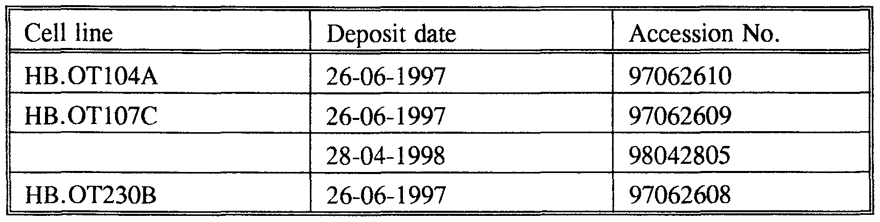
- hybridoma cell lines which have been specifically referred to above. These cell lines have been deposited at the European Collection of Animal Cell Cultures (ECACC), Centre for Applied Microbiology and Research, Salisbury, Wiltshire SP4 OJG, United Kingdom, under the accession numbers and dates shown in the following table:
- Specific antibodies and other binding molecules in accordance with the invention are useful in diagnosis and in therapy.
- the above defined antibodies and other binding molecules can be used in diagnostic applications in isolation or in combination with antibodies specifically directed to other regions ofthe HBV like the S-region.
- the antibodies directed against the S-region do recognize also the HBV variants which do lack the pre-S region.
- the antibodies and other binding molecules according to the present invention do overcome missing HBV infected individuals in the diagnosis of HBV infection. With these antibodies, the HBsAg test-concept based on the S-region of HBV could be improved. Mutant detection of HBsAg is confirmed.
- the antibodies and other binding molecules according to the present invention are useful tools for the detection of HBV expression in cells and cell extracts both in vivo and in vitro, for purification purposes and for a variety of biochemical and immunological analysis techniques to study the function of these proteins.
- a peptide comprising at least part of the amino acid sequence RDSHPQAMQWNSTTFHQALLDPRVRGLYFPAGGSSSGT.
- a preferred embodiment is a peptide comprising at least part of the amino acid sequence RDSHPQAMQWNSTTFHQAL.
- a preferred specific fragment is MQWN.
- a fragment is a peptide comprising at least part of the amino acid sequence SHPQAMQWNSTTFHQALLDPR.
- a preferred specific fragment is STTFHQA.
- a fragment is a peptide comprising at least part of the amino acid sequence ALLDPRVRGLYFPAGGSSSGT.
- a preferred specific fragment is VRGLYFPA.
- Synthetic peptides can be synthesized highly reproducible and are easily purified, thus well suited for further improvement and standardization of HBV-serology.
- Synthetic peptides have the advantage of being chemically well defined, thus allowing easy and reproducible production at high yields, well suited for application in diagnostic assays which can be manufactured and used with greater reproducibility.
- the peptides according to the present invention have the great advantage that these are of a safe non-infectious origin.
- peptide refers to a molecular chain of amino acids with a biological activity, and does not refer to a specific length of the product. Thus inter alia, proteins, fusion-proteins or -peptides oligopeptides and polypeptides are included.
- peptides according to the invention can be modified in vivo or in vitro, for example by glycosylation, amidation, carboxylation or phosphorylation.
- the term "at least a part of” as used herein means a subsequence of the identified amino acid sequence. Said part or fragment is a region having one or more antigenic determinants of the HBsAg proteins. Fragments can inter alia be produced by enzymatic cleavage of precursor molecules, using restriction endonucleases for the DNA and proteases for the (poly)peptides. Other methods include chemical synthesis of the fragments or the expression of peptide fragments by DNA fragments.
- the preparation of the peptides or fragments thereof according to the invention is effected by means of one of the known organic chemical methods for peptide synthesis or with the aid of recombinant DNA techniques which are also known in the art.
- Peptides according to the present invention can also be combined in a single molecule.
- the peptides according to the invention can likewise be prepared with the aid of recombinant DNA techniques. This possibility is of importance particularly when the peptide is incorporated in a repeating sequence ("in tandem") or when the peptide can be prepared as a constituent of a (much larger) protein or polypeptide or as a fusion protein with, for example, (part of) ⁇ - galactosidase. This type of peptides therefore likewise falls within the scope of the invention.
- the peptides or fragments thereof prepared and described above can also be used to produce antibodies, both polyclonal and monoclonal and other binding molecules.
- Methods of diagnosis in accordance with the invention can be carried out in vitro.
- a method for the diagnosis of hepatitis B comprising contacting the sample suspected to contain hepatitis B particles or antigens with the specific binding molecule as described above.
- a preferred embodiment of the present invention is directed to a method for the diagnosis of hepatitis B, the method comprising contacting the sample suspected to contain hepatitis B particles or antigens with at least one specific binding molecule directed to the S-region of HBV and at least one specific binding molecule according to the present invention.
- a particularly suitable method for the detection of HBsAg in a sample is based on a competition reaction between a peptide according to the invention provided with a labeling substance and hepatitis B particles or antigens (present in the sample) whereby the peptide and the antigen are competing with the specific binding molecule according to the present invention attached to a solid support.
- Another preferred embodiment of the present invention is directed to a method for the detection of antibodies to hepatitis B, the method comprising contacting the sample suspected to contain antibodies to hepatitis B particles or antigens with at least one peptide according to the present invention.
- supports are used to immobilise the binding molecules or peptides according to the present invention.
- Supports which can be used are, for example, the inner wall of a microtest well or a cuvette, a tube or capillary, a membrane, filter, test strip or the surface of a particle such as, for example, a latex particle, an aldehyde particle (such as a ceramic magnetizable particle with active aldehyde surface groups), an erythrocyte, a dye sol, a metal sol or metal compound as sol particle, a carrier protein such as BSA or KLH.
- Labeling substances may be radioactive or non-radioactive such as a radioactive isotope, a fluorescent compound, a chemiluminescent compound, an enzyme, a dye sol, metal sol or metal compound as sol particle.
- a radioactive isotope a fluorescent compound
- a chemiluminescent compound an enzyme
- a dye sol a metal sol or metal compound as sol particle.
- either the specific binding molecules within the scope of the invention can be labeled, or other specific binding molecules, which bind to them are labeled.
- Immunoassays including radioimmunoassays
- immunometric assays including immunometric radioassays and enzyme-linked immunosorbent assays
- In vitro assays may take many formats. Some depend upon the use of labeled specific binding molecules such as antibodies (whose use is included within the scope of the invention), whereas some detect the interaction of antibody (or other specific binding molecule) and antigen by observing the resulting precipitation. These are well known in the art.
- kits for the detection of a hepatitis B particle or antigen, the kit comprising a specific binding molecule as described above and means for detecting whether the specific binding molecule is bound to a hepatitis B particle or antigen.
- the assay methodology may for example be any of the assays referred to above.
- Competitive and, especially, sandwich immunoassay kits are preferred.
- the specific binding molecule and the detection means may be provided in separate compartments of the kit.
- the specific binding molecule may be provided bound to a solid support.
- the detection means may comprise a detectable labeled second specific binding molecule (which itself may be an antibody (monoclonal but preferably polyclonal), which bind to the bound hepatitis B particle or antigen.
- kits for the detection of a hepatitis B particle or antigen comprising at least one binding molecule directed to the S-region of HBV and at least one specific binding molecule according to the present invention and means for detecting whether the specific binding molecule is bound to a hepatitis B particle or antigen.
- kits for the detection of antibodies to hepatitis B comprising at least one peptide according to the present invention and means for detecting whether the peptide is bound to antibodies to hepatitis B.
- test kit Carrying out a sandwich reaction, for the detection of antibodies to hepatitis B the test kit may comprise, for example, a peptide according to the invention coated to a solid support, for example the inner wall of a microtest well, and either a labeled peptide according to the invention or a labeled anti-antibody.
- the test kit may comprise a peptide according to the invention coated to a solid support, and a labeled specific binding molecule according to the invention.
- the antibodies and other binding molecules according to the present invention can be useful in the development and quality control of serologic assays and for the direct detection of HBV.
- the murine anti-PreS antibodies HB.OT107C. HB.OT104A and HB.OT230B were produced by injecting Balb/c mice with native HBsAg in Freund's complete adjuvans.
- the HBsAg was obtained from sera of HBV infected donors. After two months, mice were boosted with the antigen mixed in Freund's incomplete adjuvans, which was repeated after two weeks. Three days later the spleen was removed and splenic lymphocytes were fused according to CRL manual G6-1 with P3x63Ag8653 (ATCC CRL 1580) mouse myeloma cells using polyethylene glycol 1000 according to standard methods.
- Hybridoma cells were screened for the production of anti-PreS antibodies using specific peptides and/or denatured HBsAg. Several cycles of cloning by limiting dilution were needed to achieve a stable cell line of 100% clonality.
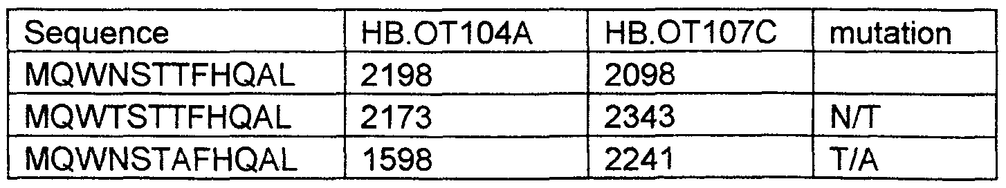
- the minimal epitope of HB.OT107C and HB.OT104A was determined by using the standard pepscan method (Geysen et al. , 1987, J. Immunol. Meth. 102, 259-274). Peptides based on sequence "MQWNSTTFHQAL” were shortened at the N- or C- terminal and tested with HB.OT104A and HB.OT107C. Similar experiments were performed with shortened peptides based on sequence "LDPRVRGLYFPA" and Mab HB.OT230B. For determination of the minimal epitope of HB.OT107C the series was extended with peptides based on sequence "STTFHQAL" shortened at the C-terminal end.
- Results shown in Table 3 indicate that the first methionine (M) at the N- terminus of the Pre-S2 sequence is essential for reactivity of Mab HB.OT104A.
- the minimal epitope probably includes at least amino acids "MQW”.
- HB.OT107C reactivity declines if the serine (S) or the threonine (T) at the C-terminus is removed.
- the epitope of HB.OT107C might include amino acids S (124) and T (126).
- Reactivity of HB.OT230B was affected by replacement of glycine (G-138) by valine (V) and phenylalanine (F-141) by leucine (L). These results are in agreement with the ala-study which showed that replacement of glycine (G-138) by alanine (A) and phenylalanine (F-141) by alanine (A) decreased the reactivity of the peptide.
- 'basic assay' only murine monoclonal antibodies are used directed to the S-region of HBsAg.
- a murine monoclonal antibody directed to the pre-S region of HBsAg is added to the monoclonal antibodies already present in the basic assay.
- microelisa wells were coated with said monoclonal antibodies.
- Each microelisa well contained an HRP-labeled anti-HBs conjugate sphere comprising antibody to HBsAg (anti-HBs) coupled to horseradish peroxidase (HRP) which serves as the conjugate with tetramethylbenzidine (TMB) and peroxide as the substrate.
- HRP-labeled anti-HBs conjugate sphere comprising antibody to HBsAg (anti-HBs) coupled to horseradish peroxidase (HRP) which serves as the conjugate with tetramethylbenzidine (TMB) and peroxide as the substrate.
- HRP-labeled anti-HBs conjugate sphere comprising antibody to HBsAg (anti-HBs) coupled to horseradish peroxidase (HRP) which serves as the conjugate with tetramethylbenzidine (TMB) and peroxide as the substrate.
- HRP horseradish peroxidase
- test sample or appropriate controls were incubated in the microelisa wells.
- the conjugate sphere dissolves in the sample and a solid phase antibody/HBsAg/enzyme-labeled antibody complex is formed. Following a wash and incubation with TMB substrate, color is produced which turns yellow when the reaction is stopped with sulfuric acid. If HBsAg is present in the sample, a color develops. However, when the sample is free of HBsAg, no or low color forms with the addition of substrate. Within limits, the amount of HBsAg in the sample is proportional to the color development. The sample was tested undiluted.
- the HBsAg-mutant patient serum is:
- Example 6 Screening recombinant HBsAg-mutant panel
- the HBsAg mutant panel was prepared as follows: Both Cos I cells and HepG2 cells were cultured as monolayer in 75 cm 2 rouxflasks in 10 ml Dulbecco's MEM medium containing 10% Foetal Calf Serum (FCS) in a 37°C incubator. Four hours before electroporation the medium was refreshed.
- FCS Foetal Calf Serum
- the cells were diluted in 5 ml medium and 10 minutes 233 g centrifuged in a Beckmann GP centrifuge.
- For Cos I cell lines 1.5 x 10 6 cells were diluted in 200 ⁇ l Phosphate Buffered Saline (PBS), for HepG2 cell lines 5 x 10 6 cells were diluted in 500 ⁇ l medium. Then 20 ⁇ l steril DNA solution of each HBsAg recombinant construct was added to the cell suspension. The mixtures were incubated on ice for 5 minutes and pipetted into a 4 mm cuvet.
- PBS Phosphate Buffered Saline
- the constructs, i.e. Cos I cell lines and HepG2 cell lines were tested seperately in two test runs.
- Run 1 for all the Cos I cell lines the cut off value for the basic assay was 126, the cut off value for the improved assay was 133.
- Run 2 for the HepG2 cell lines (wildtype and 129/133) the cut off value for the basic assay was 103, the cut off value for the improved assay was 96.
Abstract
Description


Claims
Priority Applications (5)
| Application Number | Priority Date | Filing Date | Title |
|---|---|---|---|
| AT98937559T ATE295375T1 (en) | 1997-06-27 | 1998-06-22 | ANTIBODIES AND OTHER BINDING MOLECULES AGAINST HEPATITIS B VIRUS ANTIGENS |
| DE69830174T DE69830174T2 (en) | 1997-06-27 | 1998-06-22 | ANTIBODIES AND OTHER BINDING MOLECULES AGAINST HEPATITIS B VIRUSANTIGENE |
| EP98937559A EP0993470B1 (en) | 1997-06-27 | 1998-06-22 | Antibodies and other binding molecules specific for hepatitis b viral antigens |
| AU86308/98A AU8630898A (en) | 1997-06-27 | 1998-06-22 | Antibodies and other binding molecules specific for hepatitis b viral antigens |
| US09/446,787 US6541198B1 (en) | 1997-06-27 | 1998-06-22 | Antibodies and other binding molecules specific for hepatitis B viral antigens |
Applications Claiming Priority (4)
| Application Number | Priority Date | Filing Date | Title |
|---|---|---|---|
| EP97201968 | 1997-06-27 | ||
| EP97201968.1 | 1997-06-27 | ||
| EP98201458 | 1998-05-08 | ||
| EP98201458.1 | 1998-05-08 |
Publications (2)
| Publication Number | Publication Date |
|---|---|
| WO1999000424A2 true WO1999000424A2 (en) | 1999-01-07 |
| WO1999000424A3 WO1999000424A3 (en) | 1999-04-22 |
Family
ID=26146644
Family Applications (1)
| Application Number | Title | Priority Date | Filing Date |
|---|---|---|---|
| PCT/EP1998/004018 WO1999000424A2 (en) | 1997-06-27 | 1998-06-22 | Antibodies and other binding molecules specific for hepatitis b viral antigens |
Country Status (7)
| Country | Link |
|---|---|
| US (1) | US6541198B1 (en) |
| EP (1) | EP0993470B1 (en) |
| AT (1) | ATE295375T1 (en) |
| AU (1) | AU8630898A (en) |
| DE (1) | DE69830174T2 (en) |
| ES (1) | ES2243002T3 (en) |
| WO (1) | WO1999000424A2 (en) |
Cited By (5)
| Publication number | Priority date | Publication date | Assignee | Title |
|---|---|---|---|---|
| WO2002103356A1 (en) * | 2001-06-18 | 2002-12-27 | Alexander Schoenfeld | Prophylactic article useful for both protection and diagnosis and method for use and production thereof |
| EP1297141A1 (en) * | 2000-05-29 | 2003-04-02 | Yuhan Corporation | A variable region of the monoclonal antibody against the hbv s-surface antigen and a gene encoding the same |
| KR100455902B1 (en) * | 2001-07-18 | 2004-11-12 | 주식회사 펩트론 | Surface protein preS1 peptide of HBV inducing immune activity of T-lymphocytes |
| KR101334470B1 (en) * | 2008-08-18 | 2013-11-29 | 나이키 인터내셔널 엘티디. | Orientation marker for golf club having releasable and interchangeable head and shaft connections |
| US20140323332A1 (en) * | 2013-04-26 | 2014-10-30 | Bio-Rad Laboratories, Inc. | Multiplex hepatitis b assay |
Citations (2)
| Publication number | Priority date | Publication date | Assignee | Title |
|---|---|---|---|---|
| EP0456215A1 (en) * | 1990-05-11 | 1991-11-13 | Abbott Laboratories | Monoclonal antibodies to pres2 and pres1 polypeptides of the hepatitis B viral envelope |
| EP0521348A1 (en) * | 1991-06-18 | 1993-01-07 | Korea Institute Of Science And Technology | Chimeric antibody with specificity for hepatitis B virus pre-S2 surface antigen and cell line producing same |
-
1998
- 1998-06-22 ES ES98937559T patent/ES2243002T3/en not_active Expired - Lifetime
- 1998-06-22 WO PCT/EP1998/004018 patent/WO1999000424A2/en active IP Right Grant
- 1998-06-22 EP EP98937559A patent/EP0993470B1/en not_active Expired - Lifetime
- 1998-06-22 DE DE69830174T patent/DE69830174T2/en not_active Expired - Lifetime
- 1998-06-22 US US09/446,787 patent/US6541198B1/en not_active Expired - Lifetime
- 1998-06-22 AU AU86308/98A patent/AU8630898A/en not_active Abandoned
- 1998-06-22 AT AT98937559T patent/ATE295375T1/en not_active IP Right Cessation
Patent Citations (2)
| Publication number | Priority date | Publication date | Assignee | Title |
|---|---|---|---|---|
| EP0456215A1 (en) * | 1990-05-11 | 1991-11-13 | Abbott Laboratories | Monoclonal antibodies to pres2 and pres1 polypeptides of the hepatitis B viral envelope |
| EP0521348A1 (en) * | 1991-06-18 | 1993-01-07 | Korea Institute Of Science And Technology | Chimeric antibody with specificity for hepatitis B virus pre-S2 surface antigen and cell line producing same |
Non-Patent Citations (7)
| Title |
|---|
| A. NEURATH ET AL.: "Antibodies to synthetic peptides from the Pre-S1 and Pre-S2 regions of one subtype of the hepatitis B virus (HBV) envelope protein recognize all HBV subtypes." MOLECULAR IMMUNOLOGY, vol. 24, no. 9, September 1987, pages 975-980, XP002052884 OXFORD, GB * |
| H. MEISEL ET AL.: "Fine mapping and functional characterization of two immuno-dominant regions from the preS2 sequence of hepatitis B virus." INTERVIROLOGY, vol. 37, no. 6, November 1994, pages 330-339, XP002052879 BASEL, SWITZERLAND * |
| L. MIMMS ET AL.: "Discrimination of hepatitis B (HBV) subtypes using monoclonal antibodies to the PreS1 and PreS2 domains of the viral envelope." VIROLOGY, vol. 176, no. 2, June 1990, pages 604-619, XP002052883 SAN DIEGO, CA, USA * |
| M-K. LEE ET AL.: "Epitope mapping of PreS2 of the hepatitis B virus surface antigen against a conformation-dependent monoclonal antibody using synthetic peptides." BIOCHEMISTRY AND MOLECULAR BIOLOGY INTERNATIONAL, vol. 40, no. 6, December 1996, pages 1077-1085, XP002052880 SYDNEY, AUSTRALIA * |
| M-K. LEE ET AL.: "Structure-antigenicity relationship of peptides from the Pre-S2 region of the hepatitis B virus surface antigen." BIOCHEMISTRY AND MOLECULAR BIOLOGY INTERNATIONAL, vol. 34, no. 1, September 1994, pages 159-168, XP002052881 SYDNEY, AUSTRALIA * |
| S. DASH ET AL.: "Significance of natural polymerized albumin and its receptor in hepatitis B infection of hepatocytes." HEPATOLOGY, vol. 13, no. 1, January 1991, pages 124-142, XP002052885 BALTIMORE, MD, USA * |
| S-S. PARK ET AL.: "Expression of soluble and functional single-chain antibody in Escherichia coli." MOLECULES AND CELLS, vol. 4, no. 4, 1994, pages 413-417, XP002052882 SEOUL, KOREA * |
Cited By (7)
| Publication number | Priority date | Publication date | Assignee | Title |
|---|---|---|---|---|
| EP1297141A1 (en) * | 2000-05-29 | 2003-04-02 | Yuhan Corporation | A variable region of the monoclonal antibody against the hbv s-surface antigen and a gene encoding the same |
| EP1297141A4 (en) * | 2000-05-29 | 2004-07-14 | Yuhan Corp | A variable region of the monoclonal antibody against the hbv s-surface antigen and a gene encoding the same |
| WO2002103356A1 (en) * | 2001-06-18 | 2002-12-27 | Alexander Schoenfeld | Prophylactic article useful for both protection and diagnosis and method for use and production thereof |
| KR100455902B1 (en) * | 2001-07-18 | 2004-11-12 | 주식회사 펩트론 | Surface protein preS1 peptide of HBV inducing immune activity of T-lymphocytes |
| KR101334470B1 (en) * | 2008-08-18 | 2013-11-29 | 나이키 인터내셔널 엘티디. | Orientation marker for golf club having releasable and interchangeable head and shaft connections |
| US20140323332A1 (en) * | 2013-04-26 | 2014-10-30 | Bio-Rad Laboratories, Inc. | Multiplex hepatitis b assay |
| US9927439B2 (en) * | 2013-04-26 | 2018-03-27 | Bio-Rad Laboratories, Inc. | Multiplex hepatitis B assay |
Also Published As
| Publication number | Publication date |
|---|---|
| WO1999000424A3 (en) | 1999-04-22 |
| US6541198B1 (en) | 2003-04-01 |
| EP0993470A2 (en) | 2000-04-19 |
| ATE295375T1 (en) | 2005-05-15 |
| DE69830174D1 (en) | 2005-06-16 |
| DE69830174T2 (en) | 2006-01-26 |
| ES2243002T3 (en) | 2005-11-16 |
| AU8630898A (en) | 1999-01-19 |
| EP0993470B1 (en) | 2005-05-11 |
Similar Documents
| Publication | Publication Date | Title |
|---|---|---|
| US5869232A (en) | Antigen/antibody specificity exchanger | |
| JP4351153B2 (en) | Method for simultaneously detecting antigens and antibodies of infectious microorganisms | |
| US6469143B2 (en) | Antigen/antibody specificity exchanger | |
| EP0388602B1 (en) | Monoclonal antibody for differentiating HIV-2 from HIV-1 seropositive individuals | |
| US6030616A (en) | Hepatitis B escape mutant specific binding molecules | |
| CA2252466C (en) | Hepatitis b monoclonal antibodies | |
| Wasenauer et al. | A cysteine and a hydrophobic sequence in the noncleaved portion of the pre-C leader peptide determine the biophysical properties of the secretory core protein (HBe protein) of human hepatitis B virus | |
| Schlicht et al. | The quaternary structure, antigenicity, and aggregational behavior of the secretory core protein of human hepatitis B virus are determined by its signal sequence | |
| JP2818755B2 (en) | Novel branched hybrid and cluster peptides useful for diagnosis and detection of non-A non-B hepatitis | |
| Karthigesu et al. | A hepatitis B virus variant found in the sera of immunised children induces a conformational change in the HBsAg “a” determinant | |
| AU628156B2 (en) | Monoclonal antibodies to pres2 and pres1 polypeptides of the hepatitis b viral envelope | |
| EP0993470B1 (en) | Antibodies and other binding molecules specific for hepatitis b viral antigens | |
| US20060269953A1 (en) | Novel hepatitis B virus | |
| AU646039B2 (en) | Monoclonal antibodies to Pres2 and Pres1 polypeptides of the hepatitis B viral envelope | |
| AU682335B2 (en) | Monoclonal antibodies and anti-idiotypic antibodies to hepatitis C virus | |
| KR0127148B1 (en) | A SV40 EXPRESSION VECTOR CONTAINING HBxAS AS AN EXPRESSION MARKER | |
| Wasenauer et al. | Relevance of cysteine residues for biosynthesis and antigenicity of human hepatitis B virus e protein | |
| KR20000064637A (en) | How to predict the outcome of infection with non-hepatitis B | |
| WO1994021812A2 (en) | Hepatitis b escape mutant specific binding molecules | |
| World Health Organization | The use of synthetic antigens for diagnosis of infectious diseases: report of a WHO scientific group [meeting held in Geneva from 30 November to 4 December 1987] |
Legal Events
| Date | Code | Title | Description |
|---|---|---|---|
| AK | Designated states |
Kind code of ref document: A2 Designated state(s): AU CA ID JP KR US |
|
| AL | Designated countries for regional patents |
Kind code of ref document: A2 Designated state(s): AT BE CH CY DE DK ES FI FR GB GR IE IT LU MC NL PT SE |
|
| DFPE | Request for preliminary examination filed prior to expiration of 19th month from priority date (pct application filed before 20040101) | ||
| AK | Designated states |
Kind code of ref document: A3 Designated state(s): AU CA ID JP KR US |
|
| AL | Designated countries for regional patents |
Kind code of ref document: A3 Designated state(s): AT BE CH CY DE DK ES FI FR GB GR IE IT LU MC NL PT SE |
|
| 121 | Ep: the epo has been informed by wipo that ep was designated in this application | ||
| WWE | Wipo information: entry into national phase |
Ref document number: 1998937559 Country of ref document: EP |
|
| WWE | Wipo information: entry into national phase |
Ref document number: 09446787 Country of ref document: US |
|
| NENP | Non-entry into the national phase |
Ref country code: JP Ref document number: 1999505297 Format of ref document f/p: F |
|
| WWP | Wipo information: published in national office |
Ref document number: 1998937559 Country of ref document: EP |
|
| NENP | Non-entry into the national phase |
Ref country code: CA |
|
| WWG | Wipo information: grant in national office |
Ref document number: 1998937559 Country of ref document: EP |