METHODS FOR MASS SPECTROMETRY ANALYSIS UTILIZING AN INTEGRATED MICROFLUIDICS SAMPLE PLATFORM
CROSS-REFERENCE TO RELATED APPLICATIONS
The present application claims priority to U.S. provisional application Serial No. 60/397,365 filed July 19, 2002, the disclosure of which is incorporated herein by reference in its entirety.
FIELD OF THE INVENTION
The present invention relates to methods for mass spectrometric or other such analysis in which samples are prepared and analyzed in an integrated microfluidics environment.
BACKGROUND OF THE INVENTION
Manipulating fluidic reagents and assessing the results of reagent interactions are central to chemical and biological science. Manipulations include mixing fluidic reagents, assaying products resulting from such mixtures, and separation or purification of products or reagents and the like. Assessing the results of reagent interactions can include autoradiography, spectroscopy, microscopy, photography, mass spectrometry, nuclear magnetic resonance and many other techniques for observing and recording the results of mixing reagents. A single experiment can involve literally hundreds of fluidic manipulations, product separations, result recording processes and data compilation and integration steps. Fluidic manipulations are performed using a wide variety of laboratory equipment, including various fluid heating devices, fluidic mixing devices, centrifugation equipment, molecule purification apparatus, chromatographic machinery, gel electrophoretic equipment and the like. The effects of mixing fluidic reagents are typically assessed by additional equipment relating to detection, visualization or recording of an event to be assayed, such as spectrophotometers, autoradiographic equipment, microscopes, gel scanners, computers and the like.
Because characterization of even simple chemical, biochemical, or biological phenomena requires many different types of laboratory equipment, the modern laboratory is complex, large and expensive. In addition, because so many different types of equipment are used, it has not generally been practical to integrate different types of equipment to improve automation. The need for a laboratory worker to physically perform many aspects of laboratory science imposes
sharp limits on the number of experiments which a laboratory can perform, and increases the undesirable exposure of laboratory workers to toxic or radioactive reagents. In addition, results are often analyzed manually with the selection of subsequent experiments related to initial experiments requiring consideration by a laboratory worker, severely limiting the throughput of even repetitive experimentation.
In attempts to increase laboratory throughput and to decrease exposure of laboratory workers to reagents, various strategies have been employed. For example, robotic introduction of fluids onto n icrotiter plates is commonly performed to speed mixing of reagents and to enhance experimental throughput. More recently, microscale devices for high throughput mixing and assaying of small fluid volumes have been developed. For example, U.S. Patent No. 6,046,056 entitled "High Throughput Screening Assay Systems in Microscale Fluidic Devices" reports technology related to microscale fluidic devices, especially including electrokinetic devices. The devices are generally suitable for assays relating to the interaction of biological and chemical species, including enzymes and substrates, ligands and ligand binders, receptors and ligands, antibodies and antibody ligands, as well as many other assays. Because the devices provide the ability to mix fluidic reagents and assay mixing results in a single continuous process, and because minute amounts of reagents can be assayed, these microscale devices represent a fundamental advance for laboratory science. Additional microfluidics devices, chips, and systems are reported in U.S. Patent No. 6,321,791.
Additional reasons to pursue microfluidics technology include but are not limited to: mass production of field portable and disposable microfluidics platforms at low unit cost, adaptability to high throughput via parallel assay processing, and the requirement for very small volumes of sample, reagents with generation of very small volumes of waste. However, several significant challenges remain for microfluidics technology which include, but are not limited to interfaces between macroscopic and microscopic environments, mixing of fluids without turbulence in a timely fashion, and fluid transport (Weigl et al., Adv. Drug Delivery Rev., 2003, 55, 349-337).
Mass spectrometry is a very sensitive and versatile technique for molecular characterization which is being increasingly applied in biological sciences in applications such as nucleic acid sequencing and detection of single nucleotide polymorphisms (see for example U.S. Patent Nos 6,194,144 and 6,207,370). An absolute requirement for mass spectrometry, however, is that samples are provided in a reproducible and ionizable format. In electrospray and MALDI- TOF mass spectrometry applications, microfluidics devices have been used to remove interacting
particles and ions from the sample prior to ionization. An example of such a microfluidics system is a compact disk with microfabricated desalting, elution and crystallization functionalities sequentially arranged along a radius from the outer edge if the disk towards the center, allowing the processes to be arrayed radially for parallel processing of multiple samples (Inganas et al, Micro Total Analysis Systems, 2001, Kluwer Academic, 91-92.) The preceding example however, has not been adapted for earlier stage processes of nucleic acid samples such as cell lysis, nucleic acid capture and amplification.
McMillan et al. have reported the application of microfluidics technologies to field portable disposable fluid cartridges that enable sample preparation of viruses or bacteria with processes including specimen dilution, coarse filtration, target microbe extraction/separation, microbe lysis, DNA/RNA purification and PCR or RT-PCR (real time PCR) (McMillan, et al., Microbial Biosystems: New Frontiers - Proceedings of the 8th International Symposium on Microbial Ecology Bell, C.R., Brylinsky, M., Johnson-Green P. (eds) Atlantic Canada Society for Microbial Ecology, Halifax, Canada.) It has not been taught, however, that these microfluidics technologies are compatible with integration of the additional sample process steps required for specialized biomolecular analyses carried out by sophisticated and powerful methods such as mass spectrometry.
There remains a need for development of methods that integrate microfluidics operations for rapid fluid manipulation of nucleic acids and other such biomolecules in process steps that can include cell lysis, sedimentation, capture, amplification and desalting prior to high- throughput nucleic acid characterization. The present invention addresses this need by providing a basis for integrating various combinations of nucleic acid isolation, capture and amplification microfluidics processes with a microfluidics desalting process, an essential treatment step prior to nucleic acid characterization by mass spectrometry.
SUMMARY OF THE INVENTION
The present invention provides methods of characterizing a biomolecule in a biological sample comprising introducing the biological sample to a microfluidic scale integrated fluid manipulation platform that is serially connected to a sample characterization module, and characterizing the biomolecule with the sample characterization module.
The present invention also provides microfluidic scale integrated fluid manipulation platforms comprising at least two modules selected from the group consisting of a sample preparation module, a nucleic acid amplification module, and a desalting module.
The present invention also provides biomolecule characterization apparati comprising any microfluidic scale integrated fluid manipulation platforms of the invention and a serial sample characterization module.
The present application also provides methods for determining the base composition of an unknown nucleic acid from a biological sample comprising fluidically manipulating the unknown nucleic acid from the biological sample using at least one microfluidics platform containing an interface to a mass spectrometer, wherein a nucleic acid amplification product is obtained from the fluidic manipulation, determining the molecular mass of the nucleic acid amplification product with the mass spectrometer, wherein the molecular mass is associated with a base composition, and identifying a match between the base composition of the nucleic acid amplification product and a base composition of a known bioagent, thereby determining the base composition of the unknown nucleic acid.
BRIEF DESCRIPTION OF THE DRAWINGS
Figure 1 is a schematic representation of a typical nucleic acid capture microfluidics module.
Figure 2 (top) shows a representative FTICR mass spectrum of a PCR product obtained from a sample of E. coli wherein the nucleic acid capture microfluidics module of Figure 1 was employed (bottom), an FTICR mass spectrum of the same PCR product obtained from a sample of E. coli, which was purified without the nucleic acid capture microfluidics module.
Figure 3 is a schematic representation of a typical desalting microfluidics module.
Figure 4 shows a representative FTICR mass spectrum of a PCR product obtained from a sample of Staphylococcus, which was desalted using the desalting microfluidics module of Figure 3.
Figure 5 is a schematic representation of a typical integration of sample preparation, nucleic acid capture, nucleic acid amplification, and desalting within a single platform.
Figure 6 shows a representative base composition signature (BCS) spectra from PCR products from Staphylococcus aureus (S. aureus 16S_1337F) and Bacillus anthracis (B. anthr. 16S_1337F) amplified using the same primers. The two strands differ by only two (AT— »CG) substitutions and are clearly distinguished on the basis of their BCS.
DETAILED DESCRIPTION OF EMBODIMENTS A. Overview of the Invention
The present invention provides methods for the application of integrated microscale operations in rapid fluid sample preparation of biomolecules, preferably nucleic acids, including cell lysis, sedimentation, capture, and purification wherein the biomolecules nucleic acids are characterized. Embodiments of the invention feature an integrated fluid manipulation platform which may feature any desired combination of modules for, for example, sample preparation, nucleic acid capture, nucleic acid amplification, desalting, or other such processes. The modules may be performed in serially connected platform components, or may be combined into a single component, such as a microfluidics card or chip. An output of this fluid manipulation platform may then be serially directed to a sample characterization module such as a mass spectrometer.
As used herein, the term "microfluidics" refers to the science of designing, manufacturing, and formulating devices and processes that deal with volumes of fluid which are preferably on the order of nanoliters (symbolized nl and representing units of 10" liter) or picoliters (symbolized pi and representing units of 10"12 liter). The devices themselves have dimensions ranging from millimeters (mm) down to micrometers (μm).
As used herein the term "fluidic manipulation" refers to any single step process wherein a fluid is transferred, mixed, filtered or otherwise altered in composition via introduction and/or removal of solvents, reagents or other components. The objective of fluidic manipulation in the context of this invention is the preparation of a biomolecule for characterization.
As used herein, "biological information" includes, but is not limited to, a biological sequence (such as an amino acid or nucleotide sequence), a molecular mass, and/or a base composition, bioagent identity, or any combination thereof.
Integration of microfluidics platform and modules thereof adapted for sample preparation, amplification and desalting of nucleic acids prior to detection and identification of bioagents described in examples hereinbelow will significantly enhance the efficiency and portability of the invention.
"Integrated fluid manipulation" refers to a series of fluid manipulation processes conducted serially upon the same sample aliquot within a closed system.
"Serial sample characterization" refers to a system or module which further integrates an integrated fluidic manipulation platform with a sample characterization system or module such as mass spectrometry, wherein the sample output provided by the fluidic manipulation platform is
applied directly to the sample characterization system or module via an interface within the closed system.
As used herein the terms "microfluidics system," "microfluidics card," and "microfluidics chip" and "micro-chip" refer to the physical platform upon which microfluidics operations or processes are carried out. Referring to Figure 5, examples of processes carried out with microfluidics platforms (100) described in some embodiments of the present invention include, but are not limited to, sample preparation (200), nucleic acid capture (300), nucleic acid amplification (400), and desalting (500). Components of microfluidics platforms may include, but are not limited to, sample input channels, solvent input channels, reagent input channels, fluid mixing means, cell lysis means, temperature control means, biomolecule capture membranes, and/or chromatographic separation resins.
One embodiment of the present invention is an integrated microfluidics processing platform comprising individual microfluidics platforms (modules, chips or cards) for distinct biomolecule processing steps, each of which are in fluid communication with preceding or succeeding processing steps, or an equivalent microfluidics processing system which may comprise a single platform (module, chip or card) upon which all microfluidics processing steps are integrated.
B. Sample Preparation
The process of sample preparation as defined herein comprises the release of a biomolecule of interest from a cell, bacterium, spore, or virus via processes of lysis and sedimentation of biological and inorganic particulate matter followed by isolation of said biomolecule. The step of isolation of the biomolecule of interest typically involves filtration and/or chromatographic separation of the biomolecule from biological matter.
One skilled in the art will recognize that biomolecule sample preparation can be readily performed by any combination of modules that provide particular applications such as specialized lysis required to release biomolecules from spores, bacteria, viruses, and human or animal cells or tissues.
In some embodiments in which the sample preparation is targeted toward nucleic acid biomolecules, an additional module may provide nucleic acid capture in which nucleic acids are bound to a membrane or resin. One with relevant skill in the art will recognize that DNA capture microfluidics module can be readily adapted to utilize different reagents and/or nucleic acid isolation materials for isolation of nucleic acids such as RNA and non-native oligonucleotides
[which may comprise chemical modifications such as modified bases, substituted ribose units and alternative linker groups) from a variety of sample sources which may contain biological, synthetic, or environmental contaminants.
The biological sample can be any sample taken from or including, but not limited to, samples of water, land, air, lakes, rivers, oceans, streams, water treatment systems, rainwater, groundwater, water table, reservoirs, wells, bottled water, ventilation systems, airplane cabins, schools, hospitals, mass transit locations such as subways, train stations, airports, blood and other bodily fluids and tissues, living animals, deceased animals, foodstuff, cosmetics, container, package, box, envelope, mail tube, railroad box car, organ donors, organs from donors, livestock such as cows, pigs, sheep, chickens, turkeys, goats, and the like. The biological sample can include bioagents such as, but not limited to, a bacterium, virus, parasite, fungus, spore, cell, or tissue. The biomolecule can comprise a nucleic acid, protein, carbohydrate, or lipid molecule(s).
In some embodiments, the sample preparation microfluidics module comprises a sample input channel, a plurality of solvent input channels, a fluid mixing means, a cell lysis means, and/or a sedimentation means, or any combination thereof. The sample input channel and solvent input channel(s) may be constructed as a port in the sample preparation microfluidics module, which can be amenable to communication with a syringe, pipette, eyedropper, and the like. A fluid mixing means may, for example, comprise a chamber amenable to a mixing motion such as shaking, stirring, rocking, and the like. The fluid mixing means may have a stir bar. A cell lysis means may be, for example, a sonicator or any chemical means known to lyse cells. A sedimentation means may be, for example, a chamber amenable to centrifugation or mechanical (such as a membrane or other filtration system) or chemical separation of components. Examples of each of these features are provided in the Examples.
In some embodiments, referring to Figure 1, the nucleic acid capture microfluidics module (300) may comprise reservoir for sample collection (310), a sample input channel (350), a plurality of solvent input channels or fluid loading ports (330), reagent channels (340), a fluid mixing means (390), and/or a nucleic acid capture membrane (320), a fluid waste line (360), reagent reservoirs (370), a series of valves (380), or any combination thereof. The sample input channel and solvent input channel(s) may be constructed as a port in the capture microfluidics module, which can be amenable to communication with a syringe, pipette, eyedropper, and the like. The sample input channel can also be in communication with the sample preparation microfluidics module. A fluid mixing means may, for example, comprise a chamber amenable to a mixing motion such as shaking, stirring, rocking, and the like. The fluid mixing means may
have a stir bar. A nucleic acid capture membrane can be any membrane or like material known in the art for capturing nucleic acid molecules. Examples of each of these features are provided in the Examples.
C. Nucleic Acid Amplification
Methods of nucleic acid amplification such as PCR are increasingly being implemented in clinical laboratories and offer high sensitivity and specificity. Recent advances in PCR chemistry and thermal cycling technology now enable nucleic acid analysis to be performed in a matter of minutes instead of hours. PCR technology, including RT-PCR, is familiar to those of skill in the art. Examples of other nucleic acid amplification methods that may be incorporated into microfluidics platforms and modules include, but are not limited to, ligase chain reaction and strand displacement amplification.
In some embodiments, the nucleic acid amplification module comprises a sample input channel, a plurality of solvent or reagent input channels, a fluid mixing means, and/or a temperature control means, or any combination thereof. The sample input channel, a plurality of solvent or reagent input channels, and a fluid mixing means can be present as described above. The sample input channel can also be in communication with the capture microfluidics module or the sample preparation module, or both. A temperature control means can include, for example, a thermistor, thermostat, heating and cooling elements and the like. Examples of each of these features are provided in the Examples.
D. Preparation of Samples for Specialized Methods of Analysis
Some sample analysis techniques require particular sample preparation steps. For example, mass spectrometry analysis advantageously includes a desalting step. The process of desalting, as used herein, refers to the removal of small molecule reagents and/or contaminants from a biomolecule sample. An example of desalting in the context of the present invention comprises the removal of excess reagents employed in the amplification of nucleic acids which may include nucleotide triphosphates, and various buffer salts. Removal of such agents typically involves chromatographic separations via ion exchange or gel filtration (size exclusion) resins.
In some embodiments, referring to Figure 3, the desalting microfluidics module (500) comprises a sample input channel or reservoir (510), a plurality of solvent input channels, a fluid mixing means, a series of valves (520), a desalting resin reservoir (535) having a resin start (530) and a resin end (540), and/or a sample output reservoir, an overflow reservoir (570), or any
combination thereof. The sample input channel, a plurality of solvent input channels, and a fluid nixing means can be present as described above. The sample input channel can also be in communication with the sample preparation module, nucleic acid amplification module, or nucleic acid capture module, or any combination thereof. A desalting resin reservoir can be a chamber containing any desalting resin known in the art. Examples of each of these features are provided in the Examples.
E. Sample Characterization
The process of "sample characterization" as used herein refers to the acquisition of information describing the inherent characteristics of the sample. Thus, a nucleic acid sequence or base composition, for example would be examples of information characterizing the nucleic acid. Methods employed for sample characterization, particularly for biomolecules, tend to be quite sophisticated and expensive. The level of instrumental sophistication in methods such as mass spectrometry, nuclear magnetic resonance spectroscopy, and other specialized spectroscopic methods often complicates efforts directed towards integration with specialized methods of sample preparation such as those carried out within microfluidics platforms.
The biomolecule characterization means includes, but is not limited to, any instrument that can be used to analyze a biomolecule. One such instrument is a mass spectrometer. Examples of mass spectrometers include, but are not limited to, Fourier transform ion cyclotron resonance mass spectrometry (FT-ICR-MS), ion trap, quadrupole, magnetic sector, time of flight (TOF), Q-TOF, and triple quadrupole.
F. Additional Features
The present invention also provides methods of obtaining information about a nucleic acid molecule. In some embodiments, one or more nucleic acid molecules is isolated from a biological sample by fluidically manipulating the biological sample using at least one microfluidics platform containing an interface to a nucleic acid characterization means. The one or more nucleic acid molecules is introduced to the nucleic acid characterization means by the interface upon completion of the fluidic manipulation. The one or more nucleic acid molecules are analyzed using the nucleic acid characterization means, wherein the characterization provides information about the nucleic acid molecule. Examples of nucleic acid characterization means are described above. Control of either or both of the fluid manipulation and analyzing steps can be carried out by a computer or robot. Any of the biological samples described above can be
utilized, and may contain a bacterium, virus, parasite, fungus, spore, cell or tissue. Further, any combination of the sample preparation microfluidics module, a nucleic acid capture microfluidics module, a nucleic acid amplification microfluidics module, and a desalting microfluidics module described above can be used. The biological information obtained can comprise a nucleic acid sequence, a molecular mass, or a base composition.
Another embodiment of the present invention is a microfluidics processing platform comprising individual microfluidics platforms, modules, chips or cards for distinct biomolecule processing steps, each of which are in fluid communication with preceding or succeeding processing steps, or an equivalent microfluidics processing platform which may comprise a single platform, module, chip or card, upon which all microfluidics processing steps are integrated.
In some embodiments, a microfluidics nucleic acid processing platform comprises a sample introduction unit. A sample preparation microfluidics module in fluid communication with the sample introduction unit can also be included. A nucleic acid capture microfluidics module in fluid communication with the sample preparation microfluidics module such that the product of the sample preparation microfluidics system is introduced to the nucleic acid capture microfluidics module can also be included. A nucleic acid amplification module fluidics system in fluid communication with the nucleic acid capture microfluidics module such that the product of the nucleic acid capture microfluidics module is introduced to the nucleic acid amplification module can also be included. A desalting microfluidics module in fluid communication with the nucleic acid amplification module such that the product of the nucleic acid amplification module is introduced to the desalting microfluidics module can also be included. Referring to Figure 5, an interface (600) for a means for biomolecule characterization (700) in fluid communication with the desalting microfluidics module such that the product of the desalting microfluidics module is introduced to the interface can also be included. The microfluidics nucleic acid processing modules of the present invention can comprise any combination of the aforementioned features.
In some embodiments of the present invention, one, or a plurality, or each of the aforementioned microfluidics modules resides on separate platforms. Alternately, one, or a plurality, or each of the aforementioned microfluidics modules is integrated on a single platform. The platform can be microfluidics card or chip, which are well known to the skilled artisan.
In some embodiments of the present invention, a microfluidics platform comprises any one or more of a microfluidics nucleic acid processing module, a sample preparation
microfluidics module, a nucleic acid capture microfluidics module, a nucleic acid amplification microfluidics module, a desalting microfluidics module, in combination with an intelligent primer. An "intelligent primer" is a primer that binds to a sequence region that flanks an intervening variable region. In some embodiments, these sequence regions that flank the variable region are highly conserved among different species of bioagent. For example, the sequence regions may be highly conserved among all Bacillus species. By the term "highly conserved", it is meant that the sequence regions exhibit between about 80-100%, more preferably between about 90-100% and most preferably between about 95-100% identity.
In some embodiments, the microfluidics platform comprises a body having a plurality of channels comprising an internal geometry. In some embodiments, at least one of the channels traverses at least three dimensions between its proximal and distal end. In other embodiments, at least one of the channels is interrupted by at least one reaction chamber between its proximal and distal end. In other embodiments, the microfluidics system further comprises a material transport system, such as a gas or liquid.
Another embodiment of the present invention is an instrument for preparation and characterization of biomolecules, such as nucleic acid molecules, comprising the hereinabove microfluidics processing platforms with an interface to a means for biomolecule characterization, preferably a mass spectrometer.
In some embodiments, an instrument for preparation and characterization of nucleic acids comprises any microfluidics nucleic acid processing system, such as those described above, attached to a means for biomolecule characterization by fluid or gas communication with an interface, as described above. The means for biomolecule characterization can comprise a molecular mass-determining means, such as a mass spectrometer.
In another embodiment of the present invention, application of microfluidics processing steps in rapid fluid sample preparation of nucleic acids for an extremely rapid and accurate process of detection and identification of bioagents is provided. The processes for detection and/or identification are described in the commonly owned international publication WO 02/070664, which was published September 12, 2002, the disclosure of which is incorporated herein by reference in its entirety. This process combines PCR-based amplification technology (which provides, ter alia, specificity) and a molecular mass or base composition detection mode (which provides, wter alia, speed and does not require nucleic acid sequencing of the amplified target sequence) for bioagent detection and/or identification. This embodiment of the present invention leverages ongoing biomedical research in virulence, pathogenicity, drug
resistance and genome sequencing into a method which provides greatly improved sensitivity, specificity and reliability compared to existing methods, with lower rates of false positives.
In some embodiments, a method for determining the base composition of an unknown nucleic acid molecule from a biological sample comprises fluidically manipulating an unknown nucleic acid molecule from a biological sample using at least one microfluidics platform containing an interface to a mass spectrometer. The nucleic acid molecule can be introduced to the mass spectrometer by an interface upon completion of the fluidic manipulation. A nucleic acid amplification product can be obtained from the fluidic manipulation. In some embodiments, the nucleic acid molecule is double-stranded, in which case the two strands of the nucleic acid molecule are separated to yield a first strand and a second strand. Molecular masses of the first and/or second strands can be obtained with the mass spectrometer, or other such mass- determining means. In some embodiments, a list of potential base compositions of the first and/or second strands is generated based on the molecular masses previously determined. In some embodiments, a match is identified between the complement of a potential base composition of the first strand and a potential base composition of the second strand, thereby determining the base composition of the unknown nucleic acid molecule.
In some embodiments, the methods further comprise identifying an unknown bioagent in a biological sample based on the base composition of a portion of the unknown nucleic acid molecule, wherein a pair of intelligent primers is used to obtain the amplification product. The base composition identifies the bioagent.
In some embodiments, the bioagent comprises a bacterium, virus, parasite, fungus, spore, cell or tissue. Control of any of the steps can be accomplished by a computer or robot. At least one microfluidics platform comprises a sample preparation microfluidics module, a nucleic acid capture microfluidics module, a nucleic acid amplification microfluidics module, and/or a desalting microfluidics module, or any combination thereof, such as those described above.
The present invention also provides kits comprising any of the aforementioned microfluidics platforms. The kits can be used in the field or outside a laboratory for isolation and processing of biological sample(s) known to contain, or suspected to contain a bioagent(s). The kits can comprise directions for using the same, buffers, primers, intelligent primers, mailing packages for return of the used kit to a mass detection center (any location that has a mass- determining means), and the like. The kits can contain sampling devices or sample introduction units well known in the art for obtaining various types of samples, depending on their form (i.e.,
solid, liquid, or gas), and for introducing the sample into a sample input channel of a sample preparation microfluidics platform.
While the present invention has been described with specificity in accordance with certain of its representative embodiments, the following examples serve only to illustrate the invention and are not intended to limit the same.
Another embodiment of the present invention is an instrument for preparation and characterization of biomolecules, preferably nucleic acids comprising the hereinabove microfluidics processing platform with an interface to a means for biomolecule characterization, preferably a mass spectrometer.
In another embodiment of the present invention, application of microfluidics processing steps in rapid fluid sample preparation of nucleic acids for an extremely rapid and accurate process of detection and identification of bioagents. The process for detection and identification is described in the commonly owned U.S. application Serial No. 09/798,007 filed March 2, 2001, the disclosure of which is incorporated herein by reference in its entirety. This process combines PCR-based amplification technology (which provides specificity) and a molecular mass detection mode (which provides speed and does not require nucleic acid sequencing of the amplified target sequence) for bioagent detection and identification. This embodiment of the present invention leverages ongoing biomedical research in virulence, pathogenicity, drug resistance and genome sequencing into a method which provides greatly improved sensitivity, specificity and reliability compared to existing methods, with lower rates of false positives.
Another embodiment of the present invention provides methods of analyzing a biomolecule in a biological sample. One or more biomolecules from a biological sample is fluidically manipulated using at least one microfluidics system that contains at least one interface to at least one biomolecule characterization means. In some embodiments, the one or more biomolecules is introduced to the biomolecule characterization means by the interface upon completion of the fluidic manipulation. The one or biomolecules is analyzed with the biomolecule characterization means, wherein the characterization provides biological information about the biomolecule(s).
Control of the fluid manipulation and or biomolecule analysis can, in some embodiments, be carried out by a computer or robot. Computers, robots, and systems comprising the same are well known to the skilled artisan.
In some embodiments, at least one microfluidics platform comprises a sample preparation microfluidics module, a nucleic acid capture microfluidics module, a nucleic acid amplification
nicrofluidics module, and/or a desalting microfluidics module, or any combination thereof. Representative examples of each of these systems are provided in the Examples and are known :o those skilled in the art.
EXAMPLES
Example 1: Microfluidics Biomolecule Sample Preparation Systems
A microfluidics system adapted for cell lysis (Pourahmadi et al. Clin. Chem. 2000, 46, 1511-1513) comprises three flat plastic acrylic parts sandwiched together with laser-cut thin (0.762-mm) silicone gasket layers that provide fluid sealing and surfaces for the valves and an ultrasonic horn. It contains chambers for the sample, wash buffer, lysis buffer, neutralization buffer, PCR mix, and waste. In addition, there is a chamber for holding filters for cell capture and lysis and an integrated 100-μL PCR reaction tube. The filter stack assembly consists of layers of 1.27-cm (0.5-inch) filters and glass beads: a 5 μm filter (LoProDyne; Pall Corp), a 1.2 μm filter (Nersapor, Pall Corp), glass beads (106 μm; Sigma), and a 0.2 μm filter (Durapore; Millipore). The tip of an ultrasonic horn (Selfridge and Associates) is interfaced with the cartridge by pressing against the 0.762-mm silicone gasket membrane that forms one wall of the filter stack chamber. All of the cartridge chambers are interconnected by D-shaped microfluidic lines. Fluid flow is driven pneumatically through one or more of six ports on top of the cartridge by a minipump in the instrument, and the flow rate is controlled by digital regulators (Omega). The direction of the fluid flow is controlled by nine membrane valves individually actuated by external solenoid valves pinching against the silicone membrane.
Example 2: Nucleic Acid Capture Microfluidics Systems
One example of a microfluidics system designed for nucleic acid capture is illustrated in Figure 1. The system comprises a custom-designed microfluidics card (Micronics Inc., Redmond, WA) comprising fluid entry and exit ports, pump inlets and channels appropriate for manipulation of microliter volumes of fluids. The microfluidics card comprises an appropriate laminate base, rigid support and an adhesive layer, the compositions of which must be compatible with the reagents in use. Examples of such laminate base materials include but are not limited to: polyethylene terephthalate, cyclic olefin copolymer, polypropylene, polyimide, polycarbonate and the like. A typical rigid support material is PMMA and typical adhesive layers include ACA 501F, ACA 300, ACA AR-MA-65, ACA AR-MA-70, ACA AR-8458 and transfer adhesive NT-1001.
The nucleic acid capture microfluidics system shown in Figure 1 includes four fluid
loading ports input of nucleic acid sample and reagents and valves controlling flow of sample and solvents through four channels. Fluids flowing through the channels are mixed in a fifth channel which is in line with a nucleic acid capture membrane and a reservoir for sample collection.
The reagents of a commercially-obtained microbial DNA isolation kit (ULTRACLEAN™, Mo Bio Laboratories, Inc. Carlsbad, CA) were used to effect a DNA isolation within the microfluidics system at the point immediately subsequent to initial cell lysis and centrifugation to remove cell debris (steps carried out according to the manufacturer's instructions) during the isolation of genomic DNA from a sample of E. coli. Reagent channels 1 and 2 were loaded with the M4 solution, reagent channel 3 was loaded with the MD5 solution and the DNA sample channel was loaded with the lysed E. coli sample (Cone. 240 ng/μL) in MD3 solution (See Figure 1). The nucleic acid capture microfluidics system was operated under automated computer-programmed control. Appropriate flow rates were determined without undue experimentation. Genomic DNA from the E. coli sample was captured by the membrane in the presence of the MD4 solution and was eluted from the microfluidics system with the MD5 solution. The sample thus obtained was amplified by conventional PCR. The PCR product was desalted by conventional means and analyzed by FTICR mass spectrometry and was found to be essentially identical to PCR product of E. coli DNA prepared without the microfluidics system (Figure 2), indicating the utility of the microfluidics system for nucleic acid capture in the process of nucleic acid isolation.
Example 3: Nucleic Acid Amplification Microfluidics Systems
Microfabricated silicon/glass-based devices provide functionalities of simultaneous polymerase chain reaction (PCR) target amplification and sequence-specific electrochemical (EC) detection have been successfully developed (Ming-Hung Lee et al. Lab on a Chip, 2003, 3, 100-105). The microchip-based device has a reaction chamber formed in a silicon substrate sealed by bonding to a glass substrate. Electrode materials such as gold and indium tin oxide (ITO) are patterned on the glass substrate and served as EC detection platforms where DNA probes are immobilized. Platinum temperature sensors and heaters are patterned on top of the silicon substrate for real-time, precise and rapid thermal cycling of the reaction chamber as well as for efficient target amplification by PCR. DNA analyses in the integrated PCR-EC microchip start with the asymmetric PCR amplification to produce single-stranded target amplicons, followed by immediate sequence-specific recognition of the PCR product as they hybridize to the
probe-modified electrode. Two electrochemistry-based detection techniques including metal complex intercalators and nanogold particles are employed in the microdevice to achieve a sensitive detection of target DNA analytes . With the integrated PCR-EC microdevice, the detection of trace amounts of target DNA (as few as several hundred copies) is demonstrated (Ming-Hung Lee et al. Lab on a Chip, 2003, 3, 100-105).
Example 4: Desalting Microfluidics Systems
Desalting represents another operation which is amenable to microscale process development using microfluidics cards which are similar in construction to the microfluidics card of Example 1 . An example of a microfluidics system designed for nucleic acid capture is illustrated in Figure 3 and is designed to effect the removal of excess reagents employed in the amplification of nucleic acids which may include nucleotide triphosphates and various buffer salts.
The choice of composition of the components of the custom-designed desalting microfluidics card (Micronics Inc., Redmond, WA) can be determined according to solvent compatibility as described in Example 1. A non-limiting example of the basic construction of a desalting microfluidics card is similar to that of the nucleic acid capture microfluidics card with exceptions including the replacement of the nucleic acid capture membrane with a nucleic acid desalting resin and the addition of a post-resin reservoir for holding the sample for multiple passes over the resin if necessary. Appropriate desalting resins include but are not limited to: PROP AC WAX™ (Dionex Corporation, Sunnyvale, CA) Silica PEI-300- 15 (Millipore, Billerica, MA) and POROS™ 20 PI (PerSeptive Biosystems, Framingham, MA). •
In a representative example, a 100 μL volume of a 141 nM concentration of a Staphylococcus 106 base-pair PCR product was desalted by washing in sequence with 40 mM NH OAc and 20% MeOH, followed by elution with 4% NH4OH at a flow rate of 1 μL/sec on a desalting card containing a cylinder shaped 1.9 mm length, 1.16 mm diameter column packed with POROS™ 20 PI. Using this method, typically 70 to 90% of PCR product is recovered from the desalting card. The FTICR mass spectrum of a fraction of eluate containing the desalted PCR product is shown in Figure 4 and indicates that the sample was successfully desalted.
Example 5: Integration of Microfluidics Systems
The foregoing examples 1-4 have described common processes involved in manipulation of samples of biomolecules such as nucleic acids. Added value in efficiency and portability is to
be obtained upon integration of such processes either through individual microfluidics systems in a fluid communication sequence, or having the functions of the individual microfluidics systems described herein integrated within a single, multi-functional microfluidics system such as the system depicted schematically in Figure 5 which also indicates the interface of the microfluidics system with a means for biomolecule characterization such as a mass spectrometer. One with relevant skill in the art will recognize that such interfacing functionalities can be adapted to suit a particular bioagent identification task.
Example 6: Mass spectrometry
Mass spectroscopic methods disclosed herein have been described in U.S. application Serial No. 09/798,007 filed March 2, 2001, the disclosure of which is incorporated herein by reference in its entirety.
FTICR Instrumentation: The FTICR instrument is based on a 7 tesla actively shielded superconducting magnet and modified Bruker Daltonics Apex II 70e ion optics and vacuum chamber. The spectrometer is interfaced to a LEAP PAL autosampler and a custom fluidics control system for high throughput screening applications. Samples are analyzed directly from 96- well or 384-well microtiter plates at a rate of about 1 sample/minute. The Bruker data- acquisition platform is supplemented with a lab-built ancillary NT datastation which controls the autosampler and contains an arbitrary waveform generator capable of generating complex rf- excite waveforms (frequency sweeps, filtered noise, stored waveform inverse Fourier transform (SWIFT), etc.) for sophisticated tandem MS experiments. For oligonucleotides in the 20-30-mer regime typical performance characteristics include mass resolving power in excess of 100,000 (FWHM), low ppm mass measurement errors, and an operable m/z range between 50 and 5000 m/z.
Modified ESI Source: In sample-limited analyses, analyte solutions are delivered at 150 nL/minute to a 30 mm i.d. fused-silica ESI emitter mounted on a 3-D micromanipulator. The ESI ion optics consists of a heated metal capillary, an rf-only hexapole, a skimmer cone, and an auxiliary gate electrode. The 6.2 cm rf-only hexapole is comprised of 1 mm diameter rods and is operated at a voltage of 380 Vpp at a frequency of 5 MHz. A lab-built electro-mechanical shutter can be employed to prevent the electrospray plume from entering the inlet capillary unless triggered to the "open" position via a TTL pulse from the data station. When in the "closed" position, a stable electrospray plume is maintained between the ESI emitter and the face of the shutter. The back face of the shutter arm contains an elastomeric seal that can be positioned to
form a vacuum seal with the inlet capillary. When the seal is removed, a 1 mm gap between the shutter blade and the capillary inlet allows constant pressure in the external ion reservoir regardless of whether the shutter is in the open or closed position. When the shutter is triggered, a "time slice" of ions is allowed to enter the inlet capillary and is subsequently accumulated in the external ion reservoir. The rapid response time of the ion shutter (< 25 ms) provides reproducible, user defined intervals during which ions can be injected into and accumulated in the external ion reservoir.
Apparatus for Infrared Multiphoton Dissociation A 25 watt CW CO2 laser operating at 10.6 μm has been interfaced to the spectrometer to enable infrared multiphoton dissociation (IRMPD) for oligonucleotide sequencing and other tandem MS applications. An aluminum optical bench is positioned approximately 1.5 m from the actively shielded superconducting magnet such that the laser beam is aligned with the central axis of the magnet. Using standard IR-compatible mirrors and kinematic mirror mounts, the unfocused 3 mm laser beam is aligned to traverse directly through the 3.5 mm holes in the trapping electrodes of the FTICR trapped ion cell and longitudinally traverse the hexapole region of the external ion guide finally impinging on the skimmer cone. This scheme allows IRMPD to be conducted in an m/z selective manner in the trapped ion cell (e.g. following a SWIFT isolation of the species of interest), or in a broadband mode in the high pressure region of the external ion reservoir where collisions with neutral molecules stabilize IRMPD-generated metastable fragment ions resulting in increased fragment ion yield and sequence coverage.
Example 7: Identification of Bioagents
Methods for identification of bioagents are described in U.S. application Serial No. 09/798,007 filed March 2, 2001, the disclosure of which is incorporated herein by reference in its entirety.
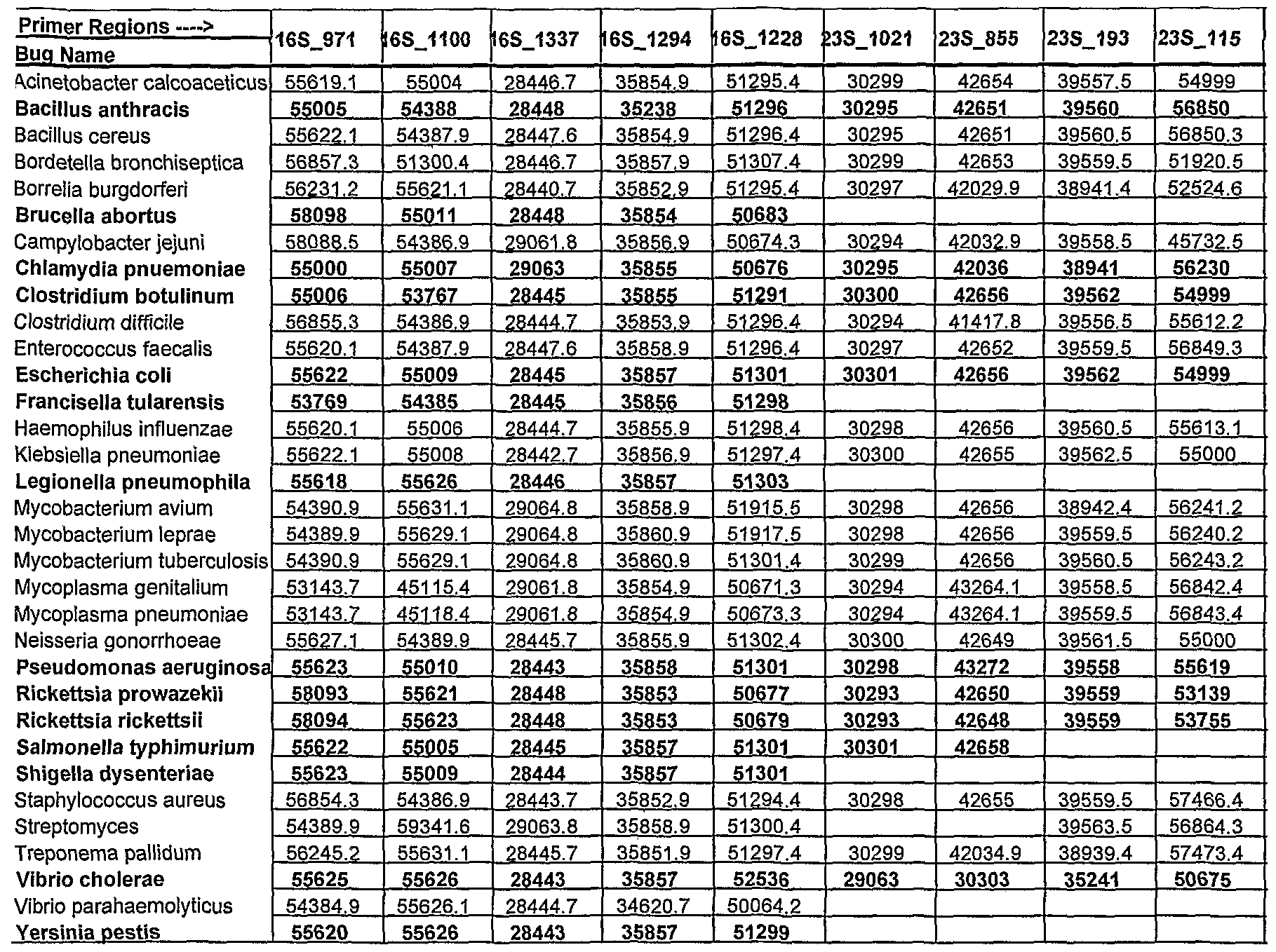
Table 1 shows a small cross section of a database of calculated molecular masses for over 9 primer sets and approximately 30 organisms. The primer sets were derived from rRNA alignment. Primer pairs were chosen so the total length of the amplified region is less than about 80-90 nucleotides. The label for each primer pair represents the starting and ending base number of the amplified region on the consensus diagram.
Included in the short bacterial database cross-section in Table 1 are many well known pathogens/biowarfare agents (shown in bold/red typeface) such as Bacillus anthracis or Yersinia pestis as well as some of the bacterial organisms found commonly in the natural environment
uch as Streptomyces. Even closely related organisms can be distinguished from each other by he appropriate choice of primers. For instance, two low G+C organisms, Bacillus anthracis and itaph aureus, can be distinguished from each other by using the primer pair defined by L6S 337 or 23S_855 (M of 4 Da).
Table 1 Cross Section Of A Database Of Calculated Molecular Masses1
Molecular mass distribution of PCR amplified regions for a selection of organisms (rows) across various primer pairs (columns). Pathogens are shown in bold. Empty cells indicate presently incomplete or missing data.
Figure 6 shows the use of ESI-FT-ICR MS for measurement of exact mass. The spectra from 46mer PCR products originating at position 1337 of the 16S rRNA from S. aureus (upper) and B. anthracis (lower) are shown. These data are from the region of the spectrum containing signals from the [M-8H+]8" charge states of the respective 5'-3' strands. The two strands differ by two (AT→CG) substitutions, and have measured masses of 14206.396 and 14208.373 ± 0.010 Da, respectively. The possible base compositions derived from the masses of the forward and reverse strands for the B. anthracis products are listed in Table 2.
Table 2 Possible base composition for B. anthracis products
Among the 16 compositions for the forward strand and the 18 compositions for the reverse strand that were calculated, only one pair (shown in bold) are complementary, corresponding to the actual base compositions of the B. anthracis PCR products.
Various modifications of the inventions, in addition to those described herein, will be apparent to those skilled in the art from the foregoing description. Such modifications are also intended to fall within the scope of the appended claims. Each reference, publication, patent, and patent application cited or referred to in the present application is incorporated herein by reference in its entirety.