WO2006073738A2 - Colorimetric sensors constructed of diacetylene materials - Google Patents
Colorimetric sensors constructed of diacetylene materials Download PDFInfo
- Publication number
- WO2006073738A2 WO2006073738A2 PCT/US2005/045607 US2005045607W WO2006073738A2 WO 2006073738 A2 WO2006073738 A2 WO 2006073738A2 US 2005045607 W US2005045607 W US 2005045607W WO 2006073738 A2 WO2006073738 A2 WO 2006073738A2
- Authority
- WO
- WIPO (PCT)
- Prior art keywords
- analyte
- buffer
- sample
- buffer solution
- probe
- Prior art date
Links
Classifications
-
- C—CHEMISTRY; METALLURGY
- C12—BIOCHEMISTRY; BEER; SPIRITS; WINE; VINEGAR; MICROBIOLOGY; ENZYMOLOGY; MUTATION OR GENETIC ENGINEERING
- C12Q—MEASURING OR TESTING PROCESSES INVOLVING ENZYMES, NUCLEIC ACIDS OR MICROORGANISMS; COMPOSITIONS OR TEST PAPERS THEREFOR; PROCESSES OF PREPARING SUCH COMPOSITIONS; CONDITION-RESPONSIVE CONTROL IN MICROBIOLOGICAL OR ENZYMOLOGICAL PROCESSES
- C12Q1/00—Measuring or testing processes involving enzymes, nucleic acids or microorganisms; Compositions therefor; Processes of preparing such compositions
- C12Q1/02—Measuring or testing processes involving enzymes, nucleic acids or microorganisms; Compositions therefor; Processes of preparing such compositions involving viable microorganisms
- C12Q1/04—Determining presence or kind of microorganism; Use of selective media for testing antibiotics or bacteriocides; Compositions containing a chemical indicator therefor
-
- G—PHYSICS
- G01—MEASURING; TESTING
- G01N—INVESTIGATING OR ANALYSING MATERIALS BY DETERMINING THEIR CHEMICAL OR PHYSICAL PROPERTIES
- G01N21/00—Investigating or analysing materials by the use of optical means, i.e. using sub-millimetre waves, infrared, visible or ultraviolet light
- G01N21/17—Systems in which incident light is modified in accordance with the properties of the material investigated
- G01N21/25—Colour; Spectral properties, i.e. comparison of effect of material on the light at two or more different wavelengths or wavelength bands
- G01N21/29—Colour; Spectral properties, i.e. comparison of effect of material on the light at two or more different wavelengths or wavelength bands using visual detection
-
- G—PHYSICS
- G01—MEASURING; TESTING
- G01N—INVESTIGATING OR ANALYSING MATERIALS BY DETERMINING THEIR CHEMICAL OR PHYSICAL PROPERTIES
- G01N21/00—Investigating or analysing materials by the use of optical means, i.e. using sub-millimetre waves, infrared, visible or ultraviolet light
- G01N21/75—Systems in which material is subjected to a chemical reaction, the progress or the result of the reaction being investigated
- G01N21/77—Systems in which material is subjected to a chemical reaction, the progress or the result of the reaction being investigated by observing the effect on a chemical indicator
- G01N21/78—Systems in which material is subjected to a chemical reaction, the progress or the result of the reaction being investigated by observing the effect on a chemical indicator producing a change of colour
-
- G—PHYSICS
- G01—MEASURING; TESTING
- G01N—INVESTIGATING OR ANALYSING MATERIALS BY DETERMINING THEIR CHEMICAL OR PHYSICAL PROPERTIES
- G01N31/00—Investigating or analysing non-biological materials by the use of the chemical methods specified in the subgroup; Apparatus specially adapted for such methods
- G01N31/22—Investigating or analysing non-biological materials by the use of the chemical methods specified in the subgroup; Apparatus specially adapted for such methods using chemical indicators
-
- G—PHYSICS
- G01—MEASURING; TESTING
- G01N—INVESTIGATING OR ANALYSING MATERIALS BY DETERMINING THEIR CHEMICAL OR PHYSICAL PROPERTIES
- G01N33/00—Investigating or analysing materials by specific methods not covered by groups G01N1/00 - G01N31/00
- G01N33/48—Biological material, e.g. blood, urine; Haemocytometers
- G01N33/50—Chemical analysis of biological material, e.g. blood, urine; Testing involving biospecific ligand binding methods; Immunological testing
- G01N33/53—Immunoassay; Biospecific binding assay; Materials therefor
- G01N33/543—Immunoassay; Biospecific binding assay; Materials therefor with an insoluble carrier for immobilising immunochemicals
- G01N33/54313—Immunoassay; Biospecific binding assay; Materials therefor with an insoluble carrier for immobilising immunochemicals the carrier being characterised by its particulate form
- G01N33/5432—Liposomes or microcapsules
-
- G—PHYSICS
- G01—MEASURING; TESTING
- G01N—INVESTIGATING OR ANALYSING MATERIALS BY DETERMINING THEIR CHEMICAL OR PHYSICAL PROPERTIES
- G01N33/00—Investigating or analysing materials by specific methods not covered by groups G01N1/00 - G01N31/00
- G01N33/48—Biological material, e.g. blood, urine; Haemocytometers
- G01N33/50—Chemical analysis of biological material, e.g. blood, urine; Testing involving biospecific ligand binding methods; Immunological testing
- G01N33/53—Immunoassay; Biospecific binding assay; Materials therefor
- G01N33/543—Immunoassay; Biospecific binding assay; Materials therefor with an insoluble carrier for immobilising immunochemicals
- G01N33/544—Immunoassay; Biospecific binding assay; Materials therefor with an insoluble carrier for immobilising immunochemicals the carrier being organic
-
- G—PHYSICS
- G01—MEASURING; TESTING
- G01N—INVESTIGATING OR ANALYSING MATERIALS BY DETERMINING THEIR CHEMICAL OR PHYSICAL PROPERTIES
- G01N33/00—Investigating or analysing materials by specific methods not covered by groups G01N1/00 - G01N31/00
- G01N33/48—Biological material, e.g. blood, urine; Haemocytometers
- G01N33/50—Chemical analysis of biological material, e.g. blood, urine; Testing involving biospecific ligand binding methods; Immunological testing
- G01N33/53—Immunoassay; Biospecific binding assay; Materials therefor
- G01N33/569—Immunoassay; Biospecific binding assay; Materials therefor for microorganisms, e.g. protozoa, bacteria, viruses
- G01N33/56911—Bacteria
- G01N33/56938—Staphylococcus
-
- G—PHYSICS
- G01—MEASURING; TESTING
- G01N—INVESTIGATING OR ANALYSING MATERIALS BY DETERMINING THEIR CHEMICAL OR PHYSICAL PROPERTIES
- G01N2333/00—Assays involving biological materials from specific organisms or of a specific nature
- G01N2333/195—Assays involving biological materials from specific organisms or of a specific nature from bacteria
- G01N2333/21—Assays involving biological materials from specific organisms or of a specific nature from bacteria from Pseudomonadaceae (F)
-
- G—PHYSICS
- G01—MEASURING; TESTING
- G01N—INVESTIGATING OR ANALYSING MATERIALS BY DETERMINING THEIR CHEMICAL OR PHYSICAL PROPERTIES
- G01N2333/00—Assays involving biological materials from specific organisms or of a specific nature
- G01N2333/195—Assays involving biological materials from specific organisms or of a specific nature from bacteria
- G01N2333/24—Assays involving biological materials from specific organisms or of a specific nature from bacteria from Enterobacteriaceae (F), e.g. Citrobacter, Serratia, Proteus, Providencia, Morganella, Yersinia
- G01N2333/245—Escherichia (G)
-
- G—PHYSICS
- G01—MEASURING; TESTING
- G01N—INVESTIGATING OR ANALYSING MATERIALS BY DETERMINING THEIR CHEMICAL OR PHYSICAL PROPERTIES
- G01N2333/00—Assays involving biological materials from specific organisms or of a specific nature
- G01N2333/195—Assays involving biological materials from specific organisms or of a specific nature from bacteria
- G01N2333/305—Assays involving biological materials from specific organisms or of a specific nature from bacteria from Micrococcaceae (F)
- G01N2333/31—Assays involving biological materials from specific organisms or of a specific nature from bacteria from Micrococcaceae (F) from Staphylococcus (G)
-
- G—PHYSICS
- G01—MEASURING; TESTING
- G01N—INVESTIGATING OR ANALYSING MATERIALS BY DETERMINING THEIR CHEMICAL OR PHYSICAL PROPERTIES
- G01N2333/00—Assays involving biological materials from specific organisms or of a specific nature
- G01N2333/435—Assays involving biological materials from specific organisms or of a specific nature from animals; from humans
- G01N2333/745—Assays involving non-enzymic blood coagulation factors
- G01N2333/75—Fibrin; Fibrinogen
Definitions
- Staphylococcus aureus which is a pathogen causing a wide spectrum of infections including: superficial lesions such as small skin abscesses and wound infections; systemic and life threatening conditions such as endocarditis, pneumonia and septicemia; as well as toxinoses such as food poisoning and toxic shock syndrome.
- S. aureus is resistant to all but a few select antibiotics.
- Diacetylenes are typically colorless as monomers in solution, and undergo addition polymerization, either thermally or by actinic radiation. As the polymerization proceeds, these compounds undergo a contrasting color change to blue or purple. When exposed to external stimuli such as heat, physical stress, or a change of solvents or counterions, polydiacetylenes exhibit a further color change produced by distortion of the planar backbone conformation. For example, polydiacetylene assemblies are known to change color from blue to red with an increase in temperature or changes in pH due to conformational changes in the conjugated backbone as described in Mino, et al., Langmuir, Vol. 8, p. 594, 1992; Chance, et al, Journal of Chemistry and Physics, VoI.
- the present invention provides a colorimetric sensor to detect the presence of analytes by spectral changes (color changes visible to the naked eye or with a colorimeter) that occur as a result of the interaction of the analytes in a manner that cause conformational changes to polydiacetylene assemblies.
- the polydiacetylene assemblies indicate the presence of an analyte in a simple yet highly sensitive manner.
- a colorimetric system for detecting an analyte comprising a colorimetric sensor comprising a receptor; a polymerized composition comprising at least one diacetylene compound (by this it is meant that the polymerized composition is formed from polymerization of the diacetylene compound); wherein the receptor is incorporated into the polymerized composition to form a transducer; and a buffer composition that mediates the interaction between the analyte and the transducer, wherein the buffer system includes two or more different buffers; wherein the transducer exhibits a color change when contacted with an analyte.
- the buffer composition is a combination of a higher ionic strength buffer with a lower ionic strength buffer.
- the buffer composition is selected from the group consisting of HEPES buffer, Imidazole buffer, PBS buffer, and combinations thereof.
- the buffer mediates the interaction of the analyte by ionic interactions with the transducer.
- the buffer composition mediates the interaction of the analyte by enhancing hydrophobic interactions with the transducer.
- the transducer may be dispersed in an aqueous solution or coated on a substrate.
- the colorimetric system further comprises a probe.
- the probe is selected from the group consisting of fibrinogen, streptavidin, IgG, and combinations thereof.
- the colorimetric system further comprises a surfactant.
- the surfactant comprises a nonionic surfactant.
- the transducer of the colorimetric system is a liposome and/or exhibits a color change upon contact with the buffer composition,
- the diacetylene compound i.e., the starting material for the polydiacetylene material
- the diacetylene compound is of the formula
- R comprises
- R comprises
- R 3 , R 8 , R 13 , R 21 , R 24 , R 31 , and R 33 are independently C 1 -C 20 alkyl;
- R 4 , R 5 , R 7 , R 14 , R 16 , R 19 , R 20 , R 22 , R 25 , and R 32 are independently C 1 -C 14 alkylene;
- R 26 are independently C 1 -C 14 alkylene, C 2 -C 8 alkenylene, or C 6 -C 13 arylene;
- R 9 is Ci- C 14 alkylene or -NR 34 -;
- R 10 , R 12 , R 27 , and R 29 are independently C 1 -C 14 alkylene or (Ci- C 14 alkylene)-( C 2 -C 8 arylene);
- R 11 and R 28 are independently C 2 -C 30 alkynyl;
- R 17 is an ester-activating group;
- R 23 is C 6 -Ci 3 arylene;
- R 30 is Ci-C 14 alkylene or -NR 36 -;
- R 34 and R 36 are C 1 -C 4 alkyl;
- p is 1-5 (herein, "diacetylene” is used to encompass compounds with two to ten C-C triple bonds); and n is 1-20; wherein R 1 and R 2 are not the same.
- the receptor in the colorimetric system comprises a phospholipid selected from the group consisting of phosphocholines, phosphoethanolamines, phosphatidylethanolamines, phosphatidylserines, phosphatidylglycerols, and combinations thereof.
- a method for the detection of an analyte includes forming a colorimetric sensor comprising a receptor and a polymerized composition comprising a diacetylene (i.e., the polymerized composition is derived from polymerization of the diacetylene), wherein the receptor is incorporated into the polymerized composition to form a transducer capable of exhibiting a color change; contacting the sensor with a probe; contacting the sensor with a sample suspected of containing a target analyte in the presence of a buffer composition comprising two or more different buffers; and observing a color change if the analyte is present.
- a colorimetric sensor comprising a receptor and a polymerized composition comprising a diacetylene (i.e., the polymerized composition is derived from polymerization of the diacetylene), wherein the receptor is incorporated into the polymerized composition to form a transducer capable of exhibiting a color change; contacting the sensor with a probe;
- a method for the detection of an analyte comprising forming a colorimetric sensor, comprising a receptor and a polymerized composition comprising a diacetylene, wherein the receptor is incorporated into the polymerized composition to form a transducer capable of exhibiting a color change in the presence of a probe; contacting the transducer with a sample suspected of containing a target analyte, and a probe that has an affinity for both the target analyte and the receptor in the presence of a buffer composition comprising two or more different buffers; and observing essentially no color change if the analyte is present.
- the probe and sample suspected for containing a target analyte may be combined to form a mixture before contacting the transducer.
- the analyte is selected from the group consisting of S. aureus, protein A, PBP2', E. coli, and Pseudomonas aeruginosa.
- the colorimetric system exhibits an observable color change within 60 minutes of contacting the transducer with an analyte.
- alkyl refers to a straight or branched chain or cyclic monovalent hydrocarbon group having a specified number of carbon atoms. Alkyl groups include those with one to twenty carbon atoms. Examples of “alkyl” as used herein include, but are not limited to, methyl, ethyl, n-propyl, n-butyl, n-pentyl, isobutyl, and isopropyl, and the like. It is to be understood that where cyclic moieties are intended, at least three carbons in said alkyl must be present. Such cyclic moieties include cyclopropyl, cyclobutyl, cyclopentyl, cyclohexyl, and cycloheptyl.
- alkylene refers to a straight or branched chain or cyclic divalent hydrocarbon group having a specified number of carbon atoms. Alkylene groups include those with one to fourteen carbon atoms. Examples of
- alkylene as used herein include, but are not limited to, methylene, ethylene, trimethylene, tetramethylene and the like. It is to be understood that where cyclic moieties are intended, at least three carbons in said alkylene must be present. Such cyclic moieties include cyclopropylene, cyclobutylene, cyclopentylene, cyclohexylene, and cycloheptylene.
- alkenylene refers to a straight or branched chain or cyclic divalent hydrocarbon group having a specified number of carbon atoms and one or more carbon—carbon double bonds. Alkenylene groups include those with two to eight carbon atoms. Examples of “alkenylene” as used herein include, but are not limited to, ethene-l,2-diyl, propene-l,3-diyl, and the like.
- arylene refers to divalent unsaturated aromatic carboxylic groups having a single ring, such as phenylene, or multiple condensed rings, such as naphthylene or anthrylene.
- Arylene groups include those with six to thirteen carbon atoms. Examples of "arylene” as used herein include, but are not limited to, benzene- 1,2-diyl, benzene- 1, 3 -diyl, benzene- 1,4-diyl, naphthalene- 1,8-diyl, and the like.
- alkylene-arylene refers to an alkylene moiety as defined above bonded to an arylene moiety as defined above.
- alkylene- arylene examples include, but are not limited to, -CH 2 -phenylene, -CH 2 CH 2 - phenylene, and -CH 2 CH 2 CH 2 -phenylene.
- alkynyl refers to a straight or branched chain or cyclic monovalent hydrocarbon group having from two to thirty carbons and at least one carbon-carbon triple bond.
- alkynyl as used herein include, but are not limited to, ethynyl, propynyl and butynyl.
- analyte(s) refers to any material that can be detected by the sensor system of the present invention. Such materials include, but are not limited to, small molecules, pathogenic and non-pathogenic organisms, toxins, membrane receptors and fragments, volatile organic compounds, enzymes and enzyme substrates, antibodies, antigens, proteins, peptides, nucleic acids, and peptide nucleic acids.
- Target analyte refers to the material targeted for detection in a sensor system.
- bacteria refers to all forms of microorganisms considered to be bacteria including cocci, bacilli, spirochetes, sheroplasts, protoplasts, etc.
- receptor refers to any molecule or assembly of molecules with an affinity for a target analyte and/or a probe.
- Receptor includes, but is not limited to, naturally occurring or synthetic receptors such as lipids, surface membrane proteins, enzymes, lectins, antibodies, recombinant proteins, synthetic proteins, nucleic acids, c-glycosides, carbohydrates, gangliosides, and chelating agents.
- the terms “assembly,” or “self-assembly,” refers to any self- ordering of diacetylene molecules and phospholipids prior to polymerization. See J. Israelachvili, Intermolecular and Surface Forces (2 nd Ed.), Academic Press, New York (1992), pp. 321-427.
- SAMs self-assembling monolayer(s) refers to any ordered ultrathin organic film formed on a given substrate by spontaneous self- ordering. A. Ulman, An Introduction to Ultrathin Organic Films, Academic Press, New York (1991), pp. 237-301.
- the term "transducer” describes a material capable of turning a recognition event such as a covalent bond or a noncovalent interaction (e.g., electrostatic interaction, polar interaction, van der Waals forces) at the molecular level into an observable signal.
- "Probe” refers to a constituent that is capable of interacting with the target analyte and/or the receptor. Accordingly, the probe is a type of "detectable binding reagent” i.e., an agent that specifically recognizes and interacts or binds with an analyte (i.e., the target analyte) and/or the receptor, wherein the probe has a property permitting detection when bound.
- Specifically interact means that detectable binding agent physically interacts with the target analyte or receptor to the substantial exclusion of other analytes also present in the sample.
- the binding of a detectable binding reagent useful according to the invention has stability permitting the measurement of the binding.
- FIG. 1 shows a schematic representation of a colorimetric sensor of the present invention.
- FIG. 2 shows a schematic representation of a colorimetric sensor array of the present invention.
- the present invention provides a colorimetric sensor system for detection of an analyte.
- the colorimetric system includes a colorimetric sensor comprising a receptor and a polymerized diacetylene material (polydiacetylene assemblies, which refer to an organized polydiacetylene structure that may (but not ntecessarily) include other components), wherein the receptor is incorporated within the polydiacetylene to form a transducer capable of providing a color change upon binding with a probe and/or analyte.
- the colorimetric sensor can function in solution or coated on a substrate.
- the diacetylene compounds of the present invention can self assemble in solution to form ordered assemblies that can be polymerized using any actinic radiation such as, for example, electromagnetic radiation in the UV or visible range of the electromagnetic spectrum.
- Polymerization of the diacetylene compounds result in polymerization reaction products that have a color in the visible spectrum less than 570 nanometers (nm), between 570nm and 600nm (including the endpoints), or greater than 600nm, depending on their conformation and exposure to external factors.
- polymerization of the diacetylene compounds disclosed herein result in meta-stable blue phase polymer networks that include a polydiacetylene backbone.
- These meta- stable blue phase polymer networks undergo a color change from bluish to reddish- orange upon exposure to external factors such as heat, a change in solvent or counterion, if available, or physical stress, for example.
- the ability of the diacetylene compounds and their polymerization products disclosed herein to undergo a visible color change upon exposure to physical stress make them candidates for the preparation of sensing devices for detection of an analyte.
- the polydiacetylene assemblies formed from the disclosed diacetylene compounds can function as a transducer in biosensing applications.
- the structural requirements of a diacetylenic molecule for a given sensing application are typically application specific.
- Features such as overall chain length, solubility, polarity, crystallinity, and presence of functional groups for further molecular modification all cooperatively determine a diacetylenic molecule's ability to serve as a useful sensing material.
- the structure of the diacetylenic compound should be capable of forming a stable dispersion in water, polymerizing efficiently to a colored material, incorporating appropriate receptor chemistry for binding to an analyte, and transducing that binding interaction by means of a color change.
- the diacetylene compounds of the present invention possess the capabilities described above and can be easily and efficiently polymerized into polydiacetylene assemblies that undergo the desired color changes. Additionally, the diacetylene compounds allow for the incorporation of large excesses of unpolymerizable material, such as a receptor described below, while still forming a stable, polymerizable solution.
- the disclosed diacetylene compounds (the starting material) can be synthesized in a rapid high-yielding fashion, including high-throughput methods of synthesis.
- the presence of functionality in the backbones of the diacetylenic compounds (the starting material) such as heteroatoms for example, provides for the possibility of easy structural elaboration in order to meet the requirements of a given sensing application.
- the diacetylenic compounds can be polymerized into the desired polydiacetylene backbone containing network by adding the diacetylene to a suitable solvent, such as water for example, sonicating the mixture, and then irradiating the solution with ultraviolet light, typically at a wavelength of 254nm. Upon polymerization the solution undergoes a color change to bluish-purple.
- a suitable solvent such as water for example, sonicating the mixture
- ultraviolet light typically at a wavelength of 254nm.
- Diacetylenes (the starting material) useful in the present invention typically contain an average carbon chain length of at least 8 with at least one functional group such as a carboxyl group, primary or tertiaty amine groups, methyl esters of carboxyl, etc.
- Suitable diacetylenes include those described in U.S. Patent No. 5,491,097 (Ribi et al.), PCT Publication No. WO 02/00920, U.S. Patent No. 6,306,598, and PCT
- the polydiacetylene assemblies include polymerized compounds resulting from the diacetylenes of the formula
- R 3 , R 8 , R 13 , R 21 , R 24 , R 31 , and R 33 are independently alkyl;
- R 4 , R 5 , R 7 , R 14 , R 16 , R 19 , R 20 , R 22 , R 25 , and R 32 are independently alkylene;
- R 6 , R 15 , R 18 , and R 26 are independently alkylene, alkenylene, or arylene;
- R 9 is alkylene or -NR 34 -;
- R 10 , R 12 , R 27 , and R 29 are
- R 1 is
- R 7 is ethylene, trimethylene, tetramethylene, pentamethylene, hexame etthhyylleennee,, hheeppttaammeetthhyylleennee,, ooccttaammeetthhyylleennee,, oorr nnoonnaammethylene, and R 6 is ethylene, trimethylene, ethenylene, or phenylene; and wherein R 2 is
- R 20 is ethylene, trimethylene, tetramethylene, pentamethylene, hexamethylene, heptamethylene, octamethylene, or nonamethylene, and wherein R 21 is undecyl, tridecyl, pentadecyl, heptadecyl; and wherein p is 1.
- the invention is inclusive of the compounds described herein including isomers, such as structural isomers and geometric isomers, salts, solvates, polymorphs, and the like.
- Diacetylenes of the Formula XXIII can be prepared as outlined in Scheme 1 where n is typically 1 to 4 and m is typically 10 to 14.
- Compounds of formula XXIII can be prepared via oxidation from compounds of formula XXII by reaction with a suitable oxidizing agent in a suitable solvent such as DMF, for example.
- suitable oxidizing agents include Jones reagent and pyridinium dichromate, for example. The aforesaid reaction is typically run for a period of time from 1 hour to 48 hours, generally 8 hours, at a temperature from 0°C to 4O 0 C, generally from 0° C to 25 0 C.
- Compounds of formula XXII can be prepared from compounds of formula XXI by reaction with a suitable acid chloride.
- suitable acid chlorides include any acid chloride that affords the desired product such as lauroyl chloride, 1 -dodecanoyl chloride, 1 -tetradecanoyl chloride, 1-hexadecanoyl chloride, and 1-octadecanoyl chloride for example.
- Suitable solvents include ether, tetrahydrofuran, dichloromethane, and chloroform, for example.
- the aforesaid reaction is typically run for a period of time from 1 hour to 24 hours, generally 3 hours, at a temperature from
- 0°C to 40°C generally from 0°C to 25°C, in the presence of a base such as trialkylamine or pyridine base.
- Diacetylenic compounds as disclosed herein can also be prepared by reacting compounds of formula XXII with an anhydride such as succinic, glutaric, or phthalic anhydride in the presence of a suitable solvent, such as toluene.
- the aforesaid reaction is typically run for a period of time from 1 hour to 24 hours, generally 15 hours, at a temperature from 5O 0 C to 125°C, generally from 100°C to 125 0 C.
- the colorimetric sensors comprising the polymerized diacetylenes can serve as the basis for the colorimetric detection of a molecular recognition event.
- the sensor can be prepared by adding a receptor to the diacetylene monomers either prior to or after polymerization.
- the receptor is capable of functionalizing the polydiacetylene assemblies through a variety of means including physical mixing, covalent bonding, and noncovalent interactions (such as electrostatic interactions, polar interactions, etc). Upon polymerization or thereafter, the receptor is effectively incorporated with the polymer network such that interaction of the receptor with an analyte results in a visible color change due to the perturbation of the conjugated ene-yne polymer backbone.
- the incorporation of the receptor with the polydiacetylene assembly provides a structural shape capable of deformation in response to interaction or binding with a probe and/or analyte.
- Particularly useful receptors are assemblies of amphiphilic molecules with typically a rod shape molecular architecture that can be characterized by a packing parameter defined as: v/(aol c ) (Israelachvili, J.N. et ah; Q. Rev. Biophys.;
- v is the volume taken up by the hydrocarbon components of the molecules (for example, the hydrocarbon chains of a phospholipid or a fatty acid)
- a 0 is the effective area taken up by the polar headgroup (for example the phosphate headgroup of a phospholipid or the carboxylic acid headgroup of a fatty acid)
- I 0 is the so-called critical length, and generally describes the length of the molecule at the temperature of its environment.
- Preferred amphiphilic molecules for a receptor are those with packing parameter v/(aol c ) values between 1/3 and 1.
- useful receptors include, but are not limited to, lipids, surface membrane proteins, enzymes, lectins, antibodies, recombinant proteins, synthetic proteins, nucleic acids, c-glycosides, carbohydrates, gangliosides, and chelating agents.
- the receptor is a phospholipid.
- Suitable phospholipids include phosphocholines (e.g., 1 ,2-dimeristoyl-sn-glycero-3-phosphocholine), phosphoethanolamines, and phosphatidylethanolamines, phosphatidylserines, and phosphatidylglycerols such as those described in Silver, Brian L., The Physical Chemistry of Membranes, Chapter 1, pp 1-24 (1985).
- the receptor is physically mixed and dispersed among the polydiacetylene to form a structure wherein the structure itself has a binding affinity for the probe and/or analyte of interest.
- Structures include, but are not limited to, liposomes, micelles, and lamellas.
- the structure is a liposome. While not intending to be bound by theory, it is believed that the phospholipid mimics a cell membrane while the polydiacetylene assemblies allow the physico-chemical changes occurring to the liposomes to be translated into a visible color change.
- the liposomes as prepared possess a well-defined morphology, size distribution, and other physical characteristics such as a well-defined surface potential.
- the ratio of receptor to diacetylene compounds (starting material)in the liposome can be varied based on the selection of materials and the desired colorimetric response. In most embodiments, the ratio of phospholipids to diacetylene compound (starting material) will be at least 25:75, and more preferably at least 40:60.
- the liposomes are composed of the diacetylene compound: HO(O)C(CH 2 ) 2 C(O)O(CH 2 ) 4 C ⁇ C-C ⁇ C(CH 2 ) 4 O(O)C(CH 2 )i 2 CH 3 [succinic acid mono- (12-tetradecanoyloxy-dodeca-5,7-diynyl) ester], and the zwitterionic phospholipid 1,2- dimeristoyl-sn-glycero-3-phosphocholine [DMPC] mixed in a 6:4 ratio.
- the discussion of the PDA systems is directed to the use of liposomes in the receptor assembly; however, this discussion also applies to other receptor assemblies, including, for example, other planar configurations.
- the liposomes are prepared by probe sonication of the material mixture suspended in a buffer solution that is referred to as the preparation buffer.
- the preparation buffer can be a low ionic strength (5mM) N-2-
- HEPES Hydroxyethylpiperazine-N'-2-efhanesulfonic acid
- the colorimetric system of the present invention is designed to exploit the way a probe can interact with the liposomes containing both a receptor, such as a phospholipids, and the polymerized diacetylenes.
- the liposomes can be used as models for biological membranes that interact with a probe, such as a protein, as described in Oellerich, S. et al; J.Phys. Chem B; 2004, 108, 3871-3878; and Zuckermann, M.J.; Heimburg T.; Biophysi. J.; 2001, 81, 2458-2472.
- proteins will adsorb to the surface of the liposomes primarily through electrostatic interactions.
- proteins continue to adsorb electrostatically to the surface of a liposome until they completely saturate or envelop the liposomes.
- both liposomes and the proteins can undergo morphological and conformational changes, until the hydrophobic segment of the proteins covering the liposome surface can begin to interact with the hydrophobic interior of the liposome structure.
- the proteins can become hydrophobically bound and penetrate the liposome structure, resulting in substantial morphological change in the liposome structure, with the size and permeability of the liposomes changing drastically.
- the layers of proteins can result in the loss of suspension stability, flocculation, and finally, precipitation.
- a charged probe can electrostatically adsorb to the polydiacetylene liposomes.
- the initial adsorption may not in itself trigger a substantial change in the size and morphology of the liposome, and thus an initially small or negligible colorimetric response, if the probe is present in excess relative to the lipid, it is likely that the probe will eventually become hydrophobically bound to the liposome and penetrate its interior membrane structure. At this point, one would expect that the large mechanical stresses imparted by the incorporation of the probe within the liposome structure would significantly change the polydiacetylene conformation, resulting in a concomitant colorimetric response readily observable.
- the probe is negatively-charged at neutral pH its capacity to interact electrostatically with the polydiacetylene liposomes is severely hindered, and the ability to generate a colorimetric response due to a hydrophobic interaction between probe and the receptor-containing polydiacetylene liposomes may be compromised.
- a high ionic strength buffer (MOOmM) at neutral pH (e.g., phosphate buffer saline (PBS), Imidazole buffer) would provide a means to decrease the surface potential of the liposomes (by screening the surface charge of the liposome), facilitating the direct hydrophobic interaction of non-charged probes with the liposomes, and resulting in the incorporation of that protein within the structure of the liposome.
- MOOmM phosphate buffer saline
- Imidazole buffer e.g., Imidazole buffer
- the higher ionic strength of the buffer because of its effect on the surface potential of the liposomes, can introduce a significant colorimetric response in the absence of a probe, it has been determined that when the probe is present, the colorimetric response is significantly enhanced due to the protein-liposome hydrophobic interactions.
- This result has very useful practical consequences: the detection time at a given limit of detection can be significantly shortened, or conversely, for a fixed assay time the limit of detection can be significantly lowered. Based on this phenomena, the probe can be selected based on its ability to interact specifically with both a given analyte target and the polydiacetylene liposome to trigger a colorimetric response.
- the colorimetric response of the polydiacteylene- containing liposome is directly proportional to the concentration of the probe or a probe-analyte complex in those cases of direct analysis.
- the selection of probe for a particular application will depend in part on the probes' size, shape, charge, hydrophobicity, and affinity towards molecules.
- the probes may be positively charged, negatively charged, or zwitterionic depending on the pH of the environment. At a pH below the isoelectric point of a probe, the probe is positively charged and above this point it is negatively charged.
- the term "isoelectric point" refers to the pH at which the probe has a net charge of zero.
- detection assays talcing into consideration not only the polydiacetylene liposome composition (e.g., choice of the phospholipid being used and the ratio of phospholipid to diacetylene), and the probe being used (e.g., polymixin, fibrinogen, antibodies), but also the aqueous environment established by the choice of a buffer system.
- polydiacetylene liposome composition e.g., choice of the phospholipid being used and the ratio of phospholipid to diacetylene
- the probe being used e.g., polymixin, fibrinogen, antibodies
- the buffer composition of the present invention provides a system capable of resisting changes in pH in the presence of other components, consisting of a conjugate acid-base pair in which the ratio of proton acceptor to proton donor is near unity.
- the buffer compositions of the present invention mediate the physical or chemical interaction between the analyte and the components of the colorimetric sensor.
- the buffer composition inhibits the interaction of the analyte with the receptor.
- the buffer composition facilitates the interaction of the analyte with the receptor.
- Buffer compositions that may be particularly useful include HEPES buffer, Imidazole buffer, and PBS buffer.
- a combination of buffers i.e., different buffers is used to adjust the appropriate ionic strength for a given application based on the selection of the probe and/or the target analyte to be detected.
- HEPES buffer which has a pH of 7.2
- polymyxin with an isoelectric point of 7.7
- Fibrinogen with an isoelectric point of 5.3, has a negative charge in the same HEPES buffer composition, which prevents adsorption or any electrostatic interaction with the polar head group of the phospholipids.
- the ionic strength alters the morphology of the liposome (or other transducer structure), to expose the hydrophobic portions.
- fibrinogen contains hydrophobic parts in the structure that interacts with the phospholipids to cause a color change.
- One convenient method of achieving the optimum balance of electrostatic and hydrophobic components in the liposome-protein interaction is to use a mixture of two or more different buffers. For example, mixing a low ionic strength organic buffer
- the mixed buffer system can be designed to provide for an optimized liposome-protein interaction.
- a mixed buffer system would also provide a way of tailoring to what extent the buffer system is an interacting versus a non-interacting buffer.
- an interacting buffer PBS, imidazole
- PBS interacting buffer
- imidazole imidazole
- HEPES liposome morphology
- surfactant component in the buffer composition that can assist the hydrophobic interaction of a probe with the colorimetric sensor.
- Surfactants that may be particularly useful in the present invention include nonionic surfactants. Polyalkoxylated, and in particular polyethoxylated, nonionic surfactants can stabilize the components of the present invention in solutions particularly well.
- Surfactants of the nonionic type that may be useful include:
- Polyethylene oxide extended sorbitan monoalkylates i.e., Polysorbates.
- a Polysorbate 20 commercially available as NIKKOL TL-IO (from Barret Products) is very effective.
- BRIJ 78 and BRIJ 700 which are stearyl alcohol ethoxylates having 20 and 100 moles of polyethylene oxide, respectively, have proven very useful.
- a ceteareth 55 which is commercially available under the trade designation PLURAFAC A-39 from BASF Corp., Performance Chemicals Div., Mt. Olive, NJ.
- Polyalkoxylated alkylphenols Useful surfactants of this type include polyethoxylated octyl or nonyl phenols having HLB values of at least about 14, which are commercially available under the trade designations ICONOL and TRITON, from BASF Corp., Performance Chemicals Div., Mt. Olive, NJ and Union Carbide Corp., Danbury, CT, respectively. Examples include TRITON XlOO (an octyl phenol having
- ethylene oxide available from Union Carbide Corp., Danbury, CT
- ICONOL NP70 and NP40 nonyl phenol having 40 and 70 moles of ethylene oxide units, respectively, available from BASF Corp., Performance Chemicals Div., Mt. Olive, NJ
- Sulfated and phosphated derivatives of these surfactants are also useful. Examples of such derivatives include ammonium nonoxynol-4-sulfate, which is commercially available under the trade designation RHODAPEX CO-436 from Rhodia, Dayton, NJ. 4. Polaxamers.
- EO EO
- PO propylene oxide
- Both EO-PO-EO blocks and PO-EO-PO blocks are expected to work well as long as the HLB is at least about 14, and preferably at least about 16.
- Such surfactants are commercially available under the trade designations PLURONIC and TETRONIC from BASF Corp.,
- PLURONIC surfactants from BASF have reported HLB values that are calculated differently than described above. In such situation, the HLB values reported by BASF should be used.
- preferred PLURONIC surfactants are L-64 and F- 127, which have HLBs of 15 and 22, respectively.
- the PLURONIC surfactants are quite effective at stabilizing the compositions of the present invention and are quite effective with iodine as the active agent, they may reduce the antimicrobial activity of compositions using povidone-iodine as the active agent.
- Polyalkoxylated esters Polyalkoxylated glycols such as ethylene glycol, propylene glycol, glycerol, and the like may be partially or completely esterified, i.e., one or more alcohols may be esterified, with a (C8-C22)alkyl carboxylic acid.
- Such polyethoxylated esters having an HLB of at least about 14, and preferably at least about 16, are suitable for use in compositions of the present invention.
- Alkyl polyglucosides such as those described in U.S. Patent No. 5,951 ,993 (Scholz et al.), starting at column 9, line 44, are compatible with the film-forming polymers of the present invention and may contribute to polymer stability.
- Examples include glucopon 425, which has a (C8-C16)alkyl chain length with an average chain length of 10.3 carbons and 1-4 glucose units.
- the detection system based on the colorimetric materials of the present invention depends on one or more of the following factors: the molecular architecture of the diacetylene compounds; the type of receptor moiety employed; the morphology (size and structure) of the liposomes or other potential aggregate structures of diacetylene and receptor molecules; the protein probe utilized; and the buffer system used to carry out the assay.
- the present invention provides a method for analysis of an analyte, which comprises contacting the abovementioned colorimetric sensor with a solution sample or surface containing an analyte and utilizing an absorption measurement or a visual observation with the naked eye to detect color change in the colorimetric sensor.
- the present invention provides a method for indirect detection of an analyte by selection of a probe with an affinity to bind with both the receptor incorporated into the polydiacetylene assemblies and the analyte. The probe selected will demonstrate a competitive affinity with the analyte.
- the probe When the analyte of interest is present, the probe will bind to the analyte rather than the receptor on the polydiacetylene backbone, resulting in a color change inversely proportional to the analyte concentration. If the analyte is absent, the probe will bind to the receptor incorporated on the polydiacetylene backbone, resulting in a color change from blue to red.
- the probe can contact the sensor after the analyte contacts the sensor, or can be mixed with the analyte prior to the mixture contacting the sensor.
- the probe and the target analyte are allowed to interact in a buffer solution, which is subsequently placed in contact with the sensor.
- the concentration of the probe free in the buffer is dependent on the amount of analyte target present: the higher the analyte concentration, the lower the remaining concentration of probe. Since the colorimetric response of the sensor is proportional to the amount of free probe available, the colorimetric response is inversely proportional to the analyte concentration.
- the probe can form a complex with the analyte which can interact directly with the sensor, yielding a direct assay where the colorimetric response is directly proportional to the concentration of analyte.
- the method of the invention comprises providing a test sample comprising the analyte in a buffer composition, providing a probe in a buffer composition, combining the test sample and the probe wherein the probe shows a greater binding affinity for the analyte than the receptor, and detecting the change with a biosensor.
- the probe could be generated in-situ by fragmenting or otherwise lysing the analyte target as discussed further below.
- the probe could also be considered a protein or protein fragment externally present on the cell wall of an organism that is available for interaction directly with the sensor. Interaction between the probe and the analyte can operate to the exclusion of interaction with the liposome. Alternatively the probe may interact with the analtye to form a complex with the resulting complex interacting with the liposome.
- the probe can be contacted with the sensor in solution or coated on a substrate.
- the probe will be any molecule with an affinity for both the target analyte and the receptor.
- Possible probes for use in the present invention include membrane disrupting peptides such as alamethicin, magainin, gramicidin, polymyxin B sulfate, and melittin; fibrinogen; streptavridin; antibodies; lectins; and combinations thereof.
- an antibody is employed as the probe.
- “Antibody” refers to an immunoglobulin having the capacity to specifically bind a given antigen inclusive of antigen binding fragments thereof.
- the term “antibody” is intended to include whole antibodies of any isotype (IgG, IgA, IgM, IgE, etc.), and fragments thereof which are also specifically reactive with a vertebrate (e.g., mammalian) protein.
- Antibodies can be fragmented using conventional techniques and the fragments screened for utility in the same manner as whole antibodies.
- the term includes segments of proteolytically-cleaved or recombinantly-prepared portions of an antibody molecule that are capable of selectively reacting with a certain protein.
- Non-limiting examples of such proteolytic and/or recombinant fragments include F(ab'), F(ab) 2 , Fv, and single chain antibodies (scFv) containing a VL and/or VH domain joined by a peptide linker.
- the scFv's can be covalently or non-covalently linked to form antibodies having two or more binding sites.
- Antibodies can be labeled with any detectable moieties known to one skilled in the art.
- S. aureus antibodies are commercially available from Sigma and Accurate Chemical.
- concentration of antibody employed is at least 2 nanograms per milliliter (ng/ml).
- the concentration of antibody is at least 100 nanograms/ml.
- a concentration of 100 micrograms/ml can be employed.
- no more than about 500 micrograms/ml are employed.
- fibrinogen is employed as the probe.
- a fibrinogen-binding protein expressed or present on/in the analyte reacts with the fibrinogen.
- S. aureus expresses the fibrinogen binding protein often referred to as clumping factor that reacts with fibrinogen when contacted.
- the concentration of fibrinogen to produce this reaction is typically at least 0.0001 wt-% and generally no more than 5 wt-%.
- Human plasma and animal (e.g., rabbit) plasma are suitable fibrinogen-containing mediums.
- Commercially available plasma products generally include an anticoagulant such as EDTA, citrate, heparin, etc.
- Fibrinogen derived from human is commercially available from Sigma Aldrich, St.
- probe concentrations can be chosen to correspond to desired concentration levels of detection.
- the method of indirect detection using the probe allows design of the system around the type and concentration of the probe for desired sensitivity in a given application. This allows the transducer to be universal to multiple analytes of interest. For example, a single transducer (polydiacetylene/receptor combination) could serve to detect multiple analytes by varying the probe in contact with the transducer in accordance with the probe's affinity for the analyte.
- Microbes i.e., microorganisms
- Gram positive bacteria such as Gram positive bacteria, Gram negative bacteria, fungi, protozoa, mycoplasma, yeast, viruses, and even lipid-enveloped viruses.
- Particularly relevant organisms include members of the families Enter vbacteriaceae, or genera Staphylococcus spp., Streptococcus spp., Pseudomonas spp., Enterococcus spp., Esherichia spp., Bacillus spp., Listeria spp., Vibrio spp., as well as herpes virus, Aspergillus spp., Fusarium spp., and Candida spp.
- Particularly virulent organisms include Staphylococcus aureus (including resistant strains such as Methicillin Resistant Staphylococcus aureus (MRSA)), S. epidermidis, Streptococcus pneumoniae, S. agalactiae, S. pyogenes, Enterococcus faecalis, Vancomycin Resistant Enterococcus (VRE), Vancomycin
- VRSA Resistant Staphylococcus aureus
- VSA Vancomycin Intermediate Staphylococcus aureus
- Bacillus anthracis Pseudomonas aeruginosa, Escherichia coli, Aspergillus niger, A. fumigatus, A. clavatus, Fusarium solani, F. oxysporum, F. chlamydosporum, Listeria monocytogenes, Vibrio cholera, V. parahemolyticus, Salmonella cholerasuis, S. typhi, S. typhimurium, Candida albicans, C. glabrata, C. krusei, and multiple drug resistant Gram negative rods (MDR).
- MDR multiple drug resistant Gram negative rods
- Gram positive bacteria such as Staphylococcus aureus.
- these can be detected by detecting the presence of a cell-wall component characteristic of the bacteria, such as a cell- wall protein.
- antibiotic resistant microbes including MRSA, VRSA, VISA, VRE, and MDR.
- these can be detected by additionally detecting the presence of an internal cell component, such as a membrane protein.
- Such microbes or other species of interest can be analyzed in a test sample that may be derived from any source, such as a physiological fluid, e.g., blood, saliva, ocular lens fluid, synovial fluid, cerebral spinal fluid, pus, sweat, exudate, urine, mucous, lactation milk, or the like.
- test sample may be derived from a body site, e.g., wound, skin, nares, scalp, nails, etc.
- test sample refers to a sample that contains the target analyte.
- the sample is a liquid or gas and more preferably, a liquid.
- sampling techniques for the detection of S. aureus. Such sampling techniques are suitable for the method of the present invention as well. It is common to obtain a sample from wiping the nares of a patient.
- a particularly preferred sampling technique includes the subject's (e.g., patient's) anterior nares swabbed with a sterile rayon swab. One swab is used to sample each subject, i.e., one swab for both nostrils.
- the sampling is performed by inserting the rayon swab (commercially available from Puritan, East Grinstead, UK under the trade designation "Pure- Wraps" dry or pre-moistened with an appropriate solution into the anterior tip of the subject's nostril and rotating the swab for two complete revolutions along the nares' mucosal surface.
- the swab is then cultured directly or extracted with an appropriate solution typically including water optionally in combination with a buffer and at least one surfactant.
- test samples may include other liquids as well as solid(s) dissolved in a liquid medium.
- Samples of interest may include process streams, water, soil, plants or other vegetation, air, surfaces (e.g., contaminated surfaces), and the like.
- the test sample e.g., liquid
- the test sample may be subjected to prior treatment, such as dilution of viscous fluids.
- the test sample e.g., liquid
- may be subjected to other methods of treatment prior to injection into the sample port such as concentration (by filtration, distillation, dialysis, or the like), dilution, filtration, inactivation of natural components, addition of reagents, chemical treatment, etc.
- One method of treatment that may enhance signal detection of the target analyte involves lysing cells to form cell-wall fragments and analyzing the cell-wall fragments, as described in U.S. Patent Publication No. 2005/0153370.
- the methods are useful for detecting one or more components of cell walls that are characteristic of a microbe, particularly Staphylococcus aureus.
- the method includes: providing a test sample including uncultured cells; lysing the uncultured cells to form a lysate including cell-wall fragments; and analyzing the cell-wall fragments for a cell-wall component characteristic of the analyte; wherein the cell-wall component characteristic of the analyte displays an enhanced signal relative to the same component in unlysed cells.
- Cell-wall components include, for example, cell-wall proteins such as protein A and microbial surface components recognizing adhesive matrix molecules (MSCRAMMs) such as fibrinogen-binding proteins (e.g., clumping factors), fibronectin-binding proteins, collagen-binding proteins, heparin/heparin-related polysaccharides binding proteins, and the like.
- MSCRAMMs adhesive matrix molecules
- Protein A and clumping factors such as fibrinogen-binding factors and clumping factors A, B, and Efb, are also particularly useful in methods of detecting the presence of Staphylococcus aureus.
- Other cell-wall components include capsular polysaccharides and cell-wall carbohydrates (e.g., teichoic acid and lipoteichoic acid).
- Lysing can include contacting the cells with a lysing agent or physically lysing the cells. Lysing can be conducted under conventional conditions, such as, for example, at a temperature of about 5 0 C to about 37 0 C, preferably at a temperature of about 15°C to about 25°C. Significantly, the lysing can occur using uncultured cells, i.e., a direct test sample, although cultured cells can be used as well.
- the test sample may include a relatively low concentration of microbes, particularly Staphylococcus aureus.
- relatively low concentrations include, for example, less than about 5 X 10 4 colony forming units ("cfu") per milliliter (cfu/ml) of microbe, less than about 5 X 10 3 cfu/ml, less than about 1000 cfu/ml, and even as low as about 500 cfu/ml.
- Microbes such as S.
- Suitable lysing agents include, for example, enzymes such as lysostaphin, lysozyme, endopeptidases, N-acetylmuramyl-L-alanine amidase, endo-beta-N- acethylglucosaminidase, and ALE-I. Various combinations of enzymes can be used if desired. Lysostaphin is particularly useful in methods of detecting the presence of Staphylococcus aureus. Other lysing agents include salts (e.g., chaotrophic salts), solubilizing agents
- reducing agents e.g., DTT, DTE, cysteine, N-acetyl cysteine
- acids e.g., HCl
- bases e.g., NaOH
- the lysed cells in the test sample can be analyzed for protein A, which is characteristic for S. aureus and can be detected with a protein A specific antibody immobilized on the biosensor surface. Additionally, lysed cells, such as S. aureus bacteria, release protein markers from the internal portion of the cells (as opposed to the cell-wall portion of the cells). Such protein markers can be detected by probes, such as an antibody.
- the test sample and probe may be combined in a variety of suitable manners.
- the probe is provided to the sensor and the test sample is provided to the colorimetric sensor as separate portions, yet in any order.
- the surface may be coated with a fibrino gen-containing solution and optionally dried.
- the test sample and probe are combined as a mixture and the mixture is provided to the colorimetric sensor.
- the probe interacts with the test sample containing the analyte before contacting the colorimetric sensor.
- the method of the invention has improved sensitivity.
- S. aureus can be detected at concentrations of 5 X 10 4 colony forming units ("cfu") per milliliter, 5 X 10 3 cfu/ml, and 5 X 10 2 cfu/ml.
- the method of the present invention can be employed to detect a target analyte at concentrations as low as 5 X 10 2 cfu/ml (e.g., any specific concentration between the stated concentrations at increments of 10 cfu/ml).
- a target analyte can also be detected at high levels as well, ranging up to as much as 5 X 10 7 cfu/ml.
- the method of the invention also advantageously result in an improved detection rate.
- the device employed herein is capable of detecting an analyte in a relatively short period of time.
- S. aureus can be detected at any of the concentrations previously described in less than 120 minutes (e.g. 90 minutes, 60 minutes, 30 minutes, 10 minutes).
- the colorimetric sensors of the present invention formed from the disclosed diacetylene compounds are amenable to a variety of applications that demand cost- effective, stable, accurate, consistent and quick diagnostics outside the laboratory setting. Applications include point-of-care testing, home testing diagnostics, military and industrial detection of air- or water-borne pathogens and VOCs, and food processing.
- the colorimetric sensors can be used for the detection of gram-negative bacteria in biological fluids to diagnose the presence of an infection.
- the presence of gram-negative bacteria in urine is indicative of a urine infection.
- a colorimetric sensor comprising the polydiacetylene assemblies of the present invention can indicate the presence of gram-negative bacteria such as S. aureus in biological fluids through color change either in a solution or as a coating on a substrate.
- the colorimetric sensors of the present invention could be paired with other known diagnostic methods to provide a multi-prong determination of the presence of bacteria or other analytes.
- the colorimetric sensors of the present invention could be used in conjunction with wound dressings to detect the presence of an infection.
- the sensor could be integrated in the dressing as a layer directly or indirectly in contact with the wound.
- the sensor could also be inserted into the dressing during use.
- the sensor could also be used as a stand-alone diagnostic in the assessment of a wound infection by analyzing the analyte extracted from a wound swab.
- a sensor comprising the polydiacetylene assemblies can be obtained without the need to form a film by the conventional LB (Langmuir-Blodgett) process before transferring it onto an appropriate support.
- the polydiacetylene assemblies can be formed on a substrate using the known LB process as described in A. Ulman, An Introduction to Ultrathin Organic Films, Academic Press, New York (1991), pp. 101-219.
- the present invention can provide biosensing capabilities in a disposable adhesive product.
- the sensors are self-contained and do not require additional instrumentation to convey a measurable result. Alternatively, use with other analytical instrumentation is possible to further enhance sensitivity, such as fluorescence with the fluorescent "red" phase developed after detection of the analyte.
- the sensors function to provide a rapid screening device, i.e., less than 30 minutes, and preferably less than 15 minutes, when the detection of a threshold presence of a specific analyte is desired.
- the sensors of the present invention are disposable and relatively inexpensive.
- the colorimetric sensor comprises a transducer formed from a receptor incorporated within the polydiacetylene assemblies in solution.
- the solution can be provided in a simple vial system, with the analyte directly added to a vial containing a solution with the transducer specific to the analyte of interest.
- the colorimetric sensor could comprise multiple vials in a kit, with each vial containing a transducer comprising polydiacetylenes assemblies with incorporated receptors particular to different analytes. For those applications in which the analyte cannot be added directly to the polydiacetylene transducer, a two-part vial system could be used.
- One compartment of the vial could contain reagents for sample preparation of the analyte physically separated from the second compartment containing the transducer formed from the polydiacetylene assemblies. Once sample preparation is complete, the physical barrier separating the compartments would be removed to allow the analyte to mix with the transducer for detection.
- the colorimetric sensor as prepared can then be coated on a solid substrate by either spotting the substrate and allowing water to evaporate, or extruding the suspension through a membrane of appropriate pore size, entrapping the polydiacetylene assemblies and resulting in a coated membrane, which is subsequently allowed to dry.
- Appropriate membranes are generally those with pore size of 200nm or less, comprising materials like polycarbonate, nylon, PTFE, polyethylene (others can be listed).
- These substrates can be either coated with a polymerized suspension of the diacetylene assemblies, or the suspension can be coated in the un-polymerized form and subsequently polymerized in the coated state.
- the colorimetric sensor is a rapid indicator in a tape or label format as depicted in FIGURE 1.
- FIGURE 1 shows a tape or label 10 coated with a pressure sensitive adhesive 20 and a transducer 30 coated on a substrate 40.
- Suitable substrates for use with the present invention can be chracteractered by contact angle measurements using millli-Q (Millipore) water and methylene iodide (Aldrich) as described in U.S. Published Application No. 2004- 0132217-AL
- Substrate 40 can include highly flat substrates, such as evaporated gold on atomically flat silicon (111) wafers, atomically flat silicon (111) wafers, or float glass, which are bare and modified with self-assembling monolayers (SAMs) to alter their surface energy in a systematic fashion; or substrates with a highly textured topography that include paper substrates, polymeric ink receptive coatings, structured polymeric films, microporous films, and membrane materials.
- SAMs self-assembling monolayers
- substrates with a highly textured topography that include paper substrates, polymeric ink receptive coatings, structured polymeric films, microporous films, and membrane materials.
- the substrate 40 exhibits advancing contact angles with methylene iodide below 50°.
- This condition corresponds to substrates characterized by a dispersive component of their surface energy greater than 40 dynes/cm.
- substrates with these properties that have an advancing contact angle with water less than 90° result in dry coatings containing a mixture of the blue and red phases.
- This condition would correspond to surfaces in which the dispersive surface energy component could be less than 40 dynes/cm but with a polar surface energy component greater than at least 10 dynes/cm.
- pressure sensitive adhesive 20 can affix tape or label 10 to a surface for direct detection of an analyte.
- Pressure sensitive adhesive 20 is isolated from transducer 30 containing the polydiacetylene assemblies to potentially minimize adverse effects.
- pressure sensitive adhesive 20 surrounds the transducer 30 located in the center of tape or label 10. In an alternate embodiment (not shown), the pressure sensitive adhesive and the transducer are combined.
- tape or label 10 will contain a transparent window on the side of tape or label 10 that does not contain pressure sensitive adhesive 20.
- the window would be centered under transducer 30 to allow the user to view the color change without removing the tape or label 10 from the surface containing the analyte.
- the tape or label 110 is shown as array 111 composed of multiple transducers 112, 113, 114, 115, and 116.
- Each of transducers 112, 113, 114, 115, and 116 could be formed from the same or different polydiacetylene assemblies with each polydiacetylene assembly incorporating the same or different receptor.
- array 111 can be designed to detect multiple analytes at various concentration levels.
- any one of transducers 112, 1 13, 114, 115 can be replaced with an alternative diagnostic test.
- Other embodiments contemplated with the present invention are provided in U.S. Serial No. 10/738,573.
- kits could contain a vial for reagant storage and mixing of the analyte before contacting the colorimetric sensor coated on a two-dimensional substrate.
- the kit could comprise a vial for reagent storage and analyte preparation, with a cap system containing the transducer of the present invention coated on a substrate.
- Colorimetric response was determined using a picture taken using a digital camera. The picture was scanned using software from Adobe Systems Incorporated (trade designation ADOBE PHOTOSHOP version 5.0, San Jose, CA) to obtain the RGB (Red, Green, Blue) channel values for each polydiacetylene sensor test.
- Diacetylene, HO(O)C(CH 2 ) 2 C(O)O(CH 2 ) 4 C C-C ⁇ C(CH 2 ) 4 O(O)C(CH 2 ) 12 CH 3, was prepared as in Example 6 of U.S. Patent Application Publication No.
- This process results in the formation of a stable liposome suspension.
- Preparative Example 2 Polymerization of the diacetylene liposome suspension
- the suspension prepared in Preparative Example 1 was filtered through a 1.2 ⁇ m syringe filter and polymerized by irradiating the sample beneath a 254 nm UV lamp (commercially available from VWR Scientific Products; West Chester, PA) at a distance of 3 cm for 10 minutes, resulting in the observation of a blue color being developed.
- a 254 nm UV lamp commercially available from VWR Scientific Products; West Chester, PA
- the suspension prepared in Preparative Example 1 was coated onto 25 (mm) diameter porous polycarbonate membranes with 200 (nm) diameter pores (Avestin, Inc. Ottawa, Canada) to make colorimetric detector samples.
- the membranes were coated using a handheld extrusion process as follows.
- the polycarbonate membrane to be coated was placed into the stainless steel chamber of a handheld extruder system, trade designation LIPOFAST, available from Avestin, Inc. (Ottawa, Canada).
- the membrane covered the bottom O-ring of the TEFLON base. Care was taken to avoid bending and/or creasing the membrane.
- the top TEFLON O-ring block was placed inside the stainless steel housing on top of the membrane. The chamber was then sealed by tightening the stainless steel caps by hand.
- a Gas Tight syringe (Hamilton 500-microliter ( ⁇ l)) was filled with a suspension of diacetylene liposomes and attached to the base and a second syringe was attached to the other cap. The liposomes of the first syringe were forced slowly through the chamber with constant even pressure.
- the membrane captured the liposomes on the surface allowing the clear buffer to flow slowly through and into second syringe. This action was considered a 1 pass coating.
- the membrane samples used as detectors in this example used 2 passes of coating. The second pass was applied like the first by a second 0.5 milliliter (ml) portion of liposome being applied to the already coated membrane.
- the second syringe containing the filtered buffer was removed and the contents were discarded.
- the stainless steel end cap was unscrewed and the TEFLON O-ring block removed.
- the wet membrane was removed and placed coated side up on a glass slide and placed in a refrigerator at 5 0 C for at least 3 hours.
- the sample was then dried in a dessiccator containing CaSO 4 for 30 minutes and exposed to 254 nanometer (nm) UV light for 30- 90 seconds.
- the PDA-coated substrate (25 millimeter (mm) circle) was cut into four quarters. Each quarter sample was used as a sample for an experiment.
- PBS buffer solution A phosphate buffer saline (PBS) solution was prepared by diluting ten-fold a 10x PBS liquid concentrate (available commercially from EMD Biosciences, San Diego CA). This results in a PBS buffer solution with the following salt composition:
- the PBS buffer solution has a pH of 7.5 at 25 0 C.
- PBS L64 buffer solution was prepared by taking the PBS buffer solution as prepared in Preparative Example 4 and adding 0.2% (w/v) of PLURONIC L64 surfactant (available from BASF Corporation, Mount Olive, NJ).
- the PBS L64 buffer solution has a pH of 7.5 at 25 0 C.
- Example 1 Solution phase detection of fibrinogen protein probe
- Fibrinogen from human plasma (available from Sigma Aldrich, St. Louis, MO, cat. No FR4129) was dissolved in Imidazole buffer at a concentration of 0.5% (w/v).
- Fibrinogen in imidazole buffer solution (100 ⁇ l) was mixed with 100 ⁇ l of the blue polydiacetylene liposome solution as prepared in Preparative Example 2.
- a control sample containing 100 ⁇ l of imidazole buffer solution without fibrinogen and 100 ⁇ l of the blue polydiacetylene liposome solution as prepared in Preparative Example 2 was also prepared. Although both samples changed from blue to red in the first 20 minutes, the suspension sample containing fibrinogen went on to flocculate and subsequently precipitate in a total of 30 minutes. The suspension of the control sample remained stable over the entire observation time.
- Example 2 Solution phase detection of Rabbit anti Staphylococcus aureus IgG antibody protein probe Rabbit anti Staphylococcus aureus IgG antibody (obtained from Accurate
- Example 3 Solution phase detection of the fibrinogen protein probe in the presence of S. aureus and PBS L64 buffer solution
- Preparative Example 2 Preparative Example 2 and 100 ⁇ l of PBS L64 buffer solution containing 10 6 cfu/ml of S. aureus bacteria as prepared in Preparative Example 6.
- a control sample was also prepared by mixing 100 ⁇ l of fibrinogen in imidazole buffer solution, 100 ⁇ l of the blue polydiacetylene liposome solution, and 100 ⁇ l of PBS L64 buffer solution without S. aureus bacteria. Both samples changed from blue to red in 30 minutes, but in contrast to Example 1, the supsensions remained stable for both samples over a 24-hour observation period.
- Example 4 Solution phase detection of the Rabbit anti Staphylococcus aureus IgG antibody protein probe in the presence of S. aureus and PBS L64 buffer solution
- the blue polydiacetylene liposome solution as prepared in Preparative Example 2 was mixed with the antibody in imidazole buffer solution as prepared in Example 2 and PBS buffer solution containing S. aureus bacteria as prepared in Preparative Example 6, using three different combinations:
- Sample 4A 100 ⁇ l of blue polydiacetylene liposome solution + 100 ⁇ l antibody in imidazole buffer solution + 100 ⁇ l of PBS buffer solution containing 10 7 cfu/ml S. aureus bacteria.
- Sample 4B 100 ⁇ l of blue polydiacetylene liposome solution +100 ⁇ l antibody in imidazole buffer solution + 100 ⁇ l of PBS buffer solution without bacteria.
- Sample 4C 100 ⁇ l of blue polydiacetylene liposome solution +100 ⁇ l imidazole buffer solution without antibody + 100 ⁇ l of PBS buffer solution without bacteria.
- Example 6 Detection of S. aureus in PBS L64 buffer solution at various concentrations using a fibrinogen protein probe in imidazole buffer solution
- Example 7 Detection of S. aureus in PBS L64 buffer solution using an antibody- streptavidin conjugated protein probe and coated samples of polydiacetylene
- Two polydiacetylene-coated substrates as prepared in Preparative Example 3 were placed at the bottom of separate wells of a 24-well microtiter plate.
- a streptavidin conjugated Rabbit anti Staphylococcus aureus IgG antibody protein probe was prepared in the following manner. The streptavidin conjugated antibody was dissolved in imidazole buffer solution at a concentration of 100 ⁇ g/ml. The following sample solutions were then prepared:
- Sample 7A 250 ⁇ l of streptavidin conjugated antibody in imidazole buffer solution + 250 ⁇ l of PBS buffer solution as prepared in Preparative Example 4.
- Example 8 Detection of streptavidin using an antibody-biotin conjugated protein probe using coated samples of polydiacetylene
- An antibody-biotin conjugated protein probe using coated samples of polydiacetylene Four polydiacetylene-coated substrates as prepared in Preparative Example 3 were placed at the bottom of separate wells of a 24- well micro titer plate.
- a biotin conjugated mouse anti protein A IgG monoclonal antibody available commercially from Sigma Aldrich, St. Louis, MO, cat. No 13-3150 protein probe was dissolved in PBS buffer solution at a concentration of 100 ⁇ g/ml.
- Streptavidin (available commercially from Jackson Immuno Research, West Grove, PA, Cat. No 016-050-084) was dissolved in PBS buffer solution at a concentration of 100 ⁇ g/ml.
- Sample 8 A - 300 ⁇ l of imidazole buffer solution.
- Sample 8C 100 ⁇ l of imidazole buffer solution + 100 ⁇ l of streptavidin in PBS buffer solution + 100 ⁇ l of biotin conjugated antibody in PBS buffer solution.
- Example 9 Detection of S. aureus in PBS L64 buffer solution at various concentrations using a fibrinogen protein probe in PBS L64 buffer solution
- Six polydiacetylene-coated substrates as prepared in Preparative Example 3 were placed at the bottom of separate wells in a 24- well microtiter plate. Fibrinogen was dissolved in PBS L64 buffer solution, at a concentration of 0.5% (w/v). Similarly, fibrinogen was also dissolved in PBS L64 buffer solution at a concentration of 0.05% (w/v). The following sample solutions were prepared:
- Sample 9C 250 ⁇ l of fibrinogen in PBS L64 buffer solution at a concentration of 0.5% + 250 ⁇ l of PBS L64 buffer solution containing 10 5 cfu/ml S. aureus bacteria.
- Sample 9D 250 ⁇ l of fibrinogen in PBS L64 buffer solution at a concentration of 0.05% + 250 ⁇ l of PBS L64 buffer solution without bacteria.
- Sample 9E 250 ⁇ l of fibrinogen in PBS L64 buffer solution at a concentration of 0.05% + 250 ⁇ l of PBS L64 buffer solution containing 10 3 cfu/ml S. aureus bacteria.
- Sample 15F 250 ⁇ l of fibrinogen in PBS L64 buffer solution at a concentration of 0.05% + 250 ⁇ l of PBS L64 buffer solution containing 10 s cfu/ml S. aureus bacteria.
- the different sample mixtures were vortexed and allowed to stand for 5 minutes and then added to separate wells containing the polydiacetylene coated substrates.
- the microtiter plate was agitated on an Eberbach Model 6000 shaker (Eberbach Corp., Ann Arbor, MI).
- a picture was taken at 30 minutes using a digital camera.
- the picture was scanned using software from Adobe Systems Incorporated (trade designation ADOBE PHOTOSHOP version 5.0, San Jose, CA).
- the data in Table 6 below reports the colorimetric response (CR) for these samples.
- Example 10 Detection of whole S. aureus in clinical samples using a fibrinogen protein probe in PBS L64 buffer solution
- Nasal swab samples from 6 patients were collected, two swabs collected from each patient for a total of 12 samples.
- the nasal swab samples were obtained by wiping the anterior nares of a patient using a sterile rayon swab (commercially available from Puritan, East Grinstead, UK under the trade designation "Pure- Wraps"). Sampling was performed by inserting the rayon swab into the anterior tip of the subject's nares and rotating the swab for two complete revolutions along the nares' mucosal surface. Each swab sample was eluted using 1 ml of PBS L64 buffer solution.
- the samples to be analyzed with the polydiacetylene sensors were prepared by mixing 250 ⁇ l of fibrinogen dissolved in PBS L64 buffer solution at a concentration of 0.5% (w/v) and 250 ⁇ l of the solution eluted from each patient swab.
- the sample solution was vortexed and allowed to stand for 5 minutes and then placed over the polydiacetylene coated sensors, which had been placed at the bottom of separate wells in a 24-well microtiter plate.
- the microtiter plate was agitated on an Eberbach Model 6000 shaker (Eberbach Corp., Ann Arbor, MI).
- a picture was taken at 45 minutes using a digital camera.
- the picture was scanned using software from Adobe Systems Incorporated (trade designation ADOBE PHOTOSHOP version 5.0, San Jose, CA).
- the data in Table 7 below reports the colorimetric response as a function of the bacteria concentration.
- Nasal swab samples from 5 patients were collected, two swabs collected from each patient for a total of 10 samples. The samples were obtained as in Example 10.
- Example 10 One sample each from 5 patients was analyzed using the coated polydiacetylene sensors as prepared in Preparative Example 3.
- the second sample from the same patient was eluted using 1 ml PBS L64 buffer solution and cultured to obtain a bacterial count as described in Example 10.
- the samples to be analyzed with the polydiacetylene sensors were prepared as follows. First, the S. aureus bacteria present in the 1 ml eluted swab sample was lysed by mixing with an equivalent volume of a lysing buffer solution consisting of lysostaphin (catalog number L-4402, Sigma- Aldrich) in PBS L64 buffer solution at a concentration of 3 ⁇ g/ml.
- Example 12 Detection of lysed S. aureus in clinical samples using a Rabbit anti Staphylococcus aureus IgG antibody protein probe in PBS L64 buffer solution
- Nasal swab samples from 6 patients were collected, two swabs collected from each patient for a total of 12 samples. Sampling was done as described in Example 10
- Example 10 One sample each from 6 patients was analyzed using the coated polydiacetylene sensors as prepared in Preparative Example 3.
- the second sample from the same patient was eluted using 1 ml PBS L64 buffer solution and cultured to obtain a bacterial count for comparison that is reported in Table 9 below.
- the culture procedure was done as described in Example 10.
- the samples to be analyzed with the polydiacetylene sensors were prepared as follows. First, the S. aureus bacteria present in the 1 ml eluted swab sample was lysed by mixing with an equivalent volume of a lysing buffer solution consisting of lysostaphin (catalog number L-4402, Sigma- Aldrich) in PBS L64 buffer solution at a concentration of 3 ⁇ g/ml. Second, 250 ⁇ l of the lysed solution was mixed with 250 ⁇ l of Rabbit anti Staphylococcus aureus IgG antibody (obtained from
- Example 13 Comparison in the detection efficiency of polydiacetylene coated sensors for lysed S. aureus versus whole S. aureus using a Rabbit anti Staphylococcus aureus IgG antibody protein probe in PBS L64 buffer solution A formulation of (60/40) diacetylene HO(O)C(CH 2 ) 2 C(O)O(CH 2 ) 4 C ⁇ C-
- the polydiacetylene coated substrate (25 millimeter (mm) circle) was cut into four quarters. Each quarter sample was used as a sample for an experiment.
- the substrates were placed in separate wells of 24-well microtiter plates.
- Whole bacteria sample solutions were prepared by mixing 250 ⁇ l PBS L64 buffer solution containing whole S. aureus bacteria ATCC 25923 with 250 ⁇ l of antibody solution.
- the antibody solution contained Rabbit an ⁇ -Staphylococcus aureus (Catalog number YVS6881, Accurate Chemical and Scientific Corp.) at a concentration of 100 ⁇ g/ml in PBS L64 buffer solution. Samples containing lysed S.
- aureus bacteria ATCC 25923 in PBS L64 buffer solution were prepared using a lysing buffer which consisted of lysostaphin (available commercially from Sigma-Aldrich, catalog number L-4402) at a concentration of 3 micrograms/milliliter in PBS L64 buffer solution.
- Lysed bacteria sample solutions consisted of 250 ⁇ l of the lysed S. aureus bacteria (ATCC 25923) in PBS L64 mixed with 250 ⁇ l of the antibody solution prepared as described above.
- the concentration of bacteria used in the test samples varied between 0 and 10 5 cfu/ml as reported in Table 10 below.
- the mixture of the bacteria and antibody solution was allowed to stand for 5 minutes and then added onto the 24-well plate containing the polydiacetylene-coated substrate.
- Control samples were also prepared for comparison.
- the control sample contained no bacteria and consisted simply of 250 ⁇ l of PBS-L64 buffer mixed with 250 ⁇ l of the antibody solution prepared as described above.
- a picture was taken every 5 minutes using a digital camera. The picture was scanned using software from Adobe Systems Incorporated (San Jose, CA), trade designation ADOBE PHOTOSHOP version 5.0).
- the data in Table 10 below shows the difference in the colorimetric response between a control sample and the bacteria containing sample (either whole or lysed), measured at 15 minutes.
- Example 14 Effect of buffer solution composition on the detection of lysed S. aureus and whole S. aureus using a Rabbit anti Staphylococcus aureus IgG antibody protein probe and coated polydiacetylene sensors.
- Example 2 containing either 10 3 cfu/ml whole S. aureus bacteria or 10 3 cfu/ml lysed S. aureus bacteria as by the lysing procedure given in Example 11 + 250 ⁇ l of HEPES buffer solution.
- Rabbit an ⁇ -Staphylococcus aureus Catalog number YVS6881, Accurate Chemical and Scientific Corp.
- Example 15 Effect of buffer solution composition on the detection of lysed S. aureus and whole S. aureus using a high concentration of a fibrinogen protein probe and coated polydiacetylene sensors.
- Sample 15H - 500 ⁇ l of HEPES buffer solution with fibrinogen available from Sigma, cat. No FR4129, Lot# 083K7604 at a concentration of 0.5% (w/v) and containing either 10 3 cfu/ml whole S 1 . aureus bacteria or 10 3 cfu/ml lysed S. aureus bacteria as by the lysing procedure given in Example 11.
- the different sample mixtures were vortexed and allowed to stand for 5 minutes and then added to separate wells containing the polydiacetylene coated substrates.
- the microtiter plate was agitated on an Eberbach Model 6000 shaker (Eberbach Corp., Ann Arbor, MI).
- a picture was taken at 40 minutes using a digital camera.
- the picture was scanned using software from Adobe Systems Incorporated (trade designation ADOBE
- Example 16 Effect of buffer solution composition on the detection of lysed S. aureus and whole S. aureus using a low concentration of a fibrinogen protein probe and coated polydiacetylene sensors.
- Sample 16D 300 ⁇ l of fibrinogen in PBS L64 buffer solution containing either 10 3 cfu/ml whole S .aureus bacteria or 10 3 cfu/ml lysed S. aureus bacteria as by the lysing procedure given in Example 11 + 200 ⁇ l of HEPES buffer solution.
- Sample 16E 250 ⁇ l of fibrinogen in PBS L64 buffer solution containing either 10 3 cfu/ml whole S. aureus bacteria or 10 3 cfu/ml lysed S. aureus bacteria as by the lysing procedure given in Example 11 + 250 ⁇ l of HEPES buffer solution.
- Sample 16H - 500 ⁇ l of HEPES buffer solution with fibrinogen available from
- sample solutions 16A-16H fibrinogen was dissolved in the buffer solutions at a concentration of 0.05% (w/v).
- a series of control sample solutions were also prepared which were identical in composition to Samples 16A-16H except that they contained no whole or lysed bacteria.
- the different sample mixtures were vortexed and allowed to stand for 5 minutes and then added to separate wells containing the polydiacetylene coated substrates.
- the microtiter plate was agitated on an Eberbach Model 6000 shaker (Eberbach Corp., Ann
- MRSA Methacyllin Resistant 5 1 . aureus
- This antibody was pre-reacted with Protein A and then exposed to lysed MRSA (3M Culture collection #360).
- the procedure for lysis of the MRSA was followed as in Example 11.
- the lysed MRSA was prepared in PBS L64 as described in Example 13.
- the concentration of bacteria lysed and used in this example were 10 5 and 10 3 cfu/ml.
- a control sample with no bacteria but containing only the lysis agent in PBS L64 was used.
- the monoclonal antibody against PBP2' was prepared in HEPES buffer at a concentration of 100 ⁇ g/ml.
- the Protein A (Zymed, SanFransisco, CA 5 catalog # 10- 1006) was also prepared in HEPES buffer at a concentration of 200 ⁇ g/ml. Two different combinations of the bacteria solution and the HEPES buffers containing the antibody and Protein A were used as described below.
- Sample 17A- 150 ⁇ l solution of the monoclonal antibody against PBP2' in HEPES buffer was mixed with 100 ⁇ l of Protein A in HEPES buffer. The vial was vortexed and allowed to stand for five minutes.
- Sample 17A was then mixed with 250 ⁇ l of the PBS L64 solution containing either the 10 3 or 10 5 cfu/ml or the control sample with no bacteria. The vial was vortexed and allowed to stand for 5 minutes. Three samples of PDA coated on polycarbonate membrane as described in Preparative Example 3 were placed at the bottom of a 24 well plate. The solutions with varying levels of bacteria were pipetted into separate wells. The color change from blue was followed and reported in Table 14 below.
- Sample 17B- 150 ⁇ l solution of the monoclonal antibody against PBP2' in HEPES buffer was mixed with 50 ⁇ l of Protein A in HEPES buffer. The vial was vortexed and allowed to stand for five minutes.
- Sample 17B was then mixed with 300 ⁇ l of the PBS L64 solution containing either the 10 3 or 10 5 cfu/ml or the control sample with no bacteria. The vial was vortexed and allowed to stand for 5 minutes. Three samples of PDA coated on polycarbonate membrane as described in Preparative Example 3 were placed at the bottom of a 24 well plate. The solutions with varying levels of bacteria were pipetted into separate wells. The color change from blue was followed and reported in Table 15 below.
- Example 11 The procedure for lysis of the MRSA was followed as in Example 11.
- the lysed MRSA was prepared in PBS L64 as described in Example 13.
- the concentration of bacteria lysed and used in this example were 10 5 and 10 3 cfu/ml.
- a control sample with no bacteria but containing only the lysis agent in PBS L64 was also used.
- the monoclonal IgGjK antibody against PBP2' was prepared in HEPES buffer at a concentration of 100 ⁇ g/ml.
- the following sample solutions were then prepared:
- Sample 18A - 250 ⁇ l solution of the monoclonal antibody against PBP2' in PBS L64 buffer solution was mixed with 250 ⁇ l of the PBS L64 buffer solution containing no bacteria. The vial was vortexed and allowed to stand for 5 minutes.
- Preparative Example 3 were placed at the bottom of a 24 well plate.
- the solutions with varying levels of bacteria were pipetted into separate wells, and the microtiter plate was agitated on an Eberbach Model 6000 shaker (Eberbach Corp., Ann Arbor, MI).
- a picture was taken at 45 minutes using a digital camera.
- the picture was scanned using software from Adobe Systems Incorporated (trade designation ADOBE PHOTOSHOP version 5.0, San Jose, CA). The colorimetric response for each sample is reported in Table 16 below.
- Example 19 Detection of E. coli in HEPES buffer solution at various concentrations using a polymyxin protein probe in HEPES buffer solution
- Polymyxin B sulfate (available commercially from Aldrich) was dissolved in HEPES buffer solution, at a concentration of 26 nanomoles/ml.
- sample 19A 500 ⁇ l of polymyxin B sulfate in HEPES buffer solution without bacteria.
- Example 20 Liposomes with tricosadiynoic acid as the diacetylene.
- Liposomes with tricosadiynoic acid were made using the procedure in Preparative Example 1. Samples of (60/40) 10,12-tricosadiynoic acid/1, 2-DMPC in 5 mmol 7.2 pH HEPES buffer were prepared to give 10 ml solutions that were ImM in diacetylene for sonification and liposome formation.
- Example 3 The grey phase liposomes were coated onto 200 run polycarbonate membranes. The thickness of the coating varied from 2, 3 & 4 passes per membrane at
- the polymerized samples were cut into four pieces.
- the sample pieces were placed at the bottom of the wells in a 24 well titer plate.
- the procedure in Example 18 was followed to create Sample solutions 20A, 2OB and 20C that had antibody plus bacteria levels of 0, 1000 cfu/ml, 100,000 cfu/ml. These solutions were exposed in duplicate to the polymerized PDA coated samples.
- the titer plate was placed in a shaker and the shaking speed was set to 60 cycles/min. The sample color change was monitored with time. None of the samples turned color to red. All of them remained blue even after overnight exposure.
Abstract
Description


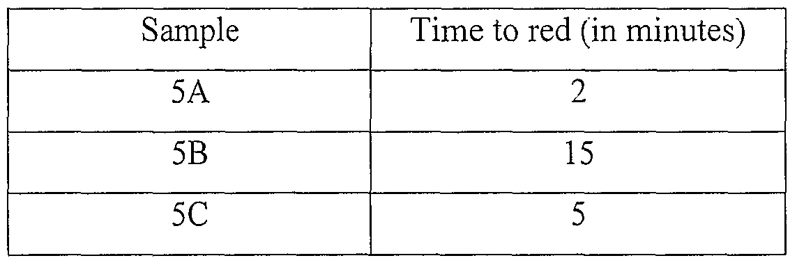


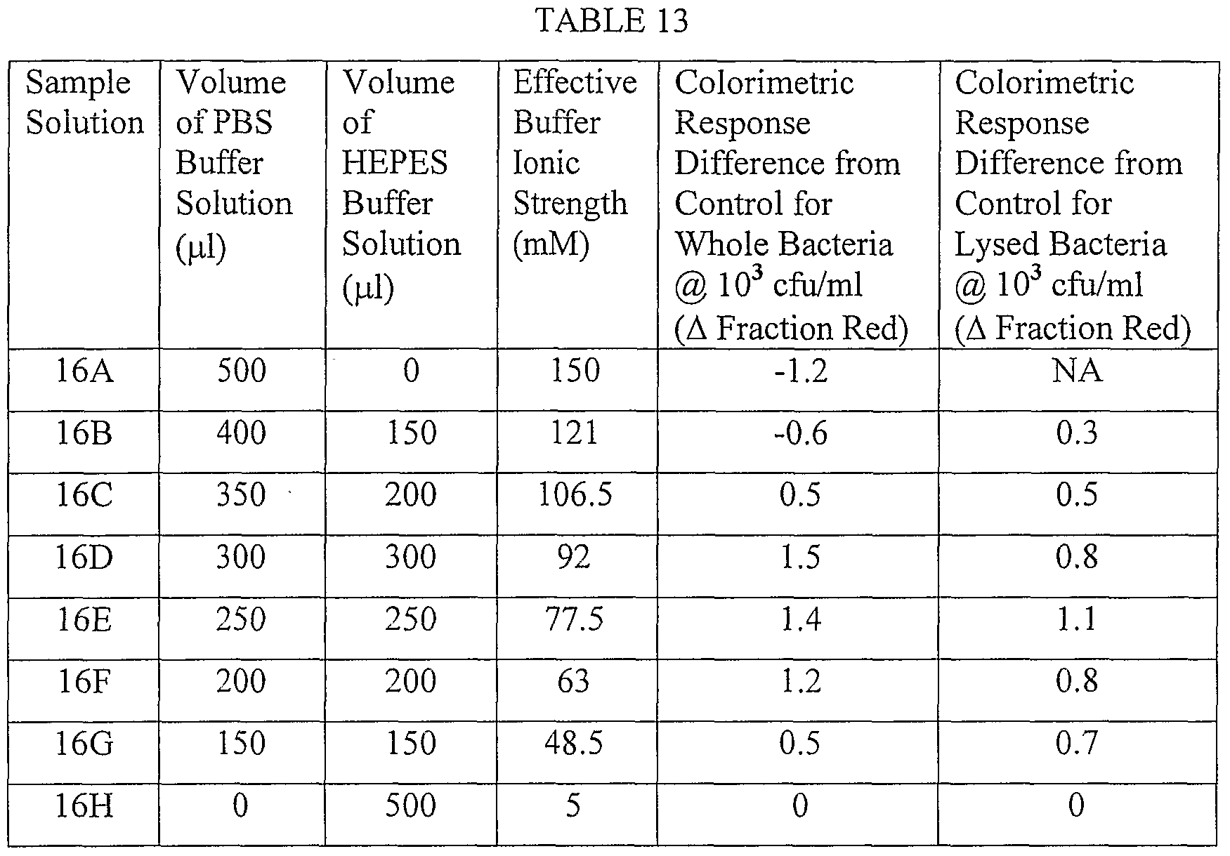


Claims
Priority Applications (9)
| Application Number | Priority Date | Filing Date | Title |
|---|---|---|---|
| CA002590867A CA2590867A1 (en) | 2004-12-17 | 2005-12-16 | Colorimetric sensors constructed of diacetylene materials |
| MX2007007179A MX2007007179A (en) | 2004-12-17 | 2005-12-16 | Colorimetric sensors constructed of diacetylene materials. |
| JP2007546936A JP2008524593A (en) | 2004-12-17 | 2005-12-16 | Colorimetric sensor constructed with diacetylene material |
| CN200580043538.XA CN101107523B (en) | 2004-12-17 | 2005-12-16 | Colorimetric sensors constructed of diacetylene materials |
| AU2005323177A AU2005323177A1 (en) | 2004-12-17 | 2005-12-16 | Colorimetric sensors constructed of diacetylene materials |
| US11/721,689 US20080193967A1 (en) | 2004-12-17 | 2005-12-16 | Colorimetric Sensor Constructed Of Diacetylene Materials |
| BRPI0519119-0A BRPI0519119A2 (en) | 2004-12-17 | 2005-12-16 | colorimetric system and method for detecting an analyte |
| EP05857091A EP1825268A2 (en) | 2004-12-17 | 2005-12-16 | Colorimetric sensors constructed of diacetylene materials |
| US13/398,882 US20120149037A1 (en) | 2004-12-17 | 2012-02-17 | Colorimetric sensors constructed of diacetylene materials |
Applications Claiming Priority (2)
| Application Number | Priority Date | Filing Date | Title |
|---|---|---|---|
| US63699304P | 2004-12-17 | 2004-12-17 | |
| US60/636,993 | 2004-12-17 |
Related Child Applications (1)
| Application Number | Title | Priority Date | Filing Date |
|---|---|---|---|
| US13/398,882 Continuation US20120149037A1 (en) | 2004-12-17 | 2012-02-17 | Colorimetric sensors constructed of diacetylene materials |
Publications (2)
| Publication Number | Publication Date |
|---|---|
| WO2006073738A2 true WO2006073738A2 (en) | 2006-07-13 |
| WO2006073738A3 WO2006073738A3 (en) | 2006-09-28 |
Family
ID=36579446
Family Applications (2)
| Application Number | Title | Priority Date | Filing Date |
|---|---|---|---|
| PCT/US2005/045607 WO2006073738A2 (en) | 2004-12-17 | 2005-12-16 | Colorimetric sensors constructed of diacetylene materials |
| PCT/US2005/046005 WO2006073782A2 (en) | 2004-12-17 | 2005-12-16 | Colorimetric sensors constructed of diacetylene materials |
Family Applications After (1)
| Application Number | Title | Priority Date | Filing Date |
|---|---|---|---|
| PCT/US2005/046005 WO2006073782A2 (en) | 2004-12-17 | 2005-12-16 | Colorimetric sensors constructed of diacetylene materials |
Country Status (11)
| Country | Link |
|---|---|
| US (3) | US20080193967A1 (en) |
| EP (2) | EP1877791A2 (en) |
| JP (2) | JP2008524593A (en) |
| CN (2) | CN101080637A (en) |
| AU (2) | AU2005323131B2 (en) |
| BR (1) | BRPI0519119A2 (en) |
| CA (2) | CA2589351A1 (en) |
| MX (1) | MX2007007179A (en) |
| TW (1) | TW200636245A (en) |
| WO (2) | WO2006073738A2 (en) |
| ZA (1) | ZA200705832B (en) |
Cited By (1)
| Publication number | Priority date | Publication date | Assignee | Title |
|---|---|---|---|---|
| JP2011504236A (en) * | 2007-11-20 | 2011-02-03 | スリーエム イノベイティブ プロパティズ カンパニー | Method for analyzing bacterial samples using a polymer sensor containing diacetylene |
Families Citing this family (48)
| Publication number | Priority date | Publication date | Assignee | Title |
|---|---|---|---|---|
| US20040126897A1 (en) * | 2002-12-19 | 2004-07-01 | 3M Innovative Properties Company | Colorimetric sensors constructed of diacetylene materials |
| US6963007B2 (en) | 2002-12-19 | 2005-11-08 | 3M Innovative Properties Company | Diacetylenic materials for sensing applications |
| AU2004314536A1 (en) * | 2003-12-30 | 2005-08-04 | 3M Innovative Properties Company | Method of enhancing signal detection of cell-wall components of cells |
| US7371511B2 (en) | 2004-08-19 | 2008-05-13 | 3M Innovative Properties Company | Polydiacetylene polymer blends |
| US7816472B2 (en) * | 2004-08-19 | 2010-10-19 | 3M Innovative Properties Company | Polydiacetylene polymer compositions and methods of manufacture |
| CA2589351A1 (en) * | 2004-12-17 | 2006-07-13 | 3M Innovative Properties Company | Colorimetric sensors constructed of diacetylene materials |
| JP5008046B2 (en) * | 2005-06-14 | 2012-08-22 | ローム株式会社 | Semiconductor device |
| TW200712495A (en) * | 2005-08-02 | 2007-04-01 | 3M Innovative Properties Co | Apparatus and method for detecting an analyte |
| TW200714898A (en) | 2005-08-02 | 2007-04-16 | 3M Innovative Properties Co | Apparatus and method for detecting an analyte |
| TW200712489A (en) * | 2005-08-02 | 2007-04-01 | 3M Innovative Properties Co | Apparatus assembly and method for detecting an analyte |
| TW200712487A (en) * | 2005-08-02 | 2007-04-01 | 3M Innovative Properties Co | Apparatus and method for detecting an analyte |
| GB0525843D0 (en) * | 2005-12-20 | 2006-02-01 | Reckitt Benckiser Uk Ltd | Improvements in or relating to compositions |
| US20080063615A1 (en) * | 2006-09-12 | 2008-03-13 | Macdonald John Gavin | Color changing skin sealant |
| US20080060550A1 (en) * | 2006-09-12 | 2008-03-13 | Macdonald Gavin | Color changing skin sealant with co-acid trigger |
| KR100847311B1 (en) | 2006-12-14 | 2008-07-21 | 안동준 | Polydiacetylene Sensor Chip Comprising DNA Sequence and Manufacturing Process thereof |
| US20080145316A1 (en) * | 2006-12-14 | 2008-06-19 | Macdonald John Gavin | Skin coating with microbial indicator |
| US8283912B2 (en) * | 2007-04-03 | 2012-10-09 | Koninklijke Philips Electronics N.V. | Sensor device with magnetic washing means |
| KR20100017232A (en) * | 2007-04-25 | 2010-02-16 | 쓰리엠 이노베이티브 프로퍼티즈 컴파니 | Compositions, methods, and devices for isolating biological materials |
| US20090030342A1 (en) * | 2007-07-27 | 2009-01-29 | 3M Innovative Properties Company | Apparatus and method for releasing a sample of material |
| WO2009029445A1 (en) * | 2007-08-27 | 2009-03-05 | 3M Innovative Properties Company | Apparatus and method for processing a fluidic sample |
| US20100257653A1 (en) * | 2007-09-06 | 2010-10-14 | Pitts Robert W | Infant wrap including body padding |
| US20090123569A1 (en) * | 2007-11-08 | 2009-05-14 | Macdonald John Gavin | Coverage indicating technology for skin sealants using tannates |
| CN101918829A (en) * | 2007-11-20 | 2010-12-15 | 3M创新有限公司 | Pick-up unit and method |
| EP2240774A2 (en) * | 2007-12-31 | 2010-10-20 | 3M Innovative Properties Company | Microorganism-capturing compositions and methods |
| ES2592205T3 (en) * | 2008-09-04 | 2016-11-28 | The Board Of Trustees Of The University Of Illinois | Colorimetric sensor matrices based on nanoporous pigments |
| EP2221592A1 (en) * | 2009-01-22 | 2010-08-25 | Stichting Dutch Polymer Institute | Multifunctional optical sensor |
| WO2011066504A1 (en) * | 2009-11-30 | 2011-06-03 | Physicians Reference Laboratory, Llc | Methods and compositions for detecting methicillin-resistant staphylococcus aureus |
| WO2011144652A2 (en) * | 2010-05-18 | 2011-11-24 | Delta Dansk Elektronik, Lys & Akustik | Method for depositing sensor material on a substrate |
| CN102243230A (en) * | 2011-04-11 | 2011-11-16 | 浙江工商大学 | Colour-changing sensor for rapidly detecting melamine and preparation method thereof |
| US9115240B2 (en) | 2011-06-02 | 2015-08-25 | University Of Maryland, College Park | Color changing polymer films for detecting chemical and biological targets |
| CN102435572B (en) * | 2011-09-20 | 2013-10-16 | 中国药科大学 | Drug-film affinity measuring method based on polydiacetylene sensor |
| WO2013170051A1 (en) * | 2012-05-09 | 2013-11-14 | Advanced Animal Diagnostics, Inc. | Sample cartridge and sample stage |
| US9052315B2 (en) | 2012-05-09 | 2015-06-09 | Advanced Animal Diagnostics, Inc. | Rapid detection of analytes in liquid samples |
| US10359614B2 (en) | 2012-07-03 | 2019-07-23 | Advanced Animal Diagnostics, Inc. | Diagnostic apparatus |
| WO2014069897A1 (en) * | 2012-11-01 | 2014-05-08 | 한양대학교 산학협력단 | Hydrocarbon identification sensor, method for manufacturing same, and application thereof |
| US9797893B2 (en) | 2013-05-09 | 2017-10-24 | Advanced Animal Diagnostics, Inc. | Rapid detection of analytes in liquid samples |
| CN106030766B (en) * | 2014-02-14 | 2019-05-28 | 株式会社樱花彩色笔 | Corona treatment detects indicator |
| JP6567863B2 (en) | 2014-09-16 | 2019-08-28 | 株式会社サクラクレパス | Ink composition for detecting plasma treatment and indicator for detecting plasma treatment |
| US10267792B2 (en) * | 2016-09-09 | 2019-04-23 | International Business Machines Corporation | Device for detecting toxic shock syndrome toxins and method of making the same |
| TWI629072B (en) * | 2017-01-13 | 2018-07-11 | 廈門聖慈醫療器材有限公司 | Suction disc |
| TWI621453B (en) | 2017-01-13 | 2018-04-21 | 廈門聖慈醫療器材有限公司 | Suction disc |
| CN109254002A (en) * | 2018-11-12 | 2019-01-22 | 四川大学 | The identification signal of biomarker is converted into the labeling method and application of visible Optical Chromatography |
| CN109738411B (en) * | 2019-01-31 | 2019-10-11 | 中南民族大学 | A kind of bionic array sensor and its application based on quantum dot fluorescence quenching |
| US11506658B2 (en) * | 2019-04-24 | 2022-11-22 | Progenitec, Inc. | System for analysis of body fluids and wound-associated biomolecules |
| CN110501316A (en) * | 2019-08-20 | 2019-11-26 | 西北农林科技大学 | A kind of poly- diacetylene liposome Pb in water environment2+Visible detection method |
| CN111595841B (en) * | 2020-05-20 | 2022-07-05 | 中国科学院新疆理化技术研究所 | Preparation method and application of array type chemical colorimetric vertical display analysis card |
| WO2022075992A1 (en) * | 2020-10-09 | 2022-04-14 | Hewlett-Packard Development Company, L.P. | Self-wicking devices |
| KR20230164416A (en) * | 2022-05-25 | 2023-12-04 | 인제대학교 산학협력단 | Composition for detecting lead ions comprising polydiacetylene liposome, method for preparing the same, and method for detecting lead ions |
Citations (8)
| Publication number | Priority date | Publication date | Assignee | Title |
|---|---|---|---|---|
| US5622872A (en) | 1989-06-15 | 1997-04-22 | Biocircuits Corporation | Analyte detection through observed optical modulation of polymerized lipid layers |
| US5753517A (en) | 1996-03-29 | 1998-05-19 | University Of British Columbia | Quantitative immunochromatographic assays |
| US6080423A (en) | 1994-08-11 | 2000-06-27 | Regents Of The University Of California | Three dimensional colorimetric assay assemblies |
| US6183722B1 (en) | 1991-11-27 | 2001-02-06 | Diatide, Inc. | Somatostatin analogs |
| US6277652B1 (en) | 1997-12-26 | 2001-08-21 | Hogy Medical Co., Ltd. | Colorimetric sensor employing polydiacetylene membrane |
| US6306598B1 (en) | 1992-11-13 | 2001-10-23 | Regents Of The University Of California | Nucleic acid-coupled colorimetric analyte detectors |
| WO2002000920A2 (en) | 2000-06-23 | 2002-01-03 | Segan Industries, Inc. | Ingestibles possessing intrinsic color change |
| US6395561B1 (en) | 1992-11-13 | 2002-05-28 | Regents Of The University Of California | Polymeric assay film for direct colorimetric detection |
Family Cites Families (52)
| Publication number | Priority date | Publication date | Assignee | Title |
|---|---|---|---|---|
| BE754567A (en) * | 1969-08-09 | 1971-02-08 | Basf Ag | IRRADIATION CURABLE MATERIALS, BASED ON POLYMERS WITH NON-SATURATED ETHYLENICALLY REPEATED GROUPS |
| US3999946A (en) * | 1976-02-23 | 1976-12-28 | Allied Chemical Corporation | Time-temperature history indicators |
| CH620655A5 (en) * | 1977-06-17 | 1980-12-15 | Loepfe Ag Geb | |
| US4189399A (en) * | 1977-07-19 | 1980-02-19 | Allied Chemical Corporation | Co-crystallized acetylenic compounds |
| US4215208A (en) * | 1977-10-05 | 1980-07-29 | Allied Chemical Corporation | Thermochromic polyacetylenes containing urethane groups |
| US4228126A (en) * | 1977-11-25 | 1980-10-14 | Allied Chemical Corporation | Diacetylene time-temperature indicators |
| US4195058A (en) * | 1978-08-30 | 1980-03-25 | Allied Chemical Corporation | Vapor permeation time-temperature indicator |
| US4238352A (en) * | 1978-11-13 | 1980-12-09 | Allied Chemical Corporation | Co-polymerized acetylenic compositions |
| US4235108A (en) * | 1978-11-13 | 1980-11-25 | Allied Chemical Corporation | Device for measuring temperature using co-crystallized acetylenic compositions |
| DE2905531A1 (en) * | 1979-02-14 | 1981-01-08 | Boehringer Mannheim Gmbh | DIAGNOSTIC AGENT FOR DETECTING LEUCOCYTES IN BODY LIQUIDS |
| US4389217A (en) * | 1979-05-11 | 1983-06-21 | Allied Corporation | Integrated time-temperature or radiation-dosage history recording device |
| US4339240A (en) * | 1980-05-07 | 1982-07-13 | Allied Corporation | Color changing polyacetylenic compounds |
| IL61951A (en) * | 1981-01-21 | 1984-01-31 | Univ Ben Gurion | Method and apparatus for detecting nitrite ions in fluids |
| US4735745A (en) * | 1982-05-03 | 1988-04-05 | Lifelines Technology, Inc. | Defrost indicator |
| US4767826A (en) * | 1985-07-18 | 1988-08-30 | Polytechnic Institute Of New York | Radiation-sensitive polymers |
| US4721769A (en) * | 1985-10-18 | 1988-01-26 | Gte Laboratories Incorporated | Diacetylene segmented copolymers |
| US4849500A (en) * | 1986-03-07 | 1989-07-18 | Gte Laboratories Incorporated | Polyamide from diacetylene dicarboxylic acid compound |
| US4916211A (en) * | 1986-03-07 | 1990-04-10 | Gte Laboratories Incorporated | Thermochromic cross polymerized polyamide-diacetylene compound |
| US4810635A (en) * | 1986-04-16 | 1989-03-07 | Miles Inc. | Specific binding assays employing label analog to reduce sample interferences |
| FR2625321B1 (en) * | 1987-12-24 | 1992-10-02 | Pasteur Institut | REAGENT FOR THE DIAGNOSIS OF STAPHYLOCOCCUS AUREUS BY AGGLUTINATION |
| US5156810A (en) * | 1989-06-15 | 1992-10-20 | Biocircuits Corporation | Biosensors employing electrical, optical and mechanical signals |
| US5672465A (en) * | 1990-04-09 | 1997-09-30 | Jp Laboratories, Inc. | Polyethyleneimine binder complex films |
| EP0475045B1 (en) * | 1990-08-06 | 1996-12-11 | Bayer Corporation | Method and device for the assay of ions |
| FR2679923B1 (en) * | 1991-08-02 | 1993-10-22 | Bio Merieux | REAGENT FOR THE IDENTIFICATION OF BACTERIA OF THE SPECIES STAPHYLOCCOCUS AUREUS. |
| US6001556A (en) * | 1992-11-13 | 1999-12-14 | The Regents Of The University Of California | Polymeric assay film for direct colorimetric detection |
| US5798215A (en) * | 1993-02-18 | 1998-08-25 | Biocircuits Corporation | Device for use in analyte detection assays |
| US5503985A (en) * | 1993-02-18 | 1996-04-02 | Cathey; Cheryl A. | Disposable device for diagnostic assays |
| US5415999A (en) * | 1993-07-09 | 1995-05-16 | Biocircuits Corporation | Fluorescent lipid polymer-macromolecular ligand compositions as detection element in ligand assays |
| US6103217A (en) * | 1994-08-11 | 2000-08-15 | The Regents Of The University Of California | Polymeric assemblies for sensitive colorimetric assays |
| AU714839B2 (en) * | 1995-02-13 | 2000-01-13 | Regents Of The University Of California, The | Three-dimensional colorimetric assay assemblies |
| CA2224798A1 (en) * | 1995-06-22 | 1997-01-09 | Matthew T. Scholz | Stable hydroalcoholic compositions |
| US5685641A (en) * | 1996-01-16 | 1997-11-11 | Ribi; Hans O. | Devices for rapid temperature detection |
| AU715973B2 (en) * | 1996-01-26 | 2000-02-10 | Regents Of The University Of California, The | Polymeric film, assay and method for direct colorimetric detection of analytes |
| DE69712754T2 (en) * | 1996-03-01 | 2002-10-17 | Univ California | INHIBITION OF SELECTINBINDING |
| CA2276091A1 (en) * | 1996-12-27 | 1998-07-09 | Mochida Pharmaceutical Co., Ltd. | Cell membrane-directed drugs |
| US6420622B1 (en) * | 1997-08-01 | 2002-07-16 | 3M Innovative Properties Company | Medical article having fluid control film |
| US6375871B1 (en) * | 1998-06-18 | 2002-04-23 | 3M Innovative Properties Company | Methods of manufacturing microfluidic articles |
| US6046455A (en) * | 1998-01-30 | 2000-04-04 | Segan Industries | Integrating ultraviolet exposure detection devices |
| US6361962B1 (en) * | 1998-10-06 | 2002-03-26 | Verseau Group | Toxin detector |
| US6472214B2 (en) * | 1999-05-26 | 2002-10-29 | Jp Labs, Inc. | Freeze monitoring device |
| US6387614B1 (en) * | 1999-07-07 | 2002-05-14 | The Regents Of The University Of California | Methods for using redox liposome biosensors |
| US6451191B1 (en) * | 1999-11-18 | 2002-09-17 | 3M Innovative Properties Company | Film based addressable programmable electronic matrix articles and methods of manufacturing and using the same |
| US7049152B2 (en) * | 2001-03-13 | 2006-05-23 | The Regents Of The University Of California | Color and shape changing polymeric ribbons and sheets |
| US6942169B2 (en) * | 2001-06-06 | 2005-09-13 | Integrated Sensing Systems | Micromachined lysing device and method for performing cell lysis |
| WO2003029479A2 (en) * | 2001-08-10 | 2003-04-10 | Regents Of The University Of California | Sensitive and rapid detection of pathogenic organisms and toxins using fluorescent polymeric lipids |
| CA2510030C (en) * | 2002-12-19 | 2012-07-10 | 3M Innovative Properties Company | Colorimetric sensors constructed of diacetylene materials |
| US6963007B2 (en) * | 2002-12-19 | 2005-11-08 | 3M Innovative Properties Company | Diacetylenic materials for sensing applications |
| US20040126897A1 (en) * | 2002-12-19 | 2004-07-01 | 3M Innovative Properties Company | Colorimetric sensors constructed of diacetylene materials |
| AU2004314536A1 (en) * | 2003-12-30 | 2005-08-04 | 3M Innovative Properties Company | Method of enhancing signal detection of cell-wall components of cells |
| US7371511B2 (en) * | 2004-08-19 | 2008-05-13 | 3M Innovative Properties Company | Polydiacetylene polymer blends |
| US7816472B2 (en) * | 2004-08-19 | 2010-10-19 | 3M Innovative Properties Company | Polydiacetylene polymer compositions and methods of manufacture |
| CA2589351A1 (en) * | 2004-12-17 | 2006-07-13 | 3M Innovative Properties Company | Colorimetric sensors constructed of diacetylene materials |
-
2005
- 2005-12-16 CA CA002589351A patent/CA2589351A1/en not_active Abandoned
- 2005-12-16 CN CNA2005800434298A patent/CN101080637A/en active Pending
- 2005-12-16 US US11/721,689 patent/US20080193967A1/en not_active Abandoned
- 2005-12-16 JP JP2007546936A patent/JP2008524593A/en not_active Ceased
- 2005-12-16 CN CN200580043538.XA patent/CN101107523B/en not_active Expired - Fee Related
- 2005-12-16 WO PCT/US2005/045607 patent/WO2006073738A2/en active Application Filing
- 2005-12-16 BR BRPI0519119-0A patent/BRPI0519119A2/en not_active IP Right Cessation
- 2005-12-16 JP JP2007547013A patent/JP2008524603A/en active Pending
- 2005-12-16 EP EP05857135A patent/EP1877791A2/en not_active Withdrawn
- 2005-12-16 CA CA002590867A patent/CA2590867A1/en not_active Abandoned
- 2005-12-16 MX MX2007007179A patent/MX2007007179A/en unknown
- 2005-12-16 EP EP05857091A patent/EP1825268A2/en not_active Withdrawn
- 2005-12-16 US US11/303,543 patent/US20060134796A1/en not_active Abandoned
- 2005-12-16 AU AU2005323131A patent/AU2005323131B2/en not_active Ceased
- 2005-12-16 TW TW094144976A patent/TW200636245A/en unknown
- 2005-12-16 AU AU2005323177A patent/AU2005323177A1/en not_active Abandoned
- 2005-12-16 WO PCT/US2005/046005 patent/WO2006073782A2/en active Application Filing
-
2007
- 2007-07-16 ZA ZA200705832A patent/ZA200705832B/en unknown
-
2012
- 2012-02-17 US US13/398,882 patent/US20120149037A1/en not_active Abandoned
Patent Citations (8)
| Publication number | Priority date | Publication date | Assignee | Title |
|---|---|---|---|---|
| US5622872A (en) | 1989-06-15 | 1997-04-22 | Biocircuits Corporation | Analyte detection through observed optical modulation of polymerized lipid layers |
| US6183722B1 (en) | 1991-11-27 | 2001-02-06 | Diatide, Inc. | Somatostatin analogs |
| US6306598B1 (en) | 1992-11-13 | 2001-10-23 | Regents Of The University Of California | Nucleic acid-coupled colorimetric analyte detectors |
| US6395561B1 (en) | 1992-11-13 | 2002-05-28 | Regents Of The University Of California | Polymeric assay film for direct colorimetric detection |
| US6080423A (en) | 1994-08-11 | 2000-06-27 | Regents Of The University Of California | Three dimensional colorimetric assay assemblies |
| US5753517A (en) | 1996-03-29 | 1998-05-19 | University Of British Columbia | Quantitative immunochromatographic assays |
| US6277652B1 (en) | 1997-12-26 | 2001-08-21 | Hogy Medical Co., Ltd. | Colorimetric sensor employing polydiacetylene membrane |
| WO2002000920A2 (en) | 2000-06-23 | 2002-01-03 | Segan Industries, Inc. | Ingestibles possessing intrinsic color change |
Non-Patent Citations (2)
| Title |
|---|
| CHANCE ET AL., JOURNAL OF CLZEMISHY AND PHYSICS |
| MINO ET AL., LANGMUIR, vol. 8, 1992, pages 594 |
Cited By (1)
| Publication number | Priority date | Publication date | Assignee | Title |
|---|---|---|---|---|
| JP2011504236A (en) * | 2007-11-20 | 2011-02-03 | スリーエム イノベイティブ プロパティズ カンパニー | Method for analyzing bacterial samples using a polymer sensor containing diacetylene |
Also Published As
| Publication number | Publication date |
|---|---|
| WO2006073782A3 (en) | 2006-11-16 |
| EP1877791A2 (en) | 2008-01-16 |
| CA2589351A1 (en) | 2006-07-13 |
| WO2006073782A9 (en) | 2007-09-27 |
| WO2006073782A2 (en) | 2006-07-13 |
| JP2008524603A (en) | 2008-07-10 |
| AU2005323131B2 (en) | 2011-09-01 |
| JP2008524593A (en) | 2008-07-10 |
| CN101107523A (en) | 2008-01-16 |
| MX2007007179A (en) | 2007-08-14 |
| BRPI0519119A2 (en) | 2008-12-23 |
| US20060134796A1 (en) | 2006-06-22 |
| US20120149037A1 (en) | 2012-06-14 |
| AU2005323177A1 (en) | 2006-07-13 |
| CN101107523B (en) | 2012-12-05 |
| CA2590867A1 (en) | 2006-07-13 |
| CN101080637A (en) | 2007-11-28 |
| ZA200705832B (en) | 2008-10-29 |
| AU2005323131A1 (en) | 2006-07-13 |
| EP1825268A2 (en) | 2007-08-29 |
| US20080193967A1 (en) | 2008-08-14 |
| WO2006073738A3 (en) | 2006-09-28 |
| TW200636245A (en) | 2006-10-16 |
Similar Documents
| Publication | Publication Date | Title |
|---|---|---|
| AU2005323131B2 (en) | Colorimetric sensors constructed of diacetylene materials | |
| CN100578226C (en) | The colorimetric sensor that constitutes by the diacetylene material | |
| Yue et al. | A facile label-free electrochemiluminescent biosensor for specific detection of Staphylococcus aureus utilizing the binding between immunoglobulin G and protein A | |
| US20110097814A1 (en) | Detection devices and methods | |
| AU2004314536A1 (en) | Method of enhancing signal detection of cell-wall components of cells | |
| US20110091903A1 (en) | Method of analyzing a sample for a bacterium using diacetylene-containing polymer sensor | |
| US11667946B2 (en) | Detection of beta-hemolytic pathogens | |
| Byzova et al. | Development of an immunochromatographic test system for the detection of Helicobacter pylori antigens | |
| US10006906B2 (en) | Detection assays and methods | |
| Zimmerman et al. | Absorbance-based assay for membrane disruption by antimicrobial peptides and synthetic copolymers using pyrroloquinoline quinone-loaded liposomes | |
| Annapure et al. | Liposomes as biosensors in the food sector | |
| AU2021464941A1 (en) | Colorimetric sensors for detection of chemical and biological contaminants |
Legal Events
| Date | Code | Title | Description |
|---|---|---|---|
| 121 | Ep: the epo has been informed by wipo that ep was designated in this application | ||
| WWE | Wipo information: entry into national phase |
Ref document number: 2005857091 Country of ref document: EP |
|
| WWE | Wipo information: entry into national phase |
Ref document number: 2590867 Country of ref document: CA |
|
| WWE | Wipo information: entry into national phase |
Ref document number: MX/a/2007/007179 Country of ref document: MX Ref document number: 11721689 Country of ref document: US |
|
| WWE | Wipo information: entry into national phase |
Ref document number: 2007546936 Country of ref document: JP Ref document number: 2005323177 Country of ref document: AU |
|
| WWE | Wipo information: entry into national phase |
Ref document number: 200580043538.X Country of ref document: CN Ref document number: 2611/CHENP/2007 Country of ref document: IN |
|
| NENP | Non-entry into the national phase |
Ref country code: DE |
|
| ENP | Entry into the national phase |
Ref document number: 2005323177 Country of ref document: AU Date of ref document: 20051216 Kind code of ref document: A |
|
| WWP | Wipo information: published in national office |
Ref document number: 2005323177 Country of ref document: AU |
|
| WWP | Wipo information: published in national office |
Ref document number: 2005857091 Country of ref document: EP |
|
| ENP | Entry into the national phase |
Ref document number: PI0519119 Country of ref document: BR |