WO2011122004A1 - Imaging apparatus and imaging method - Google Patents
Imaging apparatus and imaging method Download PDFInfo
- Publication number
- WO2011122004A1 WO2011122004A1 PCT/JP2011/001868 JP2011001868W WO2011122004A1 WO 2011122004 A1 WO2011122004 A1 WO 2011122004A1 JP 2011001868 W JP2011001868 W JP 2011001868W WO 2011122004 A1 WO2011122004 A1 WO 2011122004A1
- Authority
- WO
- WIPO (PCT)
- Prior art keywords
- imaging
- light beams
- inspection object
- measurement light
- display
- Prior art date
Links
Images


Classifications
-
- A—HUMAN NECESSITIES
- A61—MEDICAL OR VETERINARY SCIENCE; HYGIENE
- A61B—DIAGNOSIS; SURGERY; IDENTIFICATION
- A61B3/00—Apparatus for testing the eyes; Instruments for examining the eyes
- A61B3/10—Objective types, i.e. instruments for examining the eyes independent of the patients' perceptions or reactions
- A61B3/102—Objective types, i.e. instruments for examining the eyes independent of the patients' perceptions or reactions for optical coherence tomography [OCT]
-
- A—HUMAN NECESSITIES
- A61—MEDICAL OR VETERINARY SCIENCE; HYGIENE
- A61B—DIAGNOSIS; SURGERY; IDENTIFICATION
- A61B3/00—Apparatus for testing the eyes; Instruments for examining the eyes
- A61B3/10—Objective types, i.e. instruments for examining the eyes independent of the patients' perceptions or reactions
- A61B3/12—Objective types, i.e. instruments for examining the eyes independent of the patients' perceptions or reactions for looking at the eye fundus, e.g. ophthalmoscopes
-
- G—PHYSICS
- G01—MEASURING; TESTING
- G01B—MEASURING LENGTH, THICKNESS OR SIMILAR LINEAR DIMENSIONS; MEASURING ANGLES; MEASURING AREAS; MEASURING IRREGULARITIES OF SURFACES OR CONTOURS
- G01B9/00—Measuring instruments characterised by the use of optical techniques
- G01B9/02—Interferometers
Landscapes
- Health & Medical Sciences (AREA)
- Life Sciences & Earth Sciences (AREA)
- Physics & Mathematics (AREA)
- Medical Informatics (AREA)
- Surgery (AREA)
- Biophysics (AREA)
- Ophthalmology & Optometry (AREA)
- Engineering & Computer Science (AREA)
- Biomedical Technology (AREA)
- Heart & Thoracic Surgery (AREA)
- Veterinary Medicine (AREA)
- Molecular Biology (AREA)
- Public Health (AREA)
- Animal Behavior & Ethology (AREA)
- General Health & Medical Sciences (AREA)
- Radiology & Medical Imaging (AREA)
- Nuclear Medicine, Radiotherapy & Molecular Imaging (AREA)
- General Physics & Mathematics (AREA)
- Eye Examination Apparatus (AREA)
- Investigating Or Analysing Materials By Optical Means (AREA)
Abstract
Description
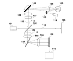
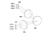
[Fig. 1B] Fig. 1B illustrates an imaging apparatus according to the first exemplary embodiment.
[Fig. 2A] Fig. 2A illustrates a screen display according to the first exemplary embodiment.
[Fig. 2B] Fig. 2B illustrates a screen display according to the first exemplary embodiment.
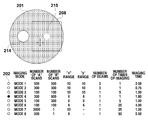
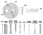
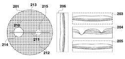
[Fig. 3A] Fig. 3A illustrates an imaging mode according to the first and a second exemplary embodiment.
[Fig. 3B] Fig. 3B illustrates an imaging mode according to the first and the second exemplary embodiments.
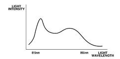
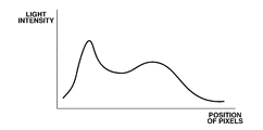
[Fig. 4A] Fig. 4A illustrates frequency characteristics of a light source and an output signal of a sensor according to the first exemplary embodiment.
[Fig. 4B] Fig. 4B illustrates frequency characteristics of a light source and an output signal of a sensor according to the first exemplary embodiment.
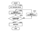
[Fig. 5] Fig. 5 is a flowchart illustrating an imaging method according to the first exemplary embodiment.
[Fig. 6A] Fig. 6A illustrates a display of optical coherence tomographic images corresponding to the number of measurement light beams according to the first exemplary embodiment.
[Fig. 6B] Fig. 6B illustrates a display of optical coherence tomographic images corresponding to the number of measurement light beams according to the first exemplary embodiment.
[Fig. 7A] Fig. 7A illustrates a screen display according to the first and the second exemplary embodiments.
[Fig. 7B] Fig. 7B illustrates a screen display according to the first and the second exemplary embodiments.
[Fig. 7C] Fig. 7C illustrates a screen display according to the first and the second exemplary embodiments.
[Fig. 7D] Fig. 7D illustrates a screen display according to the first and the second exemplary embodiments.
[Fig. 8] Fig. 8 is a flowchart illustrating an imaging method according to the second exemplary embodiment.
Claims (15)
- An imaging apparatus comprising:
an irradiation unit configured to irradiate an inspection object with a plurality of measurement light beams;
an intersection image acquisition unit configured to acquire an intersection image of the inspection object in a direction intersecting a direction in which the inspection object is irradiated with the plurality of measurement light beams;
an intersection image display control unit configured to cause a display unit to display the intersection image; and
a scanning range display control unit configured to cause the display unit to display a scanning range of the plurality of measurement light beams by being associated with the intersection image. - The imaging apparatus according to claim 1, further comprising:
an acquisition unit configured to acquire optical coherence tomographic images of the inspection object based on a plurality of combined light beams each obtained by combining a plurality of return light beams from the inspection object irradiated with the plurality of measurement light beams and a plurality of reference light beams each corresponding to the plurality of measurement light beams;
a tomographic image display control unit configured to cause the display unit to display each of the optical coherence tomographic images; and
a position display control unit configured to cause the display unit to display a position of each of the optical coherence tomographic images by being associated with the intersection image. - The imaging apparatus according to claim 1 or claim 2, wherein the scanning range display control unit causes the display unit to display the scanning ranges with different colors or shapes.
- The imaging apparatus according to any one of claims 1 to 3, wherein the inspection object is a fundus of an eye to be examined, and
the intersection image is at least one of a two-dimensional image of a surface of the fundus, an integrated image obtained by integrating at least a portion of the optical coherence tomographic images in a depth direction of the fundus, and the optical coherence tomographic images in a substantially vertical direction to the depth direction of the fundus. - The imaging apparatus according to any one of claims 1 to 4, further comprising:
a selection unit configured to select at least one of a plurality of imaging modes with different imaging conditions; and
an acquisition unit configured to acquire the optical coherence tomographic images according to the imaging mode selected by the selection unit. - The imaging apparatus according to claim 5, further comprising an imaging condition display control unit configured to cause the display unit to display a list of the plurality of imaging modes and an image including a function of the selection unit.
- The imaging apparatus according to claim 5 or claim 6, wherein at least one imaging mode among the plurality of imaging modes is set so that the number of measurement light beams irradiated to the inspection object is different from that of the other imaging modes.
- The imaging apparatus according to any one of claims 5 to 7, wherein at least one imaging mode among the plurality of imaging modes is set so that at least one value among a size of the scanning range, the number of times of imaging, and an imaging time period is set different from those in the other imaging modes.
- A method for capturing optical coherence tomographic images of an inspection object using an imaging apparatus according to any one of claims 1 to 8, the method comprising:
inputting a signal to be displayed on the display unit; and
displaying information based on the input signal on the display unit. - A method for capturing optical coherence tomographic images of an inspection object based on a plurality of combined light beams each obtained by combining a plurality of return light beams from the inspection object irradiated with a plurality of measurement light beams and a plurality of reference light beams each corresponding to the plurality of measurement light beams, the method comprising:
displaying an intersection image of the inspection object in a direction intersecting a direction in which the inspection object is irradiated with the plurality of measurement light beams; and
displaying a scanning range of the plurality of measurement light beams on the display unit by being associated with the intersection image. - A method for capturing optical coherence tomographic images of an inspection object based on a plurality of combined light beams each obtained by combining a plurality of return light beams from the inspection object irradiated with a plurality of measurement light beams and a plurality of reference light beams each corresponding to the plurality of measurement light beams, the method comprising:
selecting at least one from among a plurality of imaging modes with different imaging conditions; and
acquiring the optical coherence tomographic images according to the selected imaging mode. - A program for causing a computer to execute a method according to any one of claims 9 to 11.
- An imaging apparatus capable of capturing optical coherence tomographic images of an inspection object based on a plurality of combined light beams each obtained by combining a plurality of return light beams from the inspection object irradiated with a plurality of measurement light beams and a plurality of reference light beams each corresponding to the plurality of measurement light beams, the imaging apparatus comprising:
a selection unit configured to select at least one of a plurality of imaging modes with different imaging conditions; and
an acquisition unit configured to acquire the optical coherence tomographic images according to the imaging mode selected by the selection unit. - The imaging apparatus according to claim 13, further comprising an imaging condition display control unit configured to cause a display unit to display a list of the plurality of imaging modes and an image including a function of the selection unit.
- The imaging apparatus according to claim 13 or claim 14, wherein at least one imaging mode among the plurality of imaging modes is set so that the number of measurement light beams irradiated to the inspection object is different from that of the other imaging modes.
Priority Applications (4)
| Application Number | Priority Date | Filing Date | Title |
|---|---|---|---|
| KR1020127026813A KR101407385B1 (en) | 2010-03-31 | 2011-03-29 | Imaging apparatus and imaging method |
| US13/634,527 US8820933B2 (en) | 2010-03-31 | 2011-03-29 | Imaging apparatus and imaging method |
| CN201180017918.1A CN102834046B (en) | 2010-03-31 | 2011-03-29 | Picture pick-up device and image capture method |
| EP11716076A EP2552298A1 (en) | 2010-03-31 | 2011-03-29 | Imaging apparatus and imaging method |
Applications Claiming Priority (2)
| Application Number | Priority Date | Filing Date | Title |
|---|---|---|---|
| JP2010-082804 | 2010-03-31 | ||
| JP2010082804A JP5783681B2 (en) | 2010-03-31 | 2010-03-31 | Imaging apparatus and imaging method |
Publications (1)
| Publication Number | Publication Date |
|---|---|
| WO2011122004A1 true WO2011122004A1 (en) | 2011-10-06 |
Family
ID=44280720
Family Applications (1)
| Application Number | Title | Priority Date | Filing Date |
|---|---|---|---|
| PCT/JP2011/001868 WO2011122004A1 (en) | 2010-03-31 | 2011-03-29 | Imaging apparatus and imaging method |
Country Status (6)
| Country | Link |
|---|---|
| US (1) | US8820933B2 (en) |
| EP (1) | EP2552298A1 (en) |
| JP (1) | JP5783681B2 (en) |
| KR (1) | KR101407385B1 (en) |
| CN (2) | CN102834046B (en) |
| WO (1) | WO2011122004A1 (en) |
Cited By (3)
| Publication number | Priority date | Publication date | Assignee | Title |
|---|---|---|---|---|
| WO2014084231A1 (en) * | 2012-11-30 | 2014-06-05 | 株式会社トプコン | Fundus photographing device |
| US8870377B2 (en) | 2012-01-27 | 2014-10-28 | Canon Kabushiki Kaisha | Image processing apparatus, image processing apparatus control method, ophthalmologic apparatus, ophthalmologic apparatus control method, ophthalmologic system, and storage medium |
| EP3263016A4 (en) * | 2015-02-27 | 2018-10-24 | Kowa Company, Ltd. | Cross-section image capture device |
Families Citing this family (10)
| Publication number | Priority date | Publication date | Assignee | Title |
|---|---|---|---|---|
| JP5210443B1 (en) | 2012-01-26 | 2013-06-12 | キヤノン株式会社 | Optical tomographic imaging apparatus and control method |
| JP6460618B2 (en) * | 2013-01-31 | 2019-01-30 | キヤノン株式会社 | Optical coherence tomography apparatus and control method thereof |
| JP6300443B2 (en) * | 2013-01-31 | 2018-03-28 | キヤノン株式会社 | Optical tomographic imaging apparatus and control method thereof |
| JP6184113B2 (en) * | 2013-01-31 | 2017-08-23 | キヤノン株式会社 | Optical tomographic imaging apparatus and control method thereof |
| JP5766225B2 (en) * | 2013-02-22 | 2015-08-19 | キヤノン株式会社 | Optical tomographic imaging apparatus and control method |
| JP6585897B2 (en) * | 2015-01-29 | 2019-10-02 | 株式会社トプコン | Ophthalmic imaging equipment |
| US10709329B2 (en) * | 2015-07-30 | 2020-07-14 | Canon Kabushiki Kaisha | Image pickup apparatus and method of controlling image pickup apparatus |
| CN105913446B (en) * | 2016-05-04 | 2019-05-17 | 深圳市斯尔顿科技有限公司 | A kind of colorful line scanning fundus imaging device and colorful eye fundus image synthetic method |
| CN109715045B (en) * | 2016-09-16 | 2021-12-14 | 爱尔康公司 | Subtraction coronal optical coherence tomography imaging |
| WO2018203174A1 (en) * | 2017-05-02 | 2018-11-08 | Novartis Ag | Reconfigurable optical coherence tomography (oct) system |
Citations (5)
| Publication number | Priority date | Publication date | Assignee | Title |
|---|---|---|---|---|
| US6198540B1 (en) * | 1997-03-26 | 2001-03-06 | Kowa Company, Ltd. | Optical coherence tomography have plural reference beams of differing modulations |
| JP2008508068A (en) | 2004-08-03 | 2008-03-21 | カール ツァイス メディテック アクチエンゲゼルシャフト | Eye Fourier domain OCT ray tracing method |
| EP1908397A2 (en) * | 2006-10-04 | 2008-04-09 | Osaka University | A fundus oculi observation device |
| EP1935329A1 (en) * | 2006-12-22 | 2008-06-25 | Kabushiki Kaisha TOPCON | Fundus oculi observation device and fundus oculi image display device |
| JP2010082804A (en) | 2008-09-29 | 2010-04-15 | Pilot Ink Co Ltd | Heat discoloration writing implement |
Family Cites Families (8)
| Publication number | Priority date | Publication date | Assignee | Title |
|---|---|---|---|---|
| WO1992019930A1 (en) | 1991-04-29 | 1992-11-12 | Massachusetts Institute Of Technology | Method and apparatus for optical imaging and measurement |
| GB0425419D0 (en) * | 2004-11-18 | 2004-12-22 | Sira Ltd | Interference apparatus and method and probe |
| US8223143B2 (en) | 2006-10-27 | 2012-07-17 | Carl Zeiss Meditec, Inc. | User interface for efficiently displaying relevant OCT imaging data |
| JP5231802B2 (en) * | 2007-12-29 | 2013-07-10 | 株式会社ニデック | Ophthalmic imaging equipment |
| TW201034626A (en) * | 2008-07-21 | 2010-10-01 | Optovue Inc | Extended range imaging |
| JP4819851B2 (en) * | 2008-07-31 | 2011-11-24 | キヤノン株式会社 | Diagnosis support apparatus and method, program, and recording medium |
| JP5605999B2 (en) * | 2009-03-06 | 2014-10-15 | キヤノン株式会社 | Optical coherence tomography method and apparatus |
| JP5836564B2 (en) * | 2010-03-12 | 2015-12-24 | キヤノン株式会社 | Ophthalmic imaging apparatus, ophthalmic imaging method, and program thereof |
-
2010
- 2010-03-31 JP JP2010082804A patent/JP5783681B2/en active Active
-
2011
- 2011-03-29 WO PCT/JP2011/001868 patent/WO2011122004A1/en active Application Filing
- 2011-03-29 CN CN201180017918.1A patent/CN102834046B/en not_active Expired - Fee Related
- 2011-03-29 US US13/634,527 patent/US8820933B2/en not_active Expired - Fee Related
- 2011-03-29 EP EP11716076A patent/EP2552298A1/en not_active Withdrawn
- 2011-03-29 CN CN201510563464.8A patent/CN105266755B/en not_active Expired - Fee Related
- 2011-03-29 KR KR1020127026813A patent/KR101407385B1/en active IP Right Grant
Patent Citations (5)
| Publication number | Priority date | Publication date | Assignee | Title |
|---|---|---|---|---|
| US6198540B1 (en) * | 1997-03-26 | 2001-03-06 | Kowa Company, Ltd. | Optical coherence tomography have plural reference beams of differing modulations |
| JP2008508068A (en) | 2004-08-03 | 2008-03-21 | カール ツァイス メディテック アクチエンゲゼルシャフト | Eye Fourier domain OCT ray tracing method |
| EP1908397A2 (en) * | 2006-10-04 | 2008-04-09 | Osaka University | A fundus oculi observation device |
| EP1935329A1 (en) * | 2006-12-22 | 2008-06-25 | Kabushiki Kaisha TOPCON | Fundus oculi observation device and fundus oculi image display device |
| JP2010082804A (en) | 2008-09-29 | 2010-04-15 | Pilot Ink Co Ltd | Heat discoloration writing implement |
Cited By (4)
| Publication number | Priority date | Publication date | Assignee | Title |
|---|---|---|---|---|
| US8870377B2 (en) | 2012-01-27 | 2014-10-28 | Canon Kabushiki Kaisha | Image processing apparatus, image processing apparatus control method, ophthalmologic apparatus, ophthalmologic apparatus control method, ophthalmologic system, and storage medium |
| WO2014084231A1 (en) * | 2012-11-30 | 2014-06-05 | 株式会社トプコン | Fundus photographing device |
| EP3263016A4 (en) * | 2015-02-27 | 2018-10-24 | Kowa Company, Ltd. | Cross-section image capture device |
| US10188286B2 (en) | 2015-02-27 | 2019-01-29 | Kowa Company, Ltd. | Tomographic image capturing device |
Also Published As
| Publication number | Publication date |
|---|---|
| CN105266755A (en) | 2016-01-27 |
| CN102834046B (en) | 2015-09-02 |
| US8820933B2 (en) | 2014-09-02 |
| CN105266755B (en) | 2017-05-24 |
| EP2552298A1 (en) | 2013-02-06 |
| JP2011212203A (en) | 2011-10-27 |
| KR101407385B1 (en) | 2014-06-13 |
| US20130003018A1 (en) | 2013-01-03 |
| JP5783681B2 (en) | 2015-09-24 |
| KR20120140672A (en) | 2012-12-31 |
| CN102834046A (en) | 2012-12-19 |
Similar Documents
| Publication | Publication Date | Title |
|---|---|---|
| US8820933B2 (en) | Imaging apparatus and imaging method | |
| US10628004B2 (en) | Interactive control apparatus | |
| JP5210442B1 (en) | Optical tomographic imaging apparatus and control method | |
| JP5210443B1 (en) | Optical tomographic imaging apparatus and control method | |
| US9326679B2 (en) | Medical system | |
| JP6184113B2 (en) | Optical tomographic imaging apparatus and control method thereof | |
| US20120253183A1 (en) | Image processing apparatus, ophthalmic apparatus, and ophthalmic system | |
| US8985770B2 (en) | Ophthalmic imaging apparatus, method of controlling opthalmic apparatus and storage medium | |
| JP5506504B2 (en) | Imaging apparatus and imaging method | |
| JP6188339B2 (en) | Optical tomographic imaging apparatus and control method thereof | |
| JP2019080793A (en) | Tomographic imaging apparatus, image processing device, control method of tomographic imaging apparatus and program | |
| JP2017148541A (en) | Ophthalmologic apparatus and program for controlling the same | |
| US9538913B2 (en) | Ophthalmic system | |
| US9314156B2 (en) | Ophthalmologic apparatus and control method of ophthalmologic apparatus | |
| JP2018201749A (en) | Controller, tomographic system, control method, and program | |
| JP2017080496A (en) | Interactive controller | |
| JP5766225B2 (en) | Optical tomographic imaging apparatus and control method | |
| JP2020048688A (en) | Ophthalmologic apparatus and control method of ophthalmologic apparatus |
Legal Events
| Date | Code | Title | Description |
|---|---|---|---|
| WWE | Wipo information: entry into national phase |
Ref document number: 201180017918.1 Country of ref document: CN |
|
| 121 | Ep: the epo has been informed by wipo that ep was designated in this application |
Ref document number: 11716076 Country of ref document: EP Kind code of ref document: A1 |
|
| WWE | Wipo information: entry into national phase |
Ref document number: 13634527 Country of ref document: US |
|
| REEP | Request for entry into the european phase |
Ref document number: 2011716076 Country of ref document: EP |
|
| WWE | Wipo information: entry into national phase |
Ref document number: 2011716076 Country of ref document: EP |
|
| NENP | Non-entry into the national phase |
Ref country code: DE |
|
| ENP | Entry into the national phase |
Ref document number: 20127026813 Country of ref document: KR Kind code of ref document: A |