WO2012046149A1 - Method for increasing permeability of an epithelial barrier - Google Patents
Method for increasing permeability of an epithelial barrier Download PDFInfo
- Publication number
- WO2012046149A1 WO2012046149A1 PCT/IB2011/051863 IB2011051863W WO2012046149A1 WO 2012046149 A1 WO2012046149 A1 WO 2012046149A1 IB 2011051863 W IB2011051863 W IB 2011051863W WO 2012046149 A1 WO2012046149 A1 WO 2012046149A1
- Authority
- WO
- WIPO (PCT)
- Prior art keywords
- pattern
- nanotopography
- instance
- structures
- layer
- Prior art date
Links
- 238000000034 method Methods 0.000 title claims abstract description 102
- 230000035699 permeability Effects 0.000 title claims abstract description 46
- 230000001965 increasing effect Effects 0.000 title claims abstract description 16
- 210000004082 barrier epithelial cell Anatomy 0.000 title description 4
- 230000004890 epithelial barrier function Effects 0.000 title description 4
- 239000002086 nanomaterial Substances 0.000 claims description 43
- 239000003814 drug Substances 0.000 claims description 38
- 150000001875 compounds Chemical class 0.000 claims description 37
- 230000001413 cellular effect Effects 0.000 claims description 36
- 229940079593 drug Drugs 0.000 claims description 35
- 108090000623 proteins and genes Proteins 0.000 claims description 25
- 102000004169 proteins and genes Human genes 0.000 claims description 24
- 210000001578 tight junction Anatomy 0.000 claims description 18
- 230000001225 therapeutic effect Effects 0.000 claims description 14
- 210000002919 epithelial cell Anatomy 0.000 claims description 10
- 210000004692 intercellular junction Anatomy 0.000 claims description 5
- 102000000591 Tight Junction Proteins Human genes 0.000 claims description 3
- 108010002321 Tight Junction Proteins Proteins 0.000 claims description 3
- 239000012466 permeate Substances 0.000 claims 2
- 230000003993 interaction Effects 0.000 abstract description 22
- 210000000981 epithelium Anatomy 0.000 abstract description 17
- 210000001519 tissue Anatomy 0.000 abstract description 16
- 210000004027 cell Anatomy 0.000 description 87
- 239000010410 layer Substances 0.000 description 62
- 239000003795 chemical substances by application Substances 0.000 description 57
- -1 ZO-1 Proteins 0.000 description 52
- 239000000523 sample Substances 0.000 description 43
- 239000002356 single layer Substances 0.000 description 42
- 239000000463 material Substances 0.000 description 41
- 239000000758 substrate Substances 0.000 description 38
- 239000004743 Polypropylene Substances 0.000 description 32
- 229920001155 polypropylene Polymers 0.000 description 32
- 239000012528 membrane Substances 0.000 description 27
- 239000004793 Polystyrene Substances 0.000 description 25
- 210000004379 membrane Anatomy 0.000 description 25
- 229920002223 polystyrene Polymers 0.000 description 22
- 230000008569 process Effects 0.000 description 22
- 210000003491 skin Anatomy 0.000 description 20
- XUIMIQQOPSSXEZ-UHFFFAOYSA-N Silicon Chemical compound [Si] XUIMIQQOPSSXEZ-UHFFFAOYSA-N 0.000 description 19
- 230000000694 effects Effects 0.000 description 19
- 229910052710 silicon Inorganic materials 0.000 description 19
- 239000010703 silicon Substances 0.000 description 19
- 230000004888 barrier function Effects 0.000 description 18
- 230000014509 gene expression Effects 0.000 description 18
- 229920000642 polymer Polymers 0.000 description 18
- 102000010834 Extracellular Matrix Proteins Human genes 0.000 description 17
- 108010037362 Extracellular Matrix Proteins Proteins 0.000 description 17
- 230000015572 biosynthetic process Effects 0.000 description 14
- 238000012377 drug delivery Methods 0.000 description 14
- 239000002105 nanoparticle Substances 0.000 description 14
- 108010008165 Etanercept Proteins 0.000 description 13
- 229940027941 immunoglobulin g Drugs 0.000 description 13
- 229910052751 metal Inorganic materials 0.000 description 13
- 239000002184 metal Substances 0.000 description 13
- 239000000203 mixture Substances 0.000 description 13
- 239000000126 substance Substances 0.000 description 13
- 230000037317 transdermal delivery Effects 0.000 description 13
- 102000004127 Cytokines Human genes 0.000 description 12
- 108090000695 Cytokines Proteins 0.000 description 12
- 210000005081 epithelial layer Anatomy 0.000 description 12
- 230000001976 improved effect Effects 0.000 description 12
- 238000005530 etching Methods 0.000 description 11
- 238000012856 packing Methods 0.000 description 11
- 238000004626 scanning electron microscopy Methods 0.000 description 11
- 239000013543 active substance Substances 0.000 description 10
- 239000002585 base Substances 0.000 description 10
- 230000007423 decrease Effects 0.000 description 10
- 239000012530 fluid Substances 0.000 description 10
- 230000003746 surface roughness Effects 0.000 description 10
- KFZMGEQAYNKOFK-UHFFFAOYSA-N Isopropanol Chemical compound CC(C)O KFZMGEQAYNKOFK-UHFFFAOYSA-N 0.000 description 9
- 238000001459 lithography Methods 0.000 description 9
- 210000002966 serum Anatomy 0.000 description 9
- 108091003079 Bovine Serum Albumin Proteins 0.000 description 8
- VYPSYNLAJGMNEJ-UHFFFAOYSA-N Silicium dioxide Chemical compound O=[Si]=O VYPSYNLAJGMNEJ-UHFFFAOYSA-N 0.000 description 8
- 229940098773 bovine serum albumin Drugs 0.000 description 8
- 230000008859 change Effects 0.000 description 8
- 238000004891 communication Methods 0.000 description 8
- 230000006854 communication Effects 0.000 description 8
- 229920001577 copolymer Polymers 0.000 description 8
- 229940073621 enbrel Drugs 0.000 description 8
- 210000002744 extracellular matrix Anatomy 0.000 description 8
- 210000001650 focal adhesion Anatomy 0.000 description 8
- 230000006870 function Effects 0.000 description 8
- 230000012010 growth Effects 0.000 description 8
- 230000028993 immune response Effects 0.000 description 8
- 239000011148 porous material Substances 0.000 description 8
- 239000002094 self assembled monolayer Substances 0.000 description 8
- 239000000835 fiber Substances 0.000 description 7
- 238000009472 formulation Methods 0.000 description 7
- 239000000178 monomer Substances 0.000 description 7
- 210000000434 stratum corneum Anatomy 0.000 description 7
- XLYOFNOQVPJJNP-UHFFFAOYSA-N water Substances O XLYOFNOQVPJJNP-UHFFFAOYSA-N 0.000 description 7
- CSCPPACGZOOCGX-UHFFFAOYSA-N Acetone Chemical compound CC(C)=O CSCPPACGZOOCGX-UHFFFAOYSA-N 0.000 description 6
- 206010061218 Inflammation Diseases 0.000 description 6
- 108010002352 Interleukin-1 Proteins 0.000 description 6
- 102000000589 Interleukin-1 Human genes 0.000 description 6
- OKKJLVBELUTLKV-UHFFFAOYSA-N Methanol Chemical compound OC OKKJLVBELUTLKV-UHFFFAOYSA-N 0.000 description 6
- 230000004075 alteration Effects 0.000 description 6
- 229920001400 block copolymer Polymers 0.000 description 6
- 229910052737 gold Inorganic materials 0.000 description 6
- 239000010931 gold Substances 0.000 description 6
- 230000004054 inflammatory process Effects 0.000 description 6
- 238000004519 manufacturing process Methods 0.000 description 6
- 230000007246 mechanism Effects 0.000 description 6
- 150000002739 metals Chemical class 0.000 description 6
- 238000000059 patterning Methods 0.000 description 6
- 238000000206 photolithography Methods 0.000 description 6
- 238000001020 plasma etching Methods 0.000 description 6
- 230000004044 response Effects 0.000 description 6
- 229910001868 water Inorganic materials 0.000 description 6
- 108010035532 Collagen Proteins 0.000 description 5
- 102000008186 Collagen Human genes 0.000 description 5
- 102000003814 Interleukin-10 Human genes 0.000 description 5
- 108090000174 Interleukin-10 Proteins 0.000 description 5
- 230000015556 catabolic process Effects 0.000 description 5
- 238000005229 chemical vapour deposition Methods 0.000 description 5
- 229920001436 collagen Polymers 0.000 description 5
- 230000001276 controlling effect Effects 0.000 description 5
- 238000006731 degradation reaction Methods 0.000 description 5
- 238000013461 design Methods 0.000 description 5
- 238000009792 diffusion process Methods 0.000 description 5
- 238000000609 electron-beam lithography Methods 0.000 description 5
- 239000002158 endotoxin Substances 0.000 description 5
- 229960000403 etanercept Drugs 0.000 description 5
- PCHJSUWPFVWCPO-UHFFFAOYSA-N gold Chemical compound [Au] PCHJSUWPFVWCPO-UHFFFAOYSA-N 0.000 description 5
- 238000003384 imaging method Methods 0.000 description 5
- 210000002510 keratinocyte Anatomy 0.000 description 5
- 229920006008 lipopolysaccharide Polymers 0.000 description 5
- 210000002540 macrophage Anatomy 0.000 description 5
- 210000000498 stratum granulosum Anatomy 0.000 description 5
- 238000012876 topography Methods 0.000 description 5
- 235000012431 wafers Nutrition 0.000 description 5
- 108050007957 Cadherin Proteins 0.000 description 4
- 102000000905 Cadherin Human genes 0.000 description 4
- LFQSCWFLJHTTHZ-UHFFFAOYSA-N Ethanol Chemical compound CCO LFQSCWFLJHTTHZ-UHFFFAOYSA-N 0.000 description 4
- 102000004388 Interleukin-4 Human genes 0.000 description 4
- 108090000978 Interleukin-4 Proteins 0.000 description 4
- 108090001007 Interleukin-8 Proteins 0.000 description 4
- 102000004890 Interleukin-8 Human genes 0.000 description 4
- 108010052285 Membrane Proteins Proteins 0.000 description 4
- BELBBZDIHDAJOR-UHFFFAOYSA-N Phenolsulfonephthalein Chemical compound C1=CC(O)=CC=C1C1(C=2C=CC(O)=CC=2)C2=CC=CC=C2S(=O)(=O)O1 BELBBZDIHDAJOR-UHFFFAOYSA-N 0.000 description 4
- 210000001744 T-lymphocyte Anatomy 0.000 description 4
- 108060008682 Tumor Necrosis Factor Proteins 0.000 description 4
- 102100040247 Tumor necrosis factor Human genes 0.000 description 4
- 239000000853 adhesive Substances 0.000 description 4
- 230000001070 adhesive effect Effects 0.000 description 4
- 210000004369 blood Anatomy 0.000 description 4
- 239000008280 blood Substances 0.000 description 4
- 239000000084 colloidal system Substances 0.000 description 4
- 239000002131 composite material Substances 0.000 description 4
- 210000004292 cytoskeleton Anatomy 0.000 description 4
- 238000007772 electroless plating Methods 0.000 description 4
- GNBHRKFJIUUOQI-UHFFFAOYSA-N fluorescein Chemical compound O1C(=O)C2=CC=CC=C2C21C1=CC=C(O)C=C1OC1=CC(O)=CC=C21 GNBHRKFJIUUOQI-UHFFFAOYSA-N 0.000 description 4
- 125000000524 functional group Chemical group 0.000 description 4
- NOESYZHRGYRDHS-UHFFFAOYSA-N insulin Chemical compound N1C(=O)C(NC(=O)C(CCC(N)=O)NC(=O)C(CCC(O)=O)NC(=O)C(C(C)C)NC(=O)C(NC(=O)CN)C(C)CC)CSSCC(C(NC(CO)C(=O)NC(CC(C)C)C(=O)NC(CC=2C=CC(O)=CC=2)C(=O)NC(CCC(N)=O)C(=O)NC(CC(C)C)C(=O)NC(CCC(O)=O)C(=O)NC(CC(N)=O)C(=O)NC(CC=2C=CC(O)=CC=2)C(=O)NC(CSSCC(NC(=O)C(C(C)C)NC(=O)C(CC(C)C)NC(=O)C(CC=2C=CC(O)=CC=2)NC(=O)C(CC(C)C)NC(=O)C(C)NC(=O)C(CCC(O)=O)NC(=O)C(C(C)C)NC(=O)C(CC(C)C)NC(=O)C(CC=2NC=NC=2)NC(=O)C(CO)NC(=O)CNC2=O)C(=O)NCC(=O)NC(CCC(O)=O)C(=O)NC(CCCNC(N)=N)C(=O)NCC(=O)NC(CC=3C=CC=CC=3)C(=O)NC(CC=3C=CC=CC=3)C(=O)NC(CC=3C=CC(O)=CC=3)C(=O)NC(C(C)O)C(=O)N3C(CCC3)C(=O)NC(CCCCN)C(=O)NC(C)C(O)=O)C(=O)NC(CC(N)=O)C(O)=O)=O)NC(=O)C(C(C)CC)NC(=O)C(CO)NC(=O)C(C(C)O)NC(=O)C1CSSCC2NC(=O)C(CC(C)C)NC(=O)C(NC(=O)C(CCC(N)=O)NC(=O)C(CC(N)=O)NC(=O)C(NC(=O)C(N)CC=1C=CC=CC=1)C(C)C)CC1=CN=CN1 NOESYZHRGYRDHS-UHFFFAOYSA-N 0.000 description 4
- 210000001821 langerhans cell Anatomy 0.000 description 4
- 239000011159 matrix material Substances 0.000 description 4
- 230000003278 mimic effect Effects 0.000 description 4
- 238000001127 nanoimprint lithography Methods 0.000 description 4
- 229960003531 phenolsulfonphthalein Drugs 0.000 description 4
- 230000010076 replication Effects 0.000 description 4
- 238000001878 scanning electron micrograph Methods 0.000 description 4
- 239000007787 solid Substances 0.000 description 4
- 238000001179 sorption measurement Methods 0.000 description 4
- 238000010186 staining Methods 0.000 description 4
- UCSJYZPVAKXKNQ-HZYVHMACSA-N streptomycin Chemical compound CN[C@H]1[C@H](O)[C@@H](O)[C@H](CO)O[C@H]1O[C@@H]1[C@](C=O)(O)[C@H](C)O[C@H]1O[C@@H]1[C@@H](NC(N)=N)[C@H](O)[C@@H](NC(N)=N)[C@H](O)[C@H]1O UCSJYZPVAKXKNQ-HZYVHMACSA-N 0.000 description 4
- 238000007920 subcutaneous administration Methods 0.000 description 4
- 108091032973 (ribonucleotides)n+m Proteins 0.000 description 3
- IJGRMHOSHXDMSA-UHFFFAOYSA-N Atomic nitrogen Chemical compound N#N IJGRMHOSHXDMSA-UHFFFAOYSA-N 0.000 description 3
- YMWUJEATGCHHMB-UHFFFAOYSA-N Dichloromethane Chemical compound ClCCl YMWUJEATGCHHMB-UHFFFAOYSA-N 0.000 description 3
- AEMRFAOFKBGASW-UHFFFAOYSA-N Glycolic acid Chemical compound OCC(O)=O AEMRFAOFKBGASW-UHFFFAOYSA-N 0.000 description 3
- 102000013462 Interleukin-12 Human genes 0.000 description 3
- 108010065805 Interleukin-12 Proteins 0.000 description 3
- 102000003816 Interleukin-13 Human genes 0.000 description 3
- 108090000176 Interleukin-13 Proteins 0.000 description 3
- 108090001005 Interleukin-6 Proteins 0.000 description 3
- PXHVJJICTQNCMI-UHFFFAOYSA-N Nickel Chemical compound [Ni] PXHVJJICTQNCMI-UHFFFAOYSA-N 0.000 description 3
- 210000002867 adherens junction Anatomy 0.000 description 3
- 150000001412 amines Chemical class 0.000 description 3
- 150000001413 amino acids Chemical class 0.000 description 3
- 230000003110 anti-inflammatory effect Effects 0.000 description 3
- 238000003491 array Methods 0.000 description 3
- 238000004630 atomic force microscopy Methods 0.000 description 3
- 239000011324 bead Substances 0.000 description 3
- 239000000560 biocompatible material Substances 0.000 description 3
- 230000008499 blood brain barrier function Effects 0.000 description 3
- 210000001218 blood-brain barrier Anatomy 0.000 description 3
- 238000004113 cell culture Methods 0.000 description 3
- 239000013553 cell monolayer Substances 0.000 description 3
- 239000000919 ceramic Substances 0.000 description 3
- 239000002299 complementary DNA Substances 0.000 description 3
- 238000009826 distribution Methods 0.000 description 3
- 230000005684 electric field Effects 0.000 description 3
- 238000009713 electroplating Methods 0.000 description 3
- 238000004049 embossing Methods 0.000 description 3
- 210000002889 endothelial cell Anatomy 0.000 description 3
- 210000002615 epidermis Anatomy 0.000 description 3
- 210000004920 epithelial cell of skin Anatomy 0.000 description 3
- MHMNJMPURVTYEJ-UHFFFAOYSA-N fluorescein-5-isothiocyanate Chemical compound O1C(=O)C2=CC(N=C=S)=CC=C2C21C1=CC=C(O)C=C1OC1=CC(O)=CC=C21 MHMNJMPURVTYEJ-UHFFFAOYSA-N 0.000 description 3
- 239000005556 hormone Substances 0.000 description 3
- 229940088597 hormone Drugs 0.000 description 3
- 230000006698 induction Effects 0.000 description 3
- 230000002757 inflammatory effect Effects 0.000 description 3
- 230000003834 intracellular effect Effects 0.000 description 3
- 239000007788 liquid Substances 0.000 description 3
- 238000012423 maintenance Methods 0.000 description 3
- 125000001570 methylene group Chemical group [H]C([H])([*:1])[*:2] 0.000 description 3
- 230000004048 modification Effects 0.000 description 3
- 238000012986 modification Methods 0.000 description 3
- 229920000515 polycarbonate Polymers 0.000 description 3
- 239000004417 polycarbonate Substances 0.000 description 3
- 229920001184 polypeptide Polymers 0.000 description 3
- 102000004196 processed proteins & peptides Human genes 0.000 description 3
- 108090000765 processed proteins & peptides Proteins 0.000 description 3
- 230000005855 radiation Effects 0.000 description 3
- 230000009467 reduction Effects 0.000 description 3
- 239000000377 silicon dioxide Substances 0.000 description 3
- 235000012239 silicon dioxide Nutrition 0.000 description 3
- 239000000243 solution Substances 0.000 description 3
- 210000000437 stratum spinosum Anatomy 0.000 description 3
- 238000012360 testing method Methods 0.000 description 3
- 238000012546 transfer Methods 0.000 description 3
- 102000007469 Actins Human genes 0.000 description 2
- 108010085238 Actins Proteins 0.000 description 2
- 101100382386 Arabidopsis thaliana PPC3 gene Proteins 0.000 description 2
- VTYYLEPIZMXCLO-UHFFFAOYSA-L Calcium carbonate Chemical compound [Ca+2].[O-]C([O-])=O VTYYLEPIZMXCLO-UHFFFAOYSA-L 0.000 description 2
- BQENDLAVTKRQMS-SBBGFIFASA-L Carbenoxolone sodium Chemical compound [Na+].[Na+].C([C@H]1C2=CC(=O)[C@H]34)[C@@](C)(C([O-])=O)CC[C@]1(C)CC[C@@]2(C)[C@]4(C)CC[C@@H]1[C@]3(C)CC[C@H](OC(=O)CCC([O-])=O)C1(C)C BQENDLAVTKRQMS-SBBGFIFASA-L 0.000 description 2
- 229920001651 Cyanoacrylate Polymers 0.000 description 2
- IVOMOUWHDPKRLL-KQYNXXCUSA-N Cyclic adenosine monophosphate Chemical compound C([C@H]1O2)OP(O)(=O)O[C@H]1[C@@H](O)[C@@H]2N1C(N=CN=C2N)=C2N=C1 IVOMOUWHDPKRLL-KQYNXXCUSA-N 0.000 description 2
- 102000016911 Deoxyribonucleases Human genes 0.000 description 2
- 108010053770 Deoxyribonucleases Proteins 0.000 description 2
- 102100034578 Desmoglein-2 Human genes 0.000 description 2
- 239000006144 Dulbecco’s modified Eagle's medium Substances 0.000 description 2
- 108010014258 Elastin Proteins 0.000 description 2
- 102000016359 Fibronectins Human genes 0.000 description 2
- 108010067306 Fibronectins Proteins 0.000 description 2
- KRHYYFGTRYWZRS-UHFFFAOYSA-N Fluorane Chemical compound F KRHYYFGTRYWZRS-UHFFFAOYSA-N 0.000 description 2
- 102100031181 Glyceraldehyde-3-phosphate dehydrogenase Human genes 0.000 description 2
- HTTJABKRGRZYRN-UHFFFAOYSA-N Heparin Chemical compound OC1C(NC(=O)C)C(O)OC(COS(O)(=O)=O)C1OC1C(OS(O)(=O)=O)C(O)C(OC2C(C(OS(O)(=O)=O)C(OC3C(C(O)C(O)C(O3)C(O)=O)OS(O)(=O)=O)C(CO)O2)NS(O)(=O)=O)C(C(O)=O)O1 HTTJABKRGRZYRN-UHFFFAOYSA-N 0.000 description 2
- 101000924314 Homo sapiens Desmoglein-2 Proteins 0.000 description 2
- 102000004877 Insulin Human genes 0.000 description 2
- 108090001061 Insulin Proteins 0.000 description 2
- 101800003050 Interleukin-16 Proteins 0.000 description 2
- 102000049772 Interleukin-16 Human genes 0.000 description 2
- 108010002350 Interleukin-2 Proteins 0.000 description 2
- XEEYBQQBJWHFJM-UHFFFAOYSA-N Iron Chemical compound [Fe] XEEYBQQBJWHFJM-UHFFFAOYSA-N 0.000 description 2
- 102000018697 Membrane Proteins Human genes 0.000 description 2
- MWCLLHOVUTZFKS-UHFFFAOYSA-N Methyl cyanoacrylate Chemical compound COC(=O)C(=C)C#N MWCLLHOVUTZFKS-UHFFFAOYSA-N 0.000 description 2
- MWUXSHHQAYIFBG-UHFFFAOYSA-N Nitric oxide Chemical compound O=[N] MWUXSHHQAYIFBG-UHFFFAOYSA-N 0.000 description 2
- 102000003982 Parathyroid hormone Human genes 0.000 description 2
- 108090000445 Parathyroid hormone Proteins 0.000 description 2
- 229930182555 Penicillin Natural products 0.000 description 2
- JGSARLDLIJGVTE-MBNYWOFBSA-N Penicillin G Chemical compound N([C@H]1[C@H]2SC([C@@H](N2C1=O)C(O)=O)(C)C)C(=O)CC1=CC=CC=C1 JGSARLDLIJGVTE-MBNYWOFBSA-N 0.000 description 2
- 239000004698 Polyethylene Substances 0.000 description 2
- 101100532062 Pyrococcus furiosus (strain ATCC 43587 / DSM 3638 / JCM 8422 / Vc1) rtc gene Proteins 0.000 description 2
- 101100382400 Solanum tuberosum PPC1 gene Proteins 0.000 description 2
- PPBRXRYQALVLMV-UHFFFAOYSA-N Styrene Chemical compound C=CC1=CC=CC=C1 PPBRXRYQALVLMV-UHFFFAOYSA-N 0.000 description 2
- GWEVSGVZZGPLCZ-UHFFFAOYSA-N Titan oxide Chemical compound O=[Ti]=O GWEVSGVZZGPLCZ-UHFFFAOYSA-N 0.000 description 2
- IVOMOUWHDPKRLL-UHFFFAOYSA-N UNPD107823 Natural products O1C2COP(O)(=O)OC2C(O)C1N1C(N=CN=C2N)=C2N=C1 IVOMOUWHDPKRLL-UHFFFAOYSA-N 0.000 description 2
- 101100112137 Zea mays PEP1 gene Proteins 0.000 description 2
- 230000009471 action Effects 0.000 description 2
- 239000012790 adhesive layer Substances 0.000 description 2
- 229910052782 aluminium Inorganic materials 0.000 description 2
- XAGFODPZIPBFFR-UHFFFAOYSA-N aluminium Chemical compound [Al] XAGFODPZIPBFFR-UHFFFAOYSA-N 0.000 description 2
- 239000000427 antigen Substances 0.000 description 2
- 210000000612 antigen-presenting cell Anatomy 0.000 description 2
- 102000036639 antigens Human genes 0.000 description 2
- 108091007433 antigens Proteins 0.000 description 2
- 230000000712 assembly Effects 0.000 description 2
- 238000000429 assembly Methods 0.000 description 2
- 239000012620 biological material Substances 0.000 description 2
- 125000002915 carbonyl group Chemical group [*:2]C([*:1])=O 0.000 description 2
- 210000000170 cell membrane Anatomy 0.000 description 2
- 238000006243 chemical reaction Methods 0.000 description 2
- 239000011248 coating agent Substances 0.000 description 2
- 238000000576 coating method Methods 0.000 description 2
- 238000007906 compression Methods 0.000 description 2
- 230000006835 compression Effects 0.000 description 2
- 229910052802 copper Inorganic materials 0.000 description 2
- 239000010949 copper Substances 0.000 description 2
- 239000013078 crystal Substances 0.000 description 2
- 229940095074 cyclic amp Drugs 0.000 description 2
- 238000002716 delivery method Methods 0.000 description 2
- 238000000151 deposition Methods 0.000 description 2
- 230000008021 deposition Effects 0.000 description 2
- 210000004207 dermis Anatomy 0.000 description 2
- 238000011161 development Methods 0.000 description 2
- 230000002500 effect on skin Effects 0.000 description 2
- 238000010894 electron beam technology Methods 0.000 description 2
- 238000005370 electroosmosis Methods 0.000 description 2
- 238000005516 engineering process Methods 0.000 description 2
- 238000000407 epitaxy Methods 0.000 description 2
- 230000004907 flux Effects 0.000 description 2
- 239000011888 foil Substances 0.000 description 2
- 108020001507 fusion proteins Proteins 0.000 description 2
- 102000037865 fusion proteins Human genes 0.000 description 2
- 210000003976 gap junction Anatomy 0.000 description 2
- 239000007789 gas Substances 0.000 description 2
- 108020004445 glyceraldehyde-3-phosphate dehydrogenase Proteins 0.000 description 2
- 208000016356 hereditary diffuse gastric adenocarcinoma Diseases 0.000 description 2
- 208000024331 hereditary diffuse gastric cancer Diseases 0.000 description 2
- WQPDUTSPKFMPDP-OUMQNGNKSA-N hirudin Chemical compound C([C@@H](C(=O)N[C@@H](CCC(O)=O)C(=O)N[C@@H](CCC(O)=O)C(=O)N[C@@H]([C@@H](C)CC)C(=O)N1[C@@H](CCC1)C(=O)N[C@@H](CCC(O)=O)C(=O)N[C@@H](CCC(O)=O)C(=O)N[C@@H](CC=1C=CC(OS(O)(=O)=O)=CC=1)C(=O)N[C@@H](CC(C)C)C(=O)N[C@@H](CCC(N)=O)C(O)=O)NC(=O)[C@H](CC(O)=O)NC(=O)CNC(=O)[C@H](CC(O)=O)NC(=O)[C@H](CC(N)=O)NC(=O)[C@H](CC=1NC=NC=1)NC(=O)[C@H](CO)NC(=O)[C@H](CCC(N)=O)NC(=O)[C@H]1N(CCC1)C(=O)[C@H](CCCCN)NC(=O)[C@H]1N(CCC1)C(=O)[C@@H](NC(=O)CNC(=O)[C@H](CCC(O)=O)NC(=O)CNC(=O)[C@@H](NC(=O)[C@@H](NC(=O)[C@H]1NC(=O)[C@H](CCC(N)=O)NC(=O)[C@H](CC(N)=O)NC(=O)[C@H](CCCCN)NC(=O)[C@H](CCC(O)=O)NC(=O)CNC(=O)[C@H](CC(O)=O)NC(=O)[C@H](CO)NC(=O)CNC(=O)[C@H](CC(C)C)NC(=O)[C@H]([C@@H](C)CC)NC(=O)[C@@H]2CSSC[C@@H](C(=O)N[C@@H](CCC(O)=O)C(=O)NCC(=O)N[C@@H](CO)C(=O)N[C@@H](CC(N)=O)C(=O)N[C@H](C(=O)N[C@H](C(NCC(=O)N[C@@H](CCC(N)=O)C(=O)NCC(=O)N[C@@H](CC(N)=O)C(=O)N[C@@H](CCCCN)C(=O)N2)=O)CSSC1)C(C)C)NC(=O)[C@H](CC(C)C)NC(=O)[C@H]1NC(=O)[C@H](CC(C)C)NC(=O)[C@H](CC(N)=O)NC(=O)[C@H](CCC(N)=O)NC(=O)CNC(=O)[C@H](CO)NC(=O)[C@H](CCC(O)=O)NC(=O)[C@H]([C@@H](C)O)NC(=O)[C@@H](NC(=O)[C@H](CC(O)=O)NC(=O)[C@@H](NC(=O)[C@H](CC=2C=CC(O)=CC=2)NC(=O)[C@@H](NC(=O)[C@@H](N)C(C)C)C(C)C)[C@@H](C)O)CSSC1)C(C)C)[C@@H](C)O)[C@@H](C)O)C1=CC=CC=C1 WQPDUTSPKFMPDP-OUMQNGNKSA-N 0.000 description 2
- 238000007654 immersion Methods 0.000 description 2
- 230000006872 improvement Effects 0.000 description 2
- 238000002347 injection Methods 0.000 description 2
- 239000007924 injection Substances 0.000 description 2
- 238000001746 injection moulding Methods 0.000 description 2
- 229940125396 insulin Drugs 0.000 description 2
- 102000006495 integrins Human genes 0.000 description 2
- 108010044426 integrins Proteins 0.000 description 2
- 210000004347 intestinal mucosa Anatomy 0.000 description 2
- JVTAAEKCZFNVCJ-UHFFFAOYSA-N lactic acid Chemical compound CC(O)C(O)=O JVTAAEKCZFNVCJ-UHFFFAOYSA-N 0.000 description 2
- 239000007791 liquid phase Substances 0.000 description 2
- 238000003754 machining Methods 0.000 description 2
- 238000005259 measurement Methods 0.000 description 2
- 238000000465 moulding Methods 0.000 description 2
- 229910052759 nickel Inorganic materials 0.000 description 2
- 239000000199 parathyroid hormone Substances 0.000 description 2
- 229960001319 parathyroid hormone Drugs 0.000 description 2
- 230000036961 partial effect Effects 0.000 description 2
- 230000037361 pathway Effects 0.000 description 2
- 229940049954 penicillin Drugs 0.000 description 2
- PGMYKACGEOXYJE-UHFFFAOYSA-N pentyl acetate Chemical group CCCCCOC(C)=O PGMYKACGEOXYJE-UHFFFAOYSA-N 0.000 description 2
- 239000012071 phase Substances 0.000 description 2
- 229920002120 photoresistant polymer Polymers 0.000 description 2
- 230000000704 physical effect Effects 0.000 description 2
- 108010017843 platelet-derived growth factor A Proteins 0.000 description 2
- 229920001200 poly(ethylene-vinyl acetate) Polymers 0.000 description 2
- 229920000728 polyester Polymers 0.000 description 2
- 229920000573 polyethylene Polymers 0.000 description 2
- 238000006116 polymerization reaction Methods 0.000 description 2
- 108091033319 polynucleotide Proteins 0.000 description 2
- 102000040430 polynucleotide Human genes 0.000 description 2
- 239000002157 polynucleotide Substances 0.000 description 2
- 101150023641 ppc gene Proteins 0.000 description 2
- 230000002265 prevention Effects 0.000 description 2
- 230000000770 proinflammatory effect Effects 0.000 description 2
- 230000009257 reactivity Effects 0.000 description 2
- 230000001105 regulatory effect Effects 0.000 description 2
- 239000013545 self-assembled monolayer Substances 0.000 description 2
- 239000004065 semiconductor Substances 0.000 description 2
- 238000000926 separation method Methods 0.000 description 2
- 230000019491 signal transduction Effects 0.000 description 2
- 238000002174 soft lithography Methods 0.000 description 2
- 239000002904 solvent Substances 0.000 description 2
- 238000001228 spectrum Methods 0.000 description 2
- 238000004544 sputter deposition Methods 0.000 description 2
- 238000010561 standard procedure Methods 0.000 description 2
- 229960005322 streptomycin Drugs 0.000 description 2
- 229920001059 synthetic polymer Polymers 0.000 description 2
- 150000003573 thiols Chemical group 0.000 description 2
- 108091005703 transmembrane proteins Proteins 0.000 description 2
- 102000035160 transmembrane proteins Human genes 0.000 description 2
- 229960005486 vaccine Drugs 0.000 description 2
- 239000012808 vapor phase Substances 0.000 description 2
- SNICXCGAKADSCV-JTQLQIEISA-N (-)-Nicotine Chemical compound CN1CCC[C@H]1C1=CC=CN=C1 SNICXCGAKADSCV-JTQLQIEISA-N 0.000 description 1
- WYTZZXDRDKSJID-UHFFFAOYSA-N (3-aminopropyl)triethoxysilane Chemical compound CCO[Si](OCC)(OCC)CCCN WYTZZXDRDKSJID-UHFFFAOYSA-N 0.000 description 1
- NHBKXEKEPDILRR-UHFFFAOYSA-N 2,3-bis(butanoylsulfanyl)propyl butanoate Chemical compound CCCC(=O)OCC(SC(=O)CCC)CSC(=O)CCC NHBKXEKEPDILRR-UHFFFAOYSA-N 0.000 description 1
- 125000003821 2-(trimethylsilyl)ethoxymethyl group Chemical group [H]C([H])([H])[Si](C([H])([H])[H])(C([H])([H])[H])C([H])([H])C(OC([H])([H])[*])([H])[H] 0.000 description 1
- 229930000680 A04AD01 - Scopolamine Natural products 0.000 description 1
- 108090000020 Alpha-catenin Proteins 0.000 description 1
- 102000003730 Alpha-catenin Human genes 0.000 description 1
- 244000186140 Asperula odorata Species 0.000 description 1
- 108060000903 Beta-catenin Proteins 0.000 description 1
- 102000015735 Beta-catenin Human genes 0.000 description 1
- ZOXJGFHDIHLPTG-UHFFFAOYSA-N Boron Chemical compound [B] ZOXJGFHDIHLPTG-UHFFFAOYSA-N 0.000 description 1
- 102100021943 C-C motif chemokine 2 Human genes 0.000 description 1
- 101710155857 C-C motif chemokine 2 Proteins 0.000 description 1
- 101710085500 C-X-C motif chemokine 9 Proteins 0.000 description 1
- 102000055006 Calcitonin Human genes 0.000 description 1
- 108060001064 Calcitonin Proteins 0.000 description 1
- OKTJSMMVPCPJKN-UHFFFAOYSA-N Carbon Chemical group [C] OKTJSMMVPCPJKN-UHFFFAOYSA-N 0.000 description 1
- 108010012236 Chemokines Proteins 0.000 description 1
- 102000019034 Chemokines Human genes 0.000 description 1
- 229920001661 Chitosan Polymers 0.000 description 1
- VYZAMTAEIAYCRO-UHFFFAOYSA-N Chromium Chemical compound [Cr] VYZAMTAEIAYCRO-UHFFFAOYSA-N 0.000 description 1
- 102100033473 Cingulin Human genes 0.000 description 1
- 101710122611 Cingulin Proteins 0.000 description 1
- 102000002029 Claudin Human genes 0.000 description 1
- 108050009302 Claudin Proteins 0.000 description 1
- 206010052360 Colorectal adenocarcinoma Diseases 0.000 description 1
- RYGMFSIKBFXOCR-UHFFFAOYSA-N Copper Chemical compound [Cu] RYGMFSIKBFXOCR-UHFFFAOYSA-N 0.000 description 1
- 108020004414 DNA Proteins 0.000 description 1
- 102000018386 EGF Family of Proteins Human genes 0.000 description 1
- 108010066486 EGF Family of Proteins Proteins 0.000 description 1
- 238000008157 ELISA kit Methods 0.000 description 1
- 102000016942 Elastin Human genes 0.000 description 1
- 102100033167 Elastin Human genes 0.000 description 1
- 108010092674 Enkephalins Proteins 0.000 description 1
- 102000003951 Erythropoietin Human genes 0.000 description 1
- 108090000394 Erythropoietin Proteins 0.000 description 1
- 102000012673 Follicle Stimulating Hormone Human genes 0.000 description 1
- 108010079345 Follicle Stimulating Hormone Proteins 0.000 description 1
- 235000008526 Galium odoratum Nutrition 0.000 description 1
- 102400000321 Glucagon Human genes 0.000 description 1
- 108060003199 Glucagon Proteins 0.000 description 1
- 108010017080 Granulocyte Colony-Stimulating Factor Proteins 0.000 description 1
- 102000004269 Granulocyte Colony-Stimulating Factor Human genes 0.000 description 1
- 108010017213 Granulocyte-Macrophage Colony-Stimulating Factor Proteins 0.000 description 1
- 102100039620 Granulocyte-macrophage colony-stimulating factor Human genes 0.000 description 1
- 108010051696 Growth Hormone Proteins 0.000 description 1
- 239000000095 Growth Hormone-Releasing Hormone Substances 0.000 description 1
- 229940122853 Growth hormone antagonist Drugs 0.000 description 1
- 229920002971 Heparan sulfate Polymers 0.000 description 1
- 108010007267 Hirudins Proteins 0.000 description 1
- 102000007625 Hirudins Human genes 0.000 description 1
- 101001002634 Homo sapiens Interleukin-1 alpha Proteins 0.000 description 1
- 101000785523 Homo sapiens Tight junction protein ZO-2 Proteins 0.000 description 1
- 101000785517 Homo sapiens Tight junction protein ZO-3 Proteins 0.000 description 1
- STECJAGHUSJQJN-GAUPFVANSA-N Hyoscine Natural products C1([C@H](CO)C(=O)OC2C[C@@H]3N([C@H](C2)[C@@H]2[C@H]3O2)C)=CC=CC=C1 STECJAGHUSJQJN-GAUPFVANSA-N 0.000 description 1
- 206010020751 Hypersensitivity Diseases 0.000 description 1
- 102000026633 IL6 Human genes 0.000 description 1
- 108091058560 IL8 Proteins 0.000 description 1
- 108060003951 Immunoglobulin Proteins 0.000 description 1
- 102100034343 Integrase Human genes 0.000 description 1
- 102100026720 Interferon beta Human genes 0.000 description 1
- 108090000467 Interferon-beta Proteins 0.000 description 1
- 102000014150 Interferons Human genes 0.000 description 1
- 108010050904 Interferons Proteins 0.000 description 1
- 102100020881 Interleukin-1 alpha Human genes 0.000 description 1
- 108090000177 Interleukin-11 Proteins 0.000 description 1
- 102000015696 Interleukins Human genes 0.000 description 1
- 108010063738 Interleukins Proteins 0.000 description 1
- 102000014748 Junctional Adhesion Molecules Human genes 0.000 description 1
- 108010064064 Junctional Adhesion Molecules Proteins 0.000 description 1
- 102000011782 Keratins Human genes 0.000 description 1
- 108010076876 Keratins Proteins 0.000 description 1
- 102000007547 Laminin Human genes 0.000 description 1
- 108010085895 Laminin Proteins 0.000 description 1
- URLZCHNOLZSCCA-VABKMULXSA-N Leu-enkephalin Chemical class C([C@@H](C(=O)N[C@@H](CC(C)C)C(O)=O)NC(=O)CNC(=O)CNC(=O)[C@@H](N)CC=1C=CC(O)=CC=1)C1=CC=CC=C1 URLZCHNOLZSCCA-VABKMULXSA-N 0.000 description 1
- 239000002841 Lewis acid Substances 0.000 description 1
- 239000002879 Lewis base Substances 0.000 description 1
- 239000000232 Lipid Bilayer Substances 0.000 description 1
- 108010046938 Macrophage Colony-Stimulating Factor Proteins 0.000 description 1
- 102000007651 Macrophage Colony-Stimulating Factor Human genes 0.000 description 1
- VVQNEPGJFQJSBK-UHFFFAOYSA-N Methyl methacrylate Chemical compound COC(=O)C(C)=C VVQNEPGJFQJSBK-UHFFFAOYSA-N 0.000 description 1
- 229920001410 Microfiber Polymers 0.000 description 1
- 101710151805 Mitochondrial intermediate peptidase 1 Proteins 0.000 description 1
- 241000699670 Mus sp. Species 0.000 description 1
- STECJAGHUSJQJN-UHFFFAOYSA-N N-Methyl-scopolamin Natural products C1C(C2C3O2)N(C)C3CC1OC(=O)C(CO)C1=CC=CC=C1 STECJAGHUSJQJN-UHFFFAOYSA-N 0.000 description 1
- 102000003940 Occludin Human genes 0.000 description 1
- 108090000304 Occludin Proteins 0.000 description 1
- 108010093625 Opioid Peptides Proteins 0.000 description 1
- 102000001490 Opioid Peptides Human genes 0.000 description 1
- 241000283973 Oryctolagus cuniculus Species 0.000 description 1
- CBENFWSGALASAD-UHFFFAOYSA-N Ozone Chemical compound [O-][O+]=O CBENFWSGALASAD-UHFFFAOYSA-N 0.000 description 1
- OAICVXFJPJFONN-UHFFFAOYSA-N Phosphorus Chemical compound [P] OAICVXFJPJFONN-UHFFFAOYSA-N 0.000 description 1
- 102000001938 Plasminogen Activators Human genes 0.000 description 1
- 108010001014 Plasminogen Activators Proteins 0.000 description 1
- 102100037596 Platelet-derived growth factor subunit A Human genes 0.000 description 1
- 229920002845 Poly(methacrylic acid) Polymers 0.000 description 1
- 229920002732 Polyanhydride Polymers 0.000 description 1
- 239000002202 Polyethylene glycol Substances 0.000 description 1
- 229920000954 Polyglycolide Polymers 0.000 description 1
- 239000004820 Pressure-sensitive adhesive Substances 0.000 description 1
- 102000001253 Protein Kinase Human genes 0.000 description 1
- 102000003923 Protein Kinase C Human genes 0.000 description 1
- 108090000315 Protein Kinase C Proteins 0.000 description 1
- 108010092799 RNA-directed DNA polymerase Proteins 0.000 description 1
- 108091030071 RNAI Proteins 0.000 description 1
- 238000011529 RT qPCR Methods 0.000 description 1
- BLRPTPMANUNPDV-UHFFFAOYSA-N Silane Chemical compound [SiH4] BLRPTPMANUNPDV-UHFFFAOYSA-N 0.000 description 1
- 108020004459 Small interfering RNA Proteins 0.000 description 1
- FAPWRFPIFSIZLT-UHFFFAOYSA-M Sodium chloride Chemical compound [Na+].[Cl-] FAPWRFPIFSIZLT-UHFFFAOYSA-M 0.000 description 1
- 102100022831 Somatoliberin Human genes 0.000 description 1
- 101710142969 Somatoliberin Proteins 0.000 description 1
- 102100038803 Somatotropin Human genes 0.000 description 1
- 102100039485 Symplekin Human genes 0.000 description 1
- 101710138921 Symplekin Proteins 0.000 description 1
- 108700012920 TNF Proteins 0.000 description 1
- 102000006463 Talin Human genes 0.000 description 1
- 108010083809 Talin Proteins 0.000 description 1
- 102100026637 Tight junction protein ZO-2 Human genes 0.000 description 1
- 102100026640 Tight junction protein ZO-3 Human genes 0.000 description 1
- ATJFFYVFTNAWJD-UHFFFAOYSA-N Tin Chemical compound [Sn] ATJFFYVFTNAWJD-UHFFFAOYSA-N 0.000 description 1
- RTAQQCXQSZGOHL-UHFFFAOYSA-N Titanium Chemical compound [Ti] RTAQQCXQSZGOHL-UHFFFAOYSA-N 0.000 description 1
- 108010077465 Tropocollagen Proteins 0.000 description 1
- 102000004142 Trypsin Human genes 0.000 description 1
- 108090000631 Trypsin Proteins 0.000 description 1
- 239000004904 UV filter Substances 0.000 description 1
- 229910052770 Uranium Inorganic materials 0.000 description 1
- 102000005789 Vascular Endothelial Growth Factors Human genes 0.000 description 1
- 108010019530 Vascular Endothelial Growth Factors Proteins 0.000 description 1
- 102000003970 Vinculin Human genes 0.000 description 1
- 108090000384 Vinculin Proteins 0.000 description 1
- 238000001015 X-ray lithography Methods 0.000 description 1
- 102000011177 Zyxin Human genes 0.000 description 1
- 108010023249 Zyxin Proteins 0.000 description 1
- 238000002679 ablation Methods 0.000 description 1
- 238000010521 absorption reaction Methods 0.000 description 1
- 230000001133 acceleration Effects 0.000 description 1
- 230000003213 activating effect Effects 0.000 description 1
- 230000004913 activation Effects 0.000 description 1
- 238000004026 adhesive bonding Methods 0.000 description 1
- 239000002671 adjuvant Substances 0.000 description 1
- 230000002411 adverse Effects 0.000 description 1
- 150000001299 aldehydes Chemical class 0.000 description 1
- 229930013930 alkaloid Natural products 0.000 description 1
- 125000005376 alkyl siloxane group Chemical group 0.000 description 1
- 150000001356 alkyl thiols Chemical class 0.000 description 1
- 208000026935 allergic disease Diseases 0.000 description 1
- 239000000956 alloy Substances 0.000 description 1
- 229910045601 alloy Inorganic materials 0.000 description 1
- 150000001408 amides Chemical class 0.000 description 1
- 229940035676 analgesics Drugs 0.000 description 1
- 238000004458 analytical method Methods 0.000 description 1
- 230000033115 angiogenesis Effects 0.000 description 1
- 238000002048 anodisation reaction Methods 0.000 description 1
- 239000005557 antagonist Substances 0.000 description 1
- 239000000730 antalgic agent Substances 0.000 description 1
- 230000001430 anti-depressive effect Effects 0.000 description 1
- 230000002924 anti-infective effect Effects 0.000 description 1
- 239000002260 anti-inflammatory agent Substances 0.000 description 1
- 229940121363 anti-inflammatory agent Drugs 0.000 description 1
- 230000000692 anti-sense effect Effects 0.000 description 1
- 239000000935 antidepressant agent Substances 0.000 description 1
- 229940005513 antidepressants Drugs 0.000 description 1
- 239000003472 antidiabetic agent Substances 0.000 description 1
- 229940125708 antidiabetic agent Drugs 0.000 description 1
- 230000000890 antigenic effect Effects 0.000 description 1
- 239000000739 antihistaminic agent Substances 0.000 description 1
- 229940125715 antihistaminic agent Drugs 0.000 description 1
- 229910052787 antimony Inorganic materials 0.000 description 1
- WATWJIUSRGPENY-UHFFFAOYSA-N antimony atom Chemical compound [Sb] WATWJIUSRGPENY-UHFFFAOYSA-N 0.000 description 1
- 239000002246 antineoplastic agent Substances 0.000 description 1
- 229940041181 antineoplastic drug Drugs 0.000 description 1
- 239000003420 antiserotonin agent Substances 0.000 description 1
- 229940124977 antiviral medication Drugs 0.000 description 1
- 229910052785 arsenic Inorganic materials 0.000 description 1
- RQNWIZPPADIBDY-UHFFFAOYSA-N arsenic atom Chemical compound [As] RQNWIZPPADIBDY-UHFFFAOYSA-N 0.000 description 1
- 238000003556 assay Methods 0.000 description 1
- 239000012131 assay buffer Substances 0.000 description 1
- 210000002457 barrier cell Anatomy 0.000 description 1
- 210000002469 basement membrane Anatomy 0.000 description 1
- 230000009286 beneficial effect Effects 0.000 description 1
- 230000008901 benefit Effects 0.000 description 1
- 102000023732 binding proteins Human genes 0.000 description 1
- 108091008324 binding proteins Proteins 0.000 description 1
- 239000012867 bioactive agent Substances 0.000 description 1
- 230000033228 biological regulation Effects 0.000 description 1
- 230000005540 biological transmission Effects 0.000 description 1
- OIRCOABEOLEUMC-GEJPAHFPSA-N bivalirudin Chemical compound C([C@@H](C(=O)N[C@@H](CCC(O)=O)C(=O)N[C@@H](CCC(O)=O)C(=O)N[C@@H]([C@@H](C)CC)C(=O)N1[C@@H](CCC1)C(=O)N[C@@H](CCC(O)=O)C(=O)N[C@@H](CCC(O)=O)C(=O)N[C@@H](CC=1C=CC(O)=CC=1)C(=O)N[C@@H](CC(C)C)C(O)=O)NC(=O)[C@H](CC(O)=O)NC(=O)CNC(=O)[C@H](CC(N)=O)NC(=O)CNC(=O)CNC(=O)CNC(=O)CNC(=O)[C@H]1N(CCC1)C(=O)[C@H](CCCNC(N)=N)NC(=O)[C@H]1N(CCC1)C(=O)[C@H](N)CC=1C=CC=CC=1)C1=CC=CC=C1 OIRCOABEOLEUMC-GEJPAHFPSA-N 0.000 description 1
- 108010055460 bivalirudin Proteins 0.000 description 1
- 229960001500 bivalirudin Drugs 0.000 description 1
- 230000000740 bleeding effect Effects 0.000 description 1
- 210000000601 blood cell Anatomy 0.000 description 1
- 230000017531 blood circulation Effects 0.000 description 1
- 238000010241 blood sampling Methods 0.000 description 1
- 229910052796 boron Inorganic materials 0.000 description 1
- 210000004556 brain Anatomy 0.000 description 1
- DQXBYHZEEUGOBF-UHFFFAOYSA-N but-3-enoic acid;ethene Chemical compound C=C.OC(=O)CC=C DQXBYHZEEUGOBF-UHFFFAOYSA-N 0.000 description 1
- LLCSWKVOHICRDD-UHFFFAOYSA-N buta-1,3-diyne Chemical group C#CC#C LLCSWKVOHICRDD-UHFFFAOYSA-N 0.000 description 1
- 229960001113 butorphanol Drugs 0.000 description 1
- IFKLAQQSCNILHL-QHAWAJNXSA-N butorphanol Chemical compound N1([C@@H]2CC3=CC=C(C=C3[C@@]3([C@]2(CCCC3)O)CC1)O)CC1CCC1 IFKLAQQSCNILHL-QHAWAJNXSA-N 0.000 description 1
- BAUGPFZKDROCKT-UHFFFAOYSA-N butyl acetate;ethene Chemical compound C=C.CCCCOC(C)=O BAUGPFZKDROCKT-UHFFFAOYSA-N 0.000 description 1
- BBBFJLBPOGFECG-VJVYQDLKSA-N calcitonin Chemical compound N([C@H](C(=O)N[C@@H](CC(C)C)C(=O)NCC(=O)N[C@@H](CCCCN)C(=O)N[C@@H](CC(C)C)C(=O)N[C@@H](CO)C(=O)N[C@@H](CCC(N)=O)C(=O)N[C@@H](CCC(O)=O)C(=O)N[C@@H](CC(C)C)C(=O)N[C@@H](CC=1NC=NC=1)C(=O)N[C@@H](CCCCN)C(=O)N[C@@H](CC(C)C)C(=O)N[C@@H](CCC(N)=O)C(=O)N[C@@H]([C@@H](C)O)C(=O)N[C@@H](CC=1C=CC(O)=CC=1)C(=O)N1[C@@H](CCC1)C(=O)N[C@@H](CCCNC(N)=N)C(=O)N[C@@H]([C@@H](C)O)C(=O)N[C@@H](CC(N)=O)C(=O)N[C@@H]([C@@H](C)O)C(=O)NCC(=O)N[C@@H](CO)C(=O)NCC(=O)N[C@@H]([C@@H](C)O)C(=O)N1[C@@H](CCC1)C(N)=O)C(C)C)C(=O)[C@@H]1CSSC[C@H](N)C(=O)N[C@@H](CO)C(=O)N[C@@H](CC(N)=O)C(=O)N[C@@H](CC(C)C)C(=O)N[C@@H](CO)C(=O)N[C@@H]([C@@H](C)O)C(=O)N1 BBBFJLBPOGFECG-VJVYQDLKSA-N 0.000 description 1
- 229960004015 calcitonin Drugs 0.000 description 1
- 229910000019 calcium carbonate Inorganic materials 0.000 description 1
- 238000004364 calculation method Methods 0.000 description 1
- 229940022399 cancer vaccine Drugs 0.000 description 1
- 150000001732 carboxylic acid derivatives Chemical class 0.000 description 1
- 150000001735 carboxylic acids Chemical class 0.000 description 1
- 230000000747 cardiac effect Effects 0.000 description 1
- 239000000969 carrier Substances 0.000 description 1
- 230000008614 cellular interaction Effects 0.000 description 1
- 230000036755 cellular response Effects 0.000 description 1
- 238000005119 centrifugation Methods 0.000 description 1
- 238000012512 characterization method Methods 0.000 description 1
- 150000001793 charged compounds Chemical class 0.000 description 1
- 230000009920 chelation Effects 0.000 description 1
- 229910052804 chromium Inorganic materials 0.000 description 1
- 239000011651 chromium Substances 0.000 description 1
- 238000004140 cleaning Methods 0.000 description 1
- 238000001749 colloidal lithography Methods 0.000 description 1
- 210000002777 columnar cell Anatomy 0.000 description 1
- 210000002808 connective tissue Anatomy 0.000 description 1
- 238000011109 contamination Methods 0.000 description 1
- 239000002826 coolant Substances 0.000 description 1
- 238000001816 cooling Methods 0.000 description 1
- 210000000736 corneocyte Anatomy 0.000 description 1
- 229920006037 cross link polymer Polymers 0.000 description 1
- 230000016396 cytokine production Effects 0.000 description 1
- 210000000805 cytoplasm Anatomy 0.000 description 1
- 230000001086 cytosolic effect Effects 0.000 description 1
- 239000002254 cytotoxic agent Substances 0.000 description 1
- 229940127089 cytotoxic agent Drugs 0.000 description 1
- 231100000599 cytotoxic agent Toxicity 0.000 description 1
- 230000006378 damage Effects 0.000 description 1
- 238000012899 de-mixing Methods 0.000 description 1
- 238000000708 deep reactive-ion etching Methods 0.000 description 1
- 239000008367 deionised water Substances 0.000 description 1
- 229910021641 deionized water Inorganic materials 0.000 description 1
- 210000004443 dendritic cell Anatomy 0.000 description 1
- 230000001419 dependent effect Effects 0.000 description 1
- 229940000033 dermatological agent Drugs 0.000 description 1
- 239000003241 dermatological agent Substances 0.000 description 1
- 210000001047 desmosome Anatomy 0.000 description 1
- 238000001514 detection method Methods 0.000 description 1
- 238000003745 diagnosis Methods 0.000 description 1
- 150000001993 dienes Chemical class 0.000 description 1
- 230000004069 differentiation Effects 0.000 description 1
- 229910001873 dinitrogen Inorganic materials 0.000 description 1
- 201000010099 disease Diseases 0.000 description 1
- 208000037265 diseases, disorders, signs and symptoms Diseases 0.000 description 1
- 238000006073 displacement reaction Methods 0.000 description 1
- 238000010494 dissociation reaction Methods 0.000 description 1
- 230000005593 dissociations Effects 0.000 description 1
- 238000004090 dissolution Methods 0.000 description 1
- 229940052760 dopamine agonists Drugs 0.000 description 1
- 239000003210 dopamine receptor blocking agent Substances 0.000 description 1
- 239000003136 dopamine receptor stimulating agent Substances 0.000 description 1
- 230000003828 downregulation Effects 0.000 description 1
- 238000001312 dry etching Methods 0.000 description 1
- 230000002884 effect on inflammation Effects 0.000 description 1
- 229920002549 elastin Polymers 0.000 description 1
- 238000000454 electroless metal deposition Methods 0.000 description 1
- 239000003792 electrolyte Substances 0.000 description 1
- 238000001493 electron microscopy Methods 0.000 description 1
- 238000001962 electrophoresis Methods 0.000 description 1
- 238000001523 electrospinning Methods 0.000 description 1
- 230000009881 electrostatic interaction Effects 0.000 description 1
- 230000002708 enhancing effect Effects 0.000 description 1
- 210000004955 epithelial membrane Anatomy 0.000 description 1
- 229940105423 erythropoietin Drugs 0.000 description 1
- 125000004185 ester group Chemical group 0.000 description 1
- 150000002148 esters Chemical class 0.000 description 1
- 239000005038 ethylene vinyl acetate Substances 0.000 description 1
- 238000001704 evaporation Methods 0.000 description 1
- 230000008020 evaporation Effects 0.000 description 1
- 230000005284 excitation Effects 0.000 description 1
- 238000011049 filling Methods 0.000 description 1
- 239000006260 foam Substances 0.000 description 1
- 229940028334 follicle stimulating hormone Drugs 0.000 description 1
- 239000012634 fragment Substances 0.000 description 1
- 238000007306 functionalization reaction Methods 0.000 description 1
- 102000054078 gamma Catenin Human genes 0.000 description 1
- 108010084448 gamma Catenin Proteins 0.000 description 1
- 230000002496 gastric effect Effects 0.000 description 1
- 230000009368 gene silencing by RNA Effects 0.000 description 1
- 230000009477 glass transition Effects 0.000 description 1
- MASNOZXLGMXCHN-ZLPAWPGGSA-N glucagon Chemical compound C([C@@H](C(=O)N[C@H](C(=O)N[C@@H](CCC(N)=O)C(=O)N[C@@H](CC=1C2=CC=CC=C2NC=1)C(=O)N[C@@H](CC(C)C)C(=O)N[C@@H](CCSC)C(=O)N[C@@H](CC(N)=O)C(=O)N[C@@H]([C@@H](C)O)C(O)=O)C(C)C)NC(=O)[C@H](CC(O)=O)NC(=O)[C@H](CCC(N)=O)NC(=O)[C@H](C)NC(=O)[C@H](CCCNC(N)=N)NC(=O)[C@H](CCCNC(N)=N)NC(=O)[C@H](CO)NC(=O)[C@H](CC(O)=O)NC(=O)[C@H](CC(C)C)NC(=O)[C@H](CC=1C=CC(O)=CC=1)NC(=O)[C@H](CCCCN)NC(=O)[C@H](CO)NC(=O)[C@H](CC=1C=CC(O)=CC=1)NC(=O)[C@H](CC(O)=O)NC(=O)[C@H](CO)NC(=O)[C@@H](NC(=O)[C@H](CC=1C=CC=CC=1)NC(=O)[C@@H](NC(=O)CNC(=O)[C@H](CCC(N)=O)NC(=O)[C@H](CO)NC(=O)[C@@H](N)CC=1NC=NC=1)[C@@H](C)O)[C@@H](C)O)C1=CC=CC=C1 MASNOZXLGMXCHN-ZLPAWPGGSA-N 0.000 description 1
- 229960004666 glucagon Drugs 0.000 description 1
- 150000004676 glycans Chemical class 0.000 description 1
- 229930182470 glycoside Natural products 0.000 description 1
- 150000002338 glycosides Chemical class 0.000 description 1
- 239000000122 growth hormone Substances 0.000 description 1
- 239000001963 growth medium Substances 0.000 description 1
- 229960002897 heparin Drugs 0.000 description 1
- 229920000669 heparin Polymers 0.000 description 1
- 229940006607 hirudin Drugs 0.000 description 1
- 244000052637 human pathogen Species 0.000 description 1
- KIUKXJAPPMFGSW-MNSSHETKSA-N hyaluronan Chemical compound CC(=O)N[C@H]1[C@H](O)O[C@H](CO)[C@@H](O)C1O[C@H]1[C@H](O)[C@@H](O)[C@H](O[C@H]2[C@@H](C(O[C@H]3[C@@H]([C@@H](O)[C@H](O)[C@H](O3)C(O)=O)O)[C@H](O)[C@@H](CO)O2)NC(C)=O)[C@@H](C(O)=O)O1 KIUKXJAPPMFGSW-MNSSHETKSA-N 0.000 description 1
- 229920002674 hyaluronan Polymers 0.000 description 1
- 229940099552 hyaluronan Drugs 0.000 description 1
- 229910052739 hydrogen Inorganic materials 0.000 description 1
- 239000001257 hydrogen Substances 0.000 description 1
- 150000002433 hydrophilic molecules Chemical class 0.000 description 1
- 150000001261 hydroxy acids Chemical class 0.000 description 1
- 125000004356 hydroxy functional group Chemical group O* 0.000 description 1
- 230000009610 hypersensitivity Effects 0.000 description 1
- 210000002865 immune cell Anatomy 0.000 description 1
- 230000002163 immunogen Effects 0.000 description 1
- 102000018358 immunoglobulin Human genes 0.000 description 1
- 229940072221 immunoglobulins Drugs 0.000 description 1
- 229940125721 immunosuppressive agent Drugs 0.000 description 1
- 238000011065 in-situ storage Methods 0.000 description 1
- 230000001939 inductive effect Effects 0.000 description 1
- 230000004941 influx Effects 0.000 description 1
- 239000003112 inhibitor Substances 0.000 description 1
- 230000000977 initiatory effect Effects 0.000 description 1
- 150000002484 inorganic compounds Chemical class 0.000 description 1
- 229910010272 inorganic material Inorganic materials 0.000 description 1
- 229910052500 inorganic mineral Inorganic materials 0.000 description 1
- 229940047124 interferons Drugs 0.000 description 1
- 229940047122 interleukins Drugs 0.000 description 1
- 238000005468 ion implantation Methods 0.000 description 1
- 238000002164 ion-beam lithography Methods 0.000 description 1
- 238000010884 ion-beam technique Methods 0.000 description 1
- 229910052742 iron Inorganic materials 0.000 description 1
- 230000007794 irritation Effects 0.000 description 1
- 210000005128 keratinized epithelium Anatomy 0.000 description 1
- 235000014655 lactic acid Nutrition 0.000 description 1
- 239000004310 lactic acid Substances 0.000 description 1
- 238000003475 lamination Methods 0.000 description 1
- 150000002605 large molecules Chemical class 0.000 description 1
- 238000000608 laser ablation Methods 0.000 description 1
- 150000007517 lewis acids Chemical class 0.000 description 1
- 150000007527 lewis bases Chemical class 0.000 description 1
- 239000003446 ligand Substances 0.000 description 1
- 230000000670 limiting effect Effects 0.000 description 1
- 150000002632 lipids Chemical class 0.000 description 1
- 150000002634 lipophilic molecules Chemical class 0.000 description 1
- 230000033001 locomotion Effects 0.000 description 1
- 210000002751 lymph Anatomy 0.000 description 1
- 239000006166 lysate Substances 0.000 description 1
- 230000001404 mediated effect Effects 0.000 description 1
- 239000002609 medium Substances 0.000 description 1
- 238000004377 microelectronic Methods 0.000 description 1
- 239000003658 microfiber Substances 0.000 description 1
- 238000005459 micromachining Methods 0.000 description 1
- 239000012229 microporous material Substances 0.000 description 1
- 239000011707 mineral Substances 0.000 description 1
- 239000007758 minimum essential medium Substances 0.000 description 1
- 238000002156 mixing Methods 0.000 description 1
- 230000004899 motility Effects 0.000 description 1
- 201000003152 motion sickness Diseases 0.000 description 1
- 210000004400 mucous membrane Anatomy 0.000 description 1
- 229940035363 muscle relaxants Drugs 0.000 description 1
- 239000003158 myorelaxant agent Substances 0.000 description 1
- 239000002061 nanopillar Substances 0.000 description 1
- 230000003533 narcotic effect Effects 0.000 description 1
- 229920005615 natural polymer Polymers 0.000 description 1
- 230000007935 neutral effect Effects 0.000 description 1
- SNICXCGAKADSCV-UHFFFAOYSA-N nicotine Natural products CN1CCCC1C1=CC=CN=C1 SNICXCGAKADSCV-UHFFFAOYSA-N 0.000 description 1
- 229960002715 nicotine Drugs 0.000 description 1
- 229910052757 nitrogen Inorganic materials 0.000 description 1
- 239000012299 nitrogen atmosphere Substances 0.000 description 1
- 239000000041 non-steroidal anti-inflammatory agent Substances 0.000 description 1
- 230000006911 nucleation Effects 0.000 description 1
- 238000010899 nucleation Methods 0.000 description 1
- 239000002777 nucleoside Substances 0.000 description 1
- 150000003833 nucleoside derivatives Chemical class 0.000 description 1
- 229920001542 oligosaccharide Polymers 0.000 description 1
- 150000002482 oligosaccharides Chemical class 0.000 description 1
- 238000001543 one-way ANOVA Methods 0.000 description 1
- 239000003399 opiate peptide Substances 0.000 description 1
- 230000003647 oxidation Effects 0.000 description 1
- 238000007254 oxidation reaction Methods 0.000 description 1
- 229940094443 oxytocics prostaglandins Drugs 0.000 description 1
- 230000036407 pain Effects 0.000 description 1
- 229910052763 palladium Inorganic materials 0.000 description 1
- KDLHZDBZIXYQEI-UHFFFAOYSA-N palladium Substances [Pd] KDLHZDBZIXYQEI-UHFFFAOYSA-N 0.000 description 1
- 239000002245 particle Substances 0.000 description 1
- 230000001575 pathological effect Effects 0.000 description 1
- 239000003961 penetration enhancing agent Substances 0.000 description 1
- 230000000737 periodic effect Effects 0.000 description 1
- 230000002093 peripheral effect Effects 0.000 description 1
- 150000002989 phenols Chemical class 0.000 description 1
- 229910052698 phosphorus Inorganic materials 0.000 description 1
- 239000011574 phosphorus Substances 0.000 description 1
- 239000013612 plasmid Substances 0.000 description 1
- 229940127126 plasminogen activator Drugs 0.000 description 1
- 229920003023 plastic Polymers 0.000 description 1
- 239000004033 plastic Substances 0.000 description 1
- 229910052697 platinum Inorganic materials 0.000 description 1
- BASFCYQUMIYNBI-UHFFFAOYSA-N platinum Substances [Pt] BASFCYQUMIYNBI-UHFFFAOYSA-N 0.000 description 1
- 229920000111 poly(butyric acid) Polymers 0.000 description 1
- 229920000747 poly(lactic acid) Polymers 0.000 description 1
- 229920001306 poly(lactide-co-caprolactone) Polymers 0.000 description 1
- 229920003229 poly(methyl methacrylate) Polymers 0.000 description 1
- 229920001223 polyethylene glycol Polymers 0.000 description 1
- 229920005594 polymer fiber Polymers 0.000 description 1
- 239000002861 polymer material Substances 0.000 description 1
- 229920000307 polymer substrate Polymers 0.000 description 1
- 239000002685 polymerization catalyst Substances 0.000 description 1
- 230000000379 polymerizing effect Effects 0.000 description 1
- 239000004926 polymethyl methacrylate Substances 0.000 description 1
- 229920001282 polysaccharide Polymers 0.000 description 1
- 239000005017 polysaccharide Substances 0.000 description 1
- 229920001343 polytetrafluoroethylene Polymers 0.000 description 1
- 229920002635 polyurethane Polymers 0.000 description 1
- 239000004814 polyurethane Substances 0.000 description 1
- 229920002689 polyvinyl acetate Polymers 0.000 description 1
- 239000011118 polyvinyl acetate Substances 0.000 description 1
- 229910021426 porous silicon Inorganic materials 0.000 description 1
- 239000013641 positive control Substances 0.000 description 1
- 230000016833 positive regulation of signal transduction Effects 0.000 description 1
- OXCMYAYHXIHQOA-UHFFFAOYSA-N potassium;[2-butyl-5-chloro-3-[[4-[2-(1,2,4-triaza-3-azanidacyclopenta-1,4-dien-5-yl)phenyl]phenyl]methyl]imidazol-4-yl]methanol Chemical compound [K+].CCCCC1=NC(Cl)=C(CO)N1CC1=CC=C(C=2C(=CC=CC=2)C2=N[N-]N=N2)C=C1 OXCMYAYHXIHQOA-UHFFFAOYSA-N 0.000 description 1
- 238000002360 preparation method Methods 0.000 description 1
- GCYXWQUSHADNBF-AAEALURTSA-N preproglucagon 78-108 Chemical compound C([C@@H](C(=O)N[C@@H]([C@@H](C)CC)C(=O)N[C@@H](C)C(=O)N[C@@H](CC=1C2=CC=CC=C2NC=1)C(=O)N[C@@H](CC(C)C)C(=O)N[C@@H](C(C)C)C(=O)N[C@@H](CCCCN)C(=O)NCC(=O)N[C@@H](CCCNC(N)=N)C(=O)NCC(O)=O)NC(=O)[C@H](CCC(O)=O)NC(=O)[C@H](CCCCN)NC(=O)[C@H](C)NC(=O)[C@H](C)NC(=O)[C@H](CCC(N)=O)NC(=O)CNC(=O)[C@H](CCC(O)=O)NC(=O)[C@H](CC(C)C)NC(=O)[C@H](CC=1C=CC(O)=CC=1)NC(=O)[C@H](CO)NC(=O)[C@H](CO)NC(=O)[C@@H](NC(=O)[C@H](CC(O)=O)NC(=O)[C@H](CO)NC(=O)[C@@H](NC(=O)[C@H](CC=1C=CC=CC=1)NC(=O)[C@@H](NC(=O)CNC(=O)[C@H](CCC(O)=O)NC(=O)[C@H](C)NC(=O)[C@@H](N)CC=1N=CNC=1)[C@@H](C)O)[C@@H](C)O)C(C)C)C1=CC=CC=C1 GCYXWQUSHADNBF-AAEALURTSA-N 0.000 description 1
- 238000012545 processing Methods 0.000 description 1
- 230000035755 proliferation Effects 0.000 description 1
- 230000001737 promoting effect Effects 0.000 description 1
- 239000002089 prostaglandin antagonist Substances 0.000 description 1
- 150000003180 prostaglandins Chemical class 0.000 description 1
- 229940121649 protein inhibitor Drugs 0.000 description 1
- 239000012268 protein inhibitor Substances 0.000 description 1
- 108060006633 protein kinase Proteins 0.000 description 1
- 102000005962 receptors Human genes 0.000 description 1
- 108020003175 receptors Proteins 0.000 description 1
- 239000001044 red dye Substances 0.000 description 1
- 230000002829 reductive effect Effects 0.000 description 1
- 239000003488 releasing hormone Substances 0.000 description 1
- 238000011160 research Methods 0.000 description 1
- 230000000717 retained effect Effects 0.000 description 1
- 150000003839 salts Chemical class 0.000 description 1
- 229960002646 scopolamine Drugs 0.000 description 1
- STECJAGHUSJQJN-FWXGHANASA-N scopolamine Chemical compound C1([C@@H](CO)C(=O)O[C@H]2C[C@@H]3N([C@H](C2)[C@@H]2[C@H]3O2)C)=CC=CC=C1 STECJAGHUSJQJN-FWXGHANASA-N 0.000 description 1
- 238000007650 screen-printing Methods 0.000 description 1
- 229940125723 sedative agent Drugs 0.000 description 1
- 239000000932 sedative agent Substances 0.000 description 1
- 238000001338 self-assembly Methods 0.000 description 1
- 239000000952 serotonin receptor agonist Substances 0.000 description 1
- 230000001568 sexual effect Effects 0.000 description 1
- 230000008054 signal transmission Effects 0.000 description 1
- 230000011664 signaling Effects 0.000 description 1
- 229910000077 silane Inorganic materials 0.000 description 1
- 229910052814 silicon oxide Inorganic materials 0.000 description 1
- 239000002210 silicon-based material Substances 0.000 description 1
- 229910052709 silver Inorganic materials 0.000 description 1
- 239000010944 silver (metal) Substances 0.000 description 1
- 210000005122 simple epithelium Anatomy 0.000 description 1
- 238000005245 sintering Methods 0.000 description 1
- 150000003384 small molecules Chemical class 0.000 description 1
- 239000011780 sodium chloride Substances 0.000 description 1
- 238000000807 solvent casting Methods 0.000 description 1
- 238000000638 solvent extraction Methods 0.000 description 1
- 238000002384 solvent-assisted micromoulding Methods 0.000 description 1
- 238000002336 sorption--desorption measurement Methods 0.000 description 1
- 241000894007 species Species 0.000 description 1
- 229910001220 stainless steel Inorganic materials 0.000 description 1
- 239000010935 stainless steel Substances 0.000 description 1
- 238000003860 storage Methods 0.000 description 1
- 210000005127 stratified epithelium Anatomy 0.000 description 1
- 210000000438 stratum basale Anatomy 0.000 description 1
- 230000008093 supporting effect Effects 0.000 description 1
- 239000002344 surface layer Substances 0.000 description 1
- 239000000725 suspension Substances 0.000 description 1
- 230000002459 sustained effect Effects 0.000 description 1
- 230000008685 targeting Effects 0.000 description 1
- 229910052718 tin Inorganic materials 0.000 description 1
- 239000011135 tin Substances 0.000 description 1
- 239000010936 titanium Substances 0.000 description 1
- 229910052719 titanium Inorganic materials 0.000 description 1
- 239000004408 titanium dioxide Substances 0.000 description 1
- 239000003204 tranquilizing agent Substances 0.000 description 1
- 230000002936 tranquilizing effect Effects 0.000 description 1
- 230000026683 transduction Effects 0.000 description 1
- 238000010361 transduction Methods 0.000 description 1
- PYJJCSYBSYXGQQ-UHFFFAOYSA-N trichloro(octadecyl)silane Chemical compound CCCCCCCCCCCCCCCCCC[Si](Cl)(Cl)Cl PYJJCSYBSYXGQQ-UHFFFAOYSA-N 0.000 description 1
- 230000001960 triggered effect Effects 0.000 description 1
- 239000012588 trypsin Substances 0.000 description 1
- 229960001005 tuberculin Drugs 0.000 description 1
- 230000003827 upregulation Effects 0.000 description 1
- 210000003741 urothelium Anatomy 0.000 description 1
- 229940124549 vasodilator Drugs 0.000 description 1
- 239000003071 vasodilator agent Substances 0.000 description 1
- 244000052613 viral pathogen Species 0.000 description 1
- 238000001039 wet etching Methods 0.000 description 1
- 239000002023 wood Substances 0.000 description 1
Classifications
-
- A—HUMAN NECESSITIES
- A61—MEDICAL OR VETERINARY SCIENCE; HYGIENE
- A61M—DEVICES FOR INTRODUCING MEDIA INTO, OR ONTO, THE BODY; DEVICES FOR TRANSDUCING BODY MEDIA OR FOR TAKING MEDIA FROM THE BODY; DEVICES FOR PRODUCING OR ENDING SLEEP OR STUPOR
- A61M37/00—Other apparatus for introducing media into the body; Percutany, i.e. introducing medicines into the body by diffusion through the skin
- A61M37/0015—Other apparatus for introducing media into the body; Percutany, i.e. introducing medicines into the body by diffusion through the skin by using microneedles
-
- A—HUMAN NECESSITIES
- A61—MEDICAL OR VETERINARY SCIENCE; HYGIENE
- A61K—PREPARATIONS FOR MEDICAL, DENTAL OR TOILETRY PURPOSES
- A61K38/00—Medicinal preparations containing peptides
- A61K38/16—Peptides having more than 20 amino acids; Gastrins; Somatostatins; Melanotropins; Derivatives thereof
- A61K38/17—Peptides having more than 20 amino acids; Gastrins; Somatostatins; Melanotropins; Derivatives thereof from animals; from humans
- A61K38/177—Receptors; Cell surface antigens; Cell surface determinants
- A61K38/1793—Receptors; Cell surface antigens; Cell surface determinants for cytokines; for lymphokines; for interferons
-
- A—HUMAN NECESSITIES
- A61—MEDICAL OR VETERINARY SCIENCE; HYGIENE
- A61K—PREPARATIONS FOR MEDICAL, DENTAL OR TOILETRY PURPOSES
- A61K9/00—Medicinal preparations characterised by special physical form
- A61K9/0012—Galenical forms characterised by the site of application
- A61K9/0019—Injectable compositions; Intramuscular, intravenous, arterial, subcutaneous administration; Compositions to be administered through the skin in an invasive manner
- A61K9/0021—Intradermal administration, e.g. through microneedle arrays, needleless injectors
-
- A—HUMAN NECESSITIES
- A61—MEDICAL OR VETERINARY SCIENCE; HYGIENE
- A61N—ELECTROTHERAPY; MAGNETOTHERAPY; RADIATION THERAPY; ULTRASOUND THERAPY
- A61N1/00—Electrotherapy; Circuits therefor
- A61N1/18—Applying electric currents by contact electrodes
- A61N1/20—Applying electric currents by contact electrodes continuous direct currents
- A61N1/30—Apparatus for iontophoresis, i.e. transfer of media in ionic state by an electromotoric force into the body, or cataphoresis
- A61N1/303—Constructional details
- A61N1/306—Arrangements where at least part of the apparatus is introduced into the body
-
- A—HUMAN NECESSITIES
- A61—MEDICAL OR VETERINARY SCIENCE; HYGIENE
- A61M—DEVICES FOR INTRODUCING MEDIA INTO, OR ONTO, THE BODY; DEVICES FOR TRANSDUCING BODY MEDIA OR FOR TAKING MEDIA FROM THE BODY; DEVICES FOR PRODUCING OR ENDING SLEEP OR STUPOR
- A61M37/00—Other apparatus for introducing media into the body; Percutany, i.e. introducing medicines into the body by diffusion through the skin
- A61M2037/0007—Other apparatus for introducing media into the body; Percutany, i.e. introducing medicines into the body by diffusion through the skin having means for enhancing the permeation of substances through the epidermis, e.g. using suction or depression, electric or magnetic fields, sound waves or chemical agents
-
- A—HUMAN NECESSITIES
- A61—MEDICAL OR VETERINARY SCIENCE; HYGIENE
- A61M—DEVICES FOR INTRODUCING MEDIA INTO, OR ONTO, THE BODY; DEVICES FOR TRANSDUCING BODY MEDIA OR FOR TAKING MEDIA FROM THE BODY; DEVICES FOR PRODUCING OR ENDING SLEEP OR STUPOR
- A61M37/00—Other apparatus for introducing media into the body; Percutany, i.e. introducing medicines into the body by diffusion through the skin
- A61M37/0015—Other apparatus for introducing media into the body; Percutany, i.e. introducing medicines into the body by diffusion through the skin by using microneedles
- A61M2037/0023—Drug applicators using microneedles
-
- A—HUMAN NECESSITIES
- A61—MEDICAL OR VETERINARY SCIENCE; HYGIENE
- A61M—DEVICES FOR INTRODUCING MEDIA INTO, OR ONTO, THE BODY; DEVICES FOR TRANSDUCING BODY MEDIA OR FOR TAKING MEDIA FROM THE BODY; DEVICES FOR PRODUCING OR ENDING SLEEP OR STUPOR
- A61M37/00—Other apparatus for introducing media into the body; Percutany, i.e. introducing medicines into the body by diffusion through the skin
- A61M37/0015—Other apparatus for introducing media into the body; Percutany, i.e. introducing medicines into the body by diffusion through the skin by using microneedles
- A61M2037/003—Other apparatus for introducing media into the body; Percutany, i.e. introducing medicines into the body by diffusion through the skin by using microneedles having a lumen
-
- A—HUMAN NECESSITIES
- A61—MEDICAL OR VETERINARY SCIENCE; HYGIENE
- A61M—DEVICES FOR INTRODUCING MEDIA INTO, OR ONTO, THE BODY; DEVICES FOR TRANSDUCING BODY MEDIA OR FOR TAKING MEDIA FROM THE BODY; DEVICES FOR PRODUCING OR ENDING SLEEP OR STUPOR
- A61M37/00—Other apparatus for introducing media into the body; Percutany, i.e. introducing medicines into the body by diffusion through the skin
- A61M37/0015—Other apparatus for introducing media into the body; Percutany, i.e. introducing medicines into the body by diffusion through the skin by using microneedles
- A61M2037/0053—Methods for producing microneedles
-
- A—HUMAN NECESSITIES
- A61—MEDICAL OR VETERINARY SCIENCE; HYGIENE
- A61M—DEVICES FOR INTRODUCING MEDIA INTO, OR ONTO, THE BODY; DEVICES FOR TRANSDUCING BODY MEDIA OR FOR TAKING MEDIA FROM THE BODY; DEVICES FOR PRODUCING OR ENDING SLEEP OR STUPOR
- A61M37/00—Other apparatus for introducing media into the body; Percutany, i.e. introducing medicines into the body by diffusion through the skin
- A61M37/0015—Other apparatus for introducing media into the body; Percutany, i.e. introducing medicines into the body by diffusion through the skin by using microneedles
- A61M2037/0061—Methods for using microneedles
-
- A—HUMAN NECESSITIES
- A61—MEDICAL OR VETERINARY SCIENCE; HYGIENE
- A61M—DEVICES FOR INTRODUCING MEDIA INTO, OR ONTO, THE BODY; DEVICES FOR TRANSDUCING BODY MEDIA OR FOR TAKING MEDIA FROM THE BODY; DEVICES FOR PRODUCING OR ENDING SLEEP OR STUPOR
- A61M2205/00—General characteristics of the apparatus
- A61M2205/02—General characteristics of the apparatus characterised by a particular materials
- A61M2205/0244—Micromachined materials, e.g. made from silicon wafers, microelectromechanical systems [MEMS] or comprising nanotechnology
Definitions
- Targeted drug delivery in which an agent (e.g., a drug or a therapeutic) is provided in an active state to a specific cell or tissue type at effective
- injections are painful and both methods tend to provide • bursts of agents rather than a preferred steady-state delivery. Additionally, the human body has developed many systems to prevent the influx of foreign
- Transdermal delivery materials have been developed in an attempt to provide a painless route for delivery of active agents over a sustained period.
- a transdermal scheme must deliver an agent across the epidermis, which has evolved with a primary function of keeping foreign
- stratum corneum The outermost layer of the epidermis, the stratum corneum, has structural stability provided by overlapping corneocytes and crosslinked keratin fibers held together by coreodesmosomes and embedded within a lipid matrix, all of which provides an excellent barrier function. Beneath the stratum corneum is the stratum granulosum, within which tight junctions are formed between
- Tight junctions are barrier structures that include a network of transmembrane proteins embedded in adjacent plasma membranes (e.g., claudins, occludin, and junctional adhesion molecules) as well as multiple plaque proteins (e.g., ZO-1 , ZO-2, ZO-3, cingulin, symplekin). Tight junctions are found in internal epithelium (e.g., the intestinal epithelium, the blood-brain barrier) as well as in the stratum granulosum of the skin. Beneath both the stratum corneum and the stratum granulosum lies the stratum spinosum.
- the stratum spinosum includes Langerhans cells, which are dendritic cells that may become fully functioning antigen-presenting cells and may institute an immune response and/or a foreign body response to an invading agent.
- transdermal delivery methods are presently limited to delivery of low molecular weight agents that have a moderate lipophilicity and no charge. Even upon successful crossing of the natural boundary, problems still exist with regard to maintaining the activity level of delivered agents and avoidance of foreign body and immune response.
- microneedle devices have been found to be useful in transport of material into or across the skin.
- a microneedle device includes an array of needles that may penetrate the stratum corneum of the skin and reach an underlying layer. Examples of microneedle devices have been described in U.S. Patent No. 6,334,856 to Allen, et aL and U.S. Patent No. 7,226,439 to Prausnitz, et al.. both of which are
- chitosan has been found to be effective in opening tight junctions in the intestinal epithelium (see, e.g., Sapra, et al., AAPS Pharm. Sci. Tech., 10(1 ), March, 2009; Kaushal, et al., Sci. Pharm., 2009; 77; 877-897).
- the nanotopography of a surface adjacent to a cell has been found to affect adhesive characteristics between the two as well as to effect cell behavior including morphology, motility, cytoskeleton architecture, proliferation, and differentiation (see, e.g., Hart, et al., European Cells and Materials, Vol.
- a method for increasing the permeability of a cellular layer may include contacting the cellular layer with a surface of a device, the cellular layer comprising epithelial cells.
- the surface includes a plurality of nanostructures formed on the surface. More specifically, the nanostructures may be arranged in a predetermined pattern.
- the cellular layer may exhibit increased permeability to a drug compound.
- Fig. 1 illustrates a method for determining the TEER of a cellular layer.
- Fig. 2 illustrates one embodiment of a microneedle device.
- Fig. 3 illustrates another embodiment of a microneedle device.
- Fig. 4 illustrates one embodiment of a microneedle including a surface that defines a nanotopography.
- Fig. 5 illustrates one embodiment of a complex pattern that may be formed on a device surface.
- Fig. 6 illustrates a pattern including multiple iterations of the complex pattern of Fig. 5.
- Fig. 7 illustrates a Sierpinski triangle fractal.
- Figs. 8A-8D illustrate complex fractal and fractal-like nanotopographies.
- Fig. 9 illustrates another complex pattern that may be formed on a device surface.
- Fig. 10 illustrates exemplary packing densities as may be utilized for nano- sized structures as described herein including a square packing design (Fig. 10A), a hexagonal packing design (Fig. 10B), and a circle packing design (Fig. 10C).
- Figs. 1 1A-1 1 C schematically illustrate a nanoimprinting method as may be utilized in one embodiment in forming a device.
- Fig. 12 schematically illustrates one embodiment of a device.
- Fig. 13 is a perspective view of one embodiment of a transdermal patch prior to delivery of a drug compound.
- Fig. 14 is a front view of the patch of Fig. 13.
- Fig. 15 is a perspective view of the patch of Fig. 13 in which the release member is partially withdrawn from the patch.
- Fig. 16 is a front view of the patch of Fig. 13.
- Fig. 17 is a perspective view of the transdermal patch of Fig. 13 after removal of the release member and during use.
- Fig. 18 is a front view of the patch of Fig. 17.
- Fig. 19 is a perspective view of another embodiment of a transdermal patch prior to delivery of a drug compound.
- Fig. 20 is a front view of the patch of Fig. 19.
- Fig. 21 is a perspective view of the patch of Fig. 19 in which the release member is partially peeled away from the patch.
- Fig. 22 is a front view of the patch of Fig. 21 .
- Fig. 23 is a perspective view of the patch of Fig. 19 in which the release member is completely peeled away from the patch.
- Fig. 24 is a perspective view of the transdermal patch of Fig. 19 after removal of the release member and during use.
- Figs. 25A-25E illustrate several nanotopography patterns as described herein.
- Fig. 26 is an SEM of a film including a nanopatterned surface.
- Figs. 27A and 27B are two SEM of a film including another nanopatterned surface.
- Fig. 28 is an SEM of a film including another nanopatterned surface.
- Fig. 29 is an SEM of a film including another nanopatterned surface.
- Fig. 30 is an SEM of a film including another nanopatterned surface.
- Fig. 31 is an SEM of a film including another nanopatterned surface.
- Fig. 32 is an SEM of a film including another nanopatterned surface.
- Fig. 33 is an SEM of a film including another nanopatterned surface.
- Fig. 34 is an SEM of a film including another nanopatterned surface.
- Fig. 35 graphically illustrates the effects on permeability to bovine serum albumin (BSA) in a monolayer of cells on polystyrene films patterned with nanopatterns as described herein.
- BSA bovine serum albumin
- Fig. 36 graphically illustrates the effects on permeability to immunoglobulin- G (IgG) in a monolayer of cells on polystyrene films patterned with nanopatterns as described herein.
- IgG immunoglobulin- G
- Figs. 37A and 37B are 3D live/dead flourescein staining images showing paracellular and transcellular transport of IgG across a monolayer of cells on a polystyrene patterned surface as described herein.
- Fig. 38 graphically illustrates the effects on permeability to BSA in a monolayer of cells on polypropylene films patterned with nanopatterns as described herein.
- Fig. 39 graphically illustrates the effects on permeability to IgG in a monolayer of cells on polypropylene films patterned with nanopatterns as described herein.
- Figs. 40A and 40B are 3D live/dead flourescein staining images showing paracellular transport of IgG across a monolayer of cells on a polypropylene patterned surface as described herein.
- Figs. 41A-41 F are scanning electron microscopy (SEM) images of cells cultured on nanopatterned surfaces as described herein.
- Fig. 42 illustrates the effects on permeability to etanercept in a monolayer of cells on polypropylene or polystyrene films patterns with nanopatterns as described herein.
- Fig. 43 illustrates the increase in permeability to etanercept of a cellular layer following two hours of contact with a polypropylene or polystyrene films patterns with nanopatterns as described herein.
- Fig. 44 is an array of microneedles including a surface layer defining a pattern of nanostructures thereon.
- Fig. 45 is a single microneedle of the array of Fig. 44.
- Fig. 46 graphically illustrates the PK profile of a protein therapeutic delivered with a device as described herein.
- Fig. 47A and 47B are cross sectional images of skin following transdermal delivery of a protein therapeutic across the skin.
- Fig. 47A is a cross section of skin that was in contact with a transdermal device defining a nanotopography thereon
- Fig. 47B is a cross section of skin that was in contact with a transdermal device including no pattern of nanotopography formed thereon.
- Fig. 48 graphically illustrates the blood serum concentration of a protein therapeutic delivered with a device as described herein.
- a method for increasing the permeability of a cellular barrier that includes epithelial tissue.
- methods may increase the permeability of the epithelial tissue of the skin and may thus encourage
- transdermal delivery of a compound While not wishing to be bound to any particular theory, it is believed that disclosed methods may affect both paracellular and transcellular transport across a cellular barrier layer. For example, it is believed that disclosed methods may affect formation and maintenance of cell/cell junctions including tight junctions and/or desmosomes. Opening of the tight junctions may provide a paracellular route for improved delivery of active agents, particularly large molecular weight active agents and/or agents that exhibit high or low lipophilicity that have previously been blocked from transdermal delivery.
- the increased permeability of a cellular layer may be determined through determination of the transepithelial electrical resistance (TEER, also referred to herein as transepithelial resistance or TER) across the layer.
- TEER transepithelial electrical resistance
- TER transepithelial resistance
- this gate function of tight junctions is capable of dynamic regulation (Madara, 1988; Rubin, 1992).
- the activation of signal transduction pathways either by receptor ligands or specific membrane-permeant modulators may have striking effects on the permeability of the paracellular pathway.
- protein kinase C activation causes a substantial increase in the permeability of tight junctions in MDCK cells (Ojakian, 1981 ), an epithelial cell line. Cyclic AMP elevation decreases
- Cyclic AMP also decreases tight junction permeability in peripheral endothelial cells (Stelzner et al., 1989; Langeier et al., 1991 ).
- the permeability properties of the tight junction also depend upon the integrity of the adherens junction. Disruption of the adherens junction by removal of extracellular Ca 2+ leads to an opening of tight junctions in MDCK cells (see Martinez-Palomo et al., 1980; Gumbiner and Simons, 1986) and in endothelial cells (Rutten et al., 1987). Protein kinases appear to be involved in this indirect modulation of tight junctional integrity in MDCK cells (Citi, 1992).
- the Ca 2+ - sensitive components of the adherens junction complex are the cadherins
- the TEER of an epithelial layer may drop to less than about 95%, less than about 85%, or less than about 70% of its initial value following contact between the layer and a nanopatterned surface for a period of time.
- the TEER across the layer may be between about 90% and about 95% of its initial value.
- the TEER across the layer may be between about 80% and about 90% of its initial value, and following 120 minutes, the TEER across the layer may be between about 60% and about 75% of its initial value.
- Fig. 1 illustrates one method for determining the TEER across an epithelial layer.
- a cell layer for instance an epithelial cell monolayer, may be grown or otherwise located at the base of an apical chamber.
- the base of the apical chamber may be a microporous filter membrane, as shown, to allow flow between the two chambers and prevent individual cells from passing into the basal chamber.
- the microporous filter membrane may include pores of about 0.4 micrometers at a density of about 4 x 10 6 pores per centimeter.
- the specific parameters of the various system components are not critical, and may be varied as is known in the art.
- the apical chamber may be defined by a transwell insert that may fit into a larger well, as shown, thereby defining the basal chamber of the system.
- a first electrode 1 and a second electrode 2 may be located on either side of the epithelial cell monolayer and an ohmmeter 4 may be connected between the two electrodes.
- the ohmmeter 4 may provide the TEER across the cell monolayer.
- Systems for determining the TEER across a cell layer are known, for instance, the MillicellTM electrical resistance system (available from Millipore of Bedford, MA) may be utilized.
- nanostructures fabricated thereon may lead to a drop in TEER, which indicates an increase in layer permeability. Accordingly, following contact between an epithelial layer and a nanostructured surface, the ability to transport compounds across the layer may be greatly increased. This may improve transdermal delivery of compounds that previously could not be delivered efficiently in this fashion. For example, the transdermal delivery of high molecular weight compounds, lipophilic compounds and/or charged compounds may be improved through utilization of disclosed methods and devices.
- the device may include a plurality of nano-sized structures fabricated on the surface intended to contact an epithelial layer.
- the term 'fabricated' generally refers to a structure that has been specifically designed, engineered, and/or constructed so as to exist a surface of the device and is not to be equated with a surface feature that is merely an incidental product of the device formation process. Thus, there will be a predetermined pattern of nanostructures on the surface of the microneedles.
- Devices include those intended for use in contact with an epithelial layer, for instance transdermal patches and the like.
- the device may be constructed from a variety of materials, including metals, ceramics, semiconductors, organics, polymers, etc., as well as composites thereof.
- pharmaceutical grade stainless steel, titanium, nickel, iron, gold, tin, chromium, copper, alloys of these or other metals, silicon-based materials such as silicon dioxide, and polymers may be utilized.
- the device may be formed of a biocompatible material that is capable of carrying a pattern of structures as described herein on a surface.
- biocompatible generally refers to a material that does not substantially adversely affect the cells or tissues in the area where the device is to be delivered.
- Biocompatible materials may be synthetic or natural. Some examples of suitable biocompatible materials, which are also biodegradable, include polymers of hydroxy acids such as lactic acid and glycolic acid polylactide, polyglycolide, polylactide-co-glycolide, copolymers with polyethylene glycol, polyanhydrides, poly(ortho)esters,
- polyurethanes poly(butyric acid), poly(valeric acid), and poly(lactide-co- caprolactone).
- Other suitable materials may include, without limitation,
- the device may likewise be non-porous or porous in nature, may be homogeneous or heterogeneous across the device with regard to materials, geometry, solidity, and so forth, and may have a rigid fixed or a semi-fixed shape. Regardless of the materials employed, the device may be used for interaction with epithelial tissue, such as in delivery of a bioactive agent across a barrier layer. For example, the device may be use to transport a substance across one or more layers of the skin. During use, the device may interact with
- the device may regulate and/or modulate membrane potential, membrane proteins, and/or intercellular junctions (e.g., tight junctions, gap junctions, and/or desmasomes) so as to increase the permeability of the layer.
- membrane potential membrane proteins
- intercellular junctions e.g., tight junctions, gap junctions, and/or desmasomes
- the device may be utilized for transdermal delivery without instigating a foreign body or immune response.
- the device may be a patch that defines a nanotopography at the skin contacting surface thereof, for example a transdermal patch.
- a patch is not limited to a transdermal patch, however.
- a patch may be useful in transport of material across epithelial barriers in addition to or alternative to the skin, such as the blood-brain barrier, mucosal tissues, gastric tissues, blood and lymph vessels, and so forth.
- the device may include a microneedle.
- the device may be a transdermal patch that defines a plurality of microneedles on the skin contacting surface of the device.
- Fig. 2 illustrates a typical microneedle device 10.
- the device includes an array of individual needles 12; each formed to a size and shape so as to penetrate a biological barrier without breakage of the individual microneedles.
- Microneedles may be solid, as in Fig. 2, porous, or may include a hollow portion.
- a microneedle may include a hollow portion, e.g., an annular bore that may extend throughout all or a portion of the needle, extending parallel to the direction of the needle or branching or exiting at a side of the needle, as appropriate.
- Fig. 3 illustrates an array of microneedles 4 each including a channel 16 in a side of the needles as may be utilized for, e.g., delivery of an agent to a subdermal location.
- a channel 16 may be in at least partial alignment with an aperture in base 15 so as to form a junction between the aperture and channel 16 allowing the passage of a substance through the channel 16.
- the dimensions of the channel 16, when present, may be specifically selected to induce capillary flow of a drug compound.
- Capillary flow generally occurs when the adhesive forces of a fluid to the walls of a channel are greater than the cohesive forces between the liquid molecules.
- capillary pressure is inversely proportional to the cross-sectional dimension of the channel 16 and directly proportional to the surface tension of the liquid, multiplied by the cosine of the contact angle of the fluid in contact with the material forming the channel.
- the dimension (e.g., width, diameter, etc.) of the channel 16 may be selectively controlled, with smaller dimensions generally resulting in higher capillary pressure.
- the cross-sectional dimension of the channel typically ranges from about 1 micrometer to about 100 micrometers, in some embodiments from about 5 micrometers to about 50 micrometers, and in some embodiments, from about 10 micrometers to about 30 micrometers.
- the length of the channel may also vary to accommodate different volumes, flow rates, and dwell times for the drug compound.
- the length of the channel may be from about 10 micrometers to about 800 micrometers, in some embodiments from about 50 micrometers to about 500 micrometers, and in some embodiments, from about 100 micrometers to about 300 micrometers.
- the cross-sectional area of the channel may also vary.
- the cross- sectional area may be from about 50 square micrometers to about 1 ,000 square micrometers, in some embodiments from about 100 square micrometers to about 500 square micrometers, and in some embodiments, from about 150 square micrometers to about 350 square micrometers.
- the aspect ratio may be from about 50 square micrometers to about 1 ,000 square micrometers, in some embodiments from about 100 square micrometers to about 500 square micrometers, and in some embodiments, from about 150 square micrometers to about 350 square micrometers.
- length/cross-sectional dimension of the channel may range from about 1 to about 50, in some embodiments from about 5 to about 40, and in some embodiments from about 10 to about 20.
- the aspect ratio can be determined from the average dimensions. It should be understood that the number of microneedles shown in the figures is for illustrative purposes only. The actual number of microneedles used in a microneedle assembly may, for example, range from about 500 to about 10,000, in some embodiments from about 2,000 to about 8,000, and in some embodiments, from about 4,000 to about 6,000.
- An individual microneedle may have a straight or a tapered shaft.
- the diameter of a microneedle may be greatest at the base end of the microneedle and taper to a point at the end distal the base.
- a microneedle may also be fabricated to have a shaft that includes both a straight (untapered) portion and a tapered portion.
- a microneedle may be formed with a shaft that is circular or non-circular in cross-section.
- the cross-section of a microneedle may be polygonal (e.g. star-shaped, square, triangular), oblong, or any other shape.
- the shaft may have one or more bores and/or channels.
- the size of individual needles may be optimized depending upon the desired targeting depth, the strength requirements of the needle to avoid breakage in a particular tissue type, etc.
- the cross-sectional dimension of a transdermal microneedle may be between about 10 nanometers (nm) and 1 millimeter (mm), or between about 1 micrometer ( ⁇ ) and about 200 micrometers, or between about 10 micrometers and about 100 micrometers.
- the outer diameter may be between about 10 micrometers and about 100 micrometers and the inner diameter of a hollow needle may be between about 3 micrometers and about 80 micrometers.
- the tip typically has a radius that is less than or equal to about 1 micrometer.
- a microneedle will generally depend upon the desired application. For instance, a microneedle may be between about 1 micrometer and about 1 millimeter in length, for instance about 500 micrometers or less, or between about 10 micrometers and about 500 micrometers, or between about 30 micrometers and abut 200 micrometers.
- An array of microneedles need not include microneedles that are all identical to one another.
- An array may include a mixture of microneedles having various lengths, outer diameters, inner diameters, cross-sectional shapes, nanostructured surfaces, and/or spacings between the microneedles.
- the microneedles may be spaced apart in a uniform manner, such as in a rectangular or square grid or in concentric circles. The spacing may depend on numerous factors, including height and width of the microneedles, as well as the amount and type of any substance that is intended to be moved through the microneedles.
- a particularly useful arrangement of microneedles is a "tip-to-tip" spacing between microneedles of about 50 micrometers or more, in some embodiments about 100 to about 800 micrometers, and in some embodiments, from about 200 to about 600 micrometers.
- microneedles may be held on a substrate 20 (i.e., attached to or unitary with a substrate) such that they are oriented perpendicular or at an angle to the substrate.
- the microneedles may be oriented perpendicular to the substrate and a larger density of microneedles per unit area of substrate may be provided.
- an array of microneedles may include a mixture of microneedle orientations, heights, materials, or other parameters.
- the substrate 20 may be constructed from a rigid or flexible sheet of metal, ceramic, plastic or other material.
- the substrate 20 may vary in thickness to meet the needs of the device, such as about 1000 micrometers or less, in some embodiments from about 1 to about 500 micrometers, and in some embodiments, from about 0 to about 200 micrometers.
- a microneedle surface may define a nanotopography thereon in a random or organized pattern so as to contact an epithelial tissue and increase the permeability of the tissue.
- Fig. 4 schematically illustrates the ends of two representative microneedles 22 defining a nanotopography thereon.
- microneedles 22 define a central bore 24 as may be used for delivery of an agent via the microneedles 22.
- the surface 25 of microneedle 22 may define nanotopography 26.
- nanotopography 26 defines a random pattern on the surface 25 of the microneedle 22.
- the device may include a plurality of identical structures formed on a surface or may include different structures formed of various sizes, shapes and combinations thereof.
- a predetermined pattern of structures may include a mixture of structures having various lengths, diameters, cross-sectional shapes, and/or spacings between the structures.
- the structures may be spaced apart in a uniform manner, such as in a rectangular or square grid or in concentric circles.
- structures may vary with regard to size and/or shape and may form a complex nanotopography.
- a complex nanotopography For example, a complex
- nanotopography may define a fractal or fractal-like geometry.
- fractal generally refers to a geometric or physical structure having a fragmented shape at all scales of measurement between a greatest and a smallest scale such that certain mathematical or physical properties of the structure behave as if the dimensions of the structure are greater than the spatial dimensions.
- Mathematical or physical properties of interest may include, for example, the perimeter of a curve or the flow rate in a porous medium.
- the geometric shape of a fractal may be split into parts, each of which defines self-similarity. Additionally, a fractal has a recursive definition and has a fine structure at arbitrarily small scales.
- fractal-like generally refers to a geometric or physical structure having one or more, but not all, of the characteristics of a fractal.
- a fractal-like structure may include a geometric shape that includes self-similar parts, but may not include a fine structure at an arbitrarily small scale.
- a fractal-like geometric shape or physical structure may not decrease (or increase) in scale equally between iterations of scale, as may a fractal, though it will increase or decrease between recursive iterations of a geometric shape of the pattern.
- a fractal-like pattern may be simpler than a fractal. For instance, it may be regular and relatively easily described in traditional
- a transdermal patch surface defining a complex nanotopography may include structures of the same general shape (e.g., pillars) and the pillars may be formed to different scales of measurement (e.g., nano-scale pillars as well as micro-scale pillars).
- a patch may include at a surface structures that vary in both scale size and shape or that vary only in shape while formed to the same nano-sized scale. Additionally, structures may be formed in an organized array or in a random distribution.
- the structures may be nanostructures formed on a nano-sized scale, e.g., defining a cross-sectional dimension of less than about 500 nanometers, for instance less than about 400 nanometers, less than about 250 nanometers, or less than about 100 nanometers.
- the cross sectional dimension of the nanostructures may generally be greater than about 5 nanometers, for instance greater than about 0 nanometers, or greater than about 20 nanometers.
- nanostructures may define a cross sectional dimension between about 5
- the cross sectional dimension of a nanostructure can be determined as an average from the base to the tip of the nanostructures, or as the maximum cross sectional dimension of the structure, for example the cross sectional dimension at the base of a cone-shaped nanostructure.
- Fig. 5 illustrates one embodiment of a complex nanotopography as may be formed on a surface.
- This particular pattern includes a central large pillar 100 and surrounding pillars 102, 104, of smaller dimensions provided in a regular pattern. As may be seen, this pattern includes an iteration of pillars, each of which is formed with the same general shape, but vary with regard to horizontal dimension.
- This particular complex pattern is an example of a fractal-like pattern that does not include identical alteration in scale between successive recursive iterations.
- the pillars 102 are first nanostructures that define a horizontal dimension that is about one third that of the larger pillar 100, which is a
- the pillars 104 are second nanostructures that define a horizontal dimension that is about one half that of the pillars 102.
- a pattern that includes structures of different sizes may include larger structures having a cross-sectional dimension formed on a larger scale, e.g., microstructures having a cross-sectional dimension greater than about 500 nanometers in combination with smaller nanostructures.
- microstructures of a complex nanotopography may have a cross-sectional dimension between about 500 nanometers and about 10 micrometers, between about 600 nanometers and about 1 .5 micrometers, or between about 650 nanometers and about 1 .2 micrometers.
- the complex nanotopography may have a cross-sectional dimension between about 500 nanometers and about 10 micrometers, between about 600 nanometers and about 1 .5 micrometers, or between about 650 nanometers and about 1 .2 micrometers.
- the complex nanotopography may have a cross-sectional dimension between about 500 nanometers and about 10 micrometers, between about 600 nanometers and about 1 .5 micrometers, or between about 650 nanometers and about 1 .2 micrometers.
- nanotopography of Fig. 5 includes micro-sized pillars 100 having a cross sectional dimension of about 1 .2 micrometers.
- a pattern includes one or more larger microstructures, for instance, having a cross-sectional dimension greater than about 500 nanometers, determined either as the average cross sectional dimension of the structure or as the largest cross sectional dimension of the structure, the complex
- nanotopography will also include nanostructures, e.g., first nanostructures, second nanostructures of a different size and/or shape, etc.
- pillars 102 of the complex nanotopography of Fig. 5 have a cross-sectional dimension of about 400 nanometers
- pillars 104 have a cross-sectional dimension of about 200 nanometers.
- a nanotopography may be formed of any number of different elements.
- a pattern of elements may include two different elements, three different elements, an example of which is illustrated in Fig. 5, four different elements, or more.
- the relative proportions of the recurrence of each different element may also vary.
- the smallest elements of a pattern will be present in larger numbers than the larger elements. For instance in the pattern of Fig. 5, there are eight pillars 104 for each pillar 102, and there are eight pillars 102 for the central large pillar 100. As elements increase in size, there may generally be fewer recurrences of the element in the nanotopography.
- a first element that is about 0.5, for instance between about 0.3 and about 0.7 in cross-sectional dimension as a second, larger element may be present in the topography about five times or more than the second element.
- a first element that is approximately 0.25, or between about 0.15 and about 0.3 in cross-sectional dimension as a second, larger element may be present in the topography about 10 times or more than the second element.
- center-to- center spacing of individual structures may be between about 50 nanometers and about 1 micrometer, for instance between about 100 nanometers and about 500 nanometers.
- center-to-center spacing between structures may be on a nano-sized scale.
- the center-to-center spacing of the structures may be less than about 500 nanometers. This is not a requirement of a topography, however, and individual structures may be farther apart.
- the center-to-center spacing of structures may vary depending upon the size of the structures.
- the ratio of the average of the cross-sectional dimensions of two adjacent structures to the center-to-center spacing between those two structures may be between about 1 :1 (e.g., touching) and about 1 :4, between about 1 :1 .5 and about 1 :3.5, or between about 1 :2 and about 1 :3.
- the center to center spacing may be approximately double the average of the cross-sectional dimensions of two adjacent structures.
- two adjacent structures each having a cross-sectional dimension of about 200 nanometers may have a center-to-center spacing of about 400 nanometers.
- the ratio of the average of the diameters to the center-to-center spacing in this case is 1 :2.
- Structure spacing may be the same, i.e., equidistant, or may vary for structures in a pattern.
- the smallest structures of a pattern may be spaced apart by a first distance, and the spacing between these smallest structures and a larger structure of the pattern or between two larger structures of the pattern may be the same or different as this first distance.
- the smallest structures 104 have a center-to-center spacing of about 200 nanometers.
- the distance between the larger pillars 102 and each surrounding pillar 104 is less, about 100 nanometers.
- the distance between the largest pillar 100 and each surrounding pillar 104 is also less than the center-to-center spacing between to smallest pillars 104, about 100 nanometers.
- this is not a requirement, and all structures may be equidistant from one another or any variation in distances.
- different structures may be in contact with one another, for instance atop one another, as discussed further below, or adjacent one another and in contact with one another.
- Structures of a topography may all be formed to the same height, generally between about 10 nanometers and about 1 micrometer, but this is not a
- individual structures of a pattern may vary in size in one, two, or three dimensions. In one embodiment, some or all of the structures of a
- topography can have a height of less than about 20 micrometers, less than about 10 micrometers, or less than about 1 micrometer, for instance less than about 750 nanometers, less than about 680 nanometers, or less than about 500 nanometers.
- the structures can have a height between about 50 nanometers and about 20 micrometers or between about 100 nanometers and about 700 nanometers.
- nanostructures or microstructures can have a height between about 20 nm and about 500 nm, between about 30 nm and about 300 nm, or between about 100 nm and about 200 nm, though it should be understood that structures may be nano-sized in a cross sectional dimension and may have a height that may be measured on a micro-sized scale, for instance greater than about 500 nm.
- Micro-sized structures can have a height that is the same or different from nano-sized structures of the same pattern. For instance, micro-sized structures can have a height of between about 500 nanometers and about 20 micrometers, or between about 1 micrometer and about 10 micrometers, in another embodiment.
- Micro-sized structures may also have a cross sectional dimension on a micro-scale greater than about 500 nm, and may have a height that is on a nano-sized scale of less than about 500 nm.
- the aspect ratio of the structures (the ratio of the height of a structure to the cross sectional dimension of the structure) can be between about 0.15 and about 30, between about 0.2 and about 5, between about 0.5 and about 3.5, or between about 1 and about 2.5.
- nanostructures may have an aspect ratio falling within any of these ranges.
- the device surface may include a single instance of a pattern, as shown in Fig. 5, or may include multiple iterations of the same or different patterns.
- Fig. 6 illustrates a surface pattern including the pattern of Fig. 5 in multiple iterations over a surface.
- the increase in permeability of a contacting epithelial surface may be brought about due to the physical characteristics of a contacting surface including fabricated nanostructures as compared to a contacting surface that does not include the fabricated nanostructures.
- the fabrication of nanotopography on a surface may increase the surface area without a corresponding increase in volume, which may affect the interaction of the surface with surrounding epithelial cells and increase the permeability of the cellular layer.
- increase in the surface area to volume ratio is believed to encourage mechanical interaction between the nanotopography and surrounding proteins, e.g., extracellular matrix (ECM) proteins and/or plasma membrane proteins.
- ECM extracellular matrix
- protein generally refers to a molecular chain of amino acids that is capable of interacting structurally, enzymatically or otherwise with other proteins, polypeptides or any other organic or inorganic molecule.
- the surface area to volume ratio of a nanopatterned surface may be greater than about 10,000 cm “1 , greater than about 50,000 cm “1 , or greater than about 750,000 cm “1 . Determination of the surface area to volume ratio may be carried out according to any standard methodology as is known in the art. For instance, the specific surface area of a surface may be obtained by the physical gas adsorption method (B.E.T. method) with nitrogen as the adsorption gas, as is generally known in the art and described by Brunauer, Emmet, and Teller (J. Amer. Chem. Soc, vol. 60, Feb., 1938, pp. 309-319), incorporated herein by reference.
- B.E.T. method physical gas adsorption method
- nitrogen as the adsorption gas
- the BET surface area may be less than about 5 m 2 /g, in one embodiment, or between about 0.1 m 2 /g and about 4.5 m 2 /g.
- Values for surface area and volume may also be estimated from the geometry of molds used to form a surface, according to standard geometric calculations. For example, the volume may be estimated according to the calculated volume for each pattern element and the total number of pattern elements in a given area, e.g., over the surface of a single microneedle.
- the nanotopography may be characterized through determination of the fractal dimension of the pattern.
- the fractal dimension is a statistical quantity that gives an indication of how completely a fractal appears to fill space as the recursive iterations continue to smaller and smaller scale.
- the fractal dimension of a two dimensional structure may be represented as:
- N(e) is the number of self-similar structures needed to cover the whole object when the object is reduced by Me in each spatial direction.
- the fractal dimension is calculated as follows: log(e) log2
- the Sierpenski triangle fractal exhibits an increase in line length over the initial two dimensional equilateral triangle. Additionally, this increase in line length is not accompanied by a corresponding increase in area.
- the fractal dimension of the pattern illustrated in Fig. 5 is approximately 1.84.
- nanotopography of a surface of the device may exhibit a fractal dimension of greater than about 1 , for instance between about 1.2 and about 5, between about 1.5 and about 3, or between about 1 .5 and about 2.5.
- Figs. 8A and 8B illustrate increasing magnification images of another example of a complex nanotopography.
- the nanotopography of Figs. 8A and 8B includes an array of fibrous-like pillars 70 located on a substrate. At the distal end of each individual pillar, the pillar splits into multiple smaller fibers 60. At the distal end of each of these smaller fibers 60, each fiber splits again into multiple filaments (not visible in Figs 8A and 8B).
- Structures formed on a surface that have an aspect ratio greater than about 1 may be flexible, as are the structures illustrated in Figs. 8A and 8B, or may be stiff.
- Figs. 8C and 8D illustrate another example of a complex nanotopography.
- a plurality of pillars 72 each including an annular hollow therethrough 71 are formed on a substrate.
- a plurality of smaller pillars 62 is formed at the distal end of each hollow pillar.
- the pillars of Figs. 8C and 8D maintain their stiffness and upright orientation.
- the smaller pillars 62 of this embodiment differ in shape from the larger pillars 72.
- the smaller pillars 62 are not hollow, but are solid.
- nanotopography including structures formed to a different scale need not have all structures formed with the same shape, and structures may vary in both size and shape from the structures of a different scale.
- Figure 9 illustrates another pattern including nano-sized structures as may be formed on the device surface.
- individual pattern structures may be formed at the same general size, but with different orientations and shapes from one another.
- the device surface including a nanotopography thereon may be characterized by other methods including, without limitation, surface roughness, elastic modulus, surface energy, and so forth.
- RA average roughness
- root mean square roughness the root mean square roughness
- skewness the skewness
- kurtosis the average surface roughness
- roughness parameter as defined in the ISO 25178 series) of a surface defining a fabricated nanotopography thereon may be less than about 200 nanometers, less than about 90 nanometers, less than about 100 nanometers, or less than about 50 nanometers.
- the average surface roughness may be between about 10 nanometers and about 200 nanometers, or between about 50
- the addition of a plurality of structures forming nanotopography on a surface may decrease the elastic modulus of a material, as the addition of nano-sized structures on a surface will lead to a reduction in continuity of the surface and a related change in surface area.
- the device including nanotopography thereon may exhibit a decrease in elastic modulus of between about 35% and about 99%, for instance between about 50% and about 99%, or between about 75% and about 80%.
- the effective compression modulus of a nanopatterned surface may be less than about 50 MPa, or less than about 20 MPa. In one embodiment the effective compression modulus may be between about 0.2 MPa and about 50 MPa, between about 5 MPa and about 35 MPa, or between about 10 MPa and about 20 MPa.
- the effective shear modulus may be less than about 320 MPa, or less than about 220 MPa. For instance, the effective shear modulus may be between about 4 MPa and about 320 MPa, or between about 50 MPa and about 250 MPa, in one
- the device including nanotopography thereon may also exhibit an increase in surface energy as compared to a similar device that does not have a surface defining a pattern of nanotopography thereon.
- a surface including a nanotopography formed thereon may exhibit an increase in surface energy as compared to a similar surface of the same materials and formed according to the same methods, but for the inclusion of a pattern of nanotopography on the surface.
- the water contact angle of a surface including a nanotopography thereon may be greater than about 80°, greater than about 90°, greater than about 100°, or greater than about 110°.
- the water contact angle of a surface may be between about 80° and about 150°, between about 90° and about 130°, or between about 100° and about 120°, in one embodiment.
- the packing density of the structures may be maximized. For instance, square packing (Fig. 10A), hexagonal packing (Fig. 10B), or some variation thereof may be utilized to pattern the elements on a substrate.
- square packing Fig. 10A
- hexagonal packing Fig. 10B
- circle packing as indicated in Fig. 10C may be utilized.
- variations in packing density and determination of associated alterations in characteristics of a surface are well within the abilities of one of skill in the art.
- the device may interact with one or more components of the contacting epithelial tissue to increase permeability of the tissue.
- Epithelial tissue is one of the primary tissue types of the body.
- Epithelial tissue as may be rendered more permeable according to the present disclosure may include both simple and stratified epithelium, including both keratinized epithelium and transitional epithelium.
- epithelial tissue encompassed herein may include any cell types of an epithelial layer including, without limitation,
- keratinocytes squamous cells, columnar cells, cuboidal cells and pseudostratified cells.
- the nanotopography of the device may provide improved interaction between the device and biological components of the epithelial tissue of the delivery area.
- microneedles of a transdermal device may interact directly with ECM proteins and/or individual cells such as keratinocytes.
- Longer needles on transdermal devices may be utilized to access epithelial components of the dermis, for instance blood cells of the capillary bed.
- the surrounding tissue may be less likely to exhibit a foreign body response, which may decrease local inflammation and improve delivery of active agents.
- the device may play a more active roll in agent delivery. For instance, interaction between a
- nanotopography and the surrounding biological components may encourage delivery of high molecular weight materials, for instance through opening of tight junctions in the stratum granulosum.
- the fabricated nanotopography facilitates improved interaction with biological components through two mechanisms. According to one mechanism, a
- nanotopography may facilitate the ability of the device to mimic the ECM at a delivery site.
- the nanotopography of the device may mimic one or more components of the epithelial tissue at a delivery site.
- an epithelial cell may contact the nanotopography of the device and react in a similar fashion to typical contact with a natural structure (e.g., a basement membrane protein) that the nanotopography mimics.
- the device may directly interact with a cell to regulate or modulate (i.e., change) cell behavior, e.g., cell signal
- a nanotopography may interact with non-cellular biological components of the local epithelial tissue such as ECM proteins.
- ECM proteins may be adsorbed and desorbed from the surface of the device.
- the adsorption/desorption of ECM proteins may alter the chemistry of the local environment, which may lead to alterations in cell behavior.
- the device may indirectly affect the behavior of an epithelial cell.
- the adsorption of one or more ECM proteins at the surface of the device may indirectly regulate or modulate intracellular and/or intercellular signal transduction. Due to improved interaction with surrounding biological components, the devices may facilitate improved uptake of a delivered agent.
- the pharmacokinetic (PK) profile (i.e., the profile of absorption through the epithelial membranes) of a protein therapeutic may be enhanced through utilization of a device including a pattern of nanotopography.
- a protein therapeutic having a molecular weight of over 100 kDa for instance between about 100 kDa and about 200 kDa, or about 150 kDa, may be delivered transdermally via a patch defining a nanotopography thereon.
- a patch may be utilized to deliver a single dose of the protein therapeutic, for instance between about 200 and about 500 ⁇ _, or about 250 ⁇ .
- the recipient may exhibit a PK profile that reflects a rapid rise in blood serum concentration up to between about 500 and about 1000 nanograms therapeutic per milliliter per square centimeter of patch area, for instance between about 750 and about 850 nanograms therapeutic per milliliter per square centimeter of patch area, within about 1 to about 4 hours of
- This initial rapid rise in blood serum level which reflects rapid uptake of the therapeutic across the dermal barrier, may be followed by a less rapid decline of blood serum concentration over between about 20 and about 30 hours, for instance over about 24 hours, down to a negligible blood serum concentration of the therapeutic.
- the rapid uptake of the delivered therapeutic may be accompanied by little or no inflammation.
- the devices may also limit foreign body response and other undesirable reactions, such as inflammation. Use of previously known devices, such as transdermal patches with no nanotopography defined at the skin contacting surface, often led to local areas of inflammation and irritation.
- Structures of the nanotopography may mimic and/or interact with one or more ECM protein such as collagen, laminin, fibronectin, etc. This may directly or indirectly alter a cell membrane protein with regard to one or more characteristics such as conformation, free energy, and local density, which may in turn alter the interaction of the cell with neighbors and affect formation or maintenance of junctions between the cells, thereby increasing permeability of the cellular layer. More specifically, interaction of the device with components of an epithelial layer is believed to modulate (i.e., change) the structure of intercellular junctions of the layer, and increase paracellular transport across the layer.
- An intracellular junction may be at least one junction selected from the group consisting of tight junctions, gap junctions, and desmasomes.
- interaction between biological components and structures of a nanotopography may modulate proteins of a cellular network so as to induce the opening of tight junctions of the stratum granulosum, thereby providing improved delivery of an active agent across the epidermis, and in one particular embodiment, a high molecular weight active agent.
- the device may increase permeability of an epithelial cell layer through alteration in the focal adhesion of the cells.
- Focal adhesions are large assemblies of materials that may include 100 or more different proteins at any one time. They are dynamic in most cell types and provide a route for the transmission of information, both mechanical and chemical, from the ECM to the inner cell.
- Focal adhesions allow connection between the cytoskeleton and the ECM and are generally considered to be a signaling hub for both mechanical force and chemical signal transmission.
- the bulk of focal adhesions are beneath the cellular membrane, with connection to the ECM generally via integrins, though
- communication may also be via other transmembrane materials including hyaluronan and heparin-sulfate binding proteins.
- focal adhesion proteins that may be affected by the presence of a nanotopography in the area of the cell surface include vinculin, paxilin, talin, a-actinin, and zyxin.
- the focal adhesion proteins transmit information to the cytoskeleton, for instance via interaction with actin, and throughout the cytoplasm.
- Focal adhesions are in a constant state of flux, however, and proteins are continually associating and disassociating with the complex, relating
- Interaction between individual cells and structures of the nanotopography may induce the passage of an agent through a barrier cell and encourage transcellular transport.
- interaction with keratinocytes of the stratum corneum may encourage the partitioning of an agent into the keratinocytes, followed by diffusion through the cells and across the lipid bilayer.
- an agent may cross a cellular barrier according to both paracellular and transcellular routes, the transcellular route may be predominate for highly hydrophilic molecules, though, of course, the predominant transport path may vary depending upon the nature of the agent, hydrophilicity being only one defining characteristic.
- Cells in the local area surrounding the device may also maintain an antiinflammatory microenvironment as the device may better mimic the local environment either directly or indirectly due to protein adsorption at the surface.
- materials may be delivered by use of the device without development of a foreign body or immune response.
- microneedle surface defining nanotopography may be located in an area that includes Langerhans cells, macrophages, and/or T-cells without triggering a foreign body or immune response. Langerhans cells may take up and process an antigen to become a fully functional antigen presenting cell.
- Macrophages and T-cells play a central role in the initiation and maintenance of the immune response. Once activated by pathological or immunogenic stimuli, for instance via a Langerhans cell, T-cells may release IL-2, IL-4, INF- ⁇ , and other inflammatory cytokines. Macrophages respond in the process by releasing a host of inflammatory mediators, including TNF- , IL-1 , IL-8, IL-11 , IL-12, nitric oxide, IL- 6, GM-CSF, G-CSF, M-CSF, IFN-a, IFN- ⁇ and others. Released cytokines activate other immune cells and some may also act as independent cytotoxic agents. Excessive release of macrophage and T-cell derived inflammatory mediators may lead to damage of normal cells and surrounding tissues.
- individual cells may up- or down-regulate the production of certain cytokines, including certain chemokines.
- cytokines including certain chemokines.
- cellular response to a drug delivery device may be minimized.
- inflammation and/or foreign body response may be minimized through upregulation of one or more anti-inflammatory cytokines and/or down-regulation of one or more pro-inflammatory cytokines.
- Many cytokines have been characterized according to effect on inflammation. Proinflammatory cytokines that may demonstrate altered expression profiles when expressing cells are affected by the presence of a device including a
- nanotopography fabricated thereon may include, without limitation, IL-1 a, IL- ⁇ ⁇ , IL- 2, IL-6, IL-8, IL-10, IL-12, IL16, MIG, MIP-la, ⁇ -1 ⁇ , KC, MCP-1 , TNF-a, GM-CSI, VEGF, and the like.
- Anti-inflammatory cytokines that may demonstrate an altered expression profile may include, without limitation, IL-1 ra, IL-4, IL-10, IL-13, and the like.
- Cytokines associated with foreign body response that may demonstrate an altered expression profile may include, without limitation, IL-4, IL-10, IL-13, and so forth.
- the device may be formed according to a single-step process, i.e., the surface is formed with the nanostructures on the surface at the time of formation.
- a multi-step process may be used, in which a pattern of
- nanostructures are fabricated on a pre-formed surface.
- an array of microneedles may be first formed and then a random or non-random pattern of nanostructures may be fabricated on the surface of the formed microneedles.
- the nano-sized structures may be fabricated on the microneedle surface or on a mold surface according to any suitable nanotopography fabrication method including, without limitation,
- the nanostructures may be fabricated on the surface of a microneedle.
- a microneedle may be formed according to any standard microfabrication technique including, without limitation, lithography;
- etching techniques such as wet chemical, dry, and photoresist removal; thermal oxidation of silicon; electroplating and electroless plating; diffusion processes, such as boron, phosphorus, arsenic, and antimony diffusion; ion implantation; film deposition, such as evaporation (filament, electron beam, flash, and shadowing and step coverage), sputtering, chemical vapor deposition (CVD), epitaxy (vapor phase, liquid phase, and molecular beam), electroplating, screen printing, lamination, stereolithography, laser machining, and laser ablation (including projection ablation).
- etching techniques such as wet chemical, dry, and photoresist removal; thermal oxidation of silicon; electroplating and electroless plating; diffusion processes, such as boron, phosphorus, arsenic, and antimony diffusion; ion implantation; film deposition, such as evaporation (filament, electron beam, flash, and shadowing and step coverage), sputtering, chemical vapor deposition
- electrochemical etching of solid silicon to porous silicon is used to create extremely fine (on the order of 0.01 ⁇ ) silicon networks that may be used as piercing structures, i.e., microneedles.
- This method may use electrolytic anodization of silicon in aqueous hydrofluoric acid, potentially in combination with light, to etch channels into the silicon.
- electrolytic potential during etching, the incident light intensity, and the electrolyte concentration control over the ultimate pore structure may be achieved.
- the material not etched i.e. the silicon remaining) forms the
- Plasma etching may also be utilized, in which deep plasma etching of silicon is carried out to create microneedles with diameters on the order of 0.1 micrometer or larger. Needles may be fabricated indirectly by controlling the voltage (as in electrochemical etching).
- Lithography techniques including photolithography, e-beam lithography, X- ray lithography, and so forth may be utilized for primary pattern definition and formation of a master die. Replication may then be carried out to form the device including a nanotopography thereon.
- Common replication methods include, without limitation, solvent-assisted micromolding and casting, embossing molding, injection molding, and so forth.
- Self-assembly technologies including phase- separated block copolymer, polymer demixing and colloidal lithography techniques may also be utilized in forming a nanotopography on a surface.
- RIE reactive ion etching
- wet etching may also be employed to produce alternative profiles for fabricated nanostructures initially formed according to a different process, e.g., polymer de- mixing techniques.
- Structure diameter, shape, and pitch may be controlled via selection of appropriate materials and methods. For example, etching of metals initially evaporated onto colloidal-patterned substrates followed by colloidal lift-off generally results in prism-shaped pillars. An etching process may then be utilized to complete the structures as desired. Ordered non-spherical polymeric nanostructures may also be fabricated via temperature-controlled sintering techniques, which form a variety of ordered trigonal nanometric features in colloidal interstices following selective dissolution of polymeric nanoparticles.
- nanoimprint lithography methods utilizing ultra-high precision laser machining techniques, examples of which have been described by Hunt, et al. (U.S. Patent No. 6,995,336) and Guo, et al. (U.S. Patent No. 7,374,864), both of which are incorporated herein by reference.
- Nanoimprint lithography is a nano-scale lithography technique in which a hybrid mold is utilized which acts as both a nanoimprint lithography mold and a
- a hybrid mold 30 imprints into a substrate 32 via applied pressure to form features on a resist layer (Fig. 1 1 A).
- the surface of the substrate 32 may be heated prior to engagement with the mold 30 to a temperature above its glass transition temperature (T g ).
- T g glass transition temperature
- a flow of viscous polymer may be forced into the mold cavities to form features 34 (Fig. 1 1 B).
- the mold and substrate may then be exposed to ultraviolet light.
- the hybrid mold is generally transmissive to UV radiation save for certain obstructed areas. Thus, the UV radiation passes through transmissive portions and into the resist layer. Pressure is maintained during cooling of the mold and substrate.
- the hybrid mold 30 is then removed from the cooled substrate 32 at a temperature below T g of the substrate and polymer (Fig. 11 C).
- a silicon mold coating may be used such as trideca-(1 , ,2,2-tetrahydro)-octytrichloro silane (F 13 -TCS).
- F 13 -TCS trideca-(1 , ,2,2-tetrahydro)-octytrichloro silane
- a nanoimprinting process is a dynamic one which includes filling a mold followed by detachment of a formed polymer from the mold. To fill the mold features, the polymer temperature must be raised to a level high enough to initiate flow under the applied pressure.
- the substrate temperature may be lowered to a point where the yield strength exceeds the adhesional forces exerted by the mold.
- Structures may also be formed according to chemical addition processes. For instance, film deposition, sputtering, chemical vapor deposition (CVD), epitaxy (vapor phase, liquid phase, and molecular beam), electroplating, and so forth may be utilized for building structures on a surface.
- CVD chemical vapor deposition
- epitaxy vapor phase, liquid phase, and molecular beam
- electroplating and so forth may be utilized for building structures on a surface.
- Self-assembled monolayer processes as are known in the art may be utilized to form a pattern of structures on a surface. For instance, the ability of block copolymers to self-organize may be used to form a monolayer pattern on a surface. The pattern may then be used as a template for the growth of desired structures, e.g., colloids, according to the pattern of the monolayer.
- desired structures e.g., colloids
- a two-dimensional, cross-linked polymer network may be produced from monomers with two or more reactive sites.
- Such cross-linked monolayers have been made using self-assembling monolayer (SAM) (e.g., a gold/alkyl thiol system) or Langmuir-Blodgett (LB) monolayer techniques (Ahmed et al., Thin Solid Films 187: 141-153 (1990)) as are known in the art.
- SAM self-assembling monolayer
- LB Langmuir-Blodgett
- the monolayer may be crosslinked, which may lead to formation of a more structurally robust monolayer.
- the monomers used to form a patterned monolayer may incorporate all the structural moieties necessary to affect the desired polymerization technique and/or monolayer formation technique, as well as to influence such properties as overall solubility, dissociation methods, and lithographic methods.
- a monomer may contain at least one and more often at least two, reactive functional groups.
- a molecule used to form an organic monolayer may include any of various organic functional groups interspersed with chains of methylene groups. For instance a molecule may be a long chain carbon structure containing methylene chains to facilitate packing. The packing between methylene groups may allow weak Van der Waals bonding to occur, enhancing the stability of the monolayer produced and counteracting the entropic penalties associated with forming an ordered phase.
- terminal moieties such as hydrogen-bonding moieties may be present at one terminus of the molecules, in order to allow growth of structures on the formed monolayer, in which case the polymerizable chemical moieties may be placed in the middle of the chain or at the opposite terminus.
- Any suitable molecular recognition chemistry may be used in forming the assembly. For instance, structures may be assembled on a monolayer based on electrostatic interaction, Van der Waals interaction, metal chelation, coordination bonding (i.e., Lewis acid/base interactions), ionic bonding, covalent bonding, or hydrogen bonding.
- an additional molecule may be utilized to form the template.
- This additional molecule may have appropriate functionality at one of its termini in order to form a SAM.
- a terminal thiol may be included.
- organic molecules that may be employed to effect replication. Topochemically polymerizable moieties, such as dienes and diacetylenes, are particularly desirable as the polymerizing components. These may be interspersed with variable lengths of methylene linkers.
- LB monolayer For an LB monolayer, only one monomer molecule is needed because the molecular recognition moiety may also serve as the polar functional group for LB formation purposes. Lithography may be carried out on a LB monolayer transferred to a substrate, or directly in the trough. For example, an LB monolayer of diacetylene monomers may be patterned by UV exposure through a mask or by electron beam patterning.
- Monolayer formation may be facilitated by utilizing molecules that undergo a topochemical polymerization in the monolayer phase.
- the film By exposing the assembling film to a polymerization catalyst, the film may be grown in situ, and changed from a dynamic molecular assembly to a more robust polymerized assembly.
- Any of the techniques known in the art for monolayer patterning may be used for patterning of the monolayer.
- Techniques useful in patterning a monolayer include, but are not limited to, photolithography, e-beam techniques, focused ion- beam techniques, and soft lithography.
- Various protection schemes such as photoresist may be used for a SAM-based system.
- block copolymer patterns may be formed on gold and selectively etched to form patterns. For a two- component system, patterning may also be achieved with readily available techniques.
- Soft lithography techniques may be utilized to pattern the monolayer in which ultraviolet light and a mask may be used for patterning.
- an unpatterned base monolayer may be used as a platform for assembly of a
- the monomer monolayer may then be patterned by UV photolithography, e-beam lithography, or ion beam lithography, even though the base SAM is not patterned.
- Growth of structures on a patterned monolayer may be achieved by various growth mechanisms, such as through appropriate reduction chemistry of a metal salts and the use of seed or template-mediated nucleation.
- inorganic growth may be catalyzed at this interface by a variety of methods.
- inorganic compounds in the form of colloids bearing the shape of the patterned organic monolayer may be formed.
- calcium carbonate or silica structures may be templated by various carbonyl functionalities such as carboxylic acids and amides.
- By controlling the crystal growth conditions it is possible to control the thickness and crystal morphology of the mineral growth. Titanium dioxide may also be templated.
- Templated electroless plating techniques may be used to synthesize metals using existing organic functional groups.
- electroless metal deposition may be catalyzed on the pattern, forming patterned metallic colloids.
- Cu, Au, Ni, Ag, Pd, Pt and many other metals plateable by electroless plating conditions may be used to form metal structures in the shape of the organic monolayer.
- electroless plating conditions it is possible to control the thickness of the plated metal structures.
- Other 'bottom-up' type growth methods as are known in the art may be utilized, for example a method as described in U.S. Patent No. 7,189,435
- Tuominen et al., which is incorporated herein by reference, may be utilized.
- a conducting or semiconducting substrate for example, a metal, such as gold
- a block copolymer film for example, a block copolymer of methylmethacrylate and styrene
- one component of the copolymer forms nanoscopic cylinders in a matrix of another component of the copolymer.
- a conducting layer may then be placed on top of the copolymer to form a composite structure.
- some of the first component may be removed, for instance by exposure to UV radiation, an electron beam, or ozone, degradation, or the like to form nanoscopic pores in that region of the second component.
- copolymer structures may be formed by exposing a substrate with an imaging layer thereon, for instance an alkylsiloxane or an octadecyltrichlorosilane self assembled monolayer, to two or more beams of selected wavelengths to form interference patterns at the imaging layer to change the wettability of the imaging layer in accordance with the interference patterns.
- an imaging layer for instance an alkylsiloxane or an octadecyltrichlorosilane self assembled monolayer
- a layer of a selected block copolymer for instance a copolymer of polystyrene and poly(methyl methacrylate) may then be deposited onto the exposed imaging layer and annealed to separate the components of the copolymer in accordance with the pattern of wettability and to replicate the pattern of the imaging layer in the copolymer layer. Stripes or isolated regions of the separated components may thus be formed with periodic dimensions in the range of 100 nanometers or less.
- the device surface may include a random distribution of fabricated nanostructures.
- the surface may include additional materials, in conjunction with the fabricated nanostructures.
- a transdermal patch surface may have fabricated thereon an electrospun fibrous layer, and a random or non-random pattern of structures may be fabricated on this electrospun layer.
- Electrospinning includes of the use of a high voltage supplier to apply an electrical field to a polymer melt or solution held in a capillary tube, inducing a charge on the individual polymer molecules.
- a charge and/or dipolar orientation will be induced at the air-surface interface.
- the induction causes a force that opposes the surface tension.
- the electrostatic forces will overcome surface tension forces, and a jet of polymer material will be ejected from the capillary tube toward a conductive, grounded surface.
- the jet is elongated and accelerated by the external electric field as it leaves the capillary tube. As the jet travels in air, some of the solvent may evaporate, leaving behind charged polymer fibers which may be collected on the surface. As the fibers are collected, the individual and still wet fibers may adhere to one another, forming a nonwoven web on the surface.
- nanostructures may then be fabricated on the electrospun surface, for instance through an embossing technique utilizing a mold defining the desired
- a surface of random electrospun nano-sized fibers may further improve the desirable characteristics of a microneedle surface, e.g., one or more of surface area to volume ratio, surface roughness, surface energy, and so forth, and may provide associated benefits.
- the surface of the device may be further functionalized for improved interaction with tissues or individual cells during use.
- one or more biomolecules such as polynucleotides, polypeptides, entire proteins,
- polysaccharides, and the like may be bound to a structured surface prior to use.
- a surface including structures formed thereon may already contain suitable reactivity such that additional desired functionality may spontaneously attach to the surface with no pretreatment of the surface necessary.
- pretreatment of the structured surface prior to attachment of the desired compound may be carried out. For instance, reactivity of a structure surface may be increased through addition or creation of amine, carboxylic acid, hydroxy, aldehyde, thiol, or ester groups on the surface.
- a microneedle surface including a pattern of nanostructures formed thereon may be aminated through contact with an amine- containing compound such as 3-aminopropyltriethoxy silane in order to increase the amine functionality of the surface and bind one or more biomolecules to the surface via the added amine functionality.
- Materials as may be desirably bound to the surface of a patterned device may include ECM proteins such as laminins, tropoelastin or elastin, Tropocollagen or collagen, fibronectin, and the like.
- Short polypeptide fragments may be bound to the surface of a patterned device such as an RGD sequence, which is part of the recognition sequence of integrin binding to many ECM proteins.
- Devices may be associated with an agent for delivery via the device.
- a transdermal patch may be utilized to increase permeability of the epithelial layer and for delivery of materials beneath the stratum corneum to the stratum spinosum or the stratum germinativum, or even deeper into the dermis.
- an agent may be transported across the stratum corneum in conjunction with a microneedle, e.g., within the microneedle or at the surface of the microneedle.
- the transdermal patch may include a reservoir, e.g., a vessel, a porous matrix, etc., that may store an agent and provide the agent for delivery.
- the device may include a reservoir within the device itself.
- the device may include a hollow, or multiple pores that may carry one or more agents for delivery.
- the agent may be released from the device via degradation of a portion or the entire device or via diffusion of the agent from the device.
- Figs. 12A and 12B are perspective views of a device including a reservoir.
- the device 1 10 includes a reservoir 1 12 defined by an impermeable backing layer 1 14 and a microneedle array 1 16.
- the backing layer and the microneedle array 1 16 are joined together about the outer periphery of the device, as indicated at 1 18.
- the impermeable backing layer 14 may be joined by an adhesive, a heat seal or the like.
- the device 1 10 also includes a plurality of microneedles 120 including nanostructures formed on a surface (not shown). A release liner 122 may be removed prior to use of the device to expose microneedles 120.
- a formulation including one or more agents may be retained within the reservoir 1 12.
- Materials suitable for use as impermeable backing layer 1 14 may include materials such as polyesters, polyethylene, polypropylene and other synthetic polymers. The material is generally heat or otherwise sealable to the backing layer to provide a barrier to transverse flow of reservoir contents.
- Reservoir 1 defined by the space or gap between the impermeable backing layer 14 and the microneedle array 16, provides a storage structure in which to retain the suspension of agents to be administered.
- the reservoir may be formed from a variety of materials that are compatible with an agent to be
- natural and synthetic polymers, metals, ceramics, semiconductor materials, and composites thereof may form the reservoir.
- the reservoir may be attached to the substrate upon which the microneedles are located.
- the reservoir may be separate and removably connectable to the microneedle array or in fluid communication with the microneedle array, for instance via appropriate tubing, leur locks, etc.
- the device may include one or a plurality of reservoirs for storing agents to be delivered.
- the device may include a single reservoir that stores a single agent formulation, or the device may include multiple reservoirs, each of which stores one or more agents for delivery to all or a portion of the array of microneedles.
- Multiple reservoirs may each store a different material that may be combined for delivery.
- a first reservoir may contain an agent, e.g., a drug
- a second reservoir may contain a vehicle, e.g., saline.
- the different agents may be mixed prior to delivery. Mixing may be triggered by any means, including, for example, mechanical disruption (i.e. puncturing, degradation, or breaking), changing the porosity, or electrochemical degradation of the walls or membranes separating the chambers.
- Multiple reservoirs may contain different active agents for delivery that may be delivered in conjunction with one another or sequentially.
- the reservoir may be in fluid communication with one or more microneedles of the transdermal device, and the microneedles may define a structure (e.g., a central or lateral bore) to allow transport of delivered agents beneath the barrier layer.
- a structure e.g., a central or lateral bore
- a device may include a microneedle assembly and a reservoir assembly with flow prevention between the two prior to use.
- a device may include a release member positioned adjacent to both a reservoir and a microneedle array. The release member may be separated from the device prior to use such that during use the reservoir and the microneedle array are in fluid communication with one another. Separation may be
- a release member is shown that is configured to be detached from a transdermal patch to initiate the flow of a drug compound.
- Figs. 3-14 show a transdermal patch 300 that contains a drug delivery assembly 370 and a microneedle assembly 380.
- the drug delivery assembly 370 includes a reservoir 306 positioned adjacent to a rate control membrane 308.
- the rate control membrane may help slow down the flow rate of the drug compound upon its release. Specifically, fluidic drug compounds passing from the drug reservoir to the microneedle assembly via microfluidic channels may experience a drop in pressure that results in a reduction in flow rate. If this difference is too great, some backpressure may be created that may impede the flow of the compound and potentially overcome the capillary pressure of the fluid through the microfluidic channels. Thus, the use of the rate control membrane may ameliorate this difference in pressure and allow the drug compound to be introduced into the microneedle at a more controlled flow rate.
- the particular materials, thickness, etc. of the rate control membrane may vary based on multiple factors, such as the viscosity of the drug compound, the desired delivery time, etc.
- the rate control membrane may be fabricated from permeable, semipermeable or microporous materials that are known in the art to control the rate of drug compounds and having permeability to the permeation enhancer lower than that of drug reservoir.
- the material used to form the rate control membrane may have an average pore size of from about 50 nanometers to about 5 micrometers, in some embodiments from about 100 nanometers to about 2 micrometers, and in some embodiments, from about 300 nanometers to about 1 micrometer (e.g., about 600 nanometers).
- Suitable membrane materials include, for instance, fibrous webs (e.g., woven or nonwoven), apertured films, foams, sponges, etc., which are formed from polymers such as polyethylene,
- a particularly suitable membrane material is available from Lohmann Therapie-Systeme.
- the assembly 370 also contains an adhesive layer 304 that is positioned adjacent to the reservoir 306.
- the microneedle assembly 380 likewise includes a support 312 from which extends a plurality of microneedles 330 having channels 331 , such as described above.
- the layers of the drug delivery assembly 370 and/or the microneedle assembly 380 may be attached together if desired using any known bonding technique, such as through adhesive bonding, thermal bonding, ultrasonic bonding, etc.
- the patch 300 also contains a release member 310 that is positioned between the drug delivery assembly 370 and the microneedle assembly 380. While the release member 310 may optionally be bonded to the adjacent support 312 and/or rate control
- the release member 310 may be easily withdrawn from the patch 300.
- the release member 310 may also contain a tab portion 371 (Figs. 13-14) that extends at least partly beyond the perimeter of the patch 300 to facilitate the ability of a user to grab onto the member and pull it in the desired direction.
- the drug delivery assembly 370 of the patch 300 securely retains a drug compound 307 so that it does not flow to any significant extent into the microneedles 330.
- the patch may be "activated” by simply applying a force to the release member so that it is detached from the patch.
- Figs. 15-16 one embodiment for activating the patch 300 is shown in which the release member 310 is pulled in a longitudinal direction.
- the entire release member 310 may be removed as shown in Figs. 17-18, or it may simply be partially detached as shown in Figs. 15-16. In either case, however, the seal previously formed between the release member 310 and the aperture (not shown) of the support 312 is broken.
- a drug compound 107 may begin to flow from the drug delivery assembly 170 and into the channels 131 of the microneedles 130 via the support 1 12.
- An exemplary illustration of how the drug compound 307 flows from the reservoir 306 and into the channels 331 is shown in Figs. 17-18.
- the flow of the drug compound 307 is passively initiated and does not require any active displacement mechanisms (e.g., pumps).
- the detachment of the release member immediately initiates the flow of the drug compound to the microneedles because the drug delivery assembly is already disposed in fluid communication with the microneedle assembly. In certain embodiments, however, it may be desired to provide the user with a greater degree of control over the timing of the release of the drug compound. This may be accomplished by using a patch configuration in which the microneedle assembly is not initially in fluid
- the user may physically manipulate the two separate assemblies into fluid communication.
- the release member may be separated either before or after such physical manipulation occurs.
- Figs. 19-24 for example, one particular embodiment of a patch 200 is shown.
- Figs. 19-20 illustrate the patch 200 before use, and shows a first section 250 formed by a microneedle assembly 280 and a second section 260 formed by a drug delivery assembly 270.
- the drug delivery assembly 270 includes a reservoir 206 positioned adjacent to a rate control membrane 208 as described above. Although optional, the assembly 270 also contains an adhesive layer 204 that is positioned adjacent to the reservoir 206.
- the microneedle assembly 280 likewise includes a support 212 from which extends a plurality of microneedles 230 having channels 231 , such as described above.
- the support 212 and the rate control membrane 208 are initially positioned horizontally adjacent to each other, and a release member 210 extends over the support 212 and the rate control member 208.
- a release member 210 extends over the support 212 and the rate control member 208.
- the release member 210 is releasably attached to the support 212 and the rate control membrane 208 with an adhesive (e.g., pressure-sensitive adhesive).
- an adhesive e.g., pressure-sensitive adhesive
- the release member 210 may be peeled away and removed, such as illustrated in Figs. 21 -22, to break the seal previously formed between the release member 210 and the aperture (not shown) of the support 212. Thereafter, the second section 260 may be folded about a fold line "F" as shown by the directional arrow in Fig. 23 so that the rate control member 208 is positioned vertically adjacent to the support 212 and in fluid communication therewith. Alternatively, the first section 250 may be folded.
- folding of the sections 250 and/or 260 initiates the flow of a drug compound 207 from the drug delivery assembly 270 and into the channels 231 of the microneedles 230 via the support 212 (See Fig. 24).
- the device may deliver an agent at a rate so as to be therapeutically useful.
- a transdermal device may include a housing with microelectronics and other micro-machined structures to control the rate of delivery either according to a preprogrammed schedule or through active interface with the patient, a healthcare professional, or a biosensor.
- the device may include a material at a surface having a predetermined degradation rate, so as to control release of an agent contained within the device.
- a delivery rate may be controlled by manipulating a variety of factors, including the characteristics of the formulation to be delivered (e.g., viscosity, electric charge, and/or chemical composition); the dimensions of each device (e.g., outer diameter and the volume of any openings); the number of microneedles on a transdermal patch; the number of individual devices in a carrier matrix; the application of a driving force (e.g., a concentration gradient, a voltage gradient, a pressure gradient); the use of a valve; and so forth.
- characteristics of the formulation to be delivered e.g., viscosity, electric charge, and/or chemical composition
- the dimensions of each device e.g., outer diameter and the volume of any openings
- the number of microneedles on a transdermal patch e.g., the number of individual devices in a carrier matrix
- the application of a driving force e.g., a concentration gradient, a voltage gradient, a pressure gradient
- the use of a valve e.g.
- Transportation of agents through the device may be controlled or monitored using, for example, various combinations of valves, pumps, sensors, actuators, and microprocessors. These components may be produced using standard manufacturing or microfabrication techniques. Actuators that may be useful with the device may include micropumps, microvalves, and positioners. For instance, a microprocessor may be programmed to control a pump or valve, thereby controlling the rate of delivery.
- Electrodes are positioned on a biological surface (e.g., the skin surface), a patch surface, a microneedle, and/or a substrate adjacent a microneedle, to create a convective flow which carries oppositely charged ionic species and/or neutral molecules toward or into the delivery site.
- Flow of an agent may be manipulated by selection of the material forming the device surface.
- one or more large grooves adjacent the tissue contacting surface of the device may be used to direct the passage of drug, particularly in a liquid state.
- the physical surface properties of the device may be manipulated to either promote or inhibit transport of material along the surface, such as by controlling hydrophilicity or hydrophobicity.
- valves or gates as is known in the art.
- Valves may be repeatedly opened and closed, or they may be single-use valves.
- a breakable barrier or one-way gate may be installed in the device between a reservoir and the patterned surface. When ready to use, the barrier may be broken or gate opened to permit flow through to the microneedle surface.
- Other valves or gates used in the device may be activated thermally, electrochemically, mechanically, or magnetically to selectively initiate, modulate, or stop the flow of molecules through the device. In one embodiment, flow is controlled by using a rate-limiting membrane as a "valve.”
- any agent delivery control system including reservoirs, flow control systems, sensing systems, and so forth as are known in the art may be incorporated with devices.
- U.S. Patent Nos. 7,250,037, 7,315,758, 7,429,258, 7,582,069, and 7,611 ,481 describe reservoir and control systems as may be incorporated in devices.
- Agents as may be delivered by the device may be intended for the local area near the device or may be intended for wider distribution.
- the nanotopography of the device may improve delivery of agents while minimizing foreign body and immune response.
- the device may increase permeability of a cellular layer, which may provide a route for delivery of compounds previously prevented from transdermal delivery such as protein therapeutics.
- protein therapeutics generally refers to any biologically active proteinaceous compound including, without limitation, natural, synthetic, and recombinant compounds, fusion proteins, chimeras, and so forth, as well as compounds including the 20 standard amino acids and/or synthetic amino acids. Delivery of protein therapeutics has proven problematic in the past due to the natural barriers of the skin.
- the device may be utilized in transdermal delivery of high molecular weight agents (e.g., agents defining a molecular weight greater than about 400 Da, greater than about 10 kDa, greater than about 20 kDa, or greater than about 100 kDa, e.g., about 150 kDa).
- agents e.g., agents defining a molecular weight greater than about 400 Da, greater than about 10 kDa, greater than about 20 kDa, or greater than about 100 kDa, e.g., about 150 kDa).
- the device may provide increased efficiency and improved uptake due to increased permeability of the epithelial tissue.
- Agents may include proteinaceous agents such as insulin,
- immunoglobulins e.g., IgG, IgM, IgA, IgE), TNF-a, antiviral medications, and so forth; polynucleotide agents including plasmids, siRNA, RNAi, nucleoside anticancer drugs, vaccines, and so forth; and small molecule agents such as alkaloids, glycosides, phenols, and so forth.
- Agents may include anti-infection agents, hormones, drugs regulating cardiac action or blood flow, pain control, and so forth.
- Still other substances which may be delivered in accordance with the present disclosure are agents useful in the prevention, diagnosis, alleviation, treatment, or cure of disease.
- a non-limiting listing of agents includes anti-
- Angiogenesis agents anti-depressants, antidiabetic agents, antihistamines, antiinflammatory agents, butorphanol, calcitonin and analogs, COX-II inhibitors, dermatological agents, dopamine agonists and antagonists, enkephalins and other opioid peptides, epidermal growth factors, erythropoietin and analogs, follicle stimulating hormone, glucagon, growth hormone and analogs (including growth hormone releasing hormone), growth hormone antagonists, heparin, hirudin and hirudin analogs such as hirulog, IgE suppressors and other protein inhibitors, immunosuppressives, insulin, insulinotropin and analogs, interferons, interleukins, leutenizing hormone, leutenizing hormone releasing hormone and analogs, monoclonal or polyclonal antibodies, motion sickness preparations, muscle relaxants, narcotic analgesics, nicotine, non-steroid anti-inflammatory agents, oligosaccharides,
- Vaccine formulations may include an antigen or antigenic composition capable of eliciting an immune response against a human pathogen or from other viral pathogens.
- silicon substrates were prepared by cleaning with acetone, methanol, and isopropyl alcohol, and then coated with a 258 nanometer layer of silicon dioxide according to a chemical vapor deposition process.
- a pattern was then formed on each substrate via an electron beam lithography patterning process as is known in the art using a JEOL JBX-9300FS EBL system.
- the processing conditions were as follows:
- Beam current 1 1 nA
- Resist ZEP520A, -330 nm thickness
- a silicon dioxide etch was then carried out with an STS Advanced Oxide Etch (AOE).
- Etch time was 50 seconds utilizing 55 standard cubic centimeters per minute (seem) He, 22 seem CF 4 , 20 seem C 4 F 8 at 4 mTorr, 400 W coil, 200 W RIE and a DC Bias of 404 - 411 V.
- a silicon etch was carried out with an STS silicon oxide etch (SOE).
- Etch time was 2 minutes utilizing 20 seem Cl 2 and 5 seem Ar at 5 mTorr, 600 W coil, 50 W RIE and a DC Bias of 96 - 102 V.
- the silicon etch depth was 500 nanometers.
- a buffered oxide etchant (BOE) was used for remaining oxide removal that included a three minute BOE immersion followed by a deionized water rinse.
- An Obducat NIL-Eitre ® 6 nanoimprinter was used to form nanopatterns on a variety of polymer substrates. External water was used as coolant.
- the UV module utilized a single pulsed lamp at a wave length of between 200 and 1000 nanometers at 1.8 W/cm 2 . A UV filter of 250 - 400 nanometers was used. The exposure area was 6 inches with a maximum temperature of 200°C and 80 Bar.
- the nanoimprinter included a semi-automatic separation unit and automatic controlled demolding.
- the molds were treated with Trideca-(1 ,1 ,2,2-tetrahydro)-octytrichlorosilane (F13-TCS).
- F13-TCS Trideca-(1 ,1 ,2,2-tetrahydro)-octytrichlorosilane
- the silicon mold was first cleaned with a wash of acetone, methanol, and isopropyl alcohol and dried with a nitrogen gas.
- a Petri dish was placed on a hot plate in a nitrogen atmosphere and 1-5ml of the F13-TCS was added to the Petri dish.
- a silicon mold was placed in the Petri dish and covered for 10-15 minutes to allow the F 3 -TCS vapor to wet out the silicon mold prior to removal of the mold.
- FIG. 25A Several different nanotopography patterns were formed, schematic representations of which are illustrated in Figs. 25A-25D.
- the nanotopography pattern illustrated in Figure 25E was a surface of a flat substrate purchased from NTT Advanced Technology of Tokyo, Japan.
- the patterns were designated DN1 (Fig. 25A), DN2 (Fig. 25B), DN3 (Fig. 25C), DN4 (Fig. 25D) and NTTAT2 (Fig. 25E).
- SEM images of the molds are shown in Figs. 25A, 25B, and 25C, and images of the films are shown in Figs. 25D and 25E.
- Fig. 9 illustrates a
- nanopatterned film formed by use of the mold of Fig. 25A (DN1 ).
- the polymer features were drawn by temperature variation as previously discussed.
- the surface roughness of the pattern of Fig. 25E was found to be 34 nanometers.
- Figs. 8C and 8D The pattern illustrated in Figs. 8C and 8D was also formed according to this nanoimprinting process.
- This pattern included the pillars 72 and pillars 62, as illustrated.
- Larger pillars 72 were formed with a 3.5 micrometer ( ⁇ ) diameter and 30 ⁇ heights with center-to-center spacing of 6.8 ⁇ .
- Pillars 62 were 500 nanometers in height and 200 nanometers in diameter and a center-to-center spacing of 250 nanometers.
- the nanoimprinting process conditions used with polypropylene films are provided below in Table 2.
- Films were formed as described above in Example 1 including various different patterns and formed of either polystyrene (PS) or polypropylene (PP).
- PS polystyrene
- PP polypropylene
- the underlying substrate varied in thickness. Patterns utilized were either DN2, DN3, or DN4 utilizing formation processes as described in Example 1 .
- the pattern molds were varied with regard to hole depth and feature spacing to form a variety of differently-sized features having the designated patterns.
- BB1 (designated BB1 ) was formed by use of a 0.6 ⁇ millipore polycarbonate filter as a mold. A 25 ⁇ polypropylene film was laid over the top of the filter and was then heated to melt such that the polypropylene could flow into the pores of the filter. The mold was then cooled and the polycarbonate mold dissolved by use of a methylene chloride solvent.
- 3Feature heights are provided as the average of several individually determined feature heights
- Characterizations included formation of scanning electron micrograph (SEM), determination of surface roughness, determination of maximum measured feature height, and determination of fractal dimension.
- the atomic force microscopy (AFM) probe utilized was a series 16 silicon probe and cantilever available from ⁇ .
- the cantilever had a resonant frequency of 170 kHz, a spring constant of 40 N/m, a length of 230 ⁇ 5 ⁇ , a width of 40 ⁇ 3 ⁇ , and a thickness of 7.0 ⁇ 0.5 ⁇ .
- the probe tip was an n-type phosphorous-doped silicon probe, with a typical probe tip radius of 10 nanometers, a full tip cone angle of 40°, a total tip height of 20-25 ⁇ , and a bulk resistivity 0.01-0.05 ohm-cm.
- the surface roughness value given in Table 3 is the arithmetical mean height of the surface areal roughness parameter as defined in the ISO 25178 series.
- the Fractal Dimension was calculated for the different angles by analyzing the Fourier amplitude spectrum; for different angles the amplitude Fourier profile was extracted and the logarithm of the frequency and amplitude coordinates calculated. The fractal dimension, D, for each direction is then calculated as
- the fractal dimension may also be evaluated from 2D Fourier spectra by application of the Log Log function. If the surface is fractal the Log Log graph should be highly linear, with at negative slope (see, e.g., Fractal Surfaces, John C. Russ, Springer-Verlag New York, LLC, July, 2008).
- HaCaT human skin epithelial cells were grown in DMEM, 10% FBS, 1 % penicillin/streptomycin at 37°C, 5% CO 2 for 24 hours at a concentration of 25,000 cell/cm 2 in 6 well plates. Plates either had polypropylene nanopatterned films formed as described above in Example 1 and designate DN1 , DN2 (Sample 4 of Table 3), DN3 or untreated surface at the bottom of the well. Nanopatterned films were adhered in place with cyanoacrylate. Cells were detached from the surfaces with 1 mL of trypsin per well for 10 minutes, quenched with 1 mL growth medium (same as above), then transferred to a microfuge tube and pelleted at 1200 rpm for 7 minutes.
- cDNA was created from the RNA isolated using the RT first strand kit from SA Biosciences. Briefly, RNA was treated with DNase again at 42°C for 5 minutes. Random primers and reverse transcriptase enzyme was then added and incubated at 42°C for 15 minutes, then incubated at 95°C for 5 minutes to stop reaction.
- qPCR was then performed on the cDNA samples using RT profiler custom PCR array from SA Biosciences with primers for ILI - ⁇ , IL6, IL8, IL10, IL1 R1 , TNFa, TGFp-1 , PDGFA, GAPDH, HDGC, RTC and PPC.
- cDNA was mixed with SYBR green and water, and then added to a PCR plate pre-fixed with the correct sense and antisense primer pair for the gene of interest. The plate was then run on an ABI StepOnePlus PCR machine heated to 95°C for 10 minutes, then for 45 cycles of: 15 seconds at 95°C and 1 minute at 60°C.
- Delta delta CT analysis was performed using GAPDH as the internal control.
- HDGC, RTC and PPC levels were used as additional internal controls for activity and genomic DNA contamination.
- Table 4 presents the protein expressions obtained as the fold change in expression on nanoimprinted structures produced on polypropylene films versus expression on an unstructured film.
- Example 3 Methods as described in Example 3 were utilized to examine the expression level for several different cytokines from HaCaT human skin epithelial cells when the cells were allowed to develop on a variety of different polypropylene (PP) or polystyrene (PS) films, formed and patterned as described above.
- PP polypropylene
- PS polystyrene
- the expression level for each cytokine was compared to that from the same cell type cultured on standard tissue culture polystyrene (TCPS) and induced with lipopolysaccharide (LPS).
- TCPS tissue culture polystyrene
- LPS lipopolysaccharide
- Results are illustrated in Table 5, below. Results are provided as follows:
- HaCaT human skin epithelial cells were grown in DMEM, 10% FBS, 1 % penicillin/streptomycin at 37°C, 5% CO 2 for 24 hours at a concentration of 25,000 cell/cm 2 in 6 well plates. Plates either had a polypropylene film formed as described above in Example 1 with designation DN1 , DN2 (Sample 4 of Table 3), DN3 or an untreated surface at the bottom of the well. Films were adhered in place with cyanoacrylate.
- the permeability effects of films patterned as described herein were determined on a monolayer of Caco-2 cells (human epithelial colorectal adenocarcinoma cells).
- Films formed as described above in Example 1 were utilized including polypropylene (PP) or polystyrene (PS) films formed with patterns designated as DN2, DN3, and DN4.
- PP polypropylene
- PS polystyrene
- a fourth film, designated as BB1 (described in Example 2, above) was also used.
- the protocol was run with multiple examples of each film type.
- permeability assay is to be performed.
- Collagen coated plates are made by making a 1 :1 volume of 100% ethanol to collagen. Dry surfaces in sterile hood overnight until dry.
- t 0- time point: sample 75 ⁇ _ from the basolateral side of insert and transfer to a black-bottom 96-well plate. Replace the volume with 75 ⁇ _ of warmed PBS. Record the resistance of each well using the "chopstick" electrodes.
- the contact angles were measured by placing a drop of water on the surface according to standard practice. (See, e.g., Woodward, First Ten
- nanopatterns as described herein.
- sample no. 3 a DN3 pattern (sample no. 5), and a DN4 pattern (sample no. 6), as indicated.
- results for a non-patterned PS film marked PSUl on Fig. 35
- a layer of cells with no adjacent film marked 'cells' on Fig. 35. The results are illustrated as fold increase in permeability as a function of time
- Fig. 36A and Fig. 36B graphically illustrate the effects on permeability to immunoglobulin-G (IgG) in a monolayer of cells on polystyrene films patterned with nanopatterns as described herein.
- the film patterns included a DN2 pattern
- sample no. 3 a DN3 pattern (sample no. 5), and a DN4 pattern (sample no. 6), as indicated.
- results for a non-patterned film marked PSUl on Fig. 36A and 36B
- a layer of cells with no adjacent film marked 'cells' on Fig. 36A and 36B.
- the two figures show the data over two different time scales.
- the BSA signal was read on a fluorometer and the IgG signal was read on a spectrophotometer.
- Figs. 37A and 37B are 3D live/dead flourescein staining images showing paracellular and transcellular transport of IgG across a monolayer of cells on a polystyrene DN4 patterned surface (sample no. 6).
- Fig. 38 graphically illustrates the effects on permeability to BSA in a monolayer of cells on polypropylene films patterned with nanopatterns as
- Fig. 39 graphically illustrates the effects on permeability to IgG in a
- Figs. 40A and 40B are 3D live/dead flourescein staining images showing paracellular transport of IgG across a monolayer of cells on a polypropylene DN2 patterned surface (sample no. 4).
- Figs. 41A-41 F are scanning electron microscopy (SEM) images of Caco-2 cells cultured on nanopatterned surfaces. Specifically, Figs. 41 A and 29B illustrate Caco-2 cells on a flat polystyrene control film. Figs. 41 C and 41 D illustrate Caco-2 cells on a polystyrene film patterned with a DN2 pattern (sample no. 3) as described above, and Figs. 41 E and 41 F illustrate Caco-2 cells on a polystyrene film patterned with a DN3 pattern (sample no. 5) as described above.
- SEM scanning electron microscopy
- Fig. 42 graphically illustrates the results for cell layers grown on several different patterned substrates including both polypropylene (DN2 PP - Sample 4 of Table 3) and polystyrene (DN2 PS - Sample 3 of Table 3 and DN3 PS - Sample 1 of Table 3) as well as an unimprinted polystyrene membrane (PSUI) and a layer of cells with no membrane (cells). Results are shown as a fold change from initial permeability with time.
- An array of microneedles including a nanopatterned surface was formed. Initially, an array of microneedles as illustrated in Fig. 2 was formed on a silicon wafer via a photolithography process. Each needle included two oppositely placed side channels, aligned with one through-die hole in the base of the needle (not visible on Fig. 2).
- Microneedles were formed according to a typical micromachining process on a silicon based wafer.
- the wafers were layered with resist and/or oxide layers followed by selective etching (oxide etching, DRIE etching, iso etching), resist stripping, oxide stripping, and lithography techniques (e.g., iso lithography, hole lithography, slit lithography) according to standard methods to form the array of microneedles.
- a 5 ⁇ polypropylene film including a DN2 pattern formed thereon as described above in Example 1 the characteristics of which are described at sample 2 in Table 3 was laid over the microneedle array.
- the wafer/film structure was held on a heated vacuum box (3 in. H2O vacuum) at elevated temperature (130°C) for a period of one hour to gently pull the film over the surface of the microneedles while maintaining the
- Fig. 44 illustrates the film over the top of the array of microneedles
- Fig. 45 is a closer view of a single needle of the array including the nanopatterned film overlaying the top of the needle.
- Transdermal patches including microneedle arrays formed as described in Example 8 were formed. Patches were formed with either a DN2 pattern or a DN3 pattern on the microneedle array.
- the films defining the patterns that were applied to the microneedles are described in Table 7, below. Film 1 is equivalent to sample no. 2 of Table 3 and Film 2 is equivalent to sample no. 9 of Table 3.
- Control patches were also formed that had no pattern formed on the film subsequently applied to the array of microneedles.
- subcutaneous formulations of etanercept were prepared according to instructions from the drug supplier.
- the subcutaneous dose formulation (for the positive control) was prepared to facilitate a 4mg/kg subcutaneous drug dose.
- the concentration of Enbrer for transdermal delivery was adjusted such that an intended dosing of 200mg/kg was achieved in a 24hr period.
- a total of 10 BALB/C mice (assigned designations #1 - #10) were used in the study, 8 were transdermal ⁇ dosed with Enbrel ® (group 1 ) and 2 were subcutaneously dosed with Enbrel ® (group 2) as described in Table 8, below.
- the transdermal patches were applied to shaved skin areas and holes formed near the microneedle tips upon application of the patch to the skin.
- Transdermal patches used included both those defining a nanotopography on the surface (DN2 and DN3 patterns, as described above), as well as patches with no pattern of nanotopography.
- Samples of whole blood were collected at the time points indicated in Table 8. Approximately 100 to 200 ⁇ blood was taken via -mandibular bleeding and then centrifuged at approximately 1300 rpm for 10 minutes in a refrigerated centrifuge (set at 4°C). The resulting serum was aspirated and transferred within 30 minutes of blood collection/centrifugation to appropriately labeled tubes. The tubes were frozen and stored in the dark at ⁇ -70°C until they were analyzed for levels of Enbrel® using Human sTNF-receptor ELISA kit (R&D Systems cat# DRT200). The space time between two blood samplings on the same subject was 24 hours, to prevent unnecessary stress placed on the subject.
- Fig. 46 graphically illustrates the average PK profile of the transdermal patches that defined a nanotopography thereon.
- An average of the results for all nanotopography-including patches were used to represent the overall effect of incorporating a nanotopography in conjunction with a microneedle transdermal patch.
- the blood serum level rose rapidly to over 800 ng/mL/cm 2 of patch area within the first two hours of attachment. Following, the blood serum level gradually declined to negligible within 24 hours of attachment.
- Table 9 The data used to develop FIG. 46 is provided below in Table 9.
- Fig. 47A and 47B illustrate electron microscopy cross sectional views of the skin that was held in contact with the patches. The images were taken after the patches were removed (72 hours post-attachment).
- the sample of Fig. 47A was in contact with a patch including a nanotopography on the surface. Specifically, a DN2 pattern, as described above, was formed on the surface of the patch.
- the sample of Fig. 47B was held in contact with a transdermal patch that did not define a pattern of nanotopography on the surface. As may be seen, the sample of Fig. 47B shows signs of inflammation and a high density of macrophage presence.
- Transdermal patches including microneedle arrays formed as described in Example 8 were formed. Patches were formed with either a DN2 pattern or a DN3 pattern on the microneedle array as described in Table 7 of Example 9. Control patches were also formed that had no pattern formed on the film subsequently applied to the array of microneedles.
- Transdermal and subcutaneous formulations of etanercept (Enbrel ® ) were prepared according to instructions from the drug supplier.
Abstract
Description


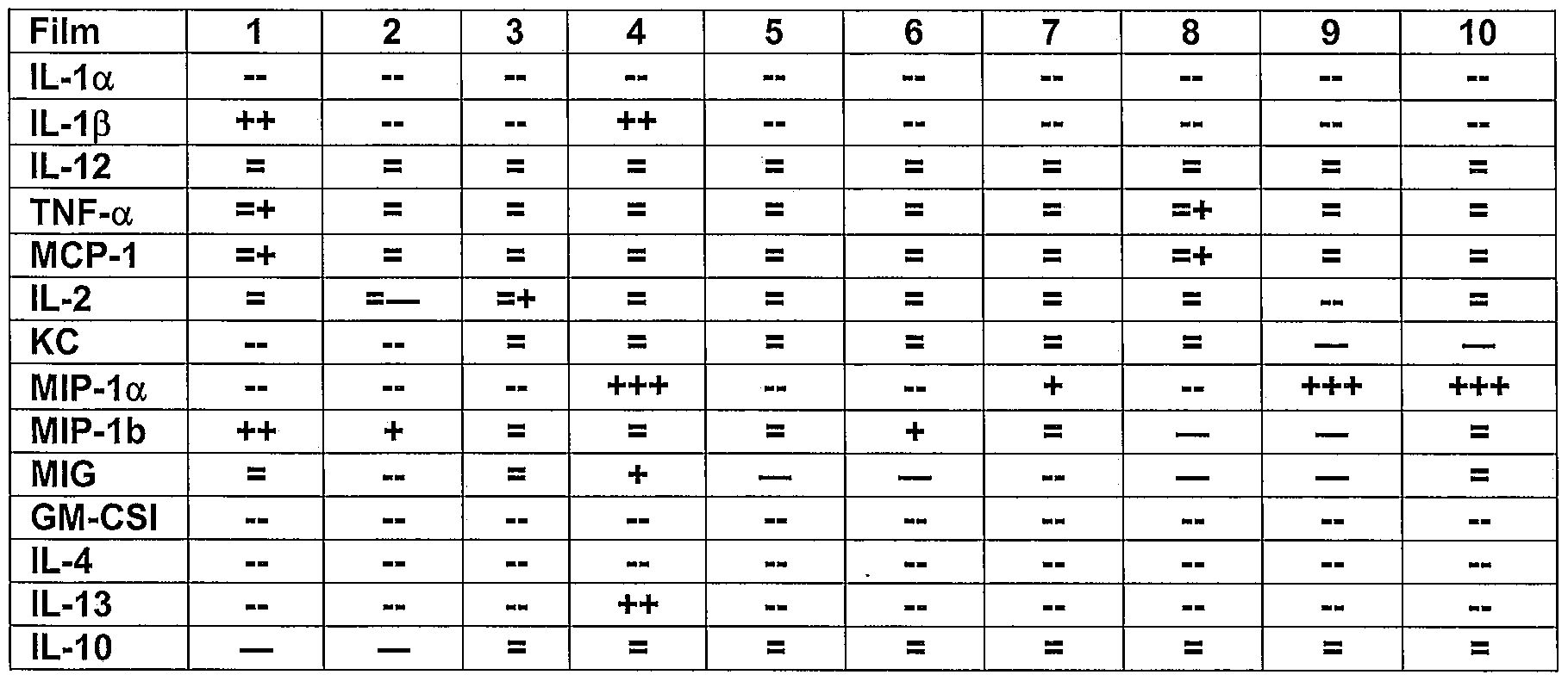


Claims
Priority Applications (8)
| Application Number | Priority Date | Filing Date | Title |
|---|---|---|---|
| AU2011311255A AU2011311255B2 (en) | 2010-04-28 | 2011-04-27 | Method for increasing permeability of an epithelial barrier |
| RU2012150725/14A RU2574137C2 (en) | 2010-04-28 | 2011-04-27 | Method for improving epithelial barrier penetration |
| CA2796965A CA2796965C (en) | 2010-04-28 | 2011-04-27 | Method for increasing permeability of an epithelial barrier |
| MX2012012567A MX2012012567A (en) | 2010-04-28 | 2011-04-27 | Method for increasing permeability of an epithelial barrier. |
| US13/641,500 US9586044B2 (en) | 2010-04-28 | 2011-04-27 | Method for increasing the permeability of an epithelial barrier |
| EP11830267.8A EP2563455A4 (en) | 2010-04-28 | 2011-04-27 | Method for increasing permeability of an epithelial barrier |
| US15/413,475 US10342965B2 (en) | 2010-04-28 | 2017-01-24 | Method for increasing the permeability of an epithelial barrier |
| US16/422,195 US11083881B2 (en) | 2010-04-28 | 2019-05-24 | Method for increasing permeability of a cellular layer of epithelial cells |
Applications Claiming Priority (6)
| Application Number | Priority Date | Filing Date | Title |
|---|---|---|---|
| US32872310P | 2010-04-28 | 2010-04-28 | |
| US61/328,723 | 2010-04-28 | ||
| US41108510P | 2010-11-08 | 2010-11-08 | |
| US61/411,085 | 2010-11-08 | ||
| US201161435963P | 2011-01-25 | 2011-01-25 | |
| US61/435,963 | 2011-01-25 |
Related Child Applications (2)
| Application Number | Title | Priority Date | Filing Date |
|---|---|---|---|
| US13/641,500 A-371-Of-International US9586044B2 (en) | 2010-04-28 | 2011-04-27 | Method for increasing the permeability of an epithelial barrier |
| US15/413,475 Continuation US10342965B2 (en) | 2010-04-28 | 2017-01-24 | Method for increasing the permeability of an epithelial barrier |
Publications (1)
| Publication Number | Publication Date |
|---|---|
| WO2012046149A1 true WO2012046149A1 (en) | 2012-04-12 |
Family
ID=45927279
Family Applications (1)
| Application Number | Title | Priority Date | Filing Date |
|---|---|---|---|
| PCT/IB2011/051863 WO2012046149A1 (en) | 2010-04-28 | 2011-04-27 | Method for increasing permeability of an epithelial barrier |
Country Status (6)
| Country | Link |
|---|---|
| US (3) | US9586044B2 (en) |
| EP (1) | EP2563455A4 (en) |
| AU (1) | AU2011311255B2 (en) |
| CA (1) | CA2796965C (en) |
| MX (2) | MX2012012567A (en) |
| WO (1) | WO2012046149A1 (en) |
Cited By (15)
| Publication number | Priority date | Publication date | Assignee | Title |
|---|---|---|---|---|
| WO2012117302A2 (en) | 2011-02-28 | 2012-09-07 | Kimberly-Clark Worldwide, Inc. | Transdermal patch containing microneedles |
| EP2771058A4 (en) * | 2011-10-27 | 2015-06-17 | Kimberly Clark Co | Implantable devices for delivery of bioactive agents |
| US9522262B2 (en) | 2010-04-28 | 2016-12-20 | Kimberly-Clark Worldwide, Inc. | Medical devices for delivery of siRNA |
| US9522263B2 (en) | 2010-04-28 | 2016-12-20 | Kimberly-Clark Worldwide, Inc. | Device for delivery of rheumatoid arthritis medication |
| US9526883B2 (en) | 2010-04-28 | 2016-12-27 | Kimberly-Clark Worldwide, Inc. | Composite microneedle array including nanostructures thereon |
| US9550053B2 (en) | 2011-10-27 | 2017-01-24 | Kimberly-Clark Worldwide, Inc. | Transdermal delivery of high viscosity bioactive agents |
| US9586044B2 (en) | 2010-04-28 | 2017-03-07 | Kimberly-Clark Worldwide, Inc. | Method for increasing the permeability of an epithelial barrier |
| US10773065B2 (en) | 2011-10-27 | 2020-09-15 | Sorrento Therapeutics, Inc. | Increased bioavailability of transdermally delivered agents |
| WO2021113585A1 (en) | 2019-12-05 | 2021-06-10 | Sorrento Therapeutics, Inc. | Method of treating cancer by administration of an anti-pd-1 or anti-pd-l1 therapeutic agent via a lymphatic delivery device |
| US11185271B2 (en) | 2013-09-13 | 2021-11-30 | University Of Utah Research Foundation | Methods of making micro-molded electrodes and arrays |
| WO2022159736A1 (en) | 2021-01-22 | 2022-07-28 | Sorrento Therapeutics, Inc. | Device for microliter-scale lymphatic delivery of coronavirus vaccines |
| WO2022192594A2 (en) | 2021-03-11 | 2022-09-15 | Sorrento Therapeutics, Inc. | Nucleic acid molecules and vaccines comprising same for the prevention and treatment of coronavirus infections and disease |
| WO2022261262A1 (en) | 2021-06-09 | 2022-12-15 | Sorrento Therapeutics, Inc. | Method of treating cancer by administration of an anti-pd-1 or anti-pd-l1 therapeutic agent via a lymphatic microneedle delivery device |
| WO2023023074A1 (en) | 2021-08-18 | 2023-02-23 | Sorrento Therapeutics, Inc. | Therapeutic agents targeting the lymphatic system |
| US11766822B2 (en) | 2019-08-20 | 2023-09-26 | 3M Innovative Properties Company | Microstructured surface with increased microorganism removal when cleaned, articles and methods |
Families Citing this family (45)
| Publication number | Priority date | Publication date | Assignee | Title |
|---|---|---|---|---|
| GB0402131D0 (en) | 2004-01-30 | 2004-03-03 | Isis Innovation | Delivery method |
| US9114238B2 (en) | 2007-04-16 | 2015-08-25 | Corium International, Inc. | Solvent-cast microprotrusion arrays containing active ingredient |
| WO2009079712A1 (en) | 2007-12-24 | 2009-07-02 | The University Of Queensland | Coating method |
| WO2011140274A2 (en) | 2010-05-04 | 2011-11-10 | Corium International, Inc. | Method and device for transdermal delivery of parathyroid hormone using a microprojection array |
| US9943673B2 (en) | 2010-07-14 | 2018-04-17 | Vaxxas Pty Limited | Patch applying apparatus |
| CA2807552A1 (en) | 2010-08-06 | 2012-02-09 | Moderna Therapeutics, Inc. | Engineered nucleic acids and methods of use thereof |
| EP4108671A1 (en) | 2010-10-01 | 2022-12-28 | ModernaTX, Inc. | Modified nucleosides, nucleotides, and nucleic acids, and uses thereof |
| AU2012236099A1 (en) | 2011-03-31 | 2013-10-03 | Moderna Therapeutics, Inc. | Delivery and formulation of engineered nucleic acids |
| US9464124B2 (en) | 2011-09-12 | 2016-10-11 | Moderna Therapeutics, Inc. | Engineered nucleic acids and methods of use thereof |
| CN103974724B (en) | 2011-10-03 | 2019-08-30 | 现代泰克斯公司 | Nucleosides, nucleotide and nucleic acid of modification and application thereof |
| AU2012323782B2 (en) | 2011-10-12 | 2017-04-06 | Vaxxas Pty Limited | Delivery device |
| CA2859387A1 (en) | 2011-12-16 | 2013-06-20 | Moderna Therapeutics, Inc. | Modified nucleoside, nucleotide, and nucleic acid compositions |
| US9572897B2 (en) | 2012-04-02 | 2017-02-21 | Modernatx, Inc. | Modified polynucleotides for the production of cytoplasmic and cytoskeletal proteins |
| AU2013243951A1 (en) | 2012-04-02 | 2014-10-30 | Moderna Therapeutics, Inc. | Modified polynucleotides for the production of secreted proteins |
| US9878056B2 (en) | 2012-04-02 | 2018-01-30 | Modernatx, Inc. | Modified polynucleotides for the production of cosmetic proteins and peptides |
| US9283287B2 (en) | 2012-04-02 | 2016-03-15 | Moderna Therapeutics, Inc. | Modified polynucleotides for the production of nuclear proteins |
| JP6144355B2 (en) | 2012-11-26 | 2017-06-07 | モデルナティエックス インコーポレイテッドModernaTX,Inc. | Chemically modified mRNA |
| US9320534B2 (en) | 2012-12-13 | 2016-04-26 | Alcon Research, Ltd. | Fine membrane forceps with integral scraping feature |
| ES2743404T3 (en) | 2012-12-21 | 2020-02-19 | Corium Inc | Matrix for therapeutic agent supply and manufacturing method |
| EP2968887B1 (en) | 2013-03-12 | 2022-05-04 | Corium, Inc. | Microprojection applicators |
| US8980864B2 (en) | 2013-03-15 | 2015-03-17 | Moderna Therapeutics, Inc. | Compositions and methods of altering cholesterol levels |
| EP2968118B1 (en) | 2013-03-15 | 2022-02-09 | Corium, Inc. | Microarray for delivery of therapeutic agent and methods of use |
| AU2014237279B2 (en) | 2013-03-15 | 2018-11-22 | Corium Pharma Solutions, Inc. | Microarray with polymer-free microstructures, methods of making, and methods of use |
| EA201690675A1 (en) | 2013-10-03 | 2016-08-31 | Модерна Терапьютикс, Инк. | POLYNUCLEOTES ENCODING THE RECEPTOR OF LOW DENSITY LIPOPROTEINS |
| US10973682B2 (en) | 2014-02-24 | 2021-04-13 | Alcon Inc. | Surgical instrument with adhesion optimized edge condition |
| EP3188714A1 (en) * | 2014-09-04 | 2017-07-12 | Corium International, Inc. | Microstructure array, methods of making, and methods of use |
| EP4218892A1 (en) | 2015-02-02 | 2023-08-02 | Vaxxas Pty Limited | Microprojection array applicator |
| US10857093B2 (en) | 2015-06-29 | 2020-12-08 | Corium, Inc. | Microarray for delivery of therapeutic agent, methods of use, and methods of making |
| AU2016297823B2 (en) | 2015-07-24 | 2020-10-22 | Sorrento Therapeutics, Inc. | Methods for better delivery of active agents to tumors |
| DK3325081T3 (en) * | 2015-07-24 | 2021-10-11 | Sorrento Therapeutics Inc | PROCEDURES FOR LYMPHATIC DELIVERY OF ACTIVE SUBSTANCES |
| US11103259B2 (en) | 2015-09-18 | 2021-08-31 | Vaxxas Pty Limited | Microprojection arrays with microprojections having large surface area profiles |
| EP3381498A4 (en) * | 2015-11-27 | 2019-07-17 | Labo Juversa Co., Ltd. | Microneedle and method for producing same |
| JP6799005B2 (en) * | 2015-12-15 | 2020-12-09 | 久光製薬株式会社 | Microneedle sheet |
| US10213144B2 (en) | 2016-01-25 | 2019-02-26 | International Business Machines Corporation | Nanopatterned biosensor electrode for enhanced sensor signal and sensitivity |
| US10376193B2 (en) | 2016-07-25 | 2019-08-13 | International Business Machines Corporation | Embedded sacrificial layer to enhance biosensor stability and lifetime for nanopatterned electrodes |
| TW201815356A (en) | 2016-10-18 | 2018-05-01 | 諾華公司 | Surgical instrument having a surface texture |
| US10161898B2 (en) | 2017-01-30 | 2018-12-25 | International Business Machines Corporation | Nanopatterned biosensor electrode for enhanced sensor signal and sensitivity |
| US10548530B2 (en) | 2017-03-01 | 2020-02-04 | International Business Machines Corporation | Biosensor calibration structure containing different sensing surface area |
| DK3606760T3 (en) | 2017-03-31 | 2023-11-06 | Vaxxas Pty Ltd | ARRANGEMENT AND PROCEDURE FOR COATING SURFACES |
| IT201700048421A1 (en) * | 2017-05-04 | 2018-11-04 | Materias S R L | DEVICE FOR THE TRANSDERMIC ADMINISTRATION OF ACTIVE MOLECULES, USES OF SUCH A DEVICE AND METHODS OF PRODUCTION OF SUCH A DEVICE AND OF ITS COMPONENTS |
| US11175128B2 (en) | 2017-06-13 | 2021-11-16 | Vaxxas Pty Limited | Quality control of substrate coatings |
| JP1601442S (en) * | 2017-07-28 | 2018-04-09 | ||
| EP3661587A4 (en) | 2017-08-04 | 2021-06-09 | Vaxxas Pty Limited | Compact high mechanical energy storage and low trigger force actuator for the delivery of microprojection array patches (map) |
| WO2019191299A1 (en) | 2018-03-27 | 2019-10-03 | San Diego State University | In situ partially degradable separation interface for fabrication of complex near net shape objects by pressure assisted sintering |
| DE102020104306A1 (en) | 2020-02-19 | 2021-08-19 | Lts Lohmann Therapie-Systeme Ag | Form element for the production of microstructures |
Citations (7)
| Publication number | Priority date | Publication date | Assignee | Title |
|---|---|---|---|---|
| US6334856B1 (en) | 1998-06-10 | 2002-01-01 | Georgia Tech Research Corporation | Microneedle devices and methods of manufacture and use thereof |
| US20040028875A1 (en) * | 2000-12-02 | 2004-02-12 | Van Rijn Cornelis Johannes Maria | Method of making a product with a micro or nano sized structure and product |
| US20060264893A1 (en) * | 1999-09-24 | 2006-11-23 | Sage Jr Burton H | Method and device for abrading skin |
| US7226439B2 (en) | 1999-06-04 | 2007-06-05 | Georgia Tech Research Corporation | Microneedle drug delivery device |
| US20080026464A1 (en) | 2003-08-18 | 2008-01-31 | Borenstein Jeffrey T | Nanotopographic Compositions and Methods for Cellular Organization in Tissue Engineered Structures |
| US20080221408A1 (en) * | 2007-03-09 | 2008-09-11 | Nellcor Puritan Bennett Llc | System and methods for optical sensing and drug delivery using microneedles |
| US20080311172A1 (en) | 2007-04-25 | 2008-12-18 | Schapira Jay N | Programmed-release, nanostructured biological construct |
Family Cites Families (186)
| Publication number | Priority date | Publication date | Assignee | Title |
|---|---|---|---|---|
| US3797494A (en) | 1969-04-01 | 1974-03-19 | Alza Corp | Bandage for the administration of drug by controlled metering through microporous materials |
| US3964482A (en) | 1971-05-17 | 1976-06-22 | Alza Corporation | Drug delivery device |
| US4031894A (en) | 1975-12-08 | 1977-06-28 | Alza Corporation | Bandage for transdermally administering scopolamine to prevent nausea |
| US4436741A (en) | 1975-12-08 | 1984-03-13 | Alza Corporation | Method for administering scopolamine transdermally |
| US4051840A (en) | 1976-01-05 | 1977-10-04 | Sinai Hospital Of Detroit | Dynamic aortic patch |
| US4201211A (en) | 1977-07-12 | 1980-05-06 | Alza Corporation | Therapeutic system for administering clonidine transdermally |
| US4379454A (en) | 1981-02-17 | 1983-04-12 | Alza Corporation | Dosage for coadministering drug and percutaneous absorption enhancer |
| US4725272A (en) | 1981-06-29 | 1988-02-16 | Alza Corporation | Novel bandage for administering beneficial drug |
| US4661105A (en) | 1981-06-29 | 1987-04-28 | Alza Corporation | Medical bandage for administering vasodilator drug |
| US5310559A (en) | 1982-09-01 | 1994-05-10 | Hercon Laboratories Corporation | Device for controlled release and delivery to mammalian tissue of pharmacologically active agents incorporating a rate controlling member which comprises an alkylene-alkyl acrylate copolymer |
| US4588580B2 (en) | 1984-07-23 | 1999-02-16 | Alaz Corp | Transdermal administration of fentanyl and device therefor |
| US4681584A (en) | 1985-05-03 | 1987-07-21 | Alza Corporation | Transdermal delivery system for delivering nitroglycerin at high transdermal fluxes |
| US4615699A (en) | 1985-05-03 | 1986-10-07 | Alza Corporation | Transdermal delivery system for delivering nitroglycerin at high transdermal fluxes |
| US4698062A (en) | 1985-10-30 | 1987-10-06 | Alza Corporation | Medical device for pulsatile transdermal delivery of biologically active agents |
| US4880633A (en) | 1986-03-12 | 1989-11-14 | Merck & Co., Inc. | Transdermal drug delivery system |
| US5344656A (en) | 1986-09-12 | 1994-09-06 | Alza Corporation | Subsaturated transdermal therapeutic system having improved release characteristics |
| US4908027A (en) | 1986-09-12 | 1990-03-13 | Alza Corporation | Subsaturated transdermal therapeutic system having improved release characteristics |
| US4832953A (en) | 1987-08-13 | 1989-05-23 | Alza Corporation | Method for preventing the formation of a crystalline hydrate in a dispersion of a liquid in a monaqueous matrix |
| US5004610A (en) | 1988-06-14 | 1991-04-02 | Alza Corporation | Subsaturated nicotine transdermal therapeutic system |
| US5364630A (en) | 1988-06-14 | 1994-11-15 | Alza Corporation | Subsaturated nicotine transdermal therapeutic system |
| US5328470A (en) | 1989-03-31 | 1994-07-12 | The Regents Of The University Of Michigan | Treatment of diseases by site-specific instillation of cells or site-specific transformation of cells and kits therefor |
| DE19525607A1 (en) | 1995-07-14 | 1997-01-16 | Boehringer Ingelheim Kg | Transcorneal drug delivery system |
| DE19730984A1 (en) | 1997-07-18 | 1999-02-11 | Siemens Ag | Radio transmission method and cordless telephone, especially for the DECT standard |
| US6471993B1 (en) | 1997-08-01 | 2002-10-29 | Massachusetts Institute Of Technology | Three-dimensional polymer matrices |
| DK1041975T3 (en) | 1997-12-22 | 2002-11-04 | Alza Corp | Speed-controlling membranes for controlled drug delivery devices |
| GB9805214D0 (en) | 1998-03-11 | 1998-05-06 | Univ Glasgow | Cell adhesion |
| GB9815819D0 (en) | 1998-07-22 | 1998-09-16 | Secr Defence | Transferring materials into cells and a microneedle array |
| US7048723B1 (en) | 1998-09-18 | 2006-05-23 | The University Of Utah Research Foundation | Surface micromachined microneedles |
| US6743211B1 (en) * | 1999-11-23 | 2004-06-01 | Georgia Tech Research Corporation | Devices and methods for enhanced microneedle penetration of biological barriers |
| US6312612B1 (en) | 1999-06-09 | 2001-11-06 | The Procter & Gamble Company | Apparatus and method for manufacturing an intracutaneous microneedle array |
| US6256533B1 (en) | 1999-06-09 | 2001-07-03 | The Procter & Gamble Company | Apparatus and method for using an intracutaneous microneedle array |
| US6569143B2 (en) | 1999-10-14 | 2003-05-27 | Becton, Dickinson And Company | Method of intradermally injecting substances |
| US20020095134A1 (en) | 1999-10-14 | 2002-07-18 | Pettis Ronald J. | Method for altering drug pharmacokinetics based on medical delivery platform |
| EP1235611B1 (en) | 1999-11-15 | 2005-09-07 | Velcro Industries B.V. | Skin attachment member |
| IL134997A0 (en) | 2000-03-09 | 2001-05-20 | Yehoshua Yeshurun | Health care system based on micro device |
| ATE450621T2 (en) | 2000-03-30 | 2009-12-15 | Whitehead Biomedical Inst | MEDIATORS OF RNA INTERFERENCE THAT ARE RNA SEQUENCE SPECIFIC |
| AU2001275138A1 (en) | 2000-06-02 | 2001-12-17 | The University Of Utah Research Foundation | Active needle devices with integrated functionality |
| US6440096B1 (en) | 2000-07-14 | 2002-08-27 | Becton, Dickinson And Co. | Microdevice and method of manufacturing a microdevice |
| US6656147B1 (en) | 2000-07-17 | 2003-12-02 | Becton, Dickinson And Company | Method and delivery device for the transdermal administration of a substance |
| US6476241B1 (en) | 2000-09-05 | 2002-11-05 | Mars Incorporated | Synthesis of 4α-arylepicatechins |
| WO2002030506A2 (en) | 2000-10-12 | 2002-04-18 | Ink Jet Technology Ltd. | Transdermal method |
| US7828827B2 (en) | 2002-05-24 | 2010-11-09 | Corium International, Inc. | Method of exfoliation of skin using closely-packed microstructures |
| US7131987B2 (en) | 2000-10-16 | 2006-11-07 | Corium International, Inc. | Microstructures and method for treating and conditioning skin which cause less irritation during exfoliation |
| US6821281B2 (en) | 2000-10-16 | 2004-11-23 | The Procter & Gamble Company | Microstructures for treating and conditioning skin |
| US6979347B1 (en) | 2000-10-23 | 2005-12-27 | Advanced Cardiovascular Systems, Inc. | Implantable drug delivery prosthesis |
| AU2002229046B2 (en) | 2000-12-11 | 2006-05-18 | President And Fellows Of Harvard College | Nanosensors |
| EP1345646A2 (en) * | 2000-12-14 | 2003-09-24 | Georgia Tech Research Corporation | Microneedle devices and production thereof |
| EP1374310A4 (en) | 2001-03-14 | 2008-02-20 | Univ Massachusetts | Nanofabrication |
| US6663820B2 (en) | 2001-03-14 | 2003-12-16 | The Procter & Gamble Company | Method of manufacturing microneedle structures using soft lithography and photolithography |
| US6591124B2 (en) | 2001-05-11 | 2003-07-08 | The Procter & Gamble Company | Portable interstitial fluid monitoring system |
| US6767341B2 (en) | 2001-06-13 | 2004-07-27 | Abbott Laboratories | Microneedles for minimally invasive drug delivery |
| SE0102736D0 (en) | 2001-08-14 | 2001-08-14 | Patrick Griss | Side opened out-of-plane microneedles for microfluidic transdermal interfacing and fabrication process of side opened out-of-plane microneedles |
| US6881203B2 (en) | 2001-09-05 | 2005-04-19 | 3M Innovative Properties Company | Microneedle arrays and methods of manufacturing the same |
| US6561143B2 (en) | 2001-09-06 | 2003-05-13 | Barry L Holtzman | Engine induction valve with reduced backflow |
| WO2003022330A2 (en) | 2001-09-12 | 2003-03-20 | Becton, Dickinson And Company | Microneedle-based pen device for drug delivery and method for using same |
| US20040087992A1 (en) | 2002-08-09 | 2004-05-06 | Vladimir Gartstein | Microstructures for delivering a composition cutaneously to skin using rotatable structures |
| EP1471953B1 (en) | 2001-09-21 | 2011-02-16 | Valeritas, Inc. | Gas pressure actuated microneedle arrays, and systems and methods relating to same |
| US6746825B2 (en) | 2001-10-05 | 2004-06-08 | Wisconsin Alumni Research Foundation | Guided self-assembly of block copolymer films on interferometrically nanopatterned substrates |
| US7429258B2 (en) | 2001-10-26 | 2008-09-30 | Massachusetts Institute Of Technology | Microneedle transport device |
| US6908453B2 (en) | 2002-01-15 | 2005-06-21 | 3M Innovative Properties Company | Microneedle devices and methods of manufacture |
| EP1490147A2 (en) | 2002-03-04 | 2004-12-29 | Nano Pass Technologies Ltd. | Devices and methods for transporting fluid across a biological barrier |
| US7115108B2 (en) | 2002-04-02 | 2006-10-03 | Becton, Dickinson And Company | Method and device for intradermally delivering a substance |
| WO2003092785A1 (en) | 2002-04-30 | 2003-11-13 | Morteza Shirkhanzadeh | Arrays of microneedles comprising porous calcium phosphate coating and bioactive agents |
| CA2493728C (en) | 2002-07-22 | 2013-10-29 | Becton, Dickinson And Company | Patch-like infusion device |
| WO2004009978A2 (en) | 2002-07-24 | 2004-01-29 | Koch Kenneth W | Methods and compositions for on-line gas turbine cleaning |
| US20040063100A1 (en) | 2002-09-30 | 2004-04-01 | Wang Chung Lin | Nanoneedle chips and the production thereof |
| WO2004033021A1 (en) | 2002-10-07 | 2004-04-22 | Biovalve Technologies, Inc. | Microneedle array patch |
| IL152912A0 (en) | 2002-11-18 | 2003-06-24 | Nanopass Ltd | Micro needle systems |
| US6995336B2 (en) | 2003-01-29 | 2006-02-07 | The Regents Of The University Of Michigan | Method for forming nanoscale features |
| US7374864B2 (en) | 2003-02-13 | 2008-05-20 | The Regents Of The University Of Michigan | Combined nanoimprinting and photolithography for micro and nano devices fabrication |
| US7578954B2 (en) | 2003-02-24 | 2009-08-25 | Corium International, Inc. | Method for manufacturing microstructures having multiple microelements with through-holes |
| US7972616B2 (en) | 2003-04-17 | 2011-07-05 | Nanosys, Inc. | Medical device applications of nanostructured surfaces |
| CA2559330A1 (en) | 2003-04-21 | 2004-11-04 | Stratagent Life Sciences | Apparatus and methods for repetitive microjet drug delivery |
| US7803574B2 (en) | 2003-05-05 | 2010-09-28 | Nanosys, Inc. | Medical device applications of nanostructured surfaces |
| US7572405B2 (en) | 2003-06-02 | 2009-08-11 | Corium International Inc. | Method for manufacturing microstructures having hollow microelements using fluidic jets during a molding operation |
| US7563451B2 (en) | 2003-07-22 | 2009-07-21 | Iowa State University Research Foundation, Inc. | Capped mesoporous silicates |
| JP4884218B2 (en) | 2003-08-01 | 2012-02-29 | バスカ,カナッグ | Laryngeal mask |
| BRPI0413867A (en) | 2003-08-26 | 2006-10-24 | Alza Corp | device and method for intradermal cell implantation |
| US7544770B2 (en) | 2003-08-29 | 2009-06-09 | Louisiana Tech Foundation, Inc. | Multilayer films, coatings, and microcapsules comprising polypeptides |
| JP2007506429A (en) | 2003-09-23 | 2007-03-22 | アトム・サイエンシズ・インコーポレーテッド | Polymeric nucleic acid hybridization probe |
| DE10353629A1 (en) | 2003-11-17 | 2005-06-16 | Lts Lohmann Therapie-Systeme Ag | Device for the transdermal administration of active substances |
| CN1905842A (en) | 2003-11-21 | 2007-01-31 | 阿尔扎公司 | Ultrasound assisted transdermal vaccine delivery method and system |
| EP1713533A4 (en) | 2003-11-21 | 2008-01-23 | Univ California | Method and/or apparatus for puncturing a surface for extraction, in situ analysis, and/or substance delivery using microneedles |
| US20050119723A1 (en) | 2003-11-28 | 2005-06-02 | Medlogics Device Corporation | Medical device with porous surface containing bioerodable bioactive composites and related methods |
| GB0402131D0 (en) | 2004-01-30 | 2004-03-03 | Isis Innovation | Delivery method |
| US7914813B2 (en) | 2004-02-03 | 2011-03-29 | Hisamitsu Pharmaceutical Co., Inc. | Interface for transdermal drug administration device |
| US9016221B2 (en) * | 2004-02-17 | 2015-04-28 | University Of Florida Research Foundation, Inc. | Surface topographies for non-toxic bioadhesion control |
| US8551391B2 (en) * | 2004-02-17 | 2013-10-08 | Avery Dennison Corporation | Method of making microneedles |
| US20070191761A1 (en) | 2004-02-23 | 2007-08-16 | 3M Innovative Properties Company | Method of molding for microneedle arrays |
| US8915957B2 (en) | 2004-03-11 | 2014-12-23 | Alcatel Lucent | Drug delivery stent |
| WO2005094526A2 (en) | 2004-03-24 | 2005-10-13 | Corium International, Inc. | Transdermal delivery device |
| CA2566462A1 (en) | 2004-05-12 | 2005-12-01 | Kamel Khalili | Compositions and methods for sirna inhibition of primate polyomavirus genes |
| US7315758B2 (en) | 2004-06-03 | 2008-01-01 | Lynntech, Inc. | Transdermal delivery of therapeutic agent |
| US20060025848A1 (en) | 2004-07-29 | 2006-02-02 | Jan Weber | Medical device having a coating layer with structural elements therein and method of making the same |
| WO2006015299A2 (en) | 2004-07-30 | 2006-02-09 | Microchips, Inc. | Multi-reservoir device for transdermal drug delivery and sensing |
| US8696619B2 (en) | 2004-08-10 | 2014-04-15 | Robert P. Schnall | Drug delivery devices |
| US20070250018A1 (en) | 2004-08-12 | 2007-10-25 | Hirotoshi Adachi | Transdermal Drug Administration System with Microneedles |
| US7316665B2 (en) | 2004-08-25 | 2008-01-08 | Becton, Dickinson And Company | Method and device for the delivery of a substance including a covering |
| DE102004041813A1 (en) | 2004-08-26 | 2006-03-02 | Siemens Ag | Surface having an adhesion reducing microstructure and method of making the same |
| SE0402100D0 (en) | 2004-08-30 | 2004-08-30 | Bonsens Ab | Molded micro-needles |
| US7449200B2 (en) | 2006-04-17 | 2008-11-11 | Gp Medical, Inc. | Nanoparticles for protein/peptide delivery and delivery means |
| US8137697B1 (en) | 2004-10-05 | 2012-03-20 | Gp Medical, Inc. | Nanoparticles for protein/peptide delivery and delivery means thereof |
| US7627938B2 (en) | 2004-10-15 | 2009-12-08 | Board Of Regents, The Univeristy Of Texas System | Tapered hollow metallic microneedle array assembly and method of making and using the same |
| EP1819379B1 (en) | 2004-11-18 | 2016-08-31 | Nanopass Technologies Ltd. | System for delivering fluid into flexible biological barrier |
| US8057842B2 (en) | 2004-11-18 | 2011-11-15 | 3M Innovative Properties Company | Method of contact coating a microneedle array |
| WO2006055799A1 (en) | 2004-11-18 | 2006-05-26 | 3M Innovative Properties Company | Masking method for coating a microneedle array |
| JP2008522875A (en) | 2004-12-07 | 2008-07-03 | スリーエム イノベイティブ プロパティズ カンパニー | Microneedle molding method |
| KR101288725B1 (en) | 2004-12-10 | 2013-07-23 | 쓰리엠 이노베이티브 프로퍼티즈 컴파니 | Medical Device |
| JPWO2006075689A1 (en) | 2005-01-14 | 2008-06-12 | 久光製薬株式会社 | Medicinal product carrying device and method for producing the same |
| US20070112548A1 (en) | 2005-02-18 | 2007-05-17 | Georgia Tech Research Corporation | Methods for fabricating micro-to-nanoscale devices via biologically-induced solid formation on biologically-derived templates, and micro-to-nanoscale structures and micro-to-nanoscale devices made thereby |
| WO2006128034A1 (en) | 2005-05-25 | 2006-11-30 | Georgia Tech Research Corporation | Microneedles and methods for microinfusion |
| EP1904158B1 (en) | 2005-06-24 | 2013-07-24 | 3M Innovative Properties Company | Collapsible patch with microneedle array |
| ATE477833T1 (en) | 2005-06-27 | 2010-09-15 | 3M Innovative Properties Co | MICRONEEDLE CARTRIDGE ASSEMBLY |
| CA2614927A1 (en) | 2005-07-25 | 2007-02-01 | Nanotechnology Victoria Pty Ltd | Microarray device |
| US8118753B2 (en) | 2005-08-18 | 2012-02-21 | Seoul National University Industry Foundation | Barb-wired micro needle made of single crystalline silicon and biopsy method and medicine injecting method using the same |
| EP1928537B1 (en) | 2005-09-02 | 2015-01-21 | Intercell USA, Inc. | Devices for transcutaneous delivery of vaccines and transdermal delivery of drugs |
| US7659252B2 (en) | 2005-09-15 | 2010-02-09 | Novomed Technologies, Inc. (Shanghai) | Transdermal delivery peptides and method of use thereof |
| US20070066934A1 (en) | 2005-09-19 | 2007-03-22 | Transport Pharmaceuticals, Inc. | Electrokinetic delivery system and methods therefor |
| EP1928539A1 (en) | 2005-09-30 | 2008-06-11 | Tti Ellebeau, Inc. | Functionalized microneedles transdermal drug delivery systems, devices, and methods |
| KR20080066712A (en) | 2005-09-30 | 2008-07-16 | 티티아이 엘뷰 가부시키가이샤 | Functionalized microneedles transdermal drug delivery systems, devices, and methods |
| US20070112309A1 (en) | 2005-11-17 | 2007-05-17 | Jerry Zucker | Withdrawal syringe |
| US20080262416A1 (en) | 2005-11-18 | 2008-10-23 | Duan Daniel C | Microneedle Arrays and Methods of Preparing Same |
| US8308960B2 (en) | 2005-12-14 | 2012-11-13 | Silex Microsystems Ab | Methods for making micro needles and applications thereof |
| US8944804B2 (en) | 2006-01-04 | 2015-02-03 | Liquidia Technologies, Inc. | Nanostructured surfaces for biomedical/biomaterial applications and processes thereof |
| US7658728B2 (en) | 2006-01-10 | 2010-02-09 | Yuzhakov Vadim V | Microneedle array, patch, and applicator for transdermal drug delivery |
| EP1992386B1 (en) | 2006-02-10 | 2011-11-09 | Hisamitsu Pharmaceutical Co., Inc. | Transdermal drug administration apparatus having microneedles |
| US8858807B2 (en) | 2006-03-24 | 2014-10-14 | 3M Innovative Properties Company | Process for making microneedles, microneedle arrays, masters, and replication tools |
| US20070224235A1 (en) | 2006-03-24 | 2007-09-27 | Barron Tenney | Medical devices having nanoporous coatings for controlled therapeutic agent delivery |
| WO2007116959A1 (en) | 2006-04-07 | 2007-10-18 | Hisamitsu Pharmaceutical Co., Inc. | Microneedle device and transdermal administration device provided with microneedles |
| CN1830496A (en) | 2006-04-10 | 2006-09-13 | 清华大学 | '-'-shaped structure three-dimensional micre solid, hollow silicone meedle orknife |
| WO2007124411A1 (en) | 2006-04-20 | 2007-11-01 | 3M Innovative Properties Company | Device for applying a microneedle array |
| WO2007131050A2 (en) | 2006-05-02 | 2007-11-15 | Georgia Tech Research Corporation | Method for drug delivery to ocular tissue using microneedle |
| AU2007288442A1 (en) | 2006-05-09 | 2008-02-28 | Apogee Technology, Inc. | Nanofiber structures on asperities for sequestering, carrying and transferring substances |
| JP5290962B2 (en) | 2006-05-17 | 2013-09-18 | デビオテック ソシエテ アノニム | Anisotropic nanoporous coating |
| US20070276318A1 (en) | 2006-05-26 | 2007-11-29 | Mit, Llp | Iontosonic-microneedle applicator apparatus and methods |
| DE102006031506A1 (en) | 2006-07-07 | 2008-01-17 | Robert Bosch Gmbh | Method of making microneedles in a Si semiconductor substrate |
| EP2062611A4 (en) | 2006-08-18 | 2010-01-06 | Toppan Printing Co Ltd | Microneedle and microneedle patch |
| DE102006040642A1 (en) | 2006-08-30 | 2008-03-13 | Robert Bosch Gmbh | Microneedles for placement in the skin for transdermal application of pharmaceuticals |
| CA2663003C (en) | 2006-09-08 | 2018-02-13 | Justin Hanes | Compositions and methods for enhancing transport through mucus |
| US20080097352A1 (en) | 2006-09-12 | 2008-04-24 | Beck Patricia A | Methods of fabricating microneedles with bio-sensory functionality |
| GB0620617D0 (en) | 2006-10-17 | 2006-11-29 | Glaxo Group Ltd | Novel device |
| US20080091226A1 (en) | 2006-10-17 | 2008-04-17 | Nanopass Technologies Ltd. | Microneedle device |
| EP2083876A4 (en) | 2006-10-25 | 2012-09-19 | Revalesio Corp | Methods of wound care and treatment |
| US7785301B2 (en) | 2006-11-28 | 2010-08-31 | Vadim V Yuzhakov | Tissue conforming microneedle array and patch for transdermal drug delivery or biological fluid collection |
| US8238995B2 (en) | 2006-12-08 | 2012-08-07 | General Electric Company | Self-adhering electrodes and methods of making the same |
| EP2124726A1 (en) | 2006-12-22 | 2009-12-02 | Medingo Ltd. | Fluid delivery with in vivo electrochemical analyte sensing |
| ES2558544T3 (en) | 2007-03-16 | 2016-02-05 | The Regents Of The University Of California | Medical implants coated on the nanostructure surface and their methods of use |
| US9114238B2 (en) | 2007-04-16 | 2015-08-25 | Corium International, Inc. | Solvent-cast microprotrusion arrays containing active ingredient |
| US20100196446A1 (en) | 2007-07-10 | 2010-08-05 | Morteza Gharib | Drug delivery and substance transfer facilitated by nano-enhanced device having aligned carbon nanotubes protruding from device surface |
| JP2010535591A (en) | 2007-08-06 | 2010-11-25 | トランスダーム, インコーポレイテッド | Microneedle array formed from polymer film |
| US20100121307A1 (en) | 2007-08-24 | 2010-05-13 | Microfabrica Inc. | Microneedles, Microneedle Arrays, Methods for Making, and Transdermal and/or Intradermal Applications |
| US20090093879A1 (en) | 2007-10-04 | 2009-04-09 | Debra Wawro | Micro- and nano-patterned surface features to reduce implant fouling and regulate wound healing |
| US20090093871A1 (en) | 2007-10-08 | 2009-04-09 | Medtronic Vascular, Inc. | Medical Implant With Internal Drug Delivery System |
| US20090099427A1 (en) | 2007-10-12 | 2009-04-16 | Arkal Medical, Inc. | Microneedle array with diverse needle configurations |
| JP5461427B2 (en) | 2007-12-17 | 2014-04-02 | ニューワールド ファーマシューティカルズ,エルエルシー | Integrated intradermal delivery, diagnosis and communication system |
| WO2009079712A1 (en) | 2007-12-24 | 2009-07-02 | The University Of Queensland | Coating method |
| EP2100850A1 (en) | 2008-03-11 | 2009-09-16 | Stichting Voor De Technische Wetenschappen | Microneedle array and a method for manufacturing microneedles |
| WO2009114719A2 (en) | 2008-03-13 | 2009-09-17 | Richmond Chemical Corporation | Apparatus and method of retaining and releasing molecules from nanostructures by an external stimulus |
| AU2009250341A1 (en) | 2008-05-23 | 2009-11-26 | The University Of Queensland | Analyte detection using a needle projection patch |
| US20100004733A1 (en) | 2008-07-02 | 2010-01-07 | Boston Scientific Scimed, Inc. | Implants Including Fractal Structures |
| CN101347652B (en) | 2008-09-09 | 2011-01-12 | 南京大学 | Method for preparing hollow micro-needle array injection syringe |
| MX349176B (en) | 2008-09-22 | 2017-07-14 | Biochemics Inc | Transdermal drug delivery using an osmolyte and vasoactive agent. |
| KR101039078B1 (en) | 2009-08-04 | 2011-06-07 | (주)마이티시스템 | Effective component delivery system having micro-needle and movable storage capsule |
| US20110021996A1 (en) | 2008-12-18 | 2011-01-27 | Miti Systems Inc. | Structure of micro-needle with side channel and manufacturing method thereof |
| EP2370141A1 (en) | 2008-12-19 | 2011-10-05 | Janisys Limited | A fluid transfer device and an active substance cartridge for the fluid transfer device, and a method for controlling the pressure at which an active substance is delivered to a subject from a fluid transfer device |
| KR101087088B1 (en) | 2008-12-29 | 2011-11-25 | 한국과학기술연구원 | Method for preparing drug-eluting stent having nano-structured pattern and drug-eluting stent prepared therefrom |
| KR101033513B1 (en) | 2009-01-20 | 2011-05-09 | (주)마이티시스템 | Case for the effective component delivery using micro-needle |
| WO2010126640A2 (en) | 2009-02-12 | 2010-11-04 | Trustees Of Tufts College | Nanoimprinting of silk fibroin structures for biomedical and biophotonic applications |
| CN102481440B (en) * | 2009-04-23 | 2014-06-25 | 新加坡国立大学 | An apparatus that includes nano-sized projections and a method for manufacture thereof |
| WO2010126174A1 (en) | 2009-05-01 | 2010-11-04 | Nanbu Plastics Co., Ltd. | Transdermal administration device |
| US8389205B2 (en) | 2009-06-11 | 2013-03-05 | International Business Machines Corporation | Patterning nano-scale patterns on a film comprising unzipping polymer chains |
| DE102009035795A1 (en) | 2009-07-31 | 2011-02-03 | Leibniz-Institut Für Neue Materialien Gemeinnützige Gmbh | Structured surfaces for implants |
| US20110144591A1 (en) | 2009-12-11 | 2011-06-16 | Ross Russell F | Transdermal Delivery Device |
| US20110306853A1 (en) | 2010-03-19 | 2011-12-15 | Michael Darryl Black | Body fluid sampling/fluid delivery device |
| MX2012012567A (en) | 2010-04-28 | 2012-11-21 | Kimberly Clark Co | Method for increasing permeability of an epithelial barrier. |
| EP2563450B1 (en) | 2010-04-28 | 2017-07-26 | Kimberly-Clark Worldwide, Inc. | Device for delivery of rheumatoid arthritis medication |
| JP5871907B2 (en) | 2010-04-28 | 2016-03-01 | キンバリー クラーク ワールドワイド インコーポレイテッド | Nanopatterned medical device with enhanced cell-cell interaction |
| RU2585138C2 (en) | 2010-04-28 | 2016-05-27 | Кимберли-Кларк Ворлдвайд, Инк. | Medical devices for delivery of sirna |
| US9943673B2 (en) | 2010-07-14 | 2018-04-17 | Vaxxas Pty Limited | Patch applying apparatus |
| EP2632613B1 (en) | 2010-10-28 | 2017-08-30 | 3M Innovative Properties Company | Engineered surfaces for reducing bacterial adhesion |
| US9017289B2 (en) | 2010-11-03 | 2015-04-28 | Covidien Lp | Transdermal fluid delivery device |
| EP2665504A4 (en) | 2011-01-18 | 2017-01-25 | Massachusetts Institute of Technology | Deployable barbed microneedle array and uses thereof |
| US8696637B2 (en) | 2011-02-28 | 2014-04-15 | Kimberly-Clark Worldwide | Transdermal patch containing microneedles |
| US20170246439A9 (en) | 2011-10-27 | 2017-08-31 | Kimberly-Clark Worldwide, Inc. | Increased Bioavailability of Transdermally Delivered Agents |
| JP6535464B2 (en) | 2011-10-27 | 2019-06-26 | ソレント・セラピューティクス・インコーポレイテッド | Implantable device for delivery of bioactive agents |
| BR112014009713A2 (en) | 2011-10-27 | 2017-04-18 | Kimberly Clark Co | transdermal administration of high viscosity bioactive agents |
-
2011
- 2011-04-27 MX MX2012012567A patent/MX2012012567A/en unknown
- 2011-04-27 EP EP11830267.8A patent/EP2563455A4/en active Pending
- 2011-04-27 US US13/641,500 patent/US9586044B2/en active Active
- 2011-04-27 AU AU2011311255A patent/AU2011311255B2/en active Active
- 2011-04-27 CA CA2796965A patent/CA2796965C/en active Active
- 2011-04-27 WO PCT/IB2011/051863 patent/WO2012046149A1/en active Application Filing
-
2012
- 2012-10-26 MX MX2022009509A patent/MX2022009509A/en unknown
-
2017
- 2017-01-24 US US15/413,475 patent/US10342965B2/en active Active
-
2019
- 2019-05-24 US US16/422,195 patent/US11083881B2/en active Active
Patent Citations (8)
| Publication number | Priority date | Publication date | Assignee | Title |
|---|---|---|---|---|
| US6334856B1 (en) | 1998-06-10 | 2002-01-01 | Georgia Tech Research Corporation | Microneedle devices and methods of manufacture and use thereof |
| US7226439B2 (en) | 1999-06-04 | 2007-06-05 | Georgia Tech Research Corporation | Microneedle drug delivery device |
| US20060264893A1 (en) * | 1999-09-24 | 2006-11-23 | Sage Jr Burton H | Method and device for abrading skin |
| US20040028875A1 (en) * | 2000-12-02 | 2004-02-12 | Van Rijn Cornelis Johannes Maria | Method of making a product with a micro or nano sized structure and product |
| US20080026464A1 (en) | 2003-08-18 | 2008-01-31 | Borenstein Jeffrey T | Nanotopographic Compositions and Methods for Cellular Organization in Tissue Engineered Structures |
| US8097456B2 (en) | 2003-08-18 | 2012-01-17 | The Charles Stark Draper Laboratory | Nanotopographic compositions and methods for cellular organization in tissue engineered structures |
| US20080221408A1 (en) * | 2007-03-09 | 2008-09-11 | Nellcor Puritan Bennett Llc | System and methods for optical sensing and drug delivery using microneedles |
| US20080311172A1 (en) | 2007-04-25 | 2008-12-18 | Schapira Jay N | Programmed-release, nanostructured biological construct |
Non-Patent Citations (6)
| Title |
|---|
| BRUNAUEREMMETTELLER, J. AMER. CHEM. SOC., vol. 60, February 1938 (1938-02-01), pages 309 - 319 |
| HART ET AL., EUROPEAN CELLS AND MATERIALS, vol. 10, no. 2, 2005 |
| KAUSHAL ET AL., SCI. PHARM., vol. 77, 2009, pages 877 - 897 |
| LIM ET AL., J R SOC INTERFACE, vol. 2, no. 2, 22 March 2005 (2005-03-22), pages 97 - 108 |
| SAPRA ET AL., AAPS PHARM. SCI. TECH., vol. 10, no. 1, March 2009 (2009-03-01) |
| YIM ET AL., BIOMATERIALS, vol. 26, no. 26, September 2005 (2005-09-01), pages 5405 - 5413 |
Cited By (37)
| Publication number | Priority date | Publication date | Assignee | Title |
|---|---|---|---|---|
| US11179555B2 (en) | 2010-04-28 | 2021-11-23 | Sorrento Therapeutics, Inc. | Nanopatterned medical device with enhanced cellular interaction |
| US10709884B2 (en) | 2010-04-28 | 2020-07-14 | Sorrento Therapeutics, Inc. | Device for delivery of rheumatoid arthritis medication |
| US11135414B2 (en) | 2010-04-28 | 2021-10-05 | Sorrento Therapeutics, Inc. | Medical devices for delivery of siRNA |
| US11083881B2 (en) | 2010-04-28 | 2021-08-10 | Sorrento Therapeutics, Inc. | Method for increasing permeability of a cellular layer of epithelial cells |
| US11565098B2 (en) | 2010-04-28 | 2023-01-31 | Sorrento Therapeutics, Inc. | Device for delivery of rheumatoid arthritis medication |
| US10806914B2 (en) | 2010-04-28 | 2020-10-20 | Sorrento Therapeutics, Inc. | Composite microneedle array including nanostructures thereon |
| US9522262B2 (en) | 2010-04-28 | 2016-12-20 | Kimberly-Clark Worldwide, Inc. | Medical devices for delivery of siRNA |
| US9522263B2 (en) | 2010-04-28 | 2016-12-20 | Kimberly-Clark Worldwide, Inc. | Device for delivery of rheumatoid arthritis medication |
| US9526883B2 (en) | 2010-04-28 | 2016-12-27 | Kimberly-Clark Worldwide, Inc. | Composite microneedle array including nanostructures thereon |
| US9545507B2 (en) | 2010-04-28 | 2017-01-17 | Kimberly-Clark Worldwide, Inc. | Injection molded microneedle array and method for forming the microneedle array |
| US10342965B2 (en) | 2010-04-28 | 2019-07-09 | Sorrento Therapeutics, Inc. | Method for increasing the permeability of an epithelial barrier |
| US9586044B2 (en) | 2010-04-28 | 2017-03-07 | Kimberly-Clark Worldwide, Inc. | Method for increasing the permeability of an epithelial barrier |
| US10029082B2 (en) | 2010-04-28 | 2018-07-24 | Kimberly-Clark Worldwide, Inc. | Device for delivery of rheumatoid arthritis medication |
| US10029084B2 (en) | 2010-04-28 | 2018-07-24 | Kimberly-Clark Worldwide, Inc. | Composite microneedle array including nanostructures thereon |
| US10029083B2 (en) | 2010-04-28 | 2018-07-24 | Kimberly-Clark Worldwide, Inc. | Medical devices for delivery of siRNA |
| US10245421B2 (en) | 2010-04-28 | 2019-04-02 | Sorrento Therapeutics, Inc. | Nanopatterned medical device with enhanced cellular interaction |
| US8696637B2 (en) | 2011-02-28 | 2014-04-15 | Kimberly-Clark Worldwide | Transdermal patch containing microneedles |
| EP2680918A2 (en) * | 2011-02-28 | 2014-01-08 | Kimberly-Clark Worldwide, Inc. | Transdermal patch containing microneedles |
| EP2680918A4 (en) * | 2011-02-28 | 2014-08-13 | Kimberly Clark Co | Transdermal patch containing microneedles |
| WO2012117302A2 (en) | 2011-02-28 | 2012-09-07 | Kimberly-Clark Worldwide, Inc. | Transdermal patch containing microneedles |
| EP2771058A4 (en) * | 2011-10-27 | 2015-06-17 | Kimberly Clark Co | Implantable devices for delivery of bioactive agents |
| US10773065B2 (en) | 2011-10-27 | 2020-09-15 | Sorrento Therapeutics, Inc. | Increased bioavailability of transdermally delivered agents |
| US10213588B2 (en) | 2011-10-27 | 2019-02-26 | Sorrento Therapeutics, Inc. | Transdermal delivery of high viscosity bioactive agents |
| EP3542851A1 (en) * | 2011-10-27 | 2019-09-25 | Sorrento Therapeutics, Inc. | Implantable devices for delivery of bioactive agents |
| US11110066B2 (en) | 2011-10-27 | 2021-09-07 | Sorrento Therapeutics, Inc. | Implantable devices for delivery of bioactive agents |
| US11129975B2 (en) | 2011-10-27 | 2021-09-28 | Sorrento Therapeutics, Inc. | Transdermal delivery of high viscosity bioactive agents |
| AU2012328038B2 (en) * | 2011-10-27 | 2016-11-17 | Kimberly-Clark Worldwide, Inc. | Implantable devices for delivery of bioactive agents |
| US11925712B2 (en) | 2011-10-27 | 2024-03-12 | Sorrento Therapeutics, Inc. | Implantable devices for delivery of bioactive agents |
| US9550053B2 (en) | 2011-10-27 | 2017-01-24 | Kimberly-Clark Worldwide, Inc. | Transdermal delivery of high viscosity bioactive agents |
| EP3995172A1 (en) * | 2011-10-27 | 2022-05-11 | Sorrento Therapeutics, Inc. | Implantable devices for delivery of bioactive agents |
| US11185271B2 (en) | 2013-09-13 | 2021-11-30 | University Of Utah Research Foundation | Methods of making micro-molded electrodes and arrays |
| US11766822B2 (en) | 2019-08-20 | 2023-09-26 | 3M Innovative Properties Company | Microstructured surface with increased microorganism removal when cleaned, articles and methods |
| WO2021113585A1 (en) | 2019-12-05 | 2021-06-10 | Sorrento Therapeutics, Inc. | Method of treating cancer by administration of an anti-pd-1 or anti-pd-l1 therapeutic agent via a lymphatic delivery device |
| WO2022159736A1 (en) | 2021-01-22 | 2022-07-28 | Sorrento Therapeutics, Inc. | Device for microliter-scale lymphatic delivery of coronavirus vaccines |
| WO2022192594A2 (en) | 2021-03-11 | 2022-09-15 | Sorrento Therapeutics, Inc. | Nucleic acid molecules and vaccines comprising same for the prevention and treatment of coronavirus infections and disease |
| WO2022261262A1 (en) | 2021-06-09 | 2022-12-15 | Sorrento Therapeutics, Inc. | Method of treating cancer by administration of an anti-pd-1 or anti-pd-l1 therapeutic agent via a lymphatic microneedle delivery device |
| WO2023023074A1 (en) | 2021-08-18 | 2023-02-23 | Sorrento Therapeutics, Inc. | Therapeutic agents targeting the lymphatic system |
Also Published As
| Publication number | Publication date |
|---|---|
| AU2011311255A1 (en) | 2012-11-08 |
| US20190275310A1 (en) | 2019-09-12 |
| AU2011311255B2 (en) | 2015-10-08 |
| MX2012012567A (en) | 2012-11-21 |
| RU2012150725A (en) | 2014-06-10 |
| US9586044B2 (en) | 2017-03-07 |
| CA2796965C (en) | 2019-04-16 |
| US20130150822A1 (en) | 2013-06-13 |
| MX2022009509A (en) | 2022-09-02 |
| US11083881B2 (en) | 2021-08-10 |
| US10342965B2 (en) | 2019-07-09 |
| EP2563455A1 (en) | 2013-03-06 |
| EP2563455A4 (en) | 2014-02-19 |
| US20170151424A1 (en) | 2017-06-01 |
| CA2796965A1 (en) | 2012-04-12 |
Similar Documents
| Publication | Publication Date | Title |
|---|---|---|
| US11083881B2 (en) | Method for increasing permeability of a cellular layer of epithelial cells | |
| US20220040464A1 (en) | Nanopatterned medical device with enhanced cellular interaction | |
| AU2011246879C1 (en) | Device for delivery of rheumatoid arthritis medication | |
| AU2015271878B2 (en) | Device for delivery of rheumatoid arthritis medication | |
| RU2574137C2 (en) | Method for improving epithelial barrier penetration | |
| EP2563454A2 (en) | Injection molded microneedle array and method for forming the microneedle array |
Legal Events
| Date | Code | Title | Description |
|---|---|---|---|
| 121 | Ep: the epo has been informed by wipo that ep was designated in this application |
Ref document number: 11830267 Country of ref document: EP Kind code of ref document: A1 |
|
| ENP | Entry into the national phase |
Ref document number: 2796965 Country of ref document: CA |
|
| WWE | Wipo information: entry into national phase |
Ref document number: 2011830267 Country of ref document: EP |
|
| WWE | Wipo information: entry into national phase |
Ref document number: MX/A/2012/012567 Country of ref document: MX |
|
| NENP | Non-entry into the national phase |
Ref country code: DE |
|
| ENP | Entry into the national phase |
Ref document number: 2011311255 Country of ref document: AU Date of ref document: 20110427 Kind code of ref document: A |
|
| ENP | Entry into the national phase |
Ref document number: 2012150725 Country of ref document: RU Kind code of ref document: A |
|
| WWE | Wipo information: entry into national phase |
Ref document number: 13641500 Country of ref document: US |