WO2015020113A1 - Method for producing pancreatic hormone-producing cell - Google Patents
Method for producing pancreatic hormone-producing cell Download PDFInfo
- Publication number
- WO2015020113A1 WO2015020113A1 PCT/JP2014/070787 JP2014070787W WO2015020113A1 WO 2015020113 A1 WO2015020113 A1 WO 2015020113A1 JP 2014070787 W JP2014070787 W JP 2014070787W WO 2015020113 A1 WO2015020113 A1 WO 2015020113A1
- Authority
- WO
- WIPO (PCT)
- Prior art keywords
- cells
- cell
- medium
- inhibitor
- producing
- Prior art date
Links
Images


Classifications
-
- C—CHEMISTRY; METALLURGY
- C12—BIOCHEMISTRY; BEER; SPIRITS; WINE; VINEGAR; MICROBIOLOGY; ENZYMOLOGY; MUTATION OR GENETIC ENGINEERING
- C12N—MICROORGANISMS OR ENZYMES; COMPOSITIONS THEREOF; PROPAGATING, PRESERVING, OR MAINTAINING MICROORGANISMS; MUTATION OR GENETIC ENGINEERING; CULTURE MEDIA
- C12N5/00—Undifferentiated human, animal or plant cells, e.g. cell lines; Tissues; Cultivation or maintenance thereof; Culture media therefor
- C12N5/06—Animal cells or tissues; Human cells or tissues
- C12N5/0602—Vertebrate cells
- C12N5/0676—Pancreatic cells
-
- C—CHEMISTRY; METALLURGY
- C12—BIOCHEMISTRY; BEER; SPIRITS; WINE; VINEGAR; MICROBIOLOGY; ENZYMOLOGY; MUTATION OR GENETIC ENGINEERING
- C12N—MICROORGANISMS OR ENZYMES; COMPOSITIONS THEREOF; PROPAGATING, PRESERVING, OR MAINTAINING MICROORGANISMS; MUTATION OR GENETIC ENGINEERING; CULTURE MEDIA
- C12N2500/00—Specific components of cell culture medium
- C12N2500/30—Organic components
- C12N2500/38—Vitamins
-
- C—CHEMISTRY; METALLURGY
- C12—BIOCHEMISTRY; BEER; SPIRITS; WINE; VINEGAR; MICROBIOLOGY; ENZYMOLOGY; MUTATION OR GENETIC ENGINEERING
- C12N—MICROORGANISMS OR ENZYMES; COMPOSITIONS THEREOF; PROPAGATING, PRESERVING, OR MAINTAINING MICROORGANISMS; MUTATION OR GENETIC ENGINEERING; CULTURE MEDIA
- C12N2501/00—Active agents used in cell culture processes, e.g. differentation
- C12N2501/01—Modulators of cAMP or cGMP, e.g. non-hydrolysable analogs, phosphodiesterase inhibitors, cholera toxin
-
- C—CHEMISTRY; METALLURGY
- C12—BIOCHEMISTRY; BEER; SPIRITS; WINE; VINEGAR; MICROBIOLOGY; ENZYMOLOGY; MUTATION OR GENETIC ENGINEERING
- C12N—MICROORGANISMS OR ENZYMES; COMPOSITIONS THEREOF; PROPAGATING, PRESERVING, OR MAINTAINING MICROORGANISMS; MUTATION OR GENETIC ENGINEERING; CULTURE MEDIA
- C12N2501/00—Active agents used in cell culture processes, e.g. differentation
- C12N2501/10—Growth factors
- C12N2501/15—Transforming growth factor beta (TGF-β)
-
- C—CHEMISTRY; METALLURGY
- C12—BIOCHEMISTRY; BEER; SPIRITS; WINE; VINEGAR; MICROBIOLOGY; ENZYMOLOGY; MUTATION OR GENETIC ENGINEERING
- C12N—MICROORGANISMS OR ENZYMES; COMPOSITIONS THEREOF; PROPAGATING, PRESERVING, OR MAINTAINING MICROORGANISMS; MUTATION OR GENETIC ENGINEERING; CULTURE MEDIA
- C12N2501/00—Active agents used in cell culture processes, e.g. differentation
- C12N2501/10—Growth factors
- C12N2501/155—Bone morphogenic proteins [BMP]; Osteogenins; Osteogenic factor; Bone inducing factor
-
- C—CHEMISTRY; METALLURGY
- C12—BIOCHEMISTRY; BEER; SPIRITS; WINE; VINEGAR; MICROBIOLOGY; ENZYMOLOGY; MUTATION OR GENETIC ENGINEERING
- C12N—MICROORGANISMS OR ENZYMES; COMPOSITIONS THEREOF; PROPAGATING, PRESERVING, OR MAINTAINING MICROORGANISMS; MUTATION OR GENETIC ENGINEERING; CULTURE MEDIA
- C12N2501/00—Active agents used in cell culture processes, e.g. differentation
- C12N2501/10—Growth factors
- C12N2501/16—Activin; Inhibin; Mullerian inhibiting substance
-
- C—CHEMISTRY; METALLURGY
- C12—BIOCHEMISTRY; BEER; SPIRITS; WINE; VINEGAR; MICROBIOLOGY; ENZYMOLOGY; MUTATION OR GENETIC ENGINEERING
- C12N—MICROORGANISMS OR ENZYMES; COMPOSITIONS THEREOF; PROPAGATING, PRESERVING, OR MAINTAINING MICROORGANISMS; MUTATION OR GENETIC ENGINEERING; CULTURE MEDIA
- C12N2501/00—Active agents used in cell culture processes, e.g. differentation
- C12N2501/30—Hormones
- C12N2501/38—Hormones with nuclear receptors
- C12N2501/385—Hormones with nuclear receptors of the family of the retinoic acid recptor, e.g. RAR, RXR; Peroxisome proliferator-activated receptor [PPAR]
-
- C—CHEMISTRY; METALLURGY
- C12—BIOCHEMISTRY; BEER; SPIRITS; WINE; VINEGAR; MICROBIOLOGY; ENZYMOLOGY; MUTATION OR GENETIC ENGINEERING
- C12N—MICROORGANISMS OR ENZYMES; COMPOSITIONS THEREOF; PROPAGATING, PRESERVING, OR MAINTAINING MICROORGANISMS; MUTATION OR GENETIC ENGINEERING; CULTURE MEDIA
- C12N2501/00—Active agents used in cell culture processes, e.g. differentation
- C12N2501/30—Hormones
- C12N2501/38—Hormones with nuclear receptors
- C12N2501/39—Steroid hormones
-
- C—CHEMISTRY; METALLURGY
- C12—BIOCHEMISTRY; BEER; SPIRITS; WINE; VINEGAR; MICROBIOLOGY; ENZYMOLOGY; MUTATION OR GENETIC ENGINEERING
- C12N—MICROORGANISMS OR ENZYMES; COMPOSITIONS THEREOF; PROPAGATING, PRESERVING, OR MAINTAINING MICROORGANISMS; MUTATION OR GENETIC ENGINEERING; CULTURE MEDIA
- C12N2501/00—Active agents used in cell culture processes, e.g. differentation
- C12N2501/70—Enzymes
- C12N2501/72—Transferases (EC 2.)
- C12N2501/727—Kinases (EC 2.7.)
-
- C—CHEMISTRY; METALLURGY
- C12—BIOCHEMISTRY; BEER; SPIRITS; WINE; VINEGAR; MICROBIOLOGY; ENZYMOLOGY; MUTATION OR GENETIC ENGINEERING
- C12N—MICROORGANISMS OR ENZYMES; COMPOSITIONS THEREOF; PROPAGATING, PRESERVING, OR MAINTAINING MICROORGANISMS; MUTATION OR GENETIC ENGINEERING; CULTURE MEDIA
- C12N2501/00—Active agents used in cell culture processes, e.g. differentation
- C12N2501/80—Neurotransmitters; Neurohormones
- C12N2501/82—Histamine
-
- C—CHEMISTRY; METALLURGY
- C12—BIOCHEMISTRY; BEER; SPIRITS; WINE; VINEGAR; MICROBIOLOGY; ENZYMOLOGY; MUTATION OR GENETIC ENGINEERING
- C12N—MICROORGANISMS OR ENZYMES; COMPOSITIONS THEREOF; PROPAGATING, PRESERVING, OR MAINTAINING MICROORGANISMS; MUTATION OR GENETIC ENGINEERING; CULTURE MEDIA
- C12N2501/00—Active agents used in cell culture processes, e.g. differentation
- C12N2501/999—Small molecules not provided for elsewhere
-
- C—CHEMISTRY; METALLURGY
- C12—BIOCHEMISTRY; BEER; SPIRITS; WINE; VINEGAR; MICROBIOLOGY; ENZYMOLOGY; MUTATION OR GENETIC ENGINEERING
- C12N—MICROORGANISMS OR ENZYMES; COMPOSITIONS THEREOF; PROPAGATING, PRESERVING, OR MAINTAINING MICROORGANISMS; MUTATION OR GENETIC ENGINEERING; CULTURE MEDIA
- C12N2506/00—Differentiation of animal cells from one lineage to another; Differentiation of pluripotent cells
- C12N2506/02—Differentiation of animal cells from one lineage to another; Differentiation of pluripotent cells from embryonic cells
-
- C—CHEMISTRY; METALLURGY
- C12—BIOCHEMISTRY; BEER; SPIRITS; WINE; VINEGAR; MICROBIOLOGY; ENZYMOLOGY; MUTATION OR GENETIC ENGINEERING
- C12N—MICROORGANISMS OR ENZYMES; COMPOSITIONS THEREOF; PROPAGATING, PRESERVING, OR MAINTAINING MICROORGANISMS; MUTATION OR GENETIC ENGINEERING; CULTURE MEDIA
- C12N2506/00—Differentiation of animal cells from one lineage to another; Differentiation of pluripotent cells
- C12N2506/45—Differentiation of animal cells from one lineage to another; Differentiation of pluripotent cells from artificially induced pluripotent stem cells
Definitions
- the present invention relates to a method for producing pancreatic hormone-producing cells.
- insulin plays an important role in promoting glucose utilization, protein synthesis, neutral fat formation and storage, lowering blood sugar levels, and maintaining blood sugar at the correct concentration.
- Glucagon plays an important role in the regulation mechanism of glucose metabolism along with insulin as a blood glucose-elevating hormone through liver glycolysis and gluconeogenesis.
- Somatostatin exerts its action through binding to the somatostatin receptor and suppresses the secretion of various hormones such as glucagon and insulin in the pancreas.
- PP is a hormone secreted from cells of the islets of Langerhans in response to meals, and is known as a satiety factor and has a function of reducing food intake and weight gain.
- Ghrelin is known to increase body weight by stimulating food intake and reducing fat oxidation.
- Diabetes mellitus is a disease that develops when insulin is deficient or its function is lost, and is difficult to cure once it has developed. Diabetes can be broadly classified into two types: type 1 diabetes (insulin-dependent diabetes) and type 2 diabetes (non-insulin-dependent diabetes).
- Type 2 diabetes is a chronic disease that develops by acquiring resistance to insulin, and is a diabetes whose lifestyle is considered to be an onset mechanism such as obesity and stress caused by overeating and lack of exercise. Type 2 diabetes often develops in middle and old age, and many diabetic patients suffer from type 2 diabetes.
- type 1 diabetes is a disease caused by destruction of ⁇ cells (insulin producing cells) due to autoimmune diseases, viral infections, etc., and insulin is not secreted into the body.
- Symptomatic treatment by insulin administration is mainly performed, but pancreas transplantation or islet transplantation is also performed as a treatment method that can automatically control blood glucose level constantly changing in the body and reduce the burden on the patient .
- pancreas transplantation or islet transplantation is also performed as a treatment method that can automatically control blood glucose level constantly changing in the body and reduce the burden on the patient .
- pancreas transplantation or islet transplantation is also performed as a treatment method that can automatically control blood glucose level constantly changing in the body and reduce the burden on the patient .
- the current situation is that there is a lack of transplantable pancreas or islets.
- the patient needs to continue to take the immunosuppressant throughout the life, and problems such as the risk of infection and side effects due to the immunosuppressant remain.
- insulin-producing cells themselves are derived from cells derived from patients outside the body, and the induced insulin-producing cells are transplanted into the patient's living body.
- a method of differentiating a patient's pancreatic tissue stem cell, taking out a patient's pancreatic duct epithelium-derived cell outside the body, and the like is known.
- Insulin-producing cells derived from patient-derived cells by these methods are advantageous in terms of safety, such as the problem of immune rejection because the cells are patient-derived.
- Methods for obtaining insulin-producing cells include methods for differentiating pluripotent stem cells such as embryonic stem (ES) cells and induced pluripotent stem (iPS) cells, methods for differentiating pancreatic tissue stem cells, pancreatic duct epithelium-derived cells There are known methods for taking out from the body and differentiating it.
- pluripotent stem cells such as embryonic stem (ES) cells and induced pluripotent stem (iPS) cells
- pancreatic tissue stem cells pancreatic duct epithelium-derived cells
- Examples of methods for inducing insulin from pluripotent stem cells include methods for inducing differentiation using activin (Activin) or retinoic acid (RA) (Patent Document 1, Non-Patent Documents 1 to 5).
- Activin activin
- RA retinoic acid
- Patent Documents 2 to 3 a method of introducing PDX1 into pluripotent stem cells and culturing
- Patents a method of producing insulin-producing cells by acting on pluripotent stem cells by appropriately combining low molecular compounds
- JP 2009-225661 A US Pat. No. 7,534,608 JP 2006-075022 A WO2011 / 082122
- An object of the present invention is to provide a method for more efficiently producing pancreatic hormone-producing cells in vitro. Specifically, pancreatic hormone-producing cells are stably produced in large quantities by inducing differentiation of pluripotent stem cells into pancreatic hormone-producing cells more efficiently.
- Non-patent Document 6 Non-patent Document 6
- a method for producing pancreatic hormone-producing cells comprising culturing pancreatic progenitor cells in a medium containing sodium cromoglycate.
- the medium is at least one selected from the group consisting of (a) an adenylate cyclase activator, a cAMP phosphodiesterase inhibitor, and a cAMP analog, (b) nicotinamide, (c) a steroid, and (d)
- the method according to [1] further comprising at least one agent selected from the group consisting of TGF ⁇ inhibitors.
- the medium is at least one selected from the group consisting of (a) an adenylate cyclase activator, a cAMP phosphodiesterase inhibitor, and a cAMP analog, (b) nicotinamide, (c) a steroid, and (d) The method according to [1] or [2], further comprising a TGF ⁇ inhibitor.
- TGF ⁇ inhibitor is 2- (3- (6-methylpyridin-2-yl) -1H-pyrazol-4-yl) -1,5-naphthyridine [2] to [2] [4] The method according to any one of [4].
- pancreatic progenitor cells are cells produced from pluripotent stem cells by the steps characterized by the following steps (1) and (2): Method described in: (1) a step of culturing pluripotent stem cells in a medium containing an activin receptor-like kinase-4,7 activator and a GSK3 inhibitor, and (2) the cells obtained in the step (1) Culturing in a medium containing (a) a retinoic acid receptor agonist, (b) a BMP inhibitor, and (c) a TGF ⁇ inhibitor.
- the method according to [7] wherein the activin receptor-like kinase-4,7 activator is activin.
- the GSK3 inhibitor is CHIR99021.
- the TGF ⁇ inhibitor used in the step (2) is SB431542.
- the BMP inhibitor is Dorsomorphin.
- the pancreatic hormone-producing cell is any one selected from the group consisting of insulin-producing cells, glucagon-producing cells, somatostatin-producing cells, and pancreatic polypeptide-producing cells. The method described.
- pancreatic hormone-producing cells are insulin-producing cells and / or glucagon-producing cells.
- pancreatic hormone-producing cell is a human cell.
- pancreatic hormone-producing cells can be efficiently produced from pancreatic progenitor cells.
- the pancreatic hormone-producing cells produced by the present invention can be used for screening compounds useful for the prevention or treatment of diseases caused by abnormalities of pancreatic hormone-producing cells such as diabetes.
- the pancreatic hormone-producing cells obtained by the production method of the present invention can be used for cell therapy for treating such diseases.
- the content rate of insulin positive cells on the 23rd day of induction (day 12 after addition of sodium cromoglycate) when human iPS cell line 201B7 is induced to differentiate by adding sodium cromoglycate at each concentration is shown.
- the immunostaining image by an anti-insulin antibody at the time of adding 0.001 mM or 10 mM sodium cromoglycate is shown.
- Measure the content of positive cells of insulin (upper figure) and C-peptide (lower figure) on the 23rd day of induction when human iPS cell strain 201B7 is induced to differentiate by adding sodium cromoglycate at various concentrations The results are shown.
- derivation at the time of differentiation-inducing each human iPS cell line (409B2, 418C1, and 201B7) by the addition or non-addition (control) of 10 mM sodium cromoglycate is shown.
- the percentage of insulin positive cells after each period of 10 mM sodium cromoglycate (4 days, 8 days, 12 days, 16 days and 20 days) is shown.
- derivation in the case of differentiation-inducing the iPS cell line by adding or not adding sodium cromoglycate (control) is shown.
- derivation at the time of differentiation induction of the iPS cell line by adding or not adding sodium cromoglycate (control) is shown.
- the schematic diagram of the process of manufacturing beta cell from a pluripotent stem cell is shown.
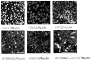
- the result of having measured the expression of the marker gene (SOX17, PDX1, NGN3, ISL-1, INS, GCG, SST, GHRE, MAFA) in the cell after each induction day (Day0, Day5, Day11, Day19) is shown.
- SOX17 or FOXA2 on day 5 of induction PDX1 on day 11 of culture, NEUROG3 on day 15 of culture, and INSULIN or C-peptide on day 19 of culture are shown.
- Per cell on the 19th day of induction cultured in 4F forskolin (10 ⁇ M), nicotinamide (10 mM), dexamethasone (10 ⁇ M), ALK5 inhibitor II (5 ⁇ M)
- 4F forskolin (10 ⁇ M), nicotinamide (10 mM), dexamethasone (10 ⁇ M), ALK5 inhibitor II (5 ⁇ M)
- Stained images of INSULIN, GLUCAGON, GHRE (GHRELIN), SST (SOMATOSTATIN), AMY (AMYLASE) and CK19 on the 19th day after induction are shown.
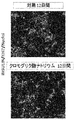
- E12.5, E14.5 and E16.5 pancreatic primordia were cultured in basal medium (medium only), 4F (forskolin (10 ⁇ M), nicotinamide (10 mM), dexamethasone (10 ⁇ M), ALK5 inhibitor II ( The results of measuring the expression of Insulin or Neurog3 after culturing in a basic medium supplemented with 5 ⁇ M)) or in a basic medium supplemented with 4F and sodium cromoglycate are shown.
- PC means positive control and indicates the expression level in adult mouse pancreatic cells.
- the schematic diagram of the process of manufacturing beta cell from a pluripotent stem cell is shown.
- FX sodium cromoglycate only
- ALK5 inhibitor II only A
- 4F, 4F and sodium cromoglycate (4FX) The result of having measured the ratio of the expression level of INSULIN with respect to the expression level of is shown.
- the present invention provides a method for producing pancreatic hormone-producing cells, comprising a step of culturing pancreatic progenitor cells in a medium containing sodium cromoglycate.
- the medium for producing the pancreatic hormone-producing cells in addition to sodium cromoglycate, (a) at least one selected from the group consisting of an adenylate cyclase activator, a cAMP phosphodiesterase inhibitor, and a cAMP analog, (b And at least one agent selected from the group consisting of: nicotinamide, (c) steroids and (d) TGF ⁇ inhibitors, (f) KGF, (g) EGF, and (h) BMP inhibitors.
- the medium used in the present invention is prepared by appropriately adding substances to the basal medium.
- basal media include IMDM medium, Medium 199 medium, Eagle'sEMinimum Essential Medium (EMEM) medium, ⁇ MEM medium, Dulbecco's modified Eagle's Medium (DMEM) medium, Improved MEM (invitrogen), Ham's F12 medium, RPMIscher's medium , Neurobasal Medium (Life Technologies), StemPro34 (invitrogen) and mixed media thereof are included.
- the basal medium may be a serum-containing medium or a serum-free medium.
- the basal medium is, for example, albumin, transferrin, Knockout Serum Replacement (KSR) (serum substitute for FBS during ES cell culture), N2 supplement (Invitrogen), B27 supplement (Invitrogen), fatty acids, insulin, It may contain one or more serum replacements such as collagen precursors, trace elements, 2-mercaptoethanol, 1-thiolglycerol, lipids, amino acids, L-glutamine, Glutamax (Invitrogen), non-essential amino acids, vitamins, It may also contain one or more substances such as growth factors, small molecule compounds, antibiotics, antioxidants, pyruvate, buffers, inorganic salts.
- KSR Knockout Serum Replacement
- the step of culturing in a medium containing sodium cromoglycate may employ any culture method of pancreatic progenitor cells, such as suspension culture or adhesion culture.
- the medium is prepared by adding sodium cromoglycate to the basal medium described above.
- a preferred basal medium is Improved MEM medium containing B-27 supplement.
- Sodium cromoglycate is a sodium salt of 1,3-bis (2-carboxychromon-5-yloxy) -2-hydroxypropane, and is available from, for example, Sigma and Wako Pure Chemical Industries.
- concentration of sodium cromoglycate used in Step 3 in the medium is usually 0.001 to 100 mM, preferably 0.01 to 50 mM, more preferably 0.01 to 20 mM. Particularly preferred is 10 mM.
- floating culture is a mode in which cells are cultured in a non-adhering state on a culture dish.
- a culture dish that has not been treated to improve adhesion to cells (for example, coating with an extracellular matrix or the like) or artificially suppresses adhesion (for example, polyhydroxyethyl) It can be performed using a culture dish coated with methacrylic acid (poly-HEMA).
- the cells When performing adhesion culture, the cells may be cultured on feeder cells or in a coated culture dish.
- the feeder cell means another cell that plays an auxiliary role used for adjusting the culture conditions of the target cell.
- fibroblast mouse fetal fibroblast (MEF), mouse
- STO SNL
- the feeder cells are preferably inactivated by a method known per se, for example, irradiation with radiation (eg gamma rays) or treatment with an anticancer agent (eg mitomycin C).
- the coating agent include matrigel (BD), collagen, gelatin, laminin, heparan sulfate proteoglycan, entactin, fragments thereof, and combinations thereof.
- substances selected from the group consisting of an adenylate cyclase activator, a cAMP phosphodiesterase inhibitor, and a cAMP analog include compounds having adenylate cyclase activity, compounds having cAMP phosphodiesterase inhibitory activity, and adenylate cyclase activity And compounds having both cAMP phosphodiesterase inhibitory activity. More specifically, forskolin, dibutyl cAMP, PACAP27 (pituitary adenylate cyclase activating polypeptide 27), IBMX (3-isobutyl-1-methylxanthine) and the like can be mentioned. Forskolin is preferred.
- the concentration in the medium is usually 0.1 to 50 ⁇ M, preferably 2 to 50 ⁇ M. is there.
- the concentration of nicotinamide in the medium is 0.1 to 20 mM, preferably 5 to 20 mM.
- the steroid examples include dexamethasone, hydrocortisone, betamethasone, and beclomethasone. Of these, dexamethasone is preferably used.
- dexamethasone is used as a steroid, the concentration in the medium is usually 0.1 to 50 ⁇ M, preferably 2 to 50 ⁇ M.
- hydrocortisone is used as a steroid, the concentration in the medium is usually 0.1 to 100 ⁇ M, preferably 1 to 50 ⁇ M.
- betamethasone is used as the steroid, the concentration in the medium is usually 0.1 to 50 ⁇ M, preferably 0.5 to 20 ⁇ M.
- beclomethasone is used as a steroid, the concentration in the medium is usually 0.1 to 50 ⁇ M, preferably 0.2 to 20 ⁇ M.
- a TGF ⁇ inhibitor is a substance that inhibits the signal transduction from binding of TGF ⁇ to the receptor to SMAD, and inhibits the binding of the receptor to the ALK family, or the phosphorylation of SMAD by the ALK family.
- Lefty-1 NCBI Accession ⁇ No. is exemplified by mouse: NM_010094, human: NM_020997
- SB431542 (4- [4- (1,3-benzodioxy).
- ALK5 inhibitor II (2- [3- [6-methylpyridin-2-yl] -1H-pyrazol-4-yl] -1,5-naphthyridine), TGF ⁇ RI kinase inhibitor VIII ( 6- [2-tert-butyl-5- [6-methyl-pyridin-2-yl] -1H-imidazol-4-yl] -quinoxaline) and derivatives thereof.
- a preferred TGF ⁇ inhibitor added to the medium containing sodium cromoglycate is ALK5 inhibitor II.
- the concentration in the medium is usually 0.5 to 100 ⁇ M, preferably 1 to 50 ⁇ M, more preferably 1 to 10 ⁇ M.
- KGF is a protein called Keratinocyte Growth Factor and is sometimes called FGF-7.
- concentration of KGF used in the present invention is 1 ng / ml to 1 ⁇ g / ml, preferably 5 ng / ml to 500 ng / ml, more preferably 10 ng / ml to 100 ng / ml.
- EGF is a protein called epidermal growth factor or Epidermal Growth Factor.
- concentration of EGF used in the present invention is 1 ng / ml to 1 ⁇ g / ml, preferably 5 ng / ml to 500 ng / ml, more preferably 10 ng / ml to 100 ng / ml.
- BMP inhibitors include proteinaceous inhibitors such as Chordin, Noggin, Follistatin, Dorsomorphin® (ie 6- [4- (2-piperidin-1-yl-ethoxy) phenyl] -3-pyridin-4-yl-pyrazolo [1,5-a] pyrimidine), derivatives thereof (P.PB. Yu et al. (2007), Circulation, 116: II_60; PB Yu et al. (2008), Nat. Chem. Biol., 4:33 -41; J. Hao et al.
- LDN-193189 ie 4- (6- (4- (piperazin-1-yl) phenyl) pyrazolo [1 , 5-a] pyrimidin-3-yl) quinoline).
- Dorsomorphin and LDN-193189 are commercially available and are available from Sigma-Aldrich and Stemgent, respectively.
- the culture days are, for example, 4 days or more, 5 days or more, 6 days or more, 7 days or more, 8 days or more, 9 days or more, 10 days or more, preferably 4 days or more and less than 20 days, More preferably, it is 8 days or more and 16 days or less.
- the pancreatic progenitor cell in the present invention means a cell having an ability to be induced into a pancreatic hormone-producing cell. Such cells can also be characterized as cells that express PDX1 and do not express insulin, glucagon, somatostatin, pancreatic polypeptide, or ghrelin.
- pancreatic progenitor cells may be obtained by isolation from in vivo or may be obtained by induction from pluripotent stem cells.
- a method including the following steps (1) to (2) is exemplified.
- the present invention also provides a method for producing pancreatic hormone-producing cells from pluripotent stem cells, which includes the following steps (1) to (3).
- Step of culturing pluripotent stem cells in a medium containing an activin receptor-like kinase-4,7 activator and a GSK3 inhibitor (2)
- the cells obtained in the step (1) are ( a) a step of culturing in a medium containing one selected from the group consisting of a retinoic acid receptor agonist, (b) a BMP inhibitor, and (c) a TGF ⁇ inhibitor (3) obtained in step (2) above Culturing the cells in a medium containing sodium cromoglycate.
- a pluripotent stem cell is a stem cell which has pluripotency that can be differentiated into many cells existing in a living body and also has a self-proliferating ability.
- pluripotent stem cells include embryonic stem (ES) cells, embryonic stem (ntES) cells derived from cloned embryos obtained by nuclear transfer, sperm stem cells (“GS cells”), embryonic germ cells (“EG cells”) "), Induced pluripotent stem (iPS) cells, cultured fibroblasts, bone marrow stem cell-derived pluripotent cells (Muse cells), and the like. From the viewpoint that pluripotent stem cells can be obtained without destroying embryos, it is preferable to use iPS cells or Muse cells.
- the pluripotent stem cell is preferably a mammalian pluripotent stem cell, more preferably a human pluripotent stem cell.
- Embryonic stem cells ES cells are stem cells established from the inner cell mass of early embryos (for example, blastocysts) of mammals such as humans and mice, and having pluripotency and proliferation ability by self-replication.
- ES cells are embryonic stem cells derived from the inner cell mass of the blastocyst, the embryo after the morula, in the 8-cell stage of a fertilized egg, and have the ability to differentiate into any cell that constitutes an adult, so-called differentiation. And ability to proliferate by self-replication.
- ES cells were discovered in mice in 1981 (MJ Evans and MH Kaufman (1981), Nature 292: 154-156), and then ES cell lines were established in primates such as humans and monkeys (JA Thomson et al. (1998), Science 282: 1145-1147; JA Thomson et al. (1995), Proc. Natl. Acad. Sci. USA, 92: 7844-7848; JA Thomson et al. (1996), Biol. Reprod , 55: 254-259; JA Thomson and VS Marshall (1998), Curr. Top. Dev. Biol., 38: 133-165 (the literature described in this paragraph is incorporated herein by reference).
- ES cells can be established by taking an inner cell mass from a blastocyst of a fertilized egg of a target animal and culturing the inner cell mass on a fibroblast feeder. In addition, maintenance of cells by subculture is performed using a culture solution to which substances such as leukemia inhibitory factor (LIF) and basic fibroblast growth factor (basic fibroblast growth factor (bFGF)) are added. It can be carried out.
- LIF leukemia inhibitory factor
- bFGF basic fibroblast growth factor
- DMEM / F-12 culture medium supplemented with 0.1 mM 2-mercaptoethanol, 0.1 mM non-essential amino acid, 2 mM L-glutamic acid, 20% KSR and 4 ng / ml bFGF is used as the culture medium for ES cell production.
- Human ES cells can be maintained in a humid atmosphere of 37 ° C., 5% CO 2 /95% air (O. Fumitaka et al. (2008), Nat. Biotechnol., 26: 215-224).
- ES cells also need to be passaged every 3-4 days, where passage is eg 0.25% trypsin and 0.1 mg / ml collagenase IV in PBS containing 1 mM CaCl 2 and 20% KSR. Can be used.
- ES cells can be generally selected by Real-Time PCR using the expression of gene markers such as alkaline phosphatase, Oct-3 / 4, Nanog as an index.
- gene markers such as alkaline phosphatase, Oct-3 / 4, Nanog
- OCT-3 / 4, NANOG, and ECAD can be used as an index (E. Kroon et al. (2008), Nat. Biotechnol., 26: 443). -452).
- sperm stem cells are testis-derived pluripotent stem cells that are the origin of spermatogenesis. Like ES cells, these cells can be induced to differentiate into various types of cells, and have characteristics such as the ability to create chimeric mice when transplanted into mouse blastocysts (M. Kanatsu-Shinohara et al. ( 2003) Biol. Reprod., 69: 612-616; K. Shinohara et al. (2004), Cell, 119: 1001-1012).
- GDNF glial cell line-derived neurotrophic factor
- Embryonic germ cells are cells that are established from embryonic primordial germ cells and have the same pluripotency as ES cells, such as LIF, bFGF, stem cell factor, etc. It can be established by culturing primordial germ cells in the presence of these substances (Y. Matsui et al. (1992), Cell, 70: 841-847; JL Resnick et al. (1992), Nature, 359: 550 -551). (The literature described in this paragraph constitutes part of this application by reference)
- the reprogramming factor is a gene specifically expressed in ES cells, its gene product or non-cording RNA, a gene that plays an important role in maintaining undifferentiation of ES cells, its gene product or non-coding RNA, or It may be constituted by a low molecular compound.
- genes included in the reprogramming factor include Oct3 / 4, Sox2, Sox1, Sox3, Sox15, Sox17, Klf4, Klf2, c-Myc, N-Myc, L-Myc, Nanog, Lin28, Fbx15, ERas, ECAT15 -2, Tcl1, beta-catenin, Lin28b, Sall1, Sall4, Esrrb, Nr5a2, Tbx3 or Glis1 etc.
- the reprogramming factor may be brought into contact with or introduced into a somatic cell by a known method depending on its form.
- somatic cells In the case of the protein form, it may be introduced into somatic cells by techniques such as lipofection, fusion with cell membrane permeable peptides (eg, HIV-derived TAT and polyarginine), and microinjection.
- cell membrane permeable peptides eg, HIV-derived TAT and polyarginine
- Virus vectors include retrovirus vectors, lentivirus vectors (cell, 126, pp.663-676, 2006; Cell, 131, pp.861-872, 2007; Science, 318, pp.1917-1920, 2007 ), Adenovirus vectors (Science, 322, 945-949, 2008), adeno-associated virus vectors, Sendai virus vectors (WO 2010/008054) and the like.
- artificial chromosome vectors examples include human artificial chromosomes (HAC), yeast artificial chromosomes (YAC), and bacterial artificial chromosomes (BAC, PAC).
- HAC human artificial chromosomes
- YAC yeast artificial chromosomes
- BAC bacterial artificial chromosomes
- a plasmid a plasmid for mammalian cells can be used (Science, 322: 949-953, 2008).
- the vector can contain regulatory sequences such as a promoter, enhancer, ribosome binding sequence, terminator, polyadenylation site, etc. so that a nuclear reprogramming substance can be expressed.
- Selective marker sequences such as kanamycin resistance gene, ampicillin resistance gene, puromycin resistance gene, thymidine kinase gene, diphtheria toxin gene, reporter gene sequences such as green fluorescent protein (GFP), ⁇ -glucuronidase (GUS), FLAG, etc. Can be included.
- GFP green fluorescent protein
- GUS ⁇ -glucuronidase
- FLAG FLAG
- the culture solution for iPS cell induction is, for example, DMEM, DMEM / F12 or DME culture solution containing 10 to 15% FBS (in addition to LIF, penicillin / streptomycin, puromycin, L-glutamine, Non-essential amino acids, ⁇ -mercaptoethanol, etc. may be included as appropriate.) Or commercially available culture media [eg, culture medium for mouse ES cell culture (TX-WES culture medium, Thrombo X), primate ES cell culture Culture medium (primate ES / iPS cell culture medium, Reprocell), serum-free medium (mTeSR, Stemcell Technology)] and the like.
- FBS penicillin / streptomycin
- puromycin puromycin
- L-glutamine Non-essential amino acids
- ⁇ -mercaptoethanol etc.
- commercially available culture media eg, culture medium for mouse ES cell culture (TX-WES culture medium, Thrombo X), primate ES cell culture Culture
- FBS-containing DMEM medium including LIF, penicillin / streptomycin, etc.
- feeder cells eg, mitomycin C-treated STO cells, SNL cells, etc.
- Puromycin, L-glutamine, non-essential amino acids, ⁇ -mercaptoethanol, etc. may be included as appropriate.
- somatic cells to be reprogrammed themselves are used (Takahashi K, et al. (2009), PLoS One.
- iPS cells may be established under hypoxic conditions (oxygen concentration of 0.1% or more and 15% or less) (Yoshida Y, et al. (2009), Cell Stem Cell. 5: 237 -241 or WO2010 / 013845). (The literature described in this paragraph constitutes part of this application by reference)
- histone deacetylase (HDAC) inhibitors for example, small molecule inhibitors such as valproic acid (VPA), trichostatin A, sodium butyrate, MC 1293, M344, HDAC siRNA and shRNA (eg, nucleic acid expression inhibitors such as HDAC1 siRNA Smartpool ⁇ (Millipore), HuSH 29mer shRNA Constructs against HDAC1), MEK inhibitors (eg, PD184352, PD98059, U0126, SL327 and PD0325901) ), Glycogen synthase kinase-3 inhibitors (eg, Bio and CHIR99021), DNA methyltransferase inhibitors (eg, 5-azacytidine), histone methyltransferase inhibitors (eg, small molecule inhibitors such as BIX-01294, Suv39hl, Nucleic acid expression inhibitors such as siRNA and shRNA for Suv39h2, SetDB
- HDAC histone deacetylase
- the culture medium is exchanged with a fresh culture medium once a day from the second day onward.
- the number of somatic cells used for nuclear reprogramming is not limited, but ranges from about 5 ⁇ 10 3 to about 5 ⁇ 10 6 cells per 100 cm 2 of culture dish.
- iPS cells can be selected according to the shape of the formed colonies.
- a culture medium selective culture medium
- a drug resistance gene that is expressed in conjunction with a gene that is expressed when somatic cells are initialized for example, Oct3 / 4, Nanog
- iPS cells can be selected by adding a luminescent substrate in the case of a luminescent enzyme gene.
- the term “somatic cell” refers to any animal cell except germline cells or pluripotent cells such as sperm, spermatocytes, eggs, oocytes, ES cells (preferably Refers to mammalian cells including humans). Somatic cells include fetal (pup) somatic cells, neonatal (pup) somatic cells, and mature healthy individual somatic cells or somatic cells of diseased individuals. Moreover, all of primary culture cells, subculture cells, and established cell lines are included.
- somatic cells are differentiated cells such as (1) tissue stem cells (somatic stem cells) such as neural stem cells, hematopoietic stem cells, mesenchymal stem cells, dental pulp stem cells, (2) tissue progenitor cells, (3) lymph Spheres, epithelial cells, endothelial cells, muscle cells, fibroblasts (skin cells, etc.), hair cells, hepatocytes, gastric mucosal cells, intestinal cells, spleen cells, pancreatic cells (exocrine pancreatic cells, etc.), brain cells, lung cells Examples thereof include kidney cells and fat cells.
- tissue stem cells such as neural stem cells, hematopoietic stem cells, mesenchymal stem cells, dental pulp stem cells
- tissue progenitor cells such as lymph Spheres, epithelial cells, endothelial cells, muscle cells, fibroblasts (skin cells, etc.), hair cells, hepatocytes, gastric mucosal cells, intestinal cells, spleen cells, pancreatic cells
- E Cloned embryo-derived ES cells obtained by nuclear transfer nt ES cells are cloned embryo-derived ES cells produced by nuclear transfer technology and have almost the same characteristics as ES cells derived from fertilized eggs (T. Wakayama et al. (2001), Science, 292: 740-743; S. Wakayama et al. (2005), Biol. Reprod., 72: 932-936; J. Byrne et al. (2007) , Nature, 450: 497-502).
- an ES cell established from an inner cell mass of a clonal embryo-derived blastocyst obtained by replacing the nucleus of an unfertilized egg with the nucleus of a somatic cell is an nt ES (nuclear transfer ES) cell.
- nt ES nuclear transfer ES
- nuclear transfer technology JB Cibelli et al. (1998), Nature Biotechnol., 16: 642-646) and ES cell production technology (above) is used (Wakayama). Seika et al. (2008), Experimental Medicine, Vol. 26, No. 5 (extra number), 47-52).
- Nuclear transfer can be initialized by injecting a somatic cell nucleus into a mammal's enucleated unfertilized egg and culturing for several hours.
- Muse cells are pluripotent stem cells produced by the method described in WO2011 / 007900. Specifically, it is a pluripotent cell obtained by subjecting fibroblasts or bone marrow stromal cells to trypsin treatment for a long time, preferably 8 hours or 16 hours, followed by suspension culture. Muse cells are positive for SSEA-3 and CD105.
- a pancreatic hormone-producing cell means a cell having the ability to produce pancreatic hormones.
- the pancreatic hormone include insulin, glucagon, somatostatin, pancreatic polypeptide, and ghrelin.
- Pancreatic hormone-producing cells need not always produce pancreatic hormones, as long as they have the ability to produce pancreatic hormones. Further, the amount of pancreatic hormone produced is not particularly limited. Examples of pancreatic hormone producing cells include insulin producing cells, glucagon producing cells, somatostatin producing cells, and pancreatic polypeptide (PP) producing cells.
- PP pancreatic polypeptide
- the method of inducing pancreatic progenitor cells from pluripotent stem cells can be performed by the following steps including Step 1 and Step 2.
- Step 1 A step of culturing pluripotent stem cells in a medium containing activin receptor-like kinase-4,7 activator and GSK3 inhibitor.
- the pluripotent stem cells are usually cultured in an adherent culture.
- pluripotent stem cells may be separated by any method and suspended culture, or adhesion culture may be performed on feeder cells or using a coated culture dish.
- a method for separating human pluripotent stem cells a conventionally known method for separating adherent cultured cells may be appropriately employed.
- a method for separating dynamically a separation solution having protease activity and collagenase activity (for example, Accutase ( (TM) and Accumax (TM), etc.) or a separation method using a separation solution having only collagenase activity.
- a method is used in which a human pluripotent stem cell is dissociated using a separation solution having protease activity and collagenase activity (particularly preferably, Accutase (TM)) and dispersed finely into a single cell.
- the human pluripotent stem cells used in step 1 are preferably colonies cultured until they are 80% or more confluent with respect to the used dish.
- the culture may be performed on feeder cells or in a coated culture dish.
- the feeder cell means another cell that plays an auxiliary role used for adjusting the culture conditions of the target cell.
- fibroblast mouse fetal fibroblast (MEF), mouse
- STO SNL
- the feeder cells are preferably inactivated by a method known per se, for example, irradiation with radiation (eg gamma rays) or treatment with an anticancer agent (eg mitomycin C).
- the coating agent include matrigel (BD), collagen, gelatin, laminin, heparan sulfate proteoglycan, entactin, and combinations thereof.
- Step 1 is started by exchanging the medium of pluripotent stem cells in culture, or by separating the pluripotent stem cells and transferring them to a culture dish for adhesion culture or suspension culture in which a predetermined medium has been introduced.
- the medium is prepared by adding an activin receptor-like kinase-4,7 activator and a GSK3 inhibitor to the basal medium described above.
- a preferred basal medium is RPMI 1640 medium with B27 supplement or RPMI 1640 medium with serum.
- the activin receptor-like kinase-4,7 activator is a substance having an activating action on ALK-4 and / or ALK-7, and examples thereof include activin, Nodal, and Myostatin.
- Activins A, B, C, D, and AB are known as activins, but any activin A, B, C, D, AB can be used in step 1.
- Activin A is particularly preferably used as the activin used in Step 1.
- activin activin derived from any mammal such as human and mouse can be used.
- activin used in Step 1 activin derived from the same animal species as the pluripotent stem cell used for differentiation is preferably used. For example, when human-derived pluripotent stem cells are used as a starting material, It is preferable to use it.
- These activins are commercially available.
- the concentration in the medium is usually 0.1 to 200 ng / ml, preferably 5 to 150 ng / ml, particularly preferably 10 to 100 ng / ml.
- a GSK3 inhibitor is defined as a substance that inhibits the kinase activity of GSK-3 ⁇ protein (for example, phosphorylation ability for ⁇ -catenin), and many are already known.
- BIO indirubin derivative BIO (also known as GSK-3 ⁇ inhibitor IX; 6-bromoindirubin 3′-oxime)
- SB216763 (3- (2,4-dichlorophenyl) -4- (1-methyl-1H-indol-3-yl), a maleimide derivative ) -1H-pyrrole-2,5-dione
- SB415286 (3-[(3-chloro-4-hydroxyphenyl) amino] -4- (2-nitrophenyl) -1H-pyrrole-2,5-dione)
- GSK-3 ⁇ inhibitor VII (4-dibromoacetophenone), a phenyl ⁇ bromomethyl ketone compound, L803-mts (also known as GSK-3 ⁇ peptide inhibitor; Myr-N-
- the concentration in the medium is usually 0.01 ⁇ M to 100 ⁇ M, preferably 0.1 ⁇ M to 10 ⁇ M, particularly preferably 1 ⁇ M to 5 ⁇ M.
- a ROCK inhibitor may be further contained.
- the ROCK inhibitor is not particularly limited as long as it can suppress the function of Rho-kinase (ROCK).
- ROCK Rho-kinase
- Y-27632 eg, Ishizaki et al., Mol. Pharmacol. 57, 976-983 (2000) Narumiya et al., Methods Enzymol. , Pharmacol. Ther. 93: See 225-232 (2002)
- Wf-536 eg, Nakajima et al., Cancer Chemother Pharmacol.
- ROCK inhibitors for example, US Patent Application Publication Nos. 2005/0209261, 2005/0192304, 2004/0014755, 2004/0002508,. 2004/0002507, 2003/0125344, 2003/0087919, and International Publications 2003/062227, 2003/059913, 2003/062225, 2002/076976 No., 2004/039796).
- one or more ROCK inhibitors may be used.
- a preferred ROCK inhibitor used in this step includes Y-27632.
- the concentration in the medium is 0.1 ⁇ M to 100 ⁇ M, preferably 1 ⁇ M to 50 ⁇ M, more preferably 5 ⁇ M to 20 ⁇ M.
- ROCK inhibitors need to be added throughout the culture period, with the intention of preventing cell death at the start of culture, and more specifically, with the intention of preventing cell death when isolating pluripotent stem cells. Instead, it may be added for 1 day or 2 days at the start of culture.
- the culture days in step 1 are, for example, 3 days or more, 4 days or more, 5 days or more, 6 days or more, 7 days or more, 8 days or more, 9 days or more, 10 days or more. There is no particular inconvenience due to long-term culture, and there is no upper limit on the number of culture days.
- the culture days are, for example, 5 days or more and 12 days or less, and preferably 5 days.
- the retinoic acid receptor (RAR) agonist used in Step 2 is a chemically synthesized retinoid, a retinoic acid receptor agonist compound having no retinoid skeleton, or a retinoic acid receptor agonist, even if it is a naturally occurring retinoid It may be a natural product having activity.
- natural retinoids having activity as RAR agonists include retinoic acid (stereoisomers of all-trans-retinoic acid (all-trans RA) and 9-cis-retinoic acid (9-cis RA) are known) Is mentioned.
- Chemically synthesized retinoids are known in the art (US Pat. No. 5,234,926, US Pat. No. 4,326,055, etc.).
- retinoic acid receptor agonist compounds having no retinoid skeleton include Am80, AM580, TTNPB, and AC55649.
- natural products having retinoic acid receptor agonist activity include honokiol and magnolol (Research Bulletin of Laboratory for Biofunction Development 9: 55-61, 2009).
- the RAR agonist used in Step 2 is preferably retinoic acid, AM580 (4-[[5,6,7,8-tetrahydro-5,5,8,8-tetramethyl-2-naphthalenyl] carboxamide] benzoic acid.
- TTNPB (4-[[E] -2- [5,6,7,8-tetrahydro-5,5,8,8-tetramethyl-2-naphthalenyl] -1-propenyl] benzoic acid), AC55649 (4′-octyl- [1,1′-biphenyl] -4-carboxylic acid), more preferably retinoic acid.
- the concentration in the medium when retinoic acid is used as the RAR agonist is usually 0.1 to 100 ⁇ M, preferably 0.5 to 10 ⁇ M.
- the concentration in the medium when TTNPB is used as the RAR agonist is usually 0.02 to 20 ⁇ M, preferably 0.05 to 10 ⁇ M.
- AM580 is used as the RAR agonist
- the concentration in the medium is usually 0.02 to 20 ⁇ M, preferably 0.05 to 10 ⁇ M.
- AC55649 is used as the RAR agonist
- the concentration in the medium is usually 0.02 to 20 ⁇ M, preferably 0.1 to 10 ⁇ M.
- BMP inhibitor examples include the aforementioned BMP inhibitors, and preferably Dorsomorphin.
- the concentration in the medium when Dorsomorphin is used as the BMP inhibitor is 0.01 ⁇ M to 100 ⁇ M, preferably 0.1 to 10 ⁇ M, and more preferably 0.5 ⁇ M to 5 ⁇ M.
- TGF ⁇ inhibitor examples include the TGF ⁇ inhibitors described above, and preferably SB431542.
- the concentration in the medium when SB431542 is used as the TGF ⁇ inhibitor is 0.01 ⁇ M to 100 ⁇ M, preferably 0.1 to 50 ⁇ M, more preferably 1 ⁇ M to 20 ⁇ M.
- KGF can be used in place of the TGF ⁇ inhibitor. Even when KGF is used in Step 2, the concentration is the same as described above from 1 ng / ml to 1 ⁇ g / ml, preferably 5 ng / ml to 500 ng / ml, more preferably 10 ng / ml. To 100 ng / ml.
- the number of culture days in step 2 is, for example, 3 days or more, 4 days or more, 5 days or more, 6 days or more, 7 days or more, 8 days or more, 9 days or more, 10 days or more. There is no particular inconvenience due to long-term culture, and there is no upper limit on the number of culture days.
- the culture days are, for example, 6 days or more and 12 days or less, and particularly preferably 6 days.
- the pancreatic hormone-producing cells obtained in the present invention are selected from the group consisting of, for example, insulin-producing cells, glucagon-producing cells, somatostatin-producing cells, and pancreatic polypeptide-producing cells, and each cell confirms the produced pancreatic hormone. Can be identified by The obtained pancreatic hormone-producing cells may be provided as a cell population containing other cell types. Alternatively, by using an antibody that specifically binds to a surface tag encoding G6PC2 (WO2010 / 037784, this document constitutes a part of this application by reference), the cells are magnetically separated using magnetic beads.
- a sorting method eg, MACS
- a method using a cell sorter using a fluorescent label e.g., a cell concentration column.
- a carrier on which an antibody or the like is immobilized e.g, a cell concentration column.
- pancreatic hormone can be confirmed by confirming the expression of pancreatic hormone protein and a polynucleotide (eg, mRNA) encoding the pancreatic hormone protein.
- the secretion of pancreatic hormone protein is a known method, for example, a method such as Western blotting analysis or ELISA method for the pancreatic hormone protein present in a cell extract or medium using an antibody that recognizes pancreatic hormone protein. Or it can measure by the method according to it.
- pancreatic hormone can be confirmed by examining the expression level of a polynucleotide encoding pancreatic hormone protein, such as mRNA.
- the amount of mRNA is measured according to a known method, for example, Northern hybridization, S1 mapping method, PCR method, quantitative RT-PCR method, DNA chip or array method, or a similar method.
- step (1) at least one selected from the group consisting of an adenylate cyclase activator, a cAMP phosphodiesterase inhibitor, and a cAMP analog, (b) nicotinamide, (c) a steroid, and (d ) It may be cultured in a medium further containing at least one selected from the group consisting of TGF ⁇ inhibitors, more preferably (a) a group consisting of an adenylate cyclase activator, a cAMP phosphodiesterase inhibitor, and a cAMP analog. It is a step of culturing in a medium further containing at least one selected from (b) nicotinamide, (c) steroid, and (d) a TGF ⁇ inhibitor.
- Candidate drugs include, for example, peptides, proteins, antibodies, non-peptide compounds, synthetic compounds, fermentation products, cell extracts, plant extracts, animal tissue extracts, and plasma.
- the test compound may form a salt.
- a salt with a physiologically acceptable acid eg, inorganic acid, organic acid
- base eg, alkali metal salt, alkaline earth metal salt, aluminum salt
- the salt include a salt with an inorganic acid (eg, hydrochloric acid, phosphoric acid, hydrobromic acid, sulfuric acid), or an organic acid (eg, acetic acid, formic acid, propionic acid, fumaric acid, maleic acid, succinic acid, tartaric acid).
- Citric acid Citric acid, malic acid, succinic acid, benzoic acid, methanesulfonic acid, benzenesulfonic acid), sodium salts, potassium salts, calcium salts, magnesium salts, barium salts, and aluminum salts.
- the present invention further provides the following method for screening pancreatic hormone secretion promoters.
- pancreatic hormone secretion promoters the same substances or methods as described above can be used to measure the expression levels of candidate drugs and pancreatic hormones.
- the pancreatic hormone-producing cells obtained using the above steps can be transplanted into a patient and used for the treatment of pancreatic diseases. Therefore, a composition for the treatment of pancreatic disease containing pancreatic hormone-producing cells obtained by the method of the present invention is also included in the present invention.
- the cells may be treated with an agent that irradiates radiation or inhibits cell growth such as mitomycin C before transplantation.
- pancreatic diseases Treatment of pancreatic diseases is carried out by directly transplanting the obtained cell suspension containing the pancreatic hormone-producing cells in physiological saline or the like to the patient's pancreas, mesentery, spleen and liver. obtain.
- the cells may be administered together with a scaffold such as polyethylene glycol, gelatin, or collagen.
- the number of cells to be administered is, for example, 1 ⁇ 10 8 to 1 ⁇ 10 10 cells / individual, preferably 5 ⁇ 10 8 to 1 ⁇ 10 10 cells / individual, more preferably 1 ⁇ 10 9 to 1 ⁇ 10 10. Cell / individual.
- iPS cells are particularly preferably used as a material for transplanting pancreatic hormone-producing cells.
- iPS cells obtained from somatic cells that are the same or substantially the same as the HLA genotype of the transplant recipient individual from the viewpoint that no rejection occurs.
- substantially the same means that the HLA genotype matches the transplanted cells to such an extent that an immune response can be suppressed by an immunosuppressive agent.
- HLA-A, HLA-B And somatic cells having an HLA type in which 3 loci of HLA-DR or 4 loci plus HLA-C are matched.
- KhES3 was received from Kyoto University and cultured by a conventional method (H. Suemori et al. (2006), Biochem. Biophys. Res. Commun., 345: 926- 932, this document is incorporated by reference).
- KhES3 was induced into insulin producing cells according to the following steps. For differentiation induction, human ES cell line is detached from individual cells using Accutase after exfoliating cells that have grown to approximately 70% confluency in the culture dish using CTK solution (Reprocell). Plate (greiner) was seeded at 2.0 ⁇ 10 5 / well and used.
- Human iPS cell 201B7 (available from iPS cell research institute, Kyoto University (Takahashi K et al, Cell. 2007 131: 861-72., this document is incorporated by reference)) was used to induce insulin-producing cells according to the following steps.
- the human iPS cell line was detached from cells grown to approximately 70% confluency in the culture dish using CTK solution, and then separated into individual cells using Accutase, and 2.0 cells in a 24-well plate. It was used after seeding at ⁇ 10 5 / well.
- Activin A 100 ng / ml
- CHIR99021 3 ⁇ M
- iPS cell lines derived from fibroblasts 418C1 and 409B2 (Okita K, et al, Nat Methods. 2011 8: 409-412) and 201B7) are available from iPS Cell Research Institute, Kyoto University. is there. Using these iPS cell lines obtained from Kyoto University, differentiation into insulin-positive cells was induced by the same steps as described above (however, the concentration of sodium cromoglycate in Step 3 was 10 mM).
- Activin A 100 ng / ml
- CHIR99021 (1 ⁇ M) and Y-27632 (10 ⁇ M) were added to an RPMI medium containing 1% B-27 and cultured for 2 days. Thereafter, the cells were further cultured for 3 days in a medium prepared by adding activin A (100 ng / ml) and CHIR99021 (1 ⁇ M) to an RPMI medium containing 1% B-27 (Day 5). At this time, the medium was changed on the third day (Day 3) and the fourth day (Day 4) with the same medium.
- the cells obtained in each step were collected, and the expression of each marker gene was confirmed using PCR (FIG. 5B).
- the expression of NGN3 and GHRE ghrelin
- SST somatostatin
- pancreatic hormone was confirmed by immunostaining (FIGS. 6A and B). As a result, it was confirmed that the expression of insulin, glucagon and somatostatin was higher when sodium cromoglycate was added in the third step.
- sodium cromoglycate has the effect of promoting the induction from in vivo pancreatic primordium cells to insulin-producing cells.
- Human iPS cells 201B7 were used to induce insulin producing cells according to the following steps. Upon induction of differentiation, human iPS cell lines were detached from cells grown to approximately 80-90% confluency in the culture dish using CTK solution, and then separated into individual cells using Accutase. The coated 24-well plate was seeded at 2.0 ⁇ 10 5 / well and used.
- Activin A 100 ng / ml
- CHIR99021 (1 ⁇ M) and Y-27632 (10 ⁇ M) were added to an RPMI medium containing 1% B-27 and cultured for 2 days. Thereafter, activin A (100 ng / ml) and CHIR99021 (1 ⁇ M) were added to an RPMI medium containing 1% B-27 and further cultured for 3 days (Day 5). At this time, the medium was changed on the third day (Day 3) and the fourth day (Day 4) with the same medium.
- sodium cromoglycate can increase the induction efficiency to insulin-producing cells even when used alone.
- pancreatic hormone-producing cells produced by this method can be used for regenerative medicine for pancreatic diseases, particularly type 1 diabetes.
Abstract
Description
[1]膵前駆細胞をクロモグリク酸ナトリウムを含む培地で培養する工程を含む、膵ホルモン産生細胞の製造方法。
[2]前記培地が、(a)アデニル酸シクラーゼ活性化剤、cAMPホスホジエステラーゼ阻害剤、およびcAMP類縁体からなる群より選択される少なくとも一種、(b)ニコチンアミド、(c)ステロイドおよび(d)TGFβ阻害剤から成るからなる群より選択される少なくとも一種の薬剤をさらに含む、[1]に記載の方法。
[3]前記培地が、(a)アデニル酸シクラーゼ活性化剤、cAMPホスホジエステラーゼ阻害剤、およびcAMP類縁体からなる群より選択される少なくとも一種、(b)ニコチンアミド、(c)ステロイドおよび(d)TGFβ阻害剤をさらに含む、[1]または[2]に記載の方法。
[4]前記アデニル酸シクラーゼ活性化剤、cAMPホスホジエステラーゼ阻害剤、およびcAMP類縁体からなる群より選択される少なくとも一種が、フォルスコリンである、[2]または[3]に記載の方法。
[5]前記ステロイドがデキサメタゾンである、[2]から[4]いずれか1項に記載の方法。
[6]前記(d)TGFβ阻害剤が、2-(3-(6-メチルピリジン-2-イル)-1H-ピラゾール-4-イル)-1,5-ナフチリジンである、[2]から[4]のいずれか1項に記載の方法。
[7]前記膵前駆細胞が、以下の工程(1)および(2)を特徴とする工程によって、多能性幹細胞から製造された細胞である、[1]から[6]のいずれか1項に記載の方法:
(1)多能性幹細胞を、アクチビン受容体様キナーゼ-4,7の活性化剤、およびGSK3阻害剤を含む培地で培養する工程、および
(2)前記工程(1)で得られた細胞を、(a)レチノイン酸受容体アゴニスト、(b)BMP阻害剤、および(c)TGFβ阻害剤を含む培地で培養する工程。
[8]前記アクチビン受容体様キナーゼ-4,7の活性化剤がアクチビンである、[7]に記載の方法。
[9]前記GSK3阻害剤が、CHIR99021である、[7]または[8]に記載の方法。
[10]前記工程(2)で用いられるTGFβ阻害剤が、SB431542である、[7]から[9]のいずれか1項に記載の方法。
[11]前記BMP阻害剤が、ドーソモルフィン(Dorsomorphin)である、[7]から[10]のいずれか1項に記載の方法。
[12]膵ホルモン産生細胞がインスリン産生細胞、グルカゴン産生細胞、ソマトスタチン産生細胞、および膵ポリペプチド産生細胞からなる群より選択されるいずれかである[1]から[11]のいずれか1項に記載の方法。
[13]膵ホルモン産生細胞がインスリン産生細胞、および/またはグルカゴン産生細胞である、[12]に記載の方法。
[14]前記膵ホルモン産生細胞が、ヒト細胞である、[1]から[13]のいずれか1項に記載の方法。 That is, the present invention provides the following.
[1] A method for producing pancreatic hormone-producing cells, comprising culturing pancreatic progenitor cells in a medium containing sodium cromoglycate.
[2] The medium is at least one selected from the group consisting of (a) an adenylate cyclase activator, a cAMP phosphodiesterase inhibitor, and a cAMP analog, (b) nicotinamide, (c) a steroid, and (d) The method according to [1], further comprising at least one agent selected from the group consisting of TGFβ inhibitors.
[3] The medium is at least one selected from the group consisting of (a) an adenylate cyclase activator, a cAMP phosphodiesterase inhibitor, and a cAMP analog, (b) nicotinamide, (c) a steroid, and (d) The method according to [1] or [2], further comprising a TGFβ inhibitor.
[4] The method according to [2] or [3], wherein at least one selected from the group consisting of the adenylate cyclase activator, cAMP phosphodiesterase inhibitor, and cAMP analog is forskolin.
[5] The method according to any one of [2] to [4], wherein the steroid is dexamethasone.
[6] The above (d) TGFβ inhibitor is 2- (3- (6-methylpyridin-2-yl) -1H-pyrazol-4-yl) -1,5-naphthyridine [2] to [2] [4] The method according to any one of [4].
[7] Any one of [1] to [6], wherein the pancreatic progenitor cells are cells produced from pluripotent stem cells by the steps characterized by the following steps (1) and (2): Method described in:
(1) a step of culturing pluripotent stem cells in a medium containing an activin receptor-like kinase-4,7 activator and a GSK3 inhibitor, and (2) the cells obtained in the step (1) Culturing in a medium containing (a) a retinoic acid receptor agonist, (b) a BMP inhibitor, and (c) a TGFβ inhibitor.
[8] The method according to [7], wherein the activin receptor-like kinase-4,7 activator is activin.
[9] The method according to [7] or [8], wherein the GSK3 inhibitor is CHIR99021.
[10] The method according to any one of [7] to [9], wherein the TGFβ inhibitor used in the step (2) is SB431542.
[11] The method according to any one of [7] to [10], wherein the BMP inhibitor is Dorsomorphin.
[12] In any one of [1] to [11], the pancreatic hormone-producing cell is any one selected from the group consisting of insulin-producing cells, glucagon-producing cells, somatostatin-producing cells, and pancreatic polypeptide-producing cells. The method described.
[13] The method according to [12], wherein the pancreatic hormone-producing cells are insulin-producing cells and / or glucagon-producing cells.
[14] The method according to any one of [1] to [13], wherein the pancreatic hormone-producing cell is a human cell.
(1)多能性幹細胞を、アクチビン受容体様キナーゼ-4,7の活性化剤、およびGSK3阻害剤を含む培地で培養する工程、および
(2)前記工程(1)で得られた細胞を、(a)レチノイン酸受容体アゴニスト、(b)BMP阻害剤、および(c)TGFβ阻害剤からなる群より選択される1種を含む培地で培養する工程。 In the case of obtaining pancreatic progenitor cells from pluripotent stem cells, a method including the following steps (1) to (2) is exemplified.
(1) a step of culturing pluripotent stem cells in a medium containing an activin receptor-like kinase-4,7 activator and a GSK3 inhibitor, and (2) the cells obtained in the step (1) Culturing in a medium containing one selected from the group consisting of: (a) a retinoic acid receptor agonist, (b) a BMP inhibitor, and (c) a TGFβ inhibitor.
(1)多能性幹細胞を、アクチビン受容体様キナーゼ-4,7の活性化剤、およびGSK3阻害剤を含む培地で培養する工程
(2)前記工程(1)で得られた細胞を、(a)レチノイン酸受容体アゴニスト、(b)BMP阻害剤、および(c)TGFβ阻害剤からなる群より選択される1種を含む培地で培養する工程
(3)前記工程(2)で得られた細胞を、(クロモグリク酸ナトリウムを含む培地で培養する工程。 That is, the present invention also provides a method for producing pancreatic hormone-producing cells from pluripotent stem cells, which includes the following steps (1) to (3).
(1) Step of culturing pluripotent stem cells in a medium containing an activin receptor-like kinase-4,7 activator and a GSK3 inhibitor (2) The cells obtained in the step (1) are ( a) a step of culturing in a medium containing one selected from the group consisting of a retinoic acid receptor agonist, (b) a BMP inhibitor, and (c) a TGFβ inhibitor (3) obtained in step (2) above Culturing the cells in a medium containing sodium cromoglycate.
ES細胞は、ヒトやマウスなどの哺乳動物の初期胚(例えば胚盤胞)の内部細胞塊から樹立された、多能性と自己複製による増殖能を有する幹細胞である。 (A) Embryonic stem cells ES cells are stem cells established from the inner cell mass of early embryos (for example, blastocysts) of mammals such as humans and mice, and having pluripotency and proliferation ability by self-replication.
精子幹細胞は、精巣由来の多能性幹細胞であり、精子形成のための起源となる細胞である。この細胞は、ES細胞と同様に、種々の系列の細胞に分化誘導可能であり、例えばマウス胚盤胞に移植するとキメラマウスを作出できるなどの性質をもつ(M. Kanatsu-Shinohara et al. (2003) Biol. Reprod., 69:612-616; K. Shinohara et al. (2004), Cell, 119:1001-1012)。神経膠細胞系由来神経栄養因子(glial cell line-derived neurotrophic factor (GDNF))を含む培養液で自己複製可能であるし、またES細胞と同様の培養条件下で継代を繰り返すことによって、精子幹細胞を得ることができる(竹林正則ら(2008),実験医学,26巻,5号(増刊),41~46頁,羊土社(東京、日本))。(本段落に記載の文献は引用により本願の一部を構成する) (B) Sperm stem cells Sperm stem cells are testis-derived pluripotent stem cells that are the origin of spermatogenesis. Like ES cells, these cells can be induced to differentiate into various types of cells, and have characteristics such as the ability to create chimeric mice when transplanted into mouse blastocysts (M. Kanatsu-Shinohara et al. ( 2003) Biol. Reprod., 69: 612-616; K. Shinohara et al. (2004), Cell, 119: 1001-1012). It is capable of self-replication in a culture medium containing glial cell line-derived neurotrophic factor (GDNF), and by repeating subculture under the same culture conditions as ES cells, Stem cells can be obtained (Masatake Takebayashi et al. (2008), Experimental Medicine, Vol. 26, No. 5 (extra number), 41-46, Yodosha (Tokyo, Japan)). (The literature described in this paragraph constitutes part of this application by reference)
胚性生殖細胞は、胎生期の始原生殖細胞から樹立される、ES細胞と同様な多能性をもつ細胞であり、LIF、bFGF、幹細胞因子(stem cell factor)などの物質の存在下で始原生殖細胞を培養することによって樹立しうる(Y. Matsui et al. (1992), Cell, 70:841-847; J.L. Resnick et al. (1992), Nature, 359:550-551)。(本段落に記載の文献は引用により本願の一部を構成する) (C) Embryonic germ cells Embryonic germ cells are cells that are established from embryonic primordial germ cells and have the same pluripotency as ES cells, such as LIF, bFGF, stem cell factor, etc. It can be established by culturing primordial germ cells in the presence of these substances (Y. Matsui et al. (1992), Cell, 70: 841-847; JL Resnick et al. (1992), Nature, 359: 550 -551). (The literature described in this paragraph constitutes part of this application by reference)
人工多能性幹(iPS)細胞は、特定の初期化因子を、体細胞に作用させることによって作製することができる、ES細胞とほぼ同等の特性を有する体細胞由来の人工の幹細胞である(K. Takahashi and S. Yamanaka (2006) Cell, 126:663-676; K. Takahashi et al. (2007), Cell, 131:861-872; J. Yu et al. (2007), Science, 318:1917-1920; Nakagawa, M.ら,Nat. Biotechnol. 26:101-106 (2008);国際公開WO 2007/069666)。初期化因子は、ES細胞に特異的に発現している遺伝子、その遺伝子産物もしくはnon-cording RNAまたはES細胞の未分化維持に重要な役割を果たす遺伝子、その遺伝子産物もしくはnon-coding RNA、あるいは低分子化合物によって構成されてもよい。初期化因子に含まれる遺伝子として、例えば、Oct3/4、Sox2、Sox1、Sox3、Sox15、Sox17、Klf4、Klf2、c-Myc、N-Myc、L-Myc、Nanog、Lin28、Fbx15、ERas、ECAT15-2、Tcl1、beta-catenin、Lin28b、Sall1、Sall4、Esrrb、Nr5a2、Tbx3またはGlis1等が例示され、これらの初期化因子は、単独で用いても良く、組み合わせて用いても良い。初期化因子の組み合わせとしては、WO2007/069666、WO2008/118820、WO2009/007852、WO2009/032194、WO2009/058413、WO2009/057831、WO2009/075119、WO2009/079007、WO2009/091659、WO2009/101084、WO2009/101407、WO2009/102983、WO2009/114949、WO2009/117439、WO2009/126250、WO2009/126251、WO2009/126655、WO2009/157593、WO2010/009015、WO2010/033906、WO2010/033920、WO2010/042800、WO2010/050626、WO 2010/056831、WO2010/068955、WO2010/098419、WO2010/102267、WO 2010/111409、WO 2010/111422、WO2010/115050、WO2010/124290、WO2010/147395、WO2010/147612、Huangfu D, et al. (2008), Nat. Biotechnol., 26: 795-797、Shi Y, et al. (2008), Cell Stem Cell, 2: 525-528、Eminli S, et al. (2008), Stem Cells. 26:2467-2474、Huangfu D, et al. (2008), Nat Biotechnol. 26:1269-1275、Shi Y, et al. (2008), Cell Stem Cell, 3, 568-574、Zhao Y, et al. (2008), Cell Stem Cell, 3:475-479、Marson A, (2008), Cell Stem Cell, 3, 132-135、Feng B, et al. (2009), Nat Cell Biol. 11:197-203、R.L. Judson et al., (2009), Nat. Biotechnol., 27:459-461、Lyssiotis CA, et al. (2009), Proc Natl Acad Sci U S A. 106:8912-8917、Kim JB, et al. (2009), Nature. 461:649-643、Ichida JK, et al. (2009), Cell Stem Cell. 5:491-503、Heng JC, et al. (2010), Cell Stem Cell. 6:167-74、Han J, et al. (2010), Nature. 463:1096-100、Mali P, et al. (2010), Stem Cells. 28:713-720、Maekawa M, et al. (2011), Nature. 474:225-9.に記載の組み合わせが例示される。(本段落に記載の文献は引用により本願の一部を構成する) (D) Artificial pluripotent stem cells Artificial pluripotent stem (iPS) cells are somatic cells that can be produced by allowing specific reprogramming factors to act on somatic cells, and have almost the same characteristics as ES cells. Is an artificial stem cell derived from K. Takahashi and S. Yamanaka (2006) Cell, 126: 663-676; K. Takahashi et al. (2007), Cell, 131: 861-872; J. Yu et al. (2007), Science, 318: 1917-1920; Nakagawa, M. et al., Nat. Biotechnol. 26: 101-106 (2008); International Publication WO 2007/069666). The reprogramming factor is a gene specifically expressed in ES cells, its gene product or non-cording RNA, a gene that plays an important role in maintaining undifferentiation of ES cells, its gene product or non-coding RNA, or It may be constituted by a low molecular compound. Examples of genes included in the reprogramming factor include Oct3 / 4, Sox2, Sox1, Sox3, Sox15, Sox17, Klf4, Klf2, c-Myc, N-Myc, L-Myc, Nanog, Lin28, Fbx15, ERas, ECAT15 -2, Tcl1, beta-catenin, Lin28b, Sall1, Sall4, Esrrb, Nr5a2, Tbx3 or Glis1 etc. are exemplified, and these reprogramming factors may be used alone or in combination. As combinations of reprogramming factors, WO2007 / 069666, WO2008 / 118820, WO2009 / 007852, WO2009 / 032194, WO2009 / 058413, WO2009 / 057831, WO2009 / 075119, WO2009 / 079007, WO2009 / 091659, WO2009 / 101084, WO2009 / 101407, WO2009 / 102983, WO2009 / 114949, WO2009 / 117439, WO2009 / 126250, WO2009 / 126251, WO2009 / 126655, WO2009 / 157593, WO2010 / 009015, WO2010 / 033906, WO2010 / 033920, WO2010 / 042800, WO2010 / 050626, WO 2010/056831, WO2010 / 068955, WO2010 / 098419, WO2010 / 102267, WO 2010/111409, WO 2010/111422, WO2010 / 115050, WO2010 / 124290, WO2010 / 147395, WO2010 / 147612, Huangfu D, et al. 2008), Nat. Biotechnol., 26: 795-797, Shi Y, et al. (2008), Cell Stem Cell, 2: 525-528, Eminli S, et al. (2008), Stem Cells. 26: 2467 -2474, Huangfu D, et al. (2008), Nat Biotechnol. 26: 1269-1275, Shi Y, et al. (2008), Cell Stem Cell, 3, 568-574, Zhao Y, et al. (2008 ), Cell Stem Cell, 3: 475-479, Marson A, (2008), Cell Stem Cell, 3, 132-135, Feng B, et al. (2009), Nat Cell Biol. 11: 197-203, RL Judson et al., (2009), Nat. Biotechnol., 27: 459-461, Lyssiotis CA, et al. (2009), Proc Natl Acad Sci US A. 106: 8912-8917, Kim JB, et al. 2009), Nature. 461: 649-643, Ichida JK, et al. (2009), Cell Stem Cell. 5: 491-503, Heng JC, et al. (2010), Cell Stem Cell. 6: 167-74 , Han J, et al. (2010), Nature. 463: 1096-100, Mali P, et al. (2010), Stem Cells. 28: 713-720, Maekawa M, et al. (2011), Nature. 474: 225-9. Is exemplified. (The literature described in this paragraph constitutes part of this application by reference)
nt ES細胞は、核移植技術によって作製されたクローン胚由来のES細胞であり、受精卵由来のES細胞とほぼ同じ特性を有している (T. Wakayama et al. (2001), Science, 292:740-743; S. Wakayama et al. (2005), Biol. Reprod., 72:932-936; J. Byrne et al. (2007), Nature, 450:497-502)。すなわち、未受精卵の核を体細胞の核と置換することによって得られたクローン胚由来の胚盤胞の内部細胞塊から樹立されたES細胞がnt ES(nuclear transfer ES)細胞である。nt ES細胞の作製のためには、核移植技術(J.B. Cibelli et al. (1998), Nature Biotechnol., 16:642-646)とES細胞作製技術(上記)との組み合わせが利用される(若山清香ら(2008),実験医学,26巻,5号(増刊), 47~52頁)。核移植においては、哺乳動物の除核した未受精卵に、体細胞の核を注入し、数時間培養することで初期化することができる。(本段落に記載の文献は引用により本願の一部を構成する) (E) Cloned embryo-derived ES cells obtained by nuclear transfer nt ES cells are cloned embryo-derived ES cells produced by nuclear transfer technology and have almost the same characteristics as ES cells derived from fertilized eggs (T. Wakayama et al. (2001), Science, 292: 740-743; S. Wakayama et al. (2005), Biol. Reprod., 72: 932-936; J. Byrne et al. (2007) , Nature, 450: 497-502). That is, an ES cell established from an inner cell mass of a clonal embryo-derived blastocyst obtained by replacing the nucleus of an unfertilized egg with the nucleus of a somatic cell is an nt ES (nuclear transfer ES) cell. For the production of nt ES cells, a combination of nuclear transfer technology (JB Cibelli et al. (1998), Nature Biotechnol., 16: 642-646) and ES cell production technology (above) is used (Wakayama). Seika et al. (2008), Experimental Medicine, Vol. 26, No. 5 (extra number), 47-52). Nuclear transfer can be initialized by injecting a somatic cell nucleus into a mammal's enucleated unfertilized egg and culturing for several hours. (The literature described in this paragraph constitutes part of this application by reference)
Muse細胞は、WO2011/007900に記載された方法にて製造された多能性幹細胞である。詳細には、線維芽細胞または骨髄間質細胞を長時間トリプシン処理、好ましくは8時間または16時間トリプシン処理した後、浮遊培養することで得られる多能性を有した細胞である。Muse細胞はSSEA-3およびCD105が陽性である。 (F) Multilineage-differentiating Stress Enduring cells (Muse cells)
Muse cells are pluripotent stem cells produced by the method described in WO2011 / 007900. Specifically, it is a pluripotent cell obtained by subjecting fibroblasts or bone marrow stromal cells to trypsin treatment for a long time, preferably 8 hours or 16 hours, followed by suspension culture. Muse cells are positive for SSEA-3 and CD105.
多能性幹細胞は通常接着培養にて培養された形態にて提供される。工程1において、多能性幹細胞を任意の方法で分離して浮遊培養を行っても、あるいはフィーダー細胞上やコーティング処理された培養皿を用いて接着培養を行ってもよい。 <
工程2は、前記工程1に続いて行われる。工程1にて得られた細胞を別の培養容器に移して培養しても、同じ培養容器にて培地を交換して培養してもよい。工程2においては浮遊培養、接着培養のいずれの培養方法を採用してもよい。工程1にて接着培養を行い、工程2を別の培養容器にて行う場合は、工程1で得られた細胞を任意の方法で分離して移せばよい。工程2の培地は、上述した基礎培地へレチノイン酸受容体アゴニスト、BMP阻害剤、およびTGFβ阻害剤を添加することによって作製される。好ましい基本培地は、B-27サプリメントを含むImproved MEM培地である。 <
(1)膵前駆細胞を、クロモグリク酸ナトリウムおよび候補薬剤を含む培地で培養する工程、
(2)前記(1)で得られた細胞と、候補薬剤を含まない以外は同じ培地で培養した細胞の膵ホルモン発現量を測定する工程、および
(3)候補薬剤を含まない培地で培養して得た細胞と比べて、候補薬剤を含む培地で培養して得た細胞の膵ホルモン産生細胞数または膵ホルモンの発現量が上昇した場合、当該候補薬剤を膵ホルモン産生細胞の誘導効率を高める薬剤として選択する工程。 The present invention further provides the following method for screening for drugs that increase the induction efficiency of pancreatic hormone-producing cells.
(1) culturing pancreatic progenitor cells in a medium containing sodium cromoglycate and a candidate drug;
(2) measuring the pancreatic hormone expression level of the cells obtained in the above (1) and cells cultured in the same medium except for not containing the candidate drug; and (3) culturing in the medium not containing the candidate drug. When the number of pancreatic hormone-producing cells or the amount of pancreatic hormone expression in cells obtained by culturing in a medium containing a candidate drug is increased, the candidate drug increases the induction efficiency of pancreatic hormone-producing cells. The process of selecting as a drug.
(1)本発明の方法で膵ホルモン産生細胞を候補薬剤を含む培地で培養する工程、
(2)前記工程(1)で得られた細胞と、候補薬剤を含まない以外は同じ培地で培養した細胞の膵ホルモン発現量を測定する工程、および
(3)候補薬剤を含まない培地で培養した細胞と比較して、候補薬剤を含む培地で培養して得た細胞の膵ホルモンの発現量が上昇した場合、当該候補薬剤を膵ホルモン分泌促進剤として選択する工程。 The present invention further provides the following method for screening pancreatic hormone secretion promoters.
(1) culturing pancreatic hormone-producing cells in a medium containing a candidate drug by the method of the present invention,
(2) a step of measuring pancreatic hormone expression levels of the cells obtained in the step (1) and cells cultured in the same medium except that the candidate drug is not included; and (3) culture in a medium not containing the candidate drug. A step of selecting the candidate drug as a pancreatic hormone secretagogue when the expression level of pancreatic hormone in cells obtained by culturing in a medium containing the candidate drug is increased as compared with the cultured cells.
ヒトES細胞株KhES3は、京都大学より受領し、従来の方法で培養した(H. Suemori et al. (2006), Biochem. Biophys. Res. Commun., 345:926-932、本文献は引用により本願の一部を構成する)。KhES3を次の工程に従ってインスリン産生細胞へと誘導した。分化誘導にあたり、ヒトES細胞株は、培養皿に対しておおよそ70%コンフルエントに増殖した細胞をCTK溶液(リプロセル)を用いて剥離させ、続いてAccutaseを用いて個々の細胞へと分離し、24well plate (greiner)に2.0×10⁵/wellにて播種して用いた。 Examination of effect of sodium cromoglycate Human ES cell line KhES3 was received from Kyoto University and cultured by a conventional method (H. Suemori et al. (2006), Biochem. Biophys. Res. Commun., 345: 926- 932, this document is incorporated by reference). KhES3 was induced into insulin producing cells according to the following steps. For differentiation induction, human ES cell line is detached from individual cells using Accutase after exfoliating cells that have grown to approximately 70% confluency in the culture dish using CTK solution (Reprocell). Plate (greiner) was seeded at 2.0 × 10 5 / well and used.
ヒトiPS細胞201B7(京都大学iPS細胞研究所より入手可能(Takahashi K et al, Cell. 2007 131:861-72.、本文献は引用により本願の一部を構成する))を用いて、次の工程に従ってインスリン産生細胞へと誘導した。分化誘導にあたり、ヒトiPS細胞株は、培養皿に対しておおよそ70%コンフルエントに増殖した細胞をCTK溶液を用いて剥離させ、続いてAccutaseを用いて個々の細胞へと分離し、24well plateに2.0×10⁵/wellにて播種して用いた。 Study in iPS cell line Human iPS cell 201B7 (available from iPS cell research institute, Kyoto University (Takahashi K et al, Cell. 2007 131: 861-72., this document is incorporated by reference)) Was used to induce insulin-producing cells according to the following steps. For differentiation induction, the human iPS cell line was detached from cells grown to approximately 70% confluency in the culture dish using CTK solution, and then separated into individual cells using Accutase, and 2.0 cells in a 24-well plate. It was used after seeding at × 10 5 / well.
3種の繊維芽細胞由来のiPS細胞株(418C1および409B2(Okita K, et al, Nat Methods. 2011 8:409-412)および201B7)は、京都大学iPS細胞研究所より入手可能である。京都大学より入手したこれらのiPS細胞株を用いて、上記と同様の工程(ただし、工程3におけるクロモグリク酸ナトリウムの濃度は、10mMとした)により、インスリン陽性細胞へと分化誘導した。 Each condition study Three types of iPS cell lines derived from fibroblasts (418C1 and 409B2 (Okita K, et al, Nat Methods. 2011 8: 409-412) and 201B7) are available from iPS Cell Research Institute, Kyoto University. is there. Using these iPS cell lines obtained from Kyoto University, differentiation into insulin-positive cells was induced by the same steps as described above (however, the concentration of sodium cromoglycate in
ヒトiPS細胞201B7を用いて、次の工程に従ってインスリン産生細胞へと誘導した。分化誘導にあたり、ヒトiPS細胞株は、培養皿に対しておおよそ80-90%コンフルエントに増殖した細胞をCTK溶液を用いて剥離させ、続いてAccutaseを用いて個々の細胞へと分離し、マトリゲルをコートした24well plateに2.0×10⁵/wellにて播種して用いた。 Examination in a separate process Using human iPS cells 201B7, induction into insulin-producing cells was performed according to the following process. Upon induction of differentiation, human iPS cell lines were detached from cells grown to approximately 80-90% confluency in the culture dish using CTK solution, and then separated into individual cells using Accutase. The coated 24-well plate was seeded at 2.0 × 10 5 / well and used.
Insulinのプロモーター下にGFPを発現するマウスの胎児E12.5、E14.5およびE16.5の膵原基を抽出し、それぞれを1%のB-27を含むImproved MEM Zinc Option培地にフォルスコリン(10μM)、ニコチンアミド(10mM)、デキサメタゾン(10μM)、ALK5阻害剤II(5μM)およびクロモグリク酸ナトリウム(10mM)を添加した培地中で、4日間浮遊培養した。その模式図を図7Aに示す。 Examination of the effect on the induction of insulin-producing cells from fetal cells Extracted fetal E12.5, E14.5, and E16.5 pancreatic primordia from mice expressing GFP under the insulin promoter, 1% of each In a medium supplemented with forskolin (10 μM), nicotinamide (10 mM), dexamethasone (10 μM), ALK5 inhibitor II (5 μM) and sodium cromoglycate (10 mM) in an improved MEM Zinc Option medium containing The suspension culture was performed for 4 days. A schematic diagram thereof is shown in FIG. 7A.
ヒトiPS細胞201B7を用いて、次の工程に従ってインスリン産生細胞へと誘導した。分化誘導にあたり、ヒトiPS細胞株は、培養皿に対しておおよそ80-90%コンフルエントに増殖した細胞をCTK溶液を用いて剥離させ、続いてAccutaseを用いて個々の細胞へと分離し、マトリゲルをコートした24well plateに2.0×10⁵/wellにて播種して用いた。 Examination of effect with sodium cromoglycate single agent Human iPS cells 201B7 were used to induce insulin producing cells according to the following steps. Upon induction of differentiation, human iPS cell lines were detached from cells grown to approximately 80-90% confluency in the culture dish using CTK solution, and then separated into individual cells using Accutase. The coated 24-well plate was seeded at 2.0 × 10 5 / well and used.
Claims (14)
- 膵前駆細胞をクロモグリク酸ナトリウムを含む培地で培養する工程を含む、膵ホルモン産生細胞の製造方法。 A method for producing pancreatic hormone-producing cells, comprising a step of culturing pancreatic progenitor cells in a medium containing sodium cromoglycate.
- 前記培地が、(a)アデニル酸シクラーゼ活性化剤、cAMPホスホジエステラーゼ阻害剤、およびcAMP類縁体からなる群より選択される少なくとも一種、(b)ニコチンアミド、(c)ステロイドおよび(d)TGFβ阻害剤から成るからなる群より選択される少なくとも一種の薬剤をさらに含む、請求項1に記載の方法。 At least one selected from the group consisting of (a) an adenylate cyclase activator, a cAMP phosphodiesterase inhibitor, and a cAMP analog, (b) nicotinamide, (c) a steroid, and (d) a TGFβ inhibitor The method of claim 1, further comprising at least one agent selected from the group consisting of:
- 前記培地が、(a)アデニル酸シクラーゼ活性化剤、cAMPホスホジエステラーゼ阻害剤、およびcAMP類縁体からなる群より選択される少なくとも一種、(b)ニコチンアミド、(c)ステロイドおよび(d)TGFβ阻害剤をさらに含む、請求項1または2に記載の方法。 At least one selected from the group consisting of (a) an adenylate cyclase activator, a cAMP phosphodiesterase inhibitor, and a cAMP analog, (b) nicotinamide, (c) a steroid, and (d) a TGFβ inhibitor The method according to claim 1, further comprising:
- 前記アデニル酸シクラーゼ活性化剤、cAMPホスホジエステラーゼ阻害剤、およびcAMP類縁体からなる群より選択される少なくとも一種が、フォルスコリンである、請求項2または3に記載の方法。 The method according to claim 2 or 3, wherein at least one selected from the group consisting of the adenylate cyclase activator, cAMP phosphodiesterase inhibitor, and cAMP analog is forskolin.
- 前記ステロイドがデキサメタゾンである、請求項2から4いずれか1項に記載の方法。 The method according to any one of claims 2 to 4, wherein the steroid is dexamethasone.
- 前記(d)TGFβ阻害剤が、2-(3-(6-メチルピリジン-2-イル)-1H-ピラゾール-4-イル)-1,5-ナフチリジンである、請求項2から4のいずれか1項に記載の方法。 5. The method according to claim 2, wherein the (d) TGFβ inhibitor is 2- (3- (6-methylpyridin-2-yl) -1H-pyrazol-4-yl) -1,5-naphthyridine. 2. The method according to item 1.
- 前記膵前駆細胞が、以下の工程(1)および(2)を特徴とする工程によって、多能性幹細胞から製造された細胞である、請求項1から6のいずれか1項に記載の方法:
(1)多能性幹細胞を、アクチビン受容体様キナーゼ-4,7の活性化剤、およびGSK3阻害剤を含む培地で培養する工程、および
(2)前記工程(1)で得られた細胞を、(a)レチノイン酸受容体アゴニスト、(b)BMP阻害剤、および(c)TGFβ阻害剤を含む培地で培養する工程。 The method according to any one of claims 1 to 6, wherein the pancreatic progenitor cells are cells produced from pluripotent stem cells by the steps characterized by the following steps (1) and (2):
(1) a step of culturing pluripotent stem cells in a medium containing an activin receptor-like kinase-4,7 activator and a GSK3 inhibitor, and (2) the cells obtained in the step (1) Culturing in a medium containing (a) a retinoic acid receptor agonist, (b) a BMP inhibitor, and (c) a TGFβ inhibitor. - 前記アクチビン受容体様キナーゼ-4,7の活性化剤がアクチビンである、請求項7に記載の方法。 8. The method of claim 7, wherein the activin receptor-like kinase-4,7 activator is activin.
- 前記GSK3阻害剤が、CHIR99021である、請求項7または8に記載の方法。 The method according to claim 7 or 8, wherein the GSK3 inhibitor is CHIR99021.
- 前記工程(2)で用いられるTGFβ阻害剤が、SB431542である、請求項7から9のいずれか1項に記載の方法。 The method according to any one of claims 7 to 9, wherein the TGFβ inhibitor used in the step (2) is SB431542.
- 前記BMP阻害剤が、ドーソモルフィン(Dorsomorphin)である、請求項7から10のいずれか1項に記載の方法。 The method according to any one of claims 7 to 10, wherein the BMP inhibitor is Dorsomorphin.
- 膵ホルモン産生細胞がインスリン産生細胞、グルカゴン産生細胞、ソマトスタチン産生細胞、および膵ポリペプチド産生細胞からなる群より選択されるいずれかである請求項1から11のいずれか1項に記載の方法。 The method according to any one of claims 1 to 11, wherein the pancreatic hormone-producing cells are any one selected from the group consisting of insulin-producing cells, glucagon-producing cells, somatostatin-producing cells, and pancreatic polypeptide-producing cells.
- 膵ホルモン産生細胞がインスリン産生細胞、および/またはグルカゴン産生細胞である、請求項12に記載の方法。 The method according to claim 12, wherein the pancreatic hormone-producing cells are insulin-producing cells and / or glucagon-producing cells.
- 前記膵ホルモン産生細胞が、ヒト細胞である、請求項1から13のいずれか1項に記載の方法。 The method according to any one of claims 1 to 13, wherein the pancreatic hormone-producing cell is a human cell.
Priority Applications (3)
| Application Number | Priority Date | Filing Date | Title |
|---|---|---|---|
| US14/909,690 US9796962B2 (en) | 2013-08-07 | 2014-08-06 | Method for generating pancreatic hormone-producing cells |
| EP14834263.7A EP3031905A4 (en) | 2013-08-07 | 2014-08-06 | Method for producing pancreatic hormone-producing cell |
| JP2015530933A JP6378183B2 (en) | 2013-08-07 | 2014-08-06 | Method for producing pancreatic hormone-producing cells |
Applications Claiming Priority (2)
| Application Number | Priority Date | Filing Date | Title |
|---|---|---|---|
| JP2013-164137 | 2013-08-07 | ||
| JP2013164137 | 2013-08-07 |
Publications (1)
| Publication Number | Publication Date |
|---|---|
| WO2015020113A1 true WO2015020113A1 (en) | 2015-02-12 |
Family
ID=52461443
Family Applications (1)
| Application Number | Title | Priority Date | Filing Date |
|---|---|---|---|
| PCT/JP2014/070787 WO2015020113A1 (en) | 2013-08-07 | 2014-08-06 | Method for producing pancreatic hormone-producing cell |
Country Status (4)
| Country | Link |
|---|---|
| US (1) | US9796962B2 (en) |
| EP (1) | EP3031905A4 (en) |
| JP (1) | JP6378183B2 (en) |
| WO (1) | WO2015020113A1 (en) |
Cited By (4)
| Publication number | Priority date | Publication date | Assignee | Title |
|---|---|---|---|---|
| WO2018021293A1 (en) * | 2016-07-26 | 2018-02-01 | 国立大学法人京都大学 | Method for producing cells positive for ptf1a |
| CN108431212A (en) * | 2015-10-29 | 2018-08-21 | 学校法人顺天堂 | The manufacturing method of pancreatic endocrine cell and transdifferentiation agent |
| WO2019124348A1 (en) * | 2017-12-19 | 2019-06-27 | 国立大学法人京都大学 | Novel method for inducing osteogenic differentiation |
| US10472610B2 (en) | 2014-05-21 | 2019-11-12 | Kyoto University | Method for generating pancreatic bud cells and therapeutic agent for pancreatic disease containing pancreatic bud cells |
Families Citing this family (6)
| Publication number | Priority date | Publication date | Assignee | Title |
|---|---|---|---|---|
| US10767164B2 (en) | 2017-03-30 | 2020-09-08 | The Research Foundation For The State University Of New York | Microenvironments for self-assembly of islet organoids from stem cells differentiation |
| WO2019142883A1 (en) | 2018-01-18 | 2019-07-25 | 第一三共株式会社 | Dihydroindolizinone derivative |
| CN112888778A (en) * | 2018-10-15 | 2021-06-01 | Cynity株式会社 | Method for producing stem/progenitor cells from cells derived from an endosymbiont tissue or organ by means of a low-molecular compound |
| US20220025336A1 (en) * | 2018-11-14 | 2022-01-27 | Kataoka Corporation | Method for producing insulin-producing cells, and composition |
| EP3882339A4 (en) * | 2018-11-14 | 2022-08-17 | Kataoka Corporation | Method for producing insulin-producing cells |
| WO2020207998A1 (en) * | 2019-04-08 | 2020-10-15 | Novo Nordisk A/S | Generation of pancreatic endoderm from stem cell derived definitive endoderm |
Citations (56)
| Publication number | Priority date | Publication date | Assignee | Title |
|---|---|---|---|---|
| US4326055A (en) | 1977-12-22 | 1982-04-20 | Hoffmann-La Roche Inc. | Stilbene derivatives |
| US5234926A (en) | 1987-03-20 | 1993-08-10 | Allergan, Inc. | Disubstituted acetylenes bearing heteroaromatic and heterobicyclic groups having retinoid like activity |
| US5843780A (en) | 1995-01-20 | 1998-12-01 | Wisconsin Alumni Research Foundation | Primate embryonic stem cells |
| WO2002076976A2 (en) | 2001-03-23 | 2002-10-03 | Bayer Corporation | Rho-kinase inhibitors |
| US20030087919A1 (en) | 2001-03-23 | 2003-05-08 | Bayer Corporation | Rho-kinase inhibitors |
| WO2003059913A1 (en) | 2002-01-10 | 2003-07-24 | Bayer Healthcare Ag | Roh-kinase inhibitors |
| WO2003062225A1 (en) | 2002-01-23 | 2003-07-31 | Bayer Pharmaceuticals Corporation | Pyrimidine derivatives as rho-kinase inhibitors |
| WO2003062227A1 (en) | 2002-01-23 | 2003-07-31 | Bayer Pharmaceuticals Corporation | Rho-kinase inhibitors |
| WO2004039796A1 (en) | 2002-10-28 | 2004-05-13 | Bayer Healthcare Ag | Heteroaryloxy-substituted phenylaminopyrimidines as rho-kinase inhibitors |
| JP2006075022A (en) | 2004-09-07 | 2006-03-23 | Foundation For Biomedical Research & Innovation | Method for obtaining cell producing pancreatic hormone |
| WO2007069666A1 (en) | 2005-12-13 | 2007-06-21 | Kyoto University | Nuclear reprogramming factor |
| US20080213885A1 (en) | 2007-01-04 | 2008-09-04 | Karl Tryggvason | Composition and method for enabling proliferation of pluripotent stem cells |
| WO2008118820A2 (en) | 2007-03-23 | 2008-10-02 | Wisconsin Alumni Research Foundation | Somatic cell reprogramming |
| WO2008144690A2 (en) * | 2007-05-21 | 2008-11-27 | Nexus Pharma, Inc. | Compositions and methods for enhancing active agent absorption |
| WO2009007852A2 (en) | 2007-06-15 | 2009-01-15 | Izumi Bio, Inc | Multipotent/pluripotent cells and methods |
| WO2009032194A1 (en) | 2007-08-31 | 2009-03-12 | Whitehead Institute For Biomedical Research | Wnt pathway stimulation in reprogramming somatic cells |
| WO2009048675A1 (en) * | 2007-07-31 | 2009-04-16 | Lifescan, Inc. | Pluripotent stem cell differentiation by using human feeder cells |
| WO2009057831A1 (en) | 2007-10-31 | 2009-05-07 | Kyoto University | Nuclear reprogramming method |
| WO2009058413A1 (en) | 2007-10-29 | 2009-05-07 | Shi-Lung Lin | Generation of human embryonic stem-like cells using intronic rna |
| US7534608B2 (en) | 2006-07-26 | 2009-05-19 | Cythera, Inc. | Methods of producing pancreatic hormones |
| WO2009075119A1 (en) | 2007-12-10 | 2009-06-18 | Kyoto University | Effective nucleus initialization method |
| WO2009079007A1 (en) | 2007-12-17 | 2009-06-25 | Gliamed, Inc. | Stem-like cells and method for reprogramming adult mammalian somatic cells |
| WO2009091659A2 (en) | 2008-01-16 | 2009-07-23 | Shi-Lung Lin | Generation of tumor-free embryonic stem-like pluripotent cells using inducible recombinant rna agents |
| WO2009102983A2 (en) | 2008-02-15 | 2009-08-20 | President And Fellows Of Harvard College | Efficient induction of pluripotent stem cells using small molecule compounds |
| WO2009101407A2 (en) | 2008-02-11 | 2009-08-20 | Cambridge Enterprise Limited | Improved reprogramming of mammalian cells, and the cells obtained |
| WO2009101084A1 (en) | 2008-02-13 | 2009-08-20 | Fondazione Telethon | Method for reprogramming differentiated cells |
| WO2009117439A2 (en) | 2008-03-17 | 2009-09-24 | The Scripps Research Institute | Combined chemical and genetic approaches for generation of induced pluripotent stem cells |
| WO2009114949A1 (en) | 2008-03-20 | 2009-09-24 | UNIVERSITé LAVAL | Methods for deprogramming somatic cells and uses thereof |
| JP2009225661A (en) | 2006-12-01 | 2009-10-08 | Okayama Univ | Method for inducing differentiation of embryonic stem cell into insulin-secreting cell, insulin-secreting cell induced by the method and use of the same |
| WO2009123349A1 (en) | 2008-03-31 | 2009-10-08 | オリエンタル酵母工業株式会社 | Method for proliferation of pluripotent stem cell |
| WO2009126655A2 (en) | 2008-04-07 | 2009-10-15 | Nupotential, Inc. | Reprogramming a cell by inducing a pluripotent gene through use of a small molecule modulator |
| WO2009146408A1 (en) | 2008-05-30 | 2009-12-03 | Summa Health Systems Llc | Methods for using tgf-b receptor inhibitors or activin-like kinase (alk) 5 inhibitors a-83-01 and sb-431542 to treat eye disease and wound healing conditions |
| WO2009157593A1 (en) | 2008-06-27 | 2009-12-30 | Kyoto University | Method of efficiently establishing induced pluripotent stem cells |
| WO2010008054A1 (en) | 2008-07-16 | 2010-01-21 | ディナベック株式会社 | Method for production of reprogrammed cell using chromosomally unintegrated virus vector |
| WO2010009015A2 (en) | 2008-07-14 | 2010-01-21 | Oklahoma Medical Research Foundation | Production of pluripotent cells through inhibition of bright/arid3a function |
| WO2010013845A1 (en) | 2008-07-30 | 2010-02-04 | Kyoto University | Method of efficiently establishing induced pluripotent stem cells |
| WO2010033906A2 (en) | 2008-09-19 | 2010-03-25 | President And Fellows Of Harvard College | Efficient induction of pluripotent stem cells using small molecule compounds |
| WO2010033920A2 (en) | 2008-09-19 | 2010-03-25 | Whitehead Institute For Biomedical Research | Compositions and methods for enhancing cell reprogramming |
| WO2010037784A1 (en) | 2008-09-30 | 2010-04-08 | Novo Nordisk A/S | G6pc2-encoded beta cell surface tags |
| WO2010042800A1 (en) | 2008-10-10 | 2010-04-15 | Nevada Cancer Institute | Methods of reprogramming somatic cells and methods of use for such cells |
| WO2010050626A1 (en) | 2008-10-30 | 2010-05-06 | Kyoto University | Method for producing induced pluripotent stem cells |
| WO2010056831A2 (en) | 2008-11-12 | 2010-05-20 | Nupotential, Inc. | Reprogramming a cell by inducing a pluripotent gene through use of an hdac modulator |
| WO2010068955A2 (en) | 2008-12-13 | 2010-06-17 | Dna Microarray | MICROENVIRONMENT NICHE ASSAY FOR CiPS SCREENING |
| WO2010098419A1 (en) | 2009-02-27 | 2010-09-02 | Kyoto University | Novel nuclear reprogramming substance |
| WO2010102267A2 (en) | 2009-03-06 | 2010-09-10 | Ipierian, Inc. | Tgf-beta pathway inhibitors for enhancement of cellular reprogramming of human cells |
| WO2010111422A2 (en) | 2009-03-25 | 2010-09-30 | The Salk Institute For Biological Studies | Induced pluripotent stem cell generation using two factors and p53 inactivation |
| WO2010111409A2 (en) | 2009-03-25 | 2010-09-30 | The Salk Institute For Biological Studies | Pluripotent stem cells |
| WO2010115050A2 (en) | 2009-04-01 | 2010-10-07 | The Regents Of The University Of California | Embryonic stem cell specific micrornas promote induced pluripotency |
| WO2010124290A2 (en) | 2009-04-24 | 2010-10-28 | Whitehead Institute For Biomedical Research | Compositions and methods for deriving or culturing pluripotent cells |
| WO2010137746A1 (en) | 2009-05-29 | 2010-12-02 | Kyoto University | Method for producing induced pluripotent stem cells and method for culturing the same |
| WO2010147612A1 (en) | 2009-06-18 | 2010-12-23 | Lixte Biotechnology, Inc. | Methods of modulating cell regulation by inhibiting p53 |
| WO2010147395A2 (en) | 2009-06-16 | 2010-12-23 | Korea Research Institute Of Bioscience And Biotechnology | Medium composition comprising neuropeptide y for the generation, maintenance, prologned undifferentiated growth of pluripotent stem cells and method of culturing pluripotent stem cell using the same |
| WO2011007900A1 (en) | 2009-07-15 | 2011-01-20 | Dezawa Mari | Pluripotent stem cell that can be isolated from body tissue |
| WO2011043405A1 (en) | 2009-10-08 | 2011-04-14 | 国立大学法人大阪大学 | Culture substratum for human pluripotent stem cells, and use thereof |
| WO2011081222A1 (en) | 2009-12-29 | 2011-07-07 | 武田薬品工業株式会社 | Method for manufacturing pancreatic-hormone-producing cells |
| WO2012020845A1 (en) * | 2010-08-09 | 2012-02-16 | 武田薬品工業株式会社 | Method of producing pancreatic hormone-producing cells |
-
2014
- 2014-08-06 JP JP2015530933A patent/JP6378183B2/en not_active Expired - Fee Related
- 2014-08-06 US US14/909,690 patent/US9796962B2/en active Active
- 2014-08-06 EP EP14834263.7A patent/EP3031905A4/en not_active Withdrawn
- 2014-08-06 WO PCT/JP2014/070787 patent/WO2015020113A1/en active Application Filing
Patent Citations (64)
| Publication number | Priority date | Publication date | Assignee | Title |
|---|---|---|---|---|
| US4326055A (en) | 1977-12-22 | 1982-04-20 | Hoffmann-La Roche Inc. | Stilbene derivatives |
| US5234926A (en) | 1987-03-20 | 1993-08-10 | Allergan, Inc. | Disubstituted acetylenes bearing heteroaromatic and heterobicyclic groups having retinoid like activity |
| US5843780A (en) | 1995-01-20 | 1998-12-01 | Wisconsin Alumni Research Foundation | Primate embryonic stem cells |
| WO2002076976A2 (en) | 2001-03-23 | 2002-10-03 | Bayer Corporation | Rho-kinase inhibitors |
| US20030087919A1 (en) | 2001-03-23 | 2003-05-08 | Bayer Corporation | Rho-kinase inhibitors |
| US20030125344A1 (en) | 2001-03-23 | 2003-07-03 | Bayer Corporation | Rho-kinase inhibitors |
| WO2003059913A1 (en) | 2002-01-10 | 2003-07-24 | Bayer Healthcare Ag | Roh-kinase inhibitors |
| US20040014755A1 (en) | 2002-01-10 | 2004-01-22 | Dhanapalan Nagarathnam | Rho-kinase inhibitors |
| WO2003062225A1 (en) | 2002-01-23 | 2003-07-31 | Bayer Pharmaceuticals Corporation | Pyrimidine derivatives as rho-kinase inhibitors |
| WO2003062227A1 (en) | 2002-01-23 | 2003-07-31 | Bayer Pharmaceuticals Corporation | Rho-kinase inhibitors |
| US20040002508A1 (en) | 2002-01-23 | 2004-01-01 | Bayer Corporation | Rho-kinase inhibitors |
| US20040002507A1 (en) | 2002-01-23 | 2004-01-01 | Bayer Corporation | Rho-kinase inhibitors |
| US20050192304A1 (en) | 2002-01-23 | 2005-09-01 | Dhanapalan Nagarathnam | Rho-kinase inhibitors |
| US20050209261A1 (en) | 2002-01-23 | 2005-09-22 | Dhanapalan Nagarathnam | Rho-kinase inhibitors |
| WO2004039796A1 (en) | 2002-10-28 | 2004-05-13 | Bayer Healthcare Ag | Heteroaryloxy-substituted phenylaminopyrimidines as rho-kinase inhibitors |
| JP2006075022A (en) | 2004-09-07 | 2006-03-23 | Foundation For Biomedical Research & Innovation | Method for obtaining cell producing pancreatic hormone |
| WO2007069666A1 (en) | 2005-12-13 | 2007-06-21 | Kyoto University | Nuclear reprogramming factor |
| US7534608B2 (en) | 2006-07-26 | 2009-05-19 | Cythera, Inc. | Methods of producing pancreatic hormones |
| JP2009225661A (en) | 2006-12-01 | 2009-10-08 | Okayama Univ | Method for inducing differentiation of embryonic stem cell into insulin-secreting cell, insulin-secreting cell induced by the method and use of the same |
| US20080213885A1 (en) | 2007-01-04 | 2008-09-04 | Karl Tryggvason | Composition and method for enabling proliferation of pluripotent stem cells |
| WO2008118820A2 (en) | 2007-03-23 | 2008-10-02 | Wisconsin Alumni Research Foundation | Somatic cell reprogramming |
| WO2008144690A2 (en) * | 2007-05-21 | 2008-11-27 | Nexus Pharma, Inc. | Compositions and methods for enhancing active agent absorption |
| WO2009007852A2 (en) | 2007-06-15 | 2009-01-15 | Izumi Bio, Inc | Multipotent/pluripotent cells and methods |
| WO2009048675A1 (en) * | 2007-07-31 | 2009-04-16 | Lifescan, Inc. | Pluripotent stem cell differentiation by using human feeder cells |
| WO2009032194A1 (en) | 2007-08-31 | 2009-03-12 | Whitehead Institute For Biomedical Research | Wnt pathway stimulation in reprogramming somatic cells |
| WO2009058413A1 (en) | 2007-10-29 | 2009-05-07 | Shi-Lung Lin | Generation of human embryonic stem-like cells using intronic rna |
| WO2009057831A1 (en) | 2007-10-31 | 2009-05-07 | Kyoto University | Nuclear reprogramming method |
| WO2009075119A1 (en) | 2007-12-10 | 2009-06-18 | Kyoto University | Effective nucleus initialization method |
| WO2009079007A1 (en) | 2007-12-17 | 2009-06-25 | Gliamed, Inc. | Stem-like cells and method for reprogramming adult mammalian somatic cells |
| WO2009091659A2 (en) | 2008-01-16 | 2009-07-23 | Shi-Lung Lin | Generation of tumor-free embryonic stem-like pluripotent cells using inducible recombinant rna agents |
| WO2009101407A2 (en) | 2008-02-11 | 2009-08-20 | Cambridge Enterprise Limited | Improved reprogramming of mammalian cells, and the cells obtained |
| WO2009101084A1 (en) | 2008-02-13 | 2009-08-20 | Fondazione Telethon | Method for reprogramming differentiated cells |
| WO2009102983A2 (en) | 2008-02-15 | 2009-08-20 | President And Fellows Of Harvard College | Efficient induction of pluripotent stem cells using small molecule compounds |
| WO2009117439A2 (en) | 2008-03-17 | 2009-09-24 | The Scripps Research Institute | Combined chemical and genetic approaches for generation of induced pluripotent stem cells |
| WO2009114949A1 (en) | 2008-03-20 | 2009-09-24 | UNIVERSITé LAVAL | Methods for deprogramming somatic cells and uses thereof |
| WO2009123349A1 (en) | 2008-03-31 | 2009-10-08 | オリエンタル酵母工業株式会社 | Method for proliferation of pluripotent stem cell |
| WO2009126655A2 (en) | 2008-04-07 | 2009-10-15 | Nupotential, Inc. | Reprogramming a cell by inducing a pluripotent gene through use of a small molecule modulator |
| WO2009126251A2 (en) | 2008-04-07 | 2009-10-15 | Nupotential, Inc. | Reprogramming a cell by inducing a pluripotent gene through use of an hdac modulator |
| WO2009126250A2 (en) | 2008-04-07 | 2009-10-15 | Nupotential, Inc. | Reprogramming a cell by inducing a pluripotent gene through rna interference |
| WO2009146408A1 (en) | 2008-05-30 | 2009-12-03 | Summa Health Systems Llc | Methods for using tgf-b receptor inhibitors or activin-like kinase (alk) 5 inhibitors a-83-01 and sb-431542 to treat eye disease and wound healing conditions |
| WO2009157593A1 (en) | 2008-06-27 | 2009-12-30 | Kyoto University | Method of efficiently establishing induced pluripotent stem cells |
| WO2010009015A2 (en) | 2008-07-14 | 2010-01-21 | Oklahoma Medical Research Foundation | Production of pluripotent cells through inhibition of bright/arid3a function |
| WO2010008054A1 (en) | 2008-07-16 | 2010-01-21 | ディナベック株式会社 | Method for production of reprogrammed cell using chromosomally unintegrated virus vector |
| WO2010013845A1 (en) | 2008-07-30 | 2010-02-04 | Kyoto University | Method of efficiently establishing induced pluripotent stem cells |
| WO2010033906A2 (en) | 2008-09-19 | 2010-03-25 | President And Fellows Of Harvard College | Efficient induction of pluripotent stem cells using small molecule compounds |
| WO2010033920A2 (en) | 2008-09-19 | 2010-03-25 | Whitehead Institute For Biomedical Research | Compositions and methods for enhancing cell reprogramming |
| WO2010037784A1 (en) | 2008-09-30 | 2010-04-08 | Novo Nordisk A/S | G6pc2-encoded beta cell surface tags |
| WO2010042800A1 (en) | 2008-10-10 | 2010-04-15 | Nevada Cancer Institute | Methods of reprogramming somatic cells and methods of use for such cells |
| WO2010050626A1 (en) | 2008-10-30 | 2010-05-06 | Kyoto University | Method for producing induced pluripotent stem cells |
| WO2010056831A2 (en) | 2008-11-12 | 2010-05-20 | Nupotential, Inc. | Reprogramming a cell by inducing a pluripotent gene through use of an hdac modulator |
| WO2010068955A2 (en) | 2008-12-13 | 2010-06-17 | Dna Microarray | MICROENVIRONMENT NICHE ASSAY FOR CiPS SCREENING |
| WO2010098419A1 (en) | 2009-02-27 | 2010-09-02 | Kyoto University | Novel nuclear reprogramming substance |
| WO2010102267A2 (en) | 2009-03-06 | 2010-09-10 | Ipierian, Inc. | Tgf-beta pathway inhibitors for enhancement of cellular reprogramming of human cells |
| WO2010111422A2 (en) | 2009-03-25 | 2010-09-30 | The Salk Institute For Biological Studies | Induced pluripotent stem cell generation using two factors and p53 inactivation |
| WO2010111409A2 (en) | 2009-03-25 | 2010-09-30 | The Salk Institute For Biological Studies | Pluripotent stem cells |
| WO2010115050A2 (en) | 2009-04-01 | 2010-10-07 | The Regents Of The University Of California | Embryonic stem cell specific micrornas promote induced pluripotency |
| WO2010124290A2 (en) | 2009-04-24 | 2010-10-28 | Whitehead Institute For Biomedical Research | Compositions and methods for deriving or culturing pluripotent cells |
| WO2010137746A1 (en) | 2009-05-29 | 2010-12-02 | Kyoto University | Method for producing induced pluripotent stem cells and method for culturing the same |
| WO2010147395A2 (en) | 2009-06-16 | 2010-12-23 | Korea Research Institute Of Bioscience And Biotechnology | Medium composition comprising neuropeptide y for the generation, maintenance, prologned undifferentiated growth of pluripotent stem cells and method of culturing pluripotent stem cell using the same |
| WO2010147612A1 (en) | 2009-06-18 | 2010-12-23 | Lixte Biotechnology, Inc. | Methods of modulating cell regulation by inhibiting p53 |
| WO2011007900A1 (en) | 2009-07-15 | 2011-01-20 | Dezawa Mari | Pluripotent stem cell that can be isolated from body tissue |
| WO2011043405A1 (en) | 2009-10-08 | 2011-04-14 | 国立大学法人大阪大学 | Culture substratum for human pluripotent stem cells, and use thereof |
| WO2011081222A1 (en) | 2009-12-29 | 2011-07-07 | 武田薬品工業株式会社 | Method for manufacturing pancreatic-hormone-producing cells |
| WO2012020845A1 (en) * | 2010-08-09 | 2012-02-16 | 武田薬品工業株式会社 | Method of producing pancreatic hormone-producing cells |
Non-Patent Citations (73)
| Title |
|---|
| 0. FUMITAKA ET AL., NAT. BIOTECHNOL., vol. 26, 2008, pages 215 - 224 |
| A. THOMSON ET AL., PROC. NATL. ACAD. SCI. USA, vol. 92, 1995, pages 7844 - 7848 |
| A. THOMSON; V. S. MARSHALL, CURR. TOP. DEV. BIOL., vol. 38, 1998, pages 133 - 165 |
| ANNUAL REPORT OF RESEARCH INSTITUTE FOR BIOLOGICAL FUNCTION, vol. 9, 2009, pages 55 - 61 |
| CELL, vol. 126, 2006, pages 663 - 676 |
| CELL, vol. 131, 2007, pages 861 - 872 |
| E. KROON ET AL., NAT. BIOTECHNOL., vol. 26, 2008, pages 443 - 452 |
| E.KROON ET AL., NATURE BIOTECHNOLOGY, vol. 26, no. 4, 2008, pages 443 - 452 |
| EMINLI S ET AL., STEM CELLS, vol. 26, 2008, pages 2467 - 2474 |
| FENG B ET AL., NAT CELL BIOL., vol. 1, no. 1, 2009, pages 197 - 203 |
| H. KAWASAKI ET AL., PROC. NATL. ACAD. SCI. USA, vol. 99, 2002, pages 1580 - 1585 |
| H. SUEMORI ET AL., BIOCHEM. BIOPHYS. RES. COMMUN., vol. 345, 2006, pages 926 - 932 |
| H. SUEMORI ET AL., DEV. DYN., vol. 222, 2001, pages 273 - 279 |
| HAN J ET AL., NATURE, vol. 463, 2010, pages 1096 - 1100 |
| HENG JC ET AL., CELL STEM CELL, vol. 6, 2010, pages 167 - 74 |
| HUANGFU D ET AL., NAT BIOTECHNOL., vol. 26, 2008, pages 1269 - 1275 |
| HUANGFU D ET AL., NAT. BIOTECHNOL., vol. 26, 2008, pages 795 - 797 |
| ICHIDA JK ET AL., CELL STEM CELL, vol. 5, 2009, pages 491 - 503 |
| ISHIZAKI ET AL., MOL. PHARMACOL., vol. 57, 2000, pages 976 - 983 |
| J. A. THOMSON ET AL., BIOL. REPROD., vol. 55, 1996, pages 254 - 259 |
| J. A. THOMSON ET AL., SCIENCE, vol. 282, 1998, pages 1145 - 1147 |
| J. B. CIBELLI ET AL., NATURE BIOTECHNOL., vol. 16, 1998, pages 642 - 646 |
| J. BYRNE ET AL., NATURE, vol. 450, 2007, pages 497 - 502 |
| J. HAO ET AL., PLOS ONE, vol. 3, no. 8, 2008, pages E2904 |
| J. L. RESNICK ET AL., NATURE, vol. 359, 1992, pages 550 - 551 |
| J. YU ET AL., SCIENCE, vol. 318, 2007, pages 1917 - 1920 |
| J.H.SHIM ET AL., DIABETOLOGIA, vol. 50, 2007, pages 1228 - 1238 |
| K. SHINOHARA ET AL., CELL, vol. 119, 2004, pages 1001 - 1012 |
| K. TAKAHASHI ET AL., CELL, vol. 131, 2007, pages 861 - 872 |
| K. TAKAHASHI; S. YAMANAKA, CELL, vol. 126, 2006, pages 663 - 676 |
| K.A.D'AMOUR ET AL., NATURE BIOTECHNOLOGY, vol. 24, no. 11, 2006, pages 1392 - 1401 |
| KIM JB ET AL., NATURE, vol. 461, 2009, pages 649 - 643 |
| KLIMANSKAYA I ET AL., NATURE, vol. 444, 2006, pages 481 - 485 |
| KUNISADA Y ET AL., STEM CELL RES., vol. 8, no. 2, 2012, pages 274 - 284 |
| LYSSIOTIS CA ET AL., PROC NATL ACAD SCI U S A., vol. 106, 2009, pages 8912 - 8917 |
| M. J. EVANS; M. H. KAUFMAN, NATURE, vol. 292, 1981, pages 154 - 156 |
| M. KANATSU-SHINOHARA ET AL., BIOL. REPROD., vol. 69, 2003, pages 612 - 616 |
| M. UENO ET AL., PROC. NATL. ACAD. SCI. USA, vol. 103, 2006, pages 9554 - 9559 |
| MAEKAWA M ET AL., NATURE, vol. 474, 2011, pages 225 - 229 |
| MALI P ET AL., STEM CELLS, vol. 28, 2010, pages 713 - 720 |
| MARSON A, CELL STEM CELL, vol. 3, 2008, pages 132 - 135 |
| MASANORI TAKEHASHI ET AL.: "Experimental Medicine", vol. 26, 2008, pages: 41 - 46 |
| NAKAGAWA, M. ET AL., NAT. BIOTECHNOL., vol. 26, 2008, pages 101 - 106 |
| NAKAJIMA ET AL., CANCER CHEMOTHER PHARMACOL., vol. 52, no. 4, 2003, pages 319 - 324 |
| NARUMIYA ET AL., METHODS ENZYMOL., vol. 325, 2000, pages 273 - 284 |
| OKITA K ET AL., NAT METHODS., vol. 8, 2011, pages 409 - 412 |
| P. B. YU ET AL., CIRCULATION, vol. 116, no. II, 2007, pages 60 |
| P.B. YU ET AL., NAT. CHEM. BIOL., vol. 4, 2008, pages 33 - 41 |
| R.K.LINDEMANN ET AL., MOL. CANCER, vol. 2, 2003, pages 20 |
| R.L. JUDSON ET AL., NAT. BIOTECH., vol. 27, 2009, pages 459 - 461 |
| R.MAEHRA ET AL., PNAS, vol. 106, no. 37, 2009, pages 15768 - 15773 |
| S. WAKAYAMA ET AL., BIOL. REPROD., vol. 72, 2005, pages 932 - 936 |
| SASAKI ET AL., PHARMACOL. THER., vol. 93, 2002, pages 225 - 232 |
| SAYAKA WAKAYAMA ET AL.: "Experimental Medicine", vol. 26, 2008, pages: 47 - 52 |
| SCIENCE, vol. 318, 2007, pages 1917 - 1920 |
| SCIENCE, vol. 322, 2008, pages 945 - 949 |
| SCIENCE, vol. 322, 2008, pages 949 - 953 |
| See also references of EP3031905A4 * |
| SHI Y ET AL., CELL STEM CELL, vol. 2, 2008, pages 525 - 528 |
| SHI Y ET AL., CELL STEM CELL, vol. 3, 2008, pages 568 - 574 |
| SUN N ET AL., PROC NATL ACAD SCI U S A., vol. 106, 2009, pages 15720 - 15725 |
| T. WAKAYAMA ET AL., SCIENCE, vol. 292, 2001, pages 740 - 743 |
| TAKAHASHI K ET AL., CELL, vol. 131, 2007, pages 861 - 872 |
| TAKAHASHI K ET AL., PLOS ONE, vol. 4, 2009, pages E8067 |
| THOMSON JA ET AL., PROC NATL. ACAD. SCI. U S A., vol. 92, 1995, pages 7844 - 7848 |
| THOMSON JA ET AL., SCIENCE, vol. 282, 1998, pages 1145 - 1147 |
| UENATA ET AL., NATURE, vol. 389, 1997, pages 990 - 994 |
| W.JIANG, CELL RESEARCH, vol. 17, 2007, pages 333 - 344 |
| WARREN L, CELL STEM CELL, vol. 7, 2010, pages 618 - 630 |
| Y. MATSUI ET AL., CELL, vol. 70, 1992, pages 841 - 847 |
| YOSHIDA Y ET AL., CELL STEM CELL, vol. 5, 2009, pages 237 - 241 |
| YOSHIYA HOSOKAWA ET AL.: "Tonyobyo no Byotai to Chiryo ni Okeru Shinpo iPS Saibo o Mochiita Tonyobyo ni Taisuru Saisei Iryo", KIDNEY AND DIALYSIS, vol. 75, no. 2, 25 August 2013 (2013-08-25), pages 268 - 273, XP008182246 * |
| ZHAO Y ET AL., CELL STEM CELL, vol. 3, 2008, pages 475 - 479 |
Cited By (8)
| Publication number | Priority date | Publication date | Assignee | Title |
|---|---|---|---|---|
| US10472610B2 (en) | 2014-05-21 | 2019-11-12 | Kyoto University | Method for generating pancreatic bud cells and therapeutic agent for pancreatic disease containing pancreatic bud cells |
| CN108431212A (en) * | 2015-10-29 | 2018-08-21 | 学校法人顺天堂 | The manufacturing method of pancreatic endocrine cell and transdifferentiation agent |
| WO2018021293A1 (en) * | 2016-07-26 | 2018-02-01 | 国立大学法人京都大学 | Method for producing cells positive for ptf1a |
| JPWO2018021293A1 (en) * | 2016-07-26 | 2019-05-09 | 国立大学法人京都大学 | Method for producing PTF1A positive cells |
| JP7065517B2 (en) | 2016-07-26 | 2022-05-12 | 国立大学法人京都大学 | Method for producing PTF1A-positive cells |
| WO2019124348A1 (en) * | 2017-12-19 | 2019-06-27 | 国立大学法人京都大学 | Novel method for inducing osteogenic differentiation |
| JPWO2019124348A1 (en) * | 2017-12-19 | 2020-12-03 | 国立大学法人京都大学 | New bone differentiation induction method |
| US11859209B2 (en) | 2017-12-19 | 2024-01-02 | Kyoto University | Method for inducing osteogenic differentiation |
Also Published As
| Publication number | Publication date |
|---|---|
| US20160289642A1 (en) | 2016-10-06 |
| EP3031905A4 (en) | 2017-04-26 |
| EP3031905A1 (en) | 2016-06-15 |
| JP6378183B2 (en) | 2018-08-22 |
| US9796962B2 (en) | 2017-10-24 |
| JPWO2015020113A1 (en) | 2017-03-02 |
Similar Documents
| Publication | Publication Date | Title |
|---|---|---|
| JP6378183B2 (en) | Method for producing pancreatic hormone-producing cells | |
| US20210284968A1 (en) | Method for inducing alveolar epithelial progenitor cells | |
| US20220186189A1 (en) | Method for inducing differentiation of alveolar epithelial cells | |
| EP2794860B1 (en) | Method for inducing differentiation of human pluripotent stem cells into intermediate mesoderm cells | |
| JP7166631B2 (en) | Novel chondrocyte induction method | |
| JP2022037232A (en) | Method for producing dopamine-producing neural progenitor cells | |
| EP2998391A1 (en) | Efficient myocardial cell induction method | |
| JP7357369B2 (en) | Novel renal progenitor cell marker and method for enriching renal progenitor cells using it | |
| US10377991B2 (en) | Method of producing airway epithelial cells | |
| JP7072756B2 (en) | Method for inducing differentiation of pluripotent stem cells into mesoderm progenitor cells and blood vascular progenitor cells |
Legal Events
| Date | Code | Title | Description |
|---|---|---|---|
| 121 | Ep: the epo has been informed by wipo that ep was designated in this application |
Ref document number: 14834263 Country of ref document: EP Kind code of ref document: A1 |
|
| ENP | Entry into the national phase |
Ref document number: 2015530933 Country of ref document: JP Kind code of ref document: A |
|
| NENP | Non-entry into the national phase |
Ref country code: DE |
|
| WWE | Wipo information: entry into national phase |
Ref document number: 2014834263 Country of ref document: EP |
|
| WWE | Wipo information: entry into national phase |
Ref document number: 14909690 Country of ref document: US |