WO2017053902A1 - High throughput process for t cell receptor target identification of natively-paired t cell receptor sequences - Google Patents
High throughput process for t cell receptor target identification of natively-paired t cell receptor sequences Download PDFInfo
- Publication number
- WO2017053902A1 WO2017053902A1 PCT/US2016/053595 US2016053595W WO2017053902A1 WO 2017053902 A1 WO2017053902 A1 WO 2017053902A1 US 2016053595 W US2016053595 W US 2016053595W WO 2017053902 A1 WO2017053902 A1 WO 2017053902A1
- Authority
- WO
- WIPO (PCT)
- Prior art keywords
- engineered
- cell
- tcr
- polypeptide
- cells
- Prior art date
Links
Classifications
-
- C—CHEMISTRY; METALLURGY
- C40—COMBINATORIAL TECHNOLOGY
- C40B—COMBINATORIAL CHEMISTRY; LIBRARIES, e.g. CHEMICAL LIBRARIES
- C40B40/00—Libraries per se, e.g. arrays, mixtures
- C40B40/04—Libraries containing only organic compounds
- C40B40/10—Libraries containing peptides or polypeptides, or derivatives thereof
-
- G—PHYSICS
- G01—MEASURING; TESTING
- G01N—INVESTIGATING OR ANALYSING MATERIALS BY DETERMINING THEIR CHEMICAL OR PHYSICAL PROPERTIES
- G01N33/00—Investigating or analysing materials by specific methods not covered by groups G01N1/00 - G01N31/00
- G01N33/48—Biological material, e.g. blood, urine; Haemocytometers
- G01N33/50—Chemical analysis of biological material, e.g. blood, urine; Testing involving biospecific ligand binding methods; Immunological testing
- G01N33/53—Immunoassay; Biospecific binding assay; Materials therefor
- G01N33/566—Immunoassay; Biospecific binding assay; Materials therefor using specific carrier or receptor proteins as ligand binding reagents where possible specific carrier or receptor proteins are classified with their target compounds
-
- A—HUMAN NECESSITIES
- A61—MEDICAL OR VETERINARY SCIENCE; HYGIENE
- A61P—SPECIFIC THERAPEUTIC ACTIVITY OF CHEMICAL COMPOUNDS OR MEDICINAL PREPARATIONS
- A61P35/00—Antineoplastic agents
-
- A—HUMAN NECESSITIES
- A61—MEDICAL OR VETERINARY SCIENCE; HYGIENE
- A61P—SPECIFIC THERAPEUTIC ACTIVITY OF CHEMICAL COMPOUNDS OR MEDICINAL PREPARATIONS
- A61P37/00—Drugs for immunological or allergic disorders
- A61P37/02—Immunomodulators
-
- C—CHEMISTRY; METALLURGY
- C07—ORGANIC CHEMISTRY
- C07K—PEPTIDES
- C07K14/00—Peptides having more than 20 amino acids; Gastrins; Somatostatins; Melanotropins; Derivatives thereof
- C07K14/435—Peptides having more than 20 amino acids; Gastrins; Somatostatins; Melanotropins; Derivatives thereof from animals; from humans
- C07K14/705—Receptors; Cell surface antigens; Cell surface determinants
- C07K14/70503—Immunoglobulin superfamily
- C07K14/7051—T-cell receptor (TcR)-CD3 complex
-
- C—CHEMISTRY; METALLURGY
- C40—COMBINATORIAL TECHNOLOGY
- C40B—COMBINATORIAL CHEMISTRY; LIBRARIES, e.g. CHEMICAL LIBRARIES
- C40B30/00—Methods of screening libraries
- C40B30/04—Methods of screening libraries by measuring the ability to specifically bind a target molecule, e.g. antibody-antigen binding, receptor-ligand binding
-
- C—CHEMISTRY; METALLURGY
- C40—COMBINATORIAL TECHNOLOGY
- C40B—COMBINATORIAL CHEMISTRY; LIBRARIES, e.g. CHEMICAL LIBRARIES
- C40B40/00—Libraries per se, e.g. arrays, mixtures
- C40B40/02—Libraries contained in or displayed by microorganisms, e.g. bacteria or animal cells; Libraries contained in or displayed by vectors, e.g. plasmids; Libraries containing only microorganisms or vectors
-
- G—PHYSICS
- G01—MEASURING; TESTING
- G01N—INVESTIGATING OR ANALYSING MATERIALS BY DETERMINING THEIR CHEMICAL OR PHYSICAL PROPERTIES
- G01N2333/00—Assays involving biological materials from specific organisms or of a specific nature
- G01N2333/435—Assays involving biological materials from specific organisms or of a specific nature from animals; from humans
- G01N2333/705—Assays involving receptors, cell surface antigens or cell surface determinants
- G01N2333/70503—Immunoglobulin superfamily, e.g. VCAMs, PECAM, LFA-3
- G01N2333/7051—T-cell receptor (TcR)-CD3 complex
-
- G—PHYSICS
- G01—MEASURING; TESTING
- G01N—INVESTIGATING OR ANALYSING MATERIALS BY DETERMINING THEIR CHEMICAL OR PHYSICAL PROPERTIES
- G01N2800/00—Detection or diagnosis of diseases
- G01N2800/70—Mechanisms involved in disease identification
- G01N2800/7023—(Hyper)proliferation
- G01N2800/7028—Cancer
Definitions
- T lymphocytes are an integral component of the adaptive immune system in humans and other animals. They compose an important portion of the cell-mediated immunity within an organism. Many T cell subsets exist, including cytotoxic T cells and helper T cells. Cytotoxic T cells (also known as CD8 + T cells) kill abnormal cells, for example virus-infected or tumor cells. Helper T cells (also known as CD4 + T cells) aid in the activation and maturation of other immune cells.
- T cell receptor T cell receptor
- T cell receptors are heterodimer proteins composed of two polypeptide chains, most commonly an alpha and beta chain, but a minority of T cells express a gamma and delta chain.
- the specific amino acid sequence of the TCR and the resultant three- dimensional structure defines the TCR antigen specificity and affinity.
- the amino acid and coding DNA sequences of the TCR chains for any individual T cell are almost always unique or at very low abundance in an organism's entire TCR repertoire, since there are a vast number of possible TCR sequences. This large sequence diversity is achieved during T cell development through a number of cellular mechanisms and is a critical aspect of the immune system's ability to respond to a huge variety of potential antigens.
- TCR target antigens are peptides which are displayed by an antigen presenting cell (APC). On the cell surface of an APC, a peptide is presented to a T cell in complex with a Major Histocompatibility Complex (MHC) protein, referred to as Human Leukocyte Antigens (HLA) in humans.
- MHC Major Histocompatibility Complex
- HLA Human Leukocyte Antigens
- MHC class I proteins display peptide antigens to cytotoxic T cells
- MHC class ⁇ proteins display peptide antigens to helper T cells.
- MHC genes are highly polymorphic, and thus the specific set of MHC proteins for any organism often differs substantially from most or all others within the species.
- a T cell interacts with an APC presenting the TCR's cognate peptide.
- the peptide must be in complex with an MHC molecule recognized by the TCR and is compatible with the T cell type, either MHC -I proteins with CD8 + T cells or MHC -II proteins with CD4 + T cells.
- MHC -I proteins with CD8 + T cells
- MHC -II proteins with CD4 + T cells.
- the T cell is activated, leading to various cellular effects.
- a method comprising identifying a plurality of natively paired TCR chains from a biological sample from a subject using high-throughput parallel single-cell sequencing; expressing a TCR polypeptide in one or more engineered cells, wherein the TCR polypeptide comprises at least one pair of the natively paired TCR chains; contacting the one or more engineered cells to one or more of a plurality of engineered antigen presenting cells (APCs), wherein each engineered APC of the plurality of engineered APCs comprises a different antigen polypeptide; identifying a reactive engineered APC of the plurality of engineered APCs, wherein the reactive engineered APC has reactivity and/or exhibits a response to the engineered cell expressing the TCR polypeptide; and determining the antigen polypeptide of the selected APC,
- a method comprising identifying a plurality of natively paired TCR chains from a biological sample from a subject using high-throughput parallel single-cell sequencing; expressing a TCR polypeptide in one or more engineered cells, wherein the TCR polypeptide comprises at least one pair of the natively paired TCR chains; contacting the one or more engineered cells to one or more of a plurality of engineered antigen presenting cells (APCs), wherein each engineered APC of the plurality of engineered APCs comprises a different antigen polypeptide; identifying an activating engineered APC of the plurality of engineered APCs, wherein the activating engineered APC activates the engineered cell expressing the TCR polypeptide; and determining the antigen polypeptide of the selected engineered APC, thereby determining a target antigen of the natively paired TCR chains.
- APCs engineered antigen presenting cells
- a method comprising identifying a plurality of natively paired TCR chains from a biological sample from a subject using high-throughput parallel single-cell sequencing; expressing a TCR polypeptide in one or more engineered cells, wherein the TCR polypeptide comprises at least one pair of the natively paired TCR chains; contacting the one or more engineered cells to one or more of a plurality of engineered antigen presenting cells (APCs), wherein each engineered APC of the plurality of engineered APCs presents a different antigen polypeptide; identifying a reactive engineered APC of the plurality of engineered APCs, wherein the reactive engineered APC has reactivity and/or exhibits a response to the engineered cell expressing the TCR polypeptide; and determining the antigen polypeptide presented on the cell surface of the selected engineered APC, thereby determining a target antigen of the natively paired TCR chains.
- APCs engineered antigen presenting cells
- a method comprising identifying a plurality of natively paired TCR chains from a biological sample from a subject using high-throughput parallel single-cell sequencing; expressing a TCR polypeptide in one or more engineered cells, wherein the TCR polypeptide comprises at least one pair of the natively paired TCR chains; contacting the one or more engineered cells to one or more of a plurality of engineered antigen presenting cells (APCs), wherein each engineered APC of the plurality of engineered APCs presents a different antigen polypeptide; identifying an activating engineered APC of the plurality of engineered APCs, wherein the activating engineered APC activates the engineered cell expressing the TCR polypeptide; and determining the antigen polypeptide presented on the cell surface of the selected engineered APC, thereby determining a target antigen of the natively paired TCR chains.
- APCs engineered antigen presenting cells
- a method comprising identifying a plurality of natively paired TCR chains from a biological sample from a subject using high-throughput parallel single-cell sequencing; a TCR polypeptide in one or more engineered cells, wherein the TCR polypeptide comprises at least pair one of the natively paired TCR chains; contacting the one or more engineered cells to a plurality of engineered antigen presenting cells (APCs), wherein each engineered APC of the plurality of engineered APCs comprises a different antigen polypeptide; identifying a reactive engineered APC of the plurality of engineered APCs, wherein the reactive engineered APC has reactivity and/or exhibits a response to the engineered cell expressing the TCR polypeptide; and determining the antigen polypeptide of the selected APC, thereby determining a target antigen of the natively paired TCR chains.
- APCs engineered antigen presenting cells
- a method comprising identifying a plurality of natively paired TCR chains from a biological sample from a subject using high-throughput parallel single-cell sequencing; expressing a TCR polypeptide in one or more engineered cells, wherein the TCR polypeptide comprises at least one pair of the natively paired TCR chains; contacting the one or more engineered cells to one or more of a plurality of engineered antigen presenting cells (APCs), wherein each engineered APC of the plurality of engineered APCs comprises a different antigen polypeptide; determining the antigen polypeptide of an engineered APC that has reactivity and/or exhibits a response to an engineered cell of the one or more engineered cells expressing a TCR polypeptide, determining a target antigen of the natively paired TCR chains, wherein the determined target antigen is the antigen polypeptide of the engineered APC that has reactivity and/or exhibits a response to the engineered cell of the
- a method comprising identifying a plurality of natively paired TCR chains from a biological sample from a subject using high-throughput parallel single-cell sequencing; expressing a TCR polypeptide in one or more engineered cells, wherein the TCR polypeptide comprises at least one pair of the identified natively paired TCR chains; contacting the one or more engineered cells to one or more of a plurality of engineered antigen presenting cells (APCs), wherein each engineered APC of the plurality of engineered APCs presents a different antigen polypeptide;
- APCs engineered antigen presenting cells
- a method comprising identifying a plurality of natively paired TCR chains from a biological sample from a subject using high-throughput parallel single-cell sequencing; expressing a TCR polypeptide in one or more engineered cells, wherein the TCR polypeptide comprises at least one pair of the identified natively paired TCR chains; contacting the one or more engineered cells to one or more of a plurality of engineered antigen presenting cells (APCs), wherein each engineered APC of the plurality of engineered APCs comprises a different antigen polypeptide; determining the antigen polypeptide of an engineered APC that activates an engineered cell of the one or more engineered cells expressing a TCR polypeptide;, determining a target antigen of the natively paired TCR chains, wherein the determined target antigen is the antigen polypeptide of the engineered APC that activates the engineered cell of the one or more engineered cells expressing a TCR polypeptide.
- APCs engineered antigen presenting cells
- a method comprising identifying a plurality of natively paired TCR chains from a biological sample from a subject using high-throughput parallel single-cell sequencing; expressing a TCR polypeptide in one or more engineered cells, wherein the TCR polypeptide comprises at least one pair of the identified natively paired TCR chains; contacting the one or more engineered cells to one or more of a plurality of engineered antigen presenting cells (APCs), wherein each engineered APC of the plurality of engineered APCs presents a different antigen polypeptide;
- APCs engineered antigen presenting cells
- a method comprising contacting one or more engineered cells to one or more of a plurality of antigen presenting cells (APCs), wherein the one or more engineered cells express a T-cell receptor (TCR) polypeptide comprising a at least one pair of natively paired TCR chains of a single immune cell from a subject; assaying for reactivity of an APC of the plurality of APCs to an engineered cell of the one or more engineered cells expressing a TCR polypeptide ; selecting an APC that has reactivity to the engineered cell of the one or more engineered cells expressing a TCR polypeptide; and determining an antigen polypeptide of the selected APC, thereby determining a target antigen of the natively paired TCR chains.
- APCs antigen presenting cells
- a method comprising contacting one or more engineered cells to one or more of a plurality of antigen presenting cells (APCs), wherein the one or more engineered cells express a T-cell receptor (TCR) polypeptide comprising at least one pair of natively paired TCR chains of a single immune cell from a subject; assaying for activation of an APC of the plurality of APCs by an engineered cell of the one or more engineered cells expressing a TCR polypeptide; selecting an APC that activates the engineered cell of the one or more engineered cells expressing a TCR polypeptide; and determining an antigen polypeptide of the selected APC, thereby determining a target antigen of the natively paired TCR chains.
- APCs antigen presenting cells
- a method comprising contacting one or more engineered cells to one or more of a plurality of antigen presenting cells (APCs), wherein the one or more engineered cells express a T-cell receptor (TCR) polypeptide comprising at least one pair of natively paired TCR chains of a single immune cell from a subject; determining an antigen polypeptide presented by an APC of the plurality of APCs that has reactivity to an engineered cell of the one or more engineered cells expressing a TCR polypeptide; and determining a target antigen of the natively paired TCR chains, wherein the determined target antigen is the determined antigen polypeptide presented by the APC of the plurality of APCs that has reactivity to an engineered cell of the one or more engineered cells expressing a TCR polypeptide.
- APCs antigen presenting cells
- a method comprising contacting an one or more engineered cells to a plurality of antigen presenting cells (APCs), wherein the one or more engineered cells express a T- cell receptor (TCR) polypeptide comprising at least one pair of natively paired TCR chains of a single immune cell from a subject; determining an antigen polypeptide presented by an activated APC of the plurality of APCs that is activated by an engineered cell of the one or more engineered cells expressing a TCR polypeptide; and determining a target antigen of the native TCR chain pair, wherein the determined target antigen is the determined antigen polypeptide presented by the activated APC of the plurality of APCs that is activated by an engineered cell of the one or more engineered cells expressing a TCR polypeptide.
- APCs antigen presenting cells
- a method comprising contacting a one or more engineered cells to a plurality of engineered antigen presenting cells (APCs) that present an antigen polypeptide associated with a disease, wherein the one or more engineered cells express a T-cell receptor (TCR) polypeptide comprising at least one pair of natively paired TCR chains of a single immune cell from a subject;
- APCs engineered antigen presenting cells
- TCR T-cell receptor
- a method comprising contacting one or more engineered cells to a sample comprising a plurality of cells, wherein the one or more engineered cells express a T-cell receptor (TCR) polypeptide comprising at least one pair of natively paired TCR chains of a single immune cell from a subject; assaying for reactivity of a cell of the plurality of cells to an engineered cell of the one or more engineered cells expressing a TCR polypeptide to a cell in the sample comprising the plurality of cells; selecting a cell from the sample comprising the plurality of cells that has reactivity to the engineered cell of the one or more engineered cells expressing a TCR polypeptide; and determining an antigen polypeptide of the selected cell, thereby determining a target antigen of the natively paired TCR chains.
- TCR T-cell receptor
- a method comprising contacting one or more engineered cells to a sample comprising a plurality of cells, wherein the one or more engineered cells express a T-cell receptor (TCR) comprising at least one pair of natively paired TCR chains of a single immune cell from a subject; assaying for activation of a cell of the plurality of cells by an engineered cell of the one or more engineered cells expressing a TCR polypeptide by a cell in the sample comprising the plurality of cells; selecting a cell from the sample comprising the plurality of cells that activates the engineered cell of the one or more engineered cells expressing a TCR polypeptide; and determining an antigen polypeptide of the selected cell, thereby determining a target antigen of the natively paired TCR chains.
- TCR T-cell receptor
- a method comprising contacting one or more engineered cells to a sample comprising a plurality of cells, wherein the one or more engineered cells express a T-cell receptor (TCR) polypeptide comprising at least one pair of natively paired TCR chains of a single immune cell from a subject; determining an antigen polypeptide presented by a cell in the sample comprising the plurality of cells that has reactivity to an engineered cell of the one or more engineered cells expressing s TCR polypeptide, determining a target antigen of the natively paired TCR chains, wherein the determined target antigen is the determined antigen polypeptide presented by the cell in the sample comprising the plurality of cells that has reactivity to an engineered cell of the one or more engineered cells expressing a TCR polypeptide.
- TCR T-cell receptor
- a method comprising contacting one or more engineered cells to a sample comprising a plurality of cells, wherein the one or more engineered cell express a T-cell receptor (TCR) polypeptide comprising at least one pair of natively paired TCR chains of a single immune cell from a subject; determining an antigen polypeptide presented by a cell in the sample comprising the plurality of cells that activates an engineered cell of the one or more engineered cells expressing a TCR polypeptide, determining a target antigen of the natively paired TCR chains, wherein the determined target antigen is the determined antigen polypeptide presented by the cell in the sample comprising the plurality of cells that activates an engineered cell of the one or more engineered cells expressing a TCR polypeptide.
- TCR T-cell receptor
- the plurality of natively paired TCR chains is identified simultaneously or from a single reaction.
- the APC is an engineered APC.
- the TCR polypeptide is expressed from an exogenous polynucleotide.
- the antigen polypeptide is expressed from an exogenous polynucleotide sequence.
- each engineered APC of the plurality of engineered APCs comprises an antigen polypeptide expressed from an exogenous polynucleotide sequence encoding a different antigen.
- the one or more engineered cells express a TCR polypeptide from an exogenous polynucleotide sequence.
- the exogenous polynucleotide sequence encodes a first T-cell receptor (TCR) chain and a second TCR chain.
- the first and second TCR chains comprise the at least one pair of natively paired TCR chains.
- the engineered APC that is activated by the engineered cell or that has reactivity to the engineered APC is for use as an immunotherapeutic
- the engineered APC comprises a plurality of engineered APCs that recognize a plurality of engineered cells.
- the plurality of engineered APCs expresses at least one MHC type. In some embodiments, the plurality of engineered APCs expresses at least two MHC types. In some embodiments, the engineered APC that is activated by the engineered cell or has reactivity to the engineered cell expresses an MHC-type specific for the TCR polypeptide of the engineered cell. In some embodiments, the plurality of engineered cells expresses a TCR polypeptide specific for an MHC-type expressed by the engineered APC in the plurality of engineered APCs.
- the antigen polypeptide associated with a disease is known.
- the one or more engineered cells comprises a first plurality of engineered cells from a first subject and a second plurality of engineered cells from a second subject, and wherein the plurality of engineered APCs comprises a first plurality of engineered APCs and a second plurality of engineered APCs.
- the method comprises contacting the first plurality of engineered cells to the first plurality of APCs and contacting the second plurality of engineered cells to the second plurality of APCs.
- the first plurality of engineered APCs and a second plurality of engineered APCs present the same antigen polypeptide.
- the first plurality of engineered APCs express a first MHC-type and the second plurality of engineered APCs express a second MHC-type.
- the first plurality of engineered cells expresses a first TCR polypeptide specific for the first MHC-type expressed by the first plurality of engineered APCs and the second plurality of engineered cells expresses a second TCR polypeptide specific for the second MHC-type expressed by the second plurality of engineered APCs.
- a library of TCR polypeptides is provided comprising the first TCR polypeptide and the second TCR polypeptide.
- the first TCR polypeptide is specific to a complex comprising the antigen polypeptide and an MHC of the first MHC -type
- the second TCR polypeptide is specific to a complex comprising the antigen polypeptide and an MHC of the second MHC -type
- the TCR polypeptide is for use as an immunotherapeutic.
- the method further comprises identifying a plurality of natively paired TCR chains from a biological sample from a subject using high-throughput parallel single-cell sequencing, wherein the plurality of natively paired TCR chains are identified simultaneously or from a single reaction. In some embodiments, the method further comprises generating one or more engineered cells expressing at least one of the identified natively paired TCR chains that were identified
- the APC is from a diseased subject.
- the sample cell is from a subject. In some embodiments, the sample cell is a cell line. In some embodiments, the sample cell is from a diseased subject. In some embodiments, the antigen presenting cell is from a subject with cancer. In some embodiments, the antigen presenting cell is from a subject infected by a virus. In some embodiments, the antigen presenting cell is from the same subject as the single immune cell. In some embodiments, the antigen presenting cell is from a subject with a major histocompatibility complex (MHC) repertoire that is similar to the MHC repertoire of the subject comprising the single immune cell.
- MHC major histocompatibility complex
- the antigen presenting cell is from a subject with a major histocompatibility complex (MHC) haplotype that is similar to the MHC haplotype of the subject comprising the single immune cell. In some embodiments, the antigen presenting cell is from a subject with a major histocompatibility complex (MHC) repertoire that is compatible with the subject comprising the single immune cell.
- the engineered APC is engineered to express MHC genes matched to the subject. In some embodiments, the engineered APC is derived from a K562 cell line. In some embodiments, the engineered cell expressing the TCR polypeptide is derived from a Jurkat cell line. In some embodiments, the antigen presenting cell is a primary cell.
- the antigen presenting cell is cell line. In some embodiments, an antigen presenting cell (APC) from a subject has reactivity to the engineered cell expressing the TCR polypeptide. In some embodiments, an antigen presenting cell (APC) from a subject is activated by the engineered cell expressing the TCR polypeptide.
- the method further comprises isolating the engineered APC.
- the determining the antigen of the engineered APC comprises sequencing a sequence of the engineered APC encoding an antigen presented by the engineered APC.
- the engineered APC is isolated by fluorescence activated cell-sorting.
- the method further comprises transfecting the exogenous polynucleotide sequence encoding a first T-cell receptor (TCR) chain and a second TCR chain. In some embodiments, the method further comprises transfecting the exogenous polynucleotide sequence encoding a different antigen. In some embodiments, the method further comprises adding a different antigen to the cell surface of the engineered APC.
- TCR T-cell receptor
- the method further comprises determining the natively paired TCR chain sequences of the single immune cell from the subject before the contacting. In some embodiments, the method further comprises determining the target antigen of each of a plurality of natively paired TCR chains. In some embodiments, the engineered cell comprises a plurality of engineered cells, each expressing a different pair of natively paired TCR chain sequences.
- the engineered cell is an engineered T-cell that does not express an endogenous TCR polypeptide. In some embodiments, the engineered cell is an engineered T-cell that does not transcribe an endogenous TCR gene. In some embodiments, the engineered cell is an engineered T-cell does not comprise an endogenous TCR gene. In some embodiments, the engineered cell is an engineered T-cell is a TCR knock-out cell. In some embodiments, the engineered cell is a T-cell. In some embodiments, the engineered cell expresses CD4. In some embodiments, the engineered cell expresses CD8.
- the antigen presenting cell comprises a plurality of antigen presenting cells, each presenting a different antigen.
- the antigen presenting cells of the assaying are in separate vessels.
- the APC comprises an exogenous reporter sequence.
- the APC comprises an exogenous reporter sequence encoding a reporter gene.
- the reporter gene is a luciferase gene.
- the reporter gene encodes a fluorescent protein.
- the reporter gene encodes a cell-surface protein.
- binding of a cell surface expressed protein ligand to a synthetic Notch (synNotch) receptor on the engineered APC causes expression of the reporter gene.
- the cell surface expressed protein ligand is expressed by the engineered cell.
- the cell surface expressed protein ligand is under control of an inducible promoter.
- the inducible promoter is a NFAT promoter.
- the synNotch receptor is cleaved and a transcriptional activator domain, such as a GAL4-VP64 domain is generated.
- the transcriptional activator domain induces expression of the reporter gene.
- the transcriptional activator domain induces expression of the reporter gene by binding to an activating sequence, such as a GAL4-UAS activating sequence.
- expression of the reporter gene indicates activation of the engineered cell by the APC.
- expression of the reporter gene indicates reactivity of the engineered APC to the engineered cell.
- the assaying comprises assaying for expression of the reporter gene.
- the assaying comprises a cDNA array, mRNA detection, or protein detection.
- the APC comprises an exogenous reporter sequence encoding a reporter gene.
- the reporter gene is a luciferase gene.
- the reporter gene encodes a fluorescent protein.
- the reporter gene encodes a cell-surface protein.
- the reporter gene encodes IL6, IL-12, IFNa, IL-23, IL-1, TNF, or IL-10.
- the cell-surface protein is CD80, CD86, MHC I, MHC II, CDl lc, CD1 lb, CD8a, OX40-L, ICOS-1, or CD40.
- the reporter gene is activated in response to
- the reporter gene is activated downstream of Notch signaling activation in the APC. In some embodiments, the reporter gene is activated downstream of PI3K/AKT signaling activation in the APC. In some embodiments, the reporter gene is activated downstream of an NF- ⁇ pathway, or a NOTCH1 signaling pathway.
- the reporter gene is activated downstream of a second receptor.
- the second receptor is activated after binding a ligand secreted by the engineered cell.
- expression of the ligand is induced after binding of a synthetic Notch (synNotch) receptor on the engineered APC.
- the engineered APC does not express an endogenous MHC polypeptide. In some embodiments, the engineered APC does not transcribe an endogenous MHC gene. In some embodiments, the engineered APC does not comprise an endogenous MHC gene.
- the first TCR chain comprises a TCR-alpha chain and the second TCR chain comprises a TCR-beta chain. In some embodiments, the first TCR chain comprises a TCR-gamma chain and the second TCR chain comprises a TCR-delta chain.
- a method of treatment comprising administering to a subject in need thereof a therapeutic targeted against a protein comprising the antigen determined according to a method described herein.
- the subject is a human.
- the therapeutic is a small molecule, a peptide, a protein, an antibody or fragment thereof, a cell therapy or an immunotherapy.
- the cell therapy comprises adoptive cell therapy.
- the method comprises administering a T cell engineered to express one or more natively paired TCR chains of the plurality of TCR chains identified according to a method described herein.
- the subject has a disease or condition.
- the disease or condition is cancer.
- FIG. 1 depicts an exemplary flow chart for a screening method of initial tumor reactivity for patient-derived TCRs.
- FIG. 2 depicts an exemplary flow chart for a method of target identification of patient-derived TCRs.
- FIG. 3 depicts an exemplary flow chart for a method of discovery of human-derived, antigen- specific TCRs.
- FIG. 4 exemplifies results from a method of single immune cell barcoding in an emulsion.
- FIG. 4A is an exemplary depiction of two aqueous streams containing cells and lysis/reaction (LR) mix being passed into oil that produces monodisperse emulsion at over 8 million droplets per hour.
- LR lysis/reaction
- FIG. 4B is an exemplary depiction showing that cells within the vessels are lysed and subjected to molecular- and droplet-specific barcoding in a single reaction.
- FIG. 4C is an exemplary depiction showing that target mRNA is reverse transcribed and template switch-tagged with a universal adaptor sequence. Subsequently PCR amplification occurs of a droplet barcode template initially diluted to ⁇ 1 molecule per droplet. Amplified barcodes are appended to template-switched cDNAs by complementary overlap extension. Products are recovered from the emulsion and purified using a biotin on the RT primer, before additional library processing steps and high throughput sequencing.
- FIG. 4D is an exemplary depiction showing that dual barcoding allows clustering of sequencing reads into their molecules and droplets of origin, reconstructing the native receptor chain pairings while minimizing sequencing errors and amplification biases.
- FIG. 5 exemplifies results from a method of BCR recovery from isolated healthy B-cells
- FIG. 5A is an exemplary depiction of droplets in which 3 million B-cells were passed into an emulsion at 0.2 cells/droplet resulting in -90% of occupied cells containing single cells.
- FIG. 5B is an exemplary depiction of V H V L pairing precision. After emulsion barcoding and sequencing, data was enriched for data from single-cell droplets and V H V L pairing precision was estimated using pair consistency among expanded clones.
- FIG. 5C is an exemplary graph of droplet barcode percentage vs. Ig isotype. Heavy chain isotype (most abundant isotype within each droplet) and light chain locus usage for 259,368 filtered V H V L pairs are shown. [0061] FIG. 5D is an exemplary graph of rank abundance of the 100 most frequent heavy chain clones in each of six independent emulsion fractions. 0.05% overall frequency is marked.
- FIG. 5E is an exemplary graph of V H VS V L expression within cells as estimated by number of captured mRNAs within each droplet barcode. 5,000 points are shown for each isotype.
- FIG. 5F is an exemplary graph of V H versus V L mutation correlation for BCR pairs and density distributions within each isotype.
- FIG. 6 exemplifies results from a method of HTV broad neutralizing antibody (bNAb) discovery
- FIG. 6A is an exemplary graph of heavy chain isotype distribution of 38,620 recovered V H V L pairs from B-cells from an HIV elite controller were entered into emulsion. A rare proportion of the IgG chains aligned well to previously known bNAbs ("PGT-like").
- FIG. 6B is an exemplary depiction of phylogenetic trees of complete VDJ amino acid sequences of known bNAbs (black) plus the newly recovered ones (red, labeled with droplet barcode), with heavy (left) and light chains (right) plotted separately.
- Potentially mismatched antibodies PGT122 and PGT123 are blue.
- FIG. 6C is an exemplary depiction of neutralization activity (IC 50 , ⁇ g/mL) of 8 newly discovered PGT-like variants against ten strains of HIV, compared to a control stock of PGT121.
- FIG. 7 exemplifies results from a method of characterization of TILs from an ovarian tumor.
- FIG. 7A is an exemplary depiction of droplets in which 400,000 unsorted dissociated cells from an ovarian tumor were entered into emulsion and BCR and TCR pairs were simultaneously recovered by emulsion barcoding.
- FIG. 7B is an exemplary graph of droplet barcodes vs. receptor chain combinations showing the numbers of all V H /V L and Va/ ⁇ combinations observed within droplet barcodes after filtering.
- FIG. 7C is an exemplary graph of droplet barcode percentage vs. heavy chain isotype distribution of recovered BCR pairs.
- FIG. 7D is an exemplary graph of V H VS V L mutation correlation for BCR pairs and density distributions within each isotype.
- FIG. 7E depicts exemplary graphs of the numbers of captured mRNAs for TCR pairs and BCR pairs overall (top) and for different isotypes (bottom).
- FIG. 7F depicts exemplary graphs of clonal analysis showing the rank abundance of the 30 most frequent BCR heavy chain clones (top) and the 30 most frequent TCR beta chain clones in each of six independent emulsion fractions. 1% and 10% overall frequency levels are shown.
- FIG. 8A depicts an exemplary co-capture of immune receptor sequences with additional mRNA and protein targets. Surface protein targets are quantified by pre-incubating cells with DNA-labeled staining antibodies prior to emulsion sequencing.
- FIG. 8B depicts an exemplary CD4 and CD8 mRNA and protein measurements on 3,682 droplet barcode TCR ⁇ ⁇ ⁇ pairs generated from healthy human T-cells.
- FIG. 8C depicts an exemplary Concordance between mRNA and protein measurements (each point is a droplet barcode linked to a TCR ⁇ ⁇ ⁇ pair).
- FIG. 8D depicts an exemplary table of simultaneous mRNA and protein detection of CD4 and CD8 from unsorted T-cells in emulsion. From 30,000 input T-cells, 3,682 TCR pairs were recovered. Frequencies of TCR pairs called as CD4 + or CD8 + by mRNA vs protein (based on molecular counting, majority rule) are shown in a matrix.
- FIG. 9A depicts an exemplary schematic of a vessel of the methods described herein.
- FIG. 9B depicts an exemplary design of oligonucleotide tag conjugated to an antibody.
- Each colored block represents a portion of the complete oligonucleotide sequence.
- the fusion sequence is used for enzymatic attachment of a droplet-specific DNA barcode inside the emulsion reaction. Only one possible arrangement of the sequences is shown, although other arrangements are compatible with the method described.
- FIG. lO exemplifies a method of immunophenotyping using antibody-oligonucleotide conjugates.
- FIG. 10A depicts an exemplary schematic showing 2 vessels each containing a single cell bound to an antibody-oligonucleotide conjugate are depicted.
- FIG. 10B depicts an exemplary schematic showing 2 vessels each containing RNA molecules from a lysed cell of a vessel from FIG. 9A.
- the RNA molecules are reverse transcribed and non-template nucleotides are added to the end of the cDNA molecule created by the reverse transcription.
- Molecular barcodes are hybridized to the non-template nucleotides added to the end of the cDNA molecule created by the reverse transcription.
- FIG. IOC depicts an exemplary schematic showing 2 vessels each containing a template barcoded polynucleotide that is amplified and attached to the cDNA of a vessel from FIG. 9B via hybridization and the cDNA is extended (top). The extended cDNA is then amplified (bottom).
- FIG. 10D depicts an exemplary schematic showing that RNA-MB-DB species with the same molecular barcode (MB) attached to the same identical RNA sequences is likely the result of PCR duplication.
- RNA-MB-DB species with two different MBs that are attached to the same identical RNA sequences are two independent RNA molecules of origin and not of PCR duplication.
- FIG. 10E depicts an exemplary schematic showing that DB-AMB-AK) species with the same antibody molecular barcode (AMB) attached to a sequence with the same droplet barcode (DB) and antigen ID barcode (AID) is likely the result of PCR duplication.
- DB1-AMB1-AID1 and DB1-AMB2- AIDl species with two different AMBs attached to sequences with the same droplet barcode (DB) and antigen ID barcode (AID) are two independent oligonucleotide molecules from two independent antibody oligonucleotide conjugate molecules each with an antibody that specifically binds to the same target antigen attached to the same single cell in a vessel, and not of PCR duplication.
- DBl-AMBn-AIDl and DBl-AMBn-AID2 species with two different AIDs attached to sequences with the same droplet barcode (DB) and a same or different antibody molecular barcodes (AMBs) are two independent oligonucleotide molecules from two independent antibody oligonucleotide conjugate molecules attached to the same single cell in a vessel, wherein one of the antibody oligonucleotide conjugate molecules has an antibody that specifically binds to a first target antigen and the other antibody oligonucleotide conjugate molecule has an antibody that specifically binds to a second target antigen.
- the term “about” or “approximately” can mean within an acceptable error range for the particular value as determined by one of ordinary skill in the art, which will depend in part on how the value is measured or determined, i.e., the limitations of the measurement system. For example, “about” can mean within 1 or more than 1 standard deviation, per the practice in the art. Alternatively, “about” can mean a range of up to 20%, up to 10%, up to 5%, or up to 1% of a given value. Alternatively, particularly with respect to biological systems or processes, the term can mean within an order of magnitude, within 5-fold, and more preferably within 2-fold, of a value.
- TCR specificity is determined by the amino acid sequences of both of its TCR chains (alpha and beta or gamma and delta). To recapitulate a TCR, for further characterization, both chain sequences are desired to be determined. These polypeptide chains are expressed independently from unique genetic loci within the T cell. TCR sequencing approaches using a bulk T cell population yield many chain sequences, but it determining the native pairing of TCR chains derived from the same original cell has remained difficult. Thus, in order to determine the complete, properly paired TCR chain sequences from an individual T cell, a single cell analysis method is important.
- a method comprises determining which TCRs are reactive to a specific tissue type of interest (e.g. tumor cells or virus-infected cells) (FIG. 1).
- a tissue type of interest e.g. tumor cells or virus-infected cells
- paired TCR chains can be cloned into a suitable expression vector or a lentiviral vector and coexpressed within an appropriate T cell line.
- a T cell line e.g. Jurkat
- these T cells can contain a reporter DNA construct that expresses a reporter gene (e.g. luciferase or fluorescent protein) upon TCR-antigen recognition and subsequent T cell activation. This can involve, for example, an NFAT protein-responsive promoter which is upregulated upon T cell activation.
- TCR-expressing T cells can be incubated with APCs of interest, either primary cells or a cell line.
- APCs of interest either primary cells or a cell line.
- These APC cells can be derived from an original host that yielded the TCR or another host with a similar MHC repertoire. This can ensure that the MHC alleles can be recognized by the TCR under investigation,.
- T cell activation/TCR reactivity can be determined by either the engineered reporter gene expression or secretion of an immune-relevant cytokine (e.g. IFN-gamma).
- an immune-relevant cytokine e.g. IFN-gamma
- a method further comprises identifying the antigen of candidate TCR hits (FIG. 2).
- TCR expression vectors are transfected into a similar T cell line as above.
- TCR expression vectors are transfected into a similar T cell line as above, but without the activation reporter.
- T cells are mixed with cell-based artificial or engineeredAPCs (aAPCs or eAPCs, respectively).
- aAPCs e.g. based on the K562 cell line
- the aAPC cell lines do not express endogenous MHC proteins.
- the aAPC cells are transfected with MHC gene(s) matched to those of the source organism of the TCR under investigation. MHC is also referred to herein as human leukocyte antigen (HLA) in humans.
- aAPCs also are engineered to contain a reporter construct which will express a reporter gene (e.g. luciferase, fluorescent protein, surface protein) upon antigen recognition by a T cell.
- a reporter gene e.g. luciferase, fluorescent protein, surface protein
- a library of antigen-expressing vectors is transfected into aAPCs.
- An antigen coding sequence can be for the peptide of interest, a minigene construct or an entire cDNA coding sequence which can be processed appropriately into peptides prior to MHC-I binding and surface display. Peptides can also be directly added to the aAPCs for MHC loading.
- the antigen library can be composed of an unbiased set of protein coding regions from the target cell of interest or can be more narrowly defined (e.g. neoantigens determined by exome sequencing, virus-derived genes).
- TCR-expressing T cells are incubated with the antigen-presenting aAPCs. Successful TCR-antigen/MHC recognition leads to the activation of the T cell and causes the aAPC reporter gene to express.
- TCR-expressing T-cells are incubated with non- engineered and/or non-artificial APCs expressing endogenous MHC.
- endogenous APCs are pulsed with a library of target antigens.
- An advantage of this method is that a T cell population expressing a single TCR can be interrogated against a large library of potential antigens.
- aAPCs displaying the target antigen of T cell TCR can express the reporter gene.
- the expression of the reporter gene can permit isolation of the aAPC.
- expression of the reporter gene can permit isolation of the aAPC by fluorescence activated cell sorting or surface marker-based methods.
- the coding sequence of the antigen expressed in each isolated aAPC can be determined, which will uncover the identity of the presented antigen and, therefore, the target of the TCR. This, ultimately, allows for the robust determination of the specificity of many TCRs against a large library of potential antigens.
- TCR sequences from a representative human population are obtained.
- a library of these is transfected or transduced into T cells.
- the T cells are incubated with different sets of aAPCs which express a specific MHC and target antigen.
- the T cell contains a reporter gene construct that is expressed upon antigen recognition and T cell activation.
- the reporter allows for the isolation of activated cells from the population through fluorescence or surface marker-based sorting. In some embodiments, such TCRs reactive against disease-relevant targets may be useful and may be applicable in the treatment of disease.
- TILs Renewed interest in TILs from the broad scientific community can be attributed to technological development in cell transfer immunotherapies, development of CAR-T against solid tumors, and the discovery of checkpoint inhibitors and their impact on tumor growth.
- These fields of research all require an understanding of the TILs involved, either to monitor TIL fluctuation and evolution in responding versus non-responding patients, or for the discovery of novel tumor antigens using TIL receptors as a tool.
- a major barrier to advancing this field of research is that only a small percentage of TILs in a tumor may be antigen specific, with a large background of non-specific immune cells present due to general inflammation.
- TCRs and BCRs are critical to sample sufficient numbers of TCRs and BCRs to identify those specifically enriched in the tumor environment, a task likely to require many thousands of cells rather than the small numbers afforded by plate-based TIL analysis.
- marker-based sorting may miss important TIL populations.
- the methods presented herein represent the first demonstration of simultaneous RNA sequencing with protein measurements from single cells in emulsion.
- This approach combines the type of information collected by flow cytometry with the digital resolution of immune sequencing, allowing direct linking of each cell's phenotype to its full-length receptor sequence.
- the method therefore promises to be a powerful addition to tumor receptor profiling, by revealing which T- and B-cell lineages represent particular cell subtypes and states such as immune suppression or "exhaustion", often a key indicator of anti-tumor reactivity.
- the approach could also be applied to discovery of antibodies specific for a desired antigen, by incubating B-cells with a DNA-barcoded target antigen prior to emulsion isolation and analysis.
- T cell receptor chain pairs and antibody immunoglobulin chain pairs are both types of immune receptors and are related evolutionarily. It is an object of the invention to generate polynucleotide libraries for high-throughput sequencing and diagnostics. It is also an object of the invention to develop human derived library panels for antibody and/or TCR discovery from patient or cohorts with specific common attributes.
- Starting material can be peripheral blood or from a tissue biopsy, from which immune cells are globally isolated or sub-sorted for naive, memory and ASC if desired.
- the disclosed invention can be applied to multiple different types of paired variable sequences, e.g., T-cell receptor chain pairs and antibody immunoglobulin chain pairs.
- Isolated cells such as immune cells
- vessels such as water in oil emulsions (droplets), in such a way to create individual picoliter compartments containing a single immune cell or less per droplet. Millions of cells can be processed for each sample, such as a biological sample from a subject, allowing high throughput in single cell sequencing technology.
- the use of a solid support, such as a bead, can be avoided using the methods described herein.
- the need to generate two separate populations of vessels can also be avoided using the methods described herein. For example, libraries of sequences can be generated in a same or a single reaction, or in a single plurality or population of vessels.
- Polynucleotides complementary to cell polynucleotides are introduced during formation of the vessels.
- a polynucleotide harboring a vessel barcode can also be introduced during formation of the vessels.
- These vessel barcoded polynucleotides can carry degenerate barcodes such that each cell polynucleotide containing a vessel barcode contains a unique identity code corresponding to the vessel they are in.
- a plurality of polynucleotides harboring a molecular barcode can also be introduced during formation of the vessels.
- molecular barcoded polynucleotides can carry degenerate barcodes such that each cell polynucleotide molecule containing a molecular barcode contains a unique identity code corresponding to a single cell polynucleotide molecule from which they came.
- the millions of single immune cells can be lysed inside the emulsion and cell transcripts, such as V H and V L and/or Va/ ⁇ and/or Vy/V5 chain transcripts, can be reverse transcribed or copied using primers, followed by tagging with a vessel barcode and a molecular barcode, and PCR amplification of the barcoded polynucleotides.
- Each V H and V L and/or Va/ ⁇ and/or Vy/V5 chain stemming from a single immune cell e.g., a B-cell or T-cell
- a single immune cell e.g., a B-cell or T-cell
- V H and V L and/or Va/ ⁇ and/or Vy/V5 chains can then be recovered from the vessels and PCR enriched in order to add next-generation sequencing (NGS) tags.
- NGS next-generation sequencing
- the library can be sequenced using a high throughput sequencing platform followed by analysis of repertoire diversity, antibody frequency, CDR3 characterization, somatic hypermutation phylogeny analysis, etc.
- a database of correctly matched V H and V L and/or Va/ ⁇ and/or Vy/V5 pairs can be generated by deconvoluting the vessel and molecular barcode sequences. Because each single immune cell are isolated in their respective vessel, for each vessel barcode observed twice, the transcripts sequenced originated from the same emulsion droplets and therefore from a unique single cell.
- the transcripts sequenced originated from a different transcript molecule from a single cell.
- the transcripts sequenced originated from a same transcript molecule from a single cell ⁇ e.g., PCR duplicates.
- a library of V H and V L and/or Va/ ⁇ and/or Vy/V5 chains recovered from the vessels can be cloned into antibody expression vectors and co-transfected for yeast display screening.
- Cloning this identical library pool is the preferred method compared to splitting a biological sample at the beginning, as some rare immune cells would only be captured in one, or the other assay.
- the library of human derived VH and VL and/or Va and ⁇ and/or Vy and V5 chains can be expressed regardless of correct or incorrect pair matching as with classic display assays.
- Yeast display can then be performed against one or more antigen targets to enrich for potential antibody candidates.
- Positive candidate antibodies emerging from display technologies can be sequenced and queried against the barcode database of matched pairs.
- Each yeast displayed V H and/or Va and/or Vy chain can be matched back to its respective V L or ⁇ or V5 chain, respectively, and each yeast displayed VL and/or ⁇ and/or V5 chain can be matched back to its respective VH or Va or Vy chain, respectively.
- These correctly paired candidates can be gene synthesized and expressed in mammalian cell lines and functionally validated against the target of interest. These candidates can be fully human antibodies and/or TCRs.
- antibody herein thus is used in the broadest sense and includes polyclonal and monoclonal antibodies, including intact antibodies and functional (antigen-binding) antibody fragments thereof, including fragment antigen binding (Fab) fragments, F(ab')2 fragments, Fab' fragments, Fv fragments, recombinant IgG (rlgG) fragments, single chain antibody fragments, including single chain variable fragments (scFv), and single domain antibodies (e.g., sdAb, sdFv, nanobody) fragments.
- Fab fragment antigen binding
- rlgG recombinant IgG
- scFv single chain variable fragments
- single domain antibodies e.g., sdAb, sdFv, nanobody
- the term encompasses genetically engineered and/or otherwise modified forms of immunoglobulins, such as intrabodies, peptibodies, chimeric antibodies, fully human antibodies, humanized antibodies, and heteroconjugate antibodies, multispecific, e.g., bispecific, antibodies, diabodies, triabodies, and tetrabodies, tandem di-scFv, tandem tri-scFv.
- antibody should be understood to encompass functional antibody fragments thereof.
- the term also encompasses intact or full-length antibodies, including antibodies of any class or sub-class, including IgG and sub-classes thereof, IgM, IgE, IgA, and IgD.
- hypervariable region or “HVR,” are known in the art to refer to non-contiguous sequences of amino acids within antibody variable regions, which confer antigen specificity and/or binding affinity.
- CDR-H1, CDR-H2, CDR-H3 CDRs in each heavy chain variable region
- CDR-Ll, CDR-L2, CDR-L3 CDR-L2, CDR-L3
- Framework regions and "FR” are known in the art to refer to the non-CDR portions of the variable regions of the heavy and light chains.
- FR-H1, FR-H2, FR-H3, and FR-H4 there are four FRs in each full-length heavy chain variable region
- FR-Ll, FR-L2, FR-L3, and FR-L4 four FRs in each full-length light chain variable region.
- the Kabat scheme is based structural alignments
- the Chothia scheme is based on structural information. Numbering for both the Kabat and Chothia schemes is based upon the most common antibody region sequence lengths, with insertions accommodated by insertion letters, for example, "30a,” and deletions appearing in some antibodies. The two schemes place certain insertions and deletions ("indels") at different positions, resulting in differential numbering.
- the Contact scheme is based on analysis of complex crystal structures and is similar in many respects to the Chothia numbering scheme.
- Table A lists exemplary position boundaries of CDR-Ll, CDR-L2, CDR-L3 and CDR- Hl, CDR-H2, CDR-H3 as identified by Kabat, Chothia, and Contact schemes, respectively (See, Kabat et al. (1991), Sequences of Proteins of Immunological Interest, 5th Ed. Public Health Service, National Institutes of Health, Bethesda, MD; and Al-Lazikani et al, (1997) JMB 273,927-948).
- residue numbering is listed using both the Kabat and Chothia numbering schemes.
- FRs are located between CDRs, for example, with FR-Ll located between CDR-Ll and CDR-L2, and so forth.
- a “CDR” or “complementary determining region,” or individual specified CDRs (e.g., “CDR-H1, CDR-H2), of a given antibody or region thereof, such as a variable region thereof, should be understood to encompass a (or the specific) complementary determining region as defined by any of the aforementioned schemes.
- a particular CDR e.g., a CDR-H3
- a CDR-H3 contains the amino acid sequence of a corresponding CDR in a given VH or VL amino acid sequence
- such a CDR has a sequence of the corresponding CDR (e.g., CDR-H3) within the variable region, as defined by any of the aforementioned schemes.
- specified CDR sequences are specified.
- FR or individual specified FR(s) e.g., FR-H1, FR-H2
- FR-H1, FR-H2 FR-H2
- FR-H1 FR-H2
- FR-H2 FR-H2
- FR-H2 FR-H2
- FR-H2 FR-H2
- FR-H2 FR-H2
- FR-H2 FR-H1
- FR-H2 FR-H2
- FR-H2 FR-H2
- the scheme for identification of a particular CDR, FR, or FRs or CDRs is specified, such as the CDR as defined by the Kabat, Chothia, or Contact method.
- the particular amino acid sequence of a CDR or FR is given.
- variable region refers to the domain of an antibody heavy or light chain that is involved in binding the antibody to antigen.
- the variable domains of the heavy chain and light chain (VH and VL, respectively) of a native antibody generally have similar structures, with each domain comprising four conserved framework regions (FRs) and three CDRs.
- FRs conserved framework regions
- a single VH or VL domain may be sufficient to confer antigen-binding specificity.
- antibodies that bind a particular antigen may be isolated using a VH or VL domain from an antibody that binds the antigen to screen a library of complementary VL or VH domains, respectively. See, e.g., Portolano et al., J. Immunol. 150:880-887 (1993); Clarkson et al., Nature 352:624-628 (1991).
- antibody fragments refers to a molecule other than an intact antibody that comprises a portion of an intact antibody that binds the antigen to which the intact antibody binds.
- antibody fragments include but are not limited to Fv, Fab, Fab', Fab'-SH, F(ab')2; diabodies; linear antibodies; single-chain antibody molecules (e.g. scFv); and multispecific antibodies formed from antibody fragments.
- the antibodies are single-chain antibody fragments comprising a variable heavy chain region and/or a variable light chain region, such as scFvs.
- TCR should be understood to encompass full TCRs as well as antigen-binding portions or antigen-binding fragments (also called MHC -peptide binding fragments) thereof.
- the TCR is an intact or full-length TCR.
- the TCR is an antigen-binding portion that is less than a full-length TCR but that binds to a specific antigenic peptide bound to (i.e., in the context of) an MHC molecule, i.e., an MHC -peptide complex.
- an antigen-binding portion or fragment of a TCR can contain only a portion of the structural domains of a full-length or intact TCR, but yet is able to bind the epitope (e.g., MHC-peptide complex) to which the full TCR binds.
- an antigen-binding portion or fragment of a TCR contains the variable domains of a TCR, such as variable a chain and variable ⁇ chain of a TCR, sufficient to form a binding site for binding to a specific MHC-peptide complex, such as generally where each chain contains three complementarity determining regions.
- Polypeptides or proteins having a binding domain which is an antigen-binding domain or is homologous to an antigen-binding domain are included.
- CDR complementarity determining region
- immunoglobulin chains e.g., heavy chains and lights chains
- the disclosed invention can be applied to multiple other different types of paired sequences, e.g., T-cell receptor chain pairs (TCRa and TCR chains and TCRy and TCR5 chains), and is not limited to immunoglobulins.
- T-cells The ability of T-cells to recognize antigens associated with various cancers or infectious organisms is conferred by its TCR, which is made up of both an alpha (a) chain and a beta ( ⁇ ) chain or a gamma ( ⁇ ) and a delta ( ⁇ ) chain.
- the proteins which make up these chains are encoded by DNA, which employs a unique mechanism for generating the tremendous diversity of the TCR.
- This multi-subunit immune recognition receptor associates with the CD3 complex and binds peptides presented by the MHC class I and ⁇ proteins on the surface of antigen-presenting cells (APCs). Binding of a TCR to the antigenic peptide on the APC is a central event in T-cell activation, which occurs at an immunological synapse at the point of contact between the T-cell and the APC.
- the TCR or other MHC-peptide binding molecule, recognizes or potentially recognizes the T cell epitope in the context of an MHC class I molecule.
- MHC class I proteins are expressed in all nucleated cells of higher vertebrates.
- the MHC class I molecule is a heterodimer composed of a 46-kDa heavy chain which is non-covalently associated with the 12-kDa light chain ⁇ -2 microglobulin.
- MHC alleles In humans, there are several MHC alleles, such as, for example, HLA-A2, HLA-Al, HLA-A3, HLA-A24, HLA-A28, HLA-A31, HLA-A33, HLA-A34, HLA-B7, HLA-B45 and HLA-Cw8.
- the sequences of MHC alleles are known and can be found, for example, at the IMGT/HLA database available at www.ebi.ac.uk/ipd/imgt/hla.
- the MHC class I allele is an HLA-A2 allele, which in some populations is expressed by approximately 50% of the population.
- the HLA-A2 allele can be an HLA-A*0201, *0202, *0203, *0206, or *0207 gene product.
- the TCR recognizes or potentially recognizes the T cell epitope in the context of an MHC class II molecule.
- MHC class ⁇ proteins are expressed in a subset of nucleated vertebrate cells, generally called antigen presenting cells (APCs).
- APCs antigen presenting cells
- MHC class II alleles such as, for example, DR1, DR3, DR4, DR7, DR52, DQ1, DQ2, DQ4, DQ8 and DPI .
- the MHC class II allele that is HLA- DRB1 *0101, an HLA-DRB*0301, HLA-DRB*0701, HLA-DRB*0401 an HLA-DQB 1 *0201.
- the sequences of MHC alleles are known and can be found, for example, at the IMGT/HLA database available at www.ebi.ac.uk/ipd/imgt/hla.
- Each TCR comprises variable complementarity determining regions (CDRs), as well as framework regions (FRs).
- CDR3 variable complementarity determining region
- FRs framework regions
- the amino acid sequence of the third complementarity-determining region (CDR3) loops of the a and ⁇ chain variable domains largely determines the sequence diversity of ⁇ T- cells arising from recombination between variable ( ⁇ ), diversity ( ⁇ ), and joining ( ⁇ ) gene segments in the ⁇ chain locus, and between analogous Va and Ja gene segments in the a chain locus, respectively.
- the existence of multiple such gene segments in the TCR a and ⁇ chain loci allows for a large number of distinct CDR3 sequences to be encoded.
- Immunoglobulins (Igs) expressed by B-cells are in some aspects proteins consisting of four polypeptide chains, two heavy chains (IgHs) and two light chains (IgLs), forming an H 2 L 2 structure.
- Each pair of IgH and IgL chains contains a hypervariable domain, consisting of a VL and a VH region, and a constant domain.
- the IgH chains of Igs are of several types, ⁇ , ⁇ , ⁇ , a, and ⁇ .
- the diversity of Igs within an individual is mainly determined by the hypervariable domain.
- the V domain of IgH chains is created by the combinatorial joining of the VH, D h , and 1 ⁇ 2 gene segments. Independent addition and deletion of nucleotides at the V H -D H , D H -JH, and V H -JH junctions during the process of Ig gene rearrangement further increases hypervariable domain sequence diversity.
- variable region refers to the domain of an antibody heavy or light chain that is involved in binding the antibody to antigen.
- the variable domains of the heavy chain and light chain (VH and VL, respectively) of a native antibody generally have similar structures, with each domain comprising four conserved framework regions (FRs) and three CDRs.
- FRs conserved framework regions
- a single VH or VL domain may be sufficient to confer antigen-binding specificity.
- antibodies that bind a particular antigen may be isolated using a VH or VL domain from an antibody that binds the antigen to screen a library of complementary VL or VH domains, respectively. See, e.g., Portolano et al., J. Immunol. 150:880-887 (1993); Clarkson et al., Nature 352:624-628 (1991).
- affinity portion refers to the portion of the affinity-oligonucleotide conjugate that interacts with a target antigen.
- exemplary affinity portions include antibodies, peptides, proteins, aptamers, small molecules, drugs, cells, and others.
- hypervariable region refers to the amino acid residues of an antibody or TCR which are responsible for antigen-binding.
- the hypervariable region comprises amino acid residues from a complementarity determining region or CDR.
- Framework or FR residues are those variable domain residues other than the hypervariable region residues as herein defined.
- antibody fragments refers to a molecule other than an intact antibody that comprises a portion of an intact antibody that binds the antigen to which the intact antibody binds.
- antibody fragments include but are not limited to Fv, Fab, Fab', Fab'-SH, F(ab')2; diabodies; linear antibodies; single-chain antibody molecules (e.g. scFv); and multispecific antibodies formed from antibody fragments.
- the antibodies are single-chain antibody fragments comprising a variable heavy chain region and/or a variable light chain region, such as scFvs.
- Single-domain antibodies are antibody fragments comprising all or a portion of the heavy chain variable domain or all or a portion of the light chain variable domain of an antibody.
- a single-domain antibody is a human single-domain antibody.
- Antibody fragments can be made by various techniques, including but not limited to proteolytic digestion of an intact antibody as well as production by recombinant host cells.
- the antibodies are recombinantly-produced fragments, such as fragments comprising arrangements that do not occur naturally, such as those with two or more antibody regions or chains joined by synthetic linkers, e.g., peptide linkers, and/or that are may not be produced by enzyme digestion of a naturally- occurring intact antibody.
- the antibody fragments are scFvs.
- TCR fragments including antigen-binding fragments.
- the TCR is an antigen-binding portion thereof, such as a variant of a full-length TCR not containing the transmembrane and/or cytoplasmic region(s) thereof, which may be referred to as a full soluble TCR.
- the TCR is a dimeric TCR (dTCR).
- the TCR is a single-chain TCR (scTCR), such as a scTCR having a structure as described in PCT patent publication numbers WO 03/020763, WO 04/033685, or WO 2011/044186.
- the TCR is a single-chain TCR fragment comprising an alpha chain variable region linked to a beta chain variable region, such as a scTv.
- an scTv is also referred to as an scFv.
- a single-chain Fv or scFv refers in some aspects to antibody or TCR fragments that comprise the variable heavy chain (V H ) and variable light chain (V L ) domains of an antibody or the variable alpha or gamma chain (Va or Vy) and variable beta or delta chain ( ⁇ or V5) domains of a TCR, wherein these domains are present in a single polypeptide chain.
- the Fv polypeptide further comprises a polypeptide linker between the V H and V L domains or Va and ⁇ domains or Vy and V5 domains which enables the sFv to form the desired structure for antigen binding.
- a diabody refers in some aspects to small antibody and/or TCR fragments with two antigen- binding sites, which fragments comprise a V H connected to a V L in the same polypeptide chain (V H -V L ) or a Va connected to a ⁇ in the same polypeptide chain (Va- ⁇ ) or a Vy connected to a V5 in the same polypeptide chain (Vy-V5).
- a bispecific antibody or bispecific TCR refers in some aspects to an antibody or TCR that shows specificities to two different types of antigens.
- the terms as used herein specifically include, without limitation, antibodies and TCRs which show binding specificity for a target antigen and to another target that facilitates delivery to a particular tissue.
- multi-specific antibodies and TCRs have two or more binding specificities.
- a linear antibody or linear TCR refers in some aspects to a pair of tandem Fd segments (e.g. , VH-CHI-VH-CHI or Va-Cai-Va-Cai) which form a pair of antigen binding regions.
- Linear antibodies and TCRs can be bispecific or monospecific, for example, as described by Zapata et al., Protein Eng.
- An antigen-binding domain refers in some aspects to one or more fragments of an antibody or TCR that retain the ability to specifically bind to an antigen.
- antibody fragments included within such terms include, but are not limited to, (i) a Fab fragment, a monovalent fragment consisting of the VL, VH, CL and CHI domains; (ii) a F(ab')2 fragment, a bivalent fragment containing two Fab fragments linked by a disulfide bridge at the hinge region; (iii) a Fd fragment consisting of the V H and C m domains; (iv) a Fv fragment containing the V L and V H domains of a single arm of an antibody, including scFvs, (v) a dAb fragment (Ward et al., (1989) Nature 341 :544 546), which containing a V H domain; and (vi) an isolated CDR.
- antibodies comprising a single heavy chain and a single light chain or TCRs with a single alpha chain or a single beta chain.
- F(ab') 2 " and "Fab"' moieties can be produced by treating an Ig with a protease such as pepsin and papain, and include antibody fragments generated by digesting immunoglobulin near the disulfide bonds existing between the hinge regions in each of the two heavy chains.
- papain cleaves IgG upstream of the disulfide bonds existing between the hinge regions in each of the two heavy chains to generate two homologous antibody fragments in which a light chain composed of V L and C L , and a heavy chain fragment composed of V H and C Hy i ( ⁇ region in the constant region of the heavy chain) are connected at their C terminal regions through a disulfide bond.
- Each of these two homologous antibody fragments is called 'Fab'.
- Pepsin also cleaves IgG downstream of the disulfide bonds existing between the hinge regions in each of the two heavy chains to generate an antibody fragment slightly larger than the fragment in which the two above-mentioned 'Fab' are connected at the hinge region. This antibody fragment is called F('ab')2.
- the Fab fragment also contains the constant domain of the light chain and the first constant domain (C H 1) of the heavy chain.
- 'Fab' fragments differ from Fab fragments by the addition of a few residues at the carboxyl terminus of the heavy chain C H 1 domain including one or more cysteine(s) from the antibody hinge region.
- Fab'-SH is the designation herein for Fab' in which the cysteine residue(s) of the constant domains bear a free thiol group.
- F(ab') 2 antibody fragments originally are produced as pairs of Fab' fragments which have hinge cysteines between them.
- Fv refers in some aspects to an antibody or TCR fragment which contains a complete antigen- recognition and antigen-binding site.
- This region consists of a dimer of one heavy chain and one light chain variable domain or one TCRa chain and one TCR- ⁇ chain or one TCRy chain and one TCR5 chain in tight, non-covalent association. It is in this configuration that the three CDRs of each variable domain interact to define an antigen-binding site on the surface of the V H -V L dimer or Va- ⁇ dimer or Vy-V5 dimer.
- a combination of one or more of the CDRs from each of the V H and V L chains or Va- ⁇ chains or Vy-V5 chains confers antigen-binding specificity to the antibody or TCR.
- the CDRH3 and CDRL3 could be sufficient to confer antigen- binding specificity to an antibody or TCR when transferred to V H and V L chains or Va and ⁇ chains or Vy-V5 chains of a recipient selected antibody, TCR, or antigen-binding fragment thereof and this combination of CDRs can be tested for binding, affinity, etc.
- variable domain or half of an Fv comprising only three CDRs specific for an antigen
- a single variable domain or half of an Fv comprising only three CDRs specific for an antigen
- the two domains of a Fv fragment (V L and V H or Va and ⁇ or Vy and V5 )
- they can be joined using recombinant methods by a synthetic linker that enables them to be made as a single protein chain in which the V L and V H or Va and ⁇ or Vy and V5 chain regions pair to form monovalent molecules
- single chain Fv scFv
- Osbourn et al. (1998) Nat.
- V H and V L sequences of specific scFv can be linked to an Fc region cDNA or genomic sequences, in order to generate expression vectors encoding complete Ig (e.g., IgG) molecules or other isotypes.
- V H and V L can also be used in the generation of Fab, Fv or other fragments of Igs using either protein chemistry or recombinant DNA technology.
- Antigen-binding polypeptides also include heavy chain dimers such as, for example, antibodies from camelids and sharks.
- Camelid and shark antibodies comprise a homodimeric pair of two chains of V-like and C-like domains (neither has a light chain). Since the V H region of a heavy chain dimer IgG in a camelid does not have to make hydrophobic interactions with a light chain, the region in the heavy chain that normally contacts a light chain is changed to hydrophilic amino acid residues in a camelid. V H domains of heavy-chain dimer IgGs are called V HH domains.
- V-NARs comprise a homodimer of one variable domain (termed a V-NAR domain) and five C-like constant domains (C-NAR domains).
- C-NAR domains C-like constant domains
- camelids the diversity of antibody repertoire is determined by the CDRs 1 , 2, and 3 in the V H or V HH regions.
- the CDR3 in the camel V HH region is characterized by its relatively long length, averaging 16 amino acids (Muyldermans et al., 1994, Protein Engineering 7(9): 1 129).
- a “humanized” antibody is an antibody in which all or substantially all CDR amino acid residues are derived from non-human CDRs and all or substantially all FR amino acid residues are derived from human FRs.
- a humanized antibody optionally may include at least a portion of an antibody constant region derived from a human antibody.
- a "humanized form" of a non-human antibody refers to a variant of the non-human antibody that has undergone humanization, typically to reduce
- some FR residues in a humanized antibody are substituted with corresponding residues from a non-human antibody (e.g., the antibody from which the CDR residues are derived), e.g., to restore or improve antibody specificity or affinity.
- a non-human antibody e.g., the antibody from which the CDR residues are derived
- human antibodies are human antibodies.
- a "human antibody” is an antibody with an amino acid sequence corresponding to that of an antibody produced by a human or a human cell, or non-human source that utilizes human antibody repertoires or other human antibody-encoding sequences, including human antibody libraries.
- the term excludes humanized forms of non-human antibodies comprising non-human antigen-binding regions, such as those in which all or substantially all CDRs are non-human.
- Human antibodies may be prepared by administering an immunogen to a transgenic animal that has been modified to produce intact human antibodies or intact antibodies with human variable regions in response to antigenic challenge. Such animals typically contain all or a portion of the human
- Human antibodies also may be derived from human antibody libraries, including phage display and cell-free libraries, containing antibody-encoding sequences derived from a human repertoire.
- monoclonal antibodies including monoclonal antibody fragments.
- the term "monoclonal antibody” as used herein refers to an antibody obtained from or within a population of substantially homogeneous antibodies, i.e., the individual antibodies comprising the population are identical, except for possible variants containing naturally occurring mutations or arising during production of a monoclonal antibody preparation, such variants generally being present in minor amounts.
- polyclonal antibody preparations which typically include different antibodies directed against different epitopes
- each monoclonal antibody of a monoclonal antibody preparation is directed against a single epitope on an antigen.
- a monoclonal antibody may be made by a variety of techniques, including but not limited to generation from a hybridoma, recombinant DNA methods, phage-display and other antibody display methods.
- polypeptide and protein are used interchangeably to refer to a polymer of amino acid residues, and are not limited to a minimum length.
- Polypeptides including the provided antibodies and antibody chains and other peptides, e.g., linkers and binding peptides, may include amino acid residues including natural and/or non-natural amino acid residues.
- the terms also include post-expression modifications of the polypeptide, for example, glycosylation, sialylation, acetylation, phosphorylation, and the like.
- the polypeptides may contain modifications with respect to a native or natural sequence, as long as the protein maintains the desired activity. These modifications may be deliberate, as through site-directed mutagenesis, or may be accidental, such as through mutations of hosts which produce the proteins or errors due to PCR amplification.
- a "germline sequence” refers to a genetic sequence from the germline (the haploid gametes and those diploid cells from which they are formed). Germline DNA contains multiple gene segments that encode a single Ig heavy or light chain, or a single TCRa or TCR- ⁇ chain, or a single TCRy or TCR5 chain. These gene segments are carried in the germ cells but cannot be transcribed and translated until they are arranged into functional genes. During B-cell and T-cell differentiation in the bone marrow, these gene segments are randomly shuffled by a dynamic genetic system capable of generating more than 10 s specificities. Most of these gene segments are published and collected by the germline database.
- Affinity refers to the equilibrium constant for the reversible binding of two agents and is expressed as K D .
- Affinity of a binding protein to a ligand such as affinity of an antibody for an epitope can be, for example, from about 100 nanomolar (nM) to about 0.1 nM, from about 100 nM to about 1 picomolar (pM), or from about 100 nM to about 1 femtomolar (fM).
- the term "avidity” refers to the resistance of a complex of two or more agents to dissociation after dilution.
- An epitope refers in some aspects to a portion of an antigen or other macromolecule capable of forming a binding interaction with the variable region binding pocket of an antibody or TCR. Such binding interactions can be manifested as an intermolecular contact with one or more amino acid residues of one or more CDRs. Antigen binding can involve, for example, a CDR3, a CDR3 pair, or in some instances, interactions of up to all six CDRs of the VH and VL chains.
- An epitope can be a linear peptide sequence (i.e., "continuous") or can be composed of noncontiguous amino acid sequences (i.e.,
- an antibody or TCR can recognize one or more amino acid sequences; therefore an epitope can define more than one distinct amino acid sequence.
- a TCR can recognize one or more amino acid sequences or epitopes in the context of an MHC. Epitopes recognized by antibodies and TCRs can be determined by peptide mapping and sequence analysis techniques well known to one of skill in the art. Binding interactions are manifested as intermolecular contacts with one or more amino acid residues of a CDR.
- reference to an antibody or TCR with specific binding refers to a situation in which an antibody or TCR will not show any significant binding to molecules other than the antigen containing the epitope recognized by the antibody or TCR.
- the term is also applicable where for example, an antigen binding domain is specific for a particular epitope which is carried by a number of antigens, in which case the selected antibody, TCR, or antigen-binding fragment thereof carrying the antigen binding domain will be able to bind to the various antigens carrying the epitope.
- preferentially binds or “specifically binds” mean that the antibodies, TCRs, or fragments thereof bind to an epitope with greater affinity than it binds unrelated amino acid sequences, and, if cross-reactive to other polypeptides containing the epitope, are not toxic at the levels at which they are formulated for administration to human use.
- such affinity is at least 1-fold greater, at least 2-fold greater, at least 3-fold greater, at least 4-fold greater, at least 5-fold greater, at least 6-fold greater, at least 7-fold greater, at least 8-fold greater, at least 9-fold greater, 10-fold greater, at least 20-fold greater, at least 30- fold greater, at least 40-fold greater, at least 50-fold greater, at least 60-fold greater, at least 70-fold greater, at least 80-fold greater, at least 90-fold greater, at least 100-fold greater, or at least 1000-fold greater than the affinity of the antibody, TCR, or fragment thereof for unrelated amino acid sequences.
- binding refers to a direct association between two molecules, due to, for example, covalent, electrostatic, hydrophobic, and ionic and/or hydrogen-bond interactions under physiological conditions, and includes interactions such as salt bridges and water bridges, as well as any other conventional means of binding.
- compositions that are
- a unit dose when used in reference to a therapeutic composition refers to physically discrete units suitable as unitary dosage for humans, each unit containing a predetermined quantity of active material calculated to produce the desired therapeutic effect in association with the required diluent; i.e., carrier, or vehicle.
- a packaging material refers to a physical structure housing the components of the kit.
- the packaging material can maintain the components sterilely and can be made of material commonly used for such purposes (e.g., paper, corrugated fiber, glass, plastic, foil, ampules, etc.).
- the label or packaging insert can include appropriate written instructions. Kits, therefore, can additionally include labels or instructions for using the kit components in any method of the invention.
- a kit can include a compound in a pack, or dispenser together with instructions for administering the compound in a method described herein.
- Prevention refers to prophylaxis, prevention of onset of symptoms, prevention of progression of a disease or disorder associated with excess levels of protein or correlated with protein activity.
- treatment and “treating” are used interchangeably and refer to, for example, stasis of symptoms, prolongation of survival, partial or full amelioration of symptoms, and partial or full eradication of a condition, disease or disorder associated with excess levels of protein or correlated with protein activity.
- treatment of cancer includes, but is not limited to, stasis, partial or total elimination of a cancerous growth or tumor.
- Treatment or partial elimination includes, for example, a fold reduction in growth or tumor size and/or volume such as about 2-fold, about 3 -fold, about 4-fold, about 5 -fold, about 10-fold, about 20-fold, about 50-fold, or any fold reduction in between.
- treatment or partial elimination can include a percent reduction in growth or tumor size and/or volume of about 1%, 2%, 3%, 4%, 5%, 10%, 20%, 30%, 40%, 50%, 60%, 70%, 80%, 90%, 95% or any percentage reduction in between.
- a neutralizing antibody or neutralizing TCR refers in some aspects to any antibody or TCR that inhibits replication of a pathogen, such as a virus or bacteria, regardless of the mechanism by which neutralization is achieved.
- An antibody repertoire or TCR repertoire refers to a collection of antibodies, TCR, or fragments thereof.
- An antibody repertoire can, for example, be used to select a particular antibody or screen for a particular property, such as binding ability, binding specificity, ability of gastrointestinal transport, stability, affinity, and the like.
- the term specifically includes antibody and TCR libraries, including all forms of combinatorial libraries, such as, for example, antibody phage display libraries, including, without limitation, single-chain Fv (scFv) and Fab antibody phage display libraries from any source, including naive, synthetic and semi-synthetic libraries.
- a polymerase chain reaction (PCR) refers to an in vitro amplification reaction of
- polynucleotide sequences by the simultaneous primer extension of complementary strands of a double stranded polynucleotide.
- PCR reactions produce copies of a template polynucleotide flanked by primer binding sites.
- the result, with two primers, is an exponential increase in template polynucleotide copy number of both strands with each cycle, because with each cycle both strands are replicated.
- the polynucleotide duplex has termini corresponding to the ends of primers used.
- PCR can comprise one or more repetitions of denaturing a template polynucleotide, annealing primers to primer binding sites, and extending the primers by a DNA or RNA polymerase in the presence of nucleotides.
- PCR comprises Reverse transcription PCR (RT-PCR), real-time PCR, nested PCR, quantitative PCR, multiplexed PCR, or the like.
- PCR does not comprise RT-PCR.
- RT-PCR comprises a PCR reaction preceded by a reverse transcription reaction and a resulting cDNA is amplified
- Nested PCR comprises a two-stage PCR wherein an amplicon of a first PCR reaction using a first set of primers becomes the sample for a second PCR reaction using a second primer set, at least one of which binds to an interior location of an amplicon of a first PCR reaction.
- Multiplexed PCR comprises a PCR reaction, wherein a plurality of polynucleotide sequences is subjected to PCR in the same reaction mixture simultaneously.
- PCR reaction volumes can be anywhere from 0.2 pL-1000
- Quantitative PCR comprises a PCR reaction designed to measure an absolute or relative amount, abundance, or concentration of one or more sequences in a sample. Quantitative measurements can include comparing one or more reference sequences or standards to a polynucleotide sequence of interest.
- the methods, kits, and compositions disclosed herein may comprise a support.
- the methods, kits, and compositions disclosed herein do not comprise a support.
- a solid support comprises one or more materials comprising one or more rigid or semi-rigid surfaces.
- the support is a non-solid support.
- the support or substrate may comprise a membrane, paper, plastic, coated surface, flat surface, glass, slide, chip, or any combination thereof.
- one or more surfaces of a support are substantially flat, although in some embodiments it may be desirable to physically separate synthesis regions for different compounds with, for example, wells, raised regions, pins, etched trenches, or the like.
- solid supports comprise beads, resins, gels, microspheres, or other geometric
- solid supports can comprises silica chips, microparticles, nanoparticles, plates, and arrays.
- the solid support can comprise the use of beads that self-assemble in micro wells.
- the solid support comprises Illumina's BeadArray Technology.
- the solid support comprises Abbott Molecular' s Bead Array technology, and Applied Microarray's FlexiPlexTM system.
- the solid support is a plate. Examples of plates include, but are not limited to, MSD multi-array plates, MSD Multi-Spot® plates, microplate, ProteOn microplate, AlphaPlate, DELFIA plate, IsoPlate, and LumaPlate.
- a support can comprise a plurality of beads.
- a support can comprise an array.
- a support can comprise a glass slide.
- the attachment of the polynucleotides to a support may comprise amine- thiol crosslinking, maleimide crosslinking, N-hydroxysuccinimide or N-hydroxysulfosuccinimide, Zenon or SiteClick.
- Attaching the labeled nucleic acids to the support may comprise attaching biotin to the plurality of polynucleotides and coating the one or more beads with streptavidin.
- the solid support is a bead.
- beads include, but are not limited to, streptavidin beads, agarose beads, magnetic beads, Dynabeads®, MACS® microbeads, antibody conjugated beads (e.g. , antiimmunoglobulin microbead), protein A conjugated beads, protein G conjugated beads, protein A/G conjugated beads, protein L conjugated beads, polynucleotide dT conjugated beads, silica beads, silicalike beads, anti-biotin microbead, anti-fluorochrome microbead, and BcMagTM Carboxy-Terminated Magnetic Beads.
- the diameter of the beads may be about 5 ⁇ , ⁇ , 20 ⁇ , 25 ⁇ , 30 ⁇ , 35 ⁇ , 40 ⁇ , 45 ⁇ or 50 ⁇ .
- the solid support may be an array or microarray.
- the solid support may comprise discrete regions.
- the solid support may be an array, e.g., an addressable array.
- nucleotide can mean a deoxyribonucleotide or ribonucleotide residue, or other similar nucleoside analogue capable of serving as a component of a primer suitable for use in an amplification reaction (e.g. , PCR reaction).
- amplification reaction e.g. , PCR reaction.
- nucleosides and derivatives thereof can be used as the building blocks of the primers described herein, except where indicated otherwise.
- nucleoside derivatives or bases that have been chemical modified to enhance their stability or usefulness in an amplification reaction, provided that the chemical modification does not interfere with their recognition by a polymerase as deoxyguanine, deoxycytosine, deoxythymidine, or deoxyadenine, as appropriate.
- nucleotide analogs can stabilize hybrid formation. In some embodiments, nucleotide analogs can destabilize hybrid formation. In some embodiments, nucleotide analogs can enhance hybridization specificity. In some embodiments, nucleotide analogs can reduce hybridization specificity.
- nucleic acid refers to either a single nucleotide or at least two nucleotides covalently linked together.
- a "polynucleotide” or “polynucleotide” or “polynucleotide” or grammatical equivalents refers to at least two nucleotides covalently linked together.
- a polynucleotide comprises a molecule containing two or more nucleotides.
- a polynucleotide comprises polymeric form of nucleotides of any length, either ribonucleotides, deoxyribonucleotides or peptide nucleic acids (PNAs), that comprise purine and pyrimidine bases, or other natural, chemically or biochemically modified, non-natural, or derivatives of nucleotide bases.
- PNAs peptide nucleic acids
- the backbone of the polynucleotide can comprise sugars and phosphate groups, or modified or substituted sugar or phosphate groups.
- a polynucleotide may comprise modified nucleotides, such as methylated nucleotides and nucleotide analogs.
- the sequence of nucleotides may be interrupted by non-nucleotide components.
- a polynucleotide can include other molecules, such as another hybridized polynucleotide.
- Polynucleotides include sequences of deoxyribonucleic acid (DNA), ribonucleic acid (RNA), or both.
- Non- limiting examples of polynucleotides include a gene, a gene fragment, an exon, an intron, intergenic DNA (including, without limitation, heterochromatic DNA), messenger RNA (mRNA), transfer RNA, ribosomal RNA, ribozymes, small interfering RNA (siRNA), cDNA, recombinant polynucleotides, branched polynucleotides, plasmids, vectors, isolated DNA of a sequence, isolated RNA of a sequence, nucleic acid probes, and primers.
- Polynucleotides can be isolated from natural sources, recombinant, or artificially synthesized.
- Polynucleotides can include nonstandard nucleotides, such as nucleotide analogs or modified nucleotides.
- nonstandard nucleotides can stabilize hybrid formation.
- nonstandard nucleotides can destabilize hybrid formation.
- nonstandard nucleotides can enhance hybridization specificity.
- nonstandard nucleotides can reduce hybridization specificity.
- nonstandard nucleotide modifications include 2' O-Me, 2' O-allyl, 2' O-propargyl, 2' O-alkyl, 2' fluoro, 2' arabino, 2' xylo, 2' fluoro arabino, phosphorothioate, phosphorodithioate, phosphoroamidates, 2' Amino, 5-alkyl-substituted pyrimidine, 3' deoxyguanosine, 5 -halo-substituted pyrimidine, alkyl-substituted purine, halo-substituted purine, bicyclic nucleotides, 2'MOE, PNA molecules, LNA-molecules, LNA-like molecules, diaminopurine, S2T, 5- fluorouracil, 5-bromouracil, 5-chlorouracil, 5-iodouracil, hypoxanthine, xantine, 4-acetylcytosine,
- a "subject”, “individual”, “host” or “patient” refers to a living organisms such as mammals.
- subjects and hosts include, but are not limited to, horses, cows, camels, sheep, pigs, goats, dogs, cats, rabbits, guinea pigs, rats, mice (e.g., humanized mice), gerbils, non-human primates (e.g., macaques), humans and the like, non-mammals, including, e.g., non-mammalian vertebrates, such as birds (e.g. , chickens or ducks) fish (e.g.
- a subject refers to a single organism (e.g., human).
- a subject or a group of individuals composing a small cohort having either a common immune factor to study and/or disease, and/or a cohort of individuals without the disease (e.g., negative/normal control) are provided.
- a subject from whom samples are obtained can either be inflicted with a disease and/or disorder (e.g., one or more allergies, infections, cancers or autoimmune disorders or the like) and can be compared against a negative control subject which is not affected by the disease.
- a disease and/or disorder e.g., one or more allergies, infections, cancers or autoimmune disorders or the like
- kits refers to a delivery system for delivering materials or reagents for carrying out a method disclosed herein.
- kits include systems that allow for the storage, transport, or delivery of reaction reagents (e.g., probes, enzymes, etc. in the appropriate containers) and/or supporting materials (e.g., buffers, written instructions for performing the assay etc.) from one location to another.
- reaction reagents e.g., probes, enzymes, etc.
- supporting materials e.g., buffers, written instructions for performing the assay etc.
- kits include one or more enclosures (e.g., boxes) containing the relevant reaction reagents and/or supporting materials.
- Such contents may be delivered to the intended recipient together or separately.
- a first container may contain an enzyme for use in an assay, while a second container contains a plurality of primers.
- a polypeptide refers in some aspects to a molecule comprising at least two amino acids.
- the polypeptide consists of a single peptide.
- a polypeptide comprises two or more peptides.
- a polypeptide can comprise at least about 2, 3, 4, 5, 6, 7, 8, 9, 10, 11, 12, 13, 14, 15, 16, 17, 18, 19, 20, 30, 40, 50, 60, 70, 80, 90, 100, 200, 300, 400, 500, 600, 700, 800, 900, or 1000 peptides or amino acids.
- Examples of polypeptides include, but are not limited to, amino acid chains, proteins, peptides, hormones, polypeptide saccharides, lipids, glycolipids,
- a sample refers in some aspects to a biological, environmental, medical, subject, or patient sample or a sample containing a polynucleotide, such as a target polynucleotide.
- a sample can be a biological sample from a subject containing RNA or DNA.
- the polynucleotides can be extracted from the biological sample, or the sample can be directly subjected to the methods without extraction or purification of the polynucleotides.
- the sample can be extracted or isolated DNA or RNA.
- a sample can also be total RNA or DNA extracted from a biological specimen, a cDNA library, viral, or genomic DNA.
- polynucleotides are isolated from a biological sample containing a variety of other components, such as proteins, lipids and non-template nucleic acids.
- Nucleic acid template molecules can be obtained from any cellular material, obtained from an animal, plant, bacterium, fungus, or any other cellular organism.
- the polynucleotides are obtained from a single cell.
- Polynucleotides can be obtained directly from an organism or from a biological sample obtained from an organism. Any tissue or body fluid specimen may be used as a source for nucleic acid for use in the invention.
- Polynucleotides can also be isolated from cultured cells, such as a primary cell culture or a cell line. The cells or tissues from which template nucleic acids are obtained can be infected with a virus or other intracellular pathogen.
- antibody or TCR-producing immune cells can be isolated from the blood or other biological samples of a subject or host, such as a human or other animal, such as a human or other animal that has been immunized or that is suffering from an infection, cancer, an autoimmune condition, or any other diseases to identify a pathogen-, tumor-, and/or disease specific antibody or TCR of potential clinical significance.
- a subject or host such as a human or other animal, such as a human or other animal that has been immunized or that is suffering from an infection, cancer, an autoimmune condition, or any other diseases to identify a pathogen-, tumor-, and/or disease specific antibody or TCR of potential clinical significance.
- the human may be diagnosed with a disease, be exhibiting symptoms of a disease, not be diagnosed with a disease, or not be exhibiting symptoms of a disease.
- the human may be one that was exposed to and/or who can make useful antibodies or TCRs against an infectious agent (e.g., viruses, bacteria, parasites, prions, etc), antigen, or disease.
- the animal may be one that was exposed to and/or who can make useful antibodies or TCRs against an infectious agent (e.g. , viruses, bacteria, parasites, prions, etc), antigen, or disease.
- an infectious agent e.g., viruses, bacteria, parasites, prions, etc
- an infectious agent e.g. , viruses, bacteria, parasites, prions, etc
- Certain immune cells from immunized hosts make antibodies or TCRs to one or more target antigens in question and/or one or more unknown antigens.
- the lymphocyte pool can be enriched for the desired immune cells by any suitable method, such as screening and sorting the cells using fluorescence-activated cell sorting (FACS), magnetic activated cell sorting (MACS), panning or other screening method to generate a plurality of immune cells from a sample, such as an immune cell library, before antibody chains are sequenced, antibodies are made, or an expression library is/are made.
- FACS fluorescence-activated cell sorting
- MCS magnetic activated cell sorting
- panning or other screening method to generate a plurality of immune cells from a sample, such as an immune cell library, before antibody chains are sequenced, antibodies are made, or an expression library is/are made.
- the immune cell library of the present invention contains at least 2 subsets of or individual immune cells expressing different antibodies or TCRs.
- the immune cell library of the present invention can contain at least 5, 10, 100, 250, 500, 750, 1000, 2500, 5000, 10000, 25000, 50000, 75000, 10000, 250000, 500000, 750000, 1000000, 2500000, 5000000, 7500000, or 10000000 subsets of or individual immune cells expressing different antibodies or TCRs.
- the methods of the present invention maximize immune cell recovery, and afford very high diversity.
- immune cells from non-immunized human or non-human donors are utilized.
- the naive repertoire of an animal (the repertoire before antigen challenge) provides the animal with antibodies or TCRs that can bind with moderate affinity (K A of about lxl 0 "6 to lxl 0 "7 M) to essentially any non-self molecule.
- the sequence diversity of antibody or TCR binding sites is not encoded directly in the germline but is assembled in a combinatorial manner from V gene segments. Immunizations trigger any immune cell making a V H -V L or Va- ⁇ or Vy-V5 combination that binds the immunogen to proliferate (clonal expansion) and to secrete the corresponding antibody as noted above.
- spleen cells and/or immune cells or other peripheral blood lymphocytes from an unimmunized subject can provide a better representation of the possible antibody or TCR repertoire, and also permits the construction of a subsequent B-cell or T-cell antibody or TCR library using any animal species.
- PBLs peripheral blood lymphocytes
- a blood volume of at least 0.001, 0.005, 0.01, 0.05, 0.1, 0.5, 1, 2, 3, 4, 5, 10, 20, 25, 30, 35, 40, 45, or 50 mL is drawn.
- the starting material is peripheral blood.
- the peripheral blood cells can be enriched for a particular cell type (e.g. , mononuclear cells; red blood cells; CD4 + cells; CD8 + cells;
- the peripheral blood cells can also be selectively depleted of a particular cell type (e.g. , mononuclear cells; red blood cells; CD4 + cells; CD8 + cells; immune cells; T cells, NK cells, or the like).
- a particular cell type e.g. , mononuclear cells; red blood cells; CD4 + cells; CD8 + cells; immune cells; T cells, NK cells, or the like.
- the starting material can be a tissue sample comprising a solid tissue, with non- limiting examples including brain, liver, lung, kidney, prostate, ovary, spleen, lymph node (including tonsil), thyroid, pancreas, heart, skeletal muscle, intestine, larynx, esophagus, and stomach.
- the starting material can be cells containing nucleic acids, immune cells, and in particular B-cells or T-cells.
- the starting material can be a sample containing nucleic acids, from any organism, from which genetic material can be obtained.
- a sample is a fluid, e.g. , blood, saliva, lymph, or urine.
- a sample can be taken from a subject with a condition.
- the subject from whom a sample is taken can be a patient, for example, a cancer patient or a patient suspected of having cancer.
- the subject can be a mammal, e.g. , a human, and can be male or female. In some cases, the female is pregnant.
- the sample can be a tumor biopsy.
- the biopsy can be performed by, for example, a health care provider, including a physician, physician assistant, nurse, veterinarian, dentist, chiropractor, paramedic, dermatologist, oncologist, gastroenterologist, or surgeon.
- non-nucleic acid materials can be removed from the starting material using enzymatic treatments (such as protease digestion).
- blood can be collected into an apparatus containing a magnesium chelator including but not limited to EDTA, and is stored at 4 °C.
- a calcium chelator including but not limited to EGTA, can be added.
- a cell lysis inhibitor is added to the blood including but not limited to formaldehyde, formaldehyde derivatives, formalin, glutaraldehyde, glutaraldehyde derivatives, a protein cross-linker, a nucleic acid cross-linker, a protein and nucleic acid cross-linker, primary amine reactive crosslinkers, sulfhydryl reactive crosslinkers, sulfhydryl addition or disulfide reduction, carbohydrate reactive crosslinkers, carboxyl reactive crosslinkers, photoreactive crosslinkers, or cleavable crosslinkers.
- the extracted material comprises single-stranded RNA, double-stranded RNA, or DNA-RNA hybrid
- these molecules can be converted to double-stranded DNA using techniques known in the field.
- reverse transcriptase can be employed to synthesize DNA from RNA molecules.
- conversion of RNA to DNA can require a prior ligation step, to ligate a linker fragment to the RNA, thereby permitting use of universal primers to initiate reverse transcription.
- the poly-A tail of an mRNA molecule for example, can be used to initiate reverse transcription.
- the methods detailed herein can be used, in some cases, to further capture, select, tag, or isolate a desired sequence.
- Nucleic acid molecules include deoxyribonucleic acid (DNA) and/or ribonucleic acid (RNA). Nucleic acid molecules can be synthetic or derived from naturally occurring sources. In one embodiment, nucleic acid molecules are isolated from a biological sample containing a variety of other components, such as proteins, lipids and non-template nucleic acids. Nucleic acid template molecules can be obtained from any cellular material, obtained from an animal, plant, bacterium, fungus, or any other cellular organism. In certain embodiments, the nucleic acid molecules are obtained from a single cell. Biological samples for use in the present invention include viral particles or preparations.
- Nucleic acid molecules can be obtained directly from an organism or from a biological sample obtained from an organism, e.g., from blood, urine, cerebrospinal fluid, seminal fluid, saliva, sputum, stool and tissue. Any tissue or body fluid specimen may be used as a source for nucleic acid for use in the invention. Nucleic acid molecules can also be isolated from cultured cells, such as a primary cell culture or a cell line. The cells or tissues from which template nucleic acids are obtained can be infected with a virus or other intracellular pathogen.
- a sample can also be total RNA extracted from a biological specimen, a cDNA library, viral, or genomic DNA.
- the nucleic acid molecules are bound as to other target molecules such as proteins, enzymes, substrates, antibodies, binding agents, beads, small molecules, peptides, or any other molecule
- nucleic acid can be extracted from a biological sample by a variety of techniques such as those described by Sambrook and Russell, Molecular Cloning: A Laboratory Manual, Third Edition, Cold Spring Harbor, N.Y. (2001).
- Nucleic acid molecules may be single-stranded, double- stranded, or double-stranded with single-stranded regions (for example, stem- and loop-structures).
- kits are commercially available for extracting DNA from biological samples (e.g., BD Biosciences Clontech (Palo Alto, CA): Epicentre Technologies (Madison, WI); Gentra Systems, Inc. (Minneapolis, MN); MicroProbe Corp. (Bothell, WA); Organon Teknika (Durham, NC); and Qiagen Inc. (Valencia, CA)).
- RNA extraction is also well known in the art (e.g., J. Sambrook et al, "Molecular Cloning: A Laboratory Manual” 1989, 21 Id Ed., Cold Spring Harbour Laboratory Press: New York) and kits for RNA extraction from bodily fluids are commercially available (e.g., Ambion, Inc. (Austin, TX); Amersham Biosciences (Piscataway, NJ); BD Biosciences Clontech (Palo Alto, CA); BioRad
- One or more samples can be from one or more sources.
- One or more of samples may be from two or more sources.
- One or more of samples may be from one or more subjects.
- One or more of samples may be from two or more subjects.
- One or more of samples may be from the same subject.
- One or more subjects may be from the same species.
- One or more subjects may be from different species.
- the one or more subjects may be healthy.
- the one or more subjects may be affected by a disease, disorder or condition.
- a sample is a fluid, such as blood, saliva, lymph, urine, cerebrospinal fluid, seminal fluid, sputum, stool, or tissue homogenates.
- a sample can be taken from a subject with a condition.
- the subject from whom a sample is taken can be a patient, for example, a cancer patient or a patient suspected of having cancer.
- the subject can be a mammal, e.g., a human, and can be male or female.
- the female is pregnant.
- the sample can be a tumor biopsy.
- the biopsy can be performed by, for example, a health care provider, including a physician, physician assistant, nurse, veterinarian, dentist,
- the polynucleotides are bound to other target molecules such as proteins, enzymes, substrates, antibodies, binding agents, beads, small molecules, peptides, or any other molecule.
- the polynucleotides are not bound to a solid support. Nucleic acids can be extracted from a biological sample by a variety of techniques (Sambrook et al, Molecular Cloning: A Laboratory Manual, Third Edition, Cold Spring Harbor, N.Y. (2001)).
- the sample is saliva. In some embodiments, the sample is whole blood. In some embodiments, in order to obtain sufficient amount of polynucleotides for testing, a blood volume of at least about 0.001, 0.005, 0.01, 0.05, 0.1, 0.5, 1, 2, 3, 4, 5, 10, 20, 25, 30, 35, 40, 45, or 50 mL is drawn. In some embodiments, blood can be collected into an apparatus containing a magnesium chelator including but not limited to EDTA, and is stored at 4° C. Optionally, a calcium chelator, including but not limited to EGTA, can be added.
- a magnesium chelator including but not limited to EDTA
- a calcium chelator including but not limited to EGTA
- a cell lysis inhibitor is added to the blood including but not limited to formaldehyde, formaldehyde derivatives, formalin, glutaraldehyde, glutaraldehyde derivatives, a protein cross-linker, a nucleic acid cross-linker, a protein and nucleic acid cross-linker, primary amine reactive crosslinkers, sulfhydryl reactive crosslinkers, sulfhydryl addition or disulfide reduction, carbohydrate reactive crosslinkers, carboxyl reactive crosslinkers, photoreactive crosslinkers, or cleavable crosslinkers.
- non-nucleic acid materials can be removed from the starting material using enzymatic treatments (such as protease digestion).
- a plurality of samples may comprise at least 2, 3, 4, 5, 10, 20, 30, 40, 50, 60, 70, 80, 90 or 100 or more samples.
- the plurality of samples may comprise at least about 100, 200, 300, 400, 500, 600, 700, 800, 900 or 1000 or more samples.
- the plurality of samples may comprise at least about 1000, 2000, 3000, 4000, 5000, 6000, 7000, 8000 samples, 9000, or 10,000 samples, or 100,000 samples, or 1,000,000 or more samples.
- the plurality of samples may comprise at least about 10,000 samples.
- the one or more polynucleotides in a first sample may be different from one or more polynucleotides in a second sample.
- the one or more polynucleotides in a first sample may be different from one or more polynucleotides in a plurality of samples.
- One or more polynucleotides in a sample can comprise at least about 80%, 85%, 90%, 95%, 96%, 97%, 98%, 99%, or 100% sequence identity.
- one or more polynucleotides in a sample can differ by less than about 100, 90, 80, 70, 60, 50, 40, 30, 25, 20, 25, 10, 9, 8, 7, 6, 5, 4, 3, 2, or 1 nucleotide or base pair.
- a plurality of polynucleotides in one or more samples of the plurality of samples can comprise two or more identical sequences. At least about 1%, 2%, 3%, 4%, 5%, 6%, 7%, 8%, 9%, 10%, 15%, 20%, 25%, 30%, 35%, 40%, 45%, 50%, 55%, 60%, 65%, 70%, 75%, 80%, 85%, 90%, 95%, 97%, 98%, 99% or 100% of the total polynucleotides in one or more of the plurality of samples can comprise the same sequence.
- a plurality of polynucleotides in one or more samples of the plurality of samples may comprise at least two different sequences.
- At least about 5%, 10 %, 15%, 20%, 25%, 30%, 35%, 40%, 45%, 50%, 55%, 60%, 65%, 70%, 75%, 80%, 81%, 82%, 83%, 84%, 85%, 86%, 87%, 88%, 89%, 90%, 91%, 92%, 93%, 94%, 95%, 96%, 97%, 98%, 99%, or 100% of the total polynucleotides in one or more of the plurality of samples may comprise at least two different sequences.
- polynucleotides are variants of each other.
- one or more polynucleotides may contain single nucleotide polymorphisms or other types of mutations.
- one or more polynucleotides are splice variants.
- a first sample may comprise one or more cells and the second sample may comprise one or more cells.
- the one or more cells of the first sample may be of the same cell type as the one or more cells of the second sample.
- the one or more cells of the first sample may be of a different cell type as one or more different cells of the plurality of samples.
- the plurality of samples may be obtained concurrently.
- a plurality of samples can be obtained at the same time.
- the plurality of samples can be obtained sequentially.
- a plurality of samples can be obtained over a course of years, e.g., 100 years, 10 years, 5 years, 4 years, 3 years, 2 years or 1 year of obtaining one or more different samples.
- One or more samples can be obtained within about one year of obtaining one or more different samples.
- One or more samples can be obtained within 12 months, 11 months, 10 months, 9 months, 8 months, 7 months, 6 months, 4 months, 3 months, 2 months or 1 month of obtaining one or more different samples.
- One or more samples can be obtained within 30 days, 28 days, 26 days, 24 days, 21 days, 20 days, 18 days, 17 days, 16 days, 15 days, 14 days, 13 days, 12 days, 11 days, 10 days, 9 days, 8 days, 7 days, 6 days, 5 days, 4 days, 3 days, 2 days or 1 day of obtaining one or more different samples.
- One or more samples can be obtained within about 24 hours, 22 hours, 20 hours, 18 hours, 16 hours, 14 hours, 12 hours, 10 hours, 8 hours, 6 hours, 4 hours, 2 hours or 1 hour of obtaining one or more different samples.
- One or more samples can be obtained within about 60 seconds, 45 seconds, 30 seconds, 20 seconds, 10 seconds, 5 seconds, 2 seconds or 1 second of obtaining one or more different samples.
- One or more samples can be obtained within less than one second of obtaining one or more different samples.
- concentrations or amounts ⁇ e.g., different number of molecules).
- the concentration or amount of one polynucleotide can be greater than the concentration or amount of another polynucleotide in the sample.
- the concentration or amount of at least one polynucleotide in the sample is at least about 1.5, 2, 3, 4, 5, 6, 7, 8, 9, 10, 11, 12, 13, 14, 15, 20, 25, 30, 35, 40, 45, 50, 60, 70, 80, 90, 100, 200, 300, 400, 500, 600, 700, 800, 900, 1000, or more times greater than the concentration or amount of at least one other polynucleotide in the sample.
- the concentration or amount of one polynucleotide is less than the concentration or amount of another polynucleotide in the sample.
- the concentration or amount of at least one polynucleotide in the sample may be at least about
- two or more samples may contain different amounts or concentrations of the polynucleotides.
- the concentration or amount of one polynucleotide in one sample may be greater than the concentration or amount of the same polynucleotide in a different sample.
- a blood sample might contain a higher amount of a particular polynucleotide than a urine sample.
- a single sample can divided into two or more subsamples. The subsamples may contain different amounts or concentrations of the same polynucleotide.
- the concentration or amount of at least one polynucleotide in one sample may be at least about 1.5, 2, 3, 4, 5, 6, 7, 8, 9, 10, 11, 12, 13, 14, 15, 20, 25, 30, 35, 40, 45, 50, 60, 70, 80, 90, 100, 200, 300, 400, 500, 600, 700, 800, 900, 1000, or more times greater than the concentration or amount of the same polynucleotide in another sample.
- the concentration or amount of one polynucleotide in one sample may be less than the concentration or amount of the same polynucleotide in a different sample.
- the concentration or amount of at least one polynucleotide in one sample may be at least about 1.5, 2, 3, 4, 5, 6, 7, 8, 9, 10,
- methods provided herein are directed to amplification and sequencing of a target polynucleotide molecule, such as a polynucleotide molecule from a cell. In some cases, methods provided herein are directed to amplification and sequencing of two or more regions of a target polynucleotide molecule. In some cases, methods provided herein are directed to amplification and sequencing of two or more target polynucleotide molecules.
- target polynucleotides are RNA. In one aspect, target polynucleotides are genomic nucleic acids. DNA derived from the genetic material in the chromosomes of a particular organism can be genomic DNA.
- target polynucleotides include sequences comprising variable regions of an antibody or TCR produced by an immune cell. In some embodiments, target polynucleotides include sequences comprising a variable region of a heavy chain of an antibody produced by an immune cell. In some embodiments, target polynucleotides include sequences comprising a variable region of a light chain of an antibody produced by an immune cell. In some embodiments, target polynucleotides include sequences comprising a variable region of an alpha chain of a TCR produced by an immune cell. In some embodiments, target polynucleotides include sequences comprising a variable region of a beta chain of a TCR produced by an immune cell.
- target polynucleotides include sequences comprising a variable region of a gamma chain of a TCR produced by an immune cell. In some embodiments, target polynucleotides include sequences comprising a variable region of a delta chain of a TCR produced by an immune cell.
- Target polynucleotides can be obtained from virtually any source and can be prepared using methods known in the art.
- target polynucleotides can be directly isolated without amplification using methods known in the art, including without limitation extracting a fragment of genomic DNA or mRNA from an organism or a cell (e.g., an immune cell) to obtain target
- a target polynucleotide can also encompass cDNA generated from RNA (such as mRNA) through reverse transcription-PCR.
- a target polynucleotide is an RNA molecule.
- a target polynucleotide is an mRNA molecule, or a cDNA produced from the mRNA molecule.
- a target polynucleotide is an mRNA molecule, or cDNA molecule produced from the mRNA molecule, from a single immune cell.
- target polynucleotides are mRNA molecules, or cDNA molecules produced from the mRNA molecules, from individual immune cells.
- target polynucleotides are mRNA molecules encoding an antibody sequence from a single immune cell. In some cases, target polynucleotides are mRNA molecules encoding heavy chain antibody sequences from individual immune cells. In some cases, target polynucleotides are mRNA molecules encoding a heavy chain antibody sequence from a single immune cell. In some cases, target
- polynucleotides are mRNA molecules encoding light chain antibody sequences from individual immune cells. In some cases, target polynucleotides are mRNA molecules encoding a light chain antibody sequence from a single immune cell. In some cases, target polynucleotides are mRNA molecules encoding antibody variable sequences from individual immune cells. In some cases, target
- polynucleotides are mRNA molecules encoding a variable antibody sequence from a single immune cell. In some cases, target polynucleotides are mRNA molecules encoding variable light chain antibody sequences from individual immune cells. In some cases, target polynucleotides are mRNA molecules encoding a variable light chain antibody sequence from a single immune cell. In some cases, target polynucleotides are mRNA molecules encoding variable heavy chain antibody sequences from individual immune cells. In some cases, target polynucleotides are mRNA molecules encoding a variable heavy chain antibody sequence from a single immune cell.
- a target polynucleotide can be a cell- free nucleic acid, e.g., DNA or RNA.
- target polynucleotides are mRNA molecules encoding variable alpha, beta, gamma, and/or delta chain TCR sequences from individual immune cells.
- Target polynucleotides include any polynucleotides of interest that are not products of an amplification reaction.
- a target polynucleotide can include a polynucleotide in a biological sample.
- target polynucleotides do not include products of a PCR reaction.
- target polynucleotides may include a polynucleotide template used to generate products of an amplification reaction, but do not include the amplification products themselves.
- target polynucleotides may include a polynucleotide template used to generate products of a reverse transcription reaction or primer extension reaction, and also include the reverse transcription reaction or primer extension reaction products themselves.
- target target polynucleotides may include a polynucleotide template used to generate products of a reverse transcription reaction or primer extension reaction, and also include the reverse transcription reaction or primer extension reaction products themselves.
- target target polynucleotides may include a polynucleotide template used to generate products
- polynucleotides include polynucleotides of interest that can be subjected to a reverse transcription reaction or a primer extension reaction.
- target polynucleotides include RNA or DNA.
- target polynucleotides include cDNA.
- target RNA polynucleotides are mRNA.
- target RNA polynucleotides are polyadenylated.
- the RNA polynucleotides are not polyadenylated.
- the target polynucleotides are DNA polynucleotides.
- the DNA polynucleotides may be genomic DNA.
- the DNA polynucleotides may comprise exons, introns, untranslated regions, or any combination thereof.
- libraries can be generated from two or more regions of a target polynucleotide.
- methods libraries can be generated from two or more target polynucleotides.
- target polynucleotides are genomic nucleic acids or DNA derived from chromosomes.
- target polynucleotides include sequences comprising a variant, such as a polymorphism or mutation.
- target polynucleotides include DNA and not RNA.
- target polynucleotides include RNA and not DNA.
- target polynucleotides include DNA and RNA.
- a target polynucleotides include DNA and RNA.
- a target polynucleotides include DNA and RNA.
- polynucleotide is an mRNA molecule.
- a target polynucleotide is a DNA molecule.
- a target polynucleotide is a single stranded polynucleotide.
- a target polynucleotide is a double stranded polynucleotide.
- a target mRNA molecule In some embodiments, a target mRNA molecule.
- a target polynucleotide is a DNA molecule.
- a target polynucleotide is a single stranded polynucleotide.
- a target polynucleotide is a double stranded polynucleotide.
- a target stranded polynucleotide is a target stranded polynucleotide.
- polynucleotide is a single strand of a double stranded polynucleotide.
- Target polynucleotides can be obtained from any biological sample and prepared using methods known in the art. In some embodiments, target polynucleotides are directly isolated without
- Methods for direct isolation are known in the art. Non-limiting examples include extracting genomic DNA or mRNA from a biological sample, organism or, cell.
- one or more target polynucleotides are purified from a biological sample.
- a target polynucleotide is not purified from the biological sample in which it is contained.
- a target polynucleotide is isolated from a biological sample.
- a target polynucleotide is not isolated from the biological sample in which it is contained.
- a target polynucleotide can be a cell-free nucleic acid.
- a target polynucleotide can be a fragmented nucleic acid.
- a target polynucleotide can be a transcribed nucleic acid.
- a target polynucleotide is a modified polynucleotide.
- a target polynucleotide is a non-modified
- a target polynucleotide is polynucleotide from a single cell. In some embodiments, target polynucleotides are from individual cells. In some embodiments, a target polynucleotide is polynucleotide from a sample containing a plurality of cells.
- a target polynucleotide encodes a biomarker sequence. In some embodiments, a target polynucleotide encodes two or more biomarker sequences. In some embodiments, a plurality of target polynucleotides encodes a biomarker sequence. In some embodiments, a plurality of target polynucleotides encodes two or more biomarker sequences. In some embodiments, a plurality of target polynucleotides encodes 3, 4, 5, 6, 7, 8, 9, 10, 20, 30, 40, 50, 60, 70, 80, 90, or 100 or more biomarker sequences.
- a plurality of target polynucleotides comprises a panel of
- a plurality of target polynucleotides comprises a panel of TCR sequences.
- a panel of immunoglobulin sequences can be V H and/or V L sequences.
- a panel of immunoglobulin or TCR sequences contains 1, 2, 3, 4, 5, 6, 7, 8, 9, or 10 immunoglobulin or TCR sequences.
- a panel of immunoglobulin or TCR sequences contains at least about 10, 20, 30, 40, 50, 60, 70, 80, 90, 100, 150, 200, 250, 300, 350, 400, 450, 500, 550, 600, 650, 700, 750, 800, 850, 900, 1000, 1500, 2000, 3000, 4000, 5000, 6000, 7000, 8000, 9000, 10,000, 11,000, 12,000, 13,000, 14,000, 15,000, 16,000, 17,000, 18,000, 19,000, 20,000, 25,000, 30,000, 35,000, 40,000, 45,000, 50,000, 60,000, 70,000, 80,000, 90,000, 100,000, 200,000, 300,000, 400,000, 500,000, 600,000, 700,000, 800,000, 900,000, lxlO 6 , 2xl0 6 , 3xl0 6 , 4xl 0 6 , 5xl0 6 , 6x10 6 , 7x10 6 , 8x10 6 , 9
- a panel of immunoglobulin or TCR sequences contains at most about 10, 20, 30, 40, 50, 60, 70, 80, 90, 100, 150, 200, 250, 300, 350, 400, 450, 500, 550, 600, 650, 700, 750, 800, 850, 900, 1000, 1500, 2000, 3000, 4000, 5000, 6000, 7000, 8000, 9000, 10,000, 11,000, 12,000, 13,000, 14,000, 15,000, 16,000, 17,000, 18,000, 19,000, 20,000, 25,000, 30,000, 35,000, 40,000, 45,000, 50,000, 60,000, 70,000, 80,000, 90,000, 100,000, 200,000, 300,000, 400,000, 500,000, 600,000, 700,000, 800,000, 900,000, lxlO 6 , 2xl0 6 , 3xl0 6 , 4xl 0 6 , 5xl0 6 , 6x10 6 , 7x10 6 , 8x10 6 , 9
- a panel of immunoglobulin or TCR sequences contains from about 10-20, 10-30, 10-40, 10-30, 10-40, 10-50, 10-60, 10-70, 10-80, 10-90, 10-100, 50-60, 50-70, 50-80, 50-90, 50-100, 100-200, 100-300, 100-400, 100-300, 100-400, 100- 500, 100-600, 100-700, 100-800, 100-900, 100-1000, 500-600, 500-700, 500-800, 500-900, 500-1000, 1000-2000, 1000-3000, 1000-4000, 1000-3000, 1000-4000, 1000-5000, 1000-6000, 1000-7000, 1000- 8000, 1000-9000, 1000-10000, 5000-6000, 5000-7000, 5000-8000, 5000-9000, 5000-10000, 1-lxlO 5 , 1- 2xl0 5 , l-3xl0 5 , l-4xl0 5 , 1-5 ⁇ 10 5 , 1-6 ⁇ 10 5 ,
- a target polynucleotide is about 10, 20, 30, 40, 50, 60, 70, 80, 90, 100, 150, 200, 250, 300, 350, 400, 450, 500, 550, 600, 650, 700, 750, 800, 850, 900, 1000, 1500, 2000, 3000, 4000, 5000, 6000, 7000, 8000, 9000, 10,000, 11,000, 12,000, 13,000, 14,000, 15,000, 16,000, 17,000, 18,000, 19,000, or 20,000 bases or base-pairs in length.
- a target polynucleotide is at least about 10, 20, 30, 40, 50, 60, 70, 80, 90, 100, 150, 200, 250, 300, 350, 400, 450, 500, 550, 600, 650, 700, 750, 800, 850, 900, 1000, 1500, 2000, 3000, 4000, 5000, 6000, 7000, 8000, 9000, 10,000, 11,000, 12,000, 13,000, 14,000, 15,000, 16,000, 17,000, 18,000, 19,000, or 20,000 bases or base-pairs in length.
- a target polynucleotide is at most about 10, 20, 30, 40, 50, 60, 70, 80, 90, 100, 150, 200, 250, 300, 350, 400, 450, 500, 550, 600, 650, 700, 750, 800, 850, 900, 1000, 1500, 2000, 3000, 4000, 5000, 6000, 7000, 8000, 9000, 10,000, 11,000, 12,000, 13,000, 14,000, 15,000, 16,000, 17,000, 18,000, 19,000, or 20,000 bases or base-pairs in length.
- a target polynucleotide is from about 10-20, 10-30, 10-40, 10-30, 10-40, 10-50, 10-60, 10-70, 10-80, 10-90, 10- 100, 50-60, 50-70, 50-80, 50-90, 50-100, 100-200, 100-300, 100-400, 100-300, 100-400, 100-500, 100- 600, 100-700, 100-800, 100-900, 100-1000, 500-600, 500-700, 500-800, 500-900, 500-1000, 1000-2000, 1000-3000, 1000-4000, 1000-3000, 1000-4000, 1000-5000, 1000-6000, 1000-7000, 1000-8000, 1000- 9000, 1000-10000, 5000-6000, 5000-7000, 5000-8000, 5000-9000, or 5000-10000 bases or base-pairs in length.
- the average length of the target polynucleotides, or fragments thereof can be less than about 100, 200, 300, 400, 500, or 800 base pairs, or less than about 5, 10, 20, 30, 40, 50, 60, 70, 80, 90, 100, 110, 120, 130, 140, 150, 160, 170, 180, 190, or 200 nucleotides, or less than about 1, 2, 5, 10, 20, 30, 40, 50, 60, 70, 80, 90, 100 kilobases.
- a target sequence from a relative short template such as a sample containing a target polynucleotide, is about 40, 45, 50, 55, 60, 65, 70, 75, 80, 85, 90, 95, or 100 bases.
- sequencing data are aligned against known or expected sequences using a database containing sequences or immunoglobulin or TCR sequences associated with a disease or condition.
- antibody expression library or "TCR expression library” or “expression library” as used herein can refer to a collection of molecules (i.e. two or more molecules) at either the nucleic acid or protein level.
- this term can refer to a collection of expression vectors which encode a plurality of antibody or TCR molecules (i.e. at the nucleic acid level) and/or can refer to a collection of antibody or TCR molecules after they have been expressed in an appropriate expression system (i.e. at the protein level).
- the expression vectors/expression library may be contained in suitable host cells in which they can be expressed.
- the antibody molecules which are encoded or expressed in the expression libraries of the invention can be in any appropriate format, e.g., may be whole antibody or TCR molecules or may be antibody or TCR fragments, e.g., single chain antibodies ⁇ e.g. scFv antibodies), Fv antibodies, Fab' antibodies, (Fab') 2 fragments, diabodies, etc.
- a "promotor sequence” is a DNA regulatory region capable of binding RNA polymerase in a cell and initiating transcription of a downstream (3 ' direction) coding sequence.
- the promoter is part of the DNA sequence. This sequence region has a start codon at its 3' terminus.
- the promoter sequence includes the minimum number of bases with elements necessary to initiate transcription at levels detectable above background. However, after the RNA polymerase binds the sequence and transcription is initiated at the start codon (3 ' terminus with a promoter), transcription proceeds downstream in the 3 ' direction.
- a transcription initiation site (conveniently defined by mapping with nuclease SI) as well as protein binding domains (consensus sequences) responsible for the binding of RNA polymerase.
- Antibody or TCR molecules identified by, derived from, selected from, or obtainable from the antibody or TCR expression libraries of the invention form a yet further aspect of the invention.
- these antibody or TCR molecules may be proteins or nucleic acids encoding antibody or TCR molecules, which nucleic acids may in turn be incorporated into an appropriate expression vector and/or be contained in a suitable host cell.
- the cDNA pool can be subjected to a PCR reaction with polynucleotides that hybridize to a constant region of the heavy chain of antibody genes and polynucleotides that hybridize to the 5' end of the V H or Va or Vy chain region of antibody or TCR genes.
- the cDNA pool can be subjected to a PCR reaction with polynucleotides that hybridize to a constant region of the heavy chain or alpha or gamma chain of antibody or TCR genes and polynucleotides that hybridize to region 5' to the 5' end of the V H or Va or Vy chain region of a barcoded polynucleotide comprising an antibody or TCR sequence.
- a PCR reaction is can also set up for the amplification of the V L or ⁇ or V5 chain pool of e.g., kappa and lambda classes.
- the cDNA pool can be subjected to a PCR reaction with polynucleotides that hybridize to a constant region of the light chain of antibody genes and polynucleotides that hybridize to the 5' end of the VL or ⁇ or V5 chain region of antibody or TCR genes.
- the cDNA pool can be subjected to a PCR reaction with polynucleotides that hybridize to a constant region of the light chain of antibody genes and polynucleotides that hybridize to region 5' to the 5' end of the VL or ⁇ or V5 chain region of a barcoded polynucleotide comprising an antibody or TCR sequence.
- polynucleotides or primers may be designed based on known and publicly available immunoglobulin or TCR gene sequence database information.
- V H and V L or Va and ⁇ or Vy and V5 sequences can be conveniently obtained from a library of V H and V L or Va and ⁇ or Vy and V5 sequences produced by PCR amplification using one or more primers that are not specific for heavy or light chain genes and, in particular, for one or both the terminal regions of the V H and V L or Va and ⁇ or Vy and V5
- V H and V L sequences can be conveniently obtained from a library of VH and VL or Va and ⁇ or Vy and V5 sequences produced by PCR amplification using primers specific to a region of the vessel barcoded polynucleotide. In some embodiments, V H and V L sequences can be conveniently obtained from a library of VH and VL or Va and ⁇ or Vy and V5 sequences produced by PCR amplification using C-gene family-specific primers or C-gene-specific primers.
- V H and V L sequences can be conveniently obtained from a library of V H and VL or Va and ⁇ or Vy and V5 sequences produced by PCR amplification using a primer set with a first primer specific to a region of the vessel barcoded polynucleotide and a second primer or plurality of second primers that are C-gene family-specific primers or C-gene-specific primers.
- VH and VL or Va and ⁇ or Vy and V5 sequences can be conveniently obtained from a library of VH and VL or Va and ⁇ or Vy and V5 sequences produced by PCR amplification using a primer set with a first primer specific to a region of the vessel barcoded polynucleotide and a second primer specific to a universal sequence.
- the resulting cDNA sequences may be amplified by PCR using one or more primers specific for immunoglobulin genes and, in particular, for one or both the terminal regions of the VH and VL or Va and ⁇ or Vy and V5 polynucleotides.
- V H and V L sequences can be obtained from a library of V H and V L or Va and ⁇ or Vy and V5 sequences produced by PCR amplification using V-gene family-specific primers or V-gene-specific primers (Nicholls et al., J. Immunol.
- V H and V L or Va and ⁇ or Vy and V5 sequences can be ligated, usually with an intervening spacer sequence (e.g., encoding an in- frame flexible peptide spacer), forming a cassette encoding a single-chain antibody).
- V region sequences can be conveniently cloned as cDNAs or PCR amplification products for immunoglobulin-express sing cells.
- V H and V L or Va and ⁇ or Vy and V5 regions are sequenced, optionally, in the methods described herein and particularly after certain steps as noted (e.g., after single cell PCR; after mammalian or other cell surface display, after FACS screening, and the like). Sequencing can be used, among other reasons, to verify that the level of diversity is at an acceptable level. Sequencing can include high- throughput sequencing, deep sequencing (in which the same gene is sequenced from a plurality of individual samples to identify differences in the sequences), or combinations of the two.
- cDNAs, barcoded polynucleotides, or PCR amplified barcoded cDNAs are not physically linked. In some embodiments, cDNAs, barcoded polynucleotides, or PCR amplified barcoded cDNAs are not physically linked in the same reaction or vessel.
- the natural V H and V L or Va and ⁇ or Vy and V5 combinations are physically linked, using, in addition to the cDNA primers, one primer or plurality of primers for the 5' end of the VH or Va or Vy gene and another primer or plurality of primers for the 5' end of the VL or ⁇ or V5 gene.
- These primers also contain complementary tails of extra sequence, to allow the self-assembly of the VH and VL or Va and ⁇ or Vy and V5 genes.
- the chance of getting mixed products is minimal because the amplification and linking reactions were performed within each cell.
- the risk of mixing can be further decreased by utilizing bulky reagents such as digoxigenin labeled nucleotides to further ensure that V region cDNA pairs do not leave the cellular compartment and intermix, but remain within the cell for PCR
- sequences are linked by hybridization of complementary terminal sequences.
- sequences may be recovered from cells for use in further method steps described herein.
- the recovered DNA can be PCR amplified using terminal primers, if necessary, and cloned into vectors which may be plasmids, phages, cosmids, phagemids, viral vectors or combinations thereof as detailed below.
- Convenient restriction enzyme sites may be incorporated into the hybridized sequences to facilitate cloning. These vectors may also be saved as a library of linked variable regions for later use.
- an expression system is chosen to facilitate this.
- bacteriophage expression systems allow for the random recombination of heavy- and light-chain sequences.
- Other suitable expression systems are known to those skilled in the art.
- chimerized refers to an immunoglobulin or TCR, wherein the heavy and light chain variable regions or Va and ⁇ or Vy and V5 regions are not of human origin and wherein the constant regions of the heavy and light chains or Va and ⁇ or Vy and V5 chains are of human origin. This is affected by amplifying and cloning the variable domains into a human Fc.
- the human Fc can be part of the vector, or in a separate molecule, and library of Fc's could also be used.
- the chimerized molecules grown in mammalian cells such as CHO cells, screened with FACS twice to enrich the cell population for cells expressing the antibody of interest.
- the chimerized antibodies or TCRs are characterized, by either sequencing followed by functional characterization, or direct functional characterization or kinetics. Growth, screening and characterization are described in detail below.
- PCR reactions are described for cloning the antibodies in the IgG form. These are preferred as they are generally associated with a more mature immune response and generally exhibit higher affinity than IgM antibodies, thereby making them more desirable for certain therapeutic and diagnostic applications.
- polynucleotides can be designed which will allow the cloning of one or more of the other forms of immunoglobulin molecules, e.g., IgM, IgA, IgE and IgD if desired or appropriate.
- the antibody or TCR expression libraries need not be generated immediately, providing the genetic material contained in the cells can be kept intact thereby enabling the library to be made at a later date.
- the cells, a cell lysate, or nucleic acid, e.g. , RNA or DNA derived therefrom can be stored until a later date by appropriate methods, e.g., by freezing, and the expression libraries generated at a later date when desired.
- the encoded antibody molecules can then be expressed in an appropriate expression system and screened using appropriate techniques which are well known and documented in the art.
- the above defined method of the invention may comprise the further steps of expressing the library of expression vectors in an appropriate expression system and screening the expressed library for antibodies with desired properties, as explained in further detail below.
- polynucleotides prepared by the methods of the disclosure which comprise a polynucleotide encoding antibody or TCR sequences can include, but are not limited to, those encoding the amino acid sequence of an antibody or TCRfragment, by itself, the noncoding sequence for the entire antibody or TCR or a portion thereof, the coding sequence for an antibody or TCR, fragment or portion, as well as additional sequences, such as the coding sequence of at least one signal leader or fusion peptide, with or without the aforementioned additional coding sequences, such as at least one intron, together with additional, non-coding sequences, including but not limited to, non-coding 5' and 3' sequences, such as the transcribed, nontranslated sequences that play a role in transcription, mRNA processing, including splicing and polyadenylation signals (for example—ribosome binding and stability of mRNA); an additional coding sequence that codes for additional amino acids, such as those that provide additional functionalities.
- additional sequence that codes for additional amino acids, such
- the primary PCR products can then optionally be subjected to a secondary PCR reaction with new polynucleotide sets that hybridize to the 5 ' and 3 ' ends of the antibody or TCR variable domains V H , V L kappa and V L lambda or Va and ⁇ or Vy and V5 (as appropriate depending on whether the primary PCR reaction with which the new polynucleotide sets are used was designed to amplify portions of the heavy or light chain antibody genes or Va or ⁇ TCR genes or Vy or V5 TCR genes).
- polynucleotides advantageously include DNA sequences specific for a defined set of restriction enzymes (i.e. restriction enzyme sites) for subsequent cloning.
- restriction enzymes i.e. restriction enzyme sites
- the selected restriction enzymes must be selected so as not to cut within human antibody or TCR V-gene segments.
- Such polynucleotides may be designed based on known and publicly available immunoglobulin or TCR gene sequence and restriction enzyme database information. However, preferred restriction enzyme sites to be included are Ncol, Hind III, Mlul and Not!
- the products of such secondary PCR reactions are repertoires of various V-heavy, V-light kappa and V-light lambda antibody fragments/domains. This type of secondary PCR reaction is therefore generally carried out when the expression library format of interest is a scFv or Fv format, wherein only the V H and V L or Va and ⁇ or Vy and V5 domains of an antibody or TCR are present.
- PCR products can also be subjected to a PCR reaction with new primer sets that hybridize to the 5' and 3' ends of the barcoded polynucleotides.
- These polynucleotides can advantageously include DNA sequences specific for a defined set of restriction enzymes (i.e. restriction enzyme sites) for subsequent cloning.
- restriction enzymes i.e. restriction enzyme sites
- the selected restriction enzymes must be selected so as not to cut within human antibody or TCR V-gene segments.
- Such polynucleotides may be designed based on known and publicly available immunoglobulin or TCR gene sequence and restriction enzyme database information. However, preferred restriction enzyme sites to be included are Ncol, Hind III, Mlul and Not! The products of such secondary PCR reactions are repertoires of various V H , V L kappa and V L lambda antibody
- heavy or light chain or Va or ⁇ chain or Vy or V5 chain Fv or Fab fragments may also be used with this system.
- a heavy or light chain or Va or ⁇ chain or Vy or V5 chain can be mutagenized followed by the addition of the complementary chain to the solution. The two chains are then allowed to combine and form a functional antibody fragment. Addition of random non-specific light or heavy chain or Va or ⁇ chain or Vy or V5 chain sequences allows for the production of a combinatorial system to generate a library of diverse members.
- Libraries of such repertoires of cloned fragments comprising the variable heavy chain or Va chain or Vy chain regions, or fragments thereof, and/or variable light chain or ⁇ chain or V5 chain regions, or fragments thereof, of antibody or TCR genes derived from the B or T lymphocytes of immuno-challenged hosts as defined herein form further aspects of the invention.
- These libraries comprising cloned variable regions may optionally be inserted into expression vectors to form expression libraries.
- the PCR reactions can be set up so as to retain all or part of the constant regions of the various antibody or TCR chains contained in the isolated immune cell population.
- the expression library format is a Fab format, wherein the heavy or alpha or gamma chain component comprises V H or Va or Vy and C H or Ca or Cj domains and the light chain or ⁇ chain or V5 chain component comprises VL or ⁇ or V5 chain and CL or C or C5 domains.
- libraries of such cloned fragments comprising all or part of the constant regions of antibody or TCR chains form further aspects of the invention.
- nucleic acids can conveniently comprise sequences in addition to a polynucleotide of the present invention.
- a multi-cloning site comprising one or more endonuclease restriction sites can be inserted into the nucleic acid to aid in isolation of the polynucleotide.
- translatable sequences can be inserted to aid in the isolation of the translated polynucleotide of the present invention.
- a hexa-histidine marker sequence provides a convenient means to purify the proteins of the present invention.
- the nucleic acid of the present invention, excluding the coding sequence is optionally a vector, adapter, or linker for cloning and/or expression of a polynucleotide of the present invention.
- Additional sequences can be added to such cloning and/or expression sequences to optimize their function in cloning and/or expression, to aid in isolation of the polynucleotide, or to improve the introduction of the polynucleotide into a cell.
- Use of cloning vectors, expression vectors, adapters, and linkers is well known in the art. (See, e.g. , Ausubel, supra; or Sambrook, supra).
- a library comprises a plurality of molecules. In some embodiments, a library comprises a plurality of
- a library comprises a plurality of primers.
- a library comprises a plurality of sequence reads from one or more polynucleotides, amplicons, or amplicon sets.
- a library can be stored and used multiple times to generate samples for analysis. Some applications include, for example, genotyping polymorphisms, studying RNA processing, and selecting clonal representatives to do sequencing according to the methods provided herein.
- Libraries comprising a plurality of polynucleotides, such as primers or libraries for sequencing or amplification, can be generated, wherein a plurality of polynucleotides comprises at least about 2, 3, 4, 5, 6, 7, 8, 9, 10, 11, 12, 13, 14, 15, 20, 25, 30, 35, 40, 45, 50, 60, 70, 80, 90, 100, 200, 300, 400, 500, 600, 700, 800, 900, 1000, 1500, 2000, 3000, 4000, 5000, 6000, 7000, 8000, 9000, 10,000, 11,000, 12,000, 13,000, 14,000, 15,000, 16,000, 17,000, 18,000, 19,000, 20,000, 30,000, 40,000, 50,000, 60,000, 70,000, 80,000, 90,000, 100,000, 200,000, 300,000, 400,000, 500,000, 600,000, 700,000, 800,000, 900,000, 1,000,000,
- libraries of polynucleotides comprise a plurality of at least about 2, 3, 4, 5, 6, 7, 8, 9, 10, 11, 12, 13, 14, 15, 20, 25, 30, 35, 40, 45, 50, 60, 70, 80, 90, 100, 200, 300, 400, 500, 600, 700, 800, 900, 1000, 1500, 2000, 3000, 4000, 5000, 6000, 7000, 8000, 9000, 10,000, 11,000, 12,000, 13,000, 14,000, 15,000, 16,000, 17,000, 18,000, 19,000, 20,000, 30,000, 40,000, 50,000, 60,000, 70,000, 80,000, 90,000, 100,000, 200,000, 300,000, 400,000, 500,000, 600,000, 700,000, 800,000, 900,000, 1,000,000, 50,000,000, 100,000,000 or more unique polynucleotides, wherein each unique polynucleotide comprises one or more molecular barcodes and vessel barcodes.
- a barcode can be a molecular barcode or a vessel barcode.
- a barcode such as a molecular barcode or a vessel barcode, can each have a length within a range of from 2 to 36 nucleotides, 4 to 36 nucleotides, or from 6 to 30 nucleotides, or from 8 to 20 nucleotides, 2 to 20 nucleotides, 4 to 20 nucleotides, or from 6 to 20 nucleotides.
- the melting temperatures of barcodes within a set are within 10 °C of one another, within 5 °C of one another, or within 2 °C of one another.
- the melting temperatures of barcodes within a set are not within 10 °C of one another, within 5 °C of one another, or within 2 °C of one another.
- barcodes are members of a minimally cross-hybridizing set.
- the nucleotide sequence of each member of such a set can be sufficiently different from that of every other member of the set that no member can form a stable duplex with the complement of any other member under stringent hybridization conditions.
- the nucleotide sequence of each member of a minimally cross-hybridizing set differs from those of every other member by at least two nucleotides. Barcode technologies are described in Winzeler et al.
- a molecular barcode comprises information that is unique to a single molecule from a single cell or from a single vessel, or two or more molecules of a plurality or library of molecules from two or more single cells or from two or more single vessels.
- a vessel barcode comprises information that is unique to polynucleotides from a single cell or from a single vessel, compared to polynucleotides from a different single cell or from a different single vessel.
- the unique information comprises a unique sequence of nucleotides.
- the sequence of the molecular barcode or a vessel barcode can be determined by determining the identity and order of the unique or random sequence of nucleotides comprising the molecular barcode or a vessel barcode.
- the unique information cannot be used to identify the sequence of a target polynucleotide.
- a molecular barcode may be attached to one target polynucleotide, but the molecular barcode cannot be used to determine the target polynucleotide to which it is attached.
- the unique information is not a known sequence linked to the identity of the sequence of a target polynucleotide.
- a vessel barcode may be attached to one or more target
- the unique information comprises a random sequence of nucleotides. In some embodiments the unique information comprises one or more unique sequences of nucleotides on a polynucleotide. In some embodiments the unique information comprises a degenerate nucleotide sequence or degenerate barcode.
- a degenerate barcode can comprise a variable nucleotide base composition or sequence. For example, a degenerate bar code can be a random sequence. In some embodiments, a complement sequence of a molecular barcode or a vessel barcode is also a molecular barcode or a vessel barcode sequence.
- a molecular barcode or vessel barcode can comprise any length of nucleotides.
- a molecular barcode or a vessel barcode can comprise at least about 2, 3, 4, 5, 6, 7, 8, 9, 10, 11, 12, 13, 14, 15, 16, 17, 18, 19, 20, 21, 22, 23, 24, 25, 26, 27, 28, 29, 30, 31, 32, 33, 34, 35, 36, 37, 38, 39, 40, 41, 42, 43, 44, 45, 46, 47, 48, 49, 50, 60, 70, 80, 90, 100, 200, 500, or 1000 nucleotides.
- a molecular barcode or a vessel barcode can comprise at most about 5, 6, 7, 8, 9, 10, 11, 12, 13, 14, 15, 16, 17, 18, 19, 20, 21, 22, 23, 24, 25, 26, 27, 28, 29, 30, 31, 32, 33, 34, 35, 36, 37, 38, 39, 40, 41, 42, 43, 44, 45, 46, 47, 48, 49, 50, 60, 70, 80, 90, 100, 200, 500, or 1000 nucleotides.
- a molecular barcode or a vessel barcode has a particular length of nucleotides.
- a molecular barcode or a vessel barcode can be about 2, 3, 4, 5, 6, 7, 8, 9, 10, 11, 12, 13, 14, 15, 16, 17, 18, 19, 20, 21, 22, 23, 24, 25, 26, 27, 28, 29, 30, 31, 32, 33, 34, 35, 36, 37, 38, 39, 40, 41, 42, 43, 44, 45, 46, 47, 48, 49, 50, 60, 70, 80, 90, 100, 200, 500, or 1000 nucleotides in length.
- each molecular barcode or a vessel barcode in a plurality of molecular barcodes or vessel barcodes has at least about 2 nucleotides.
- each molecular barcode or a vessel barcode in a plurality of molecular barcodes or vessel barcodes can be at least about 2, 3, 4, 5, 6, 7, 8, 9, 10, 11, 12, 13, 14, 15, 16, 17, 18, 19, 20, 21, 22, 23, 24, 25, 26, 27, 28, 29, 30, 31, 32, 33, 34, 35, 36,
- each molecular barcode or a vessel barcode in a plurality of molecular barcodes or vessel barcodes has at most about 1000 nucleotides.
- each molecular barcode or a vessel barcode in a plurality of molecular barcodes or vessel barcodes can be at most about 5, 6, 7, 8, 9, 10, 11, 12, 13, 14, 15, 16, 17, 18, 19, 20, 21, 22, 23, 24, 25, 26, 27, 28, 29, 30, 31, 32, 33, 34, 35, 36, 37,
- each molecular barcode or a vessel barcode in a plurality of molecular barcodes or vessel barcodes has the same length of nucleotides.
- each molecular barcode or a vessel barcode in a plurality of molecular barcodes or vessel barcodes can be 2, 3, 4, 5, 6, 7, 8, 9, 10, 11, 12, 13, 14, 15, 16, 17, 18, 19, 20, 21, 22, 23, 24, 25, 26, 27, 28, 29, 30, 31, 32, 33, 34, 35, 36, 37, 38, 39, 40, 41, 42, 43, 44, 45, 46, 47, 48, 49, 50, 60, 70, 80, 90, 100, 200, 500, or 1000 nucleotides in length.
- one or more molecular barcodes or vessel barcodes in a plurality of molecular barcodes or vessel barcodes have a different length of nucleotides.
- the number of molecular barcodes can be in excess of the total number of molecules to be labeled in a plurality of vessels.
- the number of vessel barcodes can be in excess of the total number of molecules to be labeled in a plurality of vessels.
- the number of molecular barcodes or vessel barcodes can be at least about 2, 3, 4, 5, 6, 7, 8, 9, 10, 15, 20, 30, 40, 50, 60, 70, 80, 90, or 100 times greater than the total number of molecules to be labeled in a plurality of vessels.
- the number of different molecular barcodes can be in excess of the total number of molecules to be labeled in a plurality of vessels. In some embodiments, the number of different molecular barcodes is at least about 1, 1.5, 2, 2.5, 3, 3.5, 4, 4.5, 5, 6, 7, 8, 9, 10, 15, 20, 30, 40, 50, 60, 70, 80, 90, or 100 times greater than the total number of molecules to be labeled in a plurality of vessels.
- the number of different molecular barcodes in a single vessel can be in excess of the number of different molecules to be labeled in the single vessel.
- the number of different molecular barcodes in a single vessel is at least about 1, 1.5, 2, 2.5, 3, 3.5, 4, 4.5, 5, 6, 7, 8, 9, 10, 15, 20, 30, 40, 50, 60, 70, 80, 90, or 100 times greater than the number of different molecules to be labeled in the single vessel.
- the number of different vessel barcodes can be less than the total number of molecules to be labeled in a plurality of vessels. In some embodiments, the number of different vessel barcodes is at least about 1, 1.5, 2, 2.5, 3, 3.5, 4, 4.5, 5, 6, 7, 8, 9, 10, 15, 20, 30, 40, 50, 60, 70, 80, 90, or 100 times less than the total number of molecules to be labeled in a plurality of vessels.
- the number of amplified product molecules from a vessel barcoded polynucleotide molecule in a single vessel can be in excess of the number of different molecules to be labeled in the single vessel.
- the number of amplified product molecules from a vessel barcoded polynucleotide molecule in a single vessel is at least about 1, 1.5, 2, 2.5, 3, 3.5, 4, 4.5, 5, 6, 7, 8, 9, 10, 15, 20, 30, 40, 50, 60, 70, 80, 90, or 100 times greater than the number of different molecules to be labeled in the single vessel.
- the number of vessel barcoded polynucleotide molecules in a single vessel can be less than the number of different molecules to be labeled in the single vessel. In some embodiments, the number of vessel barcoded polynucleotide molecules in a single vessel is at least about 1, 1.5, 2, 2.5, 3, 3.5, 4, 4.5, 5, 6, 7, 8, 9, 10, 15, 20, 30, 40, 50, 60, 70, 80, 90, or 100 times less than the number of different molecules to be labeled in the single vessel.
- the number of vessel barcoded polynucleotide molecules in a single vessel can be one molecule.
- the number of unamplified vessel barcoded polynucleotide molecules in a single vessel can be one molecule.
- the molecular barcodes or vessel barcodes in a population of molecular barcodes or vessel barcodes can have at least 10, 15, 20, 25, 30, 35, 40, 45, 50, 60, 70, 80, 90, 100, 200, 300, 400, 500, 600, 700, 800, 900, 1000 or more different sequences.
- the molecular barcodes or vessel barcodes in a population can have at least 2,000, 3,000, 4,000, 5,000, 6,000, 7,000, 8,000, 9,000, 10,000, 15,000, 20,000, 25,000, 30,000, 35,000, 40,000, 45,000, 50,000, 60,000, 70,000, 80,000, 90,000, 100,000, 200,000, 300,000, 400,000, 500,000, 600,000, 700,000, 800,000, 900,000, 1,000,000 or more different sequences.
- a plurality of molecular barcodes or vessel barcodes can be used to generate at least 10, 15, 20, 25, 30, 35, 40, 45, 50, 60, 70, 80, 90, 100, 200, 300, 400, 500, 600, 700, 800, 900, 1000 or more different sequences from one or more polynucleotides, such as target polynucleotides.
- a plurality of molecular barcodes or vessel barcodes can be used to generate at least 2,000, 3,000, 4,000, 5,000, 6,000, 7,000, 8,000, 9,000, 10,000, 15,000, 20,000, 25,000, 30,000, 35,000, 40,000, 45,000, 50,000, 60,000, 70,000, 80,000, 90,000, 100,000, 200,000, 300,000, 400,000, 500,000, 600,000, 700,000, 800,000, 900,000, lxl 0 6 , 2x10 6 , 3x10 6 , 4x10 6 , 5x10 6 , 6x10 6 , 7x10 6 , 8x10 6 , 9x10 6 , lxl 0 7 , 2x10 7 , 3x10 7 , 4x10 7 , 5x10 7 , 6x10 7 , 7x10 7 , 8x10 7 , 9x10 7 , lxl 0 8 , 2x10 8 , 3x10 8 , 2
- a plurality of molecular barcodes or vessel barcodes can be used to generate at least about 10, 15, 20, 25, 30, 35, 40, 45, 50, 60, 70, 80, 90, 100, 200, 300, 400, 500, 600, 700, 800, 900, 1000, 2000, 3000, 4000, 5000, 6000, 7000, 8000, 9000, 10,000, 15,000, 20,000, 25,000, 30,000, 35,000, 40,000, 45,000, 50,000, 60,000, 70,000, 80,000, 90,000, 100,000, 200,000, 300,000, 400,000, 500,000, 600,000, 700,000, 800,000, 900,000, lxlO 6 , 2x10 6 , 3x10 6 , 4x10 6 , 5x10 6 , 6x10 6 , 7x10 6 , 8x10 6 , 9x10 6 , lxl 0 7 , 2x10 7 , 3x10 7 , 4x10 7 , 5x10 7 , 6x10
- one or more molecular barcodes are used to group or bin sequences. In some embodiments, one or more molecular barcodes are used to group or bin sequences, wherein the sequences in each bin contain the same molecular barcode. In some embodiments, one or more molecular barcodes or vessel barcodes are used to group or bin sequences, wherein the sequences in each bin comprise an amplicon set. In some embodiments, one or more molecular barcodes are used to group or bin sequences, wherein the sequences in each bin comprise a plurality of sequences wherein the polynucleotides from which the plurality of sequences were generated were derived from the same polynucleotide molecule in an amplification reaction.
- one or more vessel barcodes are used to group or bin sequences. In some embodiments, one or more vessel barcodes are used to group or bin sequences, wherein the sequences in each bin contain the same vessel barcode. In some embodiments, one or more vessel barcodes are used to group or bin sequences, wherein the sequences in each bin comprise one or more amplicon sets. In some embodiments, one or more vessel barcodes are used to group or bin sequences, wherein the sequences in each bin comprise a plurality of sequences wherein the polynucleotides from which the plurality of sequences were generated were derived from the polynucleotides from a single vessel or single cell.
- one or more molecular barcodes and vessel barcodes are used to group or bin sequences. In some embodiments, one or more molecular barcodes and vessel barcodes are used to group or bin sequences, wherein the sequences in each bin contain the same molecular barcode and same vessel barcode. In some embodiments, one or more molecular barcodes and vessel barcodes are used to group or bin sequences, wherein the sequences in each bin comprise one or more amplicon sets.
- one or more molecular barcodes and vessel barcodes are used to group or bin sequences, wherein the sequences in each bin comprise a plurality of sequences wherein the polynucleotides from which the plurality of sequences were generated were derived from the same polynucleotide in an amplification reaction and from the same single cell or vessel. In some embodiments, one or more molecular barcodes and vessel barcodes are not used to align sequences.
- one or more molecular barcodes are not used to align sequences. In some embodiments, one or more molecular barcodes are used to align sequences. In some embodiments, one or more molecular barcodes are used to group or bin sequences, and a target specific region is used to align sequences. In some embodiments, one or more vessel barcodes are not used to align sequences. In some embodiments, one or more vessel barcodes are used to align sequences. In some embodiments, one or more vessel barcodes are used to group or bin sequences, and a target specific region is used to align sequences. In some embodiments, one or more molecular barcodes and vessel barcodes are used to align sequences. In some embodiments, one or more molecular barcodes and vessel barcodes are used to group or bin sequences, and a target specific region is used to align sequences.
- the aligned sequences contain the same molecular barcode. In some embodiments, the aligned sequences contain the same vessel barcode. In some embodiments, the aligned sequences contain the same molecular barcode and vessel barcode. In some embodiments, one or more molecular barcodes or vessel barcodes are used align sequences, wherein the aligned sequences comprise two or more sequences from an amplicon set.
- one or more molecular barcodes or vessel barcodes are used to align sequences, wherein the aligned sequences comprise a plurality of sequences wherein the polynucleotides from which the plurality of sequences were generated were derived from the same polynucleotide molecule in an amplification reaction. In some embodiments, one or more molecular barcodes or vessel barcodes are used to align sequences, wherein the aligned sequences comprise a plurality of sequences wherein the polynucleotides from which the plurality of sequences were generated were derived from a single cell or single vessel.
- DROPLET GENERATION Splitting a sample of a plurality of cells into small reaction volumes, coupled with molecular and vessel barcoding of polynucleotides from, or derived from, an individual cell from the plurality of cells can enable high throughput sequencing of a repertoire of sequences, such as biomarker sequences.
- a repertoire of sequences can comprise a plurality of sequences representing at least about 0.00001%, 0.00005%, 0.00010%, 0.00050%, 0.001%, 0.005%, 0.01%, 0.05%, 0.1%, 0.5%, 1%, 2%, 2.5%, 3%, 3.5%, 4%, 4.5%, 5%, 6%, 7%, 8%, 9%, 10%, 15%, 20%, 30%, 35%, 40%, 45, 50%, 55%, 60%, 65%, 70%, 75%, 80%, 85%, 90%, 95%, 98%, 99%, or 100% of the transcriptome of an organism.
- Splitting a sample of immune cells into small reaction volumes, coupled with molecular and vessel barcoding of polynucleotides from, or derived from, an individual immune cell from the plurality of immune cells can enable high throughput sequencing of a repertoire of heavy and light chain sequences. These methods can also allow for pairing of the heavy and light chains after sequencing based on the barcoded sequences. Splitting a sample into small reaction volumes as described herein can also enable the use of reduced amounts of reagents, thereby lowering the material cost of the analysis.
- the reverse transcription reaction and/or the amplification reaction are carried out in droplets, such as in droplet digital PCR.
- the invention provides fluidic compartments to contain all or a portion of a target material.
- a target material such as in droplet digital PCR.
- compartment is droplet. While reference is made to "droplets" throughout the specification, that term is used interchangeably with fluid compartment and fluid partition unless otherwise indicated. Except where indicated otherwise, “droplet” is used for convenience and any fluid partition or compartment may be used.
- the droplets used herein can include emulsion compositions (or mixtures of two or more immiscible fluids), such as described in US Patent No. 7,622,280.
- the droplets can be generated by devices described in WO/2010/036352.
- the term emulsion, as used herein, can refer to a mixture of immiscible liquids (such as oil and water). Oil-phase and/or water-in-oil emulsions allow for the compartmentalization of reaction mixtures within aqueous droplets.
- the emulsions can comprise aqueous droplets within a continuous oil phase.
- the emulsions provided herein can be oil-in-water emulsions, wherein the droplets are oil droplets within a continuous aqueous phase.
- the droplets provided herein are designed to prevent mixing between compartments, with each compartment protecting its contents from evaporation and coalescing with the contents of other compartments.
- the mixtures or emulsions described herein can be stable or unstable.
- the emulsions can be relatively stable and have minimal coalescence. Coalescence occurs when small droplets combine to form progressively larger ones. In some cases, less than 0.00001%, 0.00005%, 0.00010%, 0.00050%, 0.001%, 0.005%, 0.01%, 0.05%, 0.1%, 0.5%, 1%, 2%, 2.5%, 3%, 3.5%, 4%, 4.5%, 5%, 6%, 7%, 8%, 9%, or 10% of droplets generated from a droplet generator coalesce with other droplets.
- the emulsions can also have limited flocculation, a process by which the dispersed phase comes out of suspension in flakes.
- Droplets can be generated having an average diameter of about, less than about, or more than about, or at least about 0.001, 0.01, 0.05, 0.1, 1, 5, 10, 20, 30, 40, 50, 60, 70, 80, 100, 120, 130, 140, 150, 160, 180, 200, 300, 400, or 500 microns. Droplets can have an average diameter of about 0.001 to about 500, about 0.01 to about 500, about 0.1 to about 500, about 0.1 to about 100, about 0.01 to about 100, or about 1 to about 100 microns.
- Microfluidic methods of producing emulsion droplets using microchannel cross-flow focusing or physical agitation are known to produce either monodisperse or polydisperse emulsions.
- the droplets can be monodisperse droplets.
- the droplets can be generated such that the size of the droplets does not vary by more than plus or minus 5% of the average size of the droplets. In some cases, the droplets are generated such that the size of the droplets does not vary by more than plus or minus 2% of the average size of the droplets.
- a droplet generator can generate a population of droplets from a single sample, wherein none of the droplets vary in size by more than plus or minus about 0.1%, 0.5%, 1%, 1.5%, 2%, 2.5%, 3%, 3.5%, 4%, 4.5%, 5%, 5.5%, 6%, 6.5%, 7%, 7.5%, 8%, 8.5%, 9%, 9.5%, or 10% of the average size of the total population of droplets.
- Higher mechanical stability can be useful for microfluidic manipulations and higher-shear fluidic processing (e.g. , in microfluidic capillaries or through 90 degree turns, such as valves, in fluidic path).
- Pre- and post-thermally treated droplets or capsules can be mechanically stable to standard pipet manipulations and centrifugation.
- a droplet can be formed by flowing an oil phase through an aqueous sample.
- the aqueous phase can comprise a buffered solution and reagents for performing an amplification reaction, including cells, nucleotides, nucleotide analogues, molecular barcoded polynucleotides, vessel barcoded polynucleotides primers, template nucleic acids, and enzymes, such as a DNA polymerase, RNA polymerase, and/or reverse transcriptase.
- the aqueous phase can comprise a buffered solution and reagents for performing an
- the buffered solution can comprise about, more than about, or less than about 1, 5, 10, 15, 20, 30, 50, 100, or 200 mM Tris. In some cases, the concentration of potassium chloride can be about, more than about, or less than about 10, 20, 30, 40, 50, 60, 80, 100, 200 mM.
- the buffered solution can comprise about 15 mM Tris and 50 mM KC1.
- the nucleotides can comprise deoxyribonucleotide triphosphate molecules, including dATP, dCTP, dGTP, and dTTP, in concentrations of about, more than about, or less than about 50, 100, 200, 300, 400, 500, 600, or 700 ⁇ each.
- dUTP is added within the aqueous phase to a concentration of about, more than about, or less than about 50, 100, 200, 300, 400, 500, 600, or 700, 800, 900, or 1000 ⁇ .
- magnesium chloride or magnesium acetate is added to the aqueous phase at a concentration of about, more than about, or less than about 1.0, 2.0, 3.0, 4.0, or 5.0 mM.
- concentration of MgC can be about 3.2 mM.
- magnesium acetate or magnesium is used.
- magnesium sulfate is used.
- a non-specific blocking agent such as BSA or gelatin from bovine skin can be used, wherein the gelatin or BSA is present in a concentration range of approximately 0.1-0.9% w/v.
- Other possible blocking agents can include betalactoglobulin, casein, dry milk, or other common blocking agents. In some cases, preferred concentrations of BSA and gelatin are about 0.1% w/v.
- Primers for amplification within the aqueous phase can have a concentration of about, more than about, or less than about 0.05, 0.1, 0.2, 0.3, 0.4, 0.5, 0.6, 0.7, 0.8, 0.9, 1.0, 1.2, 1.5, 1.7, or 2.0 ⁇ .
- Primer concentration within the aqueous phase can be about 0.05 to about 2, about 0.1 to about 1.0, about 0.2 to about 1.0, about 0.3 to about 1.0, about 0.4 to about 1.0, or about 0.5 to about ⁇ . ⁇ .
- the concentration of primers can be about 0.5 ⁇ .
- Amenable ranges for target nucleic acid concentrations in PCR include, but are not limited to between about 1 pg and about 500 ng.
- the aqueous phase can also comprise additives including, but not limited to, nonspecific background/blocking nucleic acids (e.g., salmon sperm DNA), biopreservatives (e.g. sodium azide), PCR enhancers (e.g. Betaine, Trehalose, etc.), and inhibitors (e.g. RNAse inhibitors).
- nonspecific background/blocking nucleic acids e.g., salmon sperm DNA
- biopreservatives e.g. sodium azide
- PCR enhancers e.g. Betaine, Trehalose, etc.
- inhibitors e.g. RNAse inhibitors
- DMSO dimethyl sulfoxide
- glycerol betaine
- betaine monohydrate
- ⁇ , ⁇ , ⁇ - trimethylglycine [caroxy-methyl] trimethylammonium
- trehalose 7-Deaza-2'-deoxyguanosine triphosphate
- dC7GTP or 7-deaza-2'-dGTP 7-deaza-2'-dGTP
- the aqueous phase can comprise 0, 1, 2, 3, 4, 5, 6, 7, 8, 9, or 10 different additives. In other cases, the aqueous phase can comprise at least 0, 1, 2, 3, 4, 5, 6, 7, 8, 9, or 10 different additives.
- a non-ionic Ethylene Oxide/Propylene Oxide block copolymer can be added to the aqueous phase in a concentration of about 0.1%, 0.2%, 0.3%, 0.4%, 0.5%, 0.6%, 0.7%, 0.8%, 0.9%, or 1.0%.
- Common biosurfactants include non-ionic surfactants such as Pluronic F-68, Tetronics, and Zonyl FSN.
- Pluronic F-68 can be present at a concentration of about 0.5% w/v.
- magnesium sulfate can be substituted for magnesium chloride, at similar concentrations.
- a wide range of common, commercial PCR buffers from varied vendors can be substituted for the buffered solution.
- the emulsion can be formulated to produce highly monodisperse droplets having a liquid-like interfacial film that can be converted by heating into microcapsules having a solid-like interfacial film; such microcapsules can behave as bioreactors able to retain their contents through a reaction process such as PCR amplification.
- the conversion to microcapsule form can occur upon heating. For example, such conversion can occur at a temperature of greater than about 50 °C, 60 °C, 70 °C, 80 °C, 90 °C, or 95 °C. In some cases this heating occurs using a thermocycler. During the heating process, a fluid or mineral oil overlay can be used to prevent evaporation.
- the biocompatible capsules can be resistant to coalescence and/or flocculation across a wide range of thermal and mechanical processing. Following conversion, the capsules can be stored at about, more than about, or less than about 3 °C, 4 °C, 5 °C, 6 °C, 7 °C, 8 °C, 9 °C 10 °C, 15 °C, 20 °C, 25 °C, 30 °C, 35 °C, or 40 °C.
- capsules can be useful in biomedical applications, such as stable, digitized encapsulation of macromolecules, particularly aqueous biological fluids containing a mix of nucleic acids or protein, or both together; drug and vaccine delivery; biomolecular libraries; clinical imaging applications, and others.
- the microcapsules can contain one or more polynucleotides and can resist coalescence, particularly at high temperatures. Accordingly, PCR amplification reactions can occur at a very high density (e.g., number of reactions per unit volume). In some cases, greater than 100,000, 500,000, 1,000,000, 1,500,000, 2,000,000, 2,500,000, 5,000,000, or 10,000,000 separate reactions can occur per ml. In some cases, the reactions occur in a single well, e.g. , a well of a microtiter plate, without intermixing between reaction volumes.
- microcapsules can also contain other components necessary to enable a reverse transcription, primer extension, and/or PCR reaction to occur, e.g., primers, probes, dNTPs, DNA or RNA polymerases, etc. These capsules exhibit resistance to coalescence and flocculation across a wide range of thermal and mechanical processing.
- the amplifying step is carried out by performing digital PCR, such as microflui die-based digital PCR or droplet digital PCR.
- Droplets can be generated using microfluidic systems or devices.
- the "micro-" prefix for example, as “microchannel” or “microfluidic”
- the element or article includes a channel through which a fluid can flow.
- microfluidic refers to a device, apparatus or system that includes at least one microscale channel.
- Microfluidic systems and devices have been described in a variety of contexts, typically in the context of miniaturized laboratory (e.g., clinical) analysis. Other uses have been described as well. For example, International Patent Application Publication Nos. WO 01/89788; WO 2006/040551 ; WO 2006/040554; WO 2004/002627; WO 2008/063227; WO 2004/091763; WO 2005/021151 ; WO
- a droplet generally includes an amount of a first sample fluid in a second carrier fluid. Any technique known in the art for forming droplets may be used with methods of the invention.
- An exemplary method involves flowing a stream of the sample fluid containing the target material (e.g., immune cell) such that it intersects two opposing streams of flowing carrier fluid.
- the carrier fluid is immiscible with the sample fluid. Intersection of the sample fluid with the two opposing streams of flowing carrier fluid results in partitioning of the sample fluid into individual sample droplets containing the target material.
- the carrier fluid may be any fluid that is immiscible with the sample fluid.
- An exemplary carrier fluid is oil.
- the carrier fluid includes a surfactant.
- reagents for an amplification reaction such as a polymerase chain reaction (PCR), or a non-PCR based amplification reaction such as multi-strand displacement amplification, or other methods known to one of ordinary skill in the art.
- PCR polymerase chain reaction
- Suitable reagents for conducting PCR-based amplification reactions include, but are not limited to, DNA polymerases, forward and reverse primers, deoxynucleotide triphosphates (dNTPs), and one or more buffers.
- fluidic compartments are formed by providing a first fluid partition (e.g., a droplet) comprising a target material (e.g., an immune cell and/or a solid support such as a bead) and a second fluid (e.g., as a fluid stream or within droplets).
- a target material e.g., an immune cell and/or a solid support such as a bead
- second fluid e.g., as a fluid stream or within droplets.
- the first and second fluids are merged to form a droplet. Merging can be accomplished by application of an electric field to the two fluids.
- the second fluid contains reagents for conducting an amplification reaction, such as a polymerase chain reaction or a amplification reaction.
- the invention provides a method of making a library of uniquely barcoded heavy and light chain antibody sequences and/or alpha and beta chain TCR sequences and/or gamma and delta chain TCR sequences including obtaining a plurality of nucleic acid constructs in which each construct includes a unique N-mer and a functional N-mer.
- the functional N-mer can be a random N- mer, a PCR primer, a universal primer, an antibody, a sticky end, or any other sequence.
- the method can include making M sets of a number N of fluid compartments each containing one or more copies of a unique construct.
- the method can create barcode libraries of higher complexity by adding an additional construct to each compartment in a set, and repeating that for each set to produce ⁇ ⁇ ⁇ compartments each containing a unique pair of constructs.
- the pairs can be hybridized or ligated to produce new constructs.
- each unique N-mer can be adapted for identification by sequencing, probe hybridization, other methods, or a combination of methods.
- a droplet library is made up of a number of library elements that are pooled together in a single collection. Libraries may vary in complexity from a single library element to lxl 0 15 library elements or more. Each library element is one or more given components at a fixed concentration.
- the element may be, but is not limited to, cells, beads, amino acids, proteins, polypeptides, nucleic acids, polynucleotides or small molecule chemical compounds.
- the element may contain an identifier such as a molecular barcode, a vessel barcode, or both.
- a cell library element can include, but is not limited to, hybridomas, B-cells, T-cells, primary cells, cultured cell lines, cancer cells, stem cells, or any other cell type.
- Cellular library elements are prepared by encapsulating a number of cells from one to tens of thousands in individual droplets. The number of cells encapsulated is usually given by Poisson statistics from the number density of cells and volume of the droplet. However, in some cases the number deviates from Poisson statistics as described in Edd et al., "Controlled encapsulation of single-cells into monodisperse picolitre drops.” Lab Chip, 8(8): 1262-1264, 2008.
- the discreet nature of cells allows for libraries to be prepared in mass with a plurality of cell variants, such as immune cells producing one antibody or TCR each, all present in a single starting media and then that media is broken up into individual droplet capsules that contain at most one cell.
- the cells within the individual droplets capsules are then lysed, heavy chain and light chain polynucleotides and/or alpha and beta chain polynucleotides and/or gamma and delta chain polynucleotides from the lysed cells are barcoded with molecular barcodes and vessel barcodes and amplified and then combined or pooled to form a library consisting of heavy and light chain and/or alpha and beta chain and/or gamma and delta chain library elements.
- a bead based library element contains one or more beads, and may also contain other reagents, such as antibodies, enzymes or other proteins.
- the library elements can all be prepared from a single starting fluid or have a variety of starting fluids.
- the library elements will be prepared from a variety of starting fluids. It is desirable to have exactly one cell per droplet with only a few droplets containing more than one cell when starting with a plurality of cells. In some cases, variations from Poisson statistics can be achieved to provide an enhanced loading of droplets such that there are more droplets with exactly one cell per droplet and few exceptions of empty droplets or droplets containing more than one cell.
- variations from Poisson statistics can be achieved to provide an enhanced loading of droplets such that there are more droplets with exactly one vessel barcoded polynucleotide per droplet and few exceptions of empty droplets or droplets containing more than one vessel barcoded polynucleotide.
- Examples of droplet libraries are collections of droplets that have different contents, ranging from beads, cells, small molecules, DNA, primers, antibodies, and barcoded polynucleotides.
- the droplets range in size from roughly 0.5 micron to 500 micron in diameter, which corresponds to about 1 picoliter to 1 nanoliter. However, droplets can be as small as 5 microns and as large as 500 microns.
- the droplets are at less than 100 microns, about 1 micron to about 100 microns in diameter. The most preferred size is about 20 to 40 microns in diameter (10 to 100 picoliters).
- the preferred properties examined of droplet libraries include osmotic pressure balance, uniform size, and size ranges.
- the droplets comprised within the droplet library provided by the instant invention are preferably uniform in size. That is, the diameter of any droplet within the library will vary less than 5%, 4%, 3%, 2%, 1% or 0.5% when compared to the diameter of other droplets within the same library.
- the uniform size of the droplets in the library may be critical to maintain the stability and integrity of the droplets and also may be essential for the subsequent use of the droplets within the library for the various biological and chemical assays described herein.
- the invention provides a droplet library comprising a plurality of aqueous droplets within an immiscible fluid, wherein each droplet is preferably substantially uniform in size and comprises a different library element.
- the invention provides a method for forming the droplet library comprising providing a single aqueous fluid comprising different library elements, encapsulating each library element into an aqueous droplet within an immiscible fluid.
- different types of elements are pooled in a single source contained in the same medium.
- the elements are then encapsulated in droplets to generate a library of droplets wherein each droplet with a different type of bead or cell is a different library element.
- the dilution of the initial solution enables the encapsulation process.
- the droplets formed will either contain a single element or will not contain anything, i.e., be empty.
- the droplets formed will contain multiple copies of a library element.
- the elements being encapsulated are generally variants of a type.
- elements are immune cells of a blood sample, and each immune cell is encapsulated to amplify and barcode the antibody sequences of the nucleotides in the immune cells.
- emulsion library there are library elements that have different particles, i.e., cells or barcoded polynucleotides in a different medium and are encapsulated prior to pooling.
- a specified number of library elements i.e., n number of different cells or barcoded polynucleotides, is contained within different mediums.
- Each of the library elements are separately emulsified and pooled, at which point each of the n number of pooled different library elements are combined and pooled into a single pool.
- the resultant pool contains a plurality of water-in- oil emulsion droplets each containing a different type of particle.
- the droplets formed will either contain a single library element or will not contain anything, i.e., be empty. In other embodiments, the droplets formed will contain multiple copies of a library element.
- the contents of the beads follow a Poisson distribution, where there is a discrete probability distribution that expresses the probability of a number of events occurring in a fixed period of time if these events occur with a known average rate and independently of the time since the last event.
- the oils and surfactants used to create the libraries prevent the exchange of the contents of the library between droplets.
- one or more pairs of primers can be used in a amplification reaction; one primer of a primer pair can be a forward primer and one primer of a primer pair can be a reverse primer.
- a first pair of primers can be used in the amplification reaction; one primer of the first pair can be a forward primer complementary to a sequence of a first target polynucleotide molecule and one primer of the first pair can be reverse primer can be complementary to a second sequence of the first target polynucleotide molecule, and a first target locus can reside between the first sequence and the second sequence.
- the first target locus comprises a V H or Va or Vy sequence.
- a second pair of primers can be used in the amplification reaction; one primer of the second pair can be a forward primer complementary to a first sequence of a second target
- polynucleotide molecule and one primer of the second pair can be a reverse primer complementary to a second sequence of the second target polynucleotide molecule, and a second target locus can reside between the first sequence and the second sequence.
- the second target locus comprises a V L or ⁇ or V5 sequence.
- a third pair of primers can be used in the amplification reaction; one primer of the third pair can be a forward primer complementary to a first sequence of a third target polynucleotide molecule and one primer of the third pair can be a reverse primer complementary to a second sequence of the third target polynucleotide molecule, and a third target locus can reside between the first sequence and the second sequence.
- the third target locus comprises a barcode, such as a molecular barcode or vessel barcode.
- the length of the forward primer and the reverse primer can depend on the sequence of the target polynucleotide and the target locus.
- the length and/or T M of the forward primer and reverse primer can be optimized.
- a primer can be about, more than about, or less than about 10, 11, 12, 13, 14, 15, 16, 17, 18, 19, 20, 21, 22, 23, 24, 25, 26, 27, 28, 29, 30, 31, 32, 33, 34, 35, 36, 37, 38, 39, 40, 41, 42, 43, 44, 45, 46, 47, 48, 49, 50, 51, 52, 53, 54, 55, 56, 57, 58, 59, or 60 nucleotides in length.
- a primer is about 15 to about 20, about 15 to about 25, about 15 to about 30, about 15 to about 40, about 15 to about 45, about 15 to about 50, about 15 to about 55, about 15 to about 60, about 20 to about 25, about 20 to about 30, about 20 to about 35, about 20 to about 40, about 20 to about 45, about 20 to about 50, about 20 to about 55, or about 20 to about 60 nucleotides in length.
- a primer can be a single-stranded DNA prior to binding a template polynucleotide.
- the primer initially comprises double-stranded sequence.
- the appropriate length of a primer can depend on the intended use of the primer but can range from about 6 to about 50 nucleotides, or from about 15 to about 35 nucleotides. Short primer molecules can generally require cooler temperatures to form sufficiently stable hybrid complexes with a template.
- a primer need not reflect the exact sequence of the template nucleic acid, but can be sufficiently complementary to hybridize with a template.
- a primer can be partially double-stranded before binding to a template polynucleotide.
- a primer with double-stranded sequence can have a hairpin loop of about, more than about, or less than about 3, 4, 5, 6, 7, 8, 9, 10, 1 1 , 12, 13, 14, 15, 16, 17, 18, 19, or 20 bases.
- a double stranded portion of a primer can be about, more than about, less than about, or at least about 3, 4, 5, 6, 7, 8, 9, 10, 1 1 , 12, 13, 14, 15, 16, 17, 18, 19, 20, 21 , 22, 23, 24, 25, 26, 27, 28, 29, 30, 3 1 , 32, 33, 34, 35, 36, 37, 38, 39, 40, 41, 42, 43, 44, 45, 46, 47, 48, 49, or 50 base-pairs.
- the design of suitable primers for the amplification of a given target sequence is well known in the art.
- Primers can incorporate additional features that allow for the detection or immobilization of the primer but do not alter a basic property of the primer (e.g., acting as a point of initiation of DNA synthesis).
- primers can contain an additional nucleic acid sequence at the 5 ' end which does not hybridize to a target nucleic acid, but which facilitates cloning or further amplification, or sequencing of an amplified product.
- the additional sequence can comprise a primer binding site, such as a universal primer binding site.
- a region of the primer which is sufficiently complementary to a template to hybridize can be referred to herein as a hybridizing region.
- a primer utilized in methods and compositions described herein can comprise one or more universal nucleosides.
- Non-limiting examples of universal nucleosides are 5-nitroindole and inosine, as described in U.S. Appl. Pub. Nos. 2009/0325169 and 2010/0167353.
- Primers can be designed according to known parameters for avoiding secondary structures and self-hybridization. Different primer pairs can anneal and melt at about the same temperatures, for example, within 1 °C, 2 °C, 3 °C, 4 °C, 5 °C, 6 °C, 7 °C, 8 °C, 9 °C or 10 °C of another primer pair. In some cases, greater than 1, 2, 3, 4, 5, 6, 7, 8, 9, 10, 15, 20, 25, 30, 35, 40, 45, 50, 100, 200, 500, 1000, 5000, 10,000 or more primers are initially used. Such primers can hybridize to target polynucleotides described herein.
- Primers can be prepared by a variety of methods including but not limited to cloning of appropriate sequences and direct chemical synthesis using methods well known in the art (Narang et al, Methods Enzymol. 68: 90 (1979); Brown et al, Methods Enzymol. 68: 109 (1979)). Primers can also be obtained from commercial sources.
- the primers can have an identical melting temperature.
- the primers can have non-identical melting temperatures.
- the lengths of the primers can be extended or shortened at the 5 ' end or the 3 ' end to produce primers with desired melting temperatures.
- One of the primers of a primer pair can be longer than the other primer.
- the 3 ' annealing lengths of the primers, within a primer pair, can differ.
- the annealing position of each primer pair can be designed such that the sequence and length of the primer pairs yield the desired melting temperature.
- Computer programs can also be used to design primers.
- the T M (melting or annealing temperature) of each primer can be calculated using software programs.
- the annealing temperature of the primers can be recalculated and increased after any cycle of amplification, including but not limited to cycle 1, 2, 3, 4, 5, cycles 6-10, cycles 10-15, cycles 15-20, cycles 20-25, cycles 25-30, cycles 30-35, or cycles 35-40.
- the 5' half of the primers can be incorporated into the products from each loci of interest; thus the T M can be recalculated based on both the sequences of the 5' half and the 3' half of each primer.
- a primer comprises a double-stranded, single-stranded, or partially single-stranded polynucleotide that is sufficiently complementary to hybridize to a template
- a primer can be a single-stranded DNA prior to binding a template polynucleotide. In some embodiments, the primer initially comprises double-stranded sequence.
- a primer site includes the area of the template to which a primer hybridizes.
- primers are capable of acting as a point of initiation for template-directed nucleic acid synthesis. For example, primers can initiate template-directed nucleic acid synthesis when four different nucleotides and a polymerization agent or enzyme, such as DNA or RNA polymerase or reverse transcriptase.
- a primer pair includes 2 primers: a first primer with a 5' upstream region that hybridizes with a 5' end of a template sequence, and a second primer with a 3' downstream region that hybridizes with the complement of the 3' end of the template sequence.
- a primer set includes two or more primers: a first primer or first plurality of primers with a 5' upstream region that hybridizes with a 5' end of a template sequence or plurality of template sequences, and a second primer or second plurality of primers with a 3' downstream region that hybridizes with the complement of the 3' end of the template sequence or plurality of template sequences.
- a primer comprises a target specific sequence.
- a primer comprises a sample barcode sequence.
- a primer comprises a universal priming sequence. In some embodiments, a primer comprises a PCR priming sequence. In some embodiments, a primer comprises a PCR priming sequence used to initiate amplification of a polynucleotide. (Dieffenbach, PCR Primer: A Laboratory Manual, 2nd Edition (Cold Spring Harbor Press, New York (2003)). The universal primer binding site or sequence allows the attachment of a universal primer to a polynucleotide and/or amplicon.
- Universal primers are well known in the art and include, but are not limited to, -47F (M13F), alfaMF, AOX3', AOX5', BGHr, CMV-30, CMV-50, CVMf, LACrmt, lamgda gtlOF, lambda gt 10R, lambda gtl IF, lambda gtl lR, M13 rev, M13Forward(-20), M13Reverse, male, plOSEQPpQE, pA-120, pet4, pGAP Forward, pGLRVpr3, pGLpr2R, pKLAC14, pQEFS, pQERS, pucUl, pucU2, reversA, seqIREStam, seqIRESzpet, seqori, seqPCR, seqpIRES-, seqpIRES+, seqpSecTag, seqpSecTag+, seqretro+
- attach can refer to both or either covalent interactions and noncovalent interactions.
- Attachment of the universal primer to the universal primer binding site may be used for amplification, detection, and/or sequencing of the polynucleotide and/or amplicon.
- the universal primer binding site may comprise at least about 1, 2, 3, 4, 5, 6, 7, 8, 9, 10,
- the universal primer binding site comprises at least about 1500, 2000, 2500, 3000, 3500, 4000, 4500, 5000, 5500, 6000, 6500, 7000, 7500, 8000, 8500, 9000, 9500, or 10000 nucleotides or base pairs. In some embodiments, the universal primer binding site comprises 1-10, 10-20, 10-30 or 10-100 nucleotides or base pairs.
- the universal primer binding site comprises from about 1-90, 1-80, 1-70, 1-60, 1-50, 1-40, 1-30, 1-20, 1-10, 2-90, 2-80, 2-70, 2-60, 2-50, 2-40, 2-30, 2-20, 2-10, 1-900, 1-800, 1-700, 1-600, 1-500, 1-400, 1-300, 1-200, 1-100, 2-900, 2-800, 2-700, 2-600, 2-500, 2-400, 2-300, 2-200, 2-100, 5-90, 5-80, 5-70, 5-60, 5-50, 5-40, 5-30, 5-20, 5-10, 10-90, 10-80, 10-70, 10-60, 10-50, 10-40, 10-30, 10-20, 10-10, 5-900, 5-800, 5-700, 5-600, 5- 500, 5-400, 5-300, 5-200, 5-100, 10-900, 10-800, 10-700, 10-600, 10-500, 10-400, 10-300, 10-200, 10- 100, 25-900, 25-800, 25-700, 25-600, 25-500, 25-400, 25-
- Primers can have a length compatible with its use in synthesis of primer extension products.
- a primer can be a polynucleotide that is 8 to 200 nucleotides in length.
- the length of a primer can depend on the sequence of the template polynucleotide and the template locus. For example, the length and/or melting temperature (T M ) of a primer or primer set can be optimized.
- a primer can be about, more than about, or less than about 10, 11, 12, 13, 14, 15, 16, 17, 18, 19, 20, 21, 22, 23, 24, 25, 26, 27, 28, 29, 30, 31, 32, 33, 34, 35, 36, 37, 38, 39, 40, 41, 42, 43, 44, 45, 46, 47, 48, 49, 50, 51, 52, 53, 54, 55, 56, 57, 58, 59, or 60 nucleotides in length.
- primers are about 8-100 nucleotides in length, for example, 10-75, 15-60, 15-40, 18-30, 20-40, 21-50, 22-45, 25-40, 7-9, 12-15, 15-20, 15-25, 15-30, 15-45, 15-50, 15-55, 15-60, 20-25, 20-30, 20-35, 20-45, 20-50, 20-55, or 20-60 nucleotides in length and any length there between. In some embodiments, primers are at most about 10,
- one or more pairs of primers can be used in an exponential amplification reaction; one primer of a primer pair can be a forward primer and one primer of a primer pair can be a reverse primer.
- a first pair of primers can be used in the exponential amplification reaction; one primer of the first pair can be a forward primer complementary to a sequence of a first template polynucleotide molecule and one primer of the first pair can be a reverse primer complementary to a second sequence of the first template polynucleotide molecule, and a first template locus can reside between the first sequence and the second sequence.
- a second pair of primers can be used in the amplification reaction; one primer of the second pair can be a forward primer
- the second target locus comprises a variable light chain antibody sequence.
- a third pair of primers can be used in the amplification reaction; one primer of the third pair can be a forward primer complementary to a first sequence of a third template polynucleotide molecule and one primer of the third pair can be a reverse primer complementary to a second sequence of the third template polynucleotide molecule, and a third template locus can reside between the first sequence and the second sequence.
- the one or more primers can anneal to at least a portion of a plurality of template
- the one or more primers can anneal to the 3 ' end and/or 5' end of the plurality of template polynucleotides.
- the one or more primers can anneal to an internal region of the plurality of template polynucleotides.
- the internal region can be at least about 10, 1 1, 12, 13, 14, 15, 16, 17, 18, 19, 20, 21, 22, 23, 24, 25, 26, 27, 28, 29, 30, 31, 32, 33, 34, 35, 36, 37, 38, 39, 40, 41, 42, 43, 44, 45, 46, 47, 48, 49, 50, 100, 150, 200, 220, 230, 240, 250, 260, 270, 280, 290, 300, 310, 320, 330, 340, 350, 360, 370, 380, 390, 400, 410, 420, 430, 440, 450, 460, 470, 480, 490, 500, 510, 520, 530, 540, 550, 560, 570, 580, 590, 600, 650, 700, 750, 800, 850, 900 or 1000 nucleotides from the 3 ' ends or 5' ends the plurality of template polynucleotides.
- the one or more primers can comprise a fixed panel of primers.
- the one or more primers can comprise at least one or more custom primers.
- the one or more primers can comprise at least one or more control primers.
- the one or more primers can comprise at least one or more housekeeping gene primers.
- the one or more primers can comprise a universal primer.
- the universal primer can anneal to a universal primer binding site.
- the one or more custom primers anneal to an SBC, a target specific region, complements thereof, or any combination thereof.
- the one or more primers can comprise a universal primer.
- the one or more primers primer can be designed to amplify or perform primer extension, reverse transcription, linear extension, non-exponential
- the target specific region can comprise at least about 1, 2, 3, 4, 5, 6, 7, 8, 9, 10, 11, 12, 13, 14, 15, 16, 17, 18, 19, 20, 21, 22, 23, 24, 25, 26, 27, 28, 29, 30, 31, 32, 33, 34, 35, 36, 37, 38, 39, 40, 41, 42, 43, 44, 45, 46, 47, 48, 49, 50, 100, 150, 200, 220, 230, 240, 250, 260, 270, 280, 290, 300, 310, 320, 330, 340, 350, 360, 370, 380, 390, 400, 410, 420, 430, 440, 450, 460, 470, 480, 490, 500, 510, 520, 530, 540, 550, 560, 570, 580, 590, 600, 650, 700, 750, 800, 850, 900 or 1000 nucleotides or base pairs.
- the target specific region comprises at least about 1500, 2000, 2500, 3000, 3500, 4000, 4500, 5000, 5500, 6000, 6500, 7000, 7500, 8000, 8500, 9000, 9500, or 10000 nucleotides or base pairs, in some embodiments, the target specific region comprises from about 5-10, 10-15, 10-20, 10-30, 15-30, 10-75, 15-60, 15-40, 18-30, 20-40, 21-50, 22-45, 25-40, 7-9, 12-15, 15-20, 15-25, 15-30, 15-45, 15-50, 15-55, 15-60, 20-25, 20-30, 20-35, 20-45, 20-50, 20-55, 20-60, 2-900, 2-800, 2-700, 2-600, 2-500, 2-400, 2-300, 2-200, 2-100, 25-900, 25-800, 25-700, 25-600, 25-500, 25-400, 25-300, 25-200, 25-100, 100-1000, 100- 900, 100-800, 100-700
- Primers can be designed according to known parameters for avoiding secondary structures and self-hybridization.
- different primer pairs can anneal and melt at about the same temperatures, for example, within 1 °C, 2 °C, 3 °C, 4 °C, 5 °C, 6 °C, 7 °C, 8 °C, 9 °C or 10 °C of another primer pair.
- one or more primers in a plurality of primers can anneal and melt at about the same temperatures, for example, within 1, 2, 3, 4, 5, 6, 7, 8, 9 or 10°C of another primer in the plurality of primers.
- one or more primers in a plurality can anneal and melt at different temperatures than another primer in the plurality of primers.
- a plurality of primers for one or more steps of the methods described herein can comprise a plurality of primers comprising about, at most about, or at least about 1, 2, 3, 4, 5, 6, 7, 8, 9, 10, 11, 12, 13, 14, 15, 16, 17, 18, 19, 20, 30, 40, 50, 60, 70, 80, 90, 100, 200, 300, 400, 500, 600, 700, 800, 900, 1000, 1500, 2000, 3000, 4000, 5000, 6000, 7000, 8000, 9000, 10,000, 11,000, 12,000, 13,000, 14,000, 15,000, 16,000, 17,000, 18,000, 19,000, 20,000, 30,000, 40,000, 50,000, 60,000, 70,000, 80,000, 90,000, 100,000, 200,000, 300,000, 400,000, 500,000, 600,000, 700,000, 800,000, 900,000, 1,000,000,
- each primer in a plurality of primers can comprise a different target or template specific region or sequence.
- the target polynucleotides are prepared from an RNA by reverse transcription. In some cases, the target polynucleotides are prepared from a DNA by primer extension, such as using a polymerase.
- reverse transcription and PCR can be carried out in two distinct steps.
- Reverse transcription and PCR can be carried out in a single closed vessel reaction.
- three primers can be employed, one for reverse transcription and two for PCR.
- the primer for reverse transcription can bind to the mRNA 3 ' to the position of the PCR amplicon.
- the reverse transcription primer can include RNA residues or modified analogs such as 2'-0- methyl RNA bases, which will not form a substrate for RNase H when hybridized to the mRNA.
- the temperature to carry out the reverse transcription reaction depends on the reverse transcriptase being used.
- a thermostable reverse transcriptase is used and the reverse transcription reaction is carried out at about 37 °C to about 75 °C, at about 37 °C to about 50 °C, at about 37 °C to about 55 °C, at about 37 °C to about 60 °C, at about 55 °C to about 75 °C, at about 55 °C to about 60 °C, at about 37 °C, or at about 60 °C.
- a reverse transcriptase that transfers 3 or more non-template terminal nucleotides to an end of the transcribed product is used.
- a reverse transcription reaction and the PCR reaction described herein can be carried out in various formats known in the art, such as in tubes, microtiter plates, microfluidic devices, or, preferably, droplets.
- a reverse transcription reaction can be carried out in volumes ranging from 5 ⁇ L to 100 ⁇ L, or in 10 ⁇ L to 20 ⁇ L reaction volumes.
- reaction volumes can range from 1 pL to 100 nL, or 10 pL to 1 nL.
- the reverse transcription reaction is carried out in a droplet having a volume that is about or less than 1 nL.
- a PCR reaction is in a droplet having a reaction volume ranges from 1 pL to 100 nL preferably 10 pL to 1 nL.
- the PCR reaction is carried out in a droplet having a volume that is about or less than 1 nL.
- a reverse transcription reaction and a PCR reaction are carried out in the same droplet having a reaction volume ranges from 1 pL to 100 nL or 10 pL to 1 nL. In some cases, the reverse transcription reaction and the PCR reaction are carried out in a droplet having a volume that is about or less than 1 nL or a volume that is about or less than 1 pL. In some cases, a reverse transcription reaction and a PCR reaction are carried out in a different droplet. In some cases, a reverse transcription reaction and a PCR reaction are carried out in a plurality of droplets each having a reaction volume ranges from 1 pL to 100 nL or 10 pL to 1 nL. In some cases, the reverse transcription reaction and the PCR reaction are carried out in a plurality of droplets each having a volume that is about or less than 1 nL.
- a first PCR reaction is in a first droplet having a reaction volume ranges from 1 pL to 100 nL preferably 10 pL to 1 nL and a second PCR reaction is in a second droplet having a reaction volume ranges from 1 pL to 100 nL preferably 10 pL to 1 nL.
- a first PCR reaction is in a first droplet having a volume that is about or less than 1 nL
- a second PCR reaction is in a second droplet having a volume that is about or less than 1 nL.
- a first PCR reaction and a second PCR reaction are carried out in a plurality of droplets each having a reaction volume ranges from 1 pL to 100 nL or 10 pL to 1 nL. In some cases, a first PCR reaction and a second PCR reaction are carried out in a plurality of droplets each having a volume that is about or less than 1 nL.
- Target polynucleotides such as RNA
- the one or more reverse transcription primers can comprise a region complementary to a region of the RNA, such as a constant region (e.g., a heavy or light chain constant region or a poly-A tail of mRNA).
- the reverse transcription primers can comprise a first reverse transcription primer with a region complementary to a constant region of a first RNA, and a second reverse transcription primer with a region complementary to a constant region of a second RNA.
- the reverse transcription primers can comprise a first reverse transcription primer with a region complementary to a constant region of a first RNA, and one or more reverse transcription primers with a region complementary to a constant region of one or more RNAs, respectively.
- reverse transcription primers do not comprise a barcode.
- Reverse transcription primers can further comprise a region that is not complementary to a region of the RNA.
- the region that is not complementary to a region of the RNA is 5' to a region of the primers that is complementary to the RNA.
- the region that is not complementary to a region of the RNA is 3 ' to a region of the primers that is complementary to the RNA.
- the region that is not complementary to a region of the RNA is a 5' overhang region.
- the region that is not complementary to a region of the RNA comprises a priming site for amplification and/or a sequencing reaction.
- the resulting cDNA molecules can be barcoded with a molecular barcode and a vessel barcode and amplified by one or more PCR reactions, such as a first and/or a second PCR reaction.
- the first and/or second PCR reaction can utilize a pair of primers or a plurality of primer pairs.
- the first and/or second PCR reaction can utilize a plurality of forward/reverse primers and a reverse primer.
- the first and/or second PCR reaction can utilize a plurality of forward/reverse primers and a forward primer.
- a first and/or second primer of a plurality of forward/reverse primers can be a forward/reverse primer containing a region complementary to the cDNA molecules or barcoded cDNA molecules.
- a first and/or second primer of a plurality of forward/reverse primers can be a forward/reverse primer containing a region complementary to the barcoded cDNA molecules.
- a plurality of forward/reverse primers comprises one or more forward/reverse primers wherein each of the forward/reverse primers in the plurality of forward/reverse primers comprises a region complementary to one or more upstream or downstream regions to a V segment of the cDNAs or barcoded cDNAs.
- a plurality of forward/reverse primers comprises a forward/reverse primer comprising a region complementary to a upstream or downstream region to a V segment of the cDNAs or barcoded cDNAs and one or more other forward/reverse primers comprising a region complementary to one or more other upstream or downstream regions to a V segment of the cDNAs or barcoded cDNAs.
- a plurality of forward/reverse primers comprises a first and/or second forward/reverse primer comprising a region complementary to a first and/or second upstream or downstream region to a V segment of the cDNAs or barcoded cDNAs and a second forward/reverse primer comprising a region complementary to a second upstream or downstream region to a V segment of the cDNAs or barcoded cDNAs.
- a plurality of forward/reverse primers comprises a first and/or second forward/reverse primer comprising a region complementary to a first and/or second upstream or downstream region to a V segment of the cDNAs or barcoded cDNAs, a second forward/reverse primer comprising a region complementary to a second upstream or downstream region to a V segment of the cDNAs or barcoded cDNAs, and a third forward/reverse primer comprising a region complementary to a third upstream or downstream region to a V segment of the cDNAs or barcoded cDNAs, etc.
- the primers in the plurality of forward/reverse primers can be used to anneal to all possible upstream or downstream regions of all V segments expressed by the cells, such as immune B- cells or T-cells, in the sample.
- a plurality of forward/reverse primers comprises one or more forward/reverse primers wherein each of the forward/reverse primers in the plurality of forward/reverse primers comprises a region complementary to one or more upstream or downstream regions to a C segment of the cDNAs or barcoded cDNAs.
- a plurality of forward/reverse primers comprises a forward/reverse primer comprising a region complementary to a upstream or downstream region to a C segment of the cDNAs or barcoded cDNAs and one or more other forward/reverse primers comprising a region complementary to one or more other upstream or downstream regions to a C segment of the cDNAs or barcoded cDNAs.
- a plurality of forward/reverse primers comprises a first and/or second forward/reverse primer comprising a region complementary to a first and/or second upstream or downstream region to a C segment of the cDNAs or barcoded cDNAs and a second forward/reverse primer comprising a region complementary to a second upstream or downstream region to a C segment of the cDNAs or barcoded cDNAs.
- a plurality of forward/reverse primers comprises a first and/or second forward/reverse primer comprising a region complementary to a first and/or second upstream or downstream region to a C segment of the cDNAs or barcoded cDNAs, a second forward/reverse primer comprising a region complementary to a second upstream or downstream region to a C segment of the cDNAs or barcoded cDNAs, and a third forward/reverse primer comprising a region complementary to a third upstream or downstream region to a C segment of the cDNAs or barcoded cDNAs, etc.
- a plurality of forward/reverse primers comprises one or more forward/reverse primers wherein each of the forward/reverse primers in the plurality of forward/reverse primers comprises a region complementary to one or more upstream or downstream regions to a molecular barcode of the barcoded cDNAs.
- a plurality of forward/reverse primers comprises a forward/reverse primer comprising a region complementary to a upstream or downstream region to a molecular barcode of the barcoded cDNAs and one or more other forward/reverse primers comprising a region complementary to one or more other upstream or downstream regions to a molecular barcode of the barcoded cDNAs.
- a plurality of forward/reverse primers comprises a first and/or second forward/reverse primer comprising a region complementary to a first and/or second upstream or downstream region to a molecular barcode of the barcoded cDNAs and a second
- a plurality of forward/reverse primers comprises a first and/or second forward/reverse primer comprising a region complementary to a first and/or second upstream or downstream region to a molecular barcode of the barcoded cDNAs, a second forward/reverse primer comprising a region complementary to a second upstream or downstream region to a molecular barcode of the barcoded cDNAs, and a third forward/reverse primer comprising a region complementary to a third upstream or downstream region to a molecular barcode of the barcoded cDNAs, etc.
- the plurality of forward/reverse primers can be used to anneal to all possible upstream or
- downstream regions of all molecular barcodes expressed by the cells such as immune B-cells or T-cells, in the sample.
- a plurality of forward/reverse primers comprises one or more forward/reverse primers wherein each of the forward/reverse primers in the plurality of forward/reverse primers comprises a region complementary to one or more upstream or downstream regions to a vessel barcode of the barcoded cDNAs.
- a plurality of forward/reverse primers comprises a forward/reverse primer comprising a region complementary to a upstream or downstream region to a vessel barcode of the barcoded cDNAs and one or more other forward/reverse primers comprising a region complementary to one or more other upstream or downstream regions to a vessel barcode of the barcoded cDNAs.
- a plurality of forward/reverse primers comprises a first and/or second forward/reverse primer comprising a region complementary to a first and/or second upstream or downstream region to a vessel barcode of the barcoded cDNAs and a second forward/reverse primer comprising a region complementary to a second upstream or downstream region to a vessel barcode of the barcoded cDNAs.
- a plurality of forward/reverse primers comprises a first and/or second forward/reverse primer comprising a region complementary to a first and/or second upstream or downstream region to a vessel barcode of the barcoded cDNAs, a second forward/reverse primer comprising a region complementary to a second upstream or downstream region to a vessel barcode of the barcoded cDNAs, and a third forward/reverse primer comprising a region complementary to a third upstream or downstream region to a vessel barcode of the barcoded cDNAs, etc.
- the primers in the plurality of forward/reverse primers can be used to anneal to all possible upstream or downstream regions of all vessel barcodes expressed by the cells, such as immune B-cells or T-cells, in the sample.
- the forward/reverse primers in the plurality of forward/reverse primers further comprise a region that is not complementary to a region of the RNA.
- the region that is not complementary to a region of the RNA is 5' to a region of the forward/reverse primers that is
- RNA complementary to the RNA (i.e. a upstream or downstream regions of a V segment).
- the region that is not complementary to a region of the RNA is 3' to a region of the forward/reverse primers that is complementary to the RNA. In some embodiments, the region that is not complementary to a region of the RNA is a 5' overhang region. In some embodiments, the region that is not complementary to a region of the RNA comprises a priming site for amplification and/or a second sequencing reaction. In some embodiments, the region that is not complementary to a region of the RNA comprises a priming site for amplification and/or a third sequencing reaction. In some embodiments, the region that is not complementary to a region of the RNA comprises a priming site for a second and a third sequencing reaction.
- the sequence of the priming site for the second and the third sequencing reaction are the same.
- the cDNA molecules are amplified using suitable reagents known in the art.
- a region is complementary to a region of the RNA, such as the constant region or a poly-A tail of mRNA.
- the sample containing the target polynucleotide can comprise mRNA, or fragments thereof, which can be amplified.
- the average length of the mRNA, or fragments thereof can be less than about 100, 200, 300, 400, 500, or 800 base pairs, or less than about 5, 10, 20, 30, 40, 50, 60, 70, 80, 90, 100, 110, 120, 130, 140, 150, 160, 170, 180, 190, or 200 nucleotides, or less than about 1, 2, 5, 10, 20, 30, 40, 50, 60, 70, 80, 90, 100 kilobases.
- a target sequence from a relative short template such as a sample containing a template that is about 40, 45, 50, 55, 60, 65, 70, 75, 80, 85, 90, 95, or 100 bases, is amplified.
- An amplification reaction can comprise one or more additives.
- DMSO dimethyl sulfoxide
- Thermocycling reactions can be performed on samples contained in reaction volumes (e.g., droplets).
- Droplets can be polydisperse or preferably monodisperse, generated through agitation, sonication or microfluidically through a T-channel junction or other means by those familiar with the art. Densities can exceed 20,000 droplets/40ul (1 nL droplets), 200,000 droplets/40ul (100 pL droplets). The droplets can remain intact during thermocycling.
- Droplets can remain intact during thermocycling at densities of greater than about 10,000 droplets ⁇ L, 100,000 droplets ⁇ L, 200,000 droplets ⁇ L, 300,000 droplets ⁇ L, 400,000 droplets ⁇ L, 500,000 droplets ⁇ L, 600,000 droplets ⁇ L, 700,000 droplets ⁇ L, 800,000 droplets ⁇ L, 900,000 droplets ⁇ L or 1,000,000 droplets ⁇ L.
- two or more droplets do not coalesce during thermocycling.
- greater than 100 or greater than 1 ,000 droplets do not coalesce during thermocycling.
- Any DNA polymerase that catalyzes primer extension can be used, including but not limited to E. coli DNA polymerase, Klenow fragment of E. coli DNA polymerase 1, T7 DNA polymerase, T4 DNA polymerase, Taq polymerase, Pfu DNA polymerase, Vent DNA polymerase, bacteriophage 29,
- REDTaqTM Genomic DNA polymerase
- sequenase a thermostable DNA polymerase
- a hot start PCR can also be performed wherein the reaction is heated to 95°C for two minutes prior to addition of the polymerase or the polymerase can be kept inactive until the first heating step in cycle 1. Hot start PCR can be used to minimize nonspecific amplification.
- Any number of PCR cycles can be used to amplify the DNA, e.g., about, more than about, or less than about 1, 2, 3, 4, 5, 6, 7, 8, 9, 10, 11, 12, 13, 14, 15, 16, 17, 18, 19, 20, 21, 22, 23, 24, 25, 26, 27, 28, 29, 30, 31, 32, 33, 34, 35, 36, 37, 38, 39, 40, 41, 42, 43, 44 or 45 cycles.
- the number of amplification cycles can be about 1-45, 10-45, 20-45, 30- 45, 35-45, 10-40, 10-30, 10-25, 10-20, 10-15, 20-35, 25-35, 30-35, or 35-40.
- Amplification of target nucleic acids can be performed by any means known in the art.
- Target nucleic acids can be amplified by polymerase chain reaction (PCR) or isothermal DNA amplification.
- PCR techniques include, but are not limited to, quantitative PCR, quantitative fluorescent PCR (QF-PCR), multiplex fluorescent PCR (MF-PCR), real time PCR (reverse transcription-PCR), single cell PCR, restriction fragment length polymorphism PCR (PCR-RFLP), PCR- RFLP/reverse transcription-PCR-RFLP, hot start PCR, nested PCR, in situ polony PCR, in situ rolling circle amplification (RCA), digital PCR (dPCR), droplet digital PCR (ddPCR), bridge PCR, picoliter PCR and emulsion PCR.
- QF-PCR quantitative fluorescent PCR
- MF-PCR multiplex fluorescent PCR
- reverse transcription-PCR single cell PCR
- PCR-RFLP restriction fragment length polymorphism PCR
- PCR-RFLP
- LCR ligase chain reaction
- MIP molecular inversion probe
- DOP-PCR degenerate polynucleoti de- primed PCR
- NABSA nucleic acid based sequence amplification
- Amplification can be isothermal amplification, e.g. , isothermal linear amplification.
- amplification does not occur on a solid support. In some embodiments, amplification does not occur on a solid support in a droplet. In some embodiments, amplification does occur on a solid support when the amplification is not in a droplet.
- An amplification reaction can comprise one or more additives.
- tetraethyammonium chloride TSA-1
- TPrA-Cl tetrapropylammonium chloride
- non-ionic detergent e.g. , Triton X-100, Tween 20, Nonidet P-40 (NP-40)
- PREXCEL-Q PREXCEL-Q
- an amplification reaction can comprise 0, 1, 2, 3, 4, 5, 6, 7, 8, 9, or 10 different additives. In other cases, an amplification reaction can comprise at least 0, 1, 2, 3, 4, 5, 6, 7, 8, 9, or 10 different additives.
- a library of polynucleotides generated can be sequenced.
- Sequencing can be performed by any sequencing method known in the art. In some embodiments, sequencing can be performed in high throughput. Suitable next generation sequencing technologies include the 454 Life Sciences platform (Roche, Branford, CT) (Margulies et al, Nature, 437, 376-380 (2005)); lllumina's Genome Analyzer, GoldenGate Methylation Assay, or Infinium Methylation Assays, i.e., Infinium HumanMethylation 27K BeadArray or VeraCode GoldenGate methylation array (Illumina, San Diego, CA; Bibkova et al, Genome Res. 16, 383-393 (2006); and U.S. Patent Nos.
- polynucleotides are sequenced by sequencing by ligation of dye-modified probes, pyrosequencing, or single-molecule sequencing.
- Determining the sequence of a polynucleotide may be performed by sequencing methods such as Helioscope single molecule sequencing, Nanopore DNA sequencing, Lynx Therapeutics' Massively Parallel Signature Sequencing (MPSS), 454 pyro sequencing, Single Molecule real time (RNAP) sequencing, Illumina (Solexa) sequencing, SOLiD sequencing, Ion TorrentTM, Ion
- determining the sequence of polynucleotides may use sequencing platforms, including, but not limited to, Genome Analyzer IIx, HiSeq, and MiSeq offered by Illumina, Single Molecule Real Time (SMRTTM) technology, such as the PacBio RS system offered by Pacific Biosciences (California) and the Solexa Sequencer, True Single Molecule Sequencing (tSMSTM) technology such as the HeliScopeTM Sequencer offered by Helicos Inc. (Cambridge, MA). Sequencing can comprise MiSeq sequencing. Sequencing can comprise HiSeq sequencing.
- determining the sequence of a polynucleotide comprises paired-end sequencing, nanopore sequencing, high-throughput sequencing, shotgun sequencing, dye-terminator sequencing, multiple-primer DNA sequencing, primer walking, Sanger dideoxy sequencing, Maxim- Gilbert sequencing, pyrosequencing, true single molecule sequencing, or any combination thereof.
- sequence of a polynucleotide can be determined by electron microscopy or a chemical-sensitive field effect transistor (chemFET) array.
- chemFET chemical-sensitive field effect transistor
- a method can further comprise sequencing one or more polynucleotides in the library.
- a method can further comprise aligning one or more polynucleotide sequences, sequence reads, amplicon sequences, or amplicon set sequences in the library to each other.
- aligning comprises comparing a test sequence, such as a sequence read, to one or more other test sequences, reference sequences, or a combination thereof.
- aligning can be used to determine a consensus sequence from a plurality of sequences or aligned sequences.
- aligning comprises determining a consensus sequence from a plurality of sequences that each has an identical molecular barcode or vessel barcode.
- the length of a sequence aligned for comparison purposes is at least 30%, at least 40%, at least 50%, at least 60%, at least 70%, at least 80%, at least 90%, or at least 95%, of the length of a reference sequence.
- Other examples include the algorithm of Myers and Miller, CABIOS (1989), ADVANCE, ADAM, BLAT, and FASTA.
- the percent identity between two amino acid sequences can be accomplished using, for example, the GAP program in the GCG software package (Accelrys, Cambridge, UK).
- Sequencing can comprise sequencing at least about 10, 20, 30, 40, 50, 60, 70, 80, 90, 100 or more nucleotides or base pairs of the polynucleotides. In some embodiments, sequencing comprises sequencing at least about 200, 300, 400, 500, 600, 700, 800, 900, 1000, or more nucleotides or base pairs of the polynucleotides. In other instances, sequencing comprises sequencing at least about 1500, 2000, 3000, 4000, 5000, 6000, 7000, 8000, 9000, 10,000, or more nucleotides or base pairs of the
- Sequencing can comprise at least about 200, 300, 400, 500, 600, 700, 800, 900, 1000 or more sequencing reads per run.
- a sequence read comprises a sequence of nucleotides determined from a sequence or stream of data generated by a sequencing technique.
- sequencing comprises sequencing at least about 1500, 2000, 3000, 4000, 5000, 6000, 7000, 8000, 9000, 10,000, or more sequencing reads per run. Sequencing can comprise more than, less than, or equal to about 1,000,000,000 sequencing reads per run. Sequencing can comprise more than, less than, or equal to about 200,000,000 reads per run.
- the number of sequence reads used to determine a consensus sequence is from about 2-1000 sequence reads.
- the number of sequence reads used to determine a consensus sequence can be from about 2-900, 2-800, 2-700, 2-600, 2-500, 2-400, 2-300, 2-200, 2-100, 25-900, 25-800, 25-700, 25-600, 25-500, 25-400, 25-300, 25-200, 25-100, 100-1000, 100-900, 100-800, 100-700, 100-600, 100-500, 100-400, 100-300, 100-200, 200-1000, 200-900, 200-800, 200-700, 200- 600, 200-500, 200-400, 200-300, 300-1000, 300-900, 300-800, 300-700, 300-600, 300-500, 300-400, 400-1000, 400-900, 400-800, 400-700, 400-600, 400-500, 500-1000, 500-900, 500-800, 500-700, 500- 600,
- the number of sequence reads used to determine a consensus sequence is at least about 1000, 1500, 2000, 3000, 4000, 5000, 6000, 7000, 8000, 9000, 10,000, 11,000, 12,000, 13,000, 14,000, 15,000, 16,000, 17,000, 18,000, 19,000, 20,000, 25,000, 30,000,35,000, 40,000, 45,000, 50,000, 55,000, 60,000, 65,000, 70,000, 75,000, 80,000, 85,000, 90,000, 95000, 100,000, 150,000, 200,000, 250,000, 300,000, 350,000, 400,000, 450,000, 500,000, 550,000, 600,000, 650,000, 700,000, 750,000, 800,000, 850,000, 900,000, 950,000, 1,000,000, 50,000,000, or 100,000,000 reads.
- the number of sequence reads used to determine a consensus sequence is at most about 1000, 1500, 2000, 3000, 4000, 5000, 6000, 7000, 8000, 9000, 10,000, 11,000, 12,000, 13,000, 14,000, 15,000, 16,000, 17,000, 18,000, 19,000, 20,000, 25,000, 30,000,35,000, 40,000, 45,000, 50,000, 55,000, 60,000, 65,000, 70,000, 75,000, 80,000, 85,000, 90,000, 95000, 100,000, 150,000, 200,000, 250,000, 300,000, 350,000, 400,000, 450,000, 500,000, 550,000, 600,000, 650,000, 700,000, 750,000, 800,000, 850,000, 900,000, 950,000, 1,000,000, 50,000,000, or 100,000,000 reads.
- a method can comprise sequencing mis-reads.
- a method can comprise determining the number of mis-reads, such as for determining a reaction condition or designing primer sequences. Comparing the number of mis-reads generated under one or more first conditions or sets of conditions can be used to determine a preferred condition or condition set. For example, a first method can be carried out at a high salt concentration during a PCR reaction, and a second method can be carried out at a low salt concentration during a PCR reaction, wherein the first and second method are carried out substantially the same aside from the salt concentration difference.
- a lower salt reaction condition can be determined to be preferred for that particular target polynucleotide sequence or primer.
- the methods and kits disclosed herein may comprise one or more enzymes.
- enzymes include, but are not limited to ligases, reverse transcriptases, polymerases, and restriction nucleases.
- attachment of an adaptor to polynucleotides comprises the use of one or more ligases.
- ligases include, but are not limited to, DNA ligases such as DNA ligase I, DNA ligase III, DNA ligase IV, and T4 DNA ligase, and RNA ligases such as T4 RNA ligase I and T4 RNA gase II.
- the methods and kits disclosed herein may further comprise the use of one or more reverse transcriptases.
- the reverse transcriptase is a HIV-l reverse transcriptase, M-MLV reverse transcriptase, AMV reverse transcriptase, and telomerase reverse transcriptase.
- the reverse transcriptase is M-MLV reverse transcriptase.
- the methods and kits disclosed herein comprise the use of one or more proteases.
- the methods and kits disclosed herein comprise the use of one or more polymerases.
- polymerases include, but are not limited to, DNA polymerases and RNA polymerases.
- the DNA polymerase is a DNA polymerase I, DNA polymerase ⁇ , DNA polymerase ⁇ holoenzyme, and DNA polymerase IV.
- DNA polymerases include, but are not limited to, Bst 2.0 DNA Polymerase, Bst 2.0 WarmStartTM DNA Polymerase, Bst DNA Polymerase, Sulfolobus DNA Polymerase IV, Taq DNA Polymerase, 9°NTMm DNA Polymerase, Deep VentRTM (exo-) DNA Polymerase, Deep VentRTM DNA Polymerase, Hemo KlenTaqTM,
- the polymerase is an RNA polymerases such as RNA polymerase I,
- RNA polymerase II RNA polymerase II
- RNA polymerase III E. coli Poly(A) polymerase
- phi6 RNA polymerase RdRP
- Poly(U) polymerase Poly(U) polymerase, SP6 RNA polymerase, and T7 RNA polymerase.
- Artificial antigen presenting cells also referred to herein as engineered APCs (eAPC) may refer to APC cells (in some embodiments, cells based on the K562 cell line) that do not express endogenous MHC proteins.
- the aAPC cells are transfected with MHC gene(s) matched to those of the source organism of the TCR under investigation.
- aAPCs can be further transfected with an expression vector coding for peptide antigens of interest or pulsed with a library of target antigens.
- aAPCs are additionally engineered to contain a reporter gene construct which can express a reporter (e.g. luciferase, fluorescent protein, surface protein) upon antigen recognition by a T cell.
- a reporter e.g. luciferase, fluorescent protein, surface protein
- APCs (and aAPCs) that express a reporter upon recognition by and/or engagement with a T cell are deemed to have reactivity to the T cell. Expression of these reporter genes can be a method of detecting binding of an aAPC to a T cell, thereby allowing for identification of a target antigen of a TCR of interest.
- the target antigen is not known.
- the TCR comprises a TCR with known variable region sequences.
- the methods described herein can comprise identifying an unknown target antigen of a TCR with known variable region sequences of interest
- detection of an aAPC that has become activated by binding of MHC- peptide complexes to a TCR can result in expression of a reporter gene.
- the reporter is a fluorescent protein such as luciferase, red fluorescent protein, green fluorescent protein, yellow fluorescent protein, a green fluorescent protein derivative, or any engineered fluorescent protein.
- detection of the fluorescent reporter can be detected using fluorescence techniques. For example, fluorescent protein expression can be measured using a fluorescence plate reader, flow cytometry, or fluorescence microscopy.
- the activated APC can be sorted based on expression of a fluorescent reporter using a fluorescence activated cell sorter (FACS).
- FACS fluorescence activated cell sorter
- detection of an aAPC that is activated by binding of MHC -peptide complexes to a TCR can result in expression of a reporter gene that encodes for a cell-surface marker.
- APCs can upregulate or downregulate various cell surface markers upon engaging a TCR.
- the level of expression of a cell surface protein such as CD80, CD86, MHC I, MHC II, CDl lc, CD1 lb, CD8a, OX40-L, ICOS-1, or CD40 can change (e.g., increase or decrease after binding of an MHC -peptide complex to a TCR.
- detection of the cell surface reporter can be detected using techniques such as immunohistochemistry, fluorescence staining and quantification by flow cytometry, or assaying for changes in gene expression with cDNA arrays or mRNA quantification.
- the activated APC can be isolated based on expression of a cell surface reporter using magnetic activated cell sorting.
- detection of an aAPC that is activated by binding of MHC -peptide complexes to a TCR can result in expression of a reporter gene that encodes for a secreted factor such as IL6, IL-12, IFNa, IL-23, IL-1, TNF, or IL-10.
- these secreted factors can be detected by mRNA quantification, cDNA arrays, or quantification of expressed proteins by assays such as an enzyme-linked immunosorbent assay (ELISA) or an enzyme linked immunospot (ELISPOT).
- expression of a reporter gene in aAPCs can be triggered by binding of a signal by engineered T cells during T cell activation.
- APCs presenting antigens of interest can be engineered to also express a synthetic Notch (synNotch) receptor.
- the synNotch receptor is a transcellular protein with an external and an internal domain.
- synNotch can bind to target antigens at the same time as formation of the MHC -peptide TCR complex. In still other embodiments, synNotch can bind to target antigens after formation of the MHC -peptide TCR complex.
- the internal domain of the receptor is cleaved giving rise to a transcriptional activation domain that migrates to the nucleus and promotes expression of a reporter gene by the APC.
- the synthetic Notch receptor expressed by the APC recognizes a protein ligand and/or antigen expressed by a target T cell only when said T cell is activated (e.g. by TCR engagement with the appropriate MHC -peptide complex).
- engineered/artificial APC comprises a signaling system comprising a component that is able to be cleaved by one or more effector agents (e.g. an enzyme) that are secreted when an engineered T cell recognizes its cognate antigen (e.g., recognizes the MHC -peptide complex presented on the APC).
- this system is a FRET -based detection system. Exemplary systems may be found in international patent application publication number WO2015143558.
- engagement of the aAPC with the engineered T-cell results in activation of the T-cell and/or release of enzymes, cytokines, and/or chemokines.
- these enzymes, cytokines, and/or chemokines represent cytotoxic signals and/or effector functions.
- effector agents include, but are not limited to, granzymes, perforins, and granulysins.
- an engineered T cell upon contact and specific recognition of T cell receptor and MHC-epitope complex, an engineered T cell is activated and produces granules that contain a variety of effector agents, including granzyme proteases and perforins.
- granules of the activated engineered T cell are transported to the cell surface and released into the intercellular environment adjacent to the aAPC.
- perforins form pores through the cell membrane of reporter cell, thereby allowing granzymes and other effector agents to enter the cytosol of a reporter cell (e.g. aAPC).
- the signaling system and/or reporter construct may comprise a quenched fluorescent protein that may be unquenched by enzymatic cleavage (e.g. granzyme cleavage) of a peptide link that releases a fluorescent moiety to generate signal.
- the reporter construct comprises a FRET-based fluorescent protein signaling system that generates a fluorescent signal when cleaved (e.g. enzymatically cleaved) by an effector agent delivered to the aAPC by the engineered T cell.
- the reporter construct comprises a genetically-encoded CFP-YFP fusion FRET pair separated by a granzyme B (GzmB)-cleavable linker.
- a system for measuring APC activation comprises an engineered APC comprising a synthetic reporter, such as a synthetic Notch receptor.
- the synthetic reporter is a receptor comprising an intracellular domain.
- the intracellular domain can comprise a transcription activator protein.
- the intracellular domain can comprise a GAL4-VP64 domain.
- the synthetic reporter is constitutively expressed in the engineered APC.
- a system for measuring APC activation comprises an engineered T-cell comprises a gene encoding a cell surface expressed protein ligand under control of an inducible promoter, such as a NFAT promoter.
- the synthetic reporter such as a synthetic Notch receptor, can specifically bind the cell surface expressed protein ligand on the cell surface of the engineered T-cell.
- a system for measuring APC activation comprises a fluorescent reporter downstream of an activating sequence within the APC.
- the activating sequence can be GAL4-UAS.
- the T-cell does not express the protein ligand on its surface.
- the T-cell's reporter becomes active and the cell surface ligand is expressed.
- this protein ligand interacts with the extracellular domain of the synthetic Notch receptor on the APC, causing the intracellular GAL4-VP64 domain to be cleaved within the APC.
- the GAL4-VP64 domain transits to the nucleus and activates the fluorescent reporter downstream of the GAL4-UAS sequence.
- Natively paired TCR chains of interest can be transfected into T cells using expression vectors.
- the T cell line is engineered to lack endogenous TCR expression.
- exemplary T cell lines include Jurkat cell lines.
- T cells expressing a TCR of interest are cultured with aAPCs (described above) presenting a library of antigens on MHC molecules matching the host organism from which the TCR of interest was derived.
- a T cell binds an aAPC via engagement of MHC-peptide complexes via the TCR, and results in expression of a reporter gene by the APC, as described above.
- Activated APCs can be isolated using fluorescence activated cell sorting (FACS) or magnetic activated cell sorting (MACS).
- FACS fluorescence activated cell sorting
- MHC magnetic activated cell sorting
- antigenic peptides can be eluted off of the MHC molecule by treatment with an acid and/or reverse phase HPLC (RP-HPLC).
- RP-HPLC reverse phase HPLC
- the antigenic peptide can be sequenced or analyzed by mass spectrometry. This method allows rapid and simultaneous screening of a large panel of target antigens against a TCR of interest, thereby allowing for accurate identification of the target antigen of a TCR.
- pools of antigens can be deconvoluted.
- engineered T-cells expressing a TCR from a subject can be contacted with a pool of engineered APCs comprising engineered APCs expressing a plurality of antigen targets.
- engineered T-cells expressing a TCR from a subject can be contacted with a pool of engineered APCs comprising engineered APCs expressing a first antigen target and engineered APCs expressing a second antigen target.
- engineered T-cells expressing a TCR from a subject can be contacted with a first pool of engineered APCs comprising engineered APCs expressing a first antigen target and engineered APCs expressing a second antigen target, and engineered T-cells expressing the TCR can be contacted with a second pool of engineered APCs comprising engineered APCs expressing a third antigen target and engineered APCs expressing a fourth antigen target.
- engineered T-cells expressing a first TCR from a subject can be contacted with a pool of engineered APCs comprising engineered APCs expressing a first antigen target and engineered APCs expressing a second antigen target
- engineered T-cells expressing a second TCR from a subject can be contacted with a pool of engineered APCs comprising the engineered APCs expressing the first antigen target and the engineered APCs expressing the second antigen target.
- engineered T-cells expressing a first TCR from a subject can be contacted with a pool of engineered APCs comprising engineered APCs expressing a first antigen target and engineered APCs expressing a second antigen target, and engineered T-cells expressing a second TCR from the subject can be contacted with another pool of engineered APCs comprising the engineered APCs expressing a first antigen target and the engineered APCs expressing a second antigen target.
- engineered T-cells expressing a first TCR from a subject can be contacted with a first pool of engineered APCs comprising engineered APCs expressing a first antigen target and engineered APCs expressing a second antigen target, and engineered T-cells expressing the first TCR can be contacted with a second pool of engineered APCs comprising engineered APCs expressing a third antigen target and engineered APCs expressing a fourth antigen target; and engineered T-cells expressing a second TCR from a subject can be contacted with a another first pool of engineered APCs comprising the engineered APCs expressing the first antigen target and the engineered APCs expressing the second antigen target, and engineered T-cells expressing the second TCR can be contacted with another second pool of engineered APCs comprising the engineered APCs expressing the third antigen target and the engineered APCs expressing the fourth antigen target.
- the APCs in a vessel, such as a well, containing activated APCs can be replated into 2 or more vessels, such as wells, such that the two or more vessels contain a subset of the APCs from the vessel from which they came.
- the APCs in a vessel, such as a well, containing activated APCs can be replated into 2 or more vessels, such as wells, such that the two or more vessels contain a lower number of APCs than the vessel from which they came.
- the APCs in a vessel, such as a well, containing activated APCs can be replated into 2 or more vessels, such as wells, such that the two or more vessels contain a single APC.
- the replated APCs can then be assayed for activation by the engineered T-cells expressing the one or more TCRs from a subject.
- the antigen of the replated APCs that are activated can then be identified. For example, the gene encoding the antigen in replated APCs that are activated can then be amplified and sequenced.
- next generation sequencing can be used to identify the antigen.
- a subpool of a population of APCs each expressing one of "m" antigens can be plated into "o" number of wells, such that there are “n” number of antigens expressed in any well, where '"n" is less than "m”.
- the subpool size and number of wells can be balanced such that multiple wells will contain APCs expressing each of the "m” antigens.
- Each well can then be assayed with engineered T- cells expressing a TCR of interest.
- Wells containing activated APCs can then be collected. The antigen of the collected APCs can then be identified.
- DNA encoding the antigen expressed in the collected APCs can be amplified and subsequently sequenced, such as by a next generation sequencing method.
- the set of antigens in a first well containing activated APCs can be compared to the set of antigens in a second well containing activated APCs.
- the set of antigens in the well containing activated APCs can be compared to the set of antigens in each other well containing activated APCs.
- An identified antigen from a first set of antigens in a well containing activated APCs that is the same as an identified antigen in a second set of antigens in another well can be identified as a target antigen of the TCR.
- an identified antigen from the set of antigens in the well containing activated APCs that is the same as an identified antigen in each of the sets of antigens in each other well can be identified as a target antigen of the TCR.
- the present disclosure provides methods to identify human-derived TCRs that are reactive to an antigen of interest, thereby allowing for the discovery of therapeutically relevant TCRs.
- the antigen of interest can be representative of a disease condition, such as cancer.
- various TCRs of tumor infiltrating lymphocytes can be transfected into T cell lines expressing the CD4 and CD8 co-receptor.
- the T cell line is engineered to lack endogenous TCR expression.
- exemplary T cell lines include Jurkat cell lines.
- TCRs can be isolated from T cells of multiple patients, giving multiple populations of T cells.
- T cells expressing various TCRs of interest can be cultured with aAPCs presenting a target antigen of interest.
- the aAPCs are made of a multiple populations of aAPCs in which each population has can be transfected with an MHC molecule to match each T cell population present in the co-culture, all presenting the same target antigen.
- TCR-expressing T cells that induce expression of the reporter gene in the aAPCs are isolated and analyzed to determine TCRs reactive to antigen of interest..
- the identified TCRs that are reactive to a target antigen representative of a disease condition can be engineered in T cells and used as an immunotherapy.
- this T cell immunotherapy can be administered to a subject in need thereof to treat the condition.
- subject in need thereof can be a human.
- subject can have a disease in which a target antigen is expressed.
- the disease can be cancer including, B-cell lymphoma, acute lymphoblastic leukemia (ALL), chronic lymphocytic leukemia, acute myeloid leukemia, adrenocortical carcinoma, adrenal cortex cancer, AIDS-related cancers, anal cancer, appendix cancer, astrocytomas, atypical teratoid/rhabdoid tumor, basal cell carcinoma, bile duct cancer, extrahepatic, bladder cancer, bone cancer (includes ewing sarcoma and osteosarcoma and malignant fibrous histiocytoma), brain tumors, breast cancer, burkitt lymphoma, carcinoid tumor (gastrointestinal), carcinoma of unknown primary, central nervous system, lymphoma, primary, cervical cancer, cholangiocarcinoma, chronic lymphocytic leukemia (ell), chronic myelogenous leukemia (cml), chronic myeloproliferative neoplasms, colorectal cancer, cutaneous t-
- myeloproliferative neoplasms myelogenous leukemia, chronic (cml), myeloid leukemia, acute (AML), nasal cavity and paranasal sinus cancer, nasopharyngeal cancer, neuroblastoma, non-hodgkin lymphoma , non-small cell lung cancer, oral cancer, lip and oral cavity cancer and oropharyngeal cancer, osteosarcoma and malignant fibrous histiocytoma of bone, ovarian cancer, pancreatic cancer and pancreatic neuroendocrine tumors (islet cell tumors), paraganglioma, paranasal sinus and nasal cavity cancer, parathyroid cancer, penile cancer, pharyngeal cancer, pheochromocytoma, pituitary tumor, plasma cell neoplasm/multiple myeloma, pregnancy and breast cancer, primary central nervous system (CNS) lymphoma, primary peritoneal cancer, prostate cancer, rectal cancer, renal cell (
- a target antigen can be associated with any cancer.
- a sample can be taken from a patient and screened against TCRs using the methods described above.
- a target antigen can be identified by reactivity of engineered T cells.
- a patient can then be treated with a therapeutic against the target antigen.
- a patient can be administered an effective dose of a small molecule, antibody, peptide or protein biologic, or immunotherapy targeted to the identified antigen.
- the immunotherapy can include T cells engineered to express the TCR specific to the target antigen.
- a therapeutic agent against the target antigen may be a TCR or a TCR- like chimeric antigen receptor (CAR).
- a TCR-like CAR specifically binds to and/or recognizes protein epitopes in the context of MHC.
- Exemplary antigen receptors, including CARs, and methods for engineering and introducing such receptors into cells, include those described, for example, in international patent application publication numbers WO200014257, WO2013126726,
- the chimeric receptors such as TCR-like CARs, generally include an extracellular antigen binding domain, such as a portion of an antibody molecule, generally a variable heavy (VH) chain region and/or variable light (VL) chain region of the antibody, e.g., an scFv and/or scTv antibody fragment.
- VH variable heavy
- VL variable light
- TCRs such as peptide epitopes in the context of MHC, such as TCR-like scFvs and/or scTvs
- a TCR-like chimeric antigen receptor can comprise an intracellular domain comprising an intracellular domain of a T cell receptor, and an extracellular portion comprising a TCR-like antigen binding portion of an antibody, e.g., a TCR-like sFv and/or scTvs of an antibody.
- a chimeric receptor e.g. a TCR-like CAR
- the linker or spacer is derived from an immunoglobulin hinge region.
- a nucleic acid encoding a TCR-like CAR construct in some embodiments a TCR-like CAR vector, in addition to encoding an intracellular domain comprising an intracellular domain of a T cell receptor, and an extracellular portion comprising an antigen binding portion of an antibody, e.g., an sFv of an antibody and/or scTv, can comprise a promoter.
- a promoter can be a synthetic promoter that contains a U3 region of a modified MoMuLV LTR with myeloproliferative sarcoma virus enhancer.
- a promoter can be an EF1 a promoter or an EF1 promoter.
- a TCR-like CAR can comprise an intracellular domain comprising a co-stimulatory domain and an intracellular domain of a T cell receptor, and an extracellular portion comprising an antibody, such as one or more antibodies provided herein, which may be a single-chain antibody or fragment thereof.
- the antibody is or comprises an sV, an scFv, and/or an scTv.
- the TCR-like CAR may further include additional extracellular portion(s), such as a spacer and/or hinge region.
- the linkers rich in glycine and serine (and/or threonine) include at least 80, 85, 90, 91, 92, 93, 94, 95, 96, 97, 98, or 99% such amino acid(s). In some embodiments, they include at least at or about 50%, 55%, 60%, 70%, or 75%, glycine, serine, and/or threonine. In some embodiments, the linker is comprised substantially entirely of glycine, serine, and/or threonine.
- the linkers generally are between about 5 and about 50 amino acids in length, typically between at or about 10 and at or about 30, e.g., 10, 11, 12, 13, 14, 15, 16, 17, 18, 19, 20, 21, 22, 23, 24, 25, 26, 27, 28, 29, or 30, and in some examples between 10 and 25 amino acids in length.
- Exemplary linkers include linkers having various numbers of repeats of the sequence GGGGS (4GS) or GGGS (3GS), such as between 2, 3, 4, and 5 repeats of such a sequence.
- Exemplary linkers include those having or consisting of a GGGGSGGGGSGGGGS.
- Exemplary linkers further include those having or consisting of the sequence GSTSGSGKPGSGEGSTKG.
- the intracellular signaling domain includes intracellular signaling domains of costimulatory receptors such as CD28, CD137 (4-1 BB), OX40, and/or ICOS.
- the TCR-like CAR includes a primary cytoplasmic signaling sequence that regulates primary activation of the TCR complex.
- Primary cytoplasmic signaling sequences that act in a stimulatory manner may contain signaling motifs which are known as immunoreceptor tyrosine-based activation motifs or ITAMs.
- IT AM containing primary cytoplasmic signaling sequences include those derived from TCR zeta, FcR gamma, FcR beta, CD3 gamma, CD3 delta, CD3 epsilon, CD8, CD22, CD79a, CD79b, and CD66d.
- cytoplasmic signaling molecule(s) in the TCR-like CAR contain(s) a cytoplasmic signaling domain, portion thereof, or sequence derived from CD3 zeta.
- the methods include adoptive cell therapy, whereby genetically engineered cells expressing the provided antibodies, TCR-like CARs, and/or TCRs, or fragments thereof, are administered to subjects.
- Such administration can promote activation of the cells (e.g., T cell activation) in a targeted manner, such that the cells of the disease or disorder are targeted for destruction.
- the provided methods and uses include methods and uses for adoptive cell therapy.
- the methods include administration of the cells or a composition containing the cells to a subject, tissue, or cell, such as one having, at risk for, or suspected of having the disease, condition or disorder.
- the cells, populations, and compositions are administered to a subject having the particular disease or condition to be treated, e.g., via adoptive cell therapy, such as adoptive T cell therapy.
- the cells or compositions are administered to the subject, such as a subject having or at risk for the disease or condition.
- the methods thereby treat, e.g., ameliorate one or more symptom of the disease or condition.
- the cell therapy e.g., adoptive cell therapy, e.g., adoptive T cell therapy
- the cells are isolated and/or otherwise prepared from the subject who is to receive the cell therapy, or from a sample derived from such a subject.
- the cells are derived from a subject, e.g., patient, in need of a treatment and the cells, following isolation and processing are administered to the same subject.
- the cell therapy e.g., adoptive cell therapy, e.g., adoptive T cell therapy
- the cells are isolated and/or otherwise prepared from a subject other than a subject who is to receive or who ultimately receives the cell therapy, e.g., a first subject.
- the cells then are administered to a different subject, e.g., a second subject, of the same species.
- the first and second subjects are genetically identical.
- the first and second subjects are genetically similar.
- the second subject expresses the same HLA class or supertype as the first subject.
- the subject, to whom the cells, cell populations, or compositions are administered is a primate, such as a human.
- the primate is a monkey or an ape.
- the subject can be male or female and can be any suitable age, including infant, juvenile, adolescent, adult, and geriatric subjects.
- the subject is a non-primate mammal, such as a rodent.
- the patient or subject is a validated animal model for disease, adoptive cell therapy, and/or for assessing toxic outcomes such as cytokine release syndrome (CRS).
- CRS cytokine release syndrome
- the methods and kits disclosed herein may comprise the use of one or more reagents.
- reagents include, but are not limited to, PCR reagents, ligation reagents, reverse transcription reagents, enzyme reagents, hybridization reagents, sample preparation reagents, affinity capture reagents, solid supports such as beads, and reagents for nucleic acid purification and/or isolation.
- a solid support can comprise virtually any insoluble or solid material, and often a solid support composition is selected that is insoluble in water.
- a solid support can comprise or consist essentially of silica gel, glass (e.g. controlled-pore glass (CPG)), nylon, Sephadex®, Sepharose®, cellulose, a metal surface (e.g. steel, gold, silver, aluminum, silicon and copper), a magnetic material, a plastic material (e.g. , polyethylene, polypropylene, polyamide, polyester, poly vinyli dene difluoride (PVDF)) and the like.
- beads for use according to the embodiments can include an affinity moiety that allows the bead to interact with a nucleic acid molecule.
- a solid phase can comprise a member of a binding pair (e.g. avidin, streptavidin or derivative thereof).
- the bead may be a streptavi din-coated bead and a nucleic acid molecule for immobilization on the bead can include a biotin moiety.
- each polynucleotide molecule can include two affinity moieties, such as biotin, to further stabilize the polynucleotide.
- Beads can include additional features for use in immobilizing nucleic acids or that can be used in a downstream screening or selection processes.
- the bead may include a binding moiety, a fluorescent label or a fluorescent quencher.
- the bead can be magnetic.
- the solid support is a bead.
- beads include, but are not limited to, streptavidin beads, agarose beads, magnetic beads, Dynabeads®, MACS® microbeads, antibody conjugated beads (e.g., anti-immunoglobulin microbead), protein A conjugated beads, protein G conjugated beads, protein A/G conjugated beads, protein L conjugated beads, polynucleotide-dT conjugated beads, silica beads, silica-like beads, anti-biotin microbead, anti-fluoro chrome microbead, and BcMagTM Carboxy-Terminated Magnetic Beads.
- Beads or particles may be swellable (e.g., polymeric beads such as Wang resin) or non-swellable (e.g., CPG).
- a solid phase is substantially hydrophilic.
- a solid phase e.g. a bead
- a solid phase comprises a member of a binding pair (e.g. avidin, streptavidin or derivative thereof) and is substantially hydrophobic or substantially hydrophilic.
- a solid phase comprises a member of a binding pair (e.g. avidin, streptavidin or derivative thereof) and has a binding capacity greater than about 1350 picomoles of free capture agent (e.g. free biotin) per mg solid support.
- the binding capacity of solid phase comprising a member of a binding pair is greater than 800, 900, 1000, 1100, 1200, 1250, 1300, 1350, 1400, 1450, 1500, 1600, 1800, 2000 picomoles of free capture agent per mg solid support.
- beads that are suitable for the invention are gold colloids or beads such as polystyrene beads or silica beads. Substantially any bead radii may be used. Examples of beads may include beads having a radius ranging from 150 nanometers to 10 microns. Other sizes may also be used.
- the methods and kits disclosed herein may comprise the use of one or more buffers.
- buffers include, but are not limited to, wash buffers, ligation buffers, hybridization buffers, amplification buffers, and reverse transcription buffers.
- the hybridization buffer is a commercially available buffer, such as TMAC Hyb solution, SSPE hybridization solution, and
- the buffers disclosed herein may comprise one or more detergents.
- the methods and kits disclosed herein may comprise the use of one or more carriers.
- Carriers may enhance or improve the efficiency of one or more reactions disclosed herein (e.g., ligation reaction, reverse transcription, amplification, hybridization).
- Carriers may decrease or prevent non-specific loss of the molecules or any products thereof (e.g., a polynucleotide and/or amplicon).
- the carrier may decrease non-specific loss of a polynucleotide through absorption to surfaces.
- the carrier may decrease the affinity of a polynucleotide to a surface or substrate (e.g., container, Eppendorf tube, pipet tip).
- the carrier may increase the affinity of a polynucleotide to a surface or substrate (e.g., bead, array, glass, slide, chip).
- Carriers may protect the polynucleotide from degradation.
- carriers may protect an RNA molecule from ribonucleases.
- carriers may protect a DNA molecule from a DNase.
- Examples of carriers include, but are not limited to, polynucleotides such as DNA and/or RNA, or polypeptides.
- DNA carriers include plasmids, vectors, polyadenylated DNA, and DNA polynucleotides.
- RNA carriers examples include polyadenylated RNA, phage RNA, phage MS2 RNA, E.coli RNA, yeast RNA, yeast tRNA, mammalian RNA, mammalian tRNA, short polyadenylated synthetic ribonucleotides and RNA polynucleotides.
- the RNA carrier may be a polyadenylated RNA.
- the RNA carrier may be a non-polyadenylated RNA.
- the carrier is from a bacteria, yeast, or virus.
- the carrier may be a polynucleotide or a polypeptide derived from a bacteria, yeast or virus.
- the carrier is a protein from Bacillus subtilis.
- the carrier is a polynucleotide from Escherichia coli.
- the carrier is a polynucleotide or peptide from a mammal (e.g., human, mouse, goat, rat, cow, sheep, pig, dog, or rabbit), avian, amphibian, or reptile.
- Control agents may include control polynucleotides, inactive enzymes, non-specific competitors.
- control agents comprise bright hybridization, bright probe controls, nucleic acid templates, spike-in controls, PCR amplification controls.
- the PCR amplification controls may be positive controls. In other instances, the PCR amplification controls are negative controls.
- the nucleic acid template controls may be of known concentrations.
- the control agents may comprise one or more labels.
- Spike-in controls may be templates that are added to a reaction or sample.
- a spike-in template may be added to an amplification reaction.
- the spike-in template may be added to the amplification reaction any time after the first amplification cycle.
- the spike-in template is added to an amplification reaction after cycle number 2, 3, 4, 5, 6, 7, 8, 9, 10, 11, 12, 13, 14, 15, 20, 25, 30, 35, 40, 45, or 50.
- the spike-in template may be added to the amplification reaction any time before the last amplification cycle.
- the spike-in template may comprise one or more nucleotides or nucleic acid base pairs.
- the spike-in template may comprise DNA, RNA, or any combination thereof.
- the spike-in template may comprise one or more labels.
- nucleotide or nucleic acid is disclosed and discussed and a number of modifications that can be made to a number of molecules including the nucleotide or nucleic acid are discussed, each and every combination and permutation of nucleotide or nucleic acid and the modifications that are possible are specifically contemplated unless specifically indicated to the contrary.
- This concept applies to all aspects of this application including, but not limited to, steps in methods of making and using the disclosed methods and compositions.
- steps in methods of making and using the disclosed methods and compositions are if there are a variety of additional steps that can be performed it is understood that each of these additional steps can be performed with any specific embodiment or combination of embodiments of the disclosed methods, and that each such combination is specifically contemplated and should be considered disclosed.
- TILs Tumor-infiltrating lymphocytes
- BCR and TCR B-cell and T-cell receptor
- the method was validated with 3 million B-cells from healthy human blood and 350,000 B-cells from an HIV patient before processing 400,000 unsorted cells from an ovarian adenocarcinoma, recovering paired BCRs and TCRs from over 11,000 TILs.
- Our results represent the deepest sampling of any paired BCR or TCR repertoire as well as the first demonstration of simultaneous RNA sequencing and protein measurements from single-cells in emulsion.
- target mRNA is reverse transcribed with target-specific primers and a two-step DNA barcoding process attaches both molecule- specific and droplet-specific barcodes to the cDNAs.
- the dual barcoding strategy allows clustering of sequence reads into both their molecules and cells of origin. This allows extensive correction of errors and amplification biases, clone counting at both the mRNA and cellular levels, heavy chain isotype determination, and importantly, recovery of full- length, natively paired V(D)J sequences of BCR and TCRs simultaneously at extremely high throughput.
- Example 3 Large scale recovery of B-cell V R L pairs from a healthy blood sample
- the droplet and molecular barcodes were used together to collect PCR replicate reads from each original mRNA molecule, and determined a consensus for each mRNA keeping only mRNA sequences built from at least two reads. Forward and reverse reads were stitched to generate full-length products comprising the 5' UTR, complete V(D)J sequence, and constant region sufficient for isotype determination. Rearranged immunoglobulin heavy and light chain sequences were annotated with EVIGT High-VQuest and/or IgBLAST.
- the resulting dataset contained 324,988 droplet barcodes that were associated with at least one heavy chain (V H ) and one light chain (V L ) mRNA, with 229,869 distinct V H clonal lineages present as estimated by heavy chain clustering analysis. Since this raw set includes data from multi-cell as well as single-cell droplets, data from single-cell droplets was enriched by filtering out droplet barcodes linked to non-unanimous heavy or light chain V(D)J sequences (FIG. 5B). This step is made possible by the high diversity of typical immune repertoires, in which V H or V L mRNAs from two random cells will almost never match.
- the resulting enriched dataset comprised 259,368 V H V L droplet barcodes and contained 182,745 V H clonal lineages, representing comfortably the deepest sampling of a paired immune repertoire to date.
- Precision of the V H V L pairings by identifying incidences of clonal expansion was directly estimated, since clonally related cells should show consistency in their V H V L pairings.
- 2,604 V H clones were identified that were observed in more than one emulsion fraction with high confidence to be clonally expanded cells.
- the consistency of V L sequences paired with these V H clones across fractions was very high and indicated a pairing precision of 96.1%, allowing high confidence in the entire filtered dataset of 259,368 V H V L pairs.
- V H and V L sequences were invariably associated with different droplet and molecular barcodes in each fraction and thus did not represent library cross- contamination.
- the analysis may underestimate pairing precision since some B-cells are known to express multiple light chains.
- 75.0% of the 259,368 filtered V H V L droplet barcodes or "V H V L pairs" contained IgM and/or IgD (FIG. 5C), which were frequently observed together as expected given the typical 3 ⁇ 4 ⁇ 3 ⁇ 4 ⁇ + phenotype of naive B-cells. Lower but substantial fractions of IgA (18.3%) and IgG (6.6%) V H V L pairs were also found.
- V H isotypes were paired with either IgK or Ig ⁇ , in a -3:2 ratio.
- IgG and IgA pairs were almost all substantially mutated in both their V H and V L chains, whereas IgM and IgD pairs mostly showed little V H or V L mutation.
- Example 4 Recovery of known low frequency VR L pairs from an HIV elite controller.
- VH sequences from this dataset were compared to all reported broadly neutralizing antibodies (bNAbs) from this individual including PGT121 and found eight close or identical V H sequences, indicating that this family of bNAbs represents less than 0.03% of circulating B-cells.
- all light chains paired to these heavy chains were of the expected and similarly rare bNAb lineage, displaying the same 3 ⁇ 4 ⁇ - ⁇ 3-21/ ⁇ 3 rearrangement and hallmark triple codon insertion as previously reported, supporting the high accuracy and sensitivity of our method.
- the V H and V L trees show strikingly similar topology with paired V H and V L sequences occupying mirror-like positions, likely reflecting shared phylogenetic history. The variant pairs discovered here fit well with this rule.
- Example 5 B-cell and T-cell receptor pairs from tumor infiltrating lymphocytes
- the tumor IgG fraction also contained a substantial proportion of little-to non-mutated sequences that was not observed in the peripheral blood.
- TCRs and for BCRs containing IgD, similar numbers of captured mRNAs were observed per droplet barcode to the BCR results from peripheral blood (FIG. 7E, mostly ⁇ 10 per droplet barcode).
- the tumor-derived IgM, IgA and IgG pairs showed a 10 to 100-fold increased average expression level with hundreds or thousands of target mRNAs captured in many of the droplet barcodes.
- Pairing of receptor chains by droplet barcoding potentially allows capture of additional targets besides immune receptors.
- healthy T-cells into CD4 + and CD8 + populations were separated by magnetic bead enrichment and entered 20,000 cells of each type into separate emulsion runs, with primers targeting TCR alpha and beta chains and CD4 and CD8 mRNAs.
- 47.0% of 3,861 droplet barcodes containing TCR Va and ⁇ ("TCR pairs") from CD4 + isolated cells were linked to CD4 mRNA, while only 0.3% were linked to CD8 mRNA.
- 50.6% of 2,235 TCR pairs from CD8 + isolated cells were associated with CD8 mRNA, while only 0.6% were linked to CD4 mRNA.
- the DNA labels carry antibody-specific sequence tags as well as molecular barcodes and sequence complementarity to the amplified droplet barcodes, allowing emulsion droplet barcoding and molecular counting similarly to that done for mRNAs.
- the DNA labels were targeted as well as TCR, CD4 and CD8 mRNAs simultaneously. After sequencing and filtering 3,682 droplet barcodes were identified with high confidence TCR VaV pairs. Consistent with the previous experiment, roughly half (52%) of the TCR pairs could be assigned CD4 or CD8 status based on mRNA (FIG. 8B).
- B-cells were isolated using a negative selection-based human B-cell enrichment kit (Stem Cell Technologies) according to the manufacturer's instructions. Cells were passed through a 20 ⁇ cell strainer and diluted to 6.2 x 10 6 cells/mL (3-million B-cell experiment) or 3.1 x 10 6 cells/mL (PGT-donor and ovarian tumor experiments) in cell preparation buffer.
- the emulsion generation platform consisted of three Mitos P-Pumps (Dolomite Microfluidics) driven by a single air compressor, each with a Mitos Flow Rate sensor, to allow computer-controlled flow of two aqueous phases and one fluorophilic oil continuous phase into a fluorophilically-coated quartz Dolomite Small 2-Reagent chip.
- One aqueous input channel contained the cells at the required density to produce the desired cells-per-droplet occupancy level, while the second aqueous channel contained lysis and reaction mix, consisting of AbPairTM reaction buffer and oligonucleotides
- a 100 ⁇ ⁇ Hamilton Microliter syringe was used to overload a 100- ⁇ . internal diameter PEEK tubing sample loop in two injections of -100 ⁇ each of LR mix.
- a 100 ⁇ Hamilton Gastight syringe was used to load -110 ⁇ of the cell suspension into a -100 ⁇ , 0.2 mm internal diameter FEP tubing loop.
- the emulsion was formed by focused flow jetting of the aqueous phases at identical flow rates through the 2-reagent chip with simultaneous oil flow from the two oil channels in the chip.
- the emulsion leaving the chip exit channel was dripped into 0.2-mL PCR strip tubes (Eppendorf) on a cold block, after which excess oil was removed by pipetting from the bottom of the tube, 40 ⁇ of overlay solution was added (25 mM Na- EDTA, pH 8.0) and tubes were transferred to a standard thermocycler for the transcript tagging reaction.
- RNA is reverse transcribed at 42 °C with target-specific RT primers, with template-switch-based addition of a universal adaptor sequence containing a randomized molecular barcode.
- emulsions were subjected to 40 cycles of thermocy cling (each cycle: 82 °C for 10 sec, 65 °C for 25 sec) to perform PCR amplification of the droplet barcode templates, which were diluted in the initial lysis and reaction mix to 30,000 cp/ ⁇ , generating a concentration in the final mixture of 15,000 cp/ ⁇ or -1 per -65 pi droplet.
- droplet barcode comprises the Illumina read 2 ("P7") primer site, whereas the other end matches the common sequence of the universal adaptor oligonucleotide. Therefore, during PCR, template-switched cDNAs can anneal to amplified droplet barcode strands and become spliced by overlap extension to produce full- length products containing target, molecular barcode and droplet barcode sequences.
- P7 Illumina read 2
- Example 10 Emulsion breaking, cleanup, downstream PCRs, pooling and sequencing
- the overlay solution was removed by pipetting and 40 ⁇ emulsion breaking solution (1 : 1 FC-40:perfluorooctanol) were added together with 15 ⁇ lysate clearing solution (12.5 ⁇ Qiagen Protease, 2.5 ⁇ 0.5 M Na-EDTA, pH 8.0). After inverting 10 times to break the emulsion, the mixture was incubated for 15 minutes at 50 °C and 3 minutes at 95 °C to inactivate the protease. After centrifugation at 15,000 ⁇ g for 1 min to isolate the aqueous phase, the recovered material was rigorously purified to remove oligonucleotides, reagents and excess droplet barcode PCR products.
- Products were then entered into a target enrichment PCR in which primers specific to the constant regions of the B- or T-cell receptor targets were used together with a primer specific to the universal end of the droplet barcode sequence.
- This reverse primer also contained a six- base index barcode for multiplexed sequencing on the MiSeq instrument according the manufacturer's instructions.
- All targets were first amplified together for seven cycles of 98 °C 10 seconds; 64 °C 20 seconds; 72 °C 15 seconds, using Q5 Hot Start polymerase (New England Biolabs) under manufacturer-recommended conditions, including a 2 minute 98 °C polymerase activation step at the beginning of the reaction.
- V(D)J region of BCR or TCR requires stitching the two paired-end Illumina reads.
- the forward read of the 2 x 300bp kit was extended to 325bp.
- 10% phiX spike-in was used to alleviate issues of limited library diversity, since immune receptor libraries have limited diversity in the constant region primer sites.
- Illumina MiSeq reads were processed using custom pipelines built around the pRESTO package (version 0.4) to generate full length consensus sequences for mRNA molecules from each droplet, annotated with IgBLAST and/or EVIGT/HighV-QUEST, and further aligned, filtered, and processed with custom scripts and the Change-0 package to generate statistics.
- Example 13 Read processing and annotation, Isotype assignment.
- Raw read processing, V(D)J annotation and clonal assignment was performed with custom pipelines utilizing the pRESTO and Change-0 packages. Briefly, raw Illumina paired-end 325+300 bp reads were quality-controlled, primer-trimmed, and droplet- specific (DB) and molecule-specific barcodes (MB) identified via fuzzy matching of primer sites. Together, DB and MB uniquely specify a molecule of origin, and this unique molecular identifier (UMI) was used to group agreeing PCR replicate reads (minimum of two) hailing from the same molecule to generate a consensus for each mRNA sequence. Isotype-specific priming was confirmed by fuzzy matching of known isotype-specific constant regions within primer-trimmed sequences.
- UMI unique molecular identifier
- V(D)J germline segments and rearrangement structure was determined using IgBLAST and confirmed with IMGT/HighV-QUEST where appropriate, parsed by Change-0 and custom scripts. Clones were assigned via single-linkage clustering within groups of functional V(D)J sequences having matching IGHV gene, IGHJ gene, and junction length as
- Example 14 Droplet immune receptor inclusion filtering and pairing fidelity calculation
- Precision of B-cell sequence recovery from droplets can be assessed in two ways with this barcoding method: using intra-droplet mRNA sequence agreement, and via cross- fraction pairing agreement. Within each droplet, multiple mRNAs are captured per locus; expressed V(D)J sequences from one cell should agree. The presence of more than one productive VDJ and one productive VJ sequence per droplet is flagged bioinformatically as putative immune receptor inclusion or multi-cell occupancy, using a cutoff of 2% sequence diversity (mean pairwise nucleotide differences pi55 ⁇ 0.02 ) of multiple aligned V(D)J segments to define sequence agreement.
- New natively paired broadly-neutralizing antibodies (BNAbs) to HIV were discovered by mining our 38,620 V H V L pairs for similarity to known bNAb HCDR3s, VDJ sequences and Donor 17 lineages culled from the literature using tblastx, MUSCLE, and PhyML, followed by manual inspection of phylogenetic trees of full V(D)J amino acid sequence to select antibody candidates interspersing with known bNAb sequences.
- Antibody sequences were synthesized and cloned into previously described heavy and light chain vectors. Heavy and light chain plasmids were co-transfected (1 : 1 ratio) in 293 FreeStyle cells using 293fectin (Invitrogen) according to the manufacturer's protocol. Antibody supernatants were harvested four days following transfection and purified by protein-A affinity chromatography. Purified antibodies were buffer exchanged into PBS before use in further assays.
- Pseudoviruses were generated by transfection of 293T cells with an HJV-1 Env expressing plasmid and an Env-deficient genomic backbone plasmid (pSG3AEnv). Pseudoviruses were harvested 72 hr post-transfection for use in neutralization assays. Neutralizing activity was assessed using a single round of replication pseudovirus assay and TZM-B1 target cells. Briefly, TZM-B1 cells were seeded in a 96 well flat bottom plate. To this plate was added pseudovirus, which was preincubated with serial dilutions of antibody for 1 hr at 37 °C. Luciferase reporter gene expression was quantified 72 hr after infection upon lysis and addition of Bright-Glo Luciferase substrate (Promega). To determine IC 50 values, dose- response curves were fit by nonlinear regression.
- Single-stranded, 200 bp DNA oligonucleotides were designed to contain unique 5 bp antigen ID sequences and were modified with a 5' amino group (Integrated DNA Technologies).
- Mouse monoclonal, anti-human CD4 (BioLegend, #300516) and CD8a (BioLegend, #301018) antibodies were conjugated to DNA oligonucleotide tags using the Thunder-Link kit (Innova Biosciences) according to manufacturer's protocol.
- Thunder-Link kit Innova Biosciences
- two million negatively selected T-cells from peripheral blood were diluted in 400 ⁇ L cell buffer + 2 mM EDTA + 0.05% sodium azide.
- Single stranded salmon sperm DNA was added to cells to a final concentration of 200 ⁇ g/mL and cells were rotated at room temperature for five minutes. A mixture of CD4 and CD8a DNA- labeled antibodies (each to a final concentration of 5 nM) was added to the cells and incubated at room temperature for 30 minutes. Cells were washed three times with cell Buffer + 2 mM EDTA + 0.05% sodium azide + 200 ⁇ g/mL single stranded salmon sperm DNA. Cells were resuspended in cell buffer + 0.05% sodium azide prior to entry into emulsion analysis. 30,000 cells were used for emulsion sequencing.
- natively paired TCR chains are cloned into an expression vector encoding for the TCR chains and a DNA construct encoding a reporter gene, such as luciferase or a fluorescent protein that is expressed upon activation by an APC.
- T lymphocyte cell lines such as Jurkat cells, engineered to not express endogenous TCR chains are transfected with the expression vector encoding the paired TCR chain of interest.
- the CD4 or CD8 co-receptor is expressed by T lymphocyte cell line.
- TCRs are initially screened by incubation with syngeneic APCs or tumor cells, which are primary cells or an MHC-matched cell line.
- TCR reactivity is determined by the reporter gene or measurement of cytokines, such as IFNy by ELISPOT, and a candidate TCR of interest is identified.
- a candidate natively paired TCR chain of interest in some cases a natively paired TCR chain of interest identified using an initial screen as described above, is cloned into an expression vector.
- T lymphocyte cells are transfected with expression vectors encoding for the TCR of interest. In some cases, the T lymphocyte cells may lack endogenous TCR expression.
- artificial antigen presenting cells are generated by transfecting an APC cell line that does not express endogenous MHC receptors, such as the K562 cell line, with MHC genes matching the organism from which the TCR chain is derived.
- aAPCs are also engineered with a reporter gene, such as luciferase or a fluorescent protein.
- a library of antigen-expressing vectors is transfected into antigen presenting cells.
- the sequence coding for the antigen can be the exact sequence of peptides of interest, a minigene construct or an entire cDNA coding sequence.
- APCs are directly pulsed with a library of peptides of interest.
- T cells expressing the natively paired TCR chain of interest are co-cultured with aAPCs presenting a library of antigens. Engagement of T lymphocytes with aAPCs via binding of TCRs to MHC -peptide complexes results in activation of the T cells and activation of aAPC reporters. For example, TCR binding to an MHC -peptide complex leads to aAPCs expression of luciferase or a fluorescent protein. Activated aAPCs are isolated by sorting techniques, such as fluorescence activated cell sorting (FACS). Peptides are dissociated from MHC proteins by standard techniques such as treatment with a mild acid and reverse phase-HPLC (RP-HPLC). Mass spectrometry or sequence is performed to identify the antigen target of the natively paired TCR chain.
- FACS fluorescence activated cell sorting
Abstract
Description
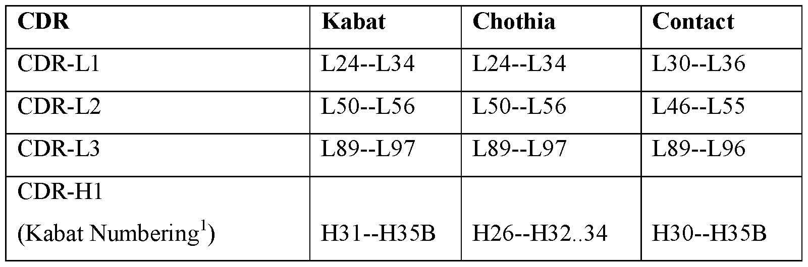
Claims
Priority Applications (8)
| Application Number | Priority Date | Filing Date | Title |
|---|---|---|---|
| CA2999885A CA2999885A1 (en) | 2015-09-25 | 2016-09-23 | High throughput process for t-cell receptor target identification of natively-paired t-cell receptor sequences |
| AU2016326734A AU2016326734B2 (en) | 2015-09-25 | 2016-09-23 | High throughput process for T cell receptor target identification of natively-paired T cell receptor sequences |
| JP2018515542A JP7341661B2 (en) | 2015-09-25 | 2016-09-23 | High-throughput process for T cell receptor target identification of natively paired T cell receptor sequences |
| MX2018003534A MX2018003534A (en) | 2015-09-25 | 2016-09-23 | High throughput process for t cell receptor target identification of natively-paired t cell receptor sequences. |
| EP16778962.7A EP3353550A1 (en) | 2015-09-25 | 2016-09-23 | High throughput process for t cell receptor target identification of natively-paired t cell receptor sequences |
| US15/762,450 US10928392B2 (en) | 2015-09-25 | 2016-09-23 | High throughput process for T cell receptor target identification of natively-paired T cell receptor sequences |
| CN201680069115.3A CN108369230B (en) | 2015-09-25 | 2016-09-23 | High throughput method for T cell receptor targeted identification of naturally paired T cell receptor sequences |
| US17/170,379 US20210293812A1 (en) | 2015-09-25 | 2021-02-08 | High throughput process for t cell receptor target identification of natively-paired t cell receptor sequences |
Applications Claiming Priority (2)
| Application Number | Priority Date | Filing Date | Title |
|---|---|---|---|
| US201562232511P | 2015-09-25 | 2015-09-25 | |
| US62/232,511 | 2015-09-25 |
Related Child Applications (2)
| Application Number | Title | Priority Date | Filing Date |
|---|---|---|---|
| US15/762,450 A-371-Of-International US10928392B2 (en) | 2015-09-25 | 2016-09-23 | High throughput process for T cell receptor target identification of natively-paired T cell receptor sequences |
| US17/170,379 Division US20210293812A1 (en) | 2015-09-25 | 2021-02-08 | High throughput process for t cell receptor target identification of natively-paired t cell receptor sequences |
Publications (1)
| Publication Number | Publication Date |
|---|---|
| WO2017053902A1 true WO2017053902A1 (en) | 2017-03-30 |
Family
ID=57121534
Family Applications (1)
| Application Number | Title | Priority Date | Filing Date |
|---|---|---|---|
| PCT/US2016/053595 WO2017053902A1 (en) | 2015-09-25 | 2016-09-23 | High throughput process for t cell receptor target identification of natively-paired t cell receptor sequences |
Country Status (8)
| Country | Link |
|---|---|
| US (2) | US10928392B2 (en) |
| EP (1) | EP3353550A1 (en) |
| JP (2) | JP7341661B2 (en) |
| CN (2) | CN108369230B (en) |
| AU (1) | AU2016326734B2 (en) |
| CA (1) | CA2999885A1 (en) |
| MX (1) | MX2018003534A (en) |
| WO (1) | WO2017053902A1 (en) |
Cited By (54)
| Publication number | Priority date | Publication date | Assignee | Title |
|---|---|---|---|---|
| US10011872B1 (en) | 2016-12-22 | 2018-07-03 | 10X Genomics, Inc. | Methods and systems for processing polynucleotides |
| WO2018218222A1 (en) | 2017-05-26 | 2018-11-29 | Goldfless Stephen Jacob | High-throughput polynucleotide library sequencing and transcriptome analysis |
| WO2019032929A1 (en) | 2017-08-09 | 2019-02-14 | Juno Therapeutics, Inc. | Methods and compositions for preparing genetically engineered cells |
| WO2019051335A1 (en) | 2017-09-07 | 2019-03-14 | Juno Therapeutics, Inc. | Methods of identifying cellular attributes related to outcomes associated with cell therapy |
| US10428326B2 (en) | 2017-01-30 | 2019-10-01 | 10X Genomics, Inc. | Methods and systems for droplet-based single cell barcoding |
| WO2019189383A1 (en) * | 2018-03-28 | 2019-10-03 | 国立大学法人 東京大学 | Analysis method for tumor-infiltrating t-cell repertoire and determination method for effectiveness of cancer therapeutic treatment using said analysis method |
| WO2019209895A1 (en) * | 2018-04-24 | 2019-10-31 | Inscripta, Inc. | Automated instrumentation for production of t-cell receptor peptide libraries |
| US10480028B2 (en) | 2014-06-26 | 2019-11-19 | 10X Genomics, Inc. | Methods and systems for processing polynucleotides |
| US10501738B2 (en) | 2018-04-24 | 2019-12-10 | Inscripta, Inc. | Automated instrumentation for production of peptide libraries |
| US10532324B1 (en) | 2018-08-14 | 2020-01-14 | Inscripta, Inc. | Instruments, modules, and methods for improved detection of edited sequences in live cells |
| US10550429B2 (en) | 2016-12-22 | 2020-02-04 | 10X Genomics, Inc. | Methods and systems for processing polynucleotides |
| WO2020033164A1 (en) * | 2018-08-08 | 2020-02-13 | Vanderbilt University | Systems and methods for simultaneous detection of antigens and antigen specific antibodies |
| WO2020036875A1 (en) * | 2018-08-13 | 2020-02-20 | Rootpath Genomics, Inc. | High throughput cloning of paired bipartite immunoreceptor polynucleotides and applications thereof |
| US10576474B2 (en) | 2018-04-13 | 2020-03-03 | Inscripta, Inc. | Automated cell processing instruments comprising reagent cartridges |
| US20200087713A1 (en) * | 2018-09-17 | 2020-03-19 | The Board Of Trustees Of The Leland Stanford Junior University | Nucleic acid quantification by temporal barcoding |
| US10604746B1 (en) | 2018-10-22 | 2020-03-31 | Inscripta, Inc. | Engineered enzymes |
| US10633627B2 (en) | 2018-08-14 | 2020-04-28 | Inscripta, Inc. | Instruments, modules, and methods for improved detection of edited sequences in live cells |
| US10689669B1 (en) | 2020-01-11 | 2020-06-23 | Inscripta, Inc. | Automated multi-module cell processing methods, instruments, and systems |
| US10704033B1 (en) | 2019-12-13 | 2020-07-07 | Inscripta, Inc. | Nucleic acid-guided nucleases |
| US10815525B2 (en) | 2016-12-22 | 2020-10-27 | 10X Genomics, Inc. | Methods and systems for processing polynucleotides |
| US10816543B2 (en) | 2018-02-12 | 2020-10-27 | 10X Genomics, Inc. | Methods and systems for analysis of major histocompatability complex |
| US10815467B2 (en) | 2019-03-25 | 2020-10-27 | Inscripta, Inc. | Simultaneous multiplex genome editing in yeast |
| US10837021B1 (en) | 2019-06-06 | 2020-11-17 | Inscripta, Inc. | Curing for recursive nucleic acid-guided cell editing |
| WO2020233503A1 (en) * | 2019-05-17 | 2020-11-26 | University Of Macau | Detection method |
| US10883095B1 (en) | 2019-12-10 | 2021-01-05 | Inscripta, Inc. | Mad nucleases |
| US10920189B2 (en) | 2019-06-21 | 2021-02-16 | Inscripta, Inc. | Genome-wide rationally-designed mutations leading to enhanced lysine production in E. coli |
| US10927385B2 (en) | 2019-06-25 | 2021-02-23 | Inscripta, Inc. | Increased nucleic-acid guided cell editing in yeast |
| US10995424B2 (en) | 2018-04-24 | 2021-05-04 | Inscripta, Inc. | Nucleic acid-guided editing of exogenous polynucleotides in heterologous cells |
| US11001831B2 (en) | 2019-03-25 | 2021-05-11 | Inscripta, Inc. | Simultaneous multiplex genome editing in yeast |
| US11008557B1 (en) | 2019-12-18 | 2021-05-18 | Inscripta, Inc. | Cascade/dCas3 complementation assays for in vivo detection of nucleic acid-guided nuclease edited cells |
| JP2021516049A (en) * | 2018-03-01 | 2021-07-01 | ザ ユニバーシティ オブ カンザス | Techniques for Producing Cell-Based Therapeutics Using Recombinant T Cell Receptor Genes |
| US11084036B2 (en) | 2016-05-13 | 2021-08-10 | 10X Genomics, Inc. | Microfluidic systems and methods of use |
| US11130970B2 (en) | 2017-06-23 | 2021-09-28 | Inscripta, Inc. | Nucleic acid-guided nucleases |
| US11135584B2 (en) | 2014-11-05 | 2021-10-05 | 10X Genomics, Inc. | Instrument systems for integrated sample processing |
| WO2021231657A1 (en) | 2020-05-13 | 2021-11-18 | Juno Therapeutics, Inc. | Methods of identifying features associated with clinical response and uses thereof |
| US11203762B2 (en) | 2019-11-19 | 2021-12-21 | Inscripta, Inc. | Methods for increasing observed editing in bacteria |
| US11214781B2 (en) | 2018-10-22 | 2022-01-04 | Inscripta, Inc. | Engineered enzyme |
| US11268061B2 (en) | 2018-08-14 | 2022-03-08 | Inscripta, Inc. | Detection of nuclease edited sequences in automated modules and instruments |
| US11268088B2 (en) | 2020-04-24 | 2022-03-08 | Inscripta, Inc. | Compositions, methods, modules and instruments for automated nucleic acid-guided nuclease editing in mammalian cells via viral delivery |
| US11293021B1 (en) | 2016-06-23 | 2022-04-05 | Inscripta, Inc. | Automated cell processing methods, modules, instruments, and systems |
| US11299731B1 (en) | 2020-09-15 | 2022-04-12 | Inscripta, Inc. | CRISPR editing to embed nucleic acid landing pads into genomes of live cells |
| US11306298B1 (en) | 2021-01-04 | 2022-04-19 | Inscripta, Inc. | Mad nucleases |
| US11332742B1 (en) | 2021-01-07 | 2022-05-17 | Inscripta, Inc. | Mad nucleases |
| US11408012B2 (en) | 2017-06-23 | 2022-08-09 | Inscripta, Inc. | Nucleic acid-guided nucleases |
| US11512297B2 (en) | 2020-11-09 | 2022-11-29 | Inscripta, Inc. | Affinity tag for recombination protein recruitment |
| US11597921B2 (en) | 2017-06-30 | 2023-03-07 | Inscripta, Inc. | Automated cell processing methods, modules, instruments, and systems |
| WO2023086787A1 (en) * | 2021-11-09 | 2023-05-19 | Janssen Biotech, Inc. | Microfluidic co-encapsulation device and system and methods for identifying t-cell receptor ligands |
| US11667932B2 (en) | 2020-01-27 | 2023-06-06 | Inscripta, Inc. | Electroporation modules and instrumentation |
| US11746162B2 (en) | 2019-04-05 | 2023-09-05 | Rootpath Genomics, Inc. | Compositions and methods for T-cell receptor gene assembly |
| WO2023183344A1 (en) * | 2022-03-21 | 2023-09-28 | Alaunos Therapeutics, Inc. | Methods for identifying neoantigen-reactive t cell receptors |
| US11787841B2 (en) | 2020-05-19 | 2023-10-17 | Inscripta, Inc. | Rationally-designed mutations to the thrA gene for enhanced lysine production in E. coli |
| US11834653B2 (en) | 2017-06-08 | 2023-12-05 | The Brigham And Women's Hospital, Inc. | Methods and compositions for identifying epitopes |
| WO2023244480A1 (en) * | 2022-06-17 | 2023-12-21 | The Children's Hospital Of Philadelphia | T lymphocyte activity screening and sequencing |
| US11884924B2 (en) | 2021-02-16 | 2024-01-30 | Inscripta, Inc. | Dual strand nucleic acid-guided nickase editing |
Families Citing this family (16)
| Publication number | Priority date | Publication date | Assignee | Title |
|---|---|---|---|---|
| CN108289954B (en) | 2015-09-24 | 2022-05-31 | 阿布维特罗有限责任公司 | HIV antibody compositions and methods of use |
| AU2016341880B2 (en) * | 2015-10-19 | 2021-01-21 | Cedars-Sinai Medical Center | Microfluidic model of the blood brain barrier |
| US10809266B2 (en) * | 2016-07-22 | 2020-10-20 | Verily Life Sciences Llc | Quantitative massively parallel proteomics |
| US10739351B2 (en) * | 2016-10-10 | 2020-08-11 | California Institute Of Technology | Functional characterization of immune repertoires |
| US20200347410A1 (en) * | 2018-01-19 | 2020-11-05 | Carsgen Therapeutics Co., Ltd. | Synnotch receptor-regulated expression of il12 |
| US11414699B2 (en) * | 2018-05-15 | 2022-08-16 | Mantra Bio, Inc. | Barcode-free single vesicle multiplexed protein and RNA analysis |
| CN109576292B (en) * | 2018-12-21 | 2022-11-29 | 深圳市爱思迪生物科技有限公司 | Construction method and application of antibody library |
| CN110222745B (en) * | 2019-05-24 | 2021-04-30 | 中南大学 | Similarity learning based and enhanced cell type identification method |
| WO2021046299A1 (en) * | 2019-09-04 | 2021-03-11 | Vanderbilt University | Methods for identification of antigen binding specificity of antibodies |
| WO2021101956A1 (en) * | 2019-11-19 | 2021-05-27 | Rootpath Genomics, Inc. | Compositions and methods for t-cell receptor identification |
| EP4159873A1 (en) | 2020-05-29 | 2023-04-05 | Riken | Method for treating cell population and method for analyzing genes included in cell population |
| CN115997125A (en) * | 2020-06-24 | 2023-04-21 | 高诚生物医药(香港)有限公司 | Method for identifying cognate pairs of ligand and receptor |
| WO2022150354A1 (en) * | 2021-01-06 | 2022-07-14 | Think Therapeutics, Inc. | Multiplexed testing of lymphocytes for antigen specificity |
| US20220341850A1 (en) * | 2021-04-23 | 2022-10-27 | Poppy Health, Inc. | System and method for characterizing, monitoring, & detecting bioaerosol presence & movement in an indoor environment |
| US11597980B2 (en) | 2021-07-08 | 2023-03-07 | Poppy Health, Inc. | System and method for detecting pathogens in an environment via an electrostatic air sampler |
| WO2023215358A1 (en) * | 2022-05-03 | 2023-11-09 | 3T Biosciences, Inc. | T-cell target discovery |
Citations (92)
| Publication number | Priority date | Publication date | Assignee | Title |
|---|---|---|---|---|
| US4690915A (en) | 1985-08-08 | 1987-09-01 | The United States Of America As Represented By The Department Of Health And Human Services | Adoptive immunotherapy as a treatment modality in humans |
| EP0404097A2 (en) | 1989-06-22 | 1990-12-27 | BEHRINGWERKE Aktiengesellschaft | Bispecific and oligospecific, mono- and oligovalent receptors, production and applications thereof |
| US4988617A (en) | 1988-03-25 | 1991-01-29 | California Institute Of Technology | Method of detecting a nucleotide change in nucleic acids |
| US5143854A (en) | 1989-06-07 | 1992-09-01 | Affymax Technologies N.V. | Large scale photolithographic solid phase synthesis of polypeptides and receptor binding screening thereof |
| US5168038A (en) | 1988-06-17 | 1992-12-01 | The Board Of Trustees Of The Leland Stanford Junior University | In situ transcription in cells and tissues |
| US5210015A (en) | 1990-08-06 | 1993-05-11 | Hoffman-La Roche Inc. | Homogeneous assay system using the nuclease activity of a nucleic acid polymerase |
| WO1993011161A1 (en) | 1991-11-25 | 1993-06-10 | Enzon, Inc. | Multivalent antigen-binding proteins |
| WO1993012227A1 (en) | 1991-12-17 | 1993-06-24 | Genpharm International, Inc. | Transgenic non-human animals capable of producing heterologous antibodies |
| US5242974A (en) | 1991-11-22 | 1993-09-07 | Affymax Technologies N.V. | Polymer reversal on solid surfaces |
| US5242794A (en) | 1984-12-13 | 1993-09-07 | Applied Biosystems, Inc. | Detection of specific sequences in nucleic acids |
| US5252743A (en) | 1989-11-13 | 1993-10-12 | Affymax Technologies N.V. | Spatially-addressable immobilization of anti-ligands on surfaces |
| US5324633A (en) | 1991-11-22 | 1994-06-28 | Affymax Technologies N.V. | Method and apparatus for measuring binding affinity |
| US5384261A (en) | 1991-11-22 | 1995-01-24 | Affymax Technologies N.V. | Very large scale immobilized polymer synthesis using mechanically directed flow paths |
| US5424186A (en) | 1989-06-07 | 1995-06-13 | Affymax Technologies N.V. | Very large scale immobilized polymer synthesis |
| US5491074A (en) | 1993-04-01 | 1996-02-13 | Affymax Technologies Nv | Association peptides |
| US5494810A (en) | 1990-05-03 | 1996-02-27 | Cornell Research Foundation, Inc. | Thermostable ligase-mediated DNA amplifications system for the detection of genetic disease |
| US5527681A (en) | 1989-06-07 | 1996-06-18 | Affymax Technologies N.V. | Immobilized molecular synthesis of systematically substituted compounds |
| US5550215A (en) | 1991-11-22 | 1996-08-27 | Holmes; Christopher P. | Polymer reversal on solid surfaces |
| US5571639A (en) | 1994-05-24 | 1996-11-05 | Affymax Technologies N.V. | Computer-aided engineering system for design of sequence arrays and lithographic masks |
| US5578832A (en) | 1994-09-02 | 1996-11-26 | Affymetrix, Inc. | Method and apparatus for imaging a sample on a device |
| US5599695A (en) | 1995-02-27 | 1997-02-04 | Affymetrix, Inc. | Printing molecular library arrays using deprotection agents solely in the vapor phase |
| US5624711A (en) | 1995-04-27 | 1997-04-29 | Affymax Technologies, N.V. | Derivatization of solid supports and methods for oligomer synthesis |
| US5631734A (en) | 1994-02-10 | 1997-05-20 | Affymetrix, Inc. | Method and apparatus for detection of fluorescently labeled materials |
| US5744305A (en) | 1989-06-07 | 1998-04-28 | Affymetrix, Inc. | Arrays of materials attached to a substrate |
| US5795716A (en) | 1994-10-21 | 1998-08-18 | Chee; Mark S. | Computer-aided visualization and analysis system for sequence evaluation |
| US5837832A (en) | 1993-06-25 | 1998-11-17 | Affymetrix, Inc. | Arrays of nucleic acid probes on biological chips |
| US5858659A (en) | 1995-11-29 | 1999-01-12 | Affymetrix, Inc. | Polymorphism detection |
| US5925517A (en) | 1993-11-12 | 1999-07-20 | The Public Health Research Institute Of The City Of New York, Inc. | Detectably labeled dual conformation oligonucleotide probes, assays and kits |
| WO1999036760A1 (en) | 1998-01-13 | 1999-07-22 | Genetic Microsystems, Inc. | Depositing fluid specimens on substrates, resulting ordered arrays, techniques for analysis of deposited arrays |
| US5936324A (en) | 1998-03-30 | 1999-08-10 | Genetic Microsystems Inc. | Moving magnet scanner |
| US5968740A (en) | 1995-07-24 | 1999-10-19 | Affymetrix, Inc. | Method of Identifying a Base in a Nucleic Acid |
| US5981956A (en) | 1996-05-16 | 1999-11-09 | Affymetrix, Inc. | Systems and methods for detection of labeled materials |
| US5981185A (en) | 1994-05-05 | 1999-11-09 | Beckman Coulter, Inc. | Oligonucleotide repeat arrays |
| US6033860A (en) | 1997-10-31 | 2000-03-07 | Affymetrix, Inc. | Expression profiles in adult and fetal organs |
| WO2000014257A1 (en) | 1998-09-04 | 2000-03-16 | Sloan-Kettering Institute For Cancer Research | Fusion receptors specific for prostate-specific membrane antigen and uses thereof |
| US6040193A (en) | 1991-11-22 | 2000-03-21 | Affymetrix, Inc. | Combinatorial strategies for polymer synthesis |
| US6090555A (en) | 1997-12-11 | 2000-07-18 | Affymetrix, Inc. | Scanned image alignment systems and methods |
| WO2000058516A2 (en) | 1999-03-26 | 2000-10-05 | Whitehead Institute For Biomedical Research | Universal arrays |
| US6174670B1 (en) | 1996-06-04 | 2001-01-16 | University Of Utah Research Foundation | Monitoring amplification of DNA during PCR |
| WO2001058593A1 (en) | 2000-02-09 | 2001-08-16 | Affymetrix, Inc. | Cleaning deposit devices that generate microarrays |
| US6306597B1 (en) | 1995-04-17 | 2001-10-23 | Lynx Therapeutics, Inc. | DNA sequencing by parallel oligonucleotide extensions |
| WO2001089788A2 (en) | 2000-05-25 | 2001-11-29 | President And Fellows Of Harvard College | Patterning of surfaces utilizing microfluidic stamps including three-dimensionally arrayed channel networks |
| US6410319B1 (en) | 1998-10-20 | 2002-06-25 | City Of Hope | CD20-specific redirected T cells and their use in cellular immunotherapy of CD20+ malignancies |
| US6451995B1 (en) | 1996-03-20 | 2002-09-17 | Sloan-Kettering Institute For Cancer Research | Single chain FV polynucleotide or peptide constructs of anti-ganglioside GD2 antibodies, cells expressing same and related methods |
| US20020131960A1 (en) | 2000-06-02 | 2002-09-19 | Michel Sadelain | Artificial antigen presenting cells and methods of use thereof |
| WO2003020763A2 (en) | 2001-08-31 | 2003-03-13 | Avidex Limited | Soluble t cell receptor |
| US6582938B1 (en) | 2001-05-11 | 2003-06-24 | Affymetrix, Inc. | Amplification of nucleic acids |
| US20030170238A1 (en) | 2002-03-07 | 2003-09-11 | Gruenberg Micheal L. | Re-activated T-cells for adoptive immunotherapy |
| WO2004002627A2 (en) | 2002-06-28 | 2004-01-08 | President And Fellows Of Harvard College | Method and apparatus for fluid dispersion |
| WO2004033685A1 (en) | 2002-10-09 | 2004-04-22 | Avidex Ltd | Single chain recombinant t cell receptors |
| US6797470B2 (en) | 1996-05-29 | 2004-09-28 | Cornell Research Foundation, Inc. | Detection of nucleic acid sequence differences using coupled ligase detection and polymerase chain reactions |
| WO2004091763A2 (en) | 2003-04-10 | 2004-10-28 | President And Fellows Of Harvard College | Formation and control of fluidic species |
| WO2005021151A1 (en) | 2003-08-27 | 2005-03-10 | President And Fellows Of Harvard College | Electronic control of fluidic species |
| WO2006040551A2 (en) | 2004-10-12 | 2006-04-20 | Medical Research Council | Compartmentalised screening by microfluidic control |
| WO2006040554A1 (en) | 2004-10-12 | 2006-04-20 | Medical Research Council | Compartmentalised combinatorial chemistry by microfluidic control |
| US7037687B2 (en) | 1998-05-01 | 2006-05-02 | Arizona Board Of Regents | Method of determining the nucleotide sequence of oligonucleotides and DNA molecules |
| US7070995B2 (en) | 2001-04-11 | 2006-07-04 | City Of Hope | CE7-specific redirected immune cells |
| US7083917B2 (en) | 1996-02-09 | 2006-08-01 | Cornell Research Foundation, Inc. | Detection of nucleic acid sequence differences using the ligase detection reaction with addressable arrays |
| WO2006096571A2 (en) | 2005-03-04 | 2006-09-14 | President And Fellows Of Harvard College | Method and apparatus for forming multiple emulsions |
| US7169560B2 (en) | 2003-11-12 | 2007-01-30 | Helicos Biosciences Corporation | Short cycle methods for sequencing polynucleotides |
| US7232656B2 (en) | 1998-07-30 | 2007-06-19 | Solexa Ltd. | Arrayed biomolecules and their use in sequencing |
| WO2007081385A2 (en) | 2006-01-11 | 2007-07-19 | Raindance Technologies, Inc. | Microfluidic devices and methods of use in the formation and control of nanoreactors |
| WO2007089541A2 (en) | 2006-01-27 | 2007-08-09 | President And Fellows Of Harvard College | Fluidic droplet coalescence |
| US20080038559A1 (en) | 2005-09-13 | 2008-02-14 | Randall True | Miniaturized microparticles |
| WO2008063227A2 (en) | 2006-05-11 | 2008-05-29 | Raindance Technologies, Inc. | Microfluidic devices |
| US7446179B2 (en) | 2000-11-07 | 2008-11-04 | City Of Hope | CD19-specific chimeric T cell receptor |
| US7446190B2 (en) | 2002-05-28 | 2008-11-04 | Sloan-Kettering Institute For Cancer Research | Nucleic acids encoding chimeric T cell receptors |
| US20090149340A1 (en) | 2007-06-25 | 2009-06-11 | True Materials, Inc. | Encoded microparticles |
| US7598035B2 (en) | 1998-02-23 | 2009-10-06 | Solexa, Inc. | Method and compositions for ordering restriction fragments |
| US7622280B2 (en) | 2001-11-16 | 2009-11-24 | 454 Life Sciences Corporation | Emulsion compositions |
| US20090325169A1 (en) | 2008-04-30 | 2009-12-31 | Integrated Dna Technologies, Inc. | Rnase h-based assays utilizing modified rna monomers |
| US7645596B2 (en) | 1998-05-01 | 2010-01-12 | Arizona Board Of Regents | Method of determining the nucleotide sequence of oligonucleotides and DNA molecules |
| WO2010036352A1 (en) | 2008-09-23 | 2010-04-01 | Quantalife, Inc | Droplet-based assay system |
| US20100167353A1 (en) | 2008-04-30 | 2010-07-01 | Integrated Dna Technologies, Inc. | Rnase h-based assays utilizing modified rna monomers |
| US7769400B2 (en) | 2004-11-22 | 2010-08-03 | Seven Networks International Oy | Connectivity function for forwarding e-mail |
| WO2011044186A1 (en) | 2009-10-06 | 2011-04-14 | The Board Of Trustees Of The University Of Illinois | Human single-chain t cell receptors |
| WO2012129514A1 (en) | 2011-03-23 | 2012-09-27 | Fred Hutchinson Cancer Research Center | Method and compositions for cellular immunotherapy |
| US8324353B2 (en) | 2001-04-30 | 2012-12-04 | City Of Hope | Chimeric immunoreceptor useful in treating human gliomas |
| US8339645B2 (en) | 2008-05-27 | 2012-12-25 | Canon Kabushiki Kaisha | Managing apparatus, image processing apparatus, and processing method for the same, wherein a first user stores a temporary object having attribute information specified but not partial-area data, at a later time an object is received from a second user that includes both partial-area data and attribute information, the storage unit is searched for the temporary object that matches attribute information of the received object, and the first user is notified in response to a match |
| EP2537416A1 (en) | 2007-03-30 | 2012-12-26 | Memorial Sloan-Kettering Cancer Center | Constitutive expression of costimulatory ligands on adoptively transferred T lymphocytes |
| US8398282B2 (en) | 2011-05-12 | 2013-03-19 | Delphi Technologies, Inc. | Vehicle front lighting assembly and systems having a variable tint electrowetting element |
| WO2013071154A1 (en) | 2011-11-11 | 2013-05-16 | Fred Hutchinson Cancer Research Center | Cyclin a1-targeted t-cell immunotherapy for cancer |
| US20130149337A1 (en) | 2003-03-11 | 2013-06-13 | City Of Hope | Method of controlling administration of cancer antigen |
| US8479118B2 (en) | 2007-12-10 | 2013-07-02 | Microsoft Corporation | Switching search providers within a browser search box |
| WO2013123061A1 (en) | 2012-02-13 | 2013-08-22 | Seattle Children's Hospital D/B/A Seattle Children's Research Institute | Bispecific chimeric antigen receptors and therapeutic uses thereof |
| WO2013126726A1 (en) | 2012-02-22 | 2013-08-29 | The Trustees Of The University Of Pennsylvania | Double transgenic t cells comprising a car and a tcr and their methods of use |
| US20130287748A1 (en) | 2010-12-09 | 2013-10-31 | The Trustees Of The University Of Pennsylvania | Use of Chimeric Antigen Receptor-Modified T-Cells to Treat Cancer |
| WO2013166321A1 (en) | 2012-05-03 | 2013-11-07 | Fred Hutchinson Cancer Research Center | Enhanced affinity t cell receptors and methods for making the same |
| WO2014031687A1 (en) | 2012-08-20 | 2014-02-27 | Jensen, Michael | Method and compositions for cellular immunotherapy |
| WO2015014869A1 (en) * | 2013-07-30 | 2015-02-05 | Biontech Ag | Tumor antigens for determining cancer therapy |
| WO2015143558A1 (en) | 2014-03-27 | 2015-10-01 | British Columbia Cancer Agency Branch | T-cell epitope identification |
| WO2016100977A1 (en) * | 2014-12-19 | 2016-06-23 | The Broad Institute Inc. | Methods for profiling the t-cel- receptor repertoire |
Family Cites Families (6)
| Publication number | Priority date | Publication date | Assignee | Title |
|---|---|---|---|---|
| CA2454937A1 (en) | 2001-07-25 | 2003-02-13 | Lorantis Limited | Modulators of notch signalling for use in immunotherapy |
| JP6192537B2 (en) * | 2010-08-06 | 2017-09-06 | ルードヴィヒ−マクシミリアン−ウニヴェルズィテート ミュンヘン | Identification of T cell target antigen |
| CN117756916A (en) | 2010-09-20 | 2024-03-26 | 生物技术细胞和基因治疗公司 | Antigen-specific T cell receptor and T cell epitope |
| ES2714523T3 (en) | 2012-09-04 | 2019-05-28 | Cellectis | Chimeric receptor of multi-chain antigens and uses thereof |
| ES2711498T3 (en) * | 2014-02-14 | 2019-05-06 | Cellectis | Immunotherapy cells designed by genetic engineering to direct an antigen present in immune cells and pathological cells |
| WO2016176322A1 (en) * | 2015-04-27 | 2016-11-03 | Abvitro Llc | Methods of sequencing, determining, pairing, and validating therapeutic agents and disease specific antigens |
-
2016
- 2016-09-23 EP EP16778962.7A patent/EP3353550A1/en active Pending
- 2016-09-23 AU AU2016326734A patent/AU2016326734B2/en active Active
- 2016-09-23 CN CN201680069115.3A patent/CN108369230B/en active Active
- 2016-09-23 MX MX2018003534A patent/MX2018003534A/en unknown
- 2016-09-23 JP JP2018515542A patent/JP7341661B2/en active Active
- 2016-09-23 WO PCT/US2016/053595 patent/WO2017053902A1/en active Application Filing
- 2016-09-23 CN CN202111003652.7A patent/CN113774495A/en active Pending
- 2016-09-23 CA CA2999885A patent/CA2999885A1/en active Pending
- 2016-09-23 US US15/762,450 patent/US10928392B2/en active Active
-
2020
- 2020-10-14 JP JP2020173375A patent/JP2021007406A/en active Pending
-
2021
- 2021-02-08 US US17/170,379 patent/US20210293812A1/en not_active Abandoned
Patent Citations (114)
| Publication number | Priority date | Publication date | Assignee | Title |
|---|---|---|---|---|
| US5242794A (en) | 1984-12-13 | 1993-09-07 | Applied Biosystems, Inc. | Detection of specific sequences in nucleic acids |
| US4690915A (en) | 1985-08-08 | 1987-09-01 | The United States Of America As Represented By The Department Of Health And Human Services | Adoptive immunotherapy as a treatment modality in humans |
| US4988617A (en) | 1988-03-25 | 1991-01-29 | California Institute Of Technology | Method of detecting a nucleotide change in nucleic acids |
| US5168038A (en) | 1988-06-17 | 1992-12-01 | The Board Of Trustees Of The Leland Stanford Junior University | In situ transcription in cells and tissues |
| US5405783A (en) | 1989-06-07 | 1995-04-11 | Affymax Technologies N.V. | Large scale photolithographic solid phase synthesis of an array of polymers |
| US5143854A (en) | 1989-06-07 | 1992-09-01 | Affymax Technologies N.V. | Large scale photolithographic solid phase synthesis of polypeptides and receptor binding screening thereof |
| US5424186A (en) | 1989-06-07 | 1995-06-13 | Affymax Technologies N.V. | Very large scale immobilized polymer synthesis |
| US5527681A (en) | 1989-06-07 | 1996-06-18 | Affymax Technologies N.V. | Immobilized molecular synthesis of systematically substituted compounds |
| US5744305A (en) | 1989-06-07 | 1998-04-28 | Affymetrix, Inc. | Arrays of materials attached to a substrate |
| EP0404097A2 (en) | 1989-06-22 | 1990-12-27 | BEHRINGWERKE Aktiengesellschaft | Bispecific and oligospecific, mono- and oligovalent receptors, production and applications thereof |
| US5451683A (en) | 1989-11-13 | 1995-09-19 | Affymax Technologies N.V. | Spatially-addressable immobilization of anti-ligands on surfaces |
| US5252743A (en) | 1989-11-13 | 1993-10-12 | Affymax Technologies N.V. | Spatially-addressable immobilization of anti-ligands on surfaces |
| US5482867A (en) | 1989-11-13 | 1996-01-09 | Affymax Technologies N.V. | Spatially-addressable immobilization of anti-ligands on surfaces |
| US5494810A (en) | 1990-05-03 | 1996-02-27 | Cornell Research Foundation, Inc. | Thermostable ligase-mediated DNA amplifications system for the detection of genetic disease |
| US5210015A (en) | 1990-08-06 | 1993-05-11 | Hoffman-La Roche Inc. | Homogeneous assay system using the nuclease activity of a nucleic acid polymerase |
| US5384261A (en) | 1991-11-22 | 1995-01-24 | Affymax Technologies N.V. | Very large scale immobilized polymer synthesis using mechanically directed flow paths |
| US5324633A (en) | 1991-11-22 | 1994-06-28 | Affymax Technologies N.V. | Method and apparatus for measuring binding affinity |
| US5242974A (en) | 1991-11-22 | 1993-09-07 | Affymax Technologies N.V. | Polymer reversal on solid surfaces |
| US5550215A (en) | 1991-11-22 | 1996-08-27 | Holmes; Christopher P. | Polymer reversal on solid surfaces |
| US6136269A (en) | 1991-11-22 | 2000-10-24 | Affymetrix, Inc. | Combinatorial kit for polymer synthesis |
| US6040193A (en) | 1991-11-22 | 2000-03-21 | Affymetrix, Inc. | Combinatorial strategies for polymer synthesis |
| WO1993011161A1 (en) | 1991-11-25 | 1993-06-10 | Enzon, Inc. | Multivalent antigen-binding proteins |
| WO1993012227A1 (en) | 1991-12-17 | 1993-06-24 | Genpharm International, Inc. | Transgenic non-human animals capable of producing heterologous antibodies |
| US5491074A (en) | 1993-04-01 | 1996-02-13 | Affymax Technologies Nv | Association peptides |
| US5837832A (en) | 1993-06-25 | 1998-11-17 | Affymetrix, Inc. | Arrays of nucleic acid probes on biological chips |
| US5925517A (en) | 1993-11-12 | 1999-07-20 | The Public Health Research Institute Of The City Of New York, Inc. | Detectably labeled dual conformation oligonucleotide probes, assays and kits |
| US5631734A (en) | 1994-02-10 | 1997-05-20 | Affymetrix, Inc. | Method and apparatus for detection of fluorescently labeled materials |
| US5981185A (en) | 1994-05-05 | 1999-11-09 | Beckman Coulter, Inc. | Oligonucleotide repeat arrays |
| US5571639A (en) | 1994-05-24 | 1996-11-05 | Affymax Technologies N.V. | Computer-aided engineering system for design of sequence arrays and lithographic masks |
| US5856101A (en) | 1994-05-24 | 1999-01-05 | Affymetrix, Inc. | Computer-aided engineering system for design of sequence arrays and lithographic masks |
| US5593839A (en) | 1994-05-24 | 1997-01-14 | Affymetrix, Inc. | Computer-aided engineering system for design of sequence arrays and lithographic masks |
| US5578832A (en) | 1994-09-02 | 1996-11-26 | Affymetrix, Inc. | Method and apparatus for imaging a sample on a device |
| US6025601A (en) | 1994-09-02 | 2000-02-15 | Affymetrix, Inc. | Method and apparatus for imaging a sample on a device |
| US5974164A (en) | 1994-10-21 | 1999-10-26 | Affymetrix, Inc. | Computer-aided visualization and analysis system for sequence evaluation |
| US5795716A (en) | 1994-10-21 | 1998-08-18 | Chee; Mark S. | Computer-aided visualization and analysis system for sequence evaluation |
| US5599695A (en) | 1995-02-27 | 1997-02-04 | Affymetrix, Inc. | Printing molecular library arrays using deprotection agents solely in the vapor phase |
| US5831070A (en) | 1995-02-27 | 1998-11-03 | Affymetrix, Inc. | Printing oligonucleotide arrays using deprotection agents solely in the vapor phase |
| US6306597B1 (en) | 1995-04-17 | 2001-10-23 | Lynx Therapeutics, Inc. | DNA sequencing by parallel oligonucleotide extensions |
| US5624711A (en) | 1995-04-27 | 1997-04-29 | Affymax Technologies, N.V. | Derivatization of solid supports and methods for oligomer synthesis |
| US5968740A (en) | 1995-07-24 | 1999-10-19 | Affymetrix, Inc. | Method of Identifying a Base in a Nucleic Acid |
| US5858659A (en) | 1995-11-29 | 1999-01-12 | Affymetrix, Inc. | Polymorphism detection |
| US7083917B2 (en) | 1996-02-09 | 2006-08-01 | Cornell Research Foundation, Inc. | Detection of nucleic acid sequence differences using the ligase detection reaction with addressable arrays |
| US6451995B1 (en) | 1996-03-20 | 2002-09-17 | Sloan-Kettering Institute For Cancer Research | Single chain FV polynucleotide or peptide constructs of anti-ganglioside GD2 antibodies, cells expressing same and related methods |
| US5981956A (en) | 1996-05-16 | 1999-11-09 | Affymetrix, Inc. | Systems and methods for detection of labeled materials |
| US7332285B2 (en) | 1996-05-29 | 2008-02-19 | Cornell Research Foundation, Inc. | Detection of nucleic acid sequence differences using coupled ligase detection and polymerase chain reactions |
| US6797470B2 (en) | 1996-05-29 | 2004-09-28 | Cornell Research Foundation, Inc. | Detection of nucleic acid sequence differences using coupled ligase detection and polymerase chain reactions |
| US7320865B2 (en) | 1996-05-29 | 2008-01-22 | Cornell Research Foundation, Inc. | Detection of nucleic acid sequence differences using coupled ligase detection and polymerase chain reactions |
| US7364858B2 (en) | 1996-05-29 | 2008-04-29 | Cornell Research Foundation, Inc. | Detection of nucleic acid sequence differences using coupled ligase detection and polymerase chain reactions |
| US7429453B2 (en) | 1996-05-29 | 2008-09-30 | Cornell Research Foundation, Inc. | Detection of nucleic acid sequence differences using coupled ligase detection and polymerase chain reactions |
| US7166434B2 (en) | 1996-05-29 | 2007-01-23 | Cornell Research Foundation, Inc. | Detection of nucleic acid sequence differences using coupled ligase detection and polymerase chain reactions |
| US6174670B1 (en) | 1996-06-04 | 2001-01-16 | University Of Utah Research Foundation | Monitoring amplification of DNA during PCR |
| US6569627B2 (en) | 1996-06-04 | 2003-05-27 | University Of Utah Research Foundation | Monitoring hybridization during PCR using SYBR™ Green I |
| US6033860A (en) | 1997-10-31 | 2000-03-07 | Affymetrix, Inc. | Expression profiles in adult and fetal organs |
| US6090555A (en) | 1997-12-11 | 2000-07-18 | Affymetrix, Inc. | Scanned image alignment systems and methods |
| WO1999036760A1 (en) | 1998-01-13 | 1999-07-22 | Genetic Microsystems, Inc. | Depositing fluid specimens on substrates, resulting ordered arrays, techniques for analysis of deposited arrays |
| US6269846B1 (en) | 1998-01-13 | 2001-08-07 | Genetic Microsystems, Inc. | Depositing fluid specimens on substrates, resulting ordered arrays, techniques for deposition of arrays |
| US7598035B2 (en) | 1998-02-23 | 2009-10-06 | Solexa, Inc. | Method and compositions for ordering restriction fragments |
| US5936324A (en) | 1998-03-30 | 1999-08-10 | Genetic Microsystems Inc. | Moving magnet scanner |
| US7645596B2 (en) | 1998-05-01 | 2010-01-12 | Arizona Board Of Regents | Method of determining the nucleotide sequence of oligonucleotides and DNA molecules |
| US7037687B2 (en) | 1998-05-01 | 2006-05-02 | Arizona Board Of Regents | Method of determining the nucleotide sequence of oligonucleotides and DNA molecules |
| US6428752B1 (en) | 1998-05-14 | 2002-08-06 | Affymetrix, Inc. | Cleaning deposit devices that form microarrays and the like |
| US7232656B2 (en) | 1998-07-30 | 2007-06-19 | Solexa Ltd. | Arrayed biomolecules and their use in sequencing |
| WO2000014257A1 (en) | 1998-09-04 | 2000-03-16 | Sloan-Kettering Institute For Cancer Research | Fusion receptors specific for prostate-specific membrane antigen and uses thereof |
| US6410319B1 (en) | 1998-10-20 | 2002-06-25 | City Of Hope | CD20-specific redirected T cells and their use in cellular immunotherapy of CD20+ malignancies |
| WO2000058516A2 (en) | 1999-03-26 | 2000-10-05 | Whitehead Institute For Biomedical Research | Universal arrays |
| US20050074787A1 (en) | 1999-03-26 | 2005-04-07 | Whitehead Institute For Biomedical Research | Universal arrays |
| WO2001058593A1 (en) | 2000-02-09 | 2001-08-16 | Affymetrix, Inc. | Cleaning deposit devices that generate microarrays |
| WO2001089788A2 (en) | 2000-05-25 | 2001-11-29 | President And Fellows Of Harvard College | Patterning of surfaces utilizing microfluidic stamps including three-dimensionally arrayed channel networks |
| US20020131960A1 (en) | 2000-06-02 | 2002-09-19 | Michel Sadelain | Artificial antigen presenting cells and methods of use thereof |
| US7446179B2 (en) | 2000-11-07 | 2008-11-04 | City Of Hope | CD19-specific chimeric T cell receptor |
| US7070995B2 (en) | 2001-04-11 | 2006-07-04 | City Of Hope | CE7-specific redirected immune cells |
| US7446191B2 (en) | 2001-04-11 | 2008-11-04 | City Of Hope | DNA construct encoding CE7-specific chimeric T cell receptor |
| US7265209B2 (en) | 2001-04-11 | 2007-09-04 | City Of Hope | CE7-specific chimeric T cell receptor |
| US7354762B2 (en) | 2001-04-11 | 2008-04-08 | City Of Hope | Method for producing CE7-specific redirected immune cells |
| US8324353B2 (en) | 2001-04-30 | 2012-12-04 | City Of Hope | Chimeric immunoreceptor useful in treating human gliomas |
| US6582938B1 (en) | 2001-05-11 | 2003-06-24 | Affymetrix, Inc. | Amplification of nucleic acids |
| WO2003020763A2 (en) | 2001-08-31 | 2003-03-13 | Avidex Limited | Soluble t cell receptor |
| US7622280B2 (en) | 2001-11-16 | 2009-11-24 | 454 Life Sciences Corporation | Emulsion compositions |
| US20030170238A1 (en) | 2002-03-07 | 2003-09-11 | Gruenberg Micheal L. | Re-activated T-cells for adoptive immunotherapy |
| US7446190B2 (en) | 2002-05-28 | 2008-11-04 | Sloan-Kettering Institute For Cancer Research | Nucleic acids encoding chimeric T cell receptors |
| WO2004002627A2 (en) | 2002-06-28 | 2004-01-08 | President And Fellows Of Harvard College | Method and apparatus for fluid dispersion |
| WO2004033685A1 (en) | 2002-10-09 | 2004-04-22 | Avidex Ltd | Single chain recombinant t cell receptors |
| US20130149337A1 (en) | 2003-03-11 | 2013-06-13 | City Of Hope | Method of controlling administration of cancer antigen |
| WO2004091763A2 (en) | 2003-04-10 | 2004-10-28 | President And Fellows Of Harvard College | Formation and control of fluidic species |
| WO2005021151A1 (en) | 2003-08-27 | 2005-03-10 | President And Fellows Of Harvard College | Electronic control of fluidic species |
| US7169560B2 (en) | 2003-11-12 | 2007-01-30 | Helicos Biosciences Corporation | Short cycle methods for sequencing polynucleotides |
| WO2006040551A2 (en) | 2004-10-12 | 2006-04-20 | Medical Research Council | Compartmentalised screening by microfluidic control |
| WO2006040554A1 (en) | 2004-10-12 | 2006-04-20 | Medical Research Council | Compartmentalised combinatorial chemistry by microfluidic control |
| US7769400B2 (en) | 2004-11-22 | 2010-08-03 | Seven Networks International Oy | Connectivity function for forwarding e-mail |
| WO2006096571A2 (en) | 2005-03-04 | 2006-09-14 | President And Fellows Of Harvard College | Method and apparatus for forming multiple emulsions |
| US20080038559A1 (en) | 2005-09-13 | 2008-02-14 | Randall True | Miniaturized microparticles |
| WO2007081385A2 (en) | 2006-01-11 | 2007-07-19 | Raindance Technologies, Inc. | Microfluidic devices and methods of use in the formation and control of nanoreactors |
| WO2007089541A2 (en) | 2006-01-27 | 2007-08-09 | President And Fellows Of Harvard College | Fluidic droplet coalescence |
| WO2008063227A2 (en) | 2006-05-11 | 2008-05-29 | Raindance Technologies, Inc. | Microfluidic devices |
| US8389282B2 (en) | 2007-03-30 | 2013-03-05 | Memorial Sloan-Kettering Cancer Center | Constitutive expression of costimulatory ligands on adoptively transferred T lymphocytes |
| EP2537416A1 (en) | 2007-03-30 | 2012-12-26 | Memorial Sloan-Kettering Cancer Center | Constitutive expression of costimulatory ligands on adoptively transferred T lymphocytes |
| US20090149340A1 (en) | 2007-06-25 | 2009-06-11 | True Materials, Inc. | Encoded microparticles |
| US8479118B2 (en) | 2007-12-10 | 2013-07-02 | Microsoft Corporation | Switching search providers within a browser search box |
| US20090325169A1 (en) | 2008-04-30 | 2009-12-31 | Integrated Dna Technologies, Inc. | Rnase h-based assays utilizing modified rna monomers |
| US20100167353A1 (en) | 2008-04-30 | 2010-07-01 | Integrated Dna Technologies, Inc. | Rnase h-based assays utilizing modified rna monomers |
| US8339645B2 (en) | 2008-05-27 | 2012-12-25 | Canon Kabushiki Kaisha | Managing apparatus, image processing apparatus, and processing method for the same, wherein a first user stores a temporary object having attribute information specified but not partial-area data, at a later time an object is received from a second user that includes both partial-area data and attribute information, the storage unit is searched for the temporary object that matches attribute information of the received object, and the first user is notified in response to a match |
| WO2010036352A1 (en) | 2008-09-23 | 2010-04-01 | Quantalife, Inc | Droplet-based assay system |
| WO2011044186A1 (en) | 2009-10-06 | 2011-04-14 | The Board Of Trustees Of The University Of Illinois | Human single-chain t cell receptors |
| US20130287748A1 (en) | 2010-12-09 | 2013-10-31 | The Trustees Of The University Of Pennsylvania | Use of Chimeric Antigen Receptor-Modified T-Cells to Treat Cancer |
| WO2012129514A1 (en) | 2011-03-23 | 2012-09-27 | Fred Hutchinson Cancer Research Center | Method and compositions for cellular immunotherapy |
| US8398282B2 (en) | 2011-05-12 | 2013-03-19 | Delphi Technologies, Inc. | Vehicle front lighting assembly and systems having a variable tint electrowetting element |
| WO2013071154A1 (en) | 2011-11-11 | 2013-05-16 | Fred Hutchinson Cancer Research Center | Cyclin a1-targeted t-cell immunotherapy for cancer |
| WO2013123061A1 (en) | 2012-02-13 | 2013-08-22 | Seattle Children's Hospital D/B/A Seattle Children's Research Institute | Bispecific chimeric antigen receptors and therapeutic uses thereof |
| WO2013126726A1 (en) | 2012-02-22 | 2013-08-29 | The Trustees Of The University Of Pennsylvania | Double transgenic t cells comprising a car and a tcr and their methods of use |
| WO2013166321A1 (en) | 2012-05-03 | 2013-11-07 | Fred Hutchinson Cancer Research Center | Enhanced affinity t cell receptors and methods for making the same |
| WO2014031687A1 (en) | 2012-08-20 | 2014-02-27 | Jensen, Michael | Method and compositions for cellular immunotherapy |
| WO2015014869A1 (en) * | 2013-07-30 | 2015-02-05 | Biontech Ag | Tumor antigens for determining cancer therapy |
| WO2015143558A1 (en) | 2014-03-27 | 2015-10-01 | British Columbia Cancer Agency Branch | T-cell epitope identification |
| WO2016100977A1 (en) * | 2014-12-19 | 2016-06-23 | The Broad Institute Inc. | Methods for profiling the t-cel- receptor repertoire |
Non-Patent Citations (64)
| Title |
|---|
| "Antibody-antigen interactions: Contact analysis and binding site topography", J. MOL. BIOL., vol. 262, pages 732 - 745 |
| "Kuby Immunology, 6th ed.,", 2007, W.H. FREEMAN AND CO., pages: 91 |
| "The Merck Manual of Diagnosis and Therapy, 18th Edition,", 2006, MERCK RESEARCH LABORATORIES |
| AL-LAZIKANI ET AL., JMB, vol. 273, 1997, pages 927 - 948 |
| ALTSCHUL, S. ET AL., NUCLEIC ACIDS RES., vol. 25, 1997, pages 3389 - 3402 |
| BECKER-ANDRE ET AL., NUCLEIC ACIDS RESEARCH, vol. 17, 1989, pages 9437 - 9446 |
| BECKER-ANDRE ET AL., NUCLEIC ACIDS RESEARCH, vol. 17, 1989, pages 9437 - 9447 |
| BENJAMIN LEWIN: "Genes IX", 2007, JONES & BARTLETT PUBLISHING |
| BIBKOVA ET AL., GENOME RES., vol. 16, 2006, pages 383 - 393 |
| BIRD ET AL., SCIENCE, vol. 242, 1988, pages 423 - 426 |
| BRENTJENS ET AL., SCI TRANSL MED., vol. 5, no. 177, 2013 |
| BROWN ET AL., METHODS ENZYMOL., vol. 68, 1979, pages 109 |
| CLARKSON ET AL., NATURE, vol. 352, 1991, pages 624 - 628 |
| DAVILA ET AL., PLOS ONE, vol. 8, no. 4, 2013, pages E61338 |
| DAVIS ET AL.: "Basic Methods in Molecular Biology", 1986, ELSEVIER SCIENCE PUBLISHING, INC |
| DEAR, BRIEF FUNCT. GENOMIC PROTEOMIC, vol. 1, no. 4, 2003, pages 397 - 416 |
| DIEFFENBACH: "PCR Primer: A Laboratory Manual, 2nd Edition", 2003, COLD SPRING HARBOR PRESS |
| DIVIACCO ET AL., GENE, vol. 122, 1992, pages 3013 - 3020 |
| EDD ET AL.: "Controlled encapsulation of single-cells into monodisperse picolitre drops", LAB CHIP, vol. 8, no. 8, 2008, pages 1262 - 1264 |
| FRED M. AUSUBEL, ET AL.: "Current Protocols in Molecular Biology (CPMB", JOHN WILEY AND SONS |
| FREEMAN ET AL., BIOTECHNIQUES, vol. 26, 1999, pages 112 - 126 |
| HARRIS ET AL., SCIENCE, vol. 320, 2008, pages 106 - 109 |
| HONEGGER A; PLIICKTHUN A: "Yet another numbering scheme for immunoglobulin variable domains: an automatic modeling and analysis tool", J MOL BIOL, vol. 309, no. 3, 8 June 2001 (2001-06-08), pages 657 - 70, XP004626893, DOI: doi:10.1006/jmbi.2001.4662 |
| HUSTON ET AL., PROC. NATL. ACAD. SCI. USA, vol. 85, 1988, pages 5879 - 5883 |
| J. B. W. HAMMOND ET AL., BIOCHEMISTRY, vol. 240, 1996, pages 298 - 300 |
| J. SAMBROOK ET AL.: "Molecular Cloning: A Laboratory Manual, 2nd Ed.,", 1989, COLD SPRING HARBOUR LABORATORY PRESS |
| JENNIE P. MATHER AND DAVID BARNES: "Animal Cell Culture Methods", vol. 57, 1998, ACADEMIC PRESS |
| JOHN E. COLIGAN, ET. AL.,: "Current Protocols in Immunology", JOHN WILEY AND SONS, INC. |
| JOHN E. COLIGAN, ET. AL.,: "Current Protocols in Protein Science (CPPS", JOHN WILEY AND SONS |
| JUAN S. BONIFACINO ET. AL.: "Current Protocols in Cell Biology", JOHN WILEY AND SONS, INC |
| KABAT ET AL.: "Sequences of Proteins of Immunological Interest 5th Ed.", 1991, NATIONAL INSTITUTES OF HEALTH |
| KARLIN, S.; ALTSCHUL, S., PROC. NATL. ACAD. SCI. USA, vol. 90, 1993, pages 5873 - 5877 |
| KENDREW ET AL.: "The Encyclopedia of Mol. Biology", 1994, BLACKWELL SCIENCE LTD., ISBN: 0-632-02182-9 |
| KOCHENDERFER ET AL., NATURE REVIEWS CLINICAL ONCOLOGY, vol. 10, 2013, pages 267 - 276 |
| LEFRANC MP ET AL.: "IMGT unique numbering for immunoglobulin and T cell receptor variable domains and Ig superfamily V-like domains", DEV COMP IMMUNOL, vol. 27, no. 1, January 2003 (2003-01-01), pages 55 - 77, XP055144492, DOI: doi:10.1016/S0145-305X(02)00039-3 |
| MACCALLUM ET AL., J. MOL. BIOL., vol. 262, 1996, pages 732 - 745 |
| MANIATIS ET AL.: "Molecular Cloning: A Laboratory Manual", 1982, COLD SPRING HARBOR LABORATORY PRESS |
| MARGULIES ET AL., NATURE, vol. 437, 2005, pages 376 - 380 |
| MCCAUGHAN ET AL., J. PATHOL., vol. 220, 2010, pages 297 - 306 |
| MUYLDERMANS ET AL., PROTEIN ENGINEERING, vol. 7, no. 9, 1994, pages 1129 |
| NARANG ET AL., METHODS ENZYMOL., vol. 68, 1979, pages 90 |
| NUCLEIC ACIDS RESEARCH, vol. 30, 2002, pages 1292 - 1305 |
| OSBOURN ET AL., NAT. BIOTECHNOL., vol. 16, 1998, pages 778 |
| P. SUNNUCKS ET AL., GENETICS, vol. 144, 1996, pages 747 - 756 |
| PORTOLANO ET AL., J. IMMUNOL., vol. 150, 1993, pages 880 - 887 |
| R. IAN FRESHNEY: "Culture of Animal Cells: A Manual of Basic Technique", 2005, WILEY-LISS |
| ROBERT A. MEYERS: "Mol. Biology and Biotechnology: a Comprehensive Desk Reference", 1995, VCH PUBLISHERS, INC. |
| ROSENBERG, NAT REV CLIN ONCOL, vol. 8, no. 10, 2011, pages 577 - 85 |
| S. GUSTINCICH ET AL., BIOTECHNIQUES, vol. 11, 1991, pages 298 - 302 |
| S. L. BERGER AND A. R. KIMMERL: "Methods in Enzymology: Guide to Molecular Cloning Techniques", vol. 152, 1987, ACADEMIC PRESS INC. |
| S. M. ALJANABI ET AL., NUCL. ACIDS RES., vol. 25, 1997, pages 4692 - 4693 |
| SADELAIN ET AL., CANCER DISCOV., vol. 3, no. 4, April 2013 (2013-04-01), pages 388 - 398 |
| SAMBROOK ET AL.: "Molecular Cloning: A Laboratory Manual (2 ed.)", 1989, COLD SPRING HARBOR LABORATORY PRESS |
| SAMBROOK ET AL.: "Molecular Cloning: A Laboratory Manual, Third Edition,", 2001, COLD SPRING HARBO |
| SAMBROOK; RUSSELL: "Molecular Cloning: A Laboratory Manual, Third Edition,", 2001, COLD SPRING HARBOR |
| SONI ET AL., CLIN. CHEM., vol. 53, 2007, pages 1996 - 2001 |
| THEMELI ET AL., NAT BIOTECHNOL., vol. 31, no. 10, 2013, pages 928 - 933 |
| TSUKAHARA ET AL., BIOCHEM BIOPHYS RES COMMUN, vol. 438, no. 1, 2013, pages 84 - 9 |
| TURTLE ET AL., CURR. OPIN. IMMUNOL., vol. 24, no. 5, October 2012 (2012-10-01), pages 633 - 39 |
| WANG ET AL., J. IMMUNOTHER., vol. 35, no. 9, 2012, pages 689 - 701 |
| WARD ET AL., NATURE, vol. 341, 1989, pages 544 - 546 |
| WU ET AL., CANCER, no. 2, 18 March 2012 (2012-03-18), pages 160 - 75 |
| ZAPATA ET AL., PROTEIN ENG., vol. 8, no. 10, 1995, pages 1057 - 1062 |
| ZIMMERMAN ET AL., BIOTECHNIQUES, vol. 21, 1996, pages 268 - 279 |
Cited By (132)
| Publication number | Priority date | Publication date | Assignee | Title |
|---|---|---|---|---|
| US10480028B2 (en) | 2014-06-26 | 2019-11-19 | 10X Genomics, Inc. | Methods and systems for processing polynucleotides |
| US11135584B2 (en) | 2014-11-05 | 2021-10-05 | 10X Genomics, Inc. | Instrument systems for integrated sample processing |
| US11084036B2 (en) | 2016-05-13 | 2021-08-10 | 10X Genomics, Inc. | Microfluidic systems and methods of use |
| US11293021B1 (en) | 2016-06-23 | 2022-04-05 | Inscripta, Inc. | Automated cell processing methods, modules, instruments, and systems |
| US10550429B2 (en) | 2016-12-22 | 2020-02-04 | 10X Genomics, Inc. | Methods and systems for processing polynucleotides |
| US10954562B2 (en) | 2016-12-22 | 2021-03-23 | 10X Genomics, Inc. | Methods and systems for processing polynucleotides |
| US10815525B2 (en) | 2016-12-22 | 2020-10-27 | 10X Genomics, Inc. | Methods and systems for processing polynucleotides |
| US10323278B2 (en) | 2016-12-22 | 2019-06-18 | 10X Genomics, Inc. | Methods and systems for processing polynucleotides |
| US11732302B2 (en) | 2016-12-22 | 2023-08-22 | 10X Genomics, Inc. | Methods and systems for processing polynucleotides |
| US10480029B2 (en) | 2016-12-22 | 2019-11-19 | 10X Genomics, Inc. | Methods and systems for processing polynucleotides |
| US10793905B2 (en) | 2016-12-22 | 2020-10-06 | 10X Genomics, Inc. | Methods and systems for processing polynucleotides |
| US10858702B2 (en) | 2016-12-22 | 2020-12-08 | 10X Genomics, Inc. | Methods and systems for processing polynucleotides |
| US11180805B2 (en) | 2016-12-22 | 2021-11-23 | 10X Genomics, Inc | Methods and systems for processing polynucleotides |
| US10011872B1 (en) | 2016-12-22 | 2018-07-03 | 10X Genomics, Inc. | Methods and systems for processing polynucleotides |
| US11248267B2 (en) | 2016-12-22 | 2022-02-15 | 10X Genomics, Inc. | Methods and systems for processing polynucleotides |
| US11193122B2 (en) | 2017-01-30 | 2021-12-07 | 10X Genomics, Inc. | Methods and systems for droplet-based single cell barcoding |
| US10428326B2 (en) | 2017-01-30 | 2019-10-01 | 10X Genomics, Inc. | Methods and systems for droplet-based single cell barcoding |
| WO2018218222A1 (en) | 2017-05-26 | 2018-11-29 | Goldfless Stephen Jacob | High-throughput polynucleotide library sequencing and transcriptome analysis |
| US11834653B2 (en) | 2017-06-08 | 2023-12-05 | The Brigham And Women's Hospital, Inc. | Methods and compositions for identifying epitopes |
| US11408012B2 (en) | 2017-06-23 | 2022-08-09 | Inscripta, Inc. | Nucleic acid-guided nucleases |
| US11697826B2 (en) | 2017-06-23 | 2023-07-11 | Inscripta, Inc. | Nucleic acid-guided nucleases |
| US11306327B1 (en) | 2017-06-23 | 2022-04-19 | Inscripta, Inc. | Nucleic acid-guided nucleases |
| US11220697B2 (en) | 2017-06-23 | 2022-01-11 | Inscripta, Inc. | Nucleic acid-guided nucleases |
| US11130970B2 (en) | 2017-06-23 | 2021-09-28 | Inscripta, Inc. | Nucleic acid-guided nucleases |
| US11597921B2 (en) | 2017-06-30 | 2023-03-07 | Inscripta, Inc. | Automated cell processing methods, modules, instruments, and systems |
| WO2019032929A1 (en) | 2017-08-09 | 2019-02-14 | Juno Therapeutics, Inc. | Methods and compositions for preparing genetically engineered cells |
| WO2019051335A1 (en) | 2017-09-07 | 2019-03-14 | Juno Therapeutics, Inc. | Methods of identifying cellular attributes related to outcomes associated with cell therapy |
| US11131664B2 (en) | 2018-02-12 | 2021-09-28 | 10X Genomics, Inc. | Methods and systems for macromolecule labeling |
| US11002731B2 (en) | 2018-02-12 | 2021-05-11 | 10X Genomics, Inc. | Methods and systems for antigen screening |
| US10816543B2 (en) | 2018-02-12 | 2020-10-27 | 10X Genomics, Inc. | Methods and systems for analysis of major histocompatability complex |
| EP3759133A4 (en) * | 2018-03-01 | 2021-12-15 | University of Kansas | Techniques for generating cell-based therapeutics using recombinant t cell receptor genes |
| JP2021516049A (en) * | 2018-03-01 | 2021-07-01 | ザ ユニバーシティ オブ カンザス | Techniques for Producing Cell-Based Therapeutics Using Recombinant T Cell Receptor Genes |
| JP7307424B2 (en) | 2018-03-28 | 2023-07-12 | 国立大学法人 東京大学 | Method for analyzing tumor-infiltrating T-cell receptor repertoire and method for determining efficacy of cancer treatment using said method |
| JPWO2019189383A1 (en) * | 2018-03-28 | 2021-07-29 | 国立大学法人 東京大学 | Method for Analyzing Tumor Infiltrating T Cell Receptor Repator and Method for Determining the Effectiveness of Cancer Treatment Treatment Using the Analysis Method |
| WO2019189383A1 (en) * | 2018-03-28 | 2019-10-03 | 国立大学法人 東京大学 | Analysis method for tumor-infiltrating t-cell repertoire and determination method for effectiveness of cancer therapeutic treatment using said analysis method |
| US10639637B1 (en) | 2018-04-13 | 2020-05-05 | Inscripta, Inc. | Automated cell processing instruments comprising reagent cartridges |
| US10737271B1 (en) | 2018-04-13 | 2020-08-11 | Inscripta, Inc. | Automated cell processing instruments comprising reagent cartridges |
| US10576474B2 (en) | 2018-04-13 | 2020-03-03 | Inscripta, Inc. | Automated cell processing instruments comprising reagent cartridges |
| US10557216B2 (en) | 2018-04-24 | 2020-02-11 | Inscripta, Inc. | Automated instrumentation for production of T-cell receptor peptide libraries |
| US11542633B2 (en) | 2018-04-24 | 2023-01-03 | Inscripta, Inc. | Nucleic acid-guided editing of exogenous polynucleotides in heterologous cells |
| US10774446B1 (en) | 2018-04-24 | 2020-09-15 | Inscripta, Inc. | Automated instrumentation for production of T-cell receptor peptide libraries |
| US10774324B2 (en) | 2018-04-24 | 2020-09-15 | Inscripta, Inc. | Automated instrumentation for production of peptide libraries |
| US11332850B2 (en) | 2018-04-24 | 2022-05-17 | Inscripta, Inc. | Nucleic acid-guided editing of exogenous polynucleotides in heterologous cells |
| US11396718B2 (en) | 2018-04-24 | 2022-07-26 | Inscripta, Inc. | Automated instrumentation for production of T-cell receptor peptide libraries |
| US11236441B2 (en) | 2018-04-24 | 2022-02-01 | Inscripta, Inc. | Nucleic acid-guided editing of exogenous polynucleotides in heterologous cells |
| US10711374B1 (en) | 2018-04-24 | 2020-07-14 | Inscripta, Inc. | Automated instrumentation for production of T-cell receptor peptide libraries |
| US11473214B2 (en) | 2018-04-24 | 2022-10-18 | Inscripta, Inc. | Automated instrumentation for production of T-cell receptor peptide libraries |
| WO2019209895A1 (en) * | 2018-04-24 | 2019-10-31 | Inscripta, Inc. | Automated instrumentation for production of t-cell receptor peptide libraries |
| US10676842B2 (en) | 2018-04-24 | 2020-06-09 | Inscripta, Inc. | Automated instrumentation for production of T-cell receptor peptide libraries |
| US11293117B2 (en) | 2018-04-24 | 2022-04-05 | Inscripta, Inc. | Automated instrumentation for production of T-cell receptor peptide libraries |
| US11555184B2 (en) | 2018-04-24 | 2023-01-17 | Inscripta, Inc. | Methods for identifying selective binding pairs |
| US11085131B1 (en) | 2018-04-24 | 2021-08-10 | Inscripta, Inc. | Nucleic acid-guided editing of exogenous polynucleotides in heterologous cells |
| US10995424B2 (en) | 2018-04-24 | 2021-05-04 | Inscripta, Inc. | Nucleic acid-guided editing of exogenous polynucleotides in heterologous cells |
| US10501738B2 (en) | 2018-04-24 | 2019-12-10 | Inscripta, Inc. | Automated instrumentation for production of peptide libraries |
| US10508273B2 (en) | 2018-04-24 | 2019-12-17 | Inscripta, Inc. | Methods for identifying selective binding pairs |
| US10526598B2 (en) | 2018-04-24 | 2020-01-07 | Inscripta, Inc. | Methods for identifying T-cell receptor antigens |
| US20210302422A1 (en) * | 2018-08-08 | 2021-09-30 | Vanderbilt University | Systems and methods for simultaneous detection of antigens and antigen specific antibodies |
| WO2020033164A1 (en) * | 2018-08-08 | 2020-02-13 | Vanderbilt University | Systems and methods for simultaneous detection of antigens and antigen specific antibodies |
| KR20210057043A (en) * | 2018-08-13 | 2021-05-20 | 루트패스 제노믹스, 인크. | High-throughput cloning of paired binary immunoreceptor polynucleotides and their application |
| WO2020036875A1 (en) * | 2018-08-13 | 2020-02-20 | Rootpath Genomics, Inc. | High throughput cloning of paired bipartite immunoreceptor polynucleotides and applications thereof |
| KR102543325B1 (en) | 2018-08-13 | 2023-06-13 | 루트패스 제노믹스, 인크. | High-throughput cloning of paired binary immunoreceptor polynucleotides and their applications |
| US11208457B2 (en) | 2018-08-13 | 2021-12-28 | Rootpath Genomics, Inc. | High throughput cloning of paired bipartite immunoreceptor polynucleotides and applications thereof |
| US10723995B1 (en) | 2018-08-14 | 2020-07-28 | Inscripta, Inc. | Instruments, modules, and methods for improved detection of edited sequences in live cells |
| US10633627B2 (en) | 2018-08-14 | 2020-04-28 | Inscripta, Inc. | Instruments, modules, and methods for improved detection of edited sequences in live cells |
| US10532324B1 (en) | 2018-08-14 | 2020-01-14 | Inscripta, Inc. | Instruments, modules, and methods for improved detection of edited sequences in live cells |
| US11268061B2 (en) | 2018-08-14 | 2022-03-08 | Inscripta, Inc. | Detection of nuclease edited sequences in automated modules and instruments |
| US10801008B1 (en) | 2018-08-14 | 2020-10-13 | Inscripta, Inc. | Instruments, modules, and methods for improved detection of edited sequences in live cells |
| US11739290B2 (en) | 2018-08-14 | 2023-08-29 | Inscripta, Inc | Instruments, modules, and methods for improved detection of edited sequences in live cells |
| US11046928B2 (en) | 2018-08-14 | 2021-06-29 | Inscripta, Inc. | Instruments, modules, and methods for improved detection of edited sequences in live cells |
| US10844344B2 (en) | 2018-08-14 | 2020-11-24 | Inscripta, Inc. | Instruments, modules, and methods for improved detection of edited sequences in live cells |
| US10760043B2 (en) | 2018-08-14 | 2020-09-01 | Inscripta, Inc. | Instruments, modules, and methods for improved detection of edited sequences in live cells |
| US10647958B2 (en) | 2018-08-14 | 2020-05-12 | Inscripta, Inc. | Instruments, modules, and methods for improved detection of edited sequences in live cells |
| US10633626B2 (en) | 2018-08-14 | 2020-04-28 | Inscripta, Inc. | Instruments, modules, and methods for improved detection of edited sequences in live cells |
| US20200087713A1 (en) * | 2018-09-17 | 2020-03-19 | The Board Of Trustees Of The Leland Stanford Junior University | Nucleic acid quantification by temporal barcoding |
| US11214781B2 (en) | 2018-10-22 | 2022-01-04 | Inscripta, Inc. | Engineered enzyme |
| US10876102B2 (en) | 2018-10-22 | 2020-12-29 | Inscripta, Inc. | Engineered enzymes |
| US10604746B1 (en) | 2018-10-22 | 2020-03-31 | Inscripta, Inc. | Engineered enzymes |
| US10640754B1 (en) | 2018-10-22 | 2020-05-05 | Inscripta, Inc. | Engineered enzymes |
| US11345903B2 (en) | 2018-10-22 | 2022-05-31 | Inscripta, Inc. | Engineered enzymes |
| US10655114B1 (en) | 2018-10-22 | 2020-05-19 | Inscripta, Inc. | Engineered enzymes |
| US11136572B2 (en) | 2019-03-25 | 2021-10-05 | Inscripta, Inc. | Simultaneous multiplex genome editing in yeast |
| US11001831B2 (en) | 2019-03-25 | 2021-05-11 | Inscripta, Inc. | Simultaneous multiplex genome editing in yeast |
| US11306299B2 (en) | 2019-03-25 | 2022-04-19 | Inscripta, Inc. | Simultaneous multiplex genome editing in yeast |
| US11149260B2 (en) | 2019-03-25 | 2021-10-19 | Inscripta, Inc. | Simultaneous multiplex genome editing in yeast |
| US11274296B2 (en) | 2019-03-25 | 2022-03-15 | Inscripta, Inc. | Simultaneous multiplex genome editing in yeast |
| US11034945B2 (en) | 2019-03-25 | 2021-06-15 | Inscripta, Inc. | Simultaneous multiplex genome editing in yeast |
| US11746347B2 (en) | 2019-03-25 | 2023-09-05 | Inscripta, Inc. | Simultaneous multiplex genome editing in yeast |
| US10815467B2 (en) | 2019-03-25 | 2020-10-27 | Inscripta, Inc. | Simultaneous multiplex genome editing in yeast |
| US11279919B2 (en) | 2019-03-25 | 2022-03-22 | Inscripta, Inc. | Simultaneous multiplex genome editing in yeast |
| US11746162B2 (en) | 2019-04-05 | 2023-09-05 | Rootpath Genomics, Inc. | Compositions and methods for T-cell receptor gene assembly |
| WO2020233503A1 (en) * | 2019-05-17 | 2020-11-26 | University Of Macau | Detection method |
| US11634719B2 (en) | 2019-06-06 | 2023-04-25 | Inscripta, Inc. | Curing for recursive nucleic acid-guided cell editing |
| US11254942B2 (en) | 2019-06-06 | 2022-02-22 | Inscripta, Inc. | Curing for recursive nucleic acid-guided cell editing |
| US10837021B1 (en) | 2019-06-06 | 2020-11-17 | Inscripta, Inc. | Curing for recursive nucleic acid-guided cell editing |
| US11053507B2 (en) | 2019-06-06 | 2021-07-06 | Inscripta, Inc. | Curing for recursive nucleic acid-guided cell editing |
| US11078458B2 (en) | 2019-06-21 | 2021-08-03 | Inscripta, Inc. | Genome-wide rationally-designed mutations leading to enhanced lysine production in E. coli |
| US10920189B2 (en) | 2019-06-21 | 2021-02-16 | Inscripta, Inc. | Genome-wide rationally-designed mutations leading to enhanced lysine production in E. coli |
| US11066675B2 (en) | 2019-06-25 | 2021-07-20 | Inscripta, Inc. | Increased nucleic-acid guided cell editing in yeast |
| US10927385B2 (en) | 2019-06-25 | 2021-02-23 | Inscripta, Inc. | Increased nucleic-acid guided cell editing in yeast |
| US11891609B2 (en) | 2019-11-19 | 2024-02-06 | Inscripta, Inc. | Methods for increasing observed editing in bacteria |
| US11203762B2 (en) | 2019-11-19 | 2021-12-21 | Inscripta, Inc. | Methods for increasing observed editing in bacteria |
| US11319542B2 (en) | 2019-11-19 | 2022-05-03 | Inscripta, Inc. | Methods for increasing observed editing in bacteria |
| US10883095B1 (en) | 2019-12-10 | 2021-01-05 | Inscripta, Inc. | Mad nucleases |
| US11174471B2 (en) | 2019-12-10 | 2021-11-16 | Inscripta, Inc. | Mad nucleases |
| US11085030B2 (en) | 2019-12-10 | 2021-08-10 | Inscripta, Inc. | MAD nucleases |
| US11053485B2 (en) | 2019-12-10 | 2021-07-06 | Inscripta, Inc. | MAD nucleases |
| US11193115B2 (en) | 2019-12-10 | 2021-12-07 | Inscripta, Inc. | Mad nucleases |
| US10745678B1 (en) | 2019-12-13 | 2020-08-18 | Inscripta, Inc. | Nucleic acid-guided nucleases |
| US10704033B1 (en) | 2019-12-13 | 2020-07-07 | Inscripta, Inc. | Nucleic acid-guided nucleases |
| US10724021B1 (en) | 2019-12-13 | 2020-07-28 | Inscripta, Inc. | Nucleic acid-guided nucleases |
| US11286471B1 (en) | 2019-12-18 | 2022-03-29 | Inscripta, Inc. | Cascade/dCas3 complementation assays for in vivo detection of nucleic acid-guided nuclease edited cells |
| US11104890B1 (en) | 2019-12-18 | 2021-08-31 | Inscripta, Inc. | Cascade/dCas3 complementation assays for in vivo detection of nucleic acid-guided nuclease edited cells |
| US11008557B1 (en) | 2019-12-18 | 2021-05-18 | Inscripta, Inc. | Cascade/dCas3 complementation assays for in vivo detection of nucleic acid-guided nuclease edited cells |
| US11198857B2 (en) | 2019-12-18 | 2021-12-14 | Inscripta, Inc. | Cascade/dCas3 complementation assays for in vivo detection of nucleic acid-guided nuclease edited cells |
| US11359187B1 (en) | 2019-12-18 | 2022-06-14 | Inscripta, Inc. | Cascade/dCas3 complementation assays for in vivo detection of nucleic acid-guided nuclease edited cells |
| US10689669B1 (en) | 2020-01-11 | 2020-06-23 | Inscripta, Inc. | Automated multi-module cell processing methods, instruments, and systems |
| US11667932B2 (en) | 2020-01-27 | 2023-06-06 | Inscripta, Inc. | Electroporation modules and instrumentation |
| US11407994B2 (en) | 2020-04-24 | 2022-08-09 | Inscripta, Inc. | Compositions, methods, modules and instruments for automated nucleic acid-guided nuclease editing in mammalian cells via viral delivery |
| US11845932B2 (en) | 2020-04-24 | 2023-12-19 | Inscripta, Inc. | Compositions, methods, modules and instruments for automated nucleic acid-guided nuclease editing in mammalian cells via viral delivery |
| US11591592B2 (en) | 2020-04-24 | 2023-02-28 | Inscripta, Inc. | Compositions, methods, modules and instruments for automated nucleic acid-guided nuclease editing in mammalian cells using microcarriers |
| US11268088B2 (en) | 2020-04-24 | 2022-03-08 | Inscripta, Inc. | Compositions, methods, modules and instruments for automated nucleic acid-guided nuclease editing in mammalian cells via viral delivery |
| WO2021231657A1 (en) | 2020-05-13 | 2021-11-18 | Juno Therapeutics, Inc. | Methods of identifying features associated with clinical response and uses thereof |
| US11787841B2 (en) | 2020-05-19 | 2023-10-17 | Inscripta, Inc. | Rationally-designed mutations to the thrA gene for enhanced lysine production in E. coli |
| US11299731B1 (en) | 2020-09-15 | 2022-04-12 | Inscripta, Inc. | CRISPR editing to embed nucleic acid landing pads into genomes of live cells |
| US11597923B2 (en) | 2020-09-15 | 2023-03-07 | Inscripta, Inc. | CRISPR editing to embed nucleic acid landing pads into genomes of live cells |
| US11512297B2 (en) | 2020-11-09 | 2022-11-29 | Inscripta, Inc. | Affinity tag for recombination protein recruitment |
| US11306298B1 (en) | 2021-01-04 | 2022-04-19 | Inscripta, Inc. | Mad nucleases |
| US11332742B1 (en) | 2021-01-07 | 2022-05-17 | Inscripta, Inc. | Mad nucleases |
| US11884924B2 (en) | 2021-02-16 | 2024-01-30 | Inscripta, Inc. | Dual strand nucleic acid-guided nickase editing |
| WO2023086787A1 (en) * | 2021-11-09 | 2023-05-19 | Janssen Biotech, Inc. | Microfluidic co-encapsulation device and system and methods for identifying t-cell receptor ligands |
| WO2023183344A1 (en) * | 2022-03-21 | 2023-09-28 | Alaunos Therapeutics, Inc. | Methods for identifying neoantigen-reactive t cell receptors |
| WO2023244480A1 (en) * | 2022-06-17 | 2023-12-21 | The Children's Hospital Of Philadelphia | T lymphocyte activity screening and sequencing |
Also Published As
| Publication number | Publication date |
|---|---|
| CN113774495A (en) | 2021-12-10 |
| CA2999885A1 (en) | 2017-03-30 |
| MX2018003534A (en) | 2019-04-25 |
| EP3353550A1 (en) | 2018-08-01 |
| CN108369230B (en) | 2021-09-17 |
| US20210293812A1 (en) | 2021-09-23 |
| JP2018533920A (en) | 2018-11-22 |
| CN108369230A (en) | 2018-08-03 |
| JP7341661B2 (en) | 2023-09-11 |
| AU2016326734A1 (en) | 2018-04-12 |
| US20190025299A1 (en) | 2019-01-24 |
| AU2016326734B2 (en) | 2022-07-07 |
| JP2021007406A (en) | 2021-01-28 |
| US10928392B2 (en) | 2021-02-23 |
Similar Documents
| Publication | Publication Date | Title |
|---|---|---|
| US20210293812A1 (en) | High throughput process for t cell receptor target identification of natively-paired t cell receptor sequences | |
| US11061030B2 (en) | Affinity-oligonucleotide conjugates and uses thereof | |
| KR102550778B1 (en) | High-throughput polynucleotide library sequencing and transcriptome analysis | |
| JP6929354B2 (en) | Affinity-oligonucleotide conjugates and their use | |
| CA2999888C (en) | Affinity-oligonucleotide conjugates and uses thereof | |
| RU2778754C2 (en) | Affinity conjugates - oligonucleotide and their applications |
Legal Events
| Date | Code | Title | Description |
|---|---|---|---|
| 121 | Ep: the epo has been informed by wipo that ep was designated in this application |
Ref document number: 16778962 Country of ref document: EP Kind code of ref document: A1 |
|
| WWE | Wipo information: entry into national phase |
Ref document number: MX/A/2018/003534 Country of ref document: MX |
|
| ENP | Entry into the national phase |
Ref document number: 2999885 Country of ref document: CA |
|
| WWE | Wipo information: entry into national phase |
Ref document number: 2018515542 Country of ref document: JP |
|
| NENP | Non-entry into the national phase |
Ref country code: DE |
|
| ENP | Entry into the national phase |
Ref document number: 2016326734 Country of ref document: AU Date of ref document: 20160923 Kind code of ref document: A |
|
| WWE | Wipo information: entry into national phase |
Ref document number: 2016778962 Country of ref document: EP |