WO2017079746A2 - Methods and compositions comprising tumor suppressor gene therapy and immune checkpoint blockade for the treatment of cancer - Google Patents
Methods and compositions comprising tumor suppressor gene therapy and immune checkpoint blockade for the treatment of cancer Download PDFInfo
- Publication number
- WO2017079746A2 WO2017079746A2 PCT/US2016/060833 US2016060833W WO2017079746A2 WO 2017079746 A2 WO2017079746 A2 WO 2017079746A2 US 2016060833 W US2016060833 W US 2016060833W WO 2017079746 A2 WO2017079746 A2 WO 2017079746A2
- Authority
- WO
- WIPO (PCT)
- Prior art keywords
- therapy
- tumor
- antibody
- inhibitor
- cancer
- Prior art date
Links
Classifications
-
- A—HUMAN NECESSITIES
- A61—MEDICAL OR VETERINARY SCIENCE; HYGIENE
- A61K—PREPARATIONS FOR MEDICAL, DENTAL OR TOILETRY PURPOSES
- A61K38/00—Medicinal preparations containing peptides
- A61K38/16—Peptides having more than 20 amino acids; Gastrins; Somatostatins; Melanotropins; Derivatives thereof
- A61K38/17—Peptides having more than 20 amino acids; Gastrins; Somatostatins; Melanotropins; Derivatives thereof from animals; from humans
- A61K38/1703—Peptides having more than 20 amino acids; Gastrins; Somatostatins; Melanotropins; Derivatives thereof from animals; from humans from vertebrates
- A61K38/1709—Peptides having more than 20 amino acids; Gastrins; Somatostatins; Melanotropins; Derivatives thereof from animals; from humans from vertebrates from mammals
- A61K38/1758—Peptides having more than 20 amino acids; Gastrins; Somatostatins; Melanotropins; Derivatives thereof from animals; from humans from vertebrates from mammals p53
-
- A—HUMAN NECESSITIES
- A61—MEDICAL OR VETERINARY SCIENCE; HYGIENE
- A61K—PREPARATIONS FOR MEDICAL, DENTAL OR TOILETRY PURPOSES
- A61K31/00—Medicinal preparations containing organic active ingredients
-
- A—HUMAN NECESSITIES
- A61—MEDICAL OR VETERINARY SCIENCE; HYGIENE
- A61K—PREPARATIONS FOR MEDICAL, DENTAL OR TOILETRY PURPOSES
- A61K35/00—Medicinal preparations containing materials or reaction products thereof with undetermined constitution
- A61K35/66—Microorganisms or materials therefrom
- A61K35/76—Viruses; Subviral particles; Bacteriophages
- A61K35/763—Herpes virus
-
- A—HUMAN NECESSITIES
- A61—MEDICAL OR VETERINARY SCIENCE; HYGIENE
- A61K—PREPARATIONS FOR MEDICAL, DENTAL OR TOILETRY PURPOSES
- A61K38/00—Medicinal preparations containing peptides
- A61K38/16—Peptides having more than 20 amino acids; Gastrins; Somatostatins; Melanotropins; Derivatives thereof
- A61K38/17—Peptides having more than 20 amino acids; Gastrins; Somatostatins; Melanotropins; Derivatives thereof from animals; from humans
- A61K38/19—Cytokines; Lymphokines; Interferons
- A61K38/193—Colony stimulating factors [CSF]
-
- A—HUMAN NECESSITIES
- A61—MEDICAL OR VETERINARY SCIENCE; HYGIENE
- A61K—PREPARATIONS FOR MEDICAL, DENTAL OR TOILETRY PURPOSES
- A61K39/00—Medicinal preparations containing antigens or antibodies
- A61K39/395—Antibodies; Immunoglobulins; Immune serum, e.g. antilymphocytic serum
- A61K39/39533—Antibodies; Immunoglobulins; Immune serum, e.g. antilymphocytic serum against materials from animals
- A61K39/3955—Antibodies; Immunoglobulins; Immune serum, e.g. antilymphocytic serum against materials from animals against proteinaceous materials, e.g. enzymes, hormones, lymphokines
-
- A—HUMAN NECESSITIES
- A61—MEDICAL OR VETERINARY SCIENCE; HYGIENE
- A61K—PREPARATIONS FOR MEDICAL, DENTAL OR TOILETRY PURPOSES
- A61K45/00—Medicinal preparations containing active ingredients not provided for in groups A61K31/00 - A61K41/00
- A61K45/06—Mixtures of active ingredients without chemical characterisation, e.g. antiphlogistics and cardiaca
-
- A—HUMAN NECESSITIES
- A61—MEDICAL OR VETERINARY SCIENCE; HYGIENE
- A61K—PREPARATIONS FOR MEDICAL, DENTAL OR TOILETRY PURPOSES
- A61K48/00—Medicinal preparations containing genetic material which is inserted into cells of the living body to treat genetic diseases; Gene therapy
-
- A—HUMAN NECESSITIES
- A61—MEDICAL OR VETERINARY SCIENCE; HYGIENE
- A61K—PREPARATIONS FOR MEDICAL, DENTAL OR TOILETRY PURPOSES
- A61K9/00—Medicinal preparations characterised by special physical form
- A61K9/0012—Galenical forms characterised by the site of application
- A61K9/0019—Injectable compositions; Intramuscular, intravenous, arterial, subcutaneous administration; Compositions to be administered through the skin in an invasive manner
-
- A—HUMAN NECESSITIES
- A61—MEDICAL OR VETERINARY SCIENCE; HYGIENE
- A61P—SPECIFIC THERAPEUTIC ACTIVITY OF CHEMICAL COMPOUNDS OR MEDICINAL PREPARATIONS
- A61P35/00—Antineoplastic agents
-
- A—HUMAN NECESSITIES
- A61—MEDICAL OR VETERINARY SCIENCE; HYGIENE
- A61P—SPECIFIC THERAPEUTIC ACTIVITY OF CHEMICAL COMPOUNDS OR MEDICINAL PREPARATIONS
- A61P35/00—Antineoplastic agents
- A61P35/04—Antineoplastic agents specific for metastasis
-
- C—CHEMISTRY; METALLURGY
- C07—ORGANIC CHEMISTRY
- C07K—PEPTIDES
- C07K16/00—Immunoglobulins [IGs], e.g. monoclonal or polyclonal antibodies
- C07K16/18—Immunoglobulins [IGs], e.g. monoclonal or polyclonal antibodies against material from animals or humans
- C07K16/28—Immunoglobulins [IGs], e.g. monoclonal or polyclonal antibodies against material from animals or humans against receptors, cell surface antigens or cell surface determinants
- C07K16/2803—Immunoglobulins [IGs], e.g. monoclonal or polyclonal antibodies against material from animals or humans against receptors, cell surface antigens or cell surface determinants against the immunoglobulin superfamily
- C07K16/2818—Immunoglobulins [IGs], e.g. monoclonal or polyclonal antibodies against material from animals or humans against receptors, cell surface antigens or cell surface determinants against the immunoglobulin superfamily against CD28 or CD152
-
- C—CHEMISTRY; METALLURGY
- C07—ORGANIC CHEMISTRY
- C07K—PEPTIDES
- C07K16/00—Immunoglobulins [IGs], e.g. monoclonal or polyclonal antibodies
- C07K16/18—Immunoglobulins [IGs], e.g. monoclonal or polyclonal antibodies against material from animals or humans
- C07K16/28—Immunoglobulins [IGs], e.g. monoclonal or polyclonal antibodies against material from animals or humans against receptors, cell surface antigens or cell surface determinants
- C07K16/2803—Immunoglobulins [IGs], e.g. monoclonal or polyclonal antibodies against material from animals or humans against receptors, cell surface antigens or cell surface determinants against the immunoglobulin superfamily
- C07K16/2827—Immunoglobulins [IGs], e.g. monoclonal or polyclonal antibodies against material from animals or humans against receptors, cell surface antigens or cell surface determinants against the immunoglobulin superfamily against B7 molecules, e.g. CD80, CD86
-
- C—CHEMISTRY; METALLURGY
- C12—BIOCHEMISTRY; BEER; SPIRITS; WINE; VINEGAR; MICROBIOLOGY; ENZYMOLOGY; MUTATION OR GENETIC ENGINEERING
- C12N—MICROORGANISMS OR ENZYMES; COMPOSITIONS THEREOF; PROPAGATING, PRESERVING, OR MAINTAINING MICROORGANISMS; MUTATION OR GENETIC ENGINEERING; CULTURE MEDIA
- C12N15/00—Mutation or genetic engineering; DNA or RNA concerning genetic engineering, vectors, e.g. plasmids, or their isolation, preparation or purification; Use of hosts therefor
- C12N15/09—Recombinant DNA-technology
- C12N15/63—Introduction of foreign genetic material using vectors; Vectors; Use of hosts therefor; Regulation of expression
- C12N15/79—Vectors or expression systems specially adapted for eukaryotic hosts
- C12N15/85—Vectors or expression systems specially adapted for eukaryotic hosts for animal cells
- C12N15/86—Viral vectors
-
- A—HUMAN NECESSITIES
- A61—MEDICAL OR VETERINARY SCIENCE; HYGIENE
- A61K—PREPARATIONS FOR MEDICAL, DENTAL OR TOILETRY PURPOSES
- A61K39/00—Medicinal preparations containing antigens or antibodies
- A61K2039/505—Medicinal preparations containing antigens or antibodies comprising antibodies
-
- A—HUMAN NECESSITIES
- A61—MEDICAL OR VETERINARY SCIENCE; HYGIENE
- A61K—PREPARATIONS FOR MEDICAL, DENTAL OR TOILETRY PURPOSES
- A61K39/00—Medicinal preparations containing antigens or antibodies
- A61K2039/505—Medicinal preparations containing antigens or antibodies comprising antibodies
- A61K2039/507—Comprising a combination of two or more separate antibodies
-
- A—HUMAN NECESSITIES
- A61—MEDICAL OR VETERINARY SCIENCE; HYGIENE
- A61K—PREPARATIONS FOR MEDICAL, DENTAL OR TOILETRY PURPOSES
- A61K2300/00—Mixtures or combinations of active ingredients, wherein at least one active ingredient is fully defined in groups A61K31/00 - A61K41/00
-
- C—CHEMISTRY; METALLURGY
- C07—ORGANIC CHEMISTRY
- C07K—PEPTIDES
- C07K2317/00—Immunoglobulins specific features
- C07K2317/70—Immunoglobulins specific features characterized by effect upon binding to a cell or to an antigen
- C07K2317/76—Antagonist effect on antigen, e.g. neutralization or inhibition of binding
-
- C—CHEMISTRY; METALLURGY
- C12—BIOCHEMISTRY; BEER; SPIRITS; WINE; VINEGAR; MICROBIOLOGY; ENZYMOLOGY; MUTATION OR GENETIC ENGINEERING
- C12N—MICROORGANISMS OR ENZYMES; COMPOSITIONS THEREOF; PROPAGATING, PRESERVING, OR MAINTAINING MICROORGANISMS; MUTATION OR GENETIC ENGINEERING; CULTURE MEDIA
- C12N2710/00—MICROORGANISMS OR ENZYMES; COMPOSITIONS THEREOF; PROPAGATING, PRESERVING, OR MAINTAINING MICROORGANISMS; MUTATION OR GENETIC ENGINEERING; CULTURE MEDIA dsDNA viruses
- C12N2710/00011—Details
- C12N2710/10011—Adenoviridae
- C12N2710/10311—Mastadenovirus, e.g. human or simian adenoviruses
- C12N2710/10332—Use of virus as therapeutic agent, other than vaccine, e.g. as cytolytic agent
-
- C—CHEMISTRY; METALLURGY
- C12—BIOCHEMISTRY; BEER; SPIRITS; WINE; VINEGAR; MICROBIOLOGY; ENZYMOLOGY; MUTATION OR GENETIC ENGINEERING
- C12N—MICROORGANISMS OR ENZYMES; COMPOSITIONS THEREOF; PROPAGATING, PRESERVING, OR MAINTAINING MICROORGANISMS; MUTATION OR GENETIC ENGINEERING; CULTURE MEDIA
- C12N2710/00—MICROORGANISMS OR ENZYMES; COMPOSITIONS THEREOF; PROPAGATING, PRESERVING, OR MAINTAINING MICROORGANISMS; MUTATION OR GENETIC ENGINEERING; CULTURE MEDIA dsDNA viruses
- C12N2710/00011—Details
- C12N2710/10011—Adenoviridae
- C12N2710/10311—Mastadenovirus, e.g. human or simian adenoviruses
- C12N2710/10341—Use of virus, viral particle or viral elements as a vector
- C12N2710/10343—Use of virus, viral particle or viral elements as a vector viral genome or elements thereof as genetic vector
-
- C—CHEMISTRY; METALLURGY
- C12—BIOCHEMISTRY; BEER; SPIRITS; WINE; VINEGAR; MICROBIOLOGY; ENZYMOLOGY; MUTATION OR GENETIC ENGINEERING
- C12N—MICROORGANISMS OR ENZYMES; COMPOSITIONS THEREOF; PROPAGATING, PRESERVING, OR MAINTAINING MICROORGANISMS; MUTATION OR GENETIC ENGINEERING; CULTURE MEDIA
- C12N2710/00—MICROORGANISMS OR ENZYMES; COMPOSITIONS THEREOF; PROPAGATING, PRESERVING, OR MAINTAINING MICROORGANISMS; MUTATION OR GENETIC ENGINEERING; CULTURE MEDIA dsDNA viruses
- C12N2710/00011—Details
- C12N2710/10011—Adenoviridae
- C12N2710/10311—Mastadenovirus, e.g. human or simian adenoviruses
- C12N2710/10371—Demonstrated in vivo effect
-
- C—CHEMISTRY; METALLURGY
- C12—BIOCHEMISTRY; BEER; SPIRITS; WINE; VINEGAR; MICROBIOLOGY; ENZYMOLOGY; MUTATION OR GENETIC ENGINEERING
- C12N—MICROORGANISMS OR ENZYMES; COMPOSITIONS THEREOF; PROPAGATING, PRESERVING, OR MAINTAINING MICROORGANISMS; MUTATION OR GENETIC ENGINEERING; CULTURE MEDIA
- C12N2710/00—MICROORGANISMS OR ENZYMES; COMPOSITIONS THEREOF; PROPAGATING, PRESERVING, OR MAINTAINING MICROORGANISMS; MUTATION OR GENETIC ENGINEERING; CULTURE MEDIA dsDNA viruses
- C12N2710/00011—Details
- C12N2710/16011—Herpesviridae
- C12N2710/16611—Simplexvirus, e.g. human herpesvirus 1, 2
- C12N2710/16632—Use of virus as therapeutic agent, other than vaccine, e.g. as cytolytic agent
-
- C—CHEMISTRY; METALLURGY
- C12—BIOCHEMISTRY; BEER; SPIRITS; WINE; VINEGAR; MICROBIOLOGY; ENZYMOLOGY; MUTATION OR GENETIC ENGINEERING
- C12N—MICROORGANISMS OR ENZYMES; COMPOSITIONS THEREOF; PROPAGATING, PRESERVING, OR MAINTAINING MICROORGANISMS; MUTATION OR GENETIC ENGINEERING; CULTURE MEDIA
- C12N2710/00—MICROORGANISMS OR ENZYMES; COMPOSITIONS THEREOF; PROPAGATING, PRESERVING, OR MAINTAINING MICROORGANISMS; MUTATION OR GENETIC ENGINEERING; CULTURE MEDIA dsDNA viruses
- C12N2710/00011—Details
- C12N2710/16011—Herpesviridae
- C12N2710/16611—Simplexvirus, e.g. human herpesvirus 1, 2
- C12N2710/16641—Use of virus, viral particle or viral elements as a vector
- C12N2710/16643—Use of virus, viral particle or viral elements as a vector viral genome or elements thereof as genetic vector
-
- C—CHEMISTRY; METALLURGY
- C12—BIOCHEMISTRY; BEER; SPIRITS; WINE; VINEGAR; MICROBIOLOGY; ENZYMOLOGY; MUTATION OR GENETIC ENGINEERING
- C12N—MICROORGANISMS OR ENZYMES; COMPOSITIONS THEREOF; PROPAGATING, PRESERVING, OR MAINTAINING MICROORGANISMS; MUTATION OR GENETIC ENGINEERING; CULTURE MEDIA
- C12N2710/00—MICROORGANISMS OR ENZYMES; COMPOSITIONS THEREOF; PROPAGATING, PRESERVING, OR MAINTAINING MICROORGANISMS; MUTATION OR GENETIC ENGINEERING; CULTURE MEDIA dsDNA viruses
- C12N2710/00011—Details
- C12N2710/24011—Poxviridae
- C12N2710/24111—Orthopoxvirus, e.g. vaccinia virus, variola
- C12N2710/24141—Use of virus, viral particle or viral elements as a vector
- C12N2710/24143—Use of virus, viral particle or viral elements as a vector viral genome or elements thereof as genetic vector
-
- Y—GENERAL TAGGING OF NEW TECHNOLOGICAL DEVELOPMENTS; GENERAL TAGGING OF CROSS-SECTIONAL TECHNOLOGIES SPANNING OVER SEVERAL SECTIONS OF THE IPC; TECHNICAL SUBJECTS COVERED BY FORMER USPC CROSS-REFERENCE ART COLLECTIONS [XRACs] AND DIGESTS
- Y02—TECHNOLOGIES OR APPLICATIONS FOR MITIGATION OR ADAPTATION AGAINST CLIMATE CHANGE
- Y02A—TECHNOLOGIES FOR ADAPTATION TO CLIMATE CHANGE
- Y02A50/00—TECHNOLOGIES FOR ADAPTATION TO CLIMATE CHANGE in human health protection, e.g. against extreme weather
- Y02A50/30—Against vector-borne diseases, e.g. mosquito-borne, fly-borne, tick-borne or waterborne diseases whose impact is exacerbated by climate change
Definitions
- the present invention relates generally to the fields of biology and medicine. More particularly, it concerns methods and compositions that combine the potency of immune checkpoint inhibitors and the expression of tumor suppressor genes.
- Malignant cells are frequently resistant to DNA damaging agents such as chemotherapy and irradiation-induced programmed cell death or apoptosis. Such resistance is generally the result of the abnormal expression of certain oncogenes or the loss of expression of tumor suppressor genes in the control of apoptosis.
- Strategies designed to replace defective tumor suppressor genes, as well as to force expression of apoptosis-inducing genes offer promise for restoring this mode of cell death in tumor cells.
- p53 which plays critical roles in several processes including cell-cycle regulation and control of apoptosis (Hartwell et al, 1994). p53 mutations are frequent in tumor cells and have been associated with cancer progression and the development of resistance to both chemotherapy and radiation therapy (Spitz et al, 1996). Preclinical studies both in vitro and in vivo have shown that restoration of wild-type (wt) p53 function can induce apoptosis in cancer cells.
- Intratumoral injection in animal models of retroviral or adenoviral wt-p53 constructs results in tumor regression for a variety of different tumor histologies, including non-small-cell lung cancer (NSCLC), leukemia, glioblastoma, and breast, liver, ovarian, colon and kidney cancers (Fujiwara et al, 1994).
- NSCLC non-small-cell lung cancer
- leukemia glioblastoma
- breast liver, ovarian, colon and kidney cancers
- the present invention provides methods and compositions of treating cancer in a subject comprising (a) administering to the subject an effective amount of a nucleic acid encoding p53 and/or a nucleic acid encoding MDA-7; and (b) administering at least one immune checkpoint inhibitor.
- a nucleic acid encoding p53 and/or a nucleic acid encoding MDA-7 comprising administering to the subject an effective amount of a nucleic acid encoding p53 and/or a nucleic acid encoding MDA-7; and (b) administering at least one immune checkpoint inhibitor.
- more than one checkpoint inhibitor is administered.
- the subject is a human.
- the at least one checkpoint inhibitor is selected from an inhibitor of CTLA-4, PD-1, PD-Ll, PD-L2, LAG-3, BTLA, B7H3, B7H4, TIM3, KIR, or A2aR.
- the at least one immune checkpoint inhibitor is an anti-CTLA-4 antibody.
- the anti-CTLA-4 antibody is tremelimumab or ipilimumab.
- the at least one immune checkpoint inhibitor is an anti-killer-cell immunoglobulin-like receptor (KIR) antibody.
- the anti-KIR antibody is lirilumab.
- the inhibitor of PD-Ll is durvalumab, atezolizumab, or avelumab. In some aspects, the inhibitor of PD-L2 is rHIgM12B7. In some aspects, the LAG3 inhibitor is IMP321, or BMS-986016. In some aspects, the inhibitor of A2aR is PBF-509.
- the at least one immune checkpoint inhibitor is a human programmed cell death 1 (PD-1) axis binding antagonist.
- the PD-1 axis binding antagonist is selected from the group consisting of a PD-1 binding antagonist, a PDLl binding antagonist and a PDL2 binding antagonist.
- the PD-1 axis binding antagonist is a PD-1 binding antagonist.
- the PD-1 binding antagonist inhibits the binding of PD-1 to PDLl and/or PDL2.
- the PD-1 binding antagonist is a monoclonal antibody or antigen binding fragment thereof.
- the PD-1 binding antagonist is nivolumab, pembrolizumab, pidilizumab, AMP-514, REGN2810, CT- 011, BMS 936559, MPDL3280A or AMP-224.
- the method further comprises providing an extracellular matrix-degrading protein.
- providing comprises administering an expression cassette encoding the extracellular matrix-degrading protein.
- the extracellular matrix-degrading protein is relaxin, hyaluronidase or decorin.
- the extracellular matrix-degrading protein is relaxin.
- the expression cassette is in a viral vector.
- the viral vector is an adenoviral vector, a retroviral vector, a vaccinia viral vector, an adeno-associated viral vector, a herpes viral vector, a vesicular stomatitis viral vector, or a polyoma viral vector.
- the extracellular matrix-degrading protein is provided before step (a).
- the expression cassette encoding the extracellular matrix- degrading protein is administered intratumorally, intraarterially, intravenously, intravascularly, intrapleuraly, intraperitoneally, intratracheally, intrathecally, intramuscularly, endoscopically, intralesionally, percutaneously, subcutaneously, regionally, stereotactically, or by direct injection or perfusion.
- the subject is administered the nucleic acid encoding p53 and/or the nucleic acid encoding MDA-7 after the at least one immune checkpoint inhibitor.
- the subject is administered the nucleic acid encoding p53 and/or the nucleic acid encoding MDA-7 before the at least one immune checkpoint inhibitor.
- the subject is administered the nucleic acid encoding p53 and/or the nucleic acid encoding MDA-7 simultaneously with the at least one immune checkpoint inhibitor.
- the adenoviral vector is administered to the subject intratumorally.
- the nucleic acid encoding p53 and/or a nucleic acid encoding MDA-7 and at least one immune checkpoint inhibitor induce abscopal effects on untreated distant tumors.
- the cancer is melanoma, non-small cell lung, small-cell lung, lung, hepatocarcinoma, retinoblastoma, astrocytoma, glioblastoma, leukemia, neuroblastoma, head, neck, breast, pancreatic, prostate, renal, bone, testicular, ovarian, mesothelioma, cervical, gastrointestinal, urogenital, respiratory tract, hematopoietic, musculoskeletal, neuroendocrine, carcinoma, sarcoma, central nervous system, peripheral nervous system, lymphoma, brain, colon or bladder cancer.
- the cancer is metastatic.
- the nucleic acid encoding p53 and/or the nucleic acid encoding MDA-7 is in an expression cassette.
- expression cassette is in a viral vector.
- the viral vector is an adenoviral vector, a retroviral vector, a vaccinia viral vector, an adeno-associated viral vector, a herpes viral vector, a vesicular stomatitis viral vector, or a polyoma viral vector.
- the viral vector is an adenoviral vector.
- the viral vector is administered at between about 10 3 and about 10 13 viral particles.
- the adenoviral vector is administered to the subject intravenously, intraarterially, intravascularly, intrapleuraly, intraperitoneally, intratracheally, intratumorally, intrathecally, intramuscularly, endoscopically, intralesionally, percutaneously, subcutaneously, regionally, stereotactically, or by direct injection or perfusion.
- the subject is administered the adenoviral vector more than once.
- the subject is administered the nucleic acid encoding p53. In other aspects, the subject is administered the nucleic acid encoding MDA-7. In certain aspects, the subject is administered the nucleic acid encoding p53 and the nucleic acid encoding MDA- 7. In some aspects, p53 and MDA-7 are under the control of a single promoter. In some embodiments, the promoter is a cytomegalovirus (CMV), SV40, or PGK.
- CMV cytomegalovirus
- SV40 SV40
- PGK PGK
- the nucleic acid is administered to the subject in a lipoplex.
- the lipoplex comprises DOTAP and at least one cholesterol, cholesterol derivative, or cholesterol mixture.
- administering comprises a local or regional injection. In other aspects, administering is via continuous infusion, intratumoral injection, or intravenous injection.
- the method further comprises administering at least one additional anticancer treatment.
- the at least one additional anticancer treatment is surgical therapy, chemotherapy (e.g., administration of a protein kinase inhibitor or a EGFR- targeted therapy), embolization therapy, chemoembolization therapy, radiation therapy, cryotherapy, hyperthermia treatment, phototherapy, radioablation therapy, hormonal therapy, immunotherapy, small molecule therapy, receptor kinase inhibitor therapy, anti- angiogenic therapy, cytokine therapy or a biological therapies such as monoclonal antibodies, siRNA, miRNA, antisense oligonucleotides, ribozymes or gene therapy.
- the immunotherapy comprises a cytokine.
- the cytokine is granulocyte macrophage colony-stimulating factor (GM-CSF), an interleukin such as IL-2, and/or an interferon such as IFN-alpha.
- GM-CSF granulocyte macrophage colony-stimulating factor
- IFN-alpha interleukin-2
- Additional approaches to boost tumor-targeted immune responses include additional immune checkpoint inhibition.
- the immune checkpoint inhibition includes anti-CTLA4, anti-PD-1 , anti-PD- Ll, anti-PD-L2, anti-TIM-3, anti-LAG-3, anti-A2aR, or anti-KIR antibodies.
- the immunotherapy comprises co-stimulatory receptor agonists such as anti-OX40 antibody, anti-GITR antibody, anti-CD 137 antibody, anti-CD40 antibody, and anti-CD27 antibody.
- the immunotherapy comprises suppression of T regulatory cells (Tregs), myeloid derived suppressor cells (MDSCs) and cancer associated fibroblasts (CAFs).
- the immunotherapy comprises stimulation of innate immune cells, such as natural killer (NK) cells, macrophages, and dendritic cells.
- Additional immune stimulatory treatments may include IDO inhibitors, TGF-beta inhibitors, IL-10 inhibitors, stimulator of interferon genes (STING) agonists, toll like receptor (TLR) agonists (e.g., TLR7, TLR8, or TLR9), tumor vaccines (e.g., whole tumor cell vaccines, peptides, and recombinant tumor associated antigen vaccines), and adoptive cellular therapies(ACT) (e.g., T cells, natural killer cells, TILs, and LAK cells).
- ACT adoptive cellular therapies
- combinations of these agents may be used such as combining immune checkpoint inhibitors, checkpoint inhibition plus agonism of T-cell costimulatory receptors, and checkpoint inhibition plus TIL ACT.
- additional anti-cancer treatment includes a combination of anti-PD-Ll immune checkpoint inhibitor (e.g., Avelumab), a 4- IBB (CD- 137) agonist (e.g. Utomilumab), and an OX40 (TNFRS4) agonist.
- anti-PD-Ll immune checkpoint inhibitor e.g., Avelumab
- 4- IBB CD- 137
- OX40 TNFFRS4
- the chemotherapy comprises a DNA damaging agent.
- the DNA damaging agent is gamma- irradiation, X-rays, UV-irradiation, microwaves, electronic emissions, adriamycin, 5- fluorouracil (5FU), capecitabine, etoposide (VP- 16). camptothecin. actinomycin-D, mitomycin C, cisplatin (CDDP), or hydrogen peroxide.
- the DNA damaging agent is 5FU or capecitabine.
- the chemotherapy comprises a cisplatin (CDDP), carboplatin, procarbazine, mechlorethamine, cyclophosphamide, camptothecin, ifosfamide, melphalan, chlorambucil, bisulfan, nitrosurea, dactinomycin, daunorubicin, doxombicin, bleomycin, plicomycin, mitomycin, etoposide (VP16), tamoxifen, taxotere, taxol, transplatinum, 5-fluorouracil, vincristin, vinblastin, methotrexate, an HDAC inhibitor or any analog or derivative variant thereof.
- CDDP cisplatin
- carboplatin carboplatin
- procarbazine mechlorethamine
- cyclophosphamide camptothecin
- ifosfamide ifosfamide
- melphalan chlorambucil
- bisulfan nitrosurea
- the at least one additional anticancer treatment is an oncolytic virus.
- the oncolytic virus is an adenovirus, adeno-associated virus, retrovirus, lentivirus, herpes virus, pox virus, vaccinia virus, vesicular stomatitis virus, polio virus, Newcastle's Disease virus, Epstein-Barr virus, influenza virus, or reovirus.
- the oncolytic virus is herpes simplex virus.
- the oncolytic virus is engineered to express a transgene, such as a cytokine.
- the cytokine is granulocyte-macrophage colony-stimulating factor (GM-CSF).
- the oncolytic virus is further defined as talimogene laherparepvec (T-VEC) (e.g., IMLYGICTM).
- T-VEC talimogene laherparepvec
- the oncolytic virus is administered before, simultaneously, or after the p53 and/or MDA-7 nucleic acids and immune checkpoint inhibitor.
- the at least one additional cancer treatment is a protein kinase inhibitor or a monoclonal antibody that inhibits receptors involved in protein kinase or growth factor signaling pathways.
- the protein kinase or receptor inhibitor can be an EGFR, VEGFR, AKT, Erbl, Erb2, ErbB, Syk, Bcr-Abl, JAK, Src, GSK-3, PI3K, Ras, Raf, MAPK, MAPKK, mTOR, c-Kit, eph receptor or BRAF inhibitor.
- the protein kinase inhibitor is a PI3K inhibitor.
- the PI3K inhibitor is a PI3K delta inhibitor.
- the protein kinase or receptor inhibitor can be Afatinib, Axitinib, Bevacizumab, Bosutinib, Cetuximab, Crizotinib, Dasatinib, Erlotinib, Fostamatinib, Gefitinib, Imatinib, Lapatinib, Lenvatinib, Mubritinib, Nilotinib, Panitumumab, Pazopanib, Pegaptanib, Ranibizumab, Ruxolitinib, Saracatinib, Sorafenib, Sunitinib, Trastuzumab, Vandetanib, AP23451, Vemurafenib, CAL101, PX-866, LY294002, rapamycin, temsirolimus, everolimus, ridaforolimus, Alvocidib, Genistein, Selumetinib, AZD-6244, Va
- the protein kinase inhibitor is an AKT inhibitor (e.g., MK-2206, GSK690693, A-443654, VQD-002, Miltefosine or Perifosine).
- EGFR-targeted therapies for use in accordance with the embodiments include, but are not limited to, inhibitors of EGFR/ErbBl/HER, ErbB2/Neu/HER2, ErbB3/HER3, and/or ErbB4/HER4.
- a wide range of such inhibitors are known and include, without limitation, tyrosine kinase inhibitors active against the receptor(s) and EGFR-binding antibodies or aptamers.
- the EGFR inhibitor can be gefitinib, erlotinib, cetuximab, matuzumab, panitumumab, AEE788; CI-1033, HKI-272, HKI-357, or EKB-569.
- the protein kinase inhibitor may be a BRAF inhibitor such as dabrafenib, or a MEK inhibitor such as trametinib.
- FIG. 1 Ad-p53 + anti-PD-1 Efficacy: Tumor Volume. A graph showing tumor volume over time in rodents receiving either phosphate buffered saline (PBS) control, anti-PD-1, Ad-p53, or the combination of Ad-p53 + anti-PD-1. There was severe tumor progression during anti-PD-1 therapy, with reversal of anti-PD-1 resistance induced by Ad-p53 therapy. There was enhanced efficacy of Ad-p53 + anti-PD-1 treatment compared to either anti-PD-1 or Ad-p53 therapy alone. By day 22, the combined treatment with Ad-p53 + anti- PD-1 induced a large decrease in tumor volume, as compared to either anti-PD-1 or Ad-p53 therapy alone.
- PBS phosphate buffered saline
- a statistical analysis of variance (ANOVA) comparison of tumor volumes for each treatment determined the anti-tumor effects of Ad-p53 + anti-PDl were synergistic as early as day 22 (p-value 0.0001), and continued through the evaluation at day 29 (p-value 0.0001)
- FIG. 2 Ad-p53 + anti-PD-1 Efficacy: Contralateral Tumor Volume. Contralateral tumor volume over time in rodents whose primary tumor had received either anti- PD-1, Ad-p53 or a combination of Ad-p53 + anti-PD-1 treatment. Consistent with the synergistic effect observed in the suppression of primary tumor growth, we also observed a statistically significant abscopal effect with decreased growth in the contralateral (secondary) tumors that did not receive tumor suppressor therapy. These findings imply that the combination treatment (Ad-p53 + anti-PDl) induced systemic immunity mediating the abscopal effects.
- FIG. 3 Ad-p53 + anti-PDl Efficacy: Survival. Kaplan-Meier survival curves for mice treated with either PBS, anti-PD-1, Ad-p53 or a combination of these agents. The results show no significant difference in the survival of animals treated with PBS or anti-PD-
- FIG. 4 Ad-IL24 + Anti-PD-1 Efficacy: Tumor Volume. A graph showing tumor volume over time in rodents receiving either PBS control, anti-PD-1, Ad-IL24, or the combination of Ad-IL24 + anti-PD-1. There was severe tumor progression during anti-PD-1 therapy with reversal of anti-PD-1 resistance by combination with Ad-IL24 therapy.
- Ad-IL24 + anti-PD-1 treatment compared to either anti-PD-1 or Ad- IL24 therapy alone.
- FIG. 5 Ad-IL24 + AntiPD-1 Efficacy: Contralateral Tumor Volume.
- Contralateral tumor volume over time in rodents whose primary tumor had received either anti- PD-1, Ad-IL24 or a combination of Ad-IL24 + anti-PD-1 treatment Consistent with the increased effects observed in the suppression of primary tumor growth by combined Ad-IL24 and anti-PD-1 treatment, we also observed a statistically significant abscopal effect with decreased growth in the contralateral (secondary) tumors that were not injected with tumor suppressor therapy.
- Ad-IL24 + anti-PD- 1 like Ad-p53 + anti-PD-1 therapy
- Contralateral tumors in animals whose primary lesion had been treated with combined Ad-IL24 and anti-PD-1 showed the greatest decrease in tumor growth.
- FIG. 7 Ad-p53 + Ad-IL24 + anti-PD-1 Efficacy: Tumor Volume. A graph showing tumor volume over time in rodents receiving either phosphate buffered saline (PBS) control, anti-PD-1, Ad-p53 + Ad-IL24, or the combination of Ad-p53 + Ad-IL24 + anti-PD-1. There was severe tumor progression during anti-PD-1 therapy, with reversal of anti-PD-1 resistance induced by Ad-p53 + Ad-IL24 therapy. There was enhanced efficacy of Ad-p53 + Ad-IL24 + anti-PD-1 treatment compared to either anti-PD-1 or Ad-p53 + Ad-IL24 therapy alone.
- PBS phosphate buffered saline
- FIG. 8 5FU + CTX + GM-CSF + anti-PD-1 Efficacy: Tumor
- ANOVA statistical analysis of variance
- FIG. 10 Ad-relaxin + Ad-IL24 + anti-PDl Efficacy: A graph showing tumor volume over time in rodents receiving either PBS control, anti-PD-1, Ad-relaxin + Ad-IL24, or the combination of Ad-relaxin + Ad-IL24 + anti-PD-1. There was severe tumor progression during anti-PD- 1 therapy with reversal of anti-PD- 1 resistance by combination with Ad-relaxin + Ad-IL24 therapy. There was enhanced efficacy of Ad-relaxin + Ad-IL24 + anti-PD-1 treatment compared to either anti-PD-1 or PBS treatment alone. A statistical analysis of variance (ANOVA) for multiple comparisons of tumor volumes on Day 11 was performed to compare treatment effects.
- ANOVA statistical analysis of variance
- FIG. 12 TAV-Ad-p53/ anti-PDLl Efficacy: Tumor Volume. A graph showing tumor volume over time in rodents receiving either PBS buffer control, anti-PD-1, TAV-Ad-p53, or the combination of TAV-Ad-p53 + anti-PD-1. There was severe tumor progression during anti-PD-1 therapy with reversal of anti-PD-1 resistance by combination with TAV-Ad-p53 therapy. TAV Ad-p53 alone and TAV Ad-p53 + anti-PD-Ll, tumor volume was significantly smaller in mice treated with TAV Ad-p53 + anti-PD-Ll compared to intratumoral buffer with intraperitoneal anti-PDLl (p ⁇ 0.05).
- the present invention overcomes challenges associated with current technologies by providing methods and compositions for altering the microenvironment of tumors to overcome resistance and to enhance anti-tumor immune responses.
- a method for the treatment of cancer by expressing p53 and/or MDA-7 in combination with at least one immune checkpoint inhibitor.
- the tumor suppressor genes are administered as replication-incompetent adenoviruses.
- the p53 gene therapy is administered in combination with an immune checkpoint inhibitor such as an anti-PDl antibody or an anti-KIR antibody to enhance innate anti-tumor immunity before the administration of the MDA-7 gene therapy in combination with an immune checkpoint inhibitor such as an anti-PD-1 antibody to induce adaptive anti-tumor immune responses.
- an immune checkpoint inhibitor such as an anti-PDl antibody or an anti-KIR antibody to enhance innate anti-tumor immunity
- an immune checkpoint inhibitor such as an anti-PD-1 antibody to induce adaptive anti-tumor immune responses.
- the p53 and MDA-7 could be administered concurrently with the immune checkpoint inhibitor.
- the inventors have determined that administering an additional therapy to degrade the tumor cell's extracellular matrix can enhance the tumor penetration of the combination therapy of the tumor suppressor gene therapy and the immune checkpoint inhibitor.
- the extracellular matrix degrading therapy is administered before the combination therapy.
- the extracellular matrix degrading therapy is relaxin gene therapy, such as adenoviral relaxin.
- the adenoviral relaxin is administered intratumorally or intraarterially.
- the methods of treatment can include additional anti-cancer therapies such as cytokines or chemotherapeutics to enhance the anti-tumor effect of the combination therapy provided herein.
- the cytokine could be granulocyte macrophage colony- stimulating factor (GM-CSF) and the chemotherapy could be 5-fluorouracil (5FU) or capecitabine or cyclophosphamide or a PI3K inhibitor.
- GM-CSF granulocyte macrophage colony- stimulating factor
- the chemotherapy could be 5-fluorouracil (5FU) or capecitabine or cyclophosphamide or a PI3K inhibitor.
- loco-regional tumor suppressor treatment reversed resistance to systemic immune checkpoint inhibitor therapy, demonstrated unexpected synergy with immune checkpoint inhibitor treatment and the combined therapies induced superior abscopal effects on distant tumors that were not treated with tumor suppressor therapy.
- the present invention provides methods of treating cancer by enhancing innate and adaptive anti-tumor immune responses as well as overcoming resistance to immune checkpoint therapy and inducing abscopal systemic treatment effects.
- essentially free in terms of a specified component, is used herein to mean that none of the specified component has been purposefully formulated into a composition and/or is present only as a contaminant or in trace amounts.
- the total amount of the specified component resulting from any unintended contamination of a composition is therefore well below 0.05%, preferably below 0.01%.
- Most preferred is a composition in which no amount of the specified component can be detected with standard analytical methods.
- wild-type refers to the naturally occurring sequence of a nucleic acid at a genetic locus in the genome of an organism, and sequences transcribed or translated from such a nucleic acid.
- wild-type also may refer to the amino acid sequence encoded by the nucleic acid.
- a genetic locus may have more than one sequence or alleles in a population of individuals, the term “wild-type” encompasses all such naturally occurring alleles.
- polymorphic means that variation exists (i.e., two or more alleles exist) at a genetic locus in the individuals of a population.
- mutant refers to a change in the sequence of a nucleic acid or its encoded protein, polypeptide, or peptide that is the result of recombinant DNA technology.
- exogenous when used in relation to a protein, gene, nucleic acid, or polynucleotide in a cell or organism refers to a protein, gene, nucleic acid, or polynucleotide that has been introduced into the cell or organism by artificial or natural means; or in relation to a cell, the term refers to a cell that was isolated and subsequently introduced to other cells or to an organism by artificial or natural means.
- An exogenous nucleic acid may be from a different organism or cell, or it may be one or more additional copies of a nucleic acid that occurs naturally within the organism or cell.
- An exogenous cell may be from a different organism, or it may be from the same organism.
- an exogenous nucleic acid is one that is in a chromosomal location different from where it would be in natural cells, or is otherwise flanked by a different nucleic acid sequence than that found in nature.
- expression construct or "expression cassette” is meant a nucleic acid molecule that is capable of directing transcription.
- An expression construct includes, at a minimum, one or more transcriptional control elements (such as promoters, enhancers or a structure functionally equivalent thereof) that direct gene expression in one or more desired cell types, tissues or organs. Additional elements, such as a transcription termination signal, may also be included.
- a “vector” or “construct” (sometimes referred to as a gene delivery system or gene transfer “vehicle”) refers to a macromolecule or complex of molecules comprising a polynucleotide to be delivered to a host cell, either in vitro or in vivo.
- a "plasmid,” a common type of a vector, is an extra-chromosomal DNA molecule separate from the chromosomal DNA that is capable of replicating independently of the chromosomal DNA. In certain cases, it is circular and double- stranded.
- An "origin of replication” (“ori") or “replication origin” is a DNA sequence, e.g., in a lymphotrophic herpes virus, that when present in a plasmid in a cell is capable of maintaining linked sequences in the plasmid and/or a site at or near where DNA synthesis initiates.
- an ori for EBV includes FR sequences (20 imperfect copies of a 30 bp repeat), and preferably DS sequences; however, other sites in EBV bind EBNA-1, e.g. , Rep* sequences can substitute for DS as an origin of replication (Kirshmaier and Sugden, 1998).
- a replication origin of EBV includes FR, DS or Rep* sequences or any functionally equivalent sequences through nucleic acid modifications or synthetic combination derived therefrom.
- the present invention may also use genetically engineered replication origin of EBV, such as by insertion or mutation of individual elements, as specifically described in Lindner, et. al. , 2008.
- a "gene,” “polynucleotide,” “coding region,” “sequence,” “segment,” “fragment,” or “transgene” that "encodes” a particular protein is a nucleic acid molecule that is transcribed and optionally also translated into a gene product, e.g. , a polypeptide, in vitro or in vivo when placed under the control of appropriate regulatory sequences.
- the coding region may be present in either a cDNA, genomic DNA, or RNA form. When present in a DNA form, the nucleic acid molecule may be single-stranded (i.e. , the sense strand) or double-stranded.
- a gene can include, but is not limited to, cDNA from prokaryotic or eukaryotic mRNA, genomic DNA sequences from prokaryotic or eukaryotic DNA, and synthetic DNA sequences.
- a transcription termination sequence will usually be located 3' to the gene sequence.
- control elements refers collectively to promoter regions, polyadenylation signals, transcription termination sequences, upstream regulatory domains, origins of replication, internal ribosome entry sites (IRES), enhancers, splice junctions, and the like, which collectively provide for the replication, transcription, post-transcriptional processing, and translation of a coding sequence in a recipient cell. Not all of these control elements need be present so long as the selected coding sequence is capable of being replicated, transcribed, and translated in an appropriate host cell.
- promoter is used herein in its ordinary sense to refer to a nucleotide region comprising a DNA regulatory sequence, wherein the regulatory sequence is derived from a gene that is capable of binding RNA polymerase and initiating transcription of a downstream (3' direction) coding sequence. It may contain genetic elements at which regulatory proteins and molecules may bind, such as RNA polymerase and other transcription factors, to initiate the specific transcription of a nucleic acid sequence.
- operatively positioned means that a promoter is in a correct functional location and/or orientation in relation to a nucleic acid sequence to control transcriptional initiation and/or expression of that sequence.
- enhancer is meant a nucleic acid sequence that, when positioned proximate to a promoter, confers increased transcription activity relative to the transcription activity resulting from the promoter in the absence of the enhancer domain.
- operably linked or co-expressed with reference to nucleic acid molecules is meant that two or more nucleic acid molecules (e.g. , a nucleic acid molecule to be transcribed, a promoter, and an enhancer element) are connected in such a way as to permit transcription of the nucleic acid molecule.
- "Operably linked” or “co-expressed” with reference to peptide and/or polypeptide molecules means that two or more peptide and/or polypeptide molecules are connected in such a way as to yield a single polypeptide chain, i.e. , a fusion polypeptide, having at least one property of each peptide and/or polypeptide component of the fusion.
- the fusion polypeptide is preferably chimeric, i.e. , composed of heterologous molecules.
- "Homology" refers to the percent of identity between two polynucleotides or two polypeptides. The correspondence between one sequence and another can be determined by techniques known in the art. For example, homology can be determined by a direct comparison of the sequence information between two polypeptide molecules by aligning the sequence information and using readily available computer programs. Alternatively, homology can be determined by hybridization of polynucleotides under conditions that promote the formation of stable duplexes between homologous regions, followed by digestion with single strand-specific nuclease(s), and size determination of the digested fragments.
- Two DNA, or two polypeptide, sequences are "substantially homologous" to each other when at least about 80%, preferably at least about 90%, and most preferably at least about 95% of the nucleotides, or amino acids, respectively match over a defined length of the molecules, as determined using the methods above.
- nucleic acid will generally refer to at least one molecule or strand of DNA, RNA or a derivative or mimic thereof, comprising at least one nucleobase, such as, for example, a naturally occurring purine or pyrimidine base found in DNA (e.g., adenine "A,” guanine “G,” thymine “T,” and cytosine “C”) or RNA (e.g. A, G, uracil “U,” and C).
- nucleobase such as, for example, a naturally occurring purine or pyrimidine base found in DNA (e.g., adenine "A,” guanine “G,” thymine “T,” and cytosine "C”) or RNA (e.g. A, G, uracil "U,” and C).
- nucleic acid encompasses the terms “oligonucleotide” and “polynucleotide.”
- oligonucleotide refers to at least one molecule of between about 3 and about 100 nucleobases in length.
- polynucleotide refers to at least one molecule of greater than about 100 nucleobases in length.
- a nucleic acid may encompass at least one double-stranded molecule or at least one triple- stranded molecule that comprises one or more complementary strand(s) or "complement(s)" of a particular sequence comprising a strand of the molecule.
- therapeutic benefit refers to anything that promotes or enhances the well-being of the patient with respect to the medical treatment of his cancer.
- a list of nonexhaustive examples of this includes extension of the patient's life by any period of time; decrease or delay in the neoplastic development of the disease; decrease in hyperproliferation; reduction in tumor growth; delay of metastases; reduction in the proliferation rate of a cancer cell or tumor cell; induction of apoptosis in any treated cell or in any cell affected by a treated cell; and a decrease in pain to the patient that can be attributed to the patient's condition.
- an "effective amount” is at least the minimum amount required to effect a measurable improvement or prevention of a particular disorder.
- An effective amount herein may vary according to factors such as the disease state, age, sex, and weight of the patient, and the ability of the antibody to elicit a desired response in the individual.
- An effective amount is also one in which any toxic or detrimental effects of the treatment are outweighed by the therapeutically beneficial effects.
- beneficial or desired results include results such as eliminating or reducing the risk, lessening the severity, or delaying the onset of the disease, including biochemical, histological and/or behavioral symptoms of the disease, its complications and intermediate pathological phenotypes presenting during development of the disease.
- beneficial or desired results include clinical results such as decreasing one or more symptoms resulting from the disease, increasing the quality of life of those suffering from the disease, decreasing the dose of other medications required to treat the disease, enhancing effect of another medication such as via targeting, delaying the progression of the disease, and/or prolonging survival.
- an effective amount of the drug may have the effect in reducing the number of cancer cells; reducing the tumor size; inhibiting (i.e., slow to some extent or desirably stop) cancer cell infiltration into peripheral organs; inhibit (i.e. , slow to some extent and desirably stop) tumor metastasis; inhibiting to some extent tumor growth; and/or relieving to some extent one or more of the symptoms associated with the disorder.
- an effective amount can be administered in one or more administrations.
- an effective amount of drug, compound, or pharmaceutical composition is an amount sufficient to accomplish prophylactic or therapeutic treatment either directly or indirectly.
- an effective amount of a drug, compound, or pharmaceutical composition may or may not be achieved in conjunction with another drug, compound, or pharmaceutical composition.
- an "effective amount" may be considered in the context of administering one or more therapeutic agents, and a single agent may be considered to be given in an effective amount if, in conjunction with one or more other agents, a desirable result may be or is achieved.
- carrier includes any and all solvents, dispersion media, vehicles, coatings, diluents, antibacterial and antifungal agents, isotonic and absorption delaying agents, buffers, carrier solutions, suspensions, colloids, and the like.
- solvents dispersion media, vehicles, coatings, diluents, antibacterial and antifungal agents, isotonic and absorption delaying agents, buffers, carrier solutions, suspensions, colloids, and the like.
- the use of such media and agents for pharmaceutical active substances is well known in the art. Except insofar as any conventional media or agent is incompatible with the active ingredient, its use in the therapeutic compositions is contemplated. Supplementary active ingredients can also be incorporated into the compositions.
- composition refers to a preparation which is in such form as to permit the biological activity of the active ingredient to be effective, and which contains no additional components which are unacceptably toxic to a subject to which the formulation would be administered. Such formulations are sterile. "Pharmaceutically acceptable” excipients (vehicles, additives) are those which can reasonably be administered to a subject mammal to provide an effective dose of the active ingredient employed. [0062] As used herein, the term “treatment” refers to clinical intervention designed to alter the natural course of the individual or cell being treated during the course of clinical pathology. Desirable effects of treatment include decreasing the rate of disease progression, ameliorating or palliating the disease state, and remission or improved prognosis.
- an individual is successfully "treated” if one or more symptoms associated with cancer are mitigated or eliminated, including, but are not limited to, reducing the proliferation of (or destroying) cancerous cells, decreasing symptoms resulting from the disease, increasing the quality of life of those suffering from the disease, decreasing the dose of other medications required to treat the disease, and/or prolonging survival of individuals.
- An "anti-cancer” agent is capable of negatively affecting a cancer cell/tumor in a subject, for example, by promoting killing of cancer cells, inducing apoptosis in cancer cells, reducing the growth rate of cancer cells, reducing the incidence or number of metastases, reducing tumor size, inhibiting tumor growth, reducing the blood supply to a tumor or cancer cells, promoting an immune response against cancer cells or a tumor, preventing or inhibiting the progression of cancer, or increasing the lifespan of a subject with cancer.
- antibody herein is used in the broadest sense and specifically covers monoclonal antibodies (including full length monoclonal antibodies), polyclonal antibodies, multispecific antibodies (e.g. , bispecific antibodies), and antibody fragments so long as they exhibit the desired biological activity.
- the term "monoclonal antibody” as used herein refers to an antibody obtained from a population of substantially homogeneous antibodies, e.g., the individual antibodies comprising the population are identical except for possible mutations, e.g., naturally occurring mutations, that may be present in minor amounts. Thus, the modifier “monoclonal” indicates the character of the antibody as not being a mixture of discrete antibodies.
- such a monoclonal antibody typically includes an antibody comprising a polypeptide sequence that binds a target, wherein the target-binding polypeptide sequence was obtained by a process that includes the selection of a single target binding polypeptide sequence from a plurality of polypeptide sequences.
- the selection process can be the selection of a unique clone from a plurality of clones, such as a pool of hybridoma clones, phage clones, or recombinant DNA clones.
- a selected target binding sequence can be further altered, for example, to improve affinity for the target, to humanize the target binding sequence, to improve its production in cell culture, to reduce its immunogenicity in vivo, to create a multispecific antibody, etc. , and that an antibody comprising the altered target binding sequence is also a monoclonal antibody of this invention.
- each monoclonal antibody of a monoclonal antibody preparation is directed against a single determinant on an antigen.
- monoclonal antibody preparations are advantageous in that they are typically uncontaminated by other immunoglobulins.
- immune checkpoint refers to a molecule such as a protein in the immune system which provides inhibitory signals to its components in order to balance immune reactions.
- Known immune checkpoint proteins comprise CTLA-4, PD-1 and its ligands PD-L1 and PD-L2 and in addition LAG- 3, BTLA, B7H3, B7H4, TIM3, KIR.
- LAG3, BTLA, B7H3, B7H4, TIM3, and KIR are recognized in the art to constitute immune checkpoint pathways similar to the CTLA-4 and PD-1 dependent pathways (see e.g. Pardoll, 2012. Nature Rev Cancer 12:252-264; Mellman et al, 2011. Nature 480:480- 489).
- PD-1 axis binding antagonist refers to a molecule that inhibits the interaction of a PD-1 axis binding partner with either one or more of its binding partners, so as to remove T-cell dysfunction resulting from signaling on the PD- 1 signaling axis - with a result being to restore or enhance T-cell function (e.g. , proliferation, cytokine production, target cell killing).
- a PD-1 axis binding antagonist includes a PD-1 binding antagonist, a PD-L1 binding antagonist and a PD-L2 binding antagonist.
- PD-1 binding antagonist refers to a molecule that decreases, blocks, inhibits, abrogates or interferes with signal transduction resulting from the interaction of PD-1 with one or more of its binding partners, such as PD-L1 and/or PD-L2.
- the PD- 1 binding antagonist is a molecule that inhibits the binding of PD- 1 to one or more of its binding partners.
- the PD-1 binding antagonist inhibits the binding of PD-1 to PD-L1 and/or PD-L2.
- PD-1 binding antagonists include anti-PD-1 antibodies, antigen binding fragments thereof, immunoadhesins, fusion proteins, oligopeptides and other molecules that decrease, block, inhibit, abrogate or interfere with signal transduction resulting from the interaction of PD-1 with PD-L1 and/or PD-L2.
- a PD-1 binding antagonist reduces the negative co- stimulatory signal mediated by or through cell surface proteins expressed on T lymphocytes mediated signaling through PD-1 so as render a dysfunctional T-cell less dysfunctional (e.g., enhancing effector responses to antigen recognition).
- the PD-1 binding antagonist is an anti-PD-1 antibody.
- a PD-1 binding antagonist is MDX-1106 (nivolumab). In another specific aspect, a PD-1 binding antagonist is MK-3475 (pembrolizumab). In another specific aspect, a PD-1 binding antagonist is CT-011 (pidilizumab). In another specific aspect, a PD-1 binding antagonist is AMP-224.
- PD-L1 binding antagonist refers to a molecule that decreases, blocks, inhibits, abrogates or interferes with signal transduction resulting from the interaction of PD-L1 with either one or more of its binding partners, such as PD-1 or B7-1.
- a PD-L1 binding antagonist is a molecule that inhibits the binding of PD-L1 to its binding partners.
- the PD-L1 binding antagonist inhibits binding of PD- Ll to PD-1 and/or B7-1.
- the PD-L1 binding antagonists include anti- PD-L1 antibodies, antigen binding fragments thereof, immunoadhesins, fusion proteins, oligopeptides and other molecules that decrease, block, inhibit, abrogate or interfere with signal transduction resulting from the interaction of PD-L1 with one or more of its binding partners, such as PD-1 or B7-1.
- a PD-L1 binding antagonist reduces the negative co- stimulatory signal mediated by or through cell surface proteins expressed on T lymphocytes mediated signaling through PD-L1 so as to render a dysfunctional T-cell less dysfunctional (e.g. , enhancing effector responses to antigen recognition).
- a PD-L1 binding antagonist is an anti-PD-Ll antibody.
- an anti-PD-Ll antibody is YW243.55.S70.
- an anti-PD-Ll antibody is MDX-1105.
- an anti-PD-Ll antibody is MPDL3280A.
- an anti-PD-Ll antibody is MEDI4736.
- PD-L2 binding antagonist refers to a molecule that decreases, blocks, inhibits, abrogates or interferes with signal transduction resulting from the interaction of PD-L2 with either one or more of its binding partners, such as PD-1.
- a PD-L2 binding antagonist is a molecule that inhibits the binding of PD-L2 to one or more of its binding partners.
- the PD-L2 binding antagonist inhibits binding of PD- L2 to PD-1.
- the PD-L2 antagonists include anti-PD-L2 antibodies, antigen binding fragments thereof, immunoadhesins, fusion proteins, oligopeptides and other molecules that decrease, block, inhibit, abrogate or interfere with signal transduction resulting from the interaction of PD-L2 with either one or more of its binding partners, such as PD-1.
- a PD-L2 binding antagonist reduces the negative co-stimulatory signal mediated by or through cell surface proteins expressed on T lymphocytes mediated signaling through PD-L2 so as render a dysfunctional T-cell less dysfunctional (e.g. , enhancing effector responses to antigen recognition).
- a PD-L2 binding antagonist is an immunoadhesin.
- An "immune checkpoint inhibitor” refers to any compound inhibiting the function of an immune checkpoint protein. Inhibition includes reduction of function and full blockade.
- the immune checkpoint protein is a human immune checkpoint protein.
- the immune checkpoint protein inhibitor in particular is an inhibitor of a human immune checkpoint protein.
- extracellular matrix degradative protein or “extracellular matrix degrading protein” refers any protein which acts on the integrity of the cell matrix, in particular exerting a total or partial degrading or destabilizing action on at least one of the constituents of the said matrix or on the bonds which unite these various constituents.
- An "abscopal effect" is referred to herein as a shrinking of tumors outside the scope of the localized treatment of a tumor.
- localized treatment with the p53 and/or IL-24 in combination with systemic treatment with an immune checkpoint therapy can result in an abscopal effect at distant untreated tumors.
- the present invention provides combination therapies for the treatment of cancer.
- Some of the combination therapies provided herein include p53 gene therapy comprising administering a wild-type p53 gene to the subject.
- Wild-type p53 is recognized as an important growth regulator in many cell types.
- the p53 gene encodes a 375-amino-acid phosphoprotein that can form complexes with host proteins such as large-T antigen and E1B. The protein is found in normal tissues and cells, but at concentrations which are minute by comparison with transformed cells or tumor tissue.
- Missense mutations are common for the p53 gene and are essential for the transforming ability of the oncogene. A single genetic change prompted by point mutations can create carcinogenic p53.
- mutant alleles appear to range from minimally dysfunctional to strongly penetrant, dominant negative alleles (Weinberg, 1991). High levels of mutant p53 have been found in many cells transformed by chemical carcinogenesis, ultraviolet radiation, and several viruses.
- the combination therapies provided herein can also additionally comprise MDA-7 gene therapy comprising administering a full-length or truncated MDA-7 gene.
- the protein product of the mda-7 gene, Interleukin (IL)-24 is a cytokine that belongs to the IL-10 family of cytokines and is also a tumor suppressor.
- the cDNA encoding the MDA-7 protein has been described by Jiang et al, 1995 (WO1995011986).
- the MDA-7 cDNA encodes an evolutionarily conserved protein of 206 amino acids with a predicted size of 23.8 kDa.
- the nucleic acid encoding MDA-7 provided herein can encode a full-length or truncated human IL-24 protein or polypeptide.
- a truncated version of MDA-7 would comprise a portion or portions of contiguous amino acid regions of the full-length sequence, but would not contain the entire sequence.
- the truncated version may be truncated by any number of contiguous amino acids at any site in the polypeptide.
- truncated versions of MDA-7 could encode amino acids from about 49 to about 206; about 75 to about 206; about 100 to about 206; about 125 to about 206; about 150 to about 206; about 175 to about 206; or about 182 to about 206 of SEQ ID NO: 1.
- MDA-7 polypeptides containing at least about 85%, 90%, and 95% of SEQ ID NO:l are within the scope of the invention.
- Methods of enhancing the anti-tumor effect of the tumor suppressor gene therapy and/or an immune checkpoint inhibitor are also provided herein.
- the delivery of the gene therapy (e.g., viral distribution) and tumor penetration are enhanced by a protein or agent which degrades the tumor cell extracellular matrix (ECM) or component thereof.
- ECM tumor cell extracellular matrix
- the extracellular matrix is a collection of extracellular molecules secreted by cells that provides structural and biochemical support to the surrounding cells. Because multicellularity evolved independently in different multicellular lineages, the composition of ECM varies between multicellular structures; however, cell adhesion, cell-to- cell communication and differentiation are common functions of the ECM. Components of the ECM that may be targeted by the extracellular matrix degradative protein include collagen, elastin, hyaluronic acid, fibronectin and laminin. A. Relaxin
- Relaxin is a 6 kDa peptide hormone that is structurally related to insulin and insulin-like growth factors. It is predominantly produced in the corpus luteum and endometrium and its serum level greatly increases during pregnancy (Sherwood et al, 1984). Relaxin is a potent inhibitor of collagen expression when collagen is overexpressed, but it does not markedly alter basal levels of collagen expression, in contrast to other collagen. It promotes the expression of various MMPs such as MMP2, MMP3, and MMP9 to degrade collagen, so that connective tissues and basal membranes are degraded to lead to the disruption of extracellular matrix of birth canal.
- MMPs such as MMP2, MMP3, and MMP9
- MMP 1 and MMP 3 expressions are also observed in lung, heart, skin, intestines, mammary gland, blood vessel and spermiduct where relaxin plays a role as an inhibitor to prevent overexpression of collagen (Qin, X., et al, 1997a; Qin, X., et al, 1997b).
- the relaxin protein can be full length relaxin or a portion of the relaxin molecule that retains biological activity as described in U.S. Pat. No. 5,023,321.
- the relaxin is recombinant human relaxin (H2) or other active agents with relaxin-like activity, such as agents that competitively displace bound relaxin from a receptor.
- Relaxin can be made by any method known to those skilled in the art, preferably as described in U.S. Patent No. 4,835,251. Relaxin analogs or derivatives thereof are described in US5811395 and peptide synthesis is described in U.S. Patent Publication No. US20110039778.
- adenoviral relaxin that may be used in the methods provided herein is described by Kim et al. (2006). Briefly, a relaxin-expressing, replication-competent (Ad-AE1B-RLX) adenovirus is generated by inserting a relaxin gene into the E3 adenoviral region.
- Ad-AE1B-RLX replication-competent
- any substance which is able to hydrolyze the polysaccharides which are generally present in extracellular matrices such as hyaluronic acid can be administered.
- the extracellular matrix degrading protein used in the present invention can be hyaluronidase.
- Hyaluronan (or hyaluronic acid) is a ubiquitous constituent of the vertebrate extracellular matrix.
- This linear polysaccharide which is based on glucuronic acid and glucosamine [D-glucuronic acid l- -3)N-acetyl-D-glucosamine(l-b-4)], is able to exert an influence on the physicochemical characteristics of the matrices by means of its property of forming very viscous solutions.
- Hyaluronic acid also interacts with various receptors and binding proteins which are located on the surface of the cells. It is involved in a large number of biological processes such as fertilization, embryonic development, cell migration and differentiation, wound-healing, inflammation, tumor growth and the formation of metastases.
- Hyaluronic acid is hydrolyzed by hyaluronidase and its hydrolysis leads to disorganization of the extracellular matrix.
- any substance possessing hyaluronidase activity is suitable for use in the present methods such as hyaluronidases as described in Kreil (Protein Sci., 1995, 4:1666-1669).
- the hyaluronidase can be a hyaluronidase which is derived from a mammalian, reptilian or hymenopteran hyaluronate glycanohydrolase, from a hyaluronate glycanohydrolase from the salivary gland of the leech, or from a bacterial, in particular streptococcal, pneumococcal and clostridial hyaluronate lyase.
- the enzymatic activity of the hyaluronidase can be assessed by conventional techniques such as those described in Hynes and Ferretti (Methods Enzymol., 1994, 235: 606-616) or Bailey and Levine (J. Pharm. Biomed. Anal., 1993, 11: 285-292).
- Decorin a small leucine-rich proteoglycan, is a ubiquitous component of the extracellular matrix and is preferentially found in association with collagen fibrils. Decorin binds to collagen fibrils and delays the lateral assembly of individual triple helical collagen molecules, resulting in the decreased diameter of the fibrils. In addition, decorin can modulate the interactions of extracellular matrix components, such as fibronectin and thrombospondin, with cells. Furthermore, decorin is capable of affecting extracellular matrix remodeling by induction of the matrix metalloproteinase collagenase.
- adenoviral decorin that may be used in the methods provided herein is described by Choi et al. (Gene Therapy, 17: 190-201, 2010). Briefly, a decorin- expressing, replication-competent (Ad-AE1B-DCNG) adenovirus is generated by inserting a decorin gene into the E3 adenoviral region. Another exemplary adenoviral decorin that may be used in the methods provided herein is described by Xu et al. (Gene Therapy, 22(3): 31—40, 2015). Similarly, a decorin-expressing, replication-competent (Ad.dcn) adenovirus is generated by inserting a decorin gene into the E3 adenoviral region. IV. Nucleic Acids
- a nucleic acid may be made by any technique known to one of ordinary skill in the art.
- Non-limiting examples of a synthetic nucleic acid, particularly a synthetic oligonucleotide include a nucleic acid made by in vitro chemical synthesis using phosphotriester, phosphite or phosphoramidite chemistry and solid phase techniques such as described in EP 266,032, or via deoxynucleoside H-phosphonate intermediates as described by Froehler et al., 1986, and U.S. Patent Serial No. 5,705,629.
- a non-limiting example of enzymatically produced nucleic acid includes one produced by enzymes in amplification reactions such as PCRTM (see for example, U.S.
- Patent 4,683,202 and U.S. Patent 4,682,195 or the synthesis of oligonucleotides described in U.S. Patent No. 5,645,897.
- a non-limiting example of a biologically produced nucleic acid includes recombinant nucleic acid production in living cells, such as recombinant DNA vector production in bacteria (see for example, Sambrook et al. 1989).
- nucleic acid(s) may be combined with other nucleic acid sequences, including but not limited to, promoters, enhancers, polyadenylation signals, restriction enzyme sites, multiple cloning sites, coding segments, and the like, to create one or more nucleic acid construct(s).
- the overall length may vary considerably between nucleic acid constructs.
- a nucleic acid segment of almost any length may be employed, with the total length preferably being limited by the ease of preparation or use in the intended recombinant nucleic acid protocol.
- Vectors provided herein are designed, primarily, to express a therapeutic tumor suppressor gene (e.g., p53 and/or MDA-7) and/or extracellular matrix degradative gene (e.g., relaxin) under the control of regulated eukaryotic promoters (i.e., constitutive, inducible, repressable, tissue-specific).
- p53 and MDA-7 may be co-expressed in a vector.
- the p53 and/or MDA-7 may be co-expressed with an extracellular matrix degradative gene.
- the vectors may contain a selectable marker if, for no other reason, to facilitate their manipulation in vitro.
- Vectors include but are not limited to, plasmids, cosmids, viruses (bacteriophage, animal viruses, and plant viruses), and artificial chromosomes (e.g., YACs), such as retroviral vectors (e.g. derived from Moloney murine leukemia virus vectors (MoMLV), MSCV, SFFV, MPSV, SNV etc), lentiviral vectors (e.g.
- adenoviral vectors including replication competent, replication deficient and gutless forms thereof, adeno-associated viral (AAV) vectors, simian virus 40 (SV-40) vectors, bovine papilloma virus vectors, Epstein-Barr virus vectors, herpes virus vectors, vaccinia virus vectors, Harvey murine sarcoma virus vectors, murine mammary tumor virus vectors, Rous sarcoma virus vectors.
- Ad adenoviral vectors including replication competent, replication deficient and gutless forms thereof, adeno-associated viral (AAV) vectors, simian virus 40 (SV-40) vectors, bovine papilloma virus vectors, Epstein-Barr virus vectors, herpes virus vectors, vaccinia virus vectors, Harvey murine sarcoma virus vectors, murine mammary tumor virus vectors, Rous sarcoma virus vectors.
- Viral vectors encoding the tumor suppressor and/or extracellular matrix degradative gene may be provided in certain aspects of the present invention.
- non-essential genes are typically replaced with a gene or coding sequence for a heterologous (or non-native) protein.
- a viral vector is a kind of expression construct that utilizes viral sequences to introduce nucleic acid and possibly proteins into a cell. The ability of certain viruses to infect cells or enter cells via receptor-mediated endocytosis, and to integrate into host cell genomes and express viral genes stably and efficiently have made them attractive candidates for the transfer of foreign nucleic acids into cells (e.g. , mammalian cells).
- Lentiviruses are complex retroviruses, which, in addition to the common retroviral genes gag, pol, and env, contain other genes with regulatory or structural function. Lentiviral vectors are well known in the art (see, for example, Naldini et al. , 1996; Zufferey et al. , 1997; Blomer * > i ⁇ /. , 1997; U.S. Patents 6,013,516 and 5,994,136).
- Recombinant lentiviral vectors are capable of infecting non-dividing cells and can be used for both in vivo and ex vivo gene transfer and expression of nucleic acid sequences.
- recombinant lentivirus capable of infecting a non-dividing cell— wherein a suitable host cell is transfected with two or more vectors carrying the packaging functions, namely gag, pol and env, as well as rev and tat— is described in U.S. Patent 5,994, 136, incorporated herein by reference.
- Adenovirus expression vector include constructs containing adenovirus sequences sufficient to (a) support packaging of the construct and (b) to ultimately express a recombinant gene construct that has been cloned therein.
- Adenovirus growth and manipulation is known to those of skill in the art, and exhibits broad host range in vitro and in vivo. This group of viruses can be obtained in high titers, e.g. , 109-1011 plaque-forming units per ml, and they are highly infective. The life cycle of adenovirus does not require integration into the host cell genome. The foreign genes delivered by adenovirus vectors are episomal and, therefore, have low genotoxicity to host cells. No side effects have been reported in studies of vaccination with wild-type adenovirus (Couch et al., 1963; Top et al. , 1971), demonstrating their safety and therapeutic potential as in vivo gene transfer vectors.
- adenovirus a 36 kb, linear, double- stranded DNA virus, allows substitution of large pieces of adenoviral DNA with foreign sequences up to 7 kb (Grunhaus and Horwitz, 1992).
- retrovirus the adenoviral infection of host cells does not result in chromosomal integration because adenoviral DNA can replicate in an episomal manner without potential genotoxicity.
- adenoviruses are structurally stable, and no genome rearrangement has been detected after extensive amplification.
- Adenovirus is particularly suitable for use as a gene transfer vector because of its mid-sized genome, ease of manipulation, high titer, wide target-cell range and high infectivity. Both ends of the viral genome contain 100-200 base pair inverted repeats (ITRs), which are cis elements necessary for viral DNA replication and packaging.
- ITRs inverted repeats
- the early (E) and late (L) regions of the genome contain different transcription units that are divided by the onset of viral DNA replication.
- the El region (E1A and E1B) encodes proteins responsible for the regulation of transcription of the viral genome and a few cellular genes. The expression of the E2 region (E2A and E2B) results in the synthesis of the proteins for viral DNA replication.
- MLP major late promoter
- TPL 5'- tripartite leader
- a recombinant adenovirus provided herein can be generated from homologous recombination between a shuttle vector and provirus vector. Due to the possible recombination between two proviral vectors, wild-type adenovirus may be generated from this process. Therefore, a single clone of virus is isolated from an individual plaque and its genomic structure is examined.
- the adenovirus vector may be replication competent, replication defective, or conditionally defective, the nature of the adenovirus vector is not believed to be crucial to the successful practice of the invention.
- the adenovirus may be of any of the 42 different known serotypes or subgroups A-F.
- Adenovirus type 5 of subgroup C is the particular starting material in order to obtain the conditional replication-defective adenovirus vector for use in the present invention. This is because Adenovirus type 5 is a human adenovirus about which a great deal of biochemical and genetic information is known, and it has historically been used for most constructions employing adenovirus as a vector.
- Nucleic acids can be introduced to adenoviral vectors as a position from which a coding sequence has been removed.
- a replication defective adenoviral vector can have the El -coding sequences removed.
- the polynucleotide encoding the gene of interest may also be inserted in lieu of the deleted E3 region in E3 replacement vectors as described by Karlsson et al. (1986) or in the E4 region where a helper cell line or helper virus complements the E4 defect.
- Generation and propagation of replication deficient adenovirus vectors can be performed with helper cell lines.
- helper cell line designated 293, was transformed from human embryonic kidney cells by Ad5 DNA fragments and constitutively expresses El proteins (Graham et al , 1977). Since the E3 region is dispensable from the adenovirus genome (Jones and Shenk, 1978), adenovirus vectors, with the help of 293 cells, carry foreign DNA in either the El, the E3, or both regions (Graham and Prevec, 1991).
- Helper cell lines may be derived from human cells such as human embryonic kidney cells, muscle cells, hematopoietic cells or other human embryonic mesenchymal or epithelial cells.
- the helper cells may be derived from the cells of other mammalian species that are permissive for human adenovirus. Such cells include, e.g. , Vero cells or other monkey embryonic mesenchymal or epithelial cells.
- a particular helper cell line is 293.
- the tumor suppressor and/or extracellular matrix degradative gene may be encoded by a retroviral vector.
- the retroviruses are a group of single- stranded RNA viruses characterized by an ability to convert their RNA to double- stranded DNA in infected cells by a process of reverse-transcription (Coffin, 1990).
- the resulting DNA then stably integrates into cellular chromosomes as a provirus and directs synthesis of viral proteins.
- the integration results in the retention of the viral gene sequences in the recipient cell and its descendants.
- the retroviral genome contains three genes, gag, pol, and env that code for capsid proteins, polymerase enzyme, and envelope components, respectively.
- a sequence found upstream from the gag gene contains a signal for packaging of the genome into virions.
- Two long terminal repeat (LTR) sequences are present at the 5' and 3' ends of the viral genome. These contain strong promoter and enhancer sequences and are also required for integration in the host cell genome (Coffin, 1990).
- a nucleic acid encoding a gene of interest is inserted into the viral genome in the place of certain viral sequences to produce a virus that is replication-defective.
- a packaging cell line containing the gag, pol, and env genes but without the LTR and packaging components is constructed (Mann et al , 1983).
- Retroviral vectors are able to infect a broad variety of cell types. However, integration and stable expression require the division of host cells (Paskind et al. , 1975).
- Adeno-associated virus is an attractive vector system for use in the present disclosure as it has a high frequency of integration and it can infect nondividing cells, thus making it useful for delivery of genes into mammalian cells (Muzyczka, 1992).
- AAV has a broad host range for infectivity (Tratschin, et al. , 1984; Laughlin, et al. , 1986; Lebkowski, et al , 1988; McLaughlin, et al, 1988), which means it is applicable for use with the present invention. Details concerning the generation and use of rAAV vectors are described in U.S. Patent No. 5, 139,941 and U.S. Patent No. 4,797,368.
- AAV is a dependent parvovirus in that it requires coinfection with another virus (either adenovirus or a member of the herpes virus family) to undergo a productive infection in cultured cells (Muzyczka, 1992).
- another virus either adenovirus or a member of the herpes virus family
- helper virus the wild-type AAV genome integrates through its ends into human chromosome 19 where it resides in a latent state as a provirus (Kotin et al, 1990; Samulski et al, 1991).
- rAAV is not restricted to chromosome 19 for integration unless the AAV Rep protein is also expressed (Shelling and Smith, 1994).
- recombinant AAV (rAAV) virus is made by cotransfecting a plasmid containing the gene of interest flanked by the two AAV terminal repeats (McLaughlin et al , 1988; Samulski et al , 1989; each incorporated herein by reference) and an expression plasmid containing the wild-type AAV coding sequences without the terminal repeats, for example pIM45 (McCarty et al , 1991).
- the cells are also infected or transfected with adenovirus or plasmids carrying the adenovirus genes required for AAV helper function.
- rAAV virus stocks made in such fashion are contaminated with adenovirus which must be physically separated from the rAAV particles (for example, by cesium chloride density centrifugation).
- adenovirus vectors containing the AAV coding regions or cell lines containing the AAV coding regions and some or all of the adenovirus helper genes could be used (Yang et al , 1994; Clark et al, 1995). Cell lines carrying the rAAV DNA as an integrated provirus can also be used (Flotte et al, 1995).
- Other Viral Vectors containing the AAV coding regions or cell lines containing the AAV coding regions and some or all of the adenovirus helper genes.
- viral vectors may be employed as constructs in the present disclosure.
- Vectors derived from viruses such as vaccinia virus (Ridgeway, 1988; Baichwal and Sugden, 1986; Coupar et al. , 1988) and herpesviruses may be employed. They offer several attractive features for various mammalian cells (Friedmann, 1989; Ridgeway, 1988; Baichwal and Sugden, 1986; Coupar et al, 1988; Horwich et al, 1990).
- VEE Venezuelan equine encephalitis
- the nucleic acid encoding chimeric CD 154 is housed within an infective virus that has been engineered to express a specific binding ligand.
- the virus particle will thus bind specifically to the cognate receptors of the target cell and deliver the contents to the cell.
- a novel approach designed to allow specific targeting of retrovirus vectors was recently developed based on the chemical modification of a retrovirus by the chemical addition of lactose residues to the viral envelope. This modification can permit the specific infection of hepatocytes via sialoglycoprotein receptors.
- Expression cassettes included in vectors useful in the present disclosure in particular contain (in a 5'-to-3' direction) a eukaryotic transcriptional promoter operably linked to a protein-coding sequence, splice signals including intervening sequences, and a transcriptional termination/polyadenylation sequence.
- the promoters and enhancers that control the transcription of protein encoding genes in eukaryotic cells are composed of multiple genetic elements. The cellular machinery is able to gather and integrate the regulatory information conveyed by each element, allowing different genes to evolve distinct, often complex patterns of transcriptional regulation.
- a promoter used in the context of the present invention includes constitutive, inducible, and tissue-specific promoters. a. Promoter/Enhancers
- the expression constructs provided herein comprise a promoter to drive expression of the tumor suppressor and/or extracellular matrix degradative gene.
- a promoter generally comprises a sequence that functions to position the start site for RNA synthesis. The best known example of this is the TATA box, but in some promoters lacking a TATA box, such as, for example, the promoter for the mammalian terminal deoxynucleotidyl transferase gene and the promoter for the SV40 late genes, a discrete element overlying the start site itself helps to fix the place of initiation. Additional promoter elements regulate the frequency of transcriptional initiation.
- these are located in the region 30-110 bp upstream of the start site, although a number of promoters have been shown to contain functional elements downstream of the start site as well.
- To bring a coding sequence "under the control of a promoter one positions the 5' end of the transcription initiation site of the transcriptional reading frame "downstream" of (i.e. , 3' of) the chosen promoter.
- the "upstream” promoter stimulates transcription of the DNA and promotes expression of the encoded RNA.
- a promoter may or may not be used in conjunction with an "enhancer,” which refers to a cis- acting regulatory sequence involved in the transcriptional activation of a nucleic acid sequence.
- a promoter may be one naturally associated with a nucleic acid sequence, as may be obtained by isolating the 5' non-coding sequences located upstream of the coding segment and/or exon.
- an enhancer may be one naturally associated with a nucleic acid sequence, located either downstream or upstream of that sequence.
- an enhancer may be one naturally associated with a nucleic acid sequence, located either downstream or upstream of that sequence.
- certain advantages will be gained by positioning the coding nucleic acid segment under the control of a recombinant or heterologous promoter, which refers to a promoter that is not normally associated with a nucleic acid sequence in its natural environment.
- a recombinant or heterologous enhancer refers also to an enhancer not normally associated with a nucleic acid sequence in its natural environment.
- promoters or enhancers may include promoters or enhancers of other genes, and promoters or enhancers isolated from any other virus, or prokaryotic or eukaryotic cell, and promoters or enhancers not "naturally occurring," i.e., containing different elements of different transcriptional regulatory regions, and/or mutations that alter expression.
- promoters that are most commonly used in recombinant DNA construction include the ⁇ -lactamase (penicillinase), lactose and tryptophan (trp) promoter systems.
- sequences may be produced using recombinant cloning and/or nucleic acid amplification technology, including PCRTM, in connection with the compositions disclosed herein (see U.S. Patent Nos. 4,683,202 and 5,928,906, each incorporated herein by reference).
- control sequences that direct transcription and/or expression of sequences within non- nuclear organelles such as mitochondria, chloroplasts, and the like, can be employed as well.
- a promoter and/or enhancer that effectively directs the expression of the DNA segment in the organelle, cell type, tissue, organ, or organism chosen for expression.
- promoters may be constitutive, tissue-specific, inducible, and/or useful under the appropriate conditions to direct high level expression of the introduced DNA segment, such as is advantageous in the large-scale production of recombinant proteins and/or peptides.
- the promoter may be heterologous or endogenous.
- any promoter/enhancer combination (as per, for example, the Eukaryotic Promoter Data Base EPDB, through world wide web at epd.isb-sib.ch/) could also be used to drive expression.
- Use of a T3, T7 or SP6 cytoplasmic expression system is another possible embodiment.
- Eukaryotic cells can support cytoplasmic transcription from certain bacterial promoters if the appropriate bacterial polymerase is provided, either as part of the delivery complex or as an additional genetic expression construct.
- Non-limiting examples of promoters include early or late viral promoters, such as, SV40 early or late promoters, cytomegalovirus (CMV) immediate early promoters, Rous Sarcoma Virus (RSV) early promoters; eukaryotic cell promoters, such as, e. g.
- beta actin promoter Ng, 1989; Quitsche et al , 1989
- GADPH promoter Alexander et al , 1988, Ercolani et al , 1988
- metallothionein promoter Karin et al , 1989; Richards et al , 1984
- concatenated response element promoters such as cyclic AMP response element promoters (ere), serum response element promoter (sre), phorbol ester promoter (TPA) and response element promoters (tre) near a minimal TATA box.
- human growth hormone promoter sequences e.g. , the human growth hormone minimal promoter described at Genbank, accession no.
- the promoter is CMV IE, dectin-1, dectin-2, human CDl lc, F4/80, SM22, RSV, SV40, Ad MLP, beta-actin, MHC class I or MHC class II promoter, however any other promoter that is useful to drive expression of the p53, MDA-7 and/or the relaxin gene is applicable to the practice of the present invention.
- methods of the disclosure also concern enhancer sequences, i.e. , nucleic acid sequences that increase a promoter's activity and that have the potential to act in cis, and regardless of their orientation, even over relatively long distances (up to several kilobases away from the target promoter).
- enhancer function is not necessarily restricted to such long distances as they may also function in close proximity to a given promoter.
- a specific initiation signal also may be used in the expression constructs provided in the present disclosure for efficient translation of coding sequences. These signals include the ATG initiation codon or adjacent sequences. Exogenous translational control signals, including the ATG initiation codon, may need to be provided. One of ordinary skill in the art would readily be capable of determining this and providing the necessary signals. It is well known that the initiation codon must be "in-frame" with the reading frame of the desired coding sequence to ensure translation of the entire insert. The exogenous translational control signals and initiation codons can be either natural or synthetic. The efficiency of expression may be enhanced by the inclusion of appropriate transcription enhancer elements.
- IRES elements are used to create multigene, or polycistronic, messages.
- IRES elements are able to bypass the ribosome scanning model of 5' methylated Cap dependent translation and begin translation at internal sites (Pelletier and Sonenberg, 1988).
- IRES elements from two members of the picornavirus family polio and encephalomyocarditis have been described (Pelletier and Sonenberg, 1988), as well an IRES from a mammalian message (Macejak and Sarnow, 1991).
- IRES elements can be linked to heterologous open reading frames. Multiple open reading frames can be transcribed together, each separated by an IRES, creating polycistronic messages.
- each open reading frame is accessible to ribosomes for efficient translation.
- Multiple genes can be efficiently expressed using a single promoter/enhancer to transcribe a single message (see U.S. Patent Nos. 5,925,565 and 5,935,819, each herein incorporated by reference).
- cleavage sequences could be used to co-express genes by linking open reading frames to form a single cistron.
- An exemplary cleavage sequence is the F2A (Foot-and-mouth diease virus 2A) or a "2A-like" sequence (e.g., Thosea asigna virus 2A; T2A) (Minskaia and Ryan, 2013).
- a vector in a host cell may contain one or more origins of replication sites (often termed "ori"), for example, a nucleic acid sequence corresponding to oriP of EBV as described above or a genetically engineered oriP with a similar or elevated function in programming, which is a specific nucleic acid sequence at which replication is initiated.
- ori origins of replication sites
- a replication origin of other extra-chromosomally replicating virus as described above or an autonomously replicating sequence (ARS) can be employed.
- cells containing a construct of the present disclosure may be identified in vitro or in vivo by including a marker in the expression vector.
- markers would confer an identifiable change to the cell permitting easy identification of cells containing the expression vector.
- a selection marker is one that confers a property that allows for selection.
- a positive selection marker is one in which the presence of the marker allows for its selection, while a negative selection marker is one in which its presence prevents its selection.
- An example of a positive selection marker is a drug resistance marker.
- a drug selection marker aids in the cloning and identification of transformants
- genes that confer resistance to neomycin, puromycin, hygromycin, DHFR, GPT, zeocin and histidinol are useful selection markers.
- other types of markers including screenable markers such as GFP, whose basis is colorimetric analysis, are also contemplated.
- screenable enzymes as negative selection markers such as herpes simplex virus thymidine kinase (tk) or chloramphenicol acetyltransferase (CAT) may be utilized.
- immunologic markers possibly in conjunction with FACS analysis.
- the marker used is not believed to be important, so long as it is capable of being expressed simultaneously with the nucleic acid encoding a gene product. Further examples of selection and screenable markers are well known to one of skill in the art.
- nucleic acid such as DNA or RNA
- any suitable methods for nucleic acid delivery for transformation of a cell as described herein or as would be known to one of ordinary skill in the art.
- Such methods include, but are not limited to, direct delivery of DNA such as by ex vivo transfection (Wilson et al , 1989, Nabel et al, 1989), by injection (U.S. Patent Nos. 5,994,624, 5,981,274, 5,945,100, 5,780,448, 5,736,524, 5,702,932, 5,656,610, 5,589,466 and 5,580,859, each incorporated herein by reference), including microinjection (Harland and Weintraub, 1985; U.S. Patent No.
- WO 94/09699 and 95/06128 U.S. Patent Nos. 5,610,042; 5,322,783 5,563,055, 5,550,318, 5,538,877 and 5,538,880, and each incorporated herein by reference); by agitation with silicon carbide fibers (Kaeppler et al, 1990; U.S. Patent Nos. 5,302,523 and 5,464,765, each incorporated herein by reference); by Agrobacterium-mediated transformation (U.S. Patent Nos. 5,591,616 and 5,563,055, each incorporated herein by reference); by desiccation/inhibition-mediated DNA uptake (Potrykus et al, 1985), and any combination of such methods. Through the application of techniques such as these, organelle(s), cell(s), tissue(s) or organism(s) may be stably or transiently transformed.
- the gene construct is introduced into target hyperproliferative cells via electroporation. Electroporation involves the exposure of cells (or tissues) and DNA (or a DNA complex) to a high-voltage electric discharge.
- electroporation conditions for hyperproliferative cells from different sources may be optimized.
- the execution of other routine adjustments will be known to those of skill in the art. See e.g., Hoffman, 1999; Heller et al. , 1996.
- the tumor suppressor and/or extracellular matrix degradative gene may be entrapped in a liposome or lipid formulation.
- Liposomes are vesicular structures characterized by a phospholipid bilayer membrane and an inner aqueous medium. Multilamellar liposomes have multiple lipid layers separated by aqueous medium.
- the lipid components undergo self-rearrangement before the formation of closed structures and entrap water and dissolved solutes between the lipid bilayers (Ghosh and Bachhawat,
- Lipid-mediated nucleic acid delivery and expression of foreign DNA in vitro has been very successful (Nicolau and Sene, 1982; Fraley et al , 1979; Nicolau et al , 1987). Wong et al. (1980) demonstrated the feasibility of lipid-mediated delivery and expression of foreign DNA in cultured chick embryo, HeLa and hepatoma cells.
- Lipid based non- viral formulations provide an alternative to adenoviral gene therapies. Although many cell culture studies have documented lipid based non- viral gene transfer, systemic gene delivery via lipid based formulations has been limited.
- a major limitation of non-viral lipid based gene delivery is the toxicity of the cationic lipids that comprise the non-viral delivery vehicle.
- the in vivo toxicity of liposomes partially explains the .discrepancy between in vitro and in vivo gene transfer results.
- Another factor contributing to this contradictory data is the difference in lipid vehicle stability in the presence and absence of serum proteins.
- the interaction between lipid vehicles and serum proteins has a dramatic impact on the stability characteristics of lipid vehicles (Yang and Huang, 1997).
- Cationic lipids attract and bind negatively charged serum proteins.
- Lipid vehicles associated with serum proteins are either dissolved or taken up by macrophages leading to their removal from circulation.
- DOTAP l,2-bis(oleoyloxy)-3-(trimethyl ammonio)propane
- Patent Application Nos. 60/135,818 and 60/133,116 discuss formulations that may be used with the present invention.
- the production of lipid formulations often is accomplished by sonication or serial extrusion of liposomal mixtures after (I) reverse phase evaporation (II) dehydration-rehydration (III) detergent dialysis and (IV) thin film hydration.
- lipid structures can be used to encapsulate compounds that are toxic (chemotherapeutics) or labile (nucleic acids) when in circulation. Lipid encapsulation has resulted in a lower toxicity and a longer serum half-life for such compounds (Gabizon et al. , 1990). Numerous disease treatments are using lipid based gene transfer strategies to enhance conventional or establish novel therapies, in particular therapies for treating hyperproliferative diseases.
- Immune checkpoints are molecules in the immune system that either turn up a signal (e.g., co-stimulatory molecules) or turn down a signal.
- Inhibitory checkpoint molecules that may be targeted by immune checkpoint blockade include adenosine A2A receptor (A2AR), B7-H3 (also known as CD276), B and T lymphocyte attenuator (BTLA), cytotoxic T- lymphocyte-associated protein 4 (CTLA-4, also known as CD152), indoleamine 2,3- dioxygenase (IDO), killer-cell immunoglobulin (KIR), lymphocyte activation gene-3 (LAG3), programmed death 1 (PD-1), T-cell immunoglobulin domain and mucin domain 3 (TIM-3) and V-domain Ig suppressor of T cell activation (VISTA).
- A2AR adenosine A2A receptor
- B7-H3 also known as CD276
- B and T lymphocyte attenuator BTLA
- CTLA-4 cytotoxic T- lymphocyte-associated protein 4
- IDO indoleamine 2,3- dioxygenase
- KIR killer-cell immunoglobulin
- the immune checkpoint inhibitors may be drugs such as small molecules, recombinant forms of ligand or receptors, or, in particular, are antibodies, such as human antibodies (e.g., International Patent Publication WO2015016718; Pardoll, Nat Rev Cancer, 12(4): 252-64, 2012; both incorporated herein by reference).
- Known inhibitors of the immune checkpoint proteins or analogs thereof may be used, in particular chimerized, humanized or human forms of antibodies may be used.
- alternative and/or equivalent names may be in use for certain antibodies mentioned in the present disclosure. Such alternative and/or equivalent names are interchangeable in the context of the present invention.
- lambrolizumab is also known under the alternative and equivalent names MK-3475 and pembrolizumab.
- any of the immune checkpoint inhibitors that are known in the art to stimulate immune responses may be used. This includes inhibitors that directly or indirectly stimulate or enhance antigen-specific T-lymphocytes.
- These immune checkpoint inhibitors include, without limitation, agents targeting immune checkpoint proteins and pathways involving PD-L2, LAG3, BTLA, B7H4 and TIM3.
- LAG3 inhibitors known in the art include soluble LAG3 (IMP321, or LAG3-Ig disclosed in WO2009044273) as well as mouse or humanized antibodies blocking human LAG3 (e.g., IMP701 disclosed in WO2008132601), or fully human antibodies blocking human LAG3 (such as disclosed in EP 2320940).
- blocking agents towards BTLA including without limitation antibodies blocking human BTLA interaction with its ligand (such as 4C7 disclosed in WO2011014438).
- agents neutralizing B7H4 including without limitation antibodies to human B7H4 (disclosed in WO 2013025779, and in WO2013067492) or soluble recombinant forms of B7H4 (such as disclosed in US20120177645).
- agents neutralizing B7-H3 including without limitation antibodies neutralizing human B7-H3 (e.g. MGA271 disclosed as BRCA84D and derivatives in US 20120294796).
- agents targeting TIM3 including without limitation antibodies targeting human TIM3 (e.g. as disclosed in WO 2013006490 A2 or the anti-human TIM3, blocking antibody F38-2E2 disclosed by Jones et al , J Exp Med. 2008; 205(12):2763-79).
- more than one immune checkpoint inhibitor e.g., anti-PD-
- p53 gene therapy and immune checkpoint inhibitors e.g., anti-KIR antibody and/or anti-PD-1 antibody
- IL24 gene therapy and immune checkpoint inhibitors e.g., anti-PD-1 antibody
- T cell dysfunction or anergy occurs concurrently with an induced and sustained expression of the inhibitory receptor, programmed death 1 polypeptide (PD-1).
- PD-1 programmed death 1 polypeptide
- therapeutic targeting of PD-1 and other molecules which signal through interactions with PD- 1, such as programmed death ligand 1 (PD-Ll) and programmed death ligand 2 (PD-L2) is provided herein.
- PD-Ll is overexpressed in many cancers and is often associated with poor prognosis (Okazaki T et al. , Intern. Immun. 2007 19(7):813).
- inhibition of the PD-L1/PD- 1 interaction in combination with p53 and/or MDA-7 gene therapy is provided herein such as to enhance CD8 + T cell-mediated killing of tumors.
- a method for treating or delaying progression of cancer in an individual comprising administering to the individual an effective amount of a PD- 1 axis binding antagonist in combination with p53 and/or MDA-7 gene therapy. Also provided herein is a method of enhancing immune function in an individual in need thereof comprising administering to the individual an effective amount of a PD- 1 axis binding antagonist and p53 and/or MDA-7 gene therapy.
- a PD-1 axis binding antagonist includes a PD-1 binding antagonist, a PDLl binding antagonist and a PDL2 binding antagonist.
- Alternative names for "PD- 1 " include CD279 and SLEB2.
- Alternative names for "PDLl " include B7-H1 , B7-4, CD274, and B7-H.
- Alternative names for "PDL2" include B7-DC, Btdc, and CD273.
- PD-1 , PDLl, and PDL2 are human PD-1 , PDLl and PDL2.
- the PD- 1 binding antagonist is a molecule that inhibits the binding of PD- 1 to its ligand binding partners.
- the PD-1 ligand binding partners are PDLl and/or PDL2.
- a PDLl binding antagonist is a molecule that inhibits the binding of PDLl to its binding partners.
- PDLl binding partners are PD-1 and/or B7-1.
- the PDL2 binding antagonist is a molecule that inhibits the binding of PDL2 to its binding partners.
- a PDL2 binding partner is PD- 1.
- the antagonist may be an antibody, an antigen binding fragment thereof, an immunoadhesion, a fusion protein, or oligopeptide.
- Exemplary antibodies are described in U.S. Patent Nos. US8735553, US8354509, and US8008449, all incorporated herein by reference.
- Other PD- 1 axis antagonists for use in the methods provided herein are known in the art such as described in U.S. Patent Application No. US20140294898, US2014022021, and US20110008369, all incorporated herein by reference.
- the PD- 1 binding antagonist is an anti-PD- 1 antibody (e.g. , a human antibody, a humanized antibody, or a chimeric antibody).
- the anti-PD-1 antibody is selected from the group consisting of nivolumab, pembrolizumab, and CT-011.
- the PD- 1 binding antagonist is an immunoadhesin (e.g., an immunoadhesin comprising an extracellular or PD-1 binding portion of PDLl or PDL2 fused to a constant region (e.g. , an Fc region of an immunoglobulin sequence).
- the PD-1 binding antagonist is AMP- 224.
- Nivolumab also known as MDX- 1106-04, MDX-1106, ONO-4538, BMS-936558, and OPDIVO ® , is an anti- PD-1 antibody described in WO2006/121168.
- Pembrolizumab also known as MK-3475, Merck 3475, lambrolizumab, KEYTRUDA ® , and SCH-900475, is an anti-PD-1 antibody described in WO2009/114335.
- CT-011 also known as hBAT or hBAT-1, is an anti-PD-1 antibody described in WO2009/101611.
- AMP-224 also known as B7-DCIg
- B7-DCIg is a PDL2-Fc fusion soluble receptor described in WO2010/027827 and WO2011/066342.
- Additional PD-1 binding antagonists include Pidilizumab, also known as CT-011, MEDI0680, also known as AMP-514, and REGN2810.
- the immune checkpoint inhibitor is a PD-L1 antagonist such as Durvalumab, also known as MEDI4736, atezolizumab, also known as MPDL3280A, or avelumab, also known as MSB00010118C.
- the immune checkpoint inhibitor is a PD-L2 antagonist such as rHIgM12B7.
- the immune checkpoint inhibitor is a LAG-3 antagonist such as, but not limited to, IMP321, and BMS-986016.
- the immune checkpoint inhibitor may be an adenosine A2a receptor (A2aR) antagonist such as PBF-509.
- the antibody described herein (such as an anti-PD-1 antibody, an anti-PDLl antibody, or an anti-PDL2 antibody) further comprises a human or murine constant region.
- the human constant region is selected from the group consisting of IgGl, IgG2, IgG2, IgG3, IgG4.
- the human constant region is IgGl.
- the murine constant region is selected from the group consisting of IgGl, IgG2A, IgG2B, IgG3.
- the antibody has reduced or minimal effector function.
- the minimal effector function results from production in prokaryotic cells.
- the minimal effector function results from an "effector-less Fc mutation" or aglycosylation.
- an antibody used herein can be aglycosylated.
- Glycosylation of antibodies is typically either N-linked or O-linked.
- N-linked refers to the attachment of the carbohydrate moiety to the side chain of an asparagine residue.
- the tripeptide sequences asparagine- X-serine and asparagine-X-threonine, where X is any amino acid except proline, are the recognition sequences for enzymatic attachment of the carbohydrate moiety to the asparagine side chain.
- X is any amino acid except proline
- O-linked glycosylation refers to the attachment of one of the sugars N-aceylgalactosamine, galactose, or xylose to a hydroxy amino acid, most commonly serine or threonine, although 5- hydroxyproline or 5 -hydroxy lysine may also be used. Removal of glycosylation sites form an antibody is conveniently accomplished by altering the amino acid sequence such that one of the above-described tripeptide sequences (for N-linked glycosylation sites) is removed. The alteration may be made by substitution of an asparagine, serine or threonine residue within the glycosylation site another amino acid residue (e.g. , glycine, alanine or a conservative substitution).
- the antibody or antigen binding fragment thereof may be made using methods known in the art, for example, by a process comprising culturing a host cell containing nucleic acid encoding any of the previously described anti-PDLl, anti-PD- 1, or anti-PDL2 antibodies or antigen-binding fragment in a form suitable for expression, under conditions suitable to produce such antibody or fragment, and recovering the antibody or fragment.
- CTLA-4 cytotoxic T-lymphocyte-associated protein 4
- CD 152 cytotoxic T-lymphocyte-associated protein 4
- the complete cDNA sequence of human CTLA-4 has the Genbank accession number L15006.
- CTLA-4 is found on the surface of T cells and acts as an "off switch when bound to CD80 or CD86 on the surface of antigen-presenting cells.
- CTLA4 is a member of the immunoglobulin superfamily that is expressed on the surface of Helper T cells and transmits an inhibitory signal to T cells.
- CTLA4 is similar to the T-cell co-stimulatory protein, CD28, and both molecules bind to CD80 and CD86, also called B7- 1 and B7-2 respectively, on antigen-presenting cells.
- CTLA4 transmits an inhibitory signal to T cells, whereas CD28 transmits a stimulatory signal.
- Intracellular CTLA4 is also found in regulatory T cells and may be important to their function. T cell activation through the T cell receptor and CD28 leads to increased expression of CTLA-4, an inhibitory receptor for B7 molecules.
- the immune checkpoint inhibitor is an anti- CTLA-4 antibody (e.g. , a human antibody, a humanized antibody, or a chimeric antibody), an antigen binding fragment thereof, an immunoadhesin, a fusion protein, or oligopeptide.
- an anti- CTLA-4 antibody e.g. , a human antibody, a humanized antibody, or a chimeric antibody
- Anti-human-CTLA-4 antibodies (or VH and/or VL domains derived therefrom) suitable for use in the present methods can be generated using methods well known in the art.
- art recognized anti-CTLA-4 antibodies can be used.
- the anti-CTLA-4 antibodies disclosed in: US 8,119,129, WO 01/14424, WO 98/42752; WO 00/37504 (CP675,206, also known as tremelimumab; formerly ticilimumab), U.S. Patent No. 6,207,156; Hurwitz et al. (1998) Proc Natl Acad Sci USA 95(17): 10067-10071 ; Camacho et al.
- An exemplary anti-CTLA-4 antibody is ipilimumab (also known as 10D1, MDX- 010, MDX- 101, and Yervoy®) or antigen binding fragments and variants thereof (see, e.g. , WOO 1/14424).
- the antibody comprises the heavy and light chain CDRs or VRs of ipilimumab. Accordingly, in one embodiment, the antibody comprises the CDR 1 , CDR2, and CDR3 domains of the VH region of ipilimumab, and the CDR 1 , CDR2 and CDR3 domains of the VL region of ipilimumab.
- the antibody competes for binding with and/or binds to the same epitope on CTLA-4 as the above- mentioned antibodies.
- the antibody has at least about 90% variable region amino acid sequence identity with the above-mentioned antibodies (e.g., at least about 90%, 95%, or 99% variable region identity with ipilimumab).
- CTLA-4 ligands and receptors such as described in U.S. Patent Nos. US5844905, US5885796 and International Patent Application Nos. WO1995001994 and WO1998042752; all incorporated herein by reference, and immunoadhesions such as described in U.S. Patent No. US8329867, incorporated herein by reference.
- KIR Killer Immunoglobulin-like Receptor
- Another immune checkpoint inhibitor for use in the present invention is an anti-KIR antibody.
- Anti-human-KIR antibodies (or VH/VL domains derived therefrom) suitable for use in the invention can be generated using methods well known in the art.
- art recognized anti-KIR antibodies can be used. The anti-
- KIR antibody can be cross-reactive with multiple inhibitory KIR receptors and potentiates the cytotoxicity of NK cells bearing one or more of these receptors.
- the anti-KIR antibody may bind to each of KIR2D2DL1, KIR2DL2, and KIR2DL3, and potentiate NK cell activity by reducing, neutralizing and/or reversing inhibition of NK cell cytotoxicity mediated by any or all of these KIRs.
- the anti-KIR antibody does not bind KIR2DS4 and/or KIR2DS3.
- monoclonal antibodies 1-7F9 also known as IPH2101
- 14F1, 1-6F1 and 1-6F5 described in WO 2006/003179, the teachings of which are hereby incorporated by reference
- Antibodies that compete with any of these art- recognized antibodies for binding to KIR also can be used.
- Additional art-recognized anti-KIR antibodies which can be used include, for example, those disclosed in WO 2005/003168, WO 2005/009465, WO 2006/072625, WO 2006/072626, WO 2007/042573, WO 2008/084106, WO 2010/065939, WO 2012/071411 and WO/2012/160448.
- an exemplary anti-KIR antibody is lirilumab (also referred to as BMS- 986015 or IPH2102).
- the anti-KIR antibody comprises the heavy and light chain complementarity determining regions (CDRs) or variable regions (VRs) of lirilumab.
- the antibody comprises the CDR1, CDR2, and CDR3 domains of the heavy chain variable (VH) region of lirilumab, and the CDR1, CDR2 and CDR3 domains of the light chain variable (VL) region of lirilumab.
- the antibody has at least about 90% variable region amino acid sequence identity with lirilumab.
- kits for treating or delaying progression of cancer in an individual comprising administering to the individual an effective amount of at least one immune checkpoint inhibitor (e.g., PD-1 axis binding antagonist and/or CTLA-4 antibody) and at least one tumor suppressor gene therapy (e.g. , p53 and/or MDA-7 gene therapy).
- at least one immune checkpoint inhibitor e.g., PD-1 axis binding antagonist and/or CTLA-4 antibody
- tumor suppressor gene therapy e.g. , p53 and/or MDA-7 gene therapy
- the treatment results in a sustained response in the individual after cessation of the treatment.
- the methods described herein may find use in treating conditions where enhanced immunogenicity is desired such as increasing tumor immunogenicity for the treatment of cancer.
- methods of enhancing immune function such as in an individual having cancer comprising administering to the individual an effective amount of an immune checkpoint inhibitor (e.g., PD- 1 axis binding antagonist and/or CTLA-4 antibody) and p53 and/or MDA-7 gene therapy.
- an immune checkpoint inhibitor e.g., PD- 1 axis binding antagonist and/or CTLA-4 antibody
- p53 and/or MDA-7 gene therapy e.g., p53 and/or MDA-7 gene therapy.
- the individual is a human.
- cancers contemplated for treatment include lung cancer, head and neck cancer, breast cancer, pancreatic cancer, prostate cancer, renal cancer, bone cancer, testicular cancer, cervical cancer, gastrointestinal cancer, lymphomas, pre-neoplastic lesions in the lung, colon cancer, melanoma, and bladder cancer.
- the individual has cancer that is resistant (has been demonstrated to be resistant) to one or more anti-cancer therapies.
- resistance to anti-cancer therapy includes recurrence of cancer or refractory cancer. Recurrence may refer to the reappearance of cancer, in the original site or a new site, after treatment.
- resistance to anti-cancer therapy includes progression of the cancer during treatment with the anti-cancer therapy.
- the cancer is at early stage or at late stage.
- the individual may have a cancer that expresses (has been shown to express e.g. , in a diagnostic test) PD-Ll biomarker.
- the patient's cancer expresses low PD-Ll biomarker.
- the patient's cancer expresses high PD-Ll biomarker.
- the PD-Ll biomarker can be detected in the sample using a method selected from the group consisting of FACS, Western blot, ELISA, immunoprecipitation, immunohistochemistry, immunofluorescence, radioimmunoassay, dot blotting, immunodetection methods, HPLC, surface plasmon resonance, optical spectroscopy, mass spectrometery, HPLC, qPCR, RT-qPCR, multiplex qPCR or RT-qPCR, RNA-seq, microarray analysis, SAGE, MassARRAY technique, and FISH, and combinations thereof.
- any of the methods described herein e.g. , combination treatments including administering an effective amount of a combination of at least one immune checkpoint inhibitor and a p53 and/or MDA-7 gene therapy may be tested in various models known in the art, such as clinical or pre -clinical models. Suitable pre-clinical models are exemplified herein and further may include without limitation ID8 ovarian cancer, GEM models, B16 melanoma, RENCA renal cell cancer, CT26 colorectal cancer, MC38 colorectal cancer, and Cloudman melanoma models of cancer. [00165] In some embodiments of the methods of the present disclosure, the cancer has low levels of T cell infiltration.
- the cancer has no detectable T cell infiltrate.
- the cancer is a non-immunogenic cancer (e.g. , non- immunogenic colorectal cancer and/or ovarian cancer).
- the combination treatment may increase T cell (e.g., CD4 + T cell, CD8 + T cell, memory T cell) priming, activation and/or proliferation relative to prior to the administration of the combination.
- activated CD4 and/or CD8 T cells in the individual are characterized by ⁇ -IF producing CD4 and/or CD8 T cells and/or enhanced cytolytic activity relative to prior to the administration of the combination.
- ⁇ -IFN may be measured by any means known in the art, including, e.g. , intracellular cytokine staining (ICS) involving cell fixation, permeabilization, and staining with an antibody against ⁇ -IF .
- Cytolytic activity may be measured by any means known in the art, e.g. , using a cell killing assay with mixed effector and target cells.
- the present disclosure is useful for any human cell that participates in an immune reaction either as a target for the immune system or as part of the immune system's response to the foreign target.
- the methods include ex vivo methods, in vivo methods, and various other methods that involve injection of polynucleotides or vectors into the host cell.
- the methods also include injection directly into the tumor or tumor bed as well as local or regional to the tumor.
- the combination therapy provided herein comprises administration of an immune checkpoint inhibitor (e.g., PD-1 axis binding antagonist and/or CTLA-4 antibody) and a p53 and/or MDA-7 gene therapy.
- the combination therapy may be administered in any suitable manner known in the art.
- an immune checkpoint inhibitor e.g., PD-1 axis binding antagonist and/or CTLA-4 antibody
- a p53 and/or MDA-7 gene therapy may be administered sequentially (at different times) or concurrently (at the same time).
- the one or more immune checkpoint inhibitors are in a separate composition as the p53 and/or MDA-7 gene therapy or expression construct thereof.
- the immune checkpoint inhibitor is in the same composition as the p53 and/or MDA-7 gene therapy.
- the subject is administered the nucleic acid encoding p53 and/or the nucleic acid encoding MDA-7 before, simultaneously, or after the at least one immune checkpoint inhibitor.
- the one or more immune checkpoint inhibitors and the p53 and/or MDA-7 gene therapy e may be administered by the same route of administration or by different routes of administration.
- the immune checkpoint inhibitor is administered intravenously, intramuscularly, subcutaneously, topically, orally, transdermally, intraperitoneally, intraorbitally, by implantation, by inhalation, intrathecally, intraventricularly, or intranasally.
- the p53 and/or MDA-7 gene therapy is administered intravenously, intramuscularly, subcutaneously, topically, orally, transdermally, intraperitoneally, intraorbitally, by implantation, by inhalation, intrathecally, intraventricularly, or intranasally.
- An effective amount of the immune checkpoint inhibitor and the p53 and/or MDA-7 gene therapy may be administered for prevention or treatment of disease.
- the appropriate dosage of immune checkpoint inhibitor and/or the p53 and/or MDA- 7 gene therapy may be determined based on the type of disease to be treated, severity and course of the disease, the clinical condition of the individual, the individual's clinical history and response to the treatment, and the discretion of the attending physician.
- combination treatment with at least one immune checkpoint inhibitor e.g., PD- 1 axis binding antagonist and/or CTLA-4 antibody
- a p53 and/or MDA-7 gene therapy are synergistic, whereby an efficacious dose of a p53 and/or MDA-7 gene therapy in the combination is reduced relative to efficacious dose of at the least one immune checkpoint inhibitor (e.g., PD-1 axis binding antagonist and/or CTLA-4 antibody) as a single agent.
- the therapeutically effective amount of the immune checkpoint inhibitor, such as an antibody, and/or the p53 and/or MDA-7 encoding nucleic acid or expression construct thereof that is administered to a human will be in the range of about 0.01 to about 50 mg/kg of patient body weight whether by one or more administrations.
- the antibody used is about 0.01 to about 45 mg/kg, about 0.01 to about 40 mg/kg, about 0.01 to about 35 mg/kg, about 0.01 to about 30 mg/kg, about 0.01 to about 25 mg/kg, about 0.01 to about 20 mg/kg, about 0.01 to about 15 mg/kg, about 0.01 to about 10 mg/kg, about 0.01 to about 5 mg/kg, or about 0.01 to about 1 mg/kg administered daily, for example.
- the antibody is administered at 15 mg/kg. However, other dosage regimens may be useful.
- an anti-PDLl antibody described herein is administered to a human at a dose of about 100 mg, about 200 mg, about 300 mg, about 400 mg, about 500 mg, about 600 mg, about 700 mg, about 800 mg, about 900 mg, about 1000 mg, about 1100 mg, about 1200 mg, about 1300 mg or about 1400 mg on day 1 of 21-day cycles.
- the dose may be administered as a single dose or as multiple doses (e.g. , 2 or 3 doses), such as infusions. The progress of this therapy is easily monitored by conventional techniques.
- Intratumoral injection, or injection into the tumor vasculature is specifically contemplated for discrete, solid, accessible tumors. Local, regional or systemic administration also may be appropriate.
- the volume to be administered will be about 4- 10 ml (in particular 10 ml), while for tumors of ⁇ 4 cm, a volume of about 1-3 ml will be used (in particular 3 ml).
- Multiple injections delivered as single dose comprise about 0.1 to about 0.5 ml volumes.
- adenoviral particles may advantageously be contacted by administering multiple injections to the tumor.
- Treatment regimens may vary as well, and often depend on tumor type, tumor location, disease progression, and health and age of the patient.
- the tumor being treated may not, at least initially, be resectable. Treatments with therapeutic viral constructs may increase the resectability of the tumor due to shrinkage at the margins or by elimination of certain particularly invasive portions. Following treatments, resection may be possible. Additional treatments subsequent to resection will serve to eliminate microscopic residual disease at the tumor site.
- the treatments may include various "unit doses.”
- Unit dose is defined as containing a predetermined-quantity of the therapeutic composition.
- the quantity to be administered, and the particular route and formulation, are within the skill of those in the clinical arts.
- a unit dose need not be administered as a single injection but may comprise continuous infusion over a set period of time.
- Unit dose of the present invention may conveniently be described in terms of plaque forming units (pfu) for a viral construct.
- Unit doses range from 10 3 , 10 4 , 10 5 , 10 6 , 10 7 , 10 8 , 10 9 , 10 10 , 10 11 , 10 12 , 10 13 pfu and higher.
- One method for the delivery of one or more expression constructs encoding human p53 and MDA-7 proteins and/or the immune checkpoint inhibitor(s) to hyperproliferative cells in the present invention is via intratumoral injection.
- the pharmaceutical compositions disclosed herein may alternatively be administered parenterally, intravenously, intradermally, intramuscularly, transdermally or even intraperitoneally as described in U.S. Patent 5,543, 158, U.S. Patent 5,641,515 and U.S. Patent 5,399,363, all incorporated herein by reference.
- Injection of nucleic acid constructs may be delivered by syringe or any other method used for injection of a solution, as long as the expression construct can pass through the particular gauge of needle required for injection.
- a novel needleless injection system has been described (U.S. Patent 5,846,233) having a nozzle defining an ampule chamber for holding the solution and an energy device for pushing the solution out of the nozzle to the site of delivery.
- a syringe system has also been described for use in gene therapy that permits multiple injections of predetermined quantities of a solution precisely at any depth (U.S. Patent 5,846,225).
- Solutions of the active compounds as free base or pharmacologically acceptable salts may be prepared in water suitably mixed with a surfactant, such as hydroxypropylcellulose.
- Dispersions may also be prepared in glycerol, liquid polyethylene glycols, and mixtures thereof and in oils. Under ordinary conditions of storage and use, these preparations contain a preservative to prevent the growth of microorganisms.
- the pharmaceutical forms suitable for injectable use include sterile aqueous solutions or dispersions and sterile powders for the extemporaneous preparation of sterile injectable solutions or dispersions (U.S. Patent 5,466,468). In all cases the form must be sterile and must be fluid to the extent that easy syringability exists.
- the carrier can be a solvent or dispersion medium containing, for example, water, ethanol, polyol (e.g., glycerol, propylene glycol, and liquid polyethylene glycol, and the like), suitable mixtures thereof, and/or vegetable oils.
- polyol e.g., glycerol, propylene glycol, and liquid polyethylene glycol, and the like
- suitable mixtures thereof e.g., glycerol, propylene glycol, and liquid polyethylene glycol, and the like
- vegetable oils e.g., glycerol, propylene glycol, and liquid polyethylene glycol, and the like
- Proper fluidity may be maintained, for example, by the use of a coating, such as lecithin, by the maintenance of the required particle size in the case of dispersion and by the use of surfactants.
- the prevention of the action of microorganisms can be brought about by various antibacterial and antifungal agents, for example, parabens, chlorobutanol, phenol, sorbic acid, thimerosal, and the like.
- isotonic agents for example, sugars or sodium chloride.
- Prolonged absorption of the injectable compositions can be brought about by the use in the compositions of agents delaying absorption, for example, aluminum monostearate and gelatin.
- aqueous solutions for parenteral administration in an aqueous solution
- the solution should be suitably buffered if necessary and the liquid diluent first rendered isotonic with sufficient saline or glucose.
- aqueous solutions are especially suitable for intravenous, intramuscular, subcutaneous, intratumoral and intraperitoneal administration.
- sterile aqueous media that can be employed will be known to those of skill in the art in light of the present disclosure.
- one dosage may be dissolved in 1 ml of isotonic NaCl solution and either added to 1000 ml of hypodermoclysis fluid or injected at the proposed site of infusion, (see for example, "Remington's Pharmaceutical Sciences” 22md Edition).
- Sterile injectable solutions are prepared by incorporating the active compounds in the required amount in the appropriate solvent with various of the other ingredients enumerated above, as required, followed by filtered sterilization.
- dispersions are prepared by incorporating the various sterilized active ingredients into a sterile vehicle which contains the basic dispersion medium and the required other ingredients from those enumerated above.
- the preferred methods of preparation are vaccuum-drying and freeze-drying techniques which yield a powder of the active ingredient plus any additional desired ingredient from a previously sterile-filtered solution thereof.
- compositions disclosed herein may be formulated in a neutral or salt form.
- Pharmaceutically-acceptable salts include the acid addition salts (formed with the free amino groups of the protein) and which are formed with inorganic acids such as, for example, hydrochloric or phosphoric acids, or such organic acids as acetic, oxalic, tartaric, mandelic, and the like. Salts formed with the free carboxyl groups can also be derived from inorganic bases such as, for example, sodium, potassium, ammonium, calcium, or ferric hydroxides, and such organic bases as isopropylamine, trimethylamine, histidine, procaine and the like.
- solutions Upon formulation, solutions will be administered in a manner compatible with the dosage formulation and in such amount as is therapeutically effective.
- the formulations are easily administered in a variety of dosage forms such as injectable solutions, drug release capsules and the like.
- the p53 and/or MDA-7 nucleic acids and the at least one immune checkpoint inhibitor can be combined with at least one additional agent effective in the treatment of cancer. More generally, these other compositions would be provided in a combined amount effective to kill or inhibit proliferation of the cell.
- This process may involve contacting the cells with the expression construct and the agent(s) or multiple factor(s) at the same time. This may be achieved by contacting the cell with a single composition or pharmacological formulation that includes both agents, or by contacting the cell with two distinct compositions or formulations, at the same time, wherein one composition includes the expression construct and the other includes the second agent(s).
- the expression construct may contact the proliferating cell and the additional therapy may affect other cells of the immune system or the tumor microenvironment to enhance anti-tumor immune responses and therapeutic efficacy.
- the at least one additional anticancer therapy may be, without limitation, a surgical therapy, chemotherapy (e.g., administration of a protein kinase inhibitor or a EGFR-targeted therapy), radiation therapy, cryotherapy, hyperthermia treatment, phototherapy, radioablation therapy, hormonal therapy, immunotherapy, small molecule therapy, receptor kinase inhibitor therapy, anti- angiogenic therapy, cytokine therapy or a biological therapies such as monoclonal antibodies, siRNA, miRNA, antisense oligonucleotides, ribozymes or gene therapy.
- chemotherapy e.g., administration of a protein kinase inhibitor or a EGFR-targeted therapy
- radiation therapy e.g., administration of a protein kinase inhibitor or a EGFR-targeted therapy
- the biological therapy may be a gene therapy, such as tumor suppressor gene therapy, a cell death protein gene therapy, a cell cycle regulator gene therapy, a cytokine gene therapy, a toxin gene therapy, an immunogene therapy, a suicide gene therapy, a prodrug gene therapy, an anti-cellular proliferation gene therapy, an enzyme gene therapy, or an anti- angiogenic factor gene therapy.
- the gene therapy may precede or follow the other agent treatment by intervals ranging from minutes to weeks. In embodiments where the other agent and expression construct are applied separately to the cell, one would generally ensure that a significant period of time did not expire between the time of each delivery, such that the agent and expression construct would still be able to exert an advantageously combined effect on the cell.
- gene therapy and immune checkpoint inhibitor is "A” and the secondary agent, i.e. chemotherapy, is "B”: A/B/A B/A/B B/B/A A/A/B A/B/B B/A/A A/B/B/B B/A/B/B/B
- Cancer therapies in general also include a variety of combination therapies with both chemical and radiation based treatments.
- Combination chemotherapies include, for example, cisplatin (CDDP), carboplatin, procarbazine, mechlorethamine, cyclophosphamide, camptothecin, ifosfamide, melphalan, chlorambucil, busulfan, nitrosurea, dactinomycin, daunorubicin, doxorubicin, bleomycin, plicomycin, mitomycin, etoposide (VP16), tamoxifen, raloxifene, estrogen receptor binding agents, taxol, gemcitabien, navelbine, famesyl-protein transferase inhibitors, transplatinum, 5-fluorouracil, vincristine, vinblastine and methotrexate, Temazolomide (an aqueous form of DTIC), or any analog or derivative variant of the foregoing.
- CDDP c
- combination chemotherapies include, for example, alkylating agents such as thiotepa and cyclosphosphamide; alkyl sulfonates such as busulfan, improsulfan and piposulfan; aziridines such as benzodopa, carboquone, meturedopa, and uredopa; ethylenimines and methylamelamines including altretamine, triethylenemelamine, trietylenephosphoramide, triethiylenethiophosphoramide and trimethylolomelamine; acetogenins (especially bullatacin and bullatacinone); a camptothecin (including the synthetic analogue topotecan); bryostatin; callystatin; CC-1065 (including its adozelesin, carze
- paclitaxel and docetaxel gemcitabine 6-thioguanine; mercaptopurine; platinum coordination complexes such as cisplatin, oxaliplatin and carboplatin; vinblastine; platinum; etoposide (VP- 16); ifosfamide; mitoxantrone; vincristine; vinorelbine; novantrone; teniposide; edatrexate; daunomycin; aminopterin; xeloda; ibandronate; irinotecan (e.g.
- compositions provided herein may be used in combination with histone deacetylase inhibitors. In certain embodiments, the compositions provided herein may be used in combination with gefitinib.
- the present embodiments may be practiced in combination with Gleevec (e.g., from about 400 to about 800 mg/day of Gleevec may be administered to a patient).
- one or more chemotherapeutic may be used in combination with the compositions provided herein.
- y-rays X-rays
- X-rays X-rays
- UV-irradiation UV-irradiation
- Dosage ranges for X-rays range from daily doses of 50 to 200 roentgens for prolonged periods of time (3 to 4 wk), to single doses of 2000 to 6000 roentgens.
- Dosage ranges for radioisotopes vary widely, and depend on the half-life of the isotope, the strength and type of radiation emitted, and the uptake by the neoplastic cells.
- Immunotherapeutics generally, rely on the use of immune effector cells and molecules to target and destroy cancer cells.
- the immune effector may be, for example, an antibody specific for some marker on the surface of a tumor cell.
- the antibody alone may serve as an effector of therapy or it may recruit other cells to actually effect cell killing.
- the antibody also may be conjugated to a drug or toxin (chemotherapeutic, radionuclide, ricin A chain, cholera toxin, pertussis toxin, etc.) and serve merely as a targeting agent.
- the effector may be a lymphocyte carrying a surface molecule that interacts, either directly or indirectly, with a tumor cell target.
- effector cells include cytotoxic T cells and NK cells as well as genetically engineered variants of these cell types modified to express chimeric antigen receptors.
- Mda-7 gene transfer to tumor cells causes tumor cell death and apoptosis.
- the apoptotic tumor cells are scavenged by reticuloendothelial cells including dendritic cells and macrophages and presented to the immune system to generate anti-tumor immunity (Rovere et al., 1999; Steinman et al., 1999).
- GM-CSF myeloid derived innate immune system cells
- PI3K inhibitors e.g., PI3K delta inhibitors
- 5FU e.g., capecitabine
- PI3K inhibitors or histone deacetylase inhibitors to remove inhibitory myeloid derived suppressor cells.
- PI3K inhibitors include, but are not limited to, LY294002, Perifosine, BKM120, Duvelisib, PX-866, BAY 80-6946, BEZ235, SF1126, GDC-0941, XL147, XL765, Palomid 529, GSK1059615, PWT33597, IC87114, TG100- 15, CAL263, PI- 103, GNE-477, CUDC-907, and AEZS- 136.
- the PI3K inhibitor is a PI3K delta inhibitor such as, but not limited to, Idelalisib, RP6530, TGR1202, and RP6503.
- the immunotherapy may also comprise the administration of an interleukin such as IL-2, or an interferon such as INFa.
- immunotherapies that can be combined with the p53 and/or MDA-7 gene therapy and immune checkpoint inhibitor are immune adjuvants (e.g., Mycobacterium bovis, Plasmodium falciparum, dinitrochlorobenzene and aromatic compounds) (U.S. Patent 5,801 ,005 ; U.S.
- Patent 5,739, 169 Hui and Hashimoto, 1998; Christodoulides et al., 1998), cytokine therapy (e.g., interferons ⁇ , ⁇ and ⁇ ; interleukins (IL-1 , IL-2), GM-CSF and TNF) (Bukowski et al., 1998; Davidson et al., 1998; Hellstrand et al., 1998) gene therapy (e.g., TNF, IL- 1, IL-2, p53) (Qin et al., 1998; Austin-Ward and Villaseca, 1998; U.S. Patent 5,830,880 and U.S.
- cytokine therapy e.g., interferons ⁇ , ⁇ and ⁇ ; interleukins (IL-1 , IL-2), GM-CSF and TNF
- gene therapy e.g., TNF, IL- 1, IL-2, p53
- Patent 5,846,945 and monoclonal antibodies (e.g., anti- ganglioside GM2, anti-HER-2, anti-pl85) (Pietras et al., 1998; Hanibuchi et al., 1998; U.S. Patent 5,824,311 ).
- Herceptin trastuzumab
- Herceptin is a chimeric (mouse-human) monoclonal antibody that blocks the HER2-neu receptor. It possesses anti-tumor activity and has been approved for use in the treatment of malignant tumors (Dillman, 1999). Combination therapy of cancer with herceptin and chemotherapy has been shown to be more effective than the individual therapies.
- one or more anti-cancer therapies may be employed with the Ad-mda7 therapy described herein.
- Additional immunotherapies that may be combined with the p53 and/or MDA-7 gene therapy and immune checkpoint inhibitor include a co- stimulatory receptor agonist, a stimulator of innate immune cells, or an activator of innate immunity.
- the co- stimulatory receptor agonist may be an anti-OX40 antibody (e.g., MEDI6469, MEDI6383, MEDI0562, and MOXR0916), anti-GITR antibody (e.g., TRX518, and MK-4166), anti- CD137 antibody (e.g., Urelumab, and PF-05082566), anti-CD40 antibody (e.g., CP-870,893, and Chi Lob 7/4), or an anti-CD27 antibody (e.g., Varlilumab, also known as CDX-1127).
- an anti-OX40 antibody e.g., MEDI6469, MEDI6383, MEDI0562, and MOXR0916
- anti-GITR antibody e.
- the stimulators of innate immune cells include, but are not limited to, a KIR monoclonal antibody (e.g., lirilumab), an inhibitor of a cytotoxicity-inhibiting receptor (e.g., NKG2A, also known as KLRC and as CD94, such as the monoclonal antibody monalizumab, and anti-CD96,also known as TACTILE), and a toll like receptor (TLR) agonist.
- the TLR agonist may be BCG, a TLR7 agonist (e.g., polyOICLC, and imiquimod), a TLR8 agonist (e.g., resiquimod), or a TLR9 agonist (e.g., CPG 7909).
- the activators of innate immune cells include IDO inhibitors, TGF inhibitor, IL-10 inhibitor.
- An exemplary activator of innate immunity is Indoximod.
- the immunotherapy is a stimulator of interferon genes (STING) agonist (Corrales et al., 2015).
- STING interferon genes
- Other immunotherapies contemplated for use in methods of the present disclosure include those described by Tchekmedyian et al, 2015, incorporated herein by reference.
- the immunotherapy may comprise suppression of T regulatory cells (Tregs), myeloid derived suppressor cells (MDSCs) and cancer associated fibroblasts (CAFs).
- the immunotherapy is a tumor vaccine (e.g., whole tumor cell vaccines, peptides, and recombinant tumor associated antigen vaccines), or adoptive cellular therapies (ACT) (e.g., T cells, natural killer cells, TILs, and LAK cells).
- T cells may be engineered with chimeric antigen receptors (CARs) or T cell receptors (TCRs) to specific tumor antigens.
- CARs chimeric antigen receptor
- TCRs T cell receptors
- a chimeric antigen receptor may refer to any engineered receptor specific for an antigen of interest that, when expressed in a T cell, confers the specificity of the CAR onto the T cell.
- a T cell expressing a chimeric antigen receptor may be introduced into a patient, as with a technique such as adoptive cell transfer.
- the T cells are activated CD4 and/or CD8 T cells in the individual which are characterized by ⁇ -IFN producing CD4 and/or CD 8 T cells and/or enhanced cytolytic activity relative to prior to the administration of the combination.
- the CD4 and/or CD8 T cells may exhibit increased release of cytokines selected from the group consisting of IFN- ⁇ , TNF-aand interleukins.
- the CD4 and/or CD8 T cells can be effector memory T cells.
- the CD4 and/or CD8 effector memory T cells are characterized by having the expression of CD44 hl s h CD62L low .
- two or more immunotherapies may be combined with the p53 and/or MDA-7 gene therapy and immune checkpoint inhibitor including additional immune checkpoint inhibitors in combination with agonists of T-cell costimulatory receptors, or in combination with TIL ACT.
- Other combinations include T-cell checkpoint blockade plus costimulatory receptor agonists, T-cell checkpoint blockade to improve innate immune cell function, checkpoint blockade plus IDO inhibition, or checkpoint blockade plus adoptive T- cell transfer.
- immunotherapy includes a combination of an anti-PD-Ll immune checkpoint inhibitor (e.g., Avelumab), a 4- 1BB (CD-137) agonist (e.g. Utomilumab), and an OX40 (TNFRS4) agonist.
- the immunotherapy may be combined with histone deacetylase (HDAC) inhibitors such as 5-azacytidine and entinostat.
- HDAC histone deacetylase
- the immunotherapy may be a cancer vaccine comprising one or more cancer antigens, in particular a protein or an immunogenic fragment thereof, DNA or RNA encoding said cancer antigen, in particular a protein or an immunogenic fragment thereof, cancer cell lysates, and/or protein preparations from tumor cells.
- a cancer antigen is an antigenic substance present in cancer cells.
- any protein produced in a cancer cell that has an abnormal structure due to mutation can act as a cancer antigen.
- cancer antigens can be products of mutated Oncogenes and tumor suppressor genes, products of other mutated genes, overexpressed or aberrantly expressed cellular proteins, cancer antigens produced by oncogenic viruses, oncofetal antigens, altered cell surface glycolipids and glycoproteins, or cell type-specific differentiation antigens.
- cancer antigens include the abnormal products of ras and p53 genes.
- Other examples include tissue differentiation antigens, mutant protein antigens, oncogenic viral antigens, cancer-testis antigens and vascular or stromal specific antigens.
- Tissue differentiation antigens are those that are specific to a certain type of tissue.
- Mutant protein antigens are likely to be much more specific to cancer cells because normal cells shouldn't contain these proteins. Normal cells will display the normal protein antigen on their MHC molecules, whereas cancer cells will display the mutant version. Some viral proteins are implicated in forming cancer, and some viral antigens are also cancer antigens. Cancer-testis antigens are antigens expressed primarily in the germ cells of the testes, but also in fetal ovaries and the trophoblast. Some cancer cells aberrantly express these proteins and therefore present these antigens, allowing attack by T- cells specific to these antigens.
- Exemplary antigens of this type are CTAG1 B and MAGEA1 as well as Rindopepimut, a 14-mer intradermal injectable peptide vaccine targeted against epidermal growth factor receptor (EGFR) vlll variant.
- Rindopepimut is particularly suitable for treating glioblastoma when used in combination with an inhibitor of the CD95/CD95L signaling system as described herein.
- proteins that are normally produced in very low quantities, but whose production is dramatically increased in cancer cells may trigger an immune response.
- An example of such a protein is the enzyme tyrosinase, which is required for melanin production. Normally tyrosinase is produced in minute quantities but its levels are very much elevated in melanoma cells.
- Oncofetal antigens are another important class of cancer antigens. Examples are alphafetoprotein (AFP) and carcinoembryonic antigen (CEA). These proteins are normally produced in the early stages of embryonic development and disappear by the time the immune system is fully developed. Thus self-tolerance does not develop against these antigens. Abnormal proteins are also produced by cells infected with oncoviruses, e.g. EBV and HPV. Cells infected by these viruses contain latent viral DNA which is transcribed and the resulting protein produces an immune response.
- a cancer vaccine may include a peptide cancer vaccine, which in some embodiments is a personalized peptide vaccine.
- the peptide cancer vaccine is a multivalent long peptide vaccine, a multi -peptide vaccine, a peptide cocktail vaccine, a hybrid peptide vaccine, or a peptide-pulsed dendritic cell vaccine
- the immunotherapy may be an antibody, such as part of a polyclonal antibody preparation, or may be a monoclonal antibody.
- the antibody may be a humanized antibody, a chimeric antibody, an antibody fragment, a bispecific antibody or a single chain antibody.
- An antibody as disclosed herein includes an antibody fragment, such as, but not limited to, Fab, Fab' and F(ab')2, Fd, single-chain Fvs (scFv), single-chain antibodies, disulfide-linked Fvs (sdfv) and fragments including either a VL or VH domain.
- the antibody or fragment thereof specifically binds epidermal growth factor receptor (EGFR1, Erb-B l), HER2/neu (Erb-B2), CD20, Vascular endothelial growth factor (VEGF), insulin- like growth factor receptor (IGF-1R), TRAIL-receptor, epithelial cell adhesion molecule, carcino-embryonic antigen, Prostate-specific membrane antigen, Mucin- 1, CD30, CD33, or CD40.
- EGFR1 epidermal growth factor receptor
- HER2/neu Erb-B2
- CD20 vascular endothelial growth factor
- VEGF Vascular endothelial growth factor
- IGF-1R insulin-like growth factor receptor
- TRAIL-receptor TRAIL-receptor
- epithelial cell adhesion molecule carcino-embryonic antigen
- Prostate-specific membrane antigen Mucin- 1, CD30, CD33, or CD40.
- Examples of monoclonal antibodies that may be used in combination with the compositions provided herein include, without limitation, trastuzumab (anti- HER2/neu antibody); Pertuzumab (anti-HER2 mAb); cetuximab (chimeric monoclonal antibody to epidermal growth factor receptor EGFR); panitumumab (anti-EGFR antibody); nimotuzumab (anti-EGFR antibody); Zalutumumab (anti-EGFR mAb); Necitumumab (anti- EGFR mAb); MDX-210 (humanized anti-HER-2 bispecific antibody); MDX-210 (humanized anti-HER-2 bispecific antibody); MDX-447 (humanized anti-EGF receptor bispecific antibody); Rituximab (chimeric murine/human anti-CD20 mAb); Obinutuzumab (anti-CD20 mAb); Ofatumumab (anti-CD20 mAb); Tositumumab-1131 (anti-CD
- PanorexTM (17-1A) murine monoclonal antibody
- Panorex (@ (17-1A) chimeric murine monoclonal antibody
- BEC2 ami-idiotypic mAb, mimics the GD epitope) (with BCG); Oncolym (Lym-1 monoclonal antibody); SMART M195 Ab, humanized 13' 1 LYM-1 (Oncolym), Ovarex (B43.13, anti- idiotypic mouse mAb); 3622W94 mAb that binds to EGP40 (17-1 A) pancarcinoma antigen on adenocarcinomas; Zenapax (SMART Anti-Tac (IL-2 receptor); SMART M195 Ab, humanized Ab, humanized); NovoMAb-G2 (pancarcinoma specific Ab); TNT (chimeric mAb to histone antigens); TNT (chimeric mAb to histone antigens); Gliomab-H (Monoclonals— Humanized
- antibodies include Zanulimumab (anti-CD4 mAb), Keliximab (anti-CD4 mAb); Ipilimumab (MDX-101; anti-CTLA-4 mAb); Tremilimumab (anti-CTLA-4 mAb); (Daclizumab (anti-CD25/IL-2R mAb); Basiliximab (anti-CD25/IL-2R mAb); MDX-1106 (anti-PDl mAb); antibody to GITR; GC1008 (anti-TGF- ⁇ antibody); metelimumab/CAT-192 (anti-TGF- ⁇ antibody); lerdelimumab/CAT-152 (anti-TGF- ⁇ antibody); ID11 (anti-TGF- ⁇ antibody); Denosumab (anti-RANKL mAb); BMS-663513 (humanized anti-4-lBB mAb); SGN-40 (humanized anti-CD40 mAb); CP870,893 (human anti-
- human monoclonal antibodies are employed in passive immunotherapy, as they produce few or no side effects in the patient.
- Human monoclonal antibodies to ganglioside antigens have been administered intralesionally to patients suffering from cutaneous recurrent melanoma (Irie & Morton, 1986). Regression was observed in six out of ten patients, following, daily or weekly, intralesional injections. In another study, moderate success was achieved from intralesional injections of two human monoclonal antibodies (Irie et al., 1989).
- Treatment protocols may include administration of lymphokines or other immune enhancers as described by Bajorin et al. (1988). The development of human monoclonal antibodies is described in further detail elsewhere in the specification. b. Active Immunotherapy
- an antigenic peptide, polypeptide or protein, or an autologous or allogenic tumor cell composition or "vaccine” is administered, generally with a distinct bacterial adjuvant (Ravindranath & Morton, 1991 ; Morton & Ravindranath, 1996; Morton et al., 1992; Mitchell et al., 1990; Mitchell et al., 1993).
- a distinct bacterial adjuvant Rosunranath & Morton, 1991 ; Morton & Ravindranath, 1996; Morton et al., 1992; Mitchell et al., 1990; Mitchell et al., 1993.
- melanoma immunotherapy those patients who elicit high IgM response often survive better than those who elicit no or low IgM antibodies (Morton et al., 1992).
- IgM antibodies are often transient antibodies and the exception to the rule appears to be anti-ganglioside or anticarbohydrate antibodies.
- the patient's circulating lymphocytes, or tumor infiltrated lymphocytes are isolated in vitro, activated by lymphokines such as IL-2 or transduced with genes for tumor necrosis, and readministered (Rosenberg et al., 1988; 1989).
- lymphokines such as IL-2 or transduced with genes for tumor necrosis
- readministered Rosenberg et al., 1988; 1989.
- the activated lymphocytes will most preferably be the patient's own cells that were earlier isolated from a blood or tumor sample and activated (or "expanded") in vitro.
- CAR T cell therapy This form of immunotherapy has produced several cases of regression of melanoma and renal carcinoma, but the percentage of responders were few compared to those who did not respond. More recently, higher response rates have been observed when such adoptive immune cellular therapies have incorporated genetically engineered T cells that express chimeric antigen receptors (CAR) termed CAR T cell therapy. Similarly, natural killer cells both autologous and allogenic have been isolated, expanded and genetically modified to express receptors or ligands to facilitate their binding and killing of tumor cells.
- CAR T cell therapy genetically engineered T cells that express chimeric antigen receptors
- agents may be used in combination with the compositions provided herein to improve the therapeutic efficacy of treatment.
- additional agents include immunomodulatory agents, agents that affect the upregulation of cell surface receptors and GAP junctions, cytostatic and differentiation agents, inhibitors of cell adhesion, or agents that increase the sensitivity of the hyperproliferative cells to apoptotic inducers.
- Immunomodulatory agents include tumor necrosis factor; interferon alpha, beta, and gamma; IL-2 and other cytokines; F42K and other cytokine analogs; or MIP-1, MIP-lbeta, MCP-1, RANTES, and other chemokines.
- cell surface receptors or their ligands such as Fas / Fas ligand, DR4 or DR5 / TRAIL would potentiate the apoptotic inducing abilities of the compositions provided herein by establishment of an autocrine or paracrine effect on hyperproliferative cells. Increases intercellular signaling by elevating the number of GAP junctions would increase the anti-hyperproliferative effects on the neighboring hyperproliferative cell population.
- cytostatic or differentiation agents can be used in combination with the compositions provided herein to improve the anti-hyerproliferative efficacy of the treatments.
- Inhibitors of cell adhesion are contemplated to improve the efficacy of the present invention.
- cell adhesion inhibitors are focal adhesion kinase (FAKs) inhibitors and Lovastatin. It is further contemplated that other agents that increase the sensitivity of a hyperproliferative cell to apoptosis, such as the antibody c225, could be used in combination with the compositions provided herein to improve the treatment efficacy.
- FAKs focal adhesion kinase
- Lovastatin Lovastatin
- the other agents may be one or more oncolytic viruses, such as an oncolytic viruses engineered to express a gene other than p53 and/or IL24, such as a cytokine.
- oncolytic viruses include adenoviruses, adeno-associated viruses, retroviruses, lentiviruses, herpes viruses, pox viruses, vaccinia viruses, vesicular stomatitis viruses, polio viruses, Newcastle's Disease viruses, Epstein-Barr viruses, influenza viruses and reoviruses.
- the other agent is talimogene laherparepvec (T-VEC) which is an oncolytic herpes simplex virus genetically engineered to express GM- CSF.
- T-VEC 7,537,924; incorporated herein by reference.
- IMLYGICTM the US FDA approved T-VEC, under the brand name IMLYGICTM, for the treatment of melanoma in patients with inoperable tumors.
- the characteristics and methods of administration of T-VEC are described in, for example, the IMLYGICTM package insert (Amgen, 2015) and U.S. Patent Publication No. US2015/0202290; both incorporated herein by reference.
- talimogene laherparepvec is typically administered by intratumoral injection into injectable cutaneous, subcutaneous, and nodal tumors at a dose of up to 4.0 ml of 10 6 plaque forming unit/mL (PFU/mL) at day 1 of week 1 followed by a dose of up to 4.0 ml of 10 8 PFU/mL at day 1 of week 4, and every 2 weeks (+ 3 days) thereafter.
- the recommended volume of talimogene laherparepvec to be injected into the tumor(s) is dependent on the size of the tumor(s) and should be determined according to the injection volume guideline.
- the p53 and/or MDA-7 nucleic acids and the at least one immune checkpoint inhibitor may be administered after, during or before T-VEC therapy, such as to reverse treatment resistance.
- exemplary oncolytic viruses include, but are not limited to, Ad5- yCD/mutTKSR39rep-hIL12, CavatakTM, CG0070, DNX-2401, G207, HF10, IMLYGICTM, JX-594, MG1-MA3, MV-NIS, OBP-301, Reolysin®, Toca 511, Oncorine, and RIGVIR.
- Other exemplary oncolytic viruses are described, for example, in International Patent Publication Nos. WO2015/027163, WO2014/138314, WO2014/047350, and WO2016/009017; all incorporated herein by reference.
- hormonal therapy may also be used in conjunction with the present embodiments or in combination with any other cancer therapy previously described.
- the use of hormones may be employed in the treatment of certain cancers such as breast, prostate, ovarian, or cervical cancer to lower the level or block the effects of certain hormones such as testosterone or estrogen. This treatment is often used in combination with at least one other cancer therapy as a treatment option or to reduce the risk of metastases
- the additional anti-cancer agent is a protein kinase inhibitor or a monoclonal antibody that inhibits receptors involved in protein kinase or growth factor signaling pathways such as an EGFR, VEGFR, AKT, Erbl, Erb2, ErbB, Syk, Bcr-Abl, JAK, Src, GSK-3, PI3K, Ras, Raf, MAPK, MAPKK, mTOR, c-Kit, eph receptor or BRAF inhibitors.
- EGFR protein kinase inhibitor or a monoclonal antibody that inhibits receptors involved in protein kinase or growth factor signaling pathways such as an EGFR, VEGFR, AKT, Erbl, Erb2, ErbB, Syk, Bcr-Abl, JAK, Src, GSK-3, PI3K, Ras, Raf, MAPK, MAPKK, mTOR, c-Kit, eph receptor or BRAF inhibitors.
- Nonlimiting examples of protein kinase or growth factor signaling pathways inhibitors include Afatinib, Axitinib, Bevacizumab, Bosutinib, Cetuximab, Crizotinib, Dasatinib, Erlotinib, Fostamatinib, Gefitinib, Imatinib, Lapatinib, Lenvatinib, Mubritinib, Nilotinib, Panitumumab, Pazopanib, Pegaptanib, Ranibizumab, Ruxolitinib, Saracatinib, Sorafenib, Sunitinib, Trastuzumab, Vandetanib, AP23451, Vemurafenib, MK-2206, GSK690693, A-443654, VQD-002, Miltefosine, Perifosine, CAL101, PX-866, LY294002, rapamycin, tem
- the PI3K inhibitor is selected from the group of PI3K inhibitors consisting of buparlisib, idelalisib, BYL-719, dactolisib, PF-05212384, pictilisib, copanlisib, copanlisib dihydrochloride, ZSTK-474, GSK-2636771 , duvelisib, GS-9820, PF- 04691502, SAR-245408, SAR-245409, sonolisib, Archexin, GDC-0032, GDC-0980, apitolisib, pilaralisib, DLBS 1425, PX-866, voxtalisib, AZD-8186, BGT-226, DS-7423, GDC- 0084, GSK-21 26458, INK-1 1 17, SAR-260301 , SF-1 1 26, AMG-319, BAY-1082439, CH- 51 3
- the additional cancer therapy can comprise an antibody, peptide, polypeptide, small molecule inhibitor, siRNA, miRNA or gene therapy which targets, for example, epidermal growth factor receptor (EGFR, EGFR1, ErbB-1, HER1), ErbB-2 (HER2/neu), ErbB-3/HER3, ErbB-4/HER4, EGFR ligand family; insulin-like growth factor receptor (IGFR) family, IGF-binding proteins (IGFBPs), IGFR ligand family (IGF-1R); platelet derived growth factor receptor (PDGFR) family, PDGFR ligand family; fibroblast growth factor receptor (FGFR) family, FGFR ligand family, vascular endothelial growth factor receptor (VEGFR) family, VEGF family; HGF receptor family: TRK receptor family; ephrin (EPH) receptor family; AXL receptor family; leukocyte tyrosine kinase (LTK) receptor family; TIE receptor family,
- EGFR epidermal growth
- An article of manufacture or a kit comprising at least one immune checkpoint inhibitor (e.g., anti-PD-1 antibody and/or anti-CTlA-4 antibody) and a nucleic acid encoding p53 and/or a nucleic acid encoding MDA-7 (e.g. ad-p53 and/or ad- MDA-7) is also provided herein.
- the article of manufacture or kit can further comprise a package insert comprising instructions for using the at least one checkpoint inhibitor in conjunction with a tumor suppressor gene therapy to treat or delay progression of cancer in an individual or to enhance immune function of an individual having cancer.
- kits may additionally comprise an extracellular matrix degrading protein or expression construct encoding the extracellular matrix degrading protein.
- the at least one immune checkpoint inhibitor e.g., anti-PD- 1 antibody and/or anti-CTLA-4 antibody
- a nucleic acid encoding p53 and/or a nucleic acid encoding MDA-7 are in the same container or separate containers.
- Suitable containers include, for example, bottles, vials, bags and syringes.
- the container may be formed from a variety of materials such as glass, plastic (such as polyvinyl chloride or polyolefin), or metal alloy (such as stainless steel or hastelloy).
- the container holds the formulation and the label on, or associated with, the container may indicate directions for use.
- the article of manufacture or kit may further include other materials desirable from a commercial and user standpoint, including other buffers, diluents, filters, needles, syringes, and package inserts with instructions for use.
- the article of manufacture further includes one or more of another agent (e.g., a chemo therapeutic agent, and anti- neoplastic agent).
- Suitable containers for the one or more agent include, for example, bottles, vials, bags and syringes.
- Example 1 Ad-p53 or Ad-IL24 Tumor Suppressor Immune Gene Therapy for Induction of Abscopal Effects and Reversal of Resistance to Prior Immunotherapy
- Viral vectors Replication-deficient human type 5 adenovirus (Ad5) encoding for expression of either the p53 or IL24 tumor suppressor genes were used for these experiments. The construction, properties and purification of the vectors have been reported elsewhere for both, Ad5/CMV p53 and IL24 vectors (Zhang 1994; Mhashilkar et al, 2001). Four doses of the viral vectors were administered intra-tumorally at 48 hour intervals. Each viral dose contained 5 x 10 10 viral particles in a volume of 50 ⁇ .
- Immune Checkpoint Inhibitors To mimic the common clinical condition of tumor progression during immune checkpoint inhibitor therapy, anti-PDl treatment, at a dose of 10 mg/kg, was begun intraperitoneally on Day 1 and administered every 3 days up to day 31. In some experiments, to evaluate the effects of tumor suppressor therapy in tumors resistant to prior immunotherapy, tumor suppressor treatment was initiated after tumor progression on anti-PD-1 therapy with the first tumor suppressor therapy dose being given 2 to 3 days after the initiation of anti-PD-1 treatment. In other experiments, tumor suppressor therapy was initiated concurrently with immune checkpoint inhibitors as initial treatment. These studies were performed in tumors known to be highly resistant to immune checkpoint inhibitor therapy. The B16F10 and B16 melanoma models are known to be highly resistant to immunotherapy.
- tumors progress on immune checkpoint inhibitor therapy similarly to control treatment with Phosphate Buffered Saline (PBS).
- PBS Phosphate Buffered Saline
- the murine lung tumor model ADS 12 is also highly resistant to immune checkpoint inhibitor treatment.
- the anti-mouse PD- 1 antibody (CD279) specifically produced for use in vivo was purchased from BioXcell (catalog # BE0146) as were antibodies to anti-PD-Ll and the immune modulator anti-LAG-3.
- loco-regional tumor suppressor treatment reversed resistance to systemic immune checkpoint inhibitor therapy, demonstrated unexpected synergy with immune checkpoint inhibitor treatment and the combined therapies induced superior abscopal effects on distant tumors that were not treated with tumor suppressor therapy.
- Ad-p53 plus checkpoint inhibitors in tumors progressing on prior immunotherapy Treatment efficacy of Ad-p53 in combination with anti-PD-1 was evaluated by tumor volume (in primary and contralateral tumors), and survival. With regards to primary tumor volume (FIG. 1), there was severe tumor progression in animals treated with anti-PD-1 monotherapy with little difference from the growth observed in the PBS treated controls.
- Ad-p53 + anti-PD-1 reversal of anti-PD-1 resistance was observed when the animals were treated with the combination therapy (Ad-p53 + anti-PD-1).
- Ad-p53 + anti-PD-1 induced a large decrease in tumor volume, as compared to either anti-PD-1 or Ad- p53 therapy alone.
- a statistical analysis of variance (ANOVA) comparison of tumor volumes for each treatment determined the anti-tumor effects of Ad-p53 + anti-PDl were synergistic as early as day 22 (p-value 0.0001), and continued through the evaluation at day 29 (p-value 0.013).
- Ad-IL24 plus checkpoint inhibitors in tumors progressing on prior immunotherapy Treatment efficacy of Ad-IL24 in combination with anti-PD- 1 was evaluated by tumor volume (in primary and contralateral tumors), and survival. With regard to tumor volume (FIG.
- Ad-IL24 alone (P 0.0021) and Ad-IL24 + anti-PD-1 (P ⁇ 0.0001) treatment groups both demonstrated a statistically significant decreased abscopal tumor growth compared to the growth rate of tumors treated with anti-PD- 1 alone.
- Ad-p53 and Ad-IL24 were combined and administered as described above using 50% of each vector's original dose in the final treatment preparation. Animals were evaluated for primary tumor volume. As shown in (FIG. 7), there was severe tumor progression in animals treated with anti-PD-1 monotherapy, whereas the combination of Ad-p53+Ad-IL24 showed reduced tumor growth. Reversal of anti-PD-1 resistance was observed in animals treated with the combination of Ad-p53 + Ad-IL24 + anti-PDl, which induced the largest decrease in primary tumor volume, as compared to either anti-PDl or Ad-p53 + Ad-IL24 therapy alone.
- Chemotherapy and Cytokine treatment were initiated on Day 3, and consisted of a single injection of the drugs (5FU and CTX), i.p., using a 1 mL syringe.
- 5FU dosing was 50 mg/kg of body weight
- cyclophosphamide dosing was 80 mg/kg of body weight.
- GM-CSF cytokine therapy was provided as recombinant murine GM-CSF dissolved in sterile ddH20 just before use and adjusted to 1XPBS. Animals were treated i.p. and the dose administered was 0.5 ⁇ g/mouse. Treatment was done twice daily, on day 3 through day 13 of the study.
- Example 4 Alteration of the Tumor Microenvironment by Ad-Relaxin to Improve Ad-IL24 Tumor Suppressor Immune Gene Therapy for Tumors Resistant to Prior Immunotherapy.
- Ad-L24 treatment was combined with a replication competent adenoviral vector expressing relaxin which degrades extracellular matrix.
- Ad-relaxin (Ad-RLX) and Ad-IL24 were combined and administered with anti-PD- 1 as described in Example 2 above using a combined dose of 2 x 10 10 vp of Ad-RLX combined with 3 x 10 10 -vp in the final treatment preparation. Animals were evaluated for primary tumor volume. As shown in (FIG. 10), there was severe tumor progression in animals treated with anti-PD-1 monotherapy, whereas the combination of Ad- RLX+Ad-IL24 showed reduced tumor growth.
- Example 5 Ad-p53 or Ad-IL24 Tumor Suppressor Immune Gene Therapy as Initial Treatment to Reverse Immunotherapy Resistance
- Ad-IL24 plus checkpoint inhibitors as initial treatment in tumors resistant to immunotherapy: Treatment efficacy of a replication incompetent Ad-IL24 and a replication competent Ad-IL24 (CTV-IL24) were evaluated in combination with anti-PD- 1 + anti-LAG-3 immunotherapy in the B16 melanoma tumor model which is known to be completely resistant to the effects of anti-PD-1 + anti-LAG3 treatment Efficacy was evaluated by animal survival.
- the increase in survival for the combined Ad-IL24/CTV-IL24 and anti-PD-1 + anti-LAG-3 therapy group was more than additive compared to the effects of Ad-IL24/CTV-IL24 and anti-PD- 1 + anti- LAG-3 therapy treatments (FIG. 11).
- Ad-p53 plus checkpoint inhibitors as initial treatment in tumors resistant to immunotherapy The study was performed in 6-12 week-old 129S4 mice using the he ADS- 12 (mouse lung carcinoma) cell line tumor model as described by Zhang et al., 2015. [00229] In this study, p53 tumor suppressor treatment was administered as Ad- p53 alone as described in Example 1 above and in a Dual Viral composition termed TAV Ad- p53 representing a mixture of Ad-p53 with the replication competent adenoviral vector TAV 255. The characteristics of TAV 255 is described in Zhang et al. These treatments were combined with an anti-PD-Ll antibody as initial therapy. It was known that the ADS-12 tumor model is highly resistant to anti-PD-Ll therapy. The study was designed to determine if TAV Ad-p53 could reverse resistance to anti-PD-Ll therapy.
- mice were injected subcutaneously into the right flank with 10 6 ADS- 12 cells suspended in phosphate buffered saline to form the "Primary Tumor".
- the target tumor volume at initiation of treatment was 50-75 mm 3 .
- treatment with intratumoral injection began on the same day.
- mice were randomized into one of the six treatment groups described in Table 1, with 5 male and 5 female mice randomized to each group. All mice were treated with intratumoral injections of test viruses or vehicle beginning when the tumor volume reached 50-75 mm3, administered on Day 1, Day 5, and Day 9. Groups 1 and 2 received vehicle, and groups 3, 4, 5, and 6 received viruses.
- Intraperitoneal Anti-PD-Ll or Phosphate Buffered Saline Injections All mice were treated with intraperitoneal injections of 200 ⁇ g anti-PD-Ll antibody or phosphate buffered saline, beginning the day after starting treatment with intratumoral injections of virus or vehicle, then every four days for 30 days (i.e. Day 2, Day 6, Day 10, Day 14, Day 18, Day 22, Day 26, Day 30). Groups 1, 3, 4, and 5 received phosphate buffered saline, and groups 2 and 6 received anti-PD-Ll antibody.
- VRX-007 is substituted for TAV 255.
- VRX-007 is an oncolytic adenoviral vector identical to Ad5, except that it lacks most of the E3 region, and overexpresses the E3-11.6K Adenovirus Death Protein (ADP).
- ADP Adenovirus Death Protein
- the locoregional and abscopal efficacy of tumor suppressor immunotherapy can be further enhanced by its combination with radiation and chemoradiation therapies.
- Animal models and treatment schedules for p53, IL24 and relaxin viral vectors, chemotherapy, cytokine therapy, immune checkpoint inhibitor treatments and their most efficacious combinations are the same as described above in Examples 1 through 5.
- animals are randomized into treatment groups that will include control (saline or PBS injection), radiation alone (5 Gray in one fraction on day 6), and the treatment groups described in Examples 1-5 above given with and without radiation (5 Gray in one fraction on day 6).
- Each treatment group contains a minimum of 5 to 10 animals. Tumor size and animal survival are measured and the data analyzed as described in Examples 1-5 above demonstrating the increased efficacy of the combination treatments with radiation.
- rRp450 a novel oncolytic herpes simplex virus vector termed rRp450 is employed as an additional therapeutic virus to enhance the efficacy of the approaches described in Examples 1-6 above.
- the rRp450 vector is engineered to replicate and selectively kill tumor cells; its structure and modifications are further described in (Aghi et al 1999). Briefly, the rRp450 vector is based on the herpes simplex virus type 1, with deletion of the gene encoding for ICP6, a peptide that provides RR 3 activity and is essential for viral replication and lysis in quiescent cells (Chase et al, 1998).
- the vector also encodes for expression of the cyclophosphamide (CPA)-sensitive rat cytochrome p450 2B 1, and of the ganciclovir (GCV)-sensitive herpes simplex virus thymidine kinase (HSV-TK) transgene.
- CPA cyclophosphamide
- GCV ganciclovir
- HSV-TK herpes simplex virus thymidine kinase
- the rRp450 vector are combined with immune checkpoint inhibitors and cyclophosphamide (CPA) and ganciclovir (GCV) therapies.
- CPA cyclophosphamide
- GCV ganciclovir
- mice are divided into the following treatment groups: Control vehicle, rRp450, CPA, GCV, anti-PDl, CPA+GCV, rRp450+CPA+GCV.
- Treatment doses and schedules are as follows: rRp450 (2.5 x 10 8 pfu, in 60 ⁇ , total volume) or CPA (100 mg/kg body weight, in a total volume of 60 ⁇ ), administered on treatment day 1 and repeated on days 3, 5, and 7.
- Animals treated with virus receive a total of 10 9 pfu; intratumoral manipulation of needle is required to ensure spread of virus.
- Animals treated with GCV receive daily i.p.
- TVEC (formerly OncoVex GM CSF ) is a replication defective HSV vector containing a human GM-CSF transgene in place of the deleted ICP34.5 gene (conferring viral replication in tumor cells but not normal cells) and a deleted ICP47 gene (resulting in suppression of the immune response to the vims).
- TVEC was created by genetically engineering a strain of herpes simplex virus 1 (HSV-1) taken from a person infected with the virus, rather than a laboratory strain (Liu et al., 2003).
- HSV-1 herpes simplex virus 1
- dv-GM derived from a laboratory Strain 17 HSV encoding a temperature- sensitive ICP4 mutant and encoding the murine GM-CSF vector.
- Infection of dv-GM into Harding-Pas sey (murine melanoma), M3 (murine melanoma), CT26 (murine colon adenocarcinoma), MCA38 (murine colon adenocarcinoma), MCA207 (murine fibrosarcoma), and GL261 (murine brain tumor) cells resulted in secretion of up to 95 pg murine GM-CSF/10 5 tumor cells/48 h (Toda et al., 2000).
- B 16 murine melanoma cells lack the receptor for HSV and thus are not an appropriate model for this agent and thus the Harding-Passey model in BL6 mice are used instead.
- This model is highly tumorigenic and tumor regression does not occur spontaneously.
- Bilateral tumors are established in BL/6 mice by implanting 1X10 6 melanoma tumor cells subcutaneously into each flank. Treatment into one flank is initiated when tumors reached 5 mm diameter (approx. 60 mm 3 ) using 2X10 5 pfu and resulted in significant inhibition of tumor growth in both inoculated and non-inoculated contralateral tumors.
- TVEC is combined with tumor suppressor immune therapies (e.g., Ad-p53 or Ad-IL24) in combination with anti-PD-1 to demonstrate increased therapeutic effects in animals previously treated with TVEC or whose tumors progressed on prior TVEC therapy.
- tumor suppressor immune therapies e.g., Ad-p53 or Ad-IL24
- TVEC animal models include A20 lymphoma models where 2X10 6 tumor cells were injected subcutaneously into each flank of Balb/c mice (Liu et al., 2003). The right tumor was treated when tumors reached approx. 60 mm 3 using doses of 1X10 6 , lxlO 7 and 1X10 8 pfu injected every other day for 3 injections total. Although all three doses showed anti- tumor effects in the primary tumor, only the highest does of 1X10 8 pfu showed regression of the contralateral tumor. [00248] Clinical studies have shown that TVEC does not improve survival or induce regression of metastases. It is likely that patients will develop resistance to the immune cell activation caused by TVEC.
- A20 lymphoma cells are inherently sensitive to TVEC.
- tumor suppressor therapy e.g., Ad-p53 or Ad-IL24
- Example 9 Applications with Vaccinia Vectors engineered with NIL deletion, IL12 expression, or in combination with PI3Kdelta/gamma inhibitors for both locoregional and systemic administration
- VVL 15-N1L-IL12 a novel oncolytic vaccinia virus termed VVL 15-N1L-IL12 is employed as an additional therapeutic virus to enhance the efficacy of the approaches described in Examples 1-9 above.
- Several strains of oncolytic vaccinia virus have been reported, for example the Western Reserve, Wyeth and Lister strains. Various deletion mutants of each of these strains have been created.
- Wang et al Patent WO2015/150809A1 have developed a TK-deficient vaccinia virus strain with an inactivated NIL gene which shows enhanced selectivity and antitumor efficacy. NIL is believed to inhibit apoptosis of infected cells as well as NF-kB activation.
- NIL gene deletion has been shown to lead to an increase in pro-inflammatory antiviral cytokines controlled by NF-kB in addition to modulating natural killer (NK) cell responses.
- the NIL deletion derivatives are described in Wang et al, 2015 (Patent WO2015/150809A1).
- GM-CSF, IL-12, IL-21, tumor suppressor and other therapeutic genes are inserted into the NIL region of the VVL 15N1L vector.
- These therapeutic "armed" VVL 15N1L vectors are used as described in Examples 1-8 above to enhance the local and abscopal effects of treatment.
- VVL 15N1L vectors are also combined with immune checkpoint inhibitors and PI3K inhibitors.
- An example incorporating PDKdelta or PI3Kgamma/delta inhibitors is described to enhance intravenous administration of viral vectors.
- Animals receive IC87114 (PI3K delta inhibitor) at concentrations of 75mg kg 1 and then three hours later intra-venous VVL 15N1L vectors at lxlO 8 PFU/mouse in ⁇ of PBS via tail vein. This treatment is given at least three times on day 0, day 3, and day 5. These treatments are combined with the same therapies as described in Examples 1-8. Tumor size and animal survival are measured and the data analyzed as described in Examples 1-8 above demonstrating the increased efficacy of the treatments combined with VVL 15N1L vectors, immune checkpoint inhibitors and PI3K inhibitors.
- Example 10 Combinations of Adeno, Vaccinia, and HSV Viral Vectors
- ViRx-007 is an oncolytic Adenovirus
- VVL 15-N1-L12 is oncolytic vaccina virus
- rRp450 is oncolytic herpes virus.
- Examples 11 and 12 Based upon the findings in the above Examples 1-10, clinical applications of tumor suppressor immune gene therapies are more fully described in Examples 11 and 12 below.
- the treatments are applied as initial cancer treatment or they are administered following the development of resistance to other therapies including immunotherapies such as TVEC or immune checkpoint inhibitor therapies, or cytokine or interleukin or radiation or chemotherapy or small molecule therapies.
- Example 11 Combination Therapy with Intra- Arterial Ad-p53 and Capecitabine and Anti-PD-1 Treatment in Patients Progressing on Previous treatments including immunotherapies
- the Phase 1 Safety stage is designed to determine the maximum tolerated dose (MTD) of Ad-p53 plus metronomic capecitabine and anti-PD-1. Following completion of the Phase 1 stage and selection of the optimal Ad-p53 dose, a randomized, controlled Phase 2 trial is conducted.
- the Phase 2 trial employs the Ad-p53 MTD defined in the Phase 1 trial.
- the Phase 2 study is designed to be an adequate and well-controlled trial to determine if Ad-p53 plus capecitabine plus immune checkpoint inhibitor therapy is superior to capecitabine plus immune checkpoint inhibitor treatment alone.
- the Ad-p53 is supplied at 2 mL volume per vial; each mL containing 1 x 10 12 viral particles (vp).
- Ad-p53 is diluted and filtered, per protocol described procedures, before administration. All patients in this study will receive intra-hepatic arterial (IHA) Ad-p53 infusions of 20 minutes duration in 100 ml volume. Ad- p53 is administered twice weekly (Monday and Thursday) during the last 6 weeks (starting on Day 15) of each 8 week cycle for at least one or more cycles.
- IHA intra-hepatic arterial
- Capecitabine (Xeloda ® ) is administered daily, orally, as 625 mg/m 2 bid continuously for the 8 week cycle for up to 2 cycles. The Xeloda is administetred throughout the cycles starting 2 weeks before the Ad-p53 in each cycle. Anti-PD-1 therapy is administered according to the FDA approved package insert instructions.
- Phase I Study Three (plus three) patients are treated with IHA Ad-p53 starting at a dose of 2.0 x 10 12 vp twice weekly (Mondays and Thursdays) for the last 6 weeks of each 8 week cycle (starting on Day 15) combined with daily oral capecitabine. Oral capecitabine treatment is administered at a dose of 625 mg/m2 two times a day (BID) continuously (metronomic) daily for each 8 week cycle starting on Day 1 (two weeks prior to the start of the Ad-p53 treatment). Patients are treated for up to 2 eight week cycles in the absence of Progressive Disease (PD), DLT or withdrawn consent. Table 5 shows a description of the dose levels to be evaluated in the Phase 1 trial. Ad-p53 is administered via IHA, twice weekly (Mondays and Thursdays) for the last 6 weeks of each cycle.
- Table 5 Dose levels to be evaluated in Phase 1 trial.
- the analysis population for the run-in Phase 1 stage will consist of all subjects receiving at least one dose of study medication. Demographic, baseline characteristics, and concomitant medications are summarized using descriptive statistics. Continuous variables is summarized by sample size (n), mean, standard deviation (SD), minimum (min), median, and maximum (max). Categorical variables are summarized by frequency and percent. Demographic, baseline characteristics, and concomitant medications is summarized separately by dose level. No formal statistical comparisons are performed. [00260] All analyses are descriptive and no formal statistical tests are conducted.
- Adverse events and their severity are classified using the National Cancer Institute (NCI; US) Common Terminology Criteria for Adverse Events (CTCAE) Version 4.0.
- NCI National Cancer Institute
- CCAE Common Terminology Criteria for Adverse Events
- TEAE treatment-emergent AEs
- Descriptive statistics such as counts and percent, are used to summarize AEs and DLTs by dose level.
- Laboratory data is graded according to CTCAE version 4.0 and summarized descriptively at baseline and at post-baseline time points by dose level.
- Descriptive statistics are used to summarize efficacy endpoints by dose level. Patients are treated with up to 2 cycles of therapy. The proportion of patients by dose level that achieve an objective response (CR+PR), along with the corresponding 2- sided 95% confidence interval is reported. In this analysis, patients who are not evaluable for response for any reason are considered as not achieving a response.
- OS Overall survival
- PFS Progression Free Survival
- CR or PR Objective response rate
- CR or PR percent of patients with best confirmed response CR or PR, using CT or MRI, and determined by a central reader per RECIST 1.1. The response must be confirmed by a subsequent determination greater than or equal to 4 weeks apart. In some sites PET is used. The evaluations and measurements are performed at screening, then at 8 week intervals starting from first treatment until PD or initiation of another or additional antitumor therapy, whichever occurs first. In addition, scans are performed at each long-term follow-up visit until progression.
- CR complete response
- PR partial response
- SD stable disease
- PD disease progression
- irRC Immune Related Response Criteria
- Anti-PD-1 treatment has become an approved therapy for melanoma patients with advanced, unresectable disease. While anti-PD-1 represents a breakthrough treatment that benefits many patients, clinical data from multiple studies indicate that the majority of patients do not respond to this therapy.
- the effect of the study drugs on: lymphocyte phenotype and serum cytokines, disease -related biomarkers, antibody responses to selected antigens, and humoral and cellular responses to tumor antigens will also be evaluated.
- tumor samples are examined for pathologic correlates of clinical activity, including (but not limited to) the abundance and characteristics of inflammatory infiltrates (e.g. , CD8 and CD4 cells and expression of Programmed Death- 1 (PD- 1) and Programmed Death-Ligand 1 (PD-L1) on lymphocytes and tumor cells, respectively).
- pathologic correlates of clinical activity including (but not limited to) the abundance and characteristics of inflammatory infiltrates (e.g. , CD8 and CD4 cells and expression of Programmed Death- 1 (PD- 1) and Programmed Death-Ligand 1 (PD-L1) on lymphocytes and tumor cells, respectively).
- PDn Non-clinically relevant progressive disease
- Clinically relevant progressive disease is defined as PD that is associated with a decline in performance status and/or in the opinion of the investigator the patient requires alternative therapy. Patients with PDr are allowed to remain on study until 24 weeks of therapy unless, in the opinion of the investigator, other treatment is warranted.
- CNS progressive disease (PDcns) is defined as progression in the central nervous system (brain).
- Ad-IL24 is provided as a frozen vial suspension
- a cutaneous lesion should be included in the first group of tumors to be treated to enhance immune effects of therapy mediated by dermal antigen presenting cells.
- An individual patient can have up to 20 lesions with no single lesion greater than 5 cm in longest diameter.
- the intent is to eventually treat all lesions with at least one cycle of Ad-IL24 therapy (twice weekly intra-tumoral injection for 3 weeks).
- Ad-IL24 therapy twice weekly intra-tumoral injection for 3 weeks.
- Each patient' s lesions are split into Ad-IL24 treatment groups with the number of lesions in each treatment group dictated by tumor diameter and dose escalation cohort such that the Ad-IL24 delivered on each treatment day will not exceed the total volume dose permitted for each treatment day specified in the dose escalation schema specified in Table 3.
- the total dose (volume) delivered to the tumor(s) will not exceed the volume specified in Table 3 and the amount injected into each individual tumor within a treatment group is dependent on the size of the tumor nodule(s) and are determined according to the following algorithm:
- the maximum volume injected into any individual lesion is 2 mL.
- the maximum dose on any one treatment day is either 2, 4 or 6 mL depending on the treatment dose escalation cohort specified in Table 2.
Abstract
Description


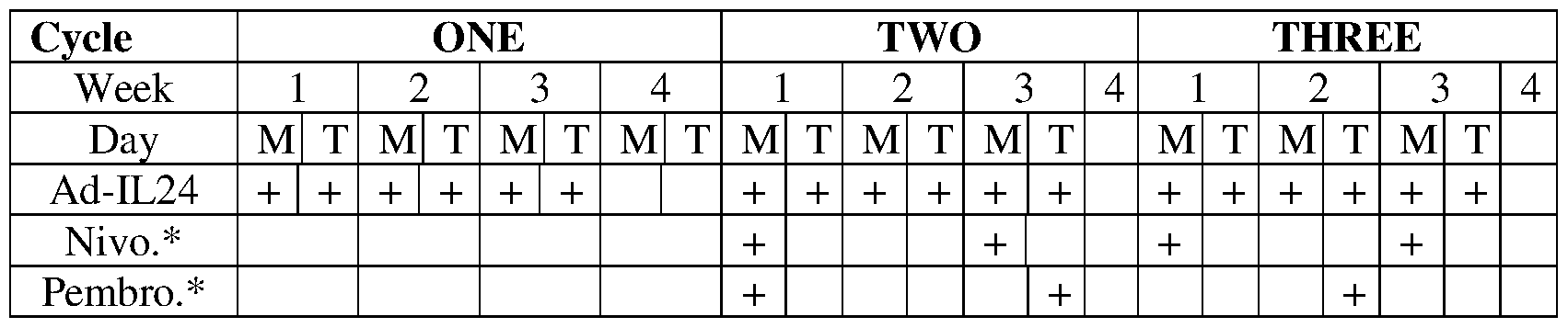


Claims
Priority Applications (7)
| Application Number | Priority Date | Filing Date | Title |
|---|---|---|---|
| AU2016349632A AU2016349632A1 (en) | 2015-11-07 | 2016-11-07 | Compositions comprising tumor suppressor gene therapy and immune checkpoint blockade for the treatment of cancer |
| KR1020187015569A KR20180104597A (en) | 2015-11-07 | 2016-11-07 | Compositions comprising tumor suppressor gene therapy and immune blockade for cancer therapy |
| EP16795531.9A EP3371221A2 (en) | 2015-11-07 | 2016-11-07 | Methods and compositions comprising tumor suppressor gene therapy and immune checkpoint blockade for the treatment of cancer |
| CN201680075823.8A CN108884159A (en) | 2015-11-07 | 2016-11-07 | The composition use for cancer treatment blocked comprising tumor suppressor gene treatment and immunologic test point |
| CA3004530A CA3004530A1 (en) | 2015-11-07 | 2016-11-07 | Methods and compositions comprising tumor suppressor gene therapy and immune checkpoint blockade for the treatment of cancer |
| JP2018543282A JP2018532810A (en) | 2015-11-07 | 2016-11-07 | Composition comprising tumor suppressor gene therapy and immune checkpoint therapy for the treatment of cancer |
| US15/773,609 US20190038713A1 (en) | 2015-11-07 | 2016-11-07 | Compositions comprising tumor suppressor gene therapy and immune checkpoint blockade for the treatment of cancer |
Applications Claiming Priority (10)
| Application Number | Priority Date | Filing Date | Title |
|---|---|---|---|
| US201562252453P | 2015-11-07 | 2015-11-07 | |
| US62/252,453 | 2015-11-07 | ||
| US201662276615P | 2016-01-08 | 2016-01-08 | |
| US62/276,615 | 2016-01-08 | ||
| US201662333817P | 2016-05-09 | 2016-05-09 | |
| US62/333,817 | 2016-05-09 | ||
| US201662345094P | 2016-06-03 | 2016-06-03 | |
| US62/345,094 | 2016-06-03 | ||
| US201662408879P | 2016-10-17 | 2016-10-17 | |
| US62/408,879 | 2016-10-17 |
Publications (2)
| Publication Number | Publication Date |
|---|---|
| WO2017079746A2 true WO2017079746A2 (en) | 2017-05-11 |
| WO2017079746A3 WO2017079746A3 (en) | 2017-06-29 |
Family
ID=58663159
Family Applications (1)
| Application Number | Title | Priority Date | Filing Date |
|---|---|---|---|
| PCT/US2016/060833 WO2017079746A2 (en) | 2015-11-07 | 2016-11-07 | Methods and compositions comprising tumor suppressor gene therapy and immune checkpoint blockade for the treatment of cancer |
Country Status (8)
| Country | Link |
|---|---|
| US (1) | US20190038713A1 (en) |
| EP (1) | EP3371221A2 (en) |
| JP (1) | JP2018532810A (en) |
| KR (1) | KR20180104597A (en) |
| CN (1) | CN108884159A (en) |
| AU (1) | AU2016349632A1 (en) |
| CA (1) | CA3004530A1 (en) |
| WO (1) | WO2017079746A2 (en) |
Cited By (16)
| Publication number | Priority date | Publication date | Assignee | Title |
|---|---|---|---|---|
| WO2019046619A1 (en) * | 2017-08-30 | 2019-03-07 | Sanford Burnham Prebys Medical Discovery Institute | Tp53 as biomarker for responsiveness to immunotherapy |
| US10287353B2 (en) | 2016-05-11 | 2019-05-14 | Huya Bioscience International, Llc | Combination therapies of HDAC inhibitors and PD-1 inhibitors |
| US10385131B2 (en) | 2016-05-11 | 2019-08-20 | Huya Bioscience International, Llc | Combination therapies of HDAC inhibitors and PD-L1 inhibitors |
| WO2019173391A1 (en) * | 2018-03-06 | 2019-09-12 | Rita Elena Serda | A high capacity platform for immunogenic cancer cell death |
| WO2020036635A2 (en) | 2018-03-19 | 2020-02-20 | Multivir Inc. | Methods and compositions comprising tumor suppressor gene therapy and cd122/cd132 agonists for the treatment of cancer |
| WO2020067085A1 (en) * | 2018-09-26 | 2020-04-02 | アステラス製薬株式会社 | Cancer therapy where oncolytic vaccinia virus and immune checkpoint inhibitor are used in combination, and pharmaceutical composition and combination drug used therein |
| WO2020077039A1 (en) * | 2018-10-12 | 2020-04-16 | Synergene Therapeutics, Inc. | Methods for reducing side effects of immunotherapy |
| CN111315398A (en) * | 2017-11-10 | 2020-06-19 | 阿尔莫生物科技股份有限公司 | Compositions and methods of use of interleukin-10 in combination with an immune checkpoint pathway inhibitor |
| WO2020198293A1 (en) * | 2019-03-25 | 2020-10-01 | Northshore University Health System | Methods and compositions comprising enhanced targeted immune gene therapy for the treatment of cancer |
| WO2020198734A1 (en) * | 2019-03-28 | 2020-10-01 | The Penn State Research Foundation | Methods, compositions relating to treatment of cancer |
| US10888594B2 (en) | 2016-05-30 | 2021-01-12 | National University Corporation Tottori University | Genetically engineered vaccinia viruses |
| US11040027B2 (en) | 2017-01-17 | 2021-06-22 | Heparegenix Gmbh | Protein kinase inhibitors for promoting liver regeneration or reducing or preventing hepatocyte death |
| US11229703B2 (en) * | 2016-04-06 | 2022-01-25 | Noxopharm Limited | Radiotherapy improvements |
| US11344589B2 (en) | 2016-05-30 | 2022-05-31 | National University Corporation Tottori University | Genetically engineered vaccinia viruses |
| US11541030B2 (en) | 2020-03-30 | 2023-01-03 | Noxopharm Limited | Methods for the treatment of inflammation associated with infection |
| US11559510B2 (en) | 2016-04-06 | 2023-01-24 | Noxopharm Limited | Isoflavonoid composition with improved pharmacokinetics |
Families Citing this family (14)
| Publication number | Priority date | Publication date | Assignee | Title |
|---|---|---|---|---|
| HRP20231299T1 (en) * | 2016-01-11 | 2024-02-02 | Queen Mary University Of London | Inhibitors of pi3k p-delta 110 for use in delivery of viruses in the treatment of cancer |
| WO2018225062A1 (en) * | 2017-06-04 | 2018-12-13 | Rappaport Family Institute For Research In The Medical Sciences | Method of predicting personalized response to cancer therapy and kit therefor |
| CN116479022A (en) * | 2018-12-12 | 2023-07-25 | 中南大学 | Recombinant CAR19-IL24 gene, lentiviral vector, CAR19-IL24-T cell and application |
| EP3911161A4 (en) * | 2019-01-16 | 2022-09-14 | Board of Regents, The University of Texas System | Methods and compositions for treating immune checkpoint inhibitor associated colitis |
| CN113993531A (en) * | 2019-05-14 | 2022-01-28 | 癌康理生物制药株式会社 | Method for applying oncolytic virus to tumor tissue and application device |
| WO2021005541A1 (en) * | 2019-07-09 | 2021-01-14 | Takeda Pharmaceutical Company Limited | Administration of sting agonist and checkpoint inhibitors |
| CN112646839A (en) * | 2019-10-10 | 2021-04-13 | 梁亚龙 | Modified adeno-associated virus |
| JP2023506443A (en) * | 2020-02-18 | 2023-02-16 | イノベイション バイオ カンパニー リミテッド | Companion diagnostic biomarker composition and companion diagnostic kit containing the same |
| KR102395580B1 (en) * | 2020-02-18 | 2022-05-10 | (주)이노베이션바이오 | Companion diagnostic biomarker composition and companion diagnostic kit comprising the same |
| CN111658778B (en) * | 2020-06-11 | 2021-11-09 | 中国科学院长春应用化学研究所 | Pharmaceutical composition, preparation method and application thereof |
| KR20230028484A (en) * | 2020-06-24 | 2023-02-28 | 피엠브이 파마슈티컬스 인코포레이티드 | Combination therapy for the treatment of cancer |
| CN116457016A (en) * | 2020-09-15 | 2023-07-18 | 默沙东有限责任公司 | Combination therapy of PD-1 antagonists and LAG3 antagonists and lenvatinib or pharmaceutically acceptable salts thereof for the treatment of cancer patients |
| CN113186225B (en) | 2021-05-13 | 2021-11-02 | 中国医学科学院北京协和医院 | PD1/PDL1 monoclonal antibody immunogenic myocarditis model and preparation method thereof |
| WO2023196899A2 (en) * | 2022-04-08 | 2023-10-12 | Global Cancer Technology | Methods and products to screen and treat glioblastoma multiforme and other cancers, including breast cancers, using a combination of pi3kinase inhibitors with checkpoint inhibitors |
Citations (105)
| Publication number | Priority date | Publication date | Assignee | Title |
|---|---|---|---|---|
| US4682195A (en) | 1985-09-30 | 1987-07-21 | General Electric Company | Insulated gate device with configured emitter contact pad |
| US4683202A (en) | 1985-03-28 | 1987-07-28 | Cetus Corporation | Process for amplifying nucleic acid sequences |
| EP0266032A1 (en) | 1986-08-29 | 1988-05-04 | Beecham Group Plc | Modified fibrinolytic enzyme |
| US4797368A (en) | 1985-03-15 | 1989-01-10 | The United States Of America As Represented By The Department Of Health And Human Services | Adeno-associated virus as eukaryotic expression vector |
| US4835251A (en) | 1986-06-23 | 1989-05-30 | Genetech, Inc. | Method of chain combination |
| US5023321A (en) | 1982-12-13 | 1991-06-11 | Howard Florey Institute Of Experimental Physiology & Medicine | Molecular cloning and characterization of a further gene sequence coding for human relaxin |
| US5139941A (en) | 1985-10-31 | 1992-08-18 | University Of Florida Research Foundation, Inc. | AAV transduction vectors |
| US5302523A (en) | 1989-06-21 | 1994-04-12 | Zeneca Limited | Transformation of plant cells |
| WO1994009699A1 (en) | 1992-10-30 | 1994-05-11 | British Technology Group Limited | Investigation of a body |
| US5322783A (en) | 1989-10-17 | 1994-06-21 | Pioneer Hi-Bred International, Inc. | Soybean transformation by microparticle bombardment |
| WO1995001994A1 (en) | 1993-07-09 | 1995-01-19 | Synergen, Inc. | Recombinant ctla4 polypeptides and methods for making the same |
| US5384253A (en) | 1990-12-28 | 1995-01-24 | Dekalb Genetics Corporation | Genetic transformation of maize cells by electroporation of cells pretreated with pectin degrading enzymes |
| WO1995006128A2 (en) | 1993-08-25 | 1995-03-02 | Dekalb Genetics Corporation | Fertile, transgenic maize plants and methods for their production |
| US5399363A (en) | 1991-01-25 | 1995-03-21 | Eastman Kodak Company | Surface modified anticancer nanoparticles |
| WO1995011986A1 (en) | 1993-10-27 | 1995-05-04 | The Trustees Of Columbia University In The City Of New York | METHOD FOR GENERATING A SUBTRACTED cDNA LIBRARY AND USES OF THE GENERATED LIBRARY |
| US5466468A (en) | 1990-04-03 | 1995-11-14 | Ciba-Geigy Corporation | Parenterally administrable liposome formulation comprising synthetic lipids |
| US5538877A (en) | 1990-01-22 | 1996-07-23 | Dekalb Genetics Corporation | Method for preparing fertile transgenic corn plants |
| US5543158A (en) | 1993-07-23 | 1996-08-06 | Massachusetts Institute Of Technology | Biodegradable injectable nanoparticles |
| US5550318A (en) | 1990-04-17 | 1996-08-27 | Dekalb Genetics Corporation | Methods and compositions for the production of stably transformed, fertile monocot plants and cells thereof |
| US5563055A (en) | 1992-07-27 | 1996-10-08 | Pioneer Hi-Bred International, Inc. | Method of Agrobacterium-mediated transformation of cultured soybean cells |
| US5580859A (en) | 1989-03-21 | 1996-12-03 | Vical Incorporated | Delivery of exogenous DNA sequences in a mammal |
| US5591616A (en) | 1992-07-07 | 1997-01-07 | Japan Tobacco, Inc. | Method for transforming monocotyledons |
| US5610042A (en) | 1991-10-07 | 1997-03-11 | Ciba-Geigy Corporation | Methods for stable transformation of wheat |
| US5641515A (en) | 1995-04-04 | 1997-06-24 | Elan Corporation, Plc | Controlled release biodegradable nanoparticles containing insulin |
| US5645897A (en) | 1992-02-15 | 1997-07-08 | Andra; Jurgen | Process and device for surface-modification by physico-chemical reactions of gases or vapors on surfaces, using highly-charged ions |
| US5656610A (en) | 1994-06-21 | 1997-08-12 | University Of Southern California | Producing a protein in a mammal by injection of a DNA-sequence into the tongue |
| US5702932A (en) | 1992-07-20 | 1997-12-30 | University Of Florida | Microinjection methods to transform arthropods with exogenous DNA |
| US5705629A (en) | 1995-10-20 | 1998-01-06 | Hybridon, Inc. | Methods for H-phosphonate synthesis of mono- and oligonucleotides |
| WO1998007408A1 (en) | 1996-08-19 | 1998-02-26 | The Government Of The United States Of America, As Represented By The Secretary, Department Of Health And Human Services | Novel liposome complexes for increased systemic delivery |
| US5736524A (en) | 1994-11-14 | 1998-04-07 | Merck & Co.,. Inc. | Polynucleotide tuberculosis vaccine |
| US5736167A (en) | 1997-02-27 | 1998-04-07 | Chang; Hui Hwa | Mold device for making safety shoe |
| US5739169A (en) | 1996-05-31 | 1998-04-14 | Procept, Incorporated | Aromatic compounds for inhibiting immune response |
| US5780448A (en) | 1995-11-07 | 1998-07-14 | Ottawa Civic Hospital Loeb Research | DNA-based vaccination of fish |
| US5789215A (en) | 1991-08-20 | 1998-08-04 | Genpharm International | Gene targeting in animal cells using isogenic DNA constructs |
| US5801005A (en) | 1993-03-17 | 1998-09-01 | University Of Washington | Immune reactivity to HER-2/neu protein for diagnosis of malignancies in which the HER-2/neu oncogene is associated |
| US5811395A (en) | 1995-06-07 | 1998-09-22 | Medical University Of South Carolina | Relaxin analogs and derivatives methods and uses thereof |
| WO1998042752A1 (en) | 1997-03-21 | 1998-10-01 | Brigham And Women's Hospital Inc. | Immunotherapeutic ctla-4 binding peptides |
| US5821337A (en) | 1991-06-14 | 1998-10-13 | Genentech, Inc. | Immunoglobulin variants |
| US5824311A (en) | 1987-11-30 | 1998-10-20 | Trustees Of The University Of Pennsylvania | Treatment of tumors with monoclonal antibodies against oncogene antigens |
| US5830880A (en) | 1994-08-26 | 1998-11-03 | Hoechst Aktiengesellschaft | Gene therapy of tumors with an endothelial cell-specific, cell cycle-dependent active compound |
| US5844905A (en) | 1996-07-09 | 1998-12-01 | International Business Machines Corporation | Extensions to distributed MAC protocols with collision avoidance using RTS/CTS exchange |
| US5846945A (en) | 1993-02-16 | 1998-12-08 | Onyx Pharmaceuticals, Inc. | Cytopathic viruses for therapy and prophylaxis of neoplasia |
| US5846225A (en) | 1997-02-19 | 1998-12-08 | Cornell Research Foundation, Inc. | Gene transfer therapy delivery device and method |
| US5846233A (en) | 1995-01-09 | 1998-12-08 | Medi-Ject Corporation | Coupling device for medical injection system |
| US5885796A (en) | 1991-06-27 | 1999-03-23 | Bristol-Myers Squibb Company | CTLA4 receptor and uses thereof |
| US5925565A (en) | 1994-07-05 | 1999-07-20 | Institut National De La Sante Et De La Recherche Medicale | Internal ribosome entry site, vector containing it and therapeutic use |
| US5928906A (en) | 1996-05-09 | 1999-07-27 | Sequenom, Inc. | Process for direct sequencing during template amplification |
| US5935819A (en) | 1992-08-27 | 1999-08-10 | Eichner; Wolfram | Process for producing a pharmaceutical preparation of PDGF-AB |
| US5945100A (en) | 1996-07-31 | 1999-08-31 | Fbp Corporation | Tumor delivery vehicles |
| US5981274A (en) | 1996-09-18 | 1999-11-09 | Tyrrell; D. Lorne J. | Recombinant hepatitis virus vectors |
| US5994624A (en) | 1997-10-20 | 1999-11-30 | Cotton Incorporated | In planta method for the production of transgenic plants |
| US5994136A (en) | 1997-12-12 | 1999-11-30 | Cell Genesys, Inc. | Method and means for producing high titer, safe, recombinant lentivirus vectors |
| US6013516A (en) | 1995-10-06 | 2000-01-11 | The Salk Institute For Biological Studies | Vector and method of use for nucleic acid delivery to non-dividing cells |
| WO2000037504A2 (en) | 1998-12-23 | 2000-06-29 | Pfizer Inc. | Human monoclonal antibodies to ctla-4 |
| WO2001014424A2 (en) | 1999-08-24 | 2001-03-01 | Medarex, Inc. | Human ctla-4 antibodies and their uses |
| US6740320B1 (en) | 1992-10-13 | 2004-05-25 | Board Of Regents, The University Of Texas System | Recombinant P53 adenovirus methods and compositions |
| WO2004058801A2 (en) | 2002-12-23 | 2004-07-15 | City Of Hope | Modified vaccinia ankara expressing p53 in cancer immunotherapy |
| WO2005003168A2 (en) | 2003-07-02 | 2005-01-13 | Novo Nordisk A/S | Methods for the production and cytotoxicity evaluation of kir2dl nk-receptor antibodies |
| WO2005009465A1 (en) | 2003-07-24 | 2005-02-03 | Innate Pharma | Methods and compositions for increasing the efficiency of therapeutic antibodies using nk cell potentiating compounds |
| WO2006003179A2 (en) | 2004-07-01 | 2006-01-12 | Novo Nordisk A/S | Antibodies binding to receptors kir2dl1, -2, 3 but not kir2ds4 and their therapeutic use |
| US7060808B1 (en) | 1995-06-07 | 2006-06-13 | Imclone Systems Incorporated | Humanized anti-EGF receptor monoclonal antibody |
| WO2006072626A1 (en) | 2005-01-06 | 2006-07-13 | Novo Nordisk A/S | Kir-binding agents and methods of use thereof |
| WO2006072625A2 (en) | 2005-01-06 | 2006-07-13 | Novo Nordisk A/S | Anti-kir combination treatments and methods |
| WO2006121168A1 (en) | 2005-05-09 | 2006-11-16 | Ono Pharmaceutical Co., Ltd. | Human monoclonal antibodies to programmed death 1(pd-1) and methods for treating cancer using anti-pd-1 antibodies alone or in combination with other immunotherapeutics |
| WO2007042573A2 (en) | 2005-10-14 | 2007-04-19 | Innate Pharma | Compositions and methods for treating proliferative disorders |
| US7223593B2 (en) | 2000-01-21 | 2007-05-29 | Biovex Limited | Herpes virus strains for gene therapy |
| WO2008084106A1 (en) | 2007-01-11 | 2008-07-17 | Novo Nordisk A/S | Anti-kir antibodies, formulations, and uses thereof |
| WO2008132601A1 (en) | 2007-04-30 | 2008-11-06 | Immutep | Cytotoxic anti-lag-3 monoclonal antibody and its use in the treatment or prevention of organ transplant rejection and autoimmune disease |
| WO2009044273A2 (en) | 2007-10-05 | 2009-04-09 | Immutep | Use of recombinant lag-3 or the derivatives thereof for eliciting monocyte immune response |
| WO2009101611A1 (en) | 2008-02-11 | 2009-08-20 | Curetech Ltd. | Monoclonal antibodies for tumor treatment |
| WO2009114335A2 (en) | 2008-03-12 | 2009-09-17 | Merck & Co., Inc. | Pd-1 binding proteins |
| WO2010027827A2 (en) | 2008-08-25 | 2010-03-11 | Amplimmune, Inc. | Targeted costimulatory polypeptides and methods of use to treat cancer |
| WO2010065939A1 (en) | 2008-12-05 | 2010-06-10 | Indiana University Research & Technology Corporation | Combination therapy to enhace nk cell mediated cytotoxicty |
| WO2011014438A1 (en) | 2009-07-31 | 2011-02-03 | N.V. Organon | Fully human antibodies to btla |
| US20110039778A1 (en) | 2009-06-01 | 2011-02-17 | Chemical & Biopharmaceutical Laboratories Of Patras S.A. | Peptide Synthesis |
| EP2320940A2 (en) | 2008-08-11 | 2011-05-18 | Medarex, Inc. | Human antibodies that bind lymphocyte activation gene-3 (lag-3), and uses thereof |
| WO2011066342A2 (en) | 2009-11-24 | 2011-06-03 | Amplimmune, Inc. | Simultaneous inhibition of pd-l1/pd-l2 |
| US20110190319A1 (en) | 2009-12-18 | 2011-08-04 | Combs Andrew P | Substituted heteroaryl fused derivatives as pi3k inhibitors |
| US8017114B2 (en) | 1999-08-24 | 2011-09-13 | Medarex, Inc. | Human CTLA-4 antibodies and their uses |
| WO2012009703A2 (en) | 2010-07-16 | 2012-01-19 | Epeius Biotechnologies Corporation | Targeted nanoparticles for cancer and other disorders |
| US8119129B2 (en) | 2008-08-01 | 2012-02-21 | Bristol-Myers Squibb Company | Combination of anti-CTLA4 antibody with dasatinib for the treatment of proliferative diseases |
| WO2012062748A1 (en) | 2010-11-11 | 2012-05-18 | Bayer Pharma Aktiengesellschaft | Aminoalcohol substituted 2,3-dihydroimidazo[1,2-c]quinazoline derivatives useful for treating hyper-proliferative disorders and diseases associated with angiogenesis |
| WO2012071411A2 (en) | 2010-11-22 | 2012-05-31 | Innate Pharma Sa | Nk cell modulating treatments and methods for treatment of hematological malignancies |
| US20120177645A1 (en) | 2009-08-31 | 2012-07-12 | Solomon Langermann | Methods and compositions for the inhibition of transplant rejection |
| WO2012146667A1 (en) | 2011-04-29 | 2012-11-01 | Almirall, S.A. | Imidazopyridine derivatives as pi3k inhibitors |
| US20120294796A1 (en) | 2010-03-04 | 2012-11-22 | Macrogenics, Inc. | Antibodies Reactive with B7-H3 and Uses Thereof |
| WO2012160448A2 (en) | 2011-05-25 | 2012-11-29 | Innate Pharma, S.A. | Anti-kir antibodies for the treatment of inflammatory disorders |
| US8329867B2 (en) | 2010-02-19 | 2012-12-11 | Xencor, Inc. | CTLA4-Ig immunoadhesins |
| WO2013006490A2 (en) | 2011-07-01 | 2013-01-10 | Cellerant Therapeutics, Inc. | Antibodies that specifically bind to tim3 |
| US8354509B2 (en) | 2007-06-18 | 2013-01-15 | Msd Oss B.V. | Antibodies to human programmed death receptor PD-1 |
| WO2013025779A1 (en) | 2011-08-15 | 2013-02-21 | Amplimmune, Inc. | Anti-b7-h4 antibodies and their uses |
| WO2013067492A1 (en) | 2011-11-03 | 2013-05-10 | The Trustees Of The University Of Pennsylvania | Isolated b7-h4 specific compositions and methods of use thereof |
| US20140022021A1 (en) | 2012-07-17 | 2014-01-23 | Murata Manufacturing Co | Power amplifier |
| WO2014047350A1 (en) | 2012-09-20 | 2014-03-27 | Morningside Technology Ventures Ltd. | Oncolytic virus encoding pd-1 binding agents and uses of the same |
| US8735553B1 (en) | 2013-09-13 | 2014-05-27 | Beigene, Ltd. | Anti-PD1 antibodies and their use as therapeutics and diagnostics |
| WO2014138314A1 (en) | 2013-03-05 | 2014-09-12 | Baylor College Of Medicine | Oncolytic virus |
| US20140294898A1 (en) | 2013-03-15 | 2014-10-02 | Bristol-Myers Squibb Company | Macrocyclic inhibitors of the pd-1/pd-l1 and cd80(b7-1)/pd-l1 protein/protein interactions |
| WO2014164942A1 (en) | 2013-03-13 | 2014-10-09 | The Regents Of The University Of Michigan | Dual mek/pi3k inhibitors and therapeutic methods using the same |
| WO2015016718A1 (en) | 2013-08-02 | 2015-02-05 | Bionovion Holding B.V. | Combining cd27 agonists and immune checkpoint inhibition for immune stimulation |
| WO2015027163A1 (en) | 2013-08-22 | 2015-02-26 | University Of Pittsburgh - Of The Commonwealth System Of Higher Education | Immuno-oncolytic therapies |
| WO2015082376A2 (en) | 2013-12-03 | 2015-06-11 | Bayer Pharma Aktiengesellschaft | Use of pi3k-inhibitors |
| US20150202290A1 (en) | 2012-08-30 | 2015-07-23 | Amgen Inc. | Method for treating melanoma using a herpes simplex virus and an immune checkpoint inhibitor |
| WO2015150809A1 (en) | 2014-04-01 | 2015-10-08 | Queen Mary University Of London | Oncolytic vaccinia virus |
| US20150291595A1 (en) | 2012-10-16 | 2015-10-15 | Almirall, S.A. | Pyrrolotriazinone derivatives as pi3k inhibitors |
| WO2016009017A1 (en) | 2014-07-16 | 2016-01-21 | Institut Gustave-Roussy | Combination of oncolytic virus with immune checkpoint modulators |
Family Cites Families (6)
| Publication number | Priority date | Publication date | Assignee | Title |
|---|---|---|---|---|
| US5710137A (en) * | 1996-08-16 | 1998-01-20 | The Trustees Of Columbia University In The City Of New York | Use of a melanoma differentiation associated gene (mda 7) for reversing a cancerous phenotype |
| CA2557326A1 (en) * | 2004-02-24 | 2005-09-09 | Introgen Therapeutics, Inc. | Combination of ad-p53 and chemotherapy for the treatment of tumours |
| US20070281041A1 (en) * | 2004-03-02 | 2007-12-06 | Introgen Therapeutics, Inc. | Compositions and Methods Involving MDA-7 for the Treatment of Cancer |
| US9682143B2 (en) * | 2012-08-14 | 2017-06-20 | Ibc Pharmaceuticals, Inc. | Combination therapy for inducing immune response to disease |
| CN105828834A (en) * | 2013-11-05 | 2016-08-03 | 同源生物服务股份有限公司 | Combinations of checkpoint inhibitors and therapeutics to treat cancer |
| CN110381997A (en) * | 2016-12-12 | 2019-10-25 | 茂体外尔公司 | For treating and preventing the method and composition comprising gene-virus therapy and immunologic test point inhibitor of cancer and infectious diseases |
-
2016
- 2016-11-07 AU AU2016349632A patent/AU2016349632A1/en not_active Abandoned
- 2016-11-07 US US15/773,609 patent/US20190038713A1/en not_active Abandoned
- 2016-11-07 KR KR1020187015569A patent/KR20180104597A/en unknown
- 2016-11-07 JP JP2018543282A patent/JP2018532810A/en active Pending
- 2016-11-07 WO PCT/US2016/060833 patent/WO2017079746A2/en active Application Filing
- 2016-11-07 EP EP16795531.9A patent/EP3371221A2/en not_active Withdrawn
- 2016-11-07 CN CN201680075823.8A patent/CN108884159A/en active Pending
- 2016-11-07 CA CA3004530A patent/CA3004530A1/en not_active Abandoned
Patent Citations (113)
| Publication number | Priority date | Publication date | Assignee | Title |
|---|---|---|---|---|
| US5023321A (en) | 1982-12-13 | 1991-06-11 | Howard Florey Institute Of Experimental Physiology & Medicine | Molecular cloning and characterization of a further gene sequence coding for human relaxin |
| US4797368A (en) | 1985-03-15 | 1989-01-10 | The United States Of America As Represented By The Department Of Health And Human Services | Adeno-associated virus as eukaryotic expression vector |
| US4683202A (en) | 1985-03-28 | 1987-07-28 | Cetus Corporation | Process for amplifying nucleic acid sequences |
| US4683202B1 (en) | 1985-03-28 | 1990-11-27 | Cetus Corp | |
| US4682195A (en) | 1985-09-30 | 1987-07-21 | General Electric Company | Insulated gate device with configured emitter contact pad |
| US5139941A (en) | 1985-10-31 | 1992-08-18 | University Of Florida Research Foundation, Inc. | AAV transduction vectors |
| US4835251A (en) | 1986-06-23 | 1989-05-30 | Genetech, Inc. | Method of chain combination |
| EP0266032A1 (en) | 1986-08-29 | 1988-05-04 | Beecham Group Plc | Modified fibrinolytic enzyme |
| US5824311A (en) | 1987-11-30 | 1998-10-20 | Trustees Of The University Of Pennsylvania | Treatment of tumors with monoclonal antibodies against oncogene antigens |
| US5589466A (en) | 1989-03-21 | 1996-12-31 | Vical Incorporated | Induction of a protective immune response in a mammal by injecting a DNA sequence |
| US5580859A (en) | 1989-03-21 | 1996-12-03 | Vical Incorporated | Delivery of exogenous DNA sequences in a mammal |
| US5302523A (en) | 1989-06-21 | 1994-04-12 | Zeneca Limited | Transformation of plant cells |
| US5464765A (en) | 1989-06-21 | 1995-11-07 | Zeneca Limited | Transformation of plant cells |
| US5322783A (en) | 1989-10-17 | 1994-06-21 | Pioneer Hi-Bred International, Inc. | Soybean transformation by microparticle bombardment |
| US5538880A (en) | 1990-01-22 | 1996-07-23 | Dekalb Genetics Corporation | Method for preparing fertile transgenic corn plants |
| US5538877A (en) | 1990-01-22 | 1996-07-23 | Dekalb Genetics Corporation | Method for preparing fertile transgenic corn plants |
| US5466468A (en) | 1990-04-03 | 1995-11-14 | Ciba-Geigy Corporation | Parenterally administrable liposome formulation comprising synthetic lipids |
| US5550318A (en) | 1990-04-17 | 1996-08-27 | Dekalb Genetics Corporation | Methods and compositions for the production of stably transformed, fertile monocot plants and cells thereof |
| US5384253A (en) | 1990-12-28 | 1995-01-24 | Dekalb Genetics Corporation | Genetic transformation of maize cells by electroporation of cells pretreated with pectin degrading enzymes |
| US5399363A (en) | 1991-01-25 | 1995-03-21 | Eastman Kodak Company | Surface modified anticancer nanoparticles |
| US5821337A (en) | 1991-06-14 | 1998-10-13 | Genentech, Inc. | Immunoglobulin variants |
| US5885796A (en) | 1991-06-27 | 1999-03-23 | Bristol-Myers Squibb Company | CTLA4 receptor and uses thereof |
| US5789215A (en) | 1991-08-20 | 1998-08-04 | Genpharm International | Gene targeting in animal cells using isogenic DNA constructs |
| US5610042A (en) | 1991-10-07 | 1997-03-11 | Ciba-Geigy Corporation | Methods for stable transformation of wheat |
| US5645897A (en) | 1992-02-15 | 1997-07-08 | Andra; Jurgen | Process and device for surface-modification by physico-chemical reactions of gases or vapors on surfaces, using highly-charged ions |
| US5591616A (en) | 1992-07-07 | 1997-01-07 | Japan Tobacco, Inc. | Method for transforming monocotyledons |
| US5702932A (en) | 1992-07-20 | 1997-12-30 | University Of Florida | Microinjection methods to transform arthropods with exogenous DNA |
| US5563055A (en) | 1992-07-27 | 1996-10-08 | Pioneer Hi-Bred International, Inc. | Method of Agrobacterium-mediated transformation of cultured soybean cells |
| US5935819A (en) | 1992-08-27 | 1999-08-10 | Eichner; Wolfram | Process for producing a pharmaceutical preparation of PDGF-AB |
| US6740320B1 (en) | 1992-10-13 | 2004-05-25 | Board Of Regents, The University Of Texas System | Recombinant P53 adenovirus methods and compositions |
| WO1994009699A1 (en) | 1992-10-30 | 1994-05-11 | British Technology Group Limited | Investigation of a body |
| US5846945A (en) | 1993-02-16 | 1998-12-08 | Onyx Pharmaceuticals, Inc. | Cytopathic viruses for therapy and prophylaxis of neoplasia |
| US5801005A (en) | 1993-03-17 | 1998-09-01 | University Of Washington | Immune reactivity to HER-2/neu protein for diagnosis of malignancies in which the HER-2/neu oncogene is associated |
| WO1995001994A1 (en) | 1993-07-09 | 1995-01-19 | Synergen, Inc. | Recombinant ctla4 polypeptides and methods for making the same |
| US5543158A (en) | 1993-07-23 | 1996-08-06 | Massachusetts Institute Of Technology | Biodegradable injectable nanoparticles |
| WO1995006128A2 (en) | 1993-08-25 | 1995-03-02 | Dekalb Genetics Corporation | Fertile, transgenic maize plants and methods for their production |
| WO1995011986A1 (en) | 1993-10-27 | 1995-05-04 | The Trustees Of Columbia University In The City Of New York | METHOD FOR GENERATING A SUBTRACTED cDNA LIBRARY AND USES OF THE GENERATED LIBRARY |
| US5656610A (en) | 1994-06-21 | 1997-08-12 | University Of Southern California | Producing a protein in a mammal by injection of a DNA-sequence into the tongue |
| US5925565A (en) | 1994-07-05 | 1999-07-20 | Institut National De La Sante Et De La Recherche Medicale | Internal ribosome entry site, vector containing it and therapeutic use |
| US5830880A (en) | 1994-08-26 | 1998-11-03 | Hoechst Aktiengesellschaft | Gene therapy of tumors with an endothelial cell-specific, cell cycle-dependent active compound |
| US5736524A (en) | 1994-11-14 | 1998-04-07 | Merck & Co.,. Inc. | Polynucleotide tuberculosis vaccine |
| US5846233A (en) | 1995-01-09 | 1998-12-08 | Medi-Ject Corporation | Coupling device for medical injection system |
| US5641515A (en) | 1995-04-04 | 1997-06-24 | Elan Corporation, Plc | Controlled release biodegradable nanoparticles containing insulin |
| US7060808B1 (en) | 1995-06-07 | 2006-06-13 | Imclone Systems Incorporated | Humanized anti-EGF receptor monoclonal antibody |
| US5811395A (en) | 1995-06-07 | 1998-09-22 | Medical University Of South Carolina | Relaxin analogs and derivatives methods and uses thereof |
| US6013516A (en) | 1995-10-06 | 2000-01-11 | The Salk Institute For Biological Studies | Vector and method of use for nucleic acid delivery to non-dividing cells |
| US5705629A (en) | 1995-10-20 | 1998-01-06 | Hybridon, Inc. | Methods for H-phosphonate synthesis of mono- and oligonucleotides |
| US5780448A (en) | 1995-11-07 | 1998-07-14 | Ottawa Civic Hospital Loeb Research | DNA-based vaccination of fish |
| US5928906A (en) | 1996-05-09 | 1999-07-27 | Sequenom, Inc. | Process for direct sequencing during template amplification |
| US5739169A (en) | 1996-05-31 | 1998-04-14 | Procept, Incorporated | Aromatic compounds for inhibiting immune response |
| US5844905A (en) | 1996-07-09 | 1998-12-01 | International Business Machines Corporation | Extensions to distributed MAC protocols with collision avoidance using RTS/CTS exchange |
| US5945100A (en) | 1996-07-31 | 1999-08-31 | Fbp Corporation | Tumor delivery vehicles |
| WO1998007408A1 (en) | 1996-08-19 | 1998-02-26 | The Government Of The United States Of America, As Represented By The Secretary, Department Of Health And Human Services | Novel liposome complexes for increased systemic delivery |
| US5981274A (en) | 1996-09-18 | 1999-11-09 | Tyrrell; D. Lorne J. | Recombinant hepatitis virus vectors |
| US5846225A (en) | 1997-02-19 | 1998-12-08 | Cornell Research Foundation, Inc. | Gene transfer therapy delivery device and method |
| US5736167A (en) | 1997-02-27 | 1998-04-07 | Chang; Hui Hwa | Mold device for making safety shoe |
| WO1998042752A1 (en) | 1997-03-21 | 1998-10-01 | Brigham And Women's Hospital Inc. | Immunotherapeutic ctla-4 binding peptides |
| US6207156B1 (en) | 1997-03-21 | 2001-03-27 | Brigham And Women's Hospital, Inc. | Specific antibodies and antibody fragments |
| US5994624A (en) | 1997-10-20 | 1999-11-30 | Cotton Incorporated | In planta method for the production of transgenic plants |
| US5994136A (en) | 1997-12-12 | 1999-11-30 | Cell Genesys, Inc. | Method and means for producing high titer, safe, recombinant lentivirus vectors |
| WO2000037504A2 (en) | 1998-12-23 | 2000-06-29 | Pfizer Inc. | Human monoclonal antibodies to ctla-4 |
| US8017114B2 (en) | 1999-08-24 | 2011-09-13 | Medarex, Inc. | Human CTLA-4 antibodies and their uses |
| WO2001014424A2 (en) | 1999-08-24 | 2001-03-01 | Medarex, Inc. | Human ctla-4 antibodies and their uses |
| US7223593B2 (en) | 2000-01-21 | 2007-05-29 | Biovex Limited | Herpes virus strains for gene therapy |
| US7537924B2 (en) | 2000-01-21 | 2009-05-26 | Biovex Limited | Virus strains |
| WO2004058801A2 (en) | 2002-12-23 | 2004-07-15 | City Of Hope | Modified vaccinia ankara expressing p53 in cancer immunotherapy |
| WO2005003168A2 (en) | 2003-07-02 | 2005-01-13 | Novo Nordisk A/S | Methods for the production and cytotoxicity evaluation of kir2dl nk-receptor antibodies |
| WO2005009465A1 (en) | 2003-07-24 | 2005-02-03 | Innate Pharma | Methods and compositions for increasing the efficiency of therapeutic antibodies using nk cell potentiating compounds |
| WO2006003179A2 (en) | 2004-07-01 | 2006-01-12 | Novo Nordisk A/S | Antibodies binding to receptors kir2dl1, -2, 3 but not kir2ds4 and their therapeutic use |
| WO2006072626A1 (en) | 2005-01-06 | 2006-07-13 | Novo Nordisk A/S | Kir-binding agents and methods of use thereof |
| WO2006072625A2 (en) | 2005-01-06 | 2006-07-13 | Novo Nordisk A/S | Anti-kir combination treatments and methods |
| WO2006121168A1 (en) | 2005-05-09 | 2006-11-16 | Ono Pharmaceutical Co., Ltd. | Human monoclonal antibodies to programmed death 1(pd-1) and methods for treating cancer using anti-pd-1 antibodies alone or in combination with other immunotherapeutics |
| US8008449B2 (en) | 2005-05-09 | 2011-08-30 | Medarex, Inc. | Human monoclonal antibodies to programmed death 1 (PD-1) and methods for treating cancer using anti-PD-1 antibodies alone or in combination with other immunotherapeutics |
| WO2007042573A2 (en) | 2005-10-14 | 2007-04-19 | Innate Pharma | Compositions and methods for treating proliferative disorders |
| WO2008084106A1 (en) | 2007-01-11 | 2008-07-17 | Novo Nordisk A/S | Anti-kir antibodies, formulations, and uses thereof |
| WO2008132601A1 (en) | 2007-04-30 | 2008-11-06 | Immutep | Cytotoxic anti-lag-3 monoclonal antibody and its use in the treatment or prevention of organ transplant rejection and autoimmune disease |
| US8354509B2 (en) | 2007-06-18 | 2013-01-15 | Msd Oss B.V. | Antibodies to human programmed death receptor PD-1 |
| WO2009044273A2 (en) | 2007-10-05 | 2009-04-09 | Immutep | Use of recombinant lag-3 or the derivatives thereof for eliciting monocyte immune response |
| WO2009101611A1 (en) | 2008-02-11 | 2009-08-20 | Curetech Ltd. | Monoclonal antibodies for tumor treatment |
| US20110008369A1 (en) | 2008-03-12 | 2011-01-13 | Finnefrock Adam C | Pd-1 binding proteins |
| WO2009114335A2 (en) | 2008-03-12 | 2009-09-17 | Merck & Co., Inc. | Pd-1 binding proteins |
| US8119129B2 (en) | 2008-08-01 | 2012-02-21 | Bristol-Myers Squibb Company | Combination of anti-CTLA4 antibody with dasatinib for the treatment of proliferative diseases |
| EP2320940A2 (en) | 2008-08-11 | 2011-05-18 | Medarex, Inc. | Human antibodies that bind lymphocyte activation gene-3 (lag-3), and uses thereof |
| WO2010027827A2 (en) | 2008-08-25 | 2010-03-11 | Amplimmune, Inc. | Targeted costimulatory polypeptides and methods of use to treat cancer |
| WO2010065939A1 (en) | 2008-12-05 | 2010-06-10 | Indiana University Research & Technology Corporation | Combination therapy to enhace nk cell mediated cytotoxicty |
| US20110039778A1 (en) | 2009-06-01 | 2011-02-17 | Chemical & Biopharmaceutical Laboratories Of Patras S.A. | Peptide Synthesis |
| WO2011014438A1 (en) | 2009-07-31 | 2011-02-03 | N.V. Organon | Fully human antibodies to btla |
| US20120177645A1 (en) | 2009-08-31 | 2012-07-12 | Solomon Langermann | Methods and compositions for the inhibition of transplant rejection |
| WO2011066342A2 (en) | 2009-11-24 | 2011-06-03 | Amplimmune, Inc. | Simultaneous inhibition of pd-l1/pd-l2 |
| US20110190319A1 (en) | 2009-12-18 | 2011-08-04 | Combs Andrew P | Substituted heteroaryl fused derivatives as pi3k inhibitors |
| US8329867B2 (en) | 2010-02-19 | 2012-12-11 | Xencor, Inc. | CTLA4-Ig immunoadhesins |
| US20120294796A1 (en) | 2010-03-04 | 2012-11-22 | Macrogenics, Inc. | Antibodies Reactive with B7-H3 and Uses Thereof |
| WO2012009703A2 (en) | 2010-07-16 | 2012-01-19 | Epeius Biotechnologies Corporation | Targeted nanoparticles for cancer and other disorders |
| WO2012062748A1 (en) | 2010-11-11 | 2012-05-18 | Bayer Pharma Aktiengesellschaft | Aminoalcohol substituted 2,3-dihydroimidazo[1,2-c]quinazoline derivatives useful for treating hyper-proliferative disorders and diseases associated with angiogenesis |
| WO2012071411A2 (en) | 2010-11-22 | 2012-05-31 | Innate Pharma Sa | Nk cell modulating treatments and methods for treatment of hematological malignancies |
| WO2012146667A1 (en) | 2011-04-29 | 2012-11-01 | Almirall, S.A. | Imidazopyridine derivatives as pi3k inhibitors |
| WO2012160448A2 (en) | 2011-05-25 | 2012-11-29 | Innate Pharma, S.A. | Anti-kir antibodies for the treatment of inflammatory disorders |
| WO2013006490A2 (en) | 2011-07-01 | 2013-01-10 | Cellerant Therapeutics, Inc. | Antibodies that specifically bind to tim3 |
| WO2013025779A1 (en) | 2011-08-15 | 2013-02-21 | Amplimmune, Inc. | Anti-b7-h4 antibodies and their uses |
| WO2013067492A1 (en) | 2011-11-03 | 2013-05-10 | The Trustees Of The University Of Pennsylvania | Isolated b7-h4 specific compositions and methods of use thereof |
| US20140022021A1 (en) | 2012-07-17 | 2014-01-23 | Murata Manufacturing Co | Power amplifier |
| US20150202290A1 (en) | 2012-08-30 | 2015-07-23 | Amgen Inc. | Method for treating melanoma using a herpes simplex virus and an immune checkpoint inhibitor |
| WO2014047350A1 (en) | 2012-09-20 | 2014-03-27 | Morningside Technology Ventures Ltd. | Oncolytic virus encoding pd-1 binding agents and uses of the same |
| US20150291595A1 (en) | 2012-10-16 | 2015-10-15 | Almirall, S.A. | Pyrrolotriazinone derivatives as pi3k inhibitors |
| WO2014138314A1 (en) | 2013-03-05 | 2014-09-12 | Baylor College Of Medicine | Oncolytic virus |
| WO2014164942A1 (en) | 2013-03-13 | 2014-10-09 | The Regents Of The University Of Michigan | Dual mek/pi3k inhibitors and therapeutic methods using the same |
| US20140294898A1 (en) | 2013-03-15 | 2014-10-02 | Bristol-Myers Squibb Company | Macrocyclic inhibitors of the pd-1/pd-l1 and cd80(b7-1)/pd-l1 protein/protein interactions |
| WO2015016718A1 (en) | 2013-08-02 | 2015-02-05 | Bionovion Holding B.V. | Combining cd27 agonists and immune checkpoint inhibition for immune stimulation |
| WO2015027163A1 (en) | 2013-08-22 | 2015-02-26 | University Of Pittsburgh - Of The Commonwealth System Of Higher Education | Immuno-oncolytic therapies |
| US8735553B1 (en) | 2013-09-13 | 2014-05-27 | Beigene, Ltd. | Anti-PD1 antibodies and their use as therapeutics and diagnostics |
| WO2015082376A2 (en) | 2013-12-03 | 2015-06-11 | Bayer Pharma Aktiengesellschaft | Use of pi3k-inhibitors |
| WO2015150809A1 (en) | 2014-04-01 | 2015-10-08 | Queen Mary University Of London | Oncolytic vaccinia virus |
| WO2016009017A1 (en) | 2014-07-16 | 2016-01-21 | Institut Gustave-Roussy | Combination of oncolytic virus with immune checkpoint modulators |
Non-Patent Citations (92)
| Title |
|---|
| AGHI ET AL., CANCER RES., vol. 59, 1999, pages 3861 - 3865 |
| AKSENTIJEVICH ET AL., HUMAN GENE THER., vol. 7, 1996, pages 1111 |
| BAICHWAL; SUGDEN: "Gene Transfer", vol. 1, 1986, PLENUM PRESS, pages: 17 - 148 |
| BAILEY; LEVINE, J. PHARM. BIOMED. ANAL., vol. 11, 1993, pages 285 - 292 |
| BOUVET ET AL., CANCER RES., vol. 58, 1998, pages 2288 - 2292 |
| BULLER ET AL., CANCER GENE THERAPY, vol. 9, 2002, pages 553 - 566 |
| CAMACHO ET AL., J CLIN ONCOLOGY, vol. 22, no. 145, 2004 |
| CANCER GENE THER., 1998, pages 444 - 448 |
| CARROLL ET AL., MOL CANCER THERAPEUTICS, vol. 1, 2001, pages 49 - 60 |
| CAUDELL ET AL., J IMMUNOL., vol. 168, 2002, pages 6041 - 6046 |
| CHADA ET AL., CANCER GENE THER., vol. 13, 2006, pages 490 - 502 |
| CHASE ET AL., NAT. BIOTECHNOL., pages 16 |
| CHEN; OKAYAMA, MOL. CELL. BIOL., vol. 7, 1987, pages 2745 - 2752 |
| CHOI ET AL., GENE THERAPY, vol. 17, 2010, pages 190 - 201 |
| CORRALES ET AL., CELL REPORTS, vol. 11, 2015, pages 1018 - 1030 |
| COUCH ET AL., AM. REV. RESP. DIS., vol. 88, 1963, pages 394 - 403 |
| DORONIN ET AL., VIROLOGY, vol. 305, 2003, pages 378 - 387 |
| FORM S-1 REGISTRATION STATEMENT, U.S. SECURITIES AND EXCHANGE COMMISSION, 2015 |
| FRALEY ET AL., PROC. NAT 'L ACAD. SCI., vol. 76, 1979, pages 3348 - 3352 |
| FUJIWARA ET AL., J NATL CANCER INST, vol. 86, 1994, pages 1458 - 1462 |
| GENNARO: "Remington's Pharmaceutical Sciences, 22nd edition", MACK PUBLISHING |
| GHIRINGHELLI ET AL., BIOMED. J., vol. 38, 2015, pages 111 - 116 |
| GRAHAM; VAN DER EB, VIROLOGY, vol. 52, 1973, pages 456 - 467 |
| GURNANI ET AL., CANCER CHEMOTHER PHARMACOL., vol. 44, no. 2, 1999, pages 143 - 151 |
| HARLAND; WEINTRAUB, J. CELL BIOL, vol. 101, 1985, pages 1094 - 1099 |
| HARTWELL ET AL., SCIENCE, vol. 266, 1994, pages 1821 - 1828 |
| HURWITZ ET AL., PROC NATL ACAD SCI USA, vol. 95, no. 17, 1998, pages 10067 - 10071 |
| HURWITZ, PROC NATL ACAD SCI., vol. 95, no. 17, 1998, pages 10067 - 10071 |
| HYNES; FERRETTI, METHODS ENZYMOL., vol. 235, 1994, pages 606 - 616 |
| IANNELLO ET AL., J EXPERIMENTAL MEDICINE, vol. 210, no. 10, pages 2057 - 2069 |
| INOUE ET AL., CANCER LETTERS, vol. 157, 2000, pages 105 - 112 |
| JIANG ET AL., PROC. NATL. ACAD. SCI., vol. 93, pages 9160 - 9165 |
| JONES ET AL., J EXP MED., vol. 205, no. 12, 2008, pages 2763 - 79 |
| KAWABE ET AL., MOL THER., vol. 6, no. 5, 2002, pages 637 - 44 |
| KAWABE ET AL., MOL. THER., vol. 6, no. 5, 2002, pages 637 - 644 |
| KIM ET AL., JOURNAL OF THE NATIONAL CANCER INSTITUTE, vol. 98, no. 20, 2006, pages 1482 - 1493 |
| KOTIN ET AL., PROC. NATL. ACAD. SCI. USA, vol. 87, 1990, pages 221 1 - 2215 |
| KREIL, PROTEIN SCI., vol. 4, 1995, pages 1666 - 1669 |
| LICHTENSTEIN ET AL., INT. REV. IMMUNOL., vol. 23, 2004, pages 75 - 111 |
| LIU ET AL., J. BIOL. CHEM., vol. 270, 1995, pages 24864 |
| LUI ET AL., GENE THERAPY, vol. 10, 2003, pages 292 - 303 |
| MANN ET AL., CELL, vol. 33, 1983, pages 153 - 159 |
| MARKOWITZ ET AL., J. VIROL., vol. 62, 1988, pages 1 120 - 1 124 |
| MCLAUGHLIN ET AL., J. VIROL., vol. 62, 1988, pages 1963 - 1973 |
| MELLMAN ET AL., NATURE, vol. 480, 2011, pages 480 - 489 |
| MHASHILKAR ET AL., MOL. MEDICINE, vol. 7, no. 4, 2001, pages 271 - 282 |
| MIYAHARA ET AL., CANCER GENE THERAPY, vol. 13, 2006, pages 753 - 761 |
| MOKYR ET AL., CANCER RES, vol. 58, 1998, pages 5301 - 5304 |
| MOKYR ET AL., CANCER RES., vol. 58, 1998, pages 5301 - 5304 |
| MUZYCZKA, CURR. TOP. MICROBIOLLMMUNOL, vol. 158, 1992, pages 97 - 129 |
| NICOLAS; RUBENSTEIN: "Vectors: A survey of molecular cloning vectors and their uses", 1988, BUTTERWORTH, pages: 493 - 513 |
| NICOLAU, METHODS ENZYMOL, vol. 149, 1987, pages 157 - 176 |
| NICOLAU; SENE, BIOCHIM. BIOPHYS. ACTA, vol. 721, 1982, pages 185 - 190 |
| NISHIKAWA ET AL., MOL. THER., vol. 9, no. 8, 2004, pages 818 - 828 |
| NISHIKAWA ET AL., ONCOGENE, vol. 23, no. 42, 2004, pages 7125 - 7131 |
| NISHIZAKI M ET AL., CLIN. CAN. RES., vol. 5, 1999, pages 1015 - 1023 |
| OHASHI M ET AL., GUT, vol. 44, 1999, pages 366 - 371 |
| OKAZAKI T ET AL., INTERN. IMMUN., vol. 19, no. 7, 2007, pages 813 |
| PARDOLL, NAT REV CANCER, vol. 12, no. 4, 2012, pages 252 - 64 |
| PARDOLL, NATURE REV CANCER, vol. 12, 2012, pages 252 - 264 |
| PHILIP ET AL., J. BIOL. CHEM., vol. 268, 1993, pages 16087 |
| QIN, X. ET AL., BIOL REPROD., vol. 56, 1997, pages 800 - 11 |
| QIN, X. ET AL., BIOL REPROD., vol. 56, 1997, pages 812 - 20 |
| RIDGEWAY: "Vectors: A survey of molecular cloning vectors and their uses", 1988, BUTTERWORTH, pages: 467 - 492 |
| RIPPE ET AL., MOL. CELL BIOL, vol. 10, 1990, pages 689 - 695 |
| ROSENBERG ET AL., NAT MED., vol. 10, no. 19, 2004, pages 909 - 15 |
| SAMULSKI ET AL., EMBO J., vol. 10, 1991, pages 3941 - 3950 |
| SAMULSKI ET AL., J VIROL, vol. 63, 1989, pages 3822 - 3828 |
| SHERWOOD ET AL., ENDOCRINOLOGY, vol. 114, 1984, pages 806 - 13 |
| SOBOL RE ET AL.: "Tp53 Gene Therapy for Cancer Treatment and Prevention", 2013, SPRINGER SCIENCE + BUSINESS MEDIA, article "Chapter 11" |
| SOLODIN ET AL., BIOCHEMISTRY, vol. 34, 1995, pages 13537 |
| SPITZ ET AL., CLIN CANCER RESEARCH, vol. 2, 1996, pages 1665 - 1671 |
| SWISHER ET AL., CLIN CANCER RESEARCH, vol. 9, 2003, pages 93 - 101 |
| TATEBE S ET AL., INT. J ONCOL., vol. 15, 1999, pages 229 - 235 |
| TCHEKMEDYIAN ET AL., ONCOLOGY, vol. 29, no. 12, 2015, pages 990 - 1002 |
| TEMIN: "n: Gene Transfer", 1986, PLENUM PRESS, pages: 149 - 188 |
| TEXTOR ET AL., CANCER RES., vol. 71, no. 18, 2011, pages 5998 - 6009 |
| THIERRY ET AL., PROC. NATL. ACAD. SCI., vol. 92, no. 21, 1995, pages 9742 - 6 |
| TIMIRYASOVA ET AL., BIOTECHNIQUES, vol. 31, no. 534, 2001, pages 6,8 - 40 |
| TODA ET AL., MOL. THERAPY, vol. 2, no. 4, 2000, pages 324 - 329 |
| TOLLEFSON ET AL., J. VIROL., vol. 70, 1996, pages 2296 - 2306 |
| TOP ET AL., J. INFECT. DIS., vol. 124, 1971, pages 155 - 160 |
| TSUKAMOTO ET AL., NATURE GENETICS, vol. 9, 1995, pages 243 |
| VINCENT ET AL., CANCER RES., vol. 70, no. 8, 2010, pages 3052 - 3061 |
| WAKU ET AL., J IMMUNOL., vol. 165, 2000, pages 5884 - 5890 |
| XU ET AL., GENE THERAPY, vol. 22, no. 3, 2015, pages 31 - 40 |
| XU ET AL., J GASTROENTEROL., vol. 48, no. 2, 2013, pages 203 - 13 |
| XUE ET AL., NATURE, vol. 445, no. 7128, 2007, pages 656 - 660 |
| YOUNG ET AL., CANCER GENE THER., vol. 20, no. 9, 2013, pages 531 - 537 |
| ZEIMET; MARTH, THE LANCER ONCOLOGY, vol. 7, 2003, pages 415 - 422 |
| ZHANG ET AL., CANCER GENE THER, vol. 22, 2015, pages 17 - 22 |
| ZHANG ET AL., CANCER GENE THER., vol. 1, 1994, pages 5 - 13 |
Cited By (23)
| Publication number | Priority date | Publication date | Assignee | Title |
|---|---|---|---|---|
| US11559510B2 (en) | 2016-04-06 | 2023-01-24 | Noxopharm Limited | Isoflavonoid composition with improved pharmacokinetics |
| US11229703B2 (en) * | 2016-04-06 | 2022-01-25 | Noxopharm Limited | Radiotherapy improvements |
| US10287353B2 (en) | 2016-05-11 | 2019-05-14 | Huya Bioscience International, Llc | Combination therapies of HDAC inhibitors and PD-1 inhibitors |
| US10385131B2 (en) | 2016-05-11 | 2019-08-20 | Huya Bioscience International, Llc | Combination therapies of HDAC inhibitors and PD-L1 inhibitors |
| US10385130B2 (en) | 2016-05-11 | 2019-08-20 | Huya Bioscience International, Llc | Combination therapies of HDAC inhibitors and PD-1 inhibitors |
| US11535670B2 (en) | 2016-05-11 | 2022-12-27 | Huyabio International, Llc | Combination therapies of HDAC inhibitors and PD-L1 inhibitors |
| US10888594B2 (en) | 2016-05-30 | 2021-01-12 | National University Corporation Tottori University | Genetically engineered vaccinia viruses |
| US11344589B2 (en) | 2016-05-30 | 2022-05-31 | National University Corporation Tottori University | Genetically engineered vaccinia viruses |
| US11040027B2 (en) | 2017-01-17 | 2021-06-22 | Heparegenix Gmbh | Protein kinase inhibitors for promoting liver regeneration or reducing or preventing hepatocyte death |
| WO2019046619A1 (en) * | 2017-08-30 | 2019-03-07 | Sanford Burnham Prebys Medical Discovery Institute | Tp53 as biomarker for responsiveness to immunotherapy |
| CN111315398A (en) * | 2017-11-10 | 2020-06-19 | 阿尔莫生物科技股份有限公司 | Compositions and methods of use of interleukin-10 in combination with an immune checkpoint pathway inhibitor |
| WO2019173391A1 (en) * | 2018-03-06 | 2019-09-12 | Rita Elena Serda | A high capacity platform for immunogenic cancer cell death |
| JP2021518408A (en) * | 2018-03-19 | 2021-08-02 | マルチビア インコーポレイテッド | Methods and Compositions Containing Tumor Suppressor Gene Therapies and CD122 / CD132 Agonists for Cancer Treatment |
| CN112020510A (en) * | 2018-03-19 | 2020-12-01 | 茂体外尔公司 | Methods and compositions comprising tumor suppressor gene therapy and CD122/CD132 agonists for treating cancer |
| WO2020036635A3 (en) * | 2018-03-19 | 2020-03-12 | Multivir Inc. | Methods and compositions comprising tumor suppressor gene therapy and cd122/cd132 agonists for the treatment of cancer |
| WO2020036635A2 (en) | 2018-03-19 | 2020-02-20 | Multivir Inc. | Methods and compositions comprising tumor suppressor gene therapy and cd122/cd132 agonists for the treatment of cancer |
| JPWO2020067085A1 (en) * | 2018-09-26 | 2021-08-30 | アステラス製薬株式会社 | Cancer therapy by combination of oncolytic vaccinia virus and immune checkpoint inhibitor, and pharmaceutical composition and combination drug for use. |
| WO2020067085A1 (en) * | 2018-09-26 | 2020-04-02 | アステラス製薬株式会社 | Cancer therapy where oncolytic vaccinia virus and immune checkpoint inhibitor are used in combination, and pharmaceutical composition and combination drug used therein |
| US11638730B2 (en) | 2018-09-26 | 2023-05-02 | Astellas Pharma Inc. | Cancer therapy by combination use of oncolytic vaccinia virus and immune checkpoint inhibitor, and pharmaceutical composition and combination medicine for use in the cancer therapy |
| WO2020077039A1 (en) * | 2018-10-12 | 2020-04-16 | Synergene Therapeutics, Inc. | Methods for reducing side effects of immunotherapy |
| WO2020198293A1 (en) * | 2019-03-25 | 2020-10-01 | Northshore University Health System | Methods and compositions comprising enhanced targeted immune gene therapy for the treatment of cancer |
| WO2020198734A1 (en) * | 2019-03-28 | 2020-10-01 | The Penn State Research Foundation | Methods, compositions relating to treatment of cancer |
| US11541030B2 (en) | 2020-03-30 | 2023-01-03 | Noxopharm Limited | Methods for the treatment of inflammation associated with infection |
Also Published As
| Publication number | Publication date |
|---|---|
| CN108884159A (en) | 2018-11-23 |
| KR20180104597A (en) | 2018-09-21 |
| US20190038713A1 (en) | 2019-02-07 |
| EP3371221A2 (en) | 2018-09-12 |
| AU2016349632A1 (en) | 2018-05-24 |
| WO2017079746A3 (en) | 2017-06-29 |
| CA3004530A1 (en) | 2017-05-11 |
| JP2018532810A (en) | 2018-11-08 |
Similar Documents
| Publication | Publication Date | Title |
|---|---|---|
| US20190038713A1 (en) | Compositions comprising tumor suppressor gene therapy and immune checkpoint blockade for the treatment of cancer | |
| US20200009203A1 (en) | Methods and compositions comprising viral gene therapy and an immune checkpoint inhibitor for treatment and prevention of cancer and infectious diseases | |
| US20210094991A1 (en) | Methods and compositions comprising tumor suppressor gene therapy and cd122/cd132 agonists for the treatment of cancer | |
| US20190185571A1 (en) | Methods and compositions for the treatment of cancer combining an anti-smic antibody and immune checkpoint inhibitors | |
| US20220226402A1 (en) | Methods and compositions comprising enhanced targeted immune gene therapy for the treatment of cancer | |
| WO2021113644A1 (en) | Combinations comprising a cd8+ t cell enhancer, an immune checkpoint inhibitor and radiotherapy for targeted and abscopal effects for the treatment of cancer | |
| US11594135B2 (en) | Methods of CD40 activation and immune checkpoint blockade | |
| WO2019183117A1 (en) | Methods and compositions comprising genetically engineered vaccines for the treatment and prevention of cancer | |
| US20240034797A1 (en) | Increasing responses to checkpoint inhibitors by extracorporeal apheresis | |
| CA3040458A1 (en) | Methods and compositions for tusc2 immunotherapy | |
| AU2019322487B2 (en) | Methods and compositions comprising tumor suppressor gene therapy and CD122/CD132 agonists for the treatment of cancer | |
| US20240042061A1 (en) | Methods and compositions comprising tumor suppressor gene therapy for the inhibition of pathogens | |
| US20220288225A1 (en) | Materials and methods for activating antigen-specific t cell responses | |
| US20200353044A1 (en) | Methods of cd40 and toll like receptor immune activation |
Legal Events
| Date | Code | Title | Description |
|---|---|---|---|
| 121 | Ep: the epo has been informed by wipo that ep was designated in this application |
Ref document number: 16795531 Country of ref document: EP Kind code of ref document: A2 |
|
| ENP | Entry into the national phase |
Ref document number: 3004530 Country of ref document: CA |
|
| WWE | Wipo information: entry into national phase |
Ref document number: 2018543282 Country of ref document: JP |
|
| NENP | Non-entry into the national phase |
Ref country code: DE |
|
| ENP | Entry into the national phase |
Ref document number: 2016349632 Country of ref document: AU Date of ref document: 20161107 Kind code of ref document: A |
|
| ENP | Entry into the national phase |
Ref document number: 20187015569 Country of ref document: KR Kind code of ref document: A |
|
| WWE | Wipo information: entry into national phase |
Ref document number: 2016795531 Country of ref document: EP |