WO2017181079A2 - Methods for monitoring and treating cancer - Google Patents
Methods for monitoring and treating cancer Download PDFInfo
- Publication number
- WO2017181079A2 WO2017181079A2 PCT/US2017/027725 US2017027725W WO2017181079A2 WO 2017181079 A2 WO2017181079 A2 WO 2017181079A2 US 2017027725 W US2017027725 W US 2017027725W WO 2017181079 A2 WO2017181079 A2 WO 2017181079A2
- Authority
- WO
- WIPO (PCT)
- Prior art keywords
- fold
- expression level
- patient
- level
- biological sample
- Prior art date
Links
- 206010028980 Neoplasm Diseases 0.000 title claims abstract description 200
- 238000000034 method Methods 0.000 title claims abstract description 199
- 201000011510 cancer Diseases 0.000 title claims abstract description 113
- 238000012544 monitoring process Methods 0.000 title claims abstract description 27
- 108010019530 Vascular Endothelial Growth Factors Proteins 0.000 claims abstract description 216
- 239000005557 antagonist Substances 0.000 claims abstract description 209
- 238000011282 treatment Methods 0.000 claims abstract description 77
- 230000004044 response Effects 0.000 claims abstract description 61
- 102000005789 Vascular Endothelial Growth Factors Human genes 0.000 claims abstract description 52
- 239000000203 mixture Substances 0.000 claims abstract description 23
- 230000014509 gene expression Effects 0.000 claims description 345
- 108090000623 proteins and genes Proteins 0.000 claims description 161
- 230000027455 binding Effects 0.000 claims description 133
- 210000004027 cell Anatomy 0.000 claims description 122
- 239000012472 biological sample Substances 0.000 claims description 119
- 101001009603 Homo sapiens Granzyme B Proteins 0.000 claims description 109
- 102100030385 Granzyme B Human genes 0.000 claims description 107
- 101000946843 Homo sapiens T-cell surface glycoprotein CD8 alpha chain Proteins 0.000 claims description 98
- 102100034922 T-cell surface glycoprotein CD8 alpha chain Human genes 0.000 claims description 98
- 230000001965 increasing effect Effects 0.000 claims description 92
- 102100025248 C-X-C motif chemokine 10 Human genes 0.000 claims description 80
- 101000858088 Homo sapiens C-X-C motif chemokine 10 Proteins 0.000 claims description 80
- 101001069921 Homo sapiens Growth-regulated alpha protein Proteins 0.000 claims description 76
- 102100034221 Growth-regulated alpha protein Human genes 0.000 claims description 74
- 101000947172 Homo sapiens C-X-C motif chemokine 9 Proteins 0.000 claims description 69
- 101001009599 Homo sapiens Granzyme A Proteins 0.000 claims description 69
- 101000599940 Homo sapiens Interferon gamma Proteins 0.000 claims description 69
- 101001109501 Homo sapiens NKG2-D type II integral membrane protein Proteins 0.000 claims description 69
- 101000987581 Homo sapiens Perforin-1 Proteins 0.000 claims description 69
- 102100036170 C-X-C motif chemokine 9 Human genes 0.000 claims description 68
- 102100030751 Eomesodermin homolog Human genes 0.000 claims description 68
- 101000858064 Homo sapiens C-X-C motif chemokine 13 Proteins 0.000 claims description 68
- 102100030386 Granzyme A Human genes 0.000 claims description 67
- 101000633784 Homo sapiens SLAM family member 7 Proteins 0.000 claims description 67
- 101000946833 Homo sapiens T-cell surface glycoprotein CD8 beta chain Proteins 0.000 claims description 67
- 102100037850 Interferon gamma Human genes 0.000 claims description 67
- 102100022680 NKG2-D type II integral membrane protein Human genes 0.000 claims description 67
- 102100028467 Perforin-1 Human genes 0.000 claims description 67
- 102100034928 T-cell surface glycoprotein CD8 beta chain Human genes 0.000 claims description 67
- 102100025277 C-X-C motif chemokine 13 Human genes 0.000 claims description 66
- 102100029198 SLAM family member 7 Human genes 0.000 claims description 66
- 108010074708 B7-H1 Antigen Proteins 0.000 claims description 61
- 229960000397 bevacizumab Drugs 0.000 claims description 58
- 241000282414 Homo sapiens Species 0.000 claims description 55
- 102000004169 proteins and genes Human genes 0.000 claims description 53
- 102100024216 Programmed cell death 1 ligand 1 Human genes 0.000 claims description 46
- 239000000523 sample Substances 0.000 claims description 39
- 239000003814 drug Substances 0.000 claims description 37
- 101000716065 Homo sapiens C-C chemokine receptor type 7 Proteins 0.000 claims description 30
- 101000797762 Homo sapiens C-C motif chemokine 5 Proteins 0.000 claims description 30
- 102100020997 Fractalkine Human genes 0.000 claims description 29
- 101000897480 Homo sapiens C-C motif chemokine 2 Proteins 0.000 claims description 29
- 101000854520 Homo sapiens Fractalkine Proteins 0.000 claims description 29
- 102100035875 C-C chemokine receptor type 5 Human genes 0.000 claims description 28
- 102100036301 C-C chemokine receptor type 7 Human genes 0.000 claims description 28
- 102100021943 C-C motif chemokine 2 Human genes 0.000 claims description 28
- 102100032367 C-C motif chemokine 5 Human genes 0.000 claims description 28
- 101710149870 C-C chemokine receptor type 5 Proteins 0.000 claims description 27
- 239000003795 chemical substances by application Substances 0.000 claims description 27
- 210000001744 T-lymphocyte Anatomy 0.000 claims description 26
- -1 use Substances 0.000 claims description 25
- 101001064167 Homo sapiens Eomesodermin homolog Proteins 0.000 claims description 21
- 229960003852 atezolizumab Drugs 0.000 claims description 20
- 229940127089 cytotoxic agent Drugs 0.000 claims description 17
- 229940124597 therapeutic agent Drugs 0.000 claims description 17
- 239000012636 effector Substances 0.000 claims description 16
- 208000006265 Renal cell carcinoma Diseases 0.000 claims description 14
- 230000002596 correlated effect Effects 0.000 claims description 14
- 238000003364 immunohistochemistry Methods 0.000 claims description 14
- 238000004519 manufacturing process Methods 0.000 claims description 13
- 230000002401 inhibitory effect Effects 0.000 claims description 10
- 239000004037 angiogenesis inhibitor Substances 0.000 claims description 9
- 102000019034 Chemokines Human genes 0.000 claims description 8
- 108010012236 Chemokines Proteins 0.000 claims description 8
- 230000012010 growth Effects 0.000 claims description 8
- 239000002254 cytotoxic agent Substances 0.000 claims description 6
- 231100000599 cytotoxic agent Toxicity 0.000 claims description 6
- 238000000684 flow cytometry Methods 0.000 claims description 6
- 108020004999 messenger RNA Proteins 0.000 claims description 6
- 208000002154 non-small cell lung carcinoma Diseases 0.000 claims description 6
- 230000002093 peripheral effect Effects 0.000 claims description 6
- 238000003753 real-time PCR Methods 0.000 claims description 6
- 208000029729 tumor suppressor gene on chromosome 11 Diseases 0.000 claims description 6
- 206010050513 Metastatic renal cell carcinoma Diseases 0.000 claims description 5
- 108700030875 Programmed Cell Death 1 Ligand 2 Proteins 0.000 claims description 5
- 102100024213 Programmed cell death 1 ligand 2 Human genes 0.000 claims description 5
- 229950009791 durvalumab Drugs 0.000 claims description 5
- 206010009944 Colon cancer Diseases 0.000 claims description 4
- 238000011529 RT qPCR Methods 0.000 claims description 4
- 208000005718 Stomach Neoplasms Diseases 0.000 claims description 4
- 206010017758 gastric cancer Diseases 0.000 claims description 4
- 230000004547 gene signature Effects 0.000 claims description 4
- 238000009169 immunotherapy Methods 0.000 claims description 4
- 208000014018 liver neoplasm Diseases 0.000 claims description 4
- 201000001441 melanoma Diseases 0.000 claims description 4
- 238000001959 radiotherapy Methods 0.000 claims description 4
- 238000003196 serial analysis of gene expression Methods 0.000 claims description 4
- 201000011549 stomach cancer Diseases 0.000 claims description 4
- 206010005003 Bladder cancer Diseases 0.000 claims description 3
- 206010006187 Breast cancer Diseases 0.000 claims description 3
- 208000026310 Breast neoplasm Diseases 0.000 claims description 3
- 208000001333 Colorectal Neoplasms Diseases 0.000 claims description 3
- 206010033128 Ovarian cancer Diseases 0.000 claims description 3
- 206010061535 Ovarian neoplasm Diseases 0.000 claims description 3
- 208000007097 Urinary Bladder Neoplasms Diseases 0.000 claims description 3
- 229950002916 avelumab Drugs 0.000 claims description 3
- 201000007270 liver cancer Diseases 0.000 claims description 3
- 201000005112 urinary bladder cancer Diseases 0.000 claims description 3
- 102100040678 Programmed cell death protein 1 Human genes 0.000 claims description 2
- 101710089372 Programmed cell death protein 1 Proteins 0.000 claims description 2
- 238000003559 RNA-seq method Methods 0.000 claims description 2
- 238000010166 immunofluorescence Methods 0.000 claims description 2
- 238000007901 in situ hybridization Methods 0.000 claims description 2
- 238000004949 mass spectrometry Methods 0.000 claims description 2
- 238000010208 microarray analysis Methods 0.000 claims description 2
- 238000003762 quantitative reverse transcription PCR Methods 0.000 claims description 2
- 238000001262 western blot Methods 0.000 claims description 2
- 101100407308 Mus musculus Pdcd1lg2 gene Proteins 0.000 claims 1
- 238000010839 reverse transcription Methods 0.000 claims 1
- 230000001225 therapeutic effect Effects 0.000 abstract description 10
- 238000002560 therapeutic procedure Methods 0.000 abstract description 8
- 238000002405 diagnostic procedure Methods 0.000 abstract description 2
- 102000009524 Vascular Endothelial Growth Factor A Human genes 0.000 description 167
- 239000000090 biomarker Substances 0.000 description 76
- 125000003275 alpha amino acid group Chemical group 0.000 description 60
- 150000007523 nucleic acids Chemical group 0.000 description 59
- 108090000765 processed proteins & peptides Proteins 0.000 description 59
- 239000000427 antigen Substances 0.000 description 58
- 108091007433 antigens Proteins 0.000 description 58
- 102000036639 antigens Human genes 0.000 description 58
- 229920001184 polypeptide Polymers 0.000 description 56
- 102000004196 processed proteins & peptides Human genes 0.000 description 56
- 101710201246 Eomesodermin Proteins 0.000 description 49
- 208000037265 diseases, disorders, signs and symptoms Diseases 0.000 description 46
- 235000018102 proteins Nutrition 0.000 description 42
- 102000039446 nucleic acids Human genes 0.000 description 38
- 108020004707 nucleic acids Proteins 0.000 description 38
- 101100519207 Mus musculus Pdcd1 gene Proteins 0.000 description 34
- 230000001900 immune effect Effects 0.000 description 34
- 210000001519 tissue Anatomy 0.000 description 28
- 238000011319 anticancer therapy Methods 0.000 description 27
- 102000040430 polynucleotide Human genes 0.000 description 27
- 108091033319 polynucleotide Proteins 0.000 description 27
- 239000002157 polynucleotide Substances 0.000 description 27
- 201000010099 disease Diseases 0.000 description 26
- 241000124008 Mammalia Species 0.000 description 24
- 108091028043 Nucleic acid sequence Proteins 0.000 description 24
- 230000007423 decrease Effects 0.000 description 24
- 239000012634 fragment Substances 0.000 description 24
- 235000001014 amino acid Nutrition 0.000 description 23
- 108060003951 Immunoglobulin Proteins 0.000 description 22
- 241000700159 Rattus Species 0.000 description 22
- 102000018358 immunoglobulin Human genes 0.000 description 22
- 241000288906 Primates Species 0.000 description 21
- 241000283984 Rodentia Species 0.000 description 21
- 239000002246 antineoplastic agent Substances 0.000 description 21
- 230000008859 change Effects 0.000 description 21
- 241000282412 Homo Species 0.000 description 20
- 241000699670 Mus sp. Species 0.000 description 20
- 241000251539 Vertebrata <Metazoa> Species 0.000 description 20
- 208000035475 disorder Diseases 0.000 description 20
- 238000012545 processing Methods 0.000 description 20
- 230000004083 survival effect Effects 0.000 description 20
- 208000008839 Kidney Neoplasms Diseases 0.000 description 19
- 206010038389 Renal cancer Diseases 0.000 description 19
- 201000010982 kidney cancer Diseases 0.000 description 19
- 229940024606 amino acid Drugs 0.000 description 18
- 150000001413 amino acids Chemical class 0.000 description 18
- 125000003729 nucleotide group Chemical group 0.000 description 18
- 108091007491 NSP3 Papain-like protease domains Proteins 0.000 description 17
- 230000008901 benefit Effects 0.000 description 16
- 230000004071 biological effect Effects 0.000 description 16
- 108020004414 DNA Proteins 0.000 description 15
- 229940002612 prodrug Drugs 0.000 description 15
- 239000000651 prodrug Substances 0.000 description 15
- 108010021625 Immunoglobulin Fragments Proteins 0.000 description 14
- 102000008394 Immunoglobulin Fragments Human genes 0.000 description 14
- 239000002773 nucleotide Substances 0.000 description 14
- NFGXHKASABOEEW-UHFFFAOYSA-N 1-methylethyl 11-methoxy-3,7,11-trimethyl-2,4-dodecadienoate Chemical compound COC(C)(C)CCCC(C)CC=CC(C)=CC(=O)OC(C)C NFGXHKASABOEEW-UHFFFAOYSA-N 0.000 description 13
- 125000000539 amino acid group Chemical group 0.000 description 13
- 108091032973 (ribonucleotides)n+m Proteins 0.000 description 12
- 108091034117 Oligonucleotide Proteins 0.000 description 12
- 102100039196 CX3C chemokine receptor 1 Human genes 0.000 description 11
- 238000003556 assay Methods 0.000 description 11
- 102000052116 epidermal growth factor receptor activity proteins Human genes 0.000 description 11
- 108700015053 epidermal growth factor receptor activity proteins Proteins 0.000 description 11
- 239000003112 inhibitor Substances 0.000 description 11
- YOHYSYJDKVYCJI-UHFFFAOYSA-N n-[3-[[6-[3-(trifluoromethyl)anilino]pyrimidin-4-yl]amino]phenyl]cyclopropanecarboxamide Chemical compound FC(F)(F)C1=CC=CC(NC=2N=CN=C(NC=3C=C(NC(=O)C4CC4)C=CC=3)C=2)=C1 YOHYSYJDKVYCJI-UHFFFAOYSA-N 0.000 description 11
- 108090000835 CX3C Chemokine Receptor 1 Proteins 0.000 description 10
- 238000004458 analytical method Methods 0.000 description 10
- 210000002865 immune cell Anatomy 0.000 description 10
- 238000002203 pretreatment Methods 0.000 description 10
- 230000009467 reduction Effects 0.000 description 10
- 230000011664 signaling Effects 0.000 description 10
- 150000003384 small molecules Chemical class 0.000 description 10
- 208000015914 Non-Hodgkin lymphomas Diseases 0.000 description 9
- 150000001875 compounds Chemical class 0.000 description 9
- 230000003247 decreasing effect Effects 0.000 description 9
- 230000006870 function Effects 0.000 description 9
- 230000003993 interaction Effects 0.000 description 9
- 230000003902 lesion Effects 0.000 description 9
- 230000035772 mutation Effects 0.000 description 9
- 238000003752 polymerase chain reaction Methods 0.000 description 9
- 241000894007 species Species 0.000 description 9
- 238000006467 substitution reaction Methods 0.000 description 9
- 208000024891 symptom Diseases 0.000 description 9
- 101100454807 Caenorhabditis elegans lgg-1 gene Proteins 0.000 description 8
- AOJJSUZBOXZQNB-TZSSRYMLSA-N Doxorubicin Chemical compound O([C@H]1C[C@@](O)(CC=2C(O)=C3C(=O)C=4C=CC=C(C=4C(=O)C3=C(O)C=21)OC)C(=O)CO)[C@H]1C[C@H](N)[C@H](O)[C@H](C)O1 AOJJSUZBOXZQNB-TZSSRYMLSA-N 0.000 description 8
- 206010027476 Metastases Diseases 0.000 description 8
- JLCPHMBAVCMARE-UHFFFAOYSA-N [3-[[3-[[3-[[3-[[3-[[3-[[3-[[3-[[3-[[3-[[3-[[5-(2-amino-6-oxo-1H-purin-9-yl)-3-[[3-[[3-[[3-[[3-[[3-[[5-(2-amino-6-oxo-1H-purin-9-yl)-3-[[5-(2-amino-6-oxo-1H-purin-9-yl)-3-hydroxyoxolan-2-yl]methoxy-hydroxyphosphoryl]oxyoxolan-2-yl]methoxy-hydroxyphosphoryl]oxy-5-(5-methyl-2,4-dioxopyrimidin-1-yl)oxolan-2-yl]methoxy-hydroxyphosphoryl]oxy-5-(6-aminopurin-9-yl)oxolan-2-yl]methoxy-hydroxyphosphoryl]oxy-5-(6-aminopurin-9-yl)oxolan-2-yl]methoxy-hydroxyphosphoryl]oxy-5-(6-aminopurin-9-yl)oxolan-2-yl]methoxy-hydroxyphosphoryl]oxy-5-(6-aminopurin-9-yl)oxolan-2-yl]methoxy-hydroxyphosphoryl]oxyoxolan-2-yl]methoxy-hydroxyphosphoryl]oxy-5-(5-methyl-2,4-dioxopyrimidin-1-yl)oxolan-2-yl]methoxy-hydroxyphosphoryl]oxy-5-(4-amino-2-oxopyrimidin-1-yl)oxolan-2-yl]methoxy-hydroxyphosphoryl]oxy-5-(5-methyl-2,4-dioxopyrimidin-1-yl)oxolan-2-yl]methoxy-hydroxyphosphoryl]oxy-5-(5-methyl-2,4-dioxopyrimidin-1-yl)oxolan-2-yl]methoxy-hydroxyphosphoryl]oxy-5-(6-aminopurin-9-yl)oxolan-2-yl]methoxy-hydroxyphosphoryl]oxy-5-(6-aminopurin-9-yl)oxolan-2-yl]methoxy-hydroxyphosphoryl]oxy-5-(4-amino-2-oxopyrimidin-1-yl)oxolan-2-yl]methoxy-hydroxyphosphoryl]oxy-5-(4-amino-2-oxopyrimidin-1-yl)oxolan-2-yl]methoxy-hydroxyphosphoryl]oxy-5-(4-amino-2-oxopyrimidin-1-yl)oxolan-2-yl]methoxy-hydroxyphosphoryl]oxy-5-(6-aminopurin-9-yl)oxolan-2-yl]methoxy-hydroxyphosphoryl]oxy-5-(4-amino-2-oxopyrimidin-1-yl)oxolan-2-yl]methyl [5-(6-aminopurin-9-yl)-2-(hydroxymethyl)oxolan-3-yl] hydrogen phosphate Polymers Cc1cn(C2CC(OP(O)(=O)OCC3OC(CC3OP(O)(=O)OCC3OC(CC3O)n3cnc4c3nc(N)[nH]c4=O)n3cnc4c3nc(N)[nH]c4=O)C(COP(O)(=O)OC3CC(OC3COP(O)(=O)OC3CC(OC3COP(O)(=O)OC3CC(OC3COP(O)(=O)OC3CC(OC3COP(O)(=O)OC3CC(OC3COP(O)(=O)OC3CC(OC3COP(O)(=O)OC3CC(OC3COP(O)(=O)OC3CC(OC3COP(O)(=O)OC3CC(OC3COP(O)(=O)OC3CC(OC3COP(O)(=O)OC3CC(OC3COP(O)(=O)OC3CC(OC3COP(O)(=O)OC3CC(OC3COP(O)(=O)OC3CC(OC3COP(O)(=O)OC3CC(OC3COP(O)(=O)OC3CC(OC3COP(O)(=O)OC3CC(OC3CO)n3cnc4c(N)ncnc34)n3ccc(N)nc3=O)n3cnc4c(N)ncnc34)n3ccc(N)nc3=O)n3ccc(N)nc3=O)n3ccc(N)nc3=O)n3cnc4c(N)ncnc34)n3cnc4c(N)ncnc34)n3cc(C)c(=O)[nH]c3=O)n3cc(C)c(=O)[nH]c3=O)n3ccc(N)nc3=O)n3cc(C)c(=O)[nH]c3=O)n3cnc4c3nc(N)[nH]c4=O)n3cnc4c(N)ncnc34)n3cnc4c(N)ncnc34)n3cnc4c(N)ncnc34)n3cnc4c(N)ncnc34)O2)c(=O)[nH]c1=O JLCPHMBAVCMARE-UHFFFAOYSA-N 0.000 description 8
- 230000004075 alteration Effects 0.000 description 8
- 230000004927 fusion Effects 0.000 description 8
- YLMAHDNUQAMNNX-UHFFFAOYSA-N imatinib methanesulfonate Chemical compound CS(O)(=O)=O.C1CN(C)CCN1CC1=CC=C(C(=O)NC=2C=C(NC=3N=C(C=CN=3)C=3C=NC=CC=3)C(C)=CC=2)C=C1 YLMAHDNUQAMNNX-UHFFFAOYSA-N 0.000 description 8
- BCFGMOOMADDAQU-UHFFFAOYSA-N lapatinib Chemical compound O1C(CNCCS(=O)(=O)C)=CC=C1C1=CC=C(N=CN=C2NC=3C=C(Cl)C(OCC=4C=C(F)C=CC=4)=CC=3)C2=C1 BCFGMOOMADDAQU-UHFFFAOYSA-N 0.000 description 8
- 239000000126 substance Substances 0.000 description 8
- 108020004705 Codon Proteins 0.000 description 7
- 108091008605 VEGF receptors Proteins 0.000 description 7
- 102000009484 Vascular Endothelial Growth Factor Receptors Human genes 0.000 description 7
- 108010081667 aflibercept Proteins 0.000 description 7
- 238000006243 chemical reaction Methods 0.000 description 7
- 230000000295 complement effect Effects 0.000 description 7
- 230000000694 effects Effects 0.000 description 7
- 230000005764 inhibitory process Effects 0.000 description 7
- 125000005647 linker group Chemical group 0.000 description 7
- 238000012423 maintenance Methods 0.000 description 7
- 230000001404 mediated effect Effects 0.000 description 7
- 230000008569 process Effects 0.000 description 7
- 239000013074 reference sample Substances 0.000 description 7
- 235000000346 sugar Nutrition 0.000 description 7
- 230000002459 sustained effect Effects 0.000 description 7
- 239000013598 vector Substances 0.000 description 7
- 102100031573 Hematopoietic progenitor cell antigen CD34 Human genes 0.000 description 6
- 101000777663 Homo sapiens Hematopoietic progenitor cell antigen CD34 Proteins 0.000 description 6
- ZDZOTLJHXYCWBA-VCVYQWHSSA-N N-debenzoyl-N-(tert-butoxycarbonyl)-10-deacetyltaxol Chemical compound O([C@H]1[C@H]2[C@@](C([C@H](O)C3=C(C)[C@@H](OC(=O)[C@H](O)[C@@H](NC(=O)OC(C)(C)C)C=4C=CC=CC=4)C[C@]1(O)C3(C)C)=O)(C)[C@@H](O)C[C@H]1OC[C@]12OC(=O)C)C(=O)C1=CC=CC=C1 ZDZOTLJHXYCWBA-VCVYQWHSSA-N 0.000 description 6
- 102100030086 Receptor tyrosine-protein kinase erbB-2 Human genes 0.000 description 6
- 230000010056 antibody-dependent cellular cytotoxicity Effects 0.000 description 6
- 230000000903 blocking effect Effects 0.000 description 6
- 239000013068 control sample Substances 0.000 description 6
- 230000000875 corresponding effect Effects 0.000 description 6
- 229940079593 drug Drugs 0.000 description 6
- AAKJLRGGTJKAMG-UHFFFAOYSA-N erlotinib Chemical compound C=12C=C(OCCOC)C(OCCOC)=CC2=NC=NC=1NC1=CC=CC(C#C)=C1 AAKJLRGGTJKAMG-UHFFFAOYSA-N 0.000 description 6
- XGALLCVXEZPNRQ-UHFFFAOYSA-N gefitinib Chemical compound C=12C=C(OCCCN3CCOCC3)C(OC)=CC2=NC=NC=1NC1=CC=C(F)C(Cl)=C1 XGALLCVXEZPNRQ-UHFFFAOYSA-N 0.000 description 6
- 230000036541 health Effects 0.000 description 6
- 230000004048 modification Effects 0.000 description 6
- 238000012986 modification Methods 0.000 description 6
- JZZFDCXSFTVOJY-UHFFFAOYSA-N n-[4-(3-chloro-4-fluoroanilino)-7-(3-morpholin-4-ylpropoxy)quinazolin-6-yl]prop-2-enamide;hydron;dichloride Chemical compound Cl.Cl.C1=C(Cl)C(F)=CC=C1NC1=NC=NC2=CC(OCCCN3CCOCC3)=C(NC(=O)C=C)C=C12 JZZFDCXSFTVOJY-UHFFFAOYSA-N 0.000 description 6
- 230000036961 partial effect Effects 0.000 description 6
- 239000013615 primer Substances 0.000 description 6
- 208000037821 progressive disease Diseases 0.000 description 6
- 102000005962 receptors Human genes 0.000 description 6
- 108020003175 receptors Proteins 0.000 description 6
- QFJCIRLUMZQUOT-HPLJOQBZSA-N sirolimus Chemical compound C1C[C@@H](O)[C@H](OC)C[C@@H]1C[C@@H](C)[C@H]1OC(=O)[C@@H]2CCCCN2C(=O)C(=O)[C@](O)(O2)[C@H](C)CC[C@H]2C[C@H](OC)/C(C)=C/C=C/C=C/[C@@H](C)C[C@@H](C)C(=O)[C@H](OC)[C@H](O)/C(C)=C/[C@@H](C)C(=O)C1 QFJCIRLUMZQUOT-HPLJOQBZSA-N 0.000 description 6
- 230000009870 specific binding Effects 0.000 description 6
- RCINICONZNJXQF-MZXODVADSA-N taxol Chemical compound O([C@@H]1[C@@]2(C[C@@H](C(C)=C(C2(C)C)[C@H](C([C@]2(C)[C@@H](O)C[C@H]3OC[C@]3([C@H]21)OC(C)=O)=O)OC(=O)C)OC(=O)[C@H](O)[C@@H](NC(=O)C=1C=CC=CC=1)C=1C=CC=CC=1)O)C(=O)C1=CC=CC=C1 RCINICONZNJXQF-MZXODVADSA-N 0.000 description 6
- 210000004881 tumor cell Anatomy 0.000 description 6
- XRYJULCDUUATMC-CYBMUJFWSA-N 4-[4-[[(1r)-1-phenylethyl]amino]-7h-pyrrolo[2,3-d]pyrimidin-6-yl]phenol Chemical compound N([C@H](C)C=1C=CC=CC=1)C(C=1C=2)=NC=NC=1NC=2C1=CC=C(O)C=C1 XRYJULCDUUATMC-CYBMUJFWSA-N 0.000 description 5
- GHASVSINZRGABV-UHFFFAOYSA-N Fluorouracil Chemical compound FC1=CNC(=O)NC1=O GHASVSINZRGABV-UHFFFAOYSA-N 0.000 description 5
- 101001012157 Homo sapiens Receptor tyrosine-protein kinase erbB-2 Proteins 0.000 description 5
- 101000808011 Homo sapiens Vascular endothelial growth factor A Proteins 0.000 description 5
- FBOZXECLQNJBKD-ZDUSSCGKSA-N L-methotrexate Chemical compound C=1N=C2N=C(N)N=C(N)C2=NC=1CN(C)C1=CC=C(C(=O)N[C@@H](CCC(O)=O)C(O)=O)C=C1 FBOZXECLQNJBKD-ZDUSSCGKSA-N 0.000 description 5
- 239000002136 L01XE07 - Lapatinib Substances 0.000 description 5
- 229930012538 Paclitaxel Natural products 0.000 description 5
- NKANXQFJJICGDU-QPLCGJKRSA-N Tamoxifen Chemical compound C=1C=CC=CC=1C(/CC)=C(C=1C=CC(OCCN(C)C)=CC=1)/C1=CC=CC=C1 NKANXQFJJICGDU-QPLCGJKRSA-N 0.000 description 5
- 108010073929 Vascular Endothelial Growth Factor A Proteins 0.000 description 5
- 229950002826 canertinib Drugs 0.000 description 5
- 238000001514 detection method Methods 0.000 description 5
- 238000005516 engineering process Methods 0.000 description 5
- 229960002949 fluorouracil Drugs 0.000 description 5
- 102000058223 human VEGFA Human genes 0.000 description 5
- 210000004408 hybridoma Anatomy 0.000 description 5
- 229960003685 imatinib mesylate Drugs 0.000 description 5
- 230000002452 interceptive effect Effects 0.000 description 5
- 238000011068 loading method Methods 0.000 description 5
- 230000009401 metastasis Effects 0.000 description 5
- 229960000485 methotrexate Drugs 0.000 description 5
- 229960001592 paclitaxel Drugs 0.000 description 5
- 230000002062 proliferating effect Effects 0.000 description 5
- 230000035755 proliferation Effects 0.000 description 5
- 230000005180 public health Effects 0.000 description 5
- 230000002829 reductive effect Effects 0.000 description 5
- 230000019491 signal transduction Effects 0.000 description 5
- WYWHKKSPHMUBEB-UHFFFAOYSA-N tioguanine Chemical compound N1C(N)=NC(=S)C2=C1N=CN2 WYWHKKSPHMUBEB-UHFFFAOYSA-N 0.000 description 5
- 210000005166 vasculature Anatomy 0.000 description 5
- STQGQHZAVUOBTE-UHFFFAOYSA-N 7-Cyan-hept-2t-en-4,6-diinsaeure Natural products C1=2C(O)=C3C(=O)C=4C(OC)=CC=CC=4C(=O)C3=C(O)C=2CC(O)(C(C)=O)CC1OC1CC(N)C(O)C(C)O1 STQGQHZAVUOBTE-UHFFFAOYSA-N 0.000 description 4
- 206010061818 Disease progression Diseases 0.000 description 4
- 102000004190 Enzymes Human genes 0.000 description 4
- 108090000790 Enzymes Proteins 0.000 description 4
- 101000746022 Homo sapiens CX3C chemokine receptor 1 Proteins 0.000 description 4
- 101000934372 Homo sapiens Macrosialin Proteins 0.000 description 4
- 239000005551 L01XE03 - Erlotinib Substances 0.000 description 4
- 102100025136 Macrosialin Human genes 0.000 description 4
- 241001465754 Metazoa Species 0.000 description 4
- NWIBSHFKIJFRCO-WUDYKRTCSA-N Mytomycin Chemical compound C1N2C(C(C(C)=C(N)C3=O)=O)=C3[C@@H](COC(N)=O)[C@@]2(OC)[C@@H]2[C@H]1N2 NWIBSHFKIJFRCO-WUDYKRTCSA-N 0.000 description 4
- 239000002253 acid Substances 0.000 description 4
- SHGAZHPCJJPHSC-YCNIQYBTSA-N all-trans-retinoic acid Chemical compound OC(=O)\C=C(/C)\C=C\C=C(/C)\C=C\C1=C(C)CCCC1(C)C SHGAZHPCJJPHSC-YCNIQYBTSA-N 0.000 description 4
- 230000015572 biosynthetic process Effects 0.000 description 4
- 210000004369 blood Anatomy 0.000 description 4
- 239000008280 blood Substances 0.000 description 4
- 230000003915 cell function Effects 0.000 description 4
- 230000004663 cell proliferation Effects 0.000 description 4
- 239000003153 chemical reaction reagent Substances 0.000 description 4
- 231100000433 cytotoxic Toxicity 0.000 description 4
- 230000001472 cytotoxic effect Effects 0.000 description 4
- STQGQHZAVUOBTE-VGBVRHCVSA-N daunorubicin Chemical compound O([C@H]1C[C@@](O)(CC=2C(O)=C3C(=O)C=4C=CC=C(C=4C(=O)C3=C(O)C=21)OC)C(C)=O)[C@H]1C[C@H](N)[C@H](O)[C@H](C)O1 STQGQHZAVUOBTE-VGBVRHCVSA-N 0.000 description 4
- 238000003745 diagnosis Methods 0.000 description 4
- 230000005750 disease progression Effects 0.000 description 4
- 229960003668 docetaxel Drugs 0.000 description 4
- 229940088598 enzyme Drugs 0.000 description 4
- 108020001507 fusion proteins Proteins 0.000 description 4
- 102000037865 fusion proteins Human genes 0.000 description 4
- 230000001976 improved effect Effects 0.000 description 4
- 238000001727 in vivo Methods 0.000 description 4
- 230000008595 infiltration Effects 0.000 description 4
- 238000001764 infiltration Methods 0.000 description 4
- 238000003780 insertion Methods 0.000 description 4
- 230000037431 insertion Effects 0.000 description 4
- 238000002372 labelling Methods 0.000 description 4
- HPJKCIUCZWXJDR-UHFFFAOYSA-N letrozole Chemical compound C1=CC(C#N)=CC=C1C(N1N=CN=C1)C1=CC=C(C#N)C=C1 HPJKCIUCZWXJDR-UHFFFAOYSA-N 0.000 description 4
- 230000001394 metastastic effect Effects 0.000 description 4
- 206010061289 metastatic neoplasm Diseases 0.000 description 4
- 125000002496 methyl group Chemical group [H]C([H])([H])* 0.000 description 4
- 210000000056 organ Anatomy 0.000 description 4
- 229960001972 panitumumab Drugs 0.000 description 4
- 210000003819 peripheral blood mononuclear cell Anatomy 0.000 description 4
- 239000008194 pharmaceutical composition Substances 0.000 description 4
- 238000002360 preparation method Methods 0.000 description 4
- 239000000047 product Substances 0.000 description 4
- ZAHRKKWIAAJSAO-UHFFFAOYSA-N rapamycin Natural products COCC(O)C(=C/C(C)C(=O)CC(OC(=O)C1CCCCN1C(=O)C(=O)C2(O)OC(CC(OC)C(=CC=CC=CC(C)CC(C)C(=O)C)C)CCC2C)C(C)CC3CCC(O)C(C3)OC)C ZAHRKKWIAAJSAO-UHFFFAOYSA-N 0.000 description 4
- 230000004043 responsiveness Effects 0.000 description 4
- 229960002930 sirolimus Drugs 0.000 description 4
- 239000007787 solid Substances 0.000 description 4
- 239000003053 toxin Substances 0.000 description 4
- 231100000765 toxin Toxicity 0.000 description 4
- 108700012359 toxins Proteins 0.000 description 4
- 230000004614 tumor growth Effects 0.000 description 4
- 229940121358 tyrosine kinase inhibitor Drugs 0.000 description 4
- 239000005483 tyrosine kinase inhibitor Substances 0.000 description 4
- 229950000578 vatalanib Drugs 0.000 description 4
- YCOYDOIWSSHVCK-UHFFFAOYSA-N vatalanib Chemical compound C1=CC(Cl)=CC=C1NC(C1=CC=CC=C11)=NN=C1CC1=CC=NC=C1 YCOYDOIWSSHVCK-UHFFFAOYSA-N 0.000 description 4
- 108091026890 Coding region Proteins 0.000 description 3
- 108010047041 Complementarity Determining Regions Proteins 0.000 description 3
- UHDGCWIWMRVCDJ-CCXZUQQUSA-N Cytarabine Chemical compound O=C1N=C(N)C=CN1[C@H]1[C@@H](O)[C@H](O)[C@@H](CO)O1 UHDGCWIWMRVCDJ-CCXZUQQUSA-N 0.000 description 3
- 206010011968 Decreased immune responsiveness Diseases 0.000 description 3
- 229940122558 EGFR antagonist Drugs 0.000 description 3
- 101000946926 Homo sapiens C-C chemokine receptor type 5 Proteins 0.000 description 3
- 239000005517 L01XE01 - Imatinib Substances 0.000 description 3
- 239000002147 L01XE04 - Sunitinib Substances 0.000 description 3
- 241001529936 Murinae Species 0.000 description 3
- 108700020796 Oncogene Proteins 0.000 description 3
- 102100037265 Podoplanin Human genes 0.000 description 3
- 101710118150 Podoplanin Proteins 0.000 description 3
- 108010029485 Protein Isoforms Proteins 0.000 description 3
- 102000001708 Protein Isoforms Human genes 0.000 description 3
- 230000018199 S phase Effects 0.000 description 3
- FOCVUCIESVLUNU-UHFFFAOYSA-N Thiotepa Chemical compound C1CN1P(N1CC1)(=S)N1CC1 FOCVUCIESVLUNU-UHFFFAOYSA-N 0.000 description 3
- JXLYSJRDGCGARV-WWYNWVTFSA-N Vinblastine Natural products O=C(O[C@H]1[C@](O)(C(=O)OC)[C@@H]2N(C)c3c(cc(c(OC)c3)[C@]3(C(=O)OC)c4[nH]c5c(c4CCN4C[C@](O)(CC)C[C@H](C3)C4)cccc5)[C@@]32[C@H]2[C@@]1(CC)C=CCN2CC3)C JXLYSJRDGCGARV-WWYNWVTFSA-N 0.000 description 3
- 239000003242 anti bacterial agent Substances 0.000 description 3
- 230000000692 anti-sense effect Effects 0.000 description 3
- 229940088710 antibiotic agent Drugs 0.000 description 3
- 229940120638 avastin Drugs 0.000 description 3
- 210000004899 c-terminal region Anatomy 0.000 description 3
- 238000004113 cell culture Methods 0.000 description 3
- 230000001413 cellular effect Effects 0.000 description 3
- 230000004540 complement-dependent cytotoxicity Effects 0.000 description 3
- 238000004590 computer program Methods 0.000 description 3
- 230000006378 damage Effects 0.000 description 3
- 229960000975 daunorubicin Drugs 0.000 description 3
- 230000034994 death Effects 0.000 description 3
- 231100000517 death Toxicity 0.000 description 3
- 238000012217 deletion Methods 0.000 description 3
- 230000037430 deletion Effects 0.000 description 3
- 239000000539 dimer Substances 0.000 description 3
- 229960004679 doxorubicin Drugs 0.000 description 3
- 230000009977 dual effect Effects 0.000 description 3
- 230000004064 dysfunction Effects 0.000 description 3
- 239000003623 enhancer Substances 0.000 description 3
- 230000002708 enhancing effect Effects 0.000 description 3
- 229960001433 erlotinib Drugs 0.000 description 3
- VJJPUSNTGOMMGY-MRVIYFEKSA-N etoposide Chemical compound COC1=C(O)C(OC)=CC([C@@H]2C3=CC=4OCOC=4C=C3[C@@H](O[C@H]3[C@@H]([C@@H](O)[C@@H]4O[C@H](C)OC[C@H]4O3)O)[C@@H]3[C@@H]2C(OC3)=O)=C1 VJJPUSNTGOMMGY-MRVIYFEKSA-N 0.000 description 3
- 238000009472 formulation Methods 0.000 description 3
- 229960002584 gefitinib Drugs 0.000 description 3
- 229960003297 gemtuzumab ozogamicin Drugs 0.000 description 3
- 229940080856 gleevec Drugs 0.000 description 3
- 230000028993 immune response Effects 0.000 description 3
- 230000036039 immunity Effects 0.000 description 3
- 229960004891 lapatinib Drugs 0.000 description 3
- GLVAUDGFNGKCSF-UHFFFAOYSA-N mercaptopurine Chemical compound S=C1NC=NC2=C1NC=N2 GLVAUDGFNGKCSF-UHFFFAOYSA-N 0.000 description 3
- 229910052751 metal Inorganic materials 0.000 description 3
- 239000002184 metal Substances 0.000 description 3
- 150000002739 metals Chemical class 0.000 description 3
- KKZJGLLVHKMTCM-UHFFFAOYSA-N mitoxantrone Chemical compound O=C1C2=C(O)C=CC(O)=C2C(=O)C2=C1C(NCCNCCO)=CC=C2NCCNCCO KKZJGLLVHKMTCM-UHFFFAOYSA-N 0.000 description 3
- 210000000822 natural killer cell Anatomy 0.000 description 3
- 230000003472 neutralizing effect Effects 0.000 description 3
- 230000001575 pathological effect Effects 0.000 description 3
- WVUNYSQLFKLYNI-AATRIKPKSA-N pelitinib Chemical compound C=12C=C(NC(=O)\C=C\CN(C)C)C(OCC)=CC2=NC=C(C#N)C=1NC1=CC=C(F)C(Cl)=C1 WVUNYSQLFKLYNI-AATRIKPKSA-N 0.000 description 3
- 229920000642 polymer Polymers 0.000 description 3
- 230000004481 post-translational protein modification Effects 0.000 description 3
- 230000002285 radioactive effect Effects 0.000 description 3
- 230000001105 regulatory effect Effects 0.000 description 3
- 229930002330 retinoic acid Natural products 0.000 description 3
- HXCHCVDVKSCDHU-PJKCJEBCSA-N s-[(2r,3s,4s,6s)-6-[[(2r,3s,4s,5r,6r)-5-[(2s,4s,5s)-5-(ethylamino)-4-methoxyoxan-2-yl]oxy-4-hydroxy-6-[[(2s,5z,9r,13e)-9-hydroxy-12-(methoxycarbonylamino)-13-[2-(methyltrisulfanyl)ethylidene]-11-oxo-2-bicyclo[7.3.1]trideca-1(12),5-dien-3,7-diynyl]oxy]-2-m Chemical compound C1[C@H](OC)[C@@H](NCC)CO[C@H]1O[C@H]1[C@H](O[C@@H]2C\3=C(NC(=O)OC)C(=O)C[C@@](C/3=C/CSSSC)(O)C#C\C=C/C#C2)O[C@H](C)[C@@H](NO[C@@H]2O[C@H](C)[C@@H](SC(=O)C=3C(=C(OC)C(O[C@H]4[C@@H]([C@H](OC)[C@@H](O)[C@H](C)O4)O)=C(I)C=3C)OC)[C@@H](O)C2)[C@@H]1O HXCHCVDVKSCDHU-PJKCJEBCSA-N 0.000 description 3
- 150000003839 salts Chemical class 0.000 description 3
- 238000012163 sequencing technique Methods 0.000 description 3
- 206010041823 squamous cell carcinoma Diseases 0.000 description 3
- 239000000758 substrate Substances 0.000 description 3
- 150000008163 sugars Chemical class 0.000 description 3
- 230000008685 targeting Effects 0.000 description 3
- 238000013518 transcription Methods 0.000 description 3
- 230000035897 transcription Effects 0.000 description 3
- 229960000241 vandetanib Drugs 0.000 description 3
- UHTHHESEBZOYNR-UHFFFAOYSA-N vandetanib Chemical compound COC1=CC(C(/N=CN2)=N/C=3C(=CC(Br)=CC=3)F)=C2C=C1OCC1CCN(C)CC1 UHTHHESEBZOYNR-UHFFFAOYSA-N 0.000 description 3
- 230000002792 vascular Effects 0.000 description 3
- 210000003556 vascular endothelial cell Anatomy 0.000 description 3
- 229960003048 vinblastine Drugs 0.000 description 3
- JXLYSJRDGCGARV-XQKSVPLYSA-N vincaleukoblastine Chemical compound C([C@@H](C[C@]1(C(=O)OC)C=2C(=CC3=C([C@]45[C@H]([C@@]([C@H](OC(C)=O)[C@]6(CC)C=CCN([C@H]56)CC4)(O)C(=O)OC)N3C)C=2)OC)C[C@@](C2)(O)CC)N2CCC2=C1NC1=CC=CC=C21 JXLYSJRDGCGARV-XQKSVPLYSA-N 0.000 description 3
- 229960004528 vincristine Drugs 0.000 description 3
- OGWKCGZFUXNPDA-XQKSVPLYSA-N vincristine Chemical compound C([N@]1C[C@@H](C[C@]2(C(=O)OC)C=3C(=CC4=C([C@]56[C@H]([C@@]([C@H](OC(C)=O)[C@]7(CC)C=CCN([C@H]67)CC5)(O)C(=O)OC)N4C=O)C=3)OC)C[C@@](C1)(O)CC)CC1=C2NC2=CC=CC=C12 OGWKCGZFUXNPDA-XQKSVPLYSA-N 0.000 description 3
- OGWKCGZFUXNPDA-UHFFFAOYSA-N vincristine Natural products C1C(CC)(O)CC(CC2(C(=O)OC)C=3C(=CC4=C(C56C(C(C(OC(C)=O)C7(CC)C=CCN(C67)CC5)(O)C(=O)OC)N4C=O)C=3)OC)CN1CCC1=C2NC2=CC=CC=C12 OGWKCGZFUXNPDA-UHFFFAOYSA-N 0.000 description 3
- YBJHBAHKTGYVGT-ZKWXMUAHSA-N (+)-Biotin Chemical compound N1C(=O)N[C@@H]2[C@H](CCCCC(=O)O)SC[C@@H]21 YBJHBAHKTGYVGT-ZKWXMUAHSA-N 0.000 description 2
- FDKXTQMXEQVLRF-ZHACJKMWSA-N (E)-dacarbazine Chemical compound CN(C)\N=N\c1[nH]cnc1C(N)=O FDKXTQMXEQVLRF-ZHACJKMWSA-N 0.000 description 2
- IAKHMKGGTNLKSZ-INIZCTEOSA-N (S)-colchicine Chemical compound C1([C@@H](NC(C)=O)CC2)=CC(=O)C(OC)=CC=C1C1=C2C=C(OC)C(OC)=C1OC IAKHMKGGTNLKSZ-INIZCTEOSA-N 0.000 description 2
- LSBDFXRDZJMBSC-UHFFFAOYSA-N 2-phenylacetamide Chemical class NC(=O)CC1=CC=CC=C1 LSBDFXRDZJMBSC-UHFFFAOYSA-N 0.000 description 2
- AOJJSUZBOXZQNB-VTZDEGQISA-N 4'-epidoxorubicin Chemical compound O([C@H]1C[C@@](O)(CC=2C(O)=C3C(=O)C=4C=CC=C(C=4C(=O)C3=C(O)C=21)OC)C(=O)CO)[C@H]1C[C@H](N)[C@@H](O)[C@H](C)O1 AOJJSUZBOXZQNB-VTZDEGQISA-N 0.000 description 2
- 102100034608 Angiopoietin-2 Human genes 0.000 description 2
- BFYIZQONLCFLEV-DAELLWKTSA-N Aromasine Chemical compound O=C1C=C[C@]2(C)[C@H]3CC[C@](C)(C(CC4)=O)[C@@H]4[C@@H]3CC(=C)C2=C1 BFYIZQONLCFLEV-DAELLWKTSA-N 0.000 description 2
- IJGRMHOSHXDMSA-UHFFFAOYSA-N Atomic nitrogen Chemical compound N#N IJGRMHOSHXDMSA-UHFFFAOYSA-N 0.000 description 2
- MLDQJTXFUGDVEO-UHFFFAOYSA-N BAY-43-9006 Chemical compound C1=NC(C(=O)NC)=CC(OC=2C=CC(NC(=O)NC=3C=C(C(Cl)=CC=3)C(F)(F)F)=CC=2)=C1 MLDQJTXFUGDVEO-UHFFFAOYSA-N 0.000 description 2
- 108010006654 Bleomycin Proteins 0.000 description 2
- 101100454808 Caenorhabditis elegans lgg-2 gene Proteins 0.000 description 2
- 101100217502 Caenorhabditis elegans lgg-3 gene Proteins 0.000 description 2
- GAGWJHPBXLXJQN-UORFTKCHSA-N Capecitabine Chemical compound C1=C(F)C(NC(=O)OCCCCC)=NC(=O)N1[C@H]1[C@H](O)[C@H](O)[C@@H](C)O1 GAGWJHPBXLXJQN-UORFTKCHSA-N 0.000 description 2
- 201000009030 Carcinoma Diseases 0.000 description 2
- 108090000994 Catalytic RNA Proteins 0.000 description 2
- 102000053642 Catalytic RNA Human genes 0.000 description 2
- 108010078239 Chemokine CX3CL1 Proteins 0.000 description 2
- 108010014419 Chemokine CXCL1 Proteins 0.000 description 2
- 102000016950 Chemokine CXCL1 Human genes 0.000 description 2
- CMSMOCZEIVJLDB-UHFFFAOYSA-N Cyclophosphamide Chemical compound ClCCN(CCCl)P1(=O)NCCCO1 CMSMOCZEIVJLDB-UHFFFAOYSA-N 0.000 description 2
- 102000004127 Cytokines Human genes 0.000 description 2
- 108090000695 Cytokines Proteins 0.000 description 2
- 102000053602 DNA Human genes 0.000 description 2
- 108010092160 Dactinomycin Proteins 0.000 description 2
- 102100033553 Delta-like protein 4 Human genes 0.000 description 2
- RTZKZFJDLAIYFH-UHFFFAOYSA-N Diethyl ether Chemical compound CCOCC RTZKZFJDLAIYFH-UHFFFAOYSA-N 0.000 description 2
- 102100021860 Endothelial cell-specific molecule 1 Human genes 0.000 description 2
- 102100032031 Epidermal growth factor-like protein 7 Human genes 0.000 description 2
- HTIJFSOGRVMCQR-UHFFFAOYSA-N Epirubicin Natural products COc1cccc2C(=O)c3c(O)c4CC(O)(CC(OC5CC(N)C(=O)C(C)O5)c4c(O)c3C(=O)c12)C(=O)CO HTIJFSOGRVMCQR-UHFFFAOYSA-N 0.000 description 2
- 108700039887 Essential Genes Proteins 0.000 description 2
- 108090000386 Fibroblast Growth Factor 1 Proteins 0.000 description 2
- 102100031706 Fibroblast growth factor 1 Human genes 0.000 description 2
- VWUXBMIQPBEWFH-WCCTWKNTSA-N Fulvestrant Chemical compound OC1=CC=C2[C@H]3CC[C@](C)([C@H](CC4)O)[C@@H]4[C@@H]3[C@H](CCCCCCCCCS(=O)CCCC(F)(F)C(F)(F)F)CC2=C1 VWUXBMIQPBEWFH-WCCTWKNTSA-N 0.000 description 2
- 102000001398 Granzyme Human genes 0.000 description 2
- 108060005986 Granzyme Proteins 0.000 description 2
- 101000924533 Homo sapiens Angiopoietin-2 Proteins 0.000 description 2
- 101100438956 Homo sapiens CD8A gene Proteins 0.000 description 2
- 101100438962 Homo sapiens CD8B gene Proteins 0.000 description 2
- 101000872077 Homo sapiens Delta-like protein 4 Proteins 0.000 description 2
- 101000897959 Homo sapiens Endothelial cell-specific molecule 1 Proteins 0.000 description 2
- 101000921195 Homo sapiens Epidermal growth factor-like protein 7 Proteins 0.000 description 2
- 108010054477 Immunoglobulin Fab Fragments Proteins 0.000 description 2
- 102000001706 Immunoglobulin Fab Fragments Human genes 0.000 description 2
- 102000006496 Immunoglobulin Heavy Chains Human genes 0.000 description 2
- 108010019476 Immunoglobulin Heavy Chains Proteins 0.000 description 2
- 108010078049 Interferon alpha-2 Proteins 0.000 description 2
- 108010074328 Interferon-gamma Proteins 0.000 description 2
- 102000008070 Interferon-gamma Human genes 0.000 description 2
- 108010050904 Interferons Proteins 0.000 description 2
- 102000014150 Interferons Human genes 0.000 description 2
- 108010065805 Interleukin-12 Proteins 0.000 description 2
- 102000013462 Interleukin-12 Human genes 0.000 description 2
- 108010002350 Interleukin-2 Proteins 0.000 description 2
- 102000000588 Interleukin-2 Human genes 0.000 description 2
- 241000764238 Isis Species 0.000 description 2
- 102100034671 L-lactate dehydrogenase A chain Human genes 0.000 description 2
- FFEARJCKVFRZRR-BYPYZUCNSA-N L-methionine Chemical compound CSCC[C@H](N)C(O)=O FFEARJCKVFRZRR-BYPYZUCNSA-N 0.000 description 2
- 239000005411 L01XE02 - Gefitinib Substances 0.000 description 2
- 239000005511 L01XE05 - Sorafenib Substances 0.000 description 2
- 108010088350 Lactate Dehydrogenase 5 Proteins 0.000 description 2
- 230000027311 M phase Effects 0.000 description 2
- 102000018697 Membrane Proteins Human genes 0.000 description 2
- 108010052285 Membrane Proteins Proteins 0.000 description 2
- 102000029749 Microtubule Human genes 0.000 description 2
- 108091022875 Microtubule Proteins 0.000 description 2
- 241000699666 Mus <mouse, genus> Species 0.000 description 2
- BTYYWOYVBXILOJ-UHFFFAOYSA-N N-{4-[(3-bromophenyl)amino]quinazolin-6-yl}but-2-ynamide Chemical compound C12=CC(NC(=O)C#CC)=CC=C2N=CN=C1NC1=CC=CC(Br)=C1 BTYYWOYVBXILOJ-UHFFFAOYSA-N 0.000 description 2
- FTFRZXFNZVCRSK-UHFFFAOYSA-N N4-(3-chloro-4-fluorophenyl)-N6-(1-methyl-4-piperidinyl)pyrimido[5,4-d]pyrimidine-4,6-diamine Chemical compound C1CN(C)CCC1NC1=NC=C(N=CN=C2NC=3C=C(Cl)C(F)=CC=3)C2=N1 FTFRZXFNZVCRSK-UHFFFAOYSA-N 0.000 description 2
- 108010038807 Oligopeptides Proteins 0.000 description 2
- 102000015636 Oligopeptides Human genes 0.000 description 2
- 241000283973 Oryctolagus cuniculus Species 0.000 description 2
- 229910019142 PO4 Inorganic materials 0.000 description 2
- 102100024616 Platelet endothelial cell adhesion molecule Human genes 0.000 description 2
- 102100033479 RAF proto-oncogene serine/threonine-protein kinase Human genes 0.000 description 2
- 101710141955 RAF proto-oncogene serine/threonine-protein kinase Proteins 0.000 description 2
- 101100372762 Rattus norvegicus Flt1 gene Proteins 0.000 description 2
- 108020004511 Recombinant DNA Proteins 0.000 description 2
- 206010039491 Sarcoma Diseases 0.000 description 2
- 206010041067 Small cell lung cancer Diseases 0.000 description 2
- 108091008874 T cell receptors Proteins 0.000 description 2
- 102000016266 T-Cell Antigen Receptors Human genes 0.000 description 2
- 229940123237 Taxane Drugs 0.000 description 2
- 241001116498 Taxus baccata Species 0.000 description 2
- 102000004243 Tubulin Human genes 0.000 description 2
- 108090000704 Tubulin Proteins 0.000 description 2
- 108010053099 Vascular Endothelial Growth Factor Receptor-2 Proteins 0.000 description 2
- 102100033177 Vascular endothelial growth factor receptor 2 Human genes 0.000 description 2
- 230000002159 abnormal effect Effects 0.000 description 2
- 150000007513 acids Chemical class 0.000 description 2
- DZBUGLKDJFMEHC-UHFFFAOYSA-N acridine Chemical compound C1=CC=CC2=CC3=CC=CC=C3N=C21 DZBUGLKDJFMEHC-UHFFFAOYSA-N 0.000 description 2
- RJURFGZVJUQBHK-UHFFFAOYSA-N actinomycin D Natural products CC1OC(=O)C(C(C)C)N(C)C(=O)CN(C)C(=O)C2CCCN2C(=O)C(C(C)C)NC(=O)C1NC(=O)C1=C(N)C(=O)C(C)=C2OC(C(C)=CC=C3C(=O)NC4C(=O)NC(C(N5CCCC5C(=O)N(C)CC(=O)N(C)C(C(C)C)C(=O)OC4C)=O)C(C)C)=C3N=C21 RJURFGZVJUQBHK-UHFFFAOYSA-N 0.000 description 2
- 230000004913 activation Effects 0.000 description 2
- 239000004480 active ingredient Substances 0.000 description 2
- 229960002833 aflibercept Drugs 0.000 description 2
- 108700025316 aldesleukin Proteins 0.000 description 2
- 229960000548 alemtuzumab Drugs 0.000 description 2
- 239000002168 alkylating agent Substances 0.000 description 2
- 229960003437 aminoglutethimide Drugs 0.000 description 2
- ROBVIMPUHSLWNV-UHFFFAOYSA-N aminoglutethimide Chemical compound C=1C=C(N)C=CC=1C1(CC)CCC(=O)NC1=O ROBVIMPUHSLWNV-UHFFFAOYSA-N 0.000 description 2
- 230000003321 amplification Effects 0.000 description 2
- YBBLVLTVTVSKRW-UHFFFAOYSA-N anastrozole Chemical compound N#CC(C)(C)C1=CC(C(C)(C#N)C)=CC(CN2N=CN=C2)=C1 YBBLVLTVTVSKRW-UHFFFAOYSA-N 0.000 description 2
- 230000033115 angiogenesis Effects 0.000 description 2
- 229940121369 angiogenesis inhibitor Drugs 0.000 description 2
- 230000002491 angiogenic effect Effects 0.000 description 2
- 229940041181 antineoplastic drug Drugs 0.000 description 2
- 238000013459 approach Methods 0.000 description 2
- 238000001574 biopsy Methods 0.000 description 2
- OYVAGSVQBOHSSS-UAPAGMARSA-O bleomycin A2 Chemical class N([C@H](C(=O)N[C@H](C)[C@@H](O)[C@H](C)C(=O)N[C@@H]([C@H](O)C)C(=O)NCCC=1SC=C(N=1)C=1SC=C(N=1)C(=O)NCCC[S+](C)C)[C@@H](O[C@H]1[C@H]([C@@H](O)[C@H](O)[C@H](CO)O1)O[C@@H]1[C@H]([C@@H](OC(N)=O)[C@H](O)[C@@H](CO)O1)O)C=1N=CNC=1)C(=O)C1=NC([C@H](CC(N)=O)NC[C@H](N)C(N)=O)=NC(N)=C1C OYVAGSVQBOHSSS-UAPAGMARSA-O 0.000 description 2
- GXJABQQUPOEUTA-RDJZCZTQSA-N bortezomib Chemical compound C([C@@H](C(=O)N[C@@H](CC(C)C)B(O)O)NC(=O)C=1N=CC=NC=1)C1=CC=CC=C1 GXJABQQUPOEUTA-RDJZCZTQSA-N 0.000 description 2
- 229930195731 calicheamicin Natural products 0.000 description 2
- 229960005395 cetuximab Drugs 0.000 description 2
- 238000002512 chemotherapy Methods 0.000 description 2
- 229960004630 chlorambucil Drugs 0.000 description 2
- JCKYGMPEJWAADB-UHFFFAOYSA-N chlorambucil Chemical compound OC(=O)CCCC1=CC=C(N(CCCl)CCCl)C=C1 JCKYGMPEJWAADB-UHFFFAOYSA-N 0.000 description 2
- 230000001684 chronic effect Effects 0.000 description 2
- 229960004316 cisplatin Drugs 0.000 description 2
- DQLATGHUWYMOKM-UHFFFAOYSA-L cisplatin Chemical compound N[Pt](N)(Cl)Cl DQLATGHUWYMOKM-UHFFFAOYSA-L 0.000 description 2
- 239000000356 contaminant Substances 0.000 description 2
- 235000012754 curcumin Nutrition 0.000 description 2
- VFLDPWHFBUODDF-FCXRPNKRSA-N curcumin Chemical compound C1=C(O)C(OC)=CC(\C=C\C(=O)CC(=O)\C=C\C=2C=C(OC)C(O)=CC=2)=C1 VFLDPWHFBUODDF-FCXRPNKRSA-N 0.000 description 2
- 235000018417 cysteine Nutrition 0.000 description 2
- 150000001945 cysteines Chemical class 0.000 description 2
- OPTASPLRGRRNAP-UHFFFAOYSA-N cytosine Chemical compound NC=1C=CNC(=O)N=1 OPTASPLRGRRNAP-UHFFFAOYSA-N 0.000 description 2
- 229960003901 dacarbazine Drugs 0.000 description 2
- 239000000032 diagnostic agent Substances 0.000 description 2
- 229940039227 diagnostic agent Drugs 0.000 description 2
- VFLDPWHFBUODDF-UHFFFAOYSA-N diferuloylmethane Natural products C1=C(O)C(OC)=CC(C=CC(=O)CC(=O)C=CC=2C=C(OC)C(O)=CC=2)=C1 VFLDPWHFBUODDF-UHFFFAOYSA-N 0.000 description 2
- 230000003292 diminished effect Effects 0.000 description 2
- 230000008034 disappearance Effects 0.000 description 2
- AUZONCFQVSMFAP-UHFFFAOYSA-N disulfiram Chemical compound CCN(CC)C(=S)SSC(=S)N(CC)CC AUZONCFQVSMFAP-UHFFFAOYSA-N 0.000 description 2
- 239000003937 drug carrier Substances 0.000 description 2
- 238000012377 drug delivery Methods 0.000 description 2
- 229940121647 egfr inhibitor Drugs 0.000 description 2
- 229960001904 epirubicin Drugs 0.000 description 2
- 229960005420 etoposide Drugs 0.000 description 2
- 238000010195 expression analysis Methods 0.000 description 2
- 239000000284 extract Substances 0.000 description 2
- 229940087476 femara Drugs 0.000 description 2
- OVBPIULPVIDEAO-LBPRGKRZSA-N folic acid Chemical compound C=1N=C2NC(N)=NC(=O)C2=NC=1CNC1=CC=C(C(=O)N[C@@H](CCC(O)=O)C(O)=O)C=C1 OVBPIULPVIDEAO-LBPRGKRZSA-N 0.000 description 2
- 230000005714 functional activity Effects 0.000 description 2
- CHPZKNULDCNCBW-UHFFFAOYSA-N gallium nitrate Chemical compound [Ga+3].[O-][N+]([O-])=O.[O-][N+]([O-])=O.[O-][N+]([O-])=O CHPZKNULDCNCBW-UHFFFAOYSA-N 0.000 description 2
- 108010037896 heparin-binding hemagglutinin Proteins 0.000 description 2
- 206010073071 hepatocellular carcinoma Diseases 0.000 description 2
- 229940088597 hormone Drugs 0.000 description 2
- 239000005556 hormone Substances 0.000 description 2
- 102000046768 human CCL2 Human genes 0.000 description 2
- 102000045341 human CCL5 Human genes 0.000 description 2
- 102000048160 human CCR5 Human genes 0.000 description 2
- 102000043839 human CCR7 Human genes 0.000 description 2
- 102000057105 human CX3CL1 Human genes 0.000 description 2
- 102000051339 human CX3CR1 Human genes 0.000 description 2
- 102000055297 human CXCL1 Human genes 0.000 description 2
- 102000046438 human CXCL10 Human genes 0.000 description 2
- 102000044132 human CXCL13 Human genes 0.000 description 2
- 102000051949 human CXCL9 Human genes 0.000 description 2
- 102000045830 human EOMES Human genes 0.000 description 2
- 102000044295 human GZMA Human genes 0.000 description 2
- 102000044290 human GZMB Human genes 0.000 description 2
- 102000043557 human IFNG Human genes 0.000 description 2
- 102000044042 human KLRK1 Human genes 0.000 description 2
- 102000056649 human PRF1 Human genes 0.000 description 2
- 102000046720 human SLAMF7 Human genes 0.000 description 2
- 125000002887 hydroxy group Chemical group [H]O* 0.000 description 2
- 229960001101 ifosfamide Drugs 0.000 description 2
- HOMGKSMUEGBAAB-UHFFFAOYSA-N ifosfamide Chemical compound ClCCNP1(=O)OCCCN1CCCl HOMGKSMUEGBAAB-UHFFFAOYSA-N 0.000 description 2
- 229940127121 immunoconjugate Drugs 0.000 description 2
- 230000005847 immunogenicity Effects 0.000 description 2
- 229940072221 immunoglobulins Drugs 0.000 description 2
- 238000000338 in vitro Methods 0.000 description 2
- 208000015181 infectious disease Diseases 0.000 description 2
- 238000001802 infusion Methods 0.000 description 2
- 238000002347 injection Methods 0.000 description 2
- 239000007924 injection Substances 0.000 description 2
- 229940047124 interferons Drugs 0.000 description 2
- 229940117681 interleukin-12 Drugs 0.000 description 2
- 229940084651 iressa Drugs 0.000 description 2
- 229960003881 letrozole Drugs 0.000 description 2
- 208000032839 leukemia Diseases 0.000 description 2
- 150000002632 lipids Chemical class 0.000 description 2
- 210000002540 macrophage Anatomy 0.000 description 2
- 230000003211 malignant effect Effects 0.000 description 2
- 239000003550 marker Substances 0.000 description 2
- 239000000463 material Substances 0.000 description 2
- 229950008001 matuzumab Drugs 0.000 description 2
- 108091008602 mbVEGFR Proteins 0.000 description 2
- HAWPXGHAZFHHAD-UHFFFAOYSA-N mechlorethamine Chemical compound ClCCN(C)CCCl HAWPXGHAZFHHAD-UHFFFAOYSA-N 0.000 description 2
- 229960004961 mechlorethamine Drugs 0.000 description 2
- SGDBTWWWUNNDEQ-LBPRGKRZSA-N melphalan Chemical compound OC(=O)[C@@H](N)CC1=CC=C(N(CCCl)CCCl)C=C1 SGDBTWWWUNNDEQ-LBPRGKRZSA-N 0.000 description 2
- 229960001924 melphalan Drugs 0.000 description 2
- 229960001428 mercaptopurine Drugs 0.000 description 2
- 229930182817 methionine Natural products 0.000 description 2
- 210000004688 microtubule Anatomy 0.000 description 2
- 229960004857 mitomycin Drugs 0.000 description 2
- 229960001156 mitoxantrone Drugs 0.000 description 2
- 239000003607 modifier Substances 0.000 description 2
- LBWFXVZLPYTWQI-IPOVEDGCSA-N n-[2-(diethylamino)ethyl]-5-[(z)-(5-fluoro-2-oxo-1h-indol-3-ylidene)methyl]-2,4-dimethyl-1h-pyrrole-3-carboxamide;(2s)-2-hydroxybutanedioic acid Chemical compound OC(=O)[C@@H](O)CC(O)=O.CCN(CC)CCNC(=O)C1=C(C)NC(\C=C/2C3=CC(F)=CC=C3NC\2=O)=C1C LBWFXVZLPYTWQI-IPOVEDGCSA-N 0.000 description 2
- QZGIWPZCWHMVQL-UIYAJPBUSA-N neocarzinostatin chromophore Chemical compound O1[C@H](C)[C@H](O)[C@H](O)[C@@H](NC)[C@H]1O[C@@H]1C/2=C/C#C[C@H]3O[C@@]3([C@@H]3OC(=O)OC3)C#CC\2=C[C@H]1OC(=O)C1=C(O)C=CC2=C(C)C=C(OC)C=C12 QZGIWPZCWHMVQL-UIYAJPBUSA-N 0.000 description 2
- 238000003199 nucleic acid amplification method Methods 0.000 description 2
- 239000002777 nucleoside Substances 0.000 description 2
- 230000037361 pathway Effects 0.000 description 2
- 229960002621 pembrolizumab Drugs 0.000 description 2
- 229960002087 pertuzumab Drugs 0.000 description 2
- 239000012071 phase Substances 0.000 description 2
- NBIIXXVUZAFLBC-UHFFFAOYSA-K phosphate Chemical compound [O-]P([O-])([O-])=O NBIIXXVUZAFLBC-UHFFFAOYSA-K 0.000 description 2
- 239000010452 phosphate Substances 0.000 description 2
- BASFCYQUMIYNBI-UHFFFAOYSA-N platinum Chemical compound [Pt] BASFCYQUMIYNBI-UHFFFAOYSA-N 0.000 description 2
- 208000017805 post-transplant lymphoproliferative disease Diseases 0.000 description 2
- 229960004618 prednisone Drugs 0.000 description 2
- XOFYZVNMUHMLCC-ZPOLXVRWSA-N prednisone Chemical compound O=C1C=C[C@]2(C)[C@H]3C(=O)C[C@](C)([C@@](CC4)(O)C(=O)CO)[C@@H]4[C@@H]3CCC2=C1 XOFYZVNMUHMLCC-ZPOLXVRWSA-N 0.000 description 2
- 238000004393 prognosis Methods 0.000 description 2
- 125000006239 protecting group Chemical group 0.000 description 2
- ZCCUUQDIBDJBTK-UHFFFAOYSA-N psoralen Chemical compound C1=C2OC(=O)C=CC2=CC2=C1OC=C2 ZCCUUQDIBDJBTK-UHFFFAOYSA-N 0.000 description 2
- RXWNCPJZOCPEPQ-NVWDDTSBSA-N puromycin Chemical compound C1=CC(OC)=CC=C1C[C@H](N)C(=O)N[C@H]1[C@@H](O)[C@H](N2C3=NC=NC(=C3N=C2)N(C)C)O[C@@H]1CO RXWNCPJZOCPEPQ-NVWDDTSBSA-N 0.000 description 2
- 238000003127 radioimmunoassay Methods 0.000 description 2
- 229960004622 raloxifene Drugs 0.000 description 2
- GZUITABIAKMVPG-UHFFFAOYSA-N raloxifene Chemical compound C1=CC(O)=CC=C1C1=C(C(=O)C=2C=CC(OCCN3CCCCC3)=CC=2)C2=CC=C(O)C=C2S1 GZUITABIAKMVPG-UHFFFAOYSA-N 0.000 description 2
- 229940099538 rapamune Drugs 0.000 description 2
- 238000011160 research Methods 0.000 description 2
- 108091092562 ribozyme Proteins 0.000 description 2
- 229960004641 rituximab Drugs 0.000 description 2
- 108091008601 sVEGFR Proteins 0.000 description 2
- 230000003248 secreting effect Effects 0.000 description 2
- 230000028327 secretion Effects 0.000 description 2
- 229940095743 selective estrogen receptor modulator Drugs 0.000 description 2
- 239000000333 selective estrogen receptor modulator Substances 0.000 description 2
- 208000000587 small cell lung carcinoma Diseases 0.000 description 2
- 208000017572 squamous cell neoplasm Diseases 0.000 description 2
- 230000000638 stimulation Effects 0.000 description 2
- PVYJZLYGTZKPJE-UHFFFAOYSA-N streptonigrin Chemical compound C=1C=C2C(=O)C(OC)=C(N)C(=O)C2=NC=1C(C=1N)=NC(C(O)=O)=C(C)C=1C1=CC=C(OC)C(OC)=C1O PVYJZLYGTZKPJE-UHFFFAOYSA-N 0.000 description 2
- 229940034785 sutent Drugs 0.000 description 2
- 238000003786 synthesis reaction Methods 0.000 description 2
- 229960001603 tamoxifen Drugs 0.000 description 2
- 229940120982 tarceva Drugs 0.000 description 2
- 229940063683 taxotere Drugs 0.000 description 2
- NRUKOCRGYNPUPR-QBPJDGROSA-N teniposide Chemical compound COC1=C(O)C(OC)=CC([C@@H]2C3=CC=4OCOC=4C=C3[C@@H](O[C@H]3[C@@H]([C@@H](O)[C@@H]4O[C@@H](OC[C@H]4O3)C=3SC=CC=3)O)[C@@H]3[C@@H]2C(OC3)=O)=C1 NRUKOCRGYNPUPR-QBPJDGROSA-N 0.000 description 2
- 238000011285 therapeutic regimen Methods 0.000 description 2
- 229960001196 thiotepa Drugs 0.000 description 2
- 229960003087 tioguanine Drugs 0.000 description 2
- 229960003989 tocilizumab Drugs 0.000 description 2
- 229960005267 tositumomab Drugs 0.000 description 2
- 238000012546 transfer Methods 0.000 description 2
- 238000013519 translation Methods 0.000 description 2
- 229960001727 tretinoin Drugs 0.000 description 2
- 239000000107 tumor biomarker Substances 0.000 description 2
- 229940094060 tykerb Drugs 0.000 description 2
- 229960005486 vaccine Drugs 0.000 description 2
- 230000003612 virological effect Effects 0.000 description 2
- XRASPMIURGNCCH-UHFFFAOYSA-N zoledronic acid Chemical compound OP(=O)(O)C(P(O)(O)=O)(O)CN1C=CN=C1 XRASPMIURGNCCH-UHFFFAOYSA-N 0.000 description 2
- 229960004276 zoledronic acid Drugs 0.000 description 2
- BMKDZUISNHGIBY-ZETCQYMHSA-N (+)-dexrazoxane Chemical compound C([C@H](C)N1CC(=O)NC(=O)C1)N1CC(=O)NC(=O)C1 BMKDZUISNHGIBY-ZETCQYMHSA-N 0.000 description 1
- DNXHEGUUPJUMQT-UHFFFAOYSA-N (+)-estrone Natural products OC1=CC=C2C3CCC(C)(C(CC4)=O)C4C3CCC2=C1 DNXHEGUUPJUMQT-UHFFFAOYSA-N 0.000 description 1
- WMBWREPUVVBILR-WIYYLYMNSA-N (-)-Epigallocatechin-3-o-gallate Chemical compound O([C@@H]1CC2=C(O)C=C(C=C2O[C@@H]1C=1C=C(O)C(O)=C(O)C=1)O)C(=O)C1=CC(O)=C(O)C(O)=C1 WMBWREPUVVBILR-WIYYLYMNSA-N 0.000 description 1
- NNJPGOLRFBJNIW-HNNXBMFYSA-N (-)-demecolcine Chemical compound C1=C(OC)C(=O)C=C2[C@@H](NC)CCC3=CC(OC)=C(OC)C(OC)=C3C2=C1 NNJPGOLRFBJNIW-HNNXBMFYSA-N 0.000 description 1
- ZIUSSTSXXLLKKK-KOBPDPAPSA-N (1e,4z,6e)-5-hydroxy-1,7-bis(4-hydroxy-3-methoxyphenyl)hepta-1,4,6-trien-3-one Chemical compound C1=C(O)C(OC)=CC(\C=C\C(\O)=C\C(=O)\C=C\C=2C=C(OC)C(O)=CC=2)=C1 ZIUSSTSXXLLKKK-KOBPDPAPSA-N 0.000 description 1
- FLWWDYNPWOSLEO-HQVZTVAUSA-N (2s)-2-[[4-[1-(2-amino-4-oxo-1h-pteridin-6-yl)ethyl-methylamino]benzoyl]amino]pentanedioic acid Chemical compound C=1N=C2NC(N)=NC(=O)C2=NC=1C(C)N(C)C1=CC=C(C(=O)N[C@@H](CCC(O)=O)C(O)=O)C=C1 FLWWDYNPWOSLEO-HQVZTVAUSA-N 0.000 description 1
- CGMTUJFWROPELF-YPAAEMCBSA-N (3E,5S)-5-[(2S)-butan-2-yl]-3-(1-hydroxyethylidene)pyrrolidine-2,4-dione Chemical compound CC[C@H](C)[C@@H]1NC(=O)\C(=C(/C)O)C1=O CGMTUJFWROPELF-YPAAEMCBSA-N 0.000 description 1
- VEEGZPWAAPPXRB-BJMVGYQFSA-N (3e)-3-(1h-imidazol-5-ylmethylidene)-1h-indol-2-one Chemical compound O=C1NC2=CC=CC=C2\C1=C/C1=CN=CN1 VEEGZPWAAPPXRB-BJMVGYQFSA-N 0.000 description 1
- TVIRNGFXQVMMGB-OFWIHYRESA-N (3s,6r,10r,13e,16s)-16-[(2r,3r,4s)-4-chloro-3-hydroxy-4-phenylbutan-2-yl]-10-[(3-chloro-4-methoxyphenyl)methyl]-6-methyl-3-(2-methylpropyl)-1,4-dioxa-8,11-diazacyclohexadec-13-ene-2,5,9,12-tetrone Chemical compound C1=C(Cl)C(OC)=CC=C1C[C@@H]1C(=O)NC[C@@H](C)C(=O)O[C@@H](CC(C)C)C(=O)O[C@H]([C@H](C)[C@@H](O)[C@@H](Cl)C=2C=CC=CC=2)C/C=C/C(=O)N1 TVIRNGFXQVMMGB-OFWIHYRESA-N 0.000 description 1
- XRBSKUSTLXISAB-XVVDYKMHSA-N (5r,6r,7r,8r)-8-hydroxy-7-(hydroxymethyl)-5-(3,4,5-trimethoxyphenyl)-5,6,7,8-tetrahydrobenzo[f][1,3]benzodioxole-6-carboxylic acid Chemical compound COC1=C(OC)C(OC)=CC([C@@H]2C3=CC=4OCOC=4C=C3[C@H](O)[C@@H](CO)[C@@H]2C(O)=O)=C1 XRBSKUSTLXISAB-XVVDYKMHSA-N 0.000 description 1
- XRBSKUSTLXISAB-UHFFFAOYSA-N (7R,7'R,8R,8'R)-form-Podophyllic acid Natural products COC1=C(OC)C(OC)=CC(C2C3=CC=4OCOC=4C=C3C(O)C(CO)C2C(O)=O)=C1 XRBSKUSTLXISAB-UHFFFAOYSA-N 0.000 description 1
- AESVUZLWRXEGEX-DKCAWCKPSA-N (7S,9R)-7-[(2S,4R,5R,6R)-4-amino-5-hydroxy-6-methyloxan-2-yl]oxy-6,9,11-trihydroxy-9-(2-hydroxyacetyl)-4-methoxy-8,10-dihydro-7H-tetracene-5,12-dione iron(3+) Chemical compound [Fe+3].COc1cccc2C(=O)c3c(O)c4C[C@@](O)(C[C@H](O[C@@H]5C[C@@H](N)[C@@H](O)[C@@H](C)O5)c4c(O)c3C(=O)c12)C(=O)CO AESVUZLWRXEGEX-DKCAWCKPSA-N 0.000 description 1
- JXVAMODRWBNUSF-KZQKBALLSA-N (7s,9r,10r)-7-[(2r,4s,5s,6s)-5-[[(2s,4as,5as,7s,9s,9ar,10ar)-2,9-dimethyl-3-oxo-4,4a,5a,6,7,9,9a,10a-octahydrodipyrano[4,2-a:4',3'-e][1,4]dioxin-7-yl]oxy]-4-(dimethylamino)-6-methyloxan-2-yl]oxy-10-[(2s,4s,5s,6s)-4-(dimethylamino)-5-hydroxy-6-methyloxan-2 Chemical compound O([C@@H]1C2=C(O)C=3C(=O)C4=CC=CC(O)=C4C(=O)C=3C(O)=C2[C@@H](O[C@@H]2O[C@@H](C)[C@@H](O[C@@H]3O[C@@H](C)[C@H]4O[C@@H]5O[C@@H](C)C(=O)C[C@@H]5O[C@H]4C3)[C@H](C2)N(C)C)C[C@]1(O)CC)[C@H]1C[C@H](N(C)C)[C@H](O)[C@H](C)O1 JXVAMODRWBNUSF-KZQKBALLSA-N 0.000 description 1
- INAUWOVKEZHHDM-PEDBPRJASA-N (7s,9s)-6,9,11-trihydroxy-9-(2-hydroxyacetyl)-7-[(2r,4s,5s,6s)-5-hydroxy-6-methyl-4-morpholin-4-yloxan-2-yl]oxy-4-methoxy-8,10-dihydro-7h-tetracene-5,12-dione;hydrochloride Chemical compound Cl.N1([C@H]2C[C@@H](O[C@@H](C)[C@H]2O)O[C@H]2C[C@@](O)(CC=3C(O)=C4C(=O)C=5C=CC=C(C=5C(=O)C4=C(O)C=32)OC)C(=O)CO)CCOCC1 INAUWOVKEZHHDM-PEDBPRJASA-N 0.000 description 1
- RCFNNLSZHVHCEK-IMHLAKCZSA-N (7s,9s)-7-(4-amino-6-methyloxan-2-yl)oxy-6,9,11-trihydroxy-9-(2-hydroxyacetyl)-4-methoxy-8,10-dihydro-7h-tetracene-5,12-dione;hydrochloride Chemical compound [Cl-].O([C@H]1C[C@@](O)(CC=2C(O)=C3C(=O)C=4C=CC=C(C=4C(=O)C3=C(O)C=21)OC)C(=O)CO)C1CC([NH3+])CC(C)O1 RCFNNLSZHVHCEK-IMHLAKCZSA-N 0.000 description 1
- NOPNWHSMQOXAEI-PUCKCBAPSA-N (7s,9s)-7-[(2r,4s,5s,6s)-4-(2,3-dihydropyrrol-1-yl)-5-hydroxy-6-methyloxan-2-yl]oxy-6,9,11-trihydroxy-9-(2-hydroxyacetyl)-4-methoxy-8,10-dihydro-7h-tetracene-5,12-dione Chemical compound N1([C@H]2C[C@@H](O[C@@H](C)[C@H]2O)O[C@H]2C[C@@](O)(CC=3C(O)=C4C(=O)C=5C=CC=C(C=5C(=O)C4=C(O)C=32)OC)C(=O)CO)CCC=C1 NOPNWHSMQOXAEI-PUCKCBAPSA-N 0.000 description 1
- FPVKHBSQESCIEP-UHFFFAOYSA-N (8S)-3-(2-deoxy-beta-D-erythro-pentofuranosyl)-3,6,7,8-tetrahydroimidazo[4,5-d][1,3]diazepin-8-ol Natural products C1C(O)C(CO)OC1N1C(NC=NCC2O)=C2N=C1 FPVKHBSQESCIEP-UHFFFAOYSA-N 0.000 description 1
- IEXUMDBQLIVNHZ-YOUGDJEHSA-N (8s,11r,13r,14s,17s)-11-[4-(dimethylamino)phenyl]-17-hydroxy-17-(3-hydroxypropyl)-13-methyl-1,2,6,7,8,11,12,14,15,16-decahydrocyclopenta[a]phenanthren-3-one Chemical compound C1=CC(N(C)C)=CC=C1[C@@H]1C2=C3CCC(=O)C=C3CC[C@H]2[C@H](CC[C@]2(O)CCCO)[C@@]2(C)C1 IEXUMDBQLIVNHZ-YOUGDJEHSA-N 0.000 description 1
- LKJPYSCBVHEWIU-KRWDZBQOSA-N (R)-bicalutamide Chemical compound C([C@@](O)(C)C(=O)NC=1C=C(C(C#N)=CC=1)C(F)(F)F)S(=O)(=O)C1=CC=C(F)C=C1 LKJPYSCBVHEWIU-KRWDZBQOSA-N 0.000 description 1
- AGNGYMCLFWQVGX-AGFFZDDWSA-N (e)-1-[(2s)-2-amino-2-carboxyethoxy]-2-diazonioethenolate Chemical compound OC(=O)[C@@H](N)CO\C([O-])=C\[N+]#N AGNGYMCLFWQVGX-AGFFZDDWSA-N 0.000 description 1
- 102000040650 (ribonucleotides)n+m Human genes 0.000 description 1
- FONKWHRXTPJODV-DNQXCXABSA-N 1,3-bis[2-[(8s)-8-(chloromethyl)-4-hydroxy-1-methyl-7,8-dihydro-3h-pyrrolo[3,2-e]indole-6-carbonyl]-1h-indol-5-yl]urea Chemical compound C1([C@H](CCl)CN2C(=O)C=3NC4=CC=C(C=C4C=3)NC(=O)NC=3C=C4C=C(NC4=CC=3)C(=O)N3C4=CC(O)=C5NC=C(C5=C4[C@H](CCl)C3)C)=C2C=C(O)C2=C1C(C)=CN2 FONKWHRXTPJODV-DNQXCXABSA-N 0.000 description 1
- WNXJIVFYUVYPPR-UHFFFAOYSA-N 1,3-dioxolane Chemical compound C1COCO1 WNXJIVFYUVYPPR-UHFFFAOYSA-N 0.000 description 1
- HJTAZXHBEBIQQX-UHFFFAOYSA-N 1,5-bis(chloromethyl)naphthalene Chemical compound C1=CC=C2C(CCl)=CC=CC2=C1CCl HJTAZXHBEBIQQX-UHFFFAOYSA-N 0.000 description 1
- SPMVMDHWKHCIDT-UHFFFAOYSA-N 1-[2-chloro-4-[(6,7-dimethoxy-4-quinolinyl)oxy]phenyl]-3-(5-methyl-3-isoxazolyl)urea Chemical compound C=12C=C(OC)C(OC)=CC2=NC=CC=1OC(C=C1Cl)=CC=C1NC(=O)NC=1C=C(C)ON=1 SPMVMDHWKHCIDT-UHFFFAOYSA-N 0.000 description 1
- ABEXEQSGABRUHS-UHFFFAOYSA-N 16-methylheptadecyl 16-methylheptadecanoate Chemical compound CC(C)CCCCCCCCCCCCCCCOC(=O)CCCCCCCCCCCCCCC(C)C ABEXEQSGABRUHS-UHFFFAOYSA-N 0.000 description 1
- APXRHPDHORGIEB-UHFFFAOYSA-N 1H-pyrazolo[4,3-d]pyrimidine Chemical class N1=CN=C2C=NNC2=C1 APXRHPDHORGIEB-UHFFFAOYSA-N 0.000 description 1
- VGONTNSXDCQUGY-RRKCRQDMSA-N 2'-deoxyinosine Chemical compound C1[C@H](O)[C@@H](CO)O[C@H]1N1C(N=CNC2=O)=C2N=C1 VGONTNSXDCQUGY-RRKCRQDMSA-N 0.000 description 1
- BTOTXLJHDSNXMW-POYBYMJQSA-N 2,3-dideoxyuridine Chemical compound O1[C@H](CO)CC[C@@H]1N1C(=O)NC(=O)C=C1 BTOTXLJHDSNXMW-POYBYMJQSA-N 0.000 description 1
- BOMZMNZEXMAQQW-UHFFFAOYSA-N 2,5,11-trimethyl-6h-pyrido[4,3-b]carbazol-2-ium-9-ol;acetate Chemical compound CC([O-])=O.C[N+]1=CC=C2C(C)=C(NC=3C4=CC(O)=CC=3)C4=C(C)C2=C1 BOMZMNZEXMAQQW-UHFFFAOYSA-N 0.000 description 1
- QXLQZLBNPTZMRK-UHFFFAOYSA-N 2-[(dimethylamino)methyl]-1-(2,4-dimethylphenyl)prop-2-en-1-one Chemical compound CN(C)CC(=C)C(=O)C1=CC=C(C)C=C1C QXLQZLBNPTZMRK-UHFFFAOYSA-N 0.000 description 1
- QCXJFISCRQIYID-IAEPZHFASA-N 2-amino-1-n-[(3s,6s,7r,10s,16s)-3-[(2s)-butan-2-yl]-7,11,14-trimethyl-2,5,9,12,15-pentaoxo-10-propan-2-yl-8-oxa-1,4,11,14-tetrazabicyclo[14.3.0]nonadecan-6-yl]-4,6-dimethyl-3-oxo-9-n-[(3s,6s,7r,10s,16s)-7,11,14-trimethyl-2,5,9,12,15-pentaoxo-3,10-di(propa Chemical compound C[C@H]1OC(=O)[C@H](C(C)C)N(C)C(=O)CN(C)C(=O)[C@@H]2CCCN2C(=O)[C@H](C(C)C)NC(=O)[C@H]1NC(=O)C1=C(N=C2C(C(=O)N[C@@H]3C(=O)N[C@H](C(N4CCC[C@H]4C(=O)N(C)CC(=O)N(C)[C@@H](C(C)C)C(=O)O[C@@H]3C)=O)[C@@H](C)CC)=C(N)C(=O)C(C)=C2O2)C2=C(C)C=C1 QCXJFISCRQIYID-IAEPZHFASA-N 0.000 description 1
- VNBAOSVONFJBKP-UHFFFAOYSA-N 2-chloro-n,n-bis(2-chloroethyl)propan-1-amine;hydrochloride Chemical compound Cl.CC(Cl)CN(CCCl)CCCl VNBAOSVONFJBKP-UHFFFAOYSA-N 0.000 description 1
- JIZRGGUCOQKGQD-UHFFFAOYSA-N 2-nitrothiophene Chemical group [O-][N+](=O)C1=CC=CS1 JIZRGGUCOQKGQD-UHFFFAOYSA-N 0.000 description 1
- AOPRXJXHLWYPQR-UHFFFAOYSA-N 2-phenoxyacetamide Chemical class NC(=O)COC1=CC=CC=C1 AOPRXJXHLWYPQR-UHFFFAOYSA-N 0.000 description 1
- YIMDLWDNDGKDTJ-QLKYHASDSA-N 3'-deamino-3'-(3-cyanomorpholin-4-yl)doxorubicin Chemical compound N1([C@H]2C[C@@H](O[C@@H](C)[C@H]2O)O[C@H]2C[C@@](O)(CC=3C(O)=C4C(=O)C=5C=CC=C(C=5C(=O)C4=C(O)C=32)OC)C(=O)CO)CCOCC1C#N YIMDLWDNDGKDTJ-QLKYHASDSA-N 0.000 description 1
- NDMPLJNOPCLANR-UHFFFAOYSA-N 3,4-dihydroxy-15-(4-hydroxy-18-methoxycarbonyl-5,18-seco-ibogamin-18-yl)-16-methoxy-1-methyl-6,7-didehydro-aspidospermidine-3-carboxylic acid methyl ester Natural products C1C(CC)(O)CC(CC2(C(=O)OC)C=3C(=CC4=C(C56C(C(C(O)C7(CC)C=CCN(C67)CC5)(O)C(=O)OC)N4C)C=3)OC)CN1CCC1=C2NC2=CC=CC=C12 NDMPLJNOPCLANR-UHFFFAOYSA-N 0.000 description 1
- PWMYMKOUNYTVQN-UHFFFAOYSA-N 3-(8,8-diethyl-2-aza-8-germaspiro[4.5]decan-2-yl)-n,n-dimethylpropan-1-amine Chemical compound C1C[Ge](CC)(CC)CCC11CN(CCCN(C)C)CC1 PWMYMKOUNYTVQN-UHFFFAOYSA-N 0.000 description 1
- UZFPOOOQHWICKY-UHFFFAOYSA-N 3-[13-[1-[1-[8,12-bis(2-carboxyethyl)-17-(1-hydroxyethyl)-3,7,13,18-tetramethyl-21,24-dihydroporphyrin-2-yl]ethoxy]ethyl]-18-(2-carboxyethyl)-8-(1-hydroxyethyl)-3,7,12,17-tetramethyl-22,23-dihydroporphyrin-2-yl]propanoic acid Chemical compound N1C(C=C2C(=C(CCC(O)=O)C(C=C3C(=C(C)C(C=C4N5)=N3)CCC(O)=O)=N2)C)=C(C)C(C(C)O)=C1C=C5C(C)=C4C(C)OC(C)C1=C(N2)C=C(N3)C(C)=C(C(O)C)C3=CC(C(C)=C3CCC(O)=O)=NC3=CC(C(CCC(O)=O)=C3C)=NC3=CC2=C1C UZFPOOOQHWICKY-UHFFFAOYSA-N 0.000 description 1
- NHFDRBXTEDBWCZ-ZROIWOOFSA-N 3-[2,4-dimethyl-5-[(z)-(2-oxo-1h-indol-3-ylidene)methyl]-1h-pyrrol-3-yl]propanoic acid Chemical compound OC(=O)CCC1=C(C)NC(\C=C/2C3=CC=CC=C3NC\2=O)=C1C NHFDRBXTEDBWCZ-ZROIWOOFSA-N 0.000 description 1
- HVCOBJNICQPDBP-UHFFFAOYSA-N 3-[3-[3,5-dihydroxy-6-methyl-4-(3,4,5-trihydroxy-6-methyloxan-2-yl)oxyoxan-2-yl]oxydecanoyloxy]decanoic acid;hydrate Chemical compound O.OC1C(OC(CC(=O)OC(CCCCCCC)CC(O)=O)CCCCCCC)OC(C)C(O)C1OC1C(O)C(O)C(O)C(C)O1 HVCOBJNICQPDBP-UHFFFAOYSA-N 0.000 description 1
- 125000004180 3-fluorophenyl group Chemical group [H]C1=C([H])C(*)=C([H])C(F)=C1[H] 0.000 description 1
- VXGRJERITKFWPL-UHFFFAOYSA-N 4',5'-Dihydropsoralen Natural products C1=C2OC(=O)C=CC2=CC2=C1OCC2 VXGRJERITKFWPL-UHFFFAOYSA-N 0.000 description 1
- CLPFFLWZZBQMAO-UHFFFAOYSA-N 4-(5,6,7,8-tetrahydroimidazo[1,5-a]pyridin-5-yl)benzonitrile Chemical compound C1=CC(C#N)=CC=C1C1N2C=NC=C2CCC1 CLPFFLWZZBQMAO-UHFFFAOYSA-N 0.000 description 1
- AKJHMTWEGVYYSE-AIRMAKDCSA-N 4-HPR Chemical compound C=1C=C(O)C=CC=1NC(=O)/C=C(\C)/C=C/C=C(C)C=CC1=C(C)CCCC1(C)C AKJHMTWEGVYYSE-AIRMAKDCSA-N 0.000 description 1
- QFVHZQCOUORWEI-UHFFFAOYSA-N 4-[(4-anilino-5-sulfonaphthalen-1-yl)diazenyl]-5-hydroxynaphthalene-2,7-disulfonic acid Chemical compound C=12C(O)=CC(S(O)(=O)=O)=CC2=CC(S(O)(=O)=O)=CC=1N=NC(C1=CC=CC(=C11)S(O)(=O)=O)=CC=C1NC1=CC=CC=C1 QFVHZQCOUORWEI-UHFFFAOYSA-N 0.000 description 1
- XXJWYDDUDKYVKI-UHFFFAOYSA-N 4-[(4-fluoro-2-methyl-1H-indol-5-yl)oxy]-6-methoxy-7-[3-(1-pyrrolidinyl)propoxy]quinazoline Chemical compound COC1=CC2=C(OC=3C(=C4C=C(C)NC4=CC=3)F)N=CN=C2C=C1OCCCN1CCCC1 XXJWYDDUDKYVKI-UHFFFAOYSA-N 0.000 description 1
- DODQJNMQWMSYGS-QPLCGJKRSA-N 4-[(z)-1-[4-[2-(dimethylamino)ethoxy]phenyl]-1-phenylbut-1-en-2-yl]phenol Chemical compound C=1C=C(O)C=CC=1C(/CC)=C(C=1C=CC(OCCN(C)C)=CC=1)/C1=CC=CC=C1 DODQJNMQWMSYGS-QPLCGJKRSA-N 0.000 description 1
- QFCXANHHBCGMAS-UHFFFAOYSA-N 4-[[4-(4-chloroanilino)furo[2,3-d]pyridazin-7-yl]oxymethyl]-n-methylpyridine-2-carboxamide Chemical compound C1=NC(C(=O)NC)=CC(COC=2C=3OC=CC=3C(NC=3C=CC(Cl)=CC=3)=NN=2)=C1 QFCXANHHBCGMAS-UHFFFAOYSA-N 0.000 description 1
- FWMNVWWHGCHHJJ-SKKKGAJSSA-N 4-amino-1-[(2r)-6-amino-2-[[(2r)-2-[[(2r)-2-[[(2r)-2-amino-3-phenylpropanoyl]amino]-3-phenylpropanoyl]amino]-4-methylpentanoyl]amino]hexanoyl]piperidine-4-carboxylic acid Chemical compound C([C@H](C(=O)N[C@H](CC(C)C)C(=O)N[C@H](CCCCN)C(=O)N1CCC(N)(CC1)C(O)=O)NC(=O)[C@H](N)CC=1C=CC=CC=1)C1=CC=CC=C1 FWMNVWWHGCHHJJ-SKKKGAJSSA-N 0.000 description 1
- TVZGACDUOSZQKY-LBPRGKRZSA-N 4-aminofolic acid Chemical compound C1=NC2=NC(N)=NC(N)=C2N=C1CNC1=CC=C(C(=O)N[C@@H](CCC(O)=O)C(O)=O)C=C1 TVZGACDUOSZQKY-LBPRGKRZSA-N 0.000 description 1
- NFBCSWGEYDCCDW-UHFFFAOYSA-N 4-n-(3-methylphenyl)quinazoline-4,6-diamine Chemical compound CC1=CC=CC(NC=2C3=CC(N)=CC=C3N=CN=2)=C1 NFBCSWGEYDCCDW-UHFFFAOYSA-N 0.000 description 1
- RONQPWQYDRPRGG-UHFFFAOYSA-N 5,6-bis(4-fluoroanilino)isoindole-1,3-dione Chemical compound C1=CC(F)=CC=C1NC(C(=C1)NC=2C=CC(F)=CC=2)=CC2=C1C(=O)NC2=O RONQPWQYDRPRGG-UHFFFAOYSA-N 0.000 description 1
- GKEYKDOLBLYGRB-LGMDPLHJSA-N 5-[2-(diethylamino)ethyl]-2-[(z)-(5-fluoro-2-oxo-1h-indol-3-ylidene)methyl]-3-methyl-6,7-dihydro-1h-pyrrolo[3,2-c]pyridin-4-one Chemical compound O=C\1NC2=CC=C(F)C=C2C/1=C/C(N1)=C(C)C2=C1CCN(CCN(CC)CC)C2=O GKEYKDOLBLYGRB-LGMDPLHJSA-N 0.000 description 1
- IDPUKCWIGUEADI-UHFFFAOYSA-N 5-[bis(2-chloroethyl)amino]uracil Chemical compound ClCCN(CCCl)C1=CNC(=O)NC1=O IDPUKCWIGUEADI-UHFFFAOYSA-N 0.000 description 1
- NMUSYJAQQFHJEW-KVTDHHQDSA-N 5-azacytidine Chemical compound O=C1N=C(N)N=CN1[C@H]1[C@H](O)[C@H](O)[C@@H](CO)O1 NMUSYJAQQFHJEW-KVTDHHQDSA-N 0.000 description 1
- FHIDNBAQOFJWCA-UAKXSSHOSA-N 5-fluorouridine Chemical compound O[C@@H]1[C@H](O)[C@@H](CO)O[C@H]1N1C(=O)NC(=O)C(F)=C1 FHIDNBAQOFJWCA-UAKXSSHOSA-N 0.000 description 1
- MJZJYWCQPMNPRM-UHFFFAOYSA-N 6,6-dimethyl-1-[3-(2,4,5-trichlorophenoxy)propoxy]-1,6-dihydro-1,3,5-triazine-2,4-diamine Chemical compound CC1(C)N=C(N)N=C(N)N1OCCCOC1=CC(Cl)=C(Cl)C=C1Cl MJZJYWCQPMNPRM-UHFFFAOYSA-N 0.000 description 1
- WYXSYVWAUAUWLD-SHUUEZRQSA-N 6-azauridine Chemical compound O[C@@H]1[C@H](O)[C@@H](CO)O[C@H]1N1C(=O)NC(=O)C=N1 WYXSYVWAUAUWLD-SHUUEZRQSA-N 0.000 description 1
- 229960005538 6-diazo-5-oxo-L-norleucine Drugs 0.000 description 1
- YCWQAMGASJSUIP-YFKPBYRVSA-N 6-diazo-5-oxo-L-norleucine Chemical compound OC(=O)[C@@H](N)CCC(=O)C=[N+]=[N-] YCWQAMGASJSUIP-YFKPBYRVSA-N 0.000 description 1
- VVIAGPKUTFNRDU-UHFFFAOYSA-N 6S-folinic acid Natural products C1NC=2NC(N)=NC(=O)C=2N(C=O)C1CNC1=CC=C(C(=O)NC(CCC(O)=O)C(O)=O)C=C1 VVIAGPKUTFNRDU-UHFFFAOYSA-N 0.000 description 1
- IADAQXMUWITWNG-UHFFFAOYSA-N 8,9-dichloro-2,3,4,5-tetrahydro-1h-benzo[c]azepine Chemical compound C1CCNCC2=C(Cl)C(Cl)=CC=C21 IADAQXMUWITWNG-UHFFFAOYSA-N 0.000 description 1
- ZGXJTSGNIOSYLO-UHFFFAOYSA-N 88755TAZ87 Chemical compound NCC(=O)CCC(O)=O ZGXJTSGNIOSYLO-UHFFFAOYSA-N 0.000 description 1
- SHGAZHPCJJPHSC-ZVCIMWCZSA-N 9-cis-retinoic acid Chemical compound OC(=O)/C=C(\C)/C=C/C=C(/C)\C=C\C1=C(C)CCCC1(C)C SHGAZHPCJJPHSC-ZVCIMWCZSA-N 0.000 description 1
- HDZZVAMISRMYHH-UHFFFAOYSA-N 9beta-Ribofuranosyl-7-deazaadenin Natural products C1=CC=2C(N)=NC=NC=2N1C1OC(CO)C(O)C1O HDZZVAMISRMYHH-UHFFFAOYSA-N 0.000 description 1
- 208000002008 AIDS-Related Lymphoma Diseases 0.000 description 1
- 208000035657 Abasia Diseases 0.000 description 1
- 102000007469 Actins Human genes 0.000 description 1
- 108010085238 Actins Proteins 0.000 description 1
- 208000024893 Acute lymphoblastic leukemia Diseases 0.000 description 1
- CEIZFXOZIQNICU-UHFFFAOYSA-N Alternaria alternata Crofton-weed toxin Natural products CCC(C)C1NC(=O)C(C(C)=O)=C1O CEIZFXOZIQNICU-UHFFFAOYSA-N 0.000 description 1
- 229930183010 Amphotericin Natural products 0.000 description 1
- QGGFZZLFKABGNL-UHFFFAOYSA-N Amphotericin A Natural products OC1C(N)C(O)C(C)OC1OC1C=CC=CC=CC=CCCC=CC=CC(C)C(O)C(C)C(C)OC(=O)CC(O)CC(O)CCC(O)C(O)CC(O)CC(O)(CC(O)C2C(O)=O)OC2C1 QGGFZZLFKABGNL-UHFFFAOYSA-N 0.000 description 1
- 206010061424 Anal cancer Diseases 0.000 description 1
- 102400000068 Angiostatin Human genes 0.000 description 1
- 108010079709 Angiostatins Proteins 0.000 description 1
- 108090000644 Angiozyme Proteins 0.000 description 1
- 108020000948 Antisense Oligonucleotides Proteins 0.000 description 1
- 108091023037 Aptamer Proteins 0.000 description 1
- 108010078554 Aromatase Proteins 0.000 description 1
- 108010024976 Asparaginase Proteins 0.000 description 1
- 102000015790 Asparaginase Human genes 0.000 description 1
- NOWKCMXCCJGMRR-UHFFFAOYSA-N Aziridine Chemical class C1CN1 NOWKCMXCCJGMRR-UHFFFAOYSA-N 0.000 description 1
- 108091008875 B cell receptors Proteins 0.000 description 1
- 102000006942 B-Cell Maturation Antigen Human genes 0.000 description 1
- 108010008014 B-Cell Maturation Antigen Proteins 0.000 description 1
- 208000010839 B-cell chronic lymphocytic leukemia Diseases 0.000 description 1
- 208000003950 B-cell lymphoma Diseases 0.000 description 1
- 230000003844 B-cell-activation Effects 0.000 description 1
- 102000008096 B7-H1 Antigen Human genes 0.000 description 1
- 241000894006 Bacteria Species 0.000 description 1
- 108091032955 Bacterial small RNA Proteins 0.000 description 1
- VGGGPCQERPFHOB-MCIONIFRSA-N Bestatin Chemical compound CC(C)C[C@H](C(O)=O)NC(=O)[C@@H](O)[C@H](N)CC1=CC=CC=C1 VGGGPCQERPFHOB-MCIONIFRSA-N 0.000 description 1
- 229940122361 Bisphosphonate Drugs 0.000 description 1
- ZOXJGFHDIHLPTG-UHFFFAOYSA-N Boron Chemical compound [B] ZOXJGFHDIHLPTG-UHFFFAOYSA-N 0.000 description 1
- 241000283690 Bos taurus Species 0.000 description 1
- 208000003174 Brain Neoplasms Diseases 0.000 description 1
- 206010055113 Breast cancer metastatic Diseases 0.000 description 1
- MBABCNBNDNGODA-LTGLSHGVSA-N Bullatacin Natural products O=C1C(C[C@H](O)CCCCCCCCCC[C@@H](O)[C@@H]2O[C@@H]([C@@H]3O[C@H]([C@@H](O)CCCCCCCCCC)CC3)CC2)=C[C@H](C)O1 MBABCNBNDNGODA-LTGLSHGVSA-N 0.000 description 1
- KGGVWMAPBXIMEM-JQFCFGFHSA-N Bullatacinone Natural products O=C(C[C@H]1C(=O)O[C@H](CCCCCCCCCC[C@H](O)[C@@H]2O[C@@H]([C@@H]3O[C@@H]([C@@H](O)CCCCCCCCCC)CC3)CC2)C1)C KGGVWMAPBXIMEM-JQFCFGFHSA-N 0.000 description 1
- KGGVWMAPBXIMEM-ZRTAFWODSA-N Bullatacinone Chemical compound O1[C@@H]([C@@H](O)CCCCCCCCCC)CC[C@@H]1[C@@H]1O[C@@H]([C@H](O)CCCCCCCCCC[C@H]2OC(=O)[C@H](CC(C)=O)C2)CC1 KGGVWMAPBXIMEM-ZRTAFWODSA-N 0.000 description 1
- 108010037003 Buserelin Proteins 0.000 description 1
- COVZYZSDYWQREU-UHFFFAOYSA-N Busulfan Chemical compound CS(=O)(=O)OCCCCOS(C)(=O)=O COVZYZSDYWQREU-UHFFFAOYSA-N 0.000 description 1
- 108700013048 CCL2 Proteins 0.000 description 1
- 102100038078 CD276 antigen Human genes 0.000 description 1
- QCMYYKRYFNMIEC-UHFFFAOYSA-N COP(O)=O Chemical class COP(O)=O QCMYYKRYFNMIEC-UHFFFAOYSA-N 0.000 description 1
- 102000004298 CX3C Chemokine Receptor 1 Human genes 0.000 description 1
- KLWPJMFMVPTNCC-UHFFFAOYSA-N Camptothecin Natural products CCC1(O)C(=O)OCC2=C1C=C3C4Nc5ccccc5C=C4CN3C2=O KLWPJMFMVPTNCC-UHFFFAOYSA-N 0.000 description 1
- 239000005461 Canertinib Substances 0.000 description 1
- 241000282472 Canis lupus familiaris Species 0.000 description 1
- GAGWJHPBXLXJQN-UHFFFAOYSA-N Capecitabine Natural products C1=C(F)C(NC(=O)OCCCCC)=NC(=O)N1C1C(O)C(O)C(C)O1 GAGWJHPBXLXJQN-UHFFFAOYSA-N 0.000 description 1
- SHHKQEUPHAENFK-UHFFFAOYSA-N Carboquone Chemical compound O=C1C(C)=C(N2CC2)C(=O)C(C(COC(N)=O)OC)=C1N1CC1 SHHKQEUPHAENFK-UHFFFAOYSA-N 0.000 description 1
- 208000024172 Cardiovascular disease Diseases 0.000 description 1
- AOCCBINRVIKJHY-UHFFFAOYSA-N Carmofur Chemical compound CCCCCCNC(=O)N1C=C(F)C(=O)NC1=O AOCCBINRVIKJHY-UHFFFAOYSA-N 0.000 description 1
- DLGOEMSEDOSKAD-UHFFFAOYSA-N Carmustine Chemical compound ClCCNC(=O)N(N=O)CCCl DLGOEMSEDOSKAD-UHFFFAOYSA-N 0.000 description 1
- 102000000844 Cell Surface Receptors Human genes 0.000 description 1
- 108010001857 Cell Surface Receptors Proteins 0.000 description 1
- 206010008342 Cervix carcinoma Diseases 0.000 description 1
- 108010008978 Chemokine CXCL10 Proteins 0.000 description 1
- 108010014231 Chemokine CXCL9 Proteins 0.000 description 1
- 102000009410 Chemokine receptor Human genes 0.000 description 1
- 108050000299 Chemokine receptor Proteins 0.000 description 1
- JWBOIMRXGHLCPP-UHFFFAOYSA-N Chloditan Chemical compound C=1C=CC=C(Cl)C=1C(C(Cl)Cl)C1=CC=C(Cl)C=C1 JWBOIMRXGHLCPP-UHFFFAOYSA-N 0.000 description 1
- MKQWTWSXVILIKJ-LXGUWJNJSA-N Chlorozotocin Chemical compound OC[C@@H](O)[C@@H](O)[C@H](O)[C@H](C=O)NC(=O)N(N=O)CCCl MKQWTWSXVILIKJ-LXGUWJNJSA-N 0.000 description 1
- KRKNYBCHXYNGOX-UHFFFAOYSA-K Citrate Chemical compound [O-]C(=O)CC(O)(CC([O-])=O)C([O-])=O KRKNYBCHXYNGOX-UHFFFAOYSA-K 0.000 description 1
- PTOAARAWEBMLNO-KVQBGUIXSA-N Cladribine Chemical compound C1=NC=2C(N)=NC(Cl)=NC=2N1[C@H]1C[C@H](O)[C@@H](CO)O1 PTOAARAWEBMLNO-KVQBGUIXSA-N 0.000 description 1
- 102100031162 Collagen alpha-1(XVIII) chain Human genes 0.000 description 1
- 108020004635 Complementary DNA Proteins 0.000 description 1
- 101150073133 Cpt1a gene Proteins 0.000 description 1
- 229930188224 Cryptophycin Natural products 0.000 description 1
- PMATZTZNYRCHOR-CGLBZJNRSA-N Cyclosporin A Chemical compound CC[C@@H]1NC(=O)[C@H]([C@H](O)[C@H](C)C\C=C\C)N(C)C(=O)[C@H](C(C)C)N(C)C(=O)[C@H](CC(C)C)N(C)C(=O)[C@H](CC(C)C)N(C)C(=O)[C@@H](C)NC(=O)[C@H](C)NC(=O)[C@H](CC(C)C)N(C)C(=O)[C@H](C(C)C)NC(=O)[C@H](CC(C)C)N(C)C(=O)CN(C)C1=O PMATZTZNYRCHOR-CGLBZJNRSA-N 0.000 description 1
- 108010036949 Cyclosporine Proteins 0.000 description 1
- WQZGKKKJIJFFOK-QTVWNMPRSA-N D-mannopyranose Chemical compound OC[C@H]1OC(O)[C@@H](O)[C@@H](O)[C@@H]1O WQZGKKKJIJFFOK-QTVWNMPRSA-N 0.000 description 1
- HMFHBZSHGGEWLO-SOOFDHNKSA-N D-ribofuranose Chemical compound OC[C@H]1OC(O)[C@H](O)[C@@H]1O HMFHBZSHGGEWLO-SOOFDHNKSA-N 0.000 description 1
- 108020003215 DNA Probes Proteins 0.000 description 1
- 239000012624 DNA alkylating agent Substances 0.000 description 1
- 239000003155 DNA primer Substances 0.000 description 1
- 239000003298 DNA probe Substances 0.000 description 1
- 229940124087 DNA topoisomerase II inhibitor Drugs 0.000 description 1
- 102000016928 DNA-directed DNA polymerase Human genes 0.000 description 1
- 108010014303 DNA-directed DNA polymerase Proteins 0.000 description 1
- 108090000626 DNA-directed RNA polymerases Proteins 0.000 description 1
- 102000004163 DNA-directed RNA polymerases Human genes 0.000 description 1
- 108010019673 Darbepoetin alfa Proteins 0.000 description 1
- WEAHRLBPCANXCN-UHFFFAOYSA-N Daunomycin Natural products CCC1(O)CC(OC2CC(N)C(O)C(C)O2)c3cc4C(=O)c5c(OC)cccc5C(=O)c4c(O)c3C1 WEAHRLBPCANXCN-UHFFFAOYSA-N 0.000 description 1
- NNJPGOLRFBJNIW-UHFFFAOYSA-N Demecolcine Natural products C1=C(OC)C(=O)C=C2C(NC)CCC3=CC(OC)=C(OC)C(OC)=C3C2=C1 NNJPGOLRFBJNIW-UHFFFAOYSA-N 0.000 description 1
- 108010002156 Depsipeptides Proteins 0.000 description 1
- AUGQEEXBDZWUJY-ZLJUKNTDSA-N Diacetoxyscirpenol Chemical compound C([C@]12[C@]3(C)[C@H](OC(C)=O)[C@@H](O)[C@H]1O[C@@H]1C=C(C)CC[C@@]13COC(=O)C)O2 AUGQEEXBDZWUJY-ZLJUKNTDSA-N 0.000 description 1
- AUGQEEXBDZWUJY-UHFFFAOYSA-N Diacetoxyscirpenol Natural products CC(=O)OCC12CCC(C)=CC1OC1C(O)C(OC(C)=O)C2(C)C11CO1 AUGQEEXBDZWUJY-UHFFFAOYSA-N 0.000 description 1
- BWGNESOTFCXPMA-UHFFFAOYSA-N Dihydrogen disulfide Chemical compound SS BWGNESOTFCXPMA-UHFFFAOYSA-N 0.000 description 1
- ZQZFYGIXNQKOAV-OCEACIFDSA-N Droloxifene Chemical compound C=1C=CC=CC=1C(/CC)=C(C=1C=C(O)C=CC=1)\C1=CC=C(OCCN(C)C)C=C1 ZQZFYGIXNQKOAV-OCEACIFDSA-N 0.000 description 1
- 229930193152 Dynemicin Natural products 0.000 description 1
- 241000196324 Embryophyta Species 0.000 description 1
- 206010014733 Endometrial cancer Diseases 0.000 description 1
- 206010014759 Endometrial neoplasm Diseases 0.000 description 1
- 108010079505 Endostatins Proteins 0.000 description 1
- AFMYMMXSQGUCBK-UHFFFAOYSA-N Endynamicin A Natural products C1#CC=CC#CC2NC(C=3C(=O)C4=C(O)C=CC(O)=C4C(=O)C=3C(O)=C3)=C3C34OC32C(C)C(C(O)=O)=C(OC)C41 AFMYMMXSQGUCBK-UHFFFAOYSA-N 0.000 description 1
- SAMRUMKYXPVKPA-VFKOLLTISA-N Enocitabine Chemical compound O=C1N=C(NC(=O)CCCCCCCCCCCCCCCCCCCCC)C=CN1[C@H]1[C@@H](O)[C@H](O)[C@@H](CO)O1 SAMRUMKYXPVKPA-VFKOLLTISA-N 0.000 description 1
- OBMLHUPNRURLOK-XGRAFVIBSA-N Epitiostanol Chemical compound C1[C@@H]2S[C@@H]2C[C@]2(C)[C@H]3CC[C@](C)([C@H](CC4)O)[C@@H]4[C@@H]3CC[C@H]21 OBMLHUPNRURLOK-XGRAFVIBSA-N 0.000 description 1
- 108010074604 Epoetin Alfa Proteins 0.000 description 1
- 241000283086 Equidae Species 0.000 description 1
- 208000000461 Esophageal Neoplasms Diseases 0.000 description 1
- 229930189413 Esperamicin Natural products 0.000 description 1
- JOYRKODLDBILNP-UHFFFAOYSA-N Ethyl urethane Chemical compound CCOC(N)=O JOYRKODLDBILNP-UHFFFAOYSA-N 0.000 description 1
- 241000272190 Falco peregrinus Species 0.000 description 1
- 108010087819 Fc receptors Proteins 0.000 description 1
- 102000009109 Fc receptors Human genes 0.000 description 1
- 241000282326 Felis catus Species 0.000 description 1
- 102100024785 Fibroblast growth factor 2 Human genes 0.000 description 1
- 108090000379 Fibroblast growth factor 2 Proteins 0.000 description 1
- 102000003972 Fibroblast growth factor 7 Human genes 0.000 description 1
- 108090000385 Fibroblast growth factor 7 Proteins 0.000 description 1
- 108010029961 Filgrastim Proteins 0.000 description 1
- 230000037057 G1 phase arrest Effects 0.000 description 1
- WMBWREPUVVBILR-UHFFFAOYSA-N GCG Natural products C=1C(O)=C(O)C(O)=CC=1C1OC2=CC(O)=CC(O)=C2CC1OC(=O)C1=CC(O)=C(O)C(O)=C1 WMBWREPUVVBILR-UHFFFAOYSA-N 0.000 description 1
- 206010017993 Gastrointestinal neoplasms Diseases 0.000 description 1
- JRZJKWGQFNTSRN-UHFFFAOYSA-N Geldanamycin Natural products C1C(C)CC(OC)C(O)C(C)C=C(C)C(OC(N)=O)C(OC)CCC=C(C)C(=O)NC2=CC(=O)C(OC)=C1C2=O JRZJKWGQFNTSRN-UHFFFAOYSA-N 0.000 description 1
- 229930186217 Glycolipid Natural products 0.000 description 1
- 102000003886 Glycoproteins Human genes 0.000 description 1
- 108090000288 Glycoproteins Proteins 0.000 description 1
- 108010069236 Goserelin Proteins 0.000 description 1
- BLCLNMBMMGCOAS-URPVMXJPSA-N Goserelin Chemical compound C([C@@H](C(=O)N[C@H](COC(C)(C)C)C(=O)N[C@@H](CC(C)C)C(=O)N[C@@H](CCCN=C(N)N)C(=O)N1[C@@H](CCC1)C(=O)NNC(N)=O)NC(=O)[C@H](CO)NC(=O)[C@H](CC=1C2=CC=CC=C2NC=1)NC(=O)[C@H](CC=1NC=NC=1)NC(=O)[C@H]1NC(=O)CC1)C1=CC=C(O)C=C1 BLCLNMBMMGCOAS-URPVMXJPSA-N 0.000 description 1
- 102100039619 Granulocyte colony-stimulating factor Human genes 0.000 description 1
- 102100039620 Granulocyte-macrophage colony-stimulating factor Human genes 0.000 description 1
- 229940125497 HER2 kinase inhibitor Drugs 0.000 description 1
- 102100028972 HLA class I histocompatibility antigen, A alpha chain Human genes 0.000 description 1
- 102100028976 HLA class I histocompatibility antigen, B alpha chain Human genes 0.000 description 1
- 102100028971 HLA class I histocompatibility antigen, C alpha chain Human genes 0.000 description 1
- 102100028970 HLA class I histocompatibility antigen, alpha chain E Human genes 0.000 description 1
- 102100028966 HLA class I histocompatibility antigen, alpha chain F Human genes 0.000 description 1
- 102100028967 HLA class I histocompatibility antigen, alpha chain G Human genes 0.000 description 1
- 108010075704 HLA-A Antigens Proteins 0.000 description 1
- 102000025850 HLA-A2 Antigen Human genes 0.000 description 1
- 108010074032 HLA-A2 Antigen Proteins 0.000 description 1
- 108010058607 HLA-B Antigens Proteins 0.000 description 1
- 108010052199 HLA-C Antigens Proteins 0.000 description 1
- 108010024164 HLA-G Antigens Proteins 0.000 description 1
- 101000851181 Homo sapiens Epidermal growth factor receptor Proteins 0.000 description 1
- 101000986085 Homo sapiens HLA class I histocompatibility antigen, alpha chain E Proteins 0.000 description 1
- 101000986080 Homo sapiens HLA class I histocompatibility antigen, alpha chain F Proteins 0.000 description 1
- 101000878605 Homo sapiens Low affinity immunoglobulin epsilon Fc receptor Proteins 0.000 description 1
- 101001001487 Homo sapiens Phosphatidylinositol-glycan biosynthesis class F protein Proteins 0.000 description 1
- 101000595923 Homo sapiens Placenta growth factor Proteins 0.000 description 1
- 101000610605 Homo sapiens Tumor necrosis factor receptor superfamily member 10A Proteins 0.000 description 1
- 101000851018 Homo sapiens Vascular endothelial growth factor receptor 1 Proteins 0.000 description 1
- 101000851007 Homo sapiens Vascular endothelial growth factor receptor 2 Proteins 0.000 description 1
- 101000851030 Homo sapiens Vascular endothelial growth factor receptor 3 Proteins 0.000 description 1
- VSNHCAURESNICA-UHFFFAOYSA-N Hydroxyurea Chemical compound NC(=O)NO VSNHCAURESNICA-UHFFFAOYSA-N 0.000 description 1
- 229940126049 IMC-1 Drugs 0.000 description 1
- MPBVHIBUJCELCL-UHFFFAOYSA-N Ibandronate Chemical compound CCCCCN(C)CCC(O)(P(O)(O)=O)P(O)(O)=O MPBVHIBUJCELCL-UHFFFAOYSA-N 0.000 description 1
- XDXDZDZNSLXDNA-TZNDIEGXSA-N Idarubicin Chemical compound C1[C@H](N)[C@H](O)[C@H](C)O[C@H]1O[C@@H]1C2=C(O)C(C(=O)C3=CC=CC=C3C3=O)=C3C(O)=C2C[C@@](O)(C(C)=O)C1 XDXDZDZNSLXDNA-TZNDIEGXSA-N 0.000 description 1
- XDXDZDZNSLXDNA-UHFFFAOYSA-N Idarubicin Natural products C1C(N)C(O)C(C)OC1OC1C2=C(O)C(C(=O)C3=CC=CC=C3C3=O)=C3C(O)=C2CC(O)(C(C)=O)C1 XDXDZDZNSLXDNA-UHFFFAOYSA-N 0.000 description 1
- 102000009786 Immunoglobulin Constant Regions Human genes 0.000 description 1
- 108010009817 Immunoglobulin Constant Regions Proteins 0.000 description 1
- 108010067060 Immunoglobulin Variable Region Proteins 0.000 description 1
- 102000017727 Immunoglobulin Variable Region Human genes 0.000 description 1
- 102100030694 Interleukin-11 Human genes 0.000 description 1
- 239000003798 L01XE11 - Pazopanib Substances 0.000 description 1
- 239000002118 L01XE12 - Vandetanib Substances 0.000 description 1
- 239000002176 L01XE26 - Cabozantinib Substances 0.000 description 1
- 229920001491 Lentinan Polymers 0.000 description 1
- 108010000817 Leuprolide Proteins 0.000 description 1
- HLFSDGLLUJUHTE-SNVBAGLBSA-N Levamisole Chemical compound C1([C@H]2CN3CCSC3=N2)=CC=CC=C1 HLFSDGLLUJUHTE-SNVBAGLBSA-N 0.000 description 1
- GQYIWUVLTXOXAJ-UHFFFAOYSA-N Lomustine Chemical compound ClCCN(N=O)C(=O)NC1CCCCC1 GQYIWUVLTXOXAJ-UHFFFAOYSA-N 0.000 description 1
- 102100038007 Low affinity immunoglobulin epsilon Fc receptor Human genes 0.000 description 1
- 206010058467 Lung neoplasm malignant Diseases 0.000 description 1
- 206010025312 Lymphoma AIDS related Diseases 0.000 description 1
- 206010025323 Lymphomas Diseases 0.000 description 1
- 239000004472 Lysine Substances 0.000 description 1
- KDXKERNSBIXSRK-UHFFFAOYSA-N Lysine Natural products NCCCCC(N)C(O)=O KDXKERNSBIXSRK-UHFFFAOYSA-N 0.000 description 1
- 102000043136 MAP kinase family Human genes 0.000 description 1
- 108091054455 MAP kinase family Proteins 0.000 description 1
- 241000282553 Macaca Species 0.000 description 1
- 208000025205 Mantle-Cell Lymphoma Diseases 0.000 description 1
- VJRAUFKOOPNFIQ-UHFFFAOYSA-N Marcellomycin Natural products C12=C(O)C=3C(=O)C4=C(O)C=CC(O)=C4C(=O)C=3C=C2C(C(=O)OC)C(CC)(O)CC1OC(OC1C)CC(N(C)C)C1OC(OC1C)CC(O)C1OC1CC(O)C(O)C(C)O1 VJRAUFKOOPNFIQ-UHFFFAOYSA-N 0.000 description 1
- 229930126263 Maytansine Natural products 0.000 description 1
- 208000000172 Medulloblastoma Diseases 0.000 description 1
- 208000006395 Meigs Syndrome Diseases 0.000 description 1
- 206010027139 Meigs' syndrome Diseases 0.000 description 1
- IVDYZAAPOLNZKG-KWHRADDSSA-N Mepitiostane Chemical compound O([C@@H]1[C@]2(CC[C@@H]3[C@@]4(C)C[C@H]5S[C@H]5C[C@@H]4CC[C@H]3[C@@H]2CC1)C)C1(OC)CCCC1 IVDYZAAPOLNZKG-KWHRADDSSA-N 0.000 description 1
- 208000002030 Merkel cell carcinoma Diseases 0.000 description 1
- XOGTZOOQQBDUSI-UHFFFAOYSA-M Mesna Chemical compound [Na+].[O-]S(=O)(=O)CCS XOGTZOOQQBDUSI-UHFFFAOYSA-M 0.000 description 1
- 206010027406 Mesothelioma Diseases 0.000 description 1
- QXKHYNVANLEOEG-UHFFFAOYSA-N Methoxsalen Chemical compound C1=CC(=O)OC2=C1C=C1C=COC1=C2OC QXKHYNVANLEOEG-UHFFFAOYSA-N 0.000 description 1
- VFKZTMPDYBFSTM-KVTDHHQDSA-N Mitobronitol Chemical compound BrC[C@@H](O)[C@@H](O)[C@H](O)[C@H](O)CBr VFKZTMPDYBFSTM-KVTDHHQDSA-N 0.000 description 1
- 229930192392 Mitomycin Natural products 0.000 description 1
- HRHKSTOGXBBQCB-UHFFFAOYSA-N Mitomycin E Natural products O=C1C(N)=C(C)C(=O)C2=C1C(COC(N)=O)C1(OC)C3N(C)C3CN12 HRHKSTOGXBBQCB-UHFFFAOYSA-N 0.000 description 1
- 208000031888 Mycoses Diseases 0.000 description 1
- HRNLUBSXIHFDHP-UHFFFAOYSA-N N-(2-aminophenyl)-4-[[[4-(3-pyridinyl)-2-pyrimidinyl]amino]methyl]benzamide Chemical compound NC1=CC=CC=C1NC(=O)C(C=C1)=CC=C1CNC1=NC=CC(C=2C=NC=CC=2)=N1 HRNLUBSXIHFDHP-UHFFFAOYSA-N 0.000 description 1
- OVBPIULPVIDEAO-UHFFFAOYSA-N N-Pteroyl-L-glutaminsaeure Natural products C=1N=C2NC(N)=NC(=O)C2=NC=1CNC1=CC=C(C(=O)NC(CCC(O)=O)C(O)=O)C=C1 OVBPIULPVIDEAO-UHFFFAOYSA-N 0.000 description 1
- WVUNYSQLFKLYNI-UHFFFAOYSA-N N-[4-(3-chloro-4-fluoroanilino)-3-cyano-7-ethoxy-6-quinolinyl]-4-(dimethylamino)-2-butenamide Chemical compound C=12C=C(NC(=O)C=CCN(C)C)C(OCC)=CC2=NC=C(C#N)C=1NC1=CC=C(F)C(Cl)=C1 WVUNYSQLFKLYNI-UHFFFAOYSA-N 0.000 description 1
- CXQHYVUVSFXTMY-UHFFFAOYSA-N N1'-[3-fluoro-4-[[6-methoxy-7-[3-(4-morpholinyl)propoxy]-4-quinolinyl]oxy]phenyl]-N1-(4-fluorophenyl)cyclopropane-1,1-dicarboxamide Chemical compound C1=CN=C2C=C(OCCCN3CCOCC3)C(OC)=CC2=C1OC(C(=C1)F)=CC=C1NC(=O)C1(C(=O)NC=2C=CC(F)=CC=2)CC1 CXQHYVUVSFXTMY-UHFFFAOYSA-N 0.000 description 1
- 108010001657 NK Cell Lectin-Like Receptor Subfamily K Proteins 0.000 description 1
- 102000000812 NK Cell Lectin-Like Receptor Subfamily K Human genes 0.000 description 1
- 208000003788 Neoplasm Micrometastasis Diseases 0.000 description 1
- SYNHCENRCUAUNM-UHFFFAOYSA-N Nitrogen mustard N-oxide hydrochloride Chemical compound Cl.ClCC[N+]([O-])(C)CCCl SYNHCENRCUAUNM-UHFFFAOYSA-N 0.000 description 1
- KGTDRFCXGRULNK-UHFFFAOYSA-N Nogalamycin Natural products COC1C(OC)(C)C(OC)C(C)OC1OC1C2=C(O)C(C(=O)C3=C(O)C=C4C5(C)OC(C(C(C5O)N(C)C)O)OC4=C3C3=O)=C3C=C2C(C(=O)OC)C(C)(O)C1 KGTDRFCXGRULNK-UHFFFAOYSA-N 0.000 description 1
- 101710163270 Nuclease Proteins 0.000 description 1
- 206010030113 Oedema Diseases 0.000 description 1
- 206010030155 Oesophageal carcinoma Diseases 0.000 description 1
- 229930187135 Olivomycin Natural products 0.000 description 1
- 102000043276 Oncogene Human genes 0.000 description 1
- 229940123751 PD-L1 antagonist Drugs 0.000 description 1
- VREZDOWOLGNDPW-ALTGWBOUSA-N Pancratistatin Chemical compound C1=C2[C@H]3[C@@H](O)[C@H](O)[C@@H](O)[C@@H](O)[C@@H]3NC(=O)C2=C(O)C2=C1OCO2 VREZDOWOLGNDPW-ALTGWBOUSA-N 0.000 description 1
- VREZDOWOLGNDPW-MYVCAWNPSA-N Pancratistatin Natural products O=C1N[C@H]2[C@H](O)[C@H](O)[C@H](O)[C@H](O)[C@@H]2c2c1c(O)c1OCOc1c2 VREZDOWOLGNDPW-MYVCAWNPSA-N 0.000 description 1
- 206010061902 Pancreatic neoplasm Diseases 0.000 description 1
- 108090000526 Papain Proteins 0.000 description 1
- 241001494479 Pecora Species 0.000 description 1
- 108010057150 Peplomycin Proteins 0.000 description 1
- 102000057297 Pepsin A Human genes 0.000 description 1
- 108090000284 Pepsin A Proteins 0.000 description 1
- 108010056995 Perforin Proteins 0.000 description 1
- 102000004503 Perforin Human genes 0.000 description 1
- 208000037581 Persistent Infection Diseases 0.000 description 1
- 206010057249 Phagocytosis Diseases 0.000 description 1
- 206010048734 Phakomatosis Diseases 0.000 description 1
- ABLZXFCXXLZCGV-UHFFFAOYSA-N Phosphorous acid Chemical group OP(O)=O ABLZXFCXXLZCGV-UHFFFAOYSA-N 0.000 description 1
- 108091000080 Phosphotransferase Proteins 0.000 description 1
- KMSKQZKKOZQFFG-HSUXVGOQSA-N Pirarubicin Chemical compound O([C@H]1[C@@H](N)C[C@@H](O[C@H]1C)O[C@H]1C[C@@](O)(CC=2C(O)=C3C(=O)C=4C=CC=C(C=4C(=O)C3=C(O)C=21)OC)C(=O)CO)[C@H]1CCCCO1 KMSKQZKKOZQFFG-HSUXVGOQSA-N 0.000 description 1
- 102100035194 Placenta growth factor Human genes 0.000 description 1
- 108010038512 Platelet-Derived Growth Factor Proteins 0.000 description 1
- 102000010780 Platelet-Derived Growth Factor Human genes 0.000 description 1
- 108010051742 Platelet-Derived Growth Factor beta Receptor Proteins 0.000 description 1
- 102100026547 Platelet-derived growth factor receptor beta Human genes 0.000 description 1
- 241000276498 Pollachius virens Species 0.000 description 1
- HFVNWDWLWUCIHC-GUPDPFMOSA-N Prednimustine Chemical compound O=C([C@@]1(O)CC[C@H]2[C@H]3[C@@H]([C@]4(C=CC(=O)C=C4CC3)C)[C@@H](O)C[C@@]21C)COC(=O)CCCC1=CC=C(N(CCCl)CCCl)C=C1 HFVNWDWLWUCIHC-GUPDPFMOSA-N 0.000 description 1
- 206010036790 Productive cough Diseases 0.000 description 1
- 206010060862 Prostate cancer Diseases 0.000 description 1
- 208000000236 Prostatic Neoplasms Diseases 0.000 description 1
- 239000004365 Protease Substances 0.000 description 1
- 108010076504 Protein Sorting Signals Proteins 0.000 description 1
- 102100024924 Protein kinase C alpha type Human genes 0.000 description 1
- 101710109947 Protein kinase C alpha type Proteins 0.000 description 1
- 229940127361 Receptor Tyrosine Kinase Inhibitors Drugs 0.000 description 1
- 101710100968 Receptor tyrosine-protein kinase erbB-2 Proteins 0.000 description 1
- 108010008281 Recombinant Fusion Proteins Proteins 0.000 description 1
- 102000007056 Recombinant Fusion Proteins Human genes 0.000 description 1
- 208000015634 Rectal Neoplasms Diseases 0.000 description 1
- 206010070308 Refractory cancer Diseases 0.000 description 1
- 201000000582 Retinoblastoma Diseases 0.000 description 1
- OWPCHSCAPHNHAV-UHFFFAOYSA-N Rhizoxin Natural products C1C(O)C2(C)OC2C=CC(C)C(OC(=O)C2)CC2CC2OC2C(=O)OC1C(C)C(OC)C(C)=CC=CC(C)=CC1=COC(C)=N1 OWPCHSCAPHNHAV-UHFFFAOYSA-N 0.000 description 1
- 108091028664 Ribonucleotide Proteins 0.000 description 1
- PYMYPHUHKUWMLA-LMVFSUKVSA-N Ribose Natural products OC[C@@H](O)[C@@H](O)[C@@H](O)C=O PYMYPHUHKUWMLA-LMVFSUKVSA-N 0.000 description 1
- NSFWWJIQIKBZMJ-YKNYLIOZSA-N Roridin A Chemical compound C([C@]12[C@]3(C)[C@H]4C[C@H]1O[C@@H]1C=C(C)CC[C@@]13COC(=O)[C@@H](O)[C@H](C)CCO[C@H](\C=C\C=C/C(=O)O4)[C@H](O)C)O2 NSFWWJIQIKBZMJ-YKNYLIOZSA-N 0.000 description 1
- 101710083287 SLAM family member 7 Proteins 0.000 description 1
- 206010061934 Salivary gland cancer Diseases 0.000 description 1
- 229940124639 Selective inhibitor Drugs 0.000 description 1
- BQCADISMDOOEFD-UHFFFAOYSA-N Silver Chemical compound [Ag] BQCADISMDOOEFD-UHFFFAOYSA-N 0.000 description 1
- 108020004682 Single-Stranded DNA Proteins 0.000 description 1
- 108010090804 Streptavidin Proteins 0.000 description 1
- 241000282887 Suidae Species 0.000 description 1
- QAOWNCQODCNURD-UHFFFAOYSA-L Sulfate Chemical compound [O-]S([O-])(=O)=O QAOWNCQODCNURD-UHFFFAOYSA-L 0.000 description 1
- 230000017274 T cell anergy Effects 0.000 description 1
- BXFOFFBJRFZBQZ-QYWOHJEZSA-N T-2 toxin Chemical compound C([C@@]12[C@]3(C)[C@H](OC(C)=O)[C@@H](O)[C@H]1O[C@H]1[C@]3(COC(C)=O)C[C@@H](C(=C1)C)OC(=O)CC(C)C)O2 BXFOFFBJRFZBQZ-QYWOHJEZSA-N 0.000 description 1
- 229940126624 Tacatuzumab tetraxetan Drugs 0.000 description 1
- NAVMQTYZDKMPEU-UHFFFAOYSA-N Targretin Chemical compound CC1=CC(C(CCC2(C)C)(C)C)=C2C=C1C(=C)C1=CC=C(C(O)=O)C=C1 NAVMQTYZDKMPEU-UHFFFAOYSA-N 0.000 description 1
- BPEGJWRSRHCHSN-UHFFFAOYSA-N Temozolomide Chemical compound O=C1N(C)N=NC2=C(C(N)=O)N=CN21 BPEGJWRSRHCHSN-UHFFFAOYSA-N 0.000 description 1
- CGMTUJFWROPELF-UHFFFAOYSA-N Tenuazonic acid Natural products CCC(C)C1NC(=O)C(=C(C)/O)C1=O CGMTUJFWROPELF-UHFFFAOYSA-N 0.000 description 1
- 208000024313 Testicular Neoplasms Diseases 0.000 description 1
- 206010057644 Testis cancer Diseases 0.000 description 1
- RYYWUUFWQRZTIU-UHFFFAOYSA-N Thiophosphoric acid Chemical class OP(O)(S)=O RYYWUUFWQRZTIU-UHFFFAOYSA-N 0.000 description 1
- 208000024770 Thyroid neoplasm Diseases 0.000 description 1
- 101710183280 Topoisomerase Proteins 0.000 description 1
- 239000000317 Topoisomerase II Inhibitor Substances 0.000 description 1
- IWEQQRMGNVVKQW-OQKDUQJOSA-N Toremifene citrate Chemical compound OC(=O)CC(O)(C(O)=O)CC(O)=O.C1=CC(OCCN(C)C)=CC=C1C(\C=1C=CC=CC=1)=C(\CCCl)C1=CC=CC=C1 IWEQQRMGNVVKQW-OQKDUQJOSA-N 0.000 description 1
- 108020004566 Transfer RNA Proteins 0.000 description 1
- 102400001320 Transforming growth factor alpha Human genes 0.000 description 1
- 101800004564 Transforming growth factor alpha Proteins 0.000 description 1
- UMILHIMHKXVDGH-UHFFFAOYSA-N Triethylene glycol diglycidyl ether Chemical compound C1OC1COCCOCCOCCOCC1CO1 UMILHIMHKXVDGH-UHFFFAOYSA-N 0.000 description 1
- FYAMXEPQQLNQDM-UHFFFAOYSA-N Tris(1-aziridinyl)phosphine oxide Chemical compound C1CN1P(N1CC1)(=O)N1CC1 FYAMXEPQQLNQDM-UHFFFAOYSA-N 0.000 description 1
- 102100040113 Tumor necrosis factor receptor superfamily member 10A Human genes 0.000 description 1
- 208000006105 Uterine Cervical Neoplasms Diseases 0.000 description 1
- 108010073925 Vascular Endothelial Growth Factor B Proteins 0.000 description 1
- 108010073923 Vascular Endothelial Growth Factor C Proteins 0.000 description 1
- 102100038217 Vascular endothelial growth factor B Human genes 0.000 description 1
- 102100038232 Vascular endothelial growth factor C Human genes 0.000 description 1
- 102100033178 Vascular endothelial growth factor receptor 1 Human genes 0.000 description 1
- 102100033179 Vascular endothelial growth factor receptor 3 Human genes 0.000 description 1
- 208000014070 Vestibular schwannoma Diseases 0.000 description 1
- 241000863480 Vinca Species 0.000 description 1
- 229940122803 Vinca alkaloid Drugs 0.000 description 1
- 206010047741 Vulval cancer Diseases 0.000 description 1
- 208000033559 Waldenström macroglobulinemia Diseases 0.000 description 1
- PCWZKQSKUXXDDJ-UHFFFAOYSA-N Xanthotoxin Natural products COCc1c2OC(=O)C=Cc2cc3ccoc13 PCWZKQSKUXXDDJ-UHFFFAOYSA-N 0.000 description 1
- SPJCRMJCFSJKDE-ZWBUGVOYSA-N [(3s,8s,9s,10r,13r,14s,17r)-10,13-dimethyl-17-[(2r)-6-methylheptan-2-yl]-2,3,4,7,8,9,11,12,14,15,16,17-dodecahydro-1h-cyclopenta[a]phenanthren-3-yl] 2-[4-[bis(2-chloroethyl)amino]phenyl]acetate Chemical compound O([C@@H]1CC2=CC[C@H]3[C@@H]4CC[C@@H]([C@]4(CC[C@@H]3[C@@]2(C)CC1)C)[C@H](C)CCCC(C)C)C(=O)CC1=CC=C(N(CCCl)CCCl)C=C1 SPJCRMJCFSJKDE-ZWBUGVOYSA-N 0.000 description 1
- IFJUINDAXYAPTO-UUBSBJJBSA-N [(8r,9s,13s,14s,17s)-17-[2-[4-[4-[bis(2-chloroethyl)amino]phenyl]butanoyloxy]acetyl]oxy-13-methyl-6,7,8,9,11,12,14,15,16,17-decahydrocyclopenta[a]phenanthren-3-yl] benzoate Chemical compound C([C@@H]1[C@@H](C2=CC=3)CC[C@]4([C@H]1CC[C@@H]4OC(=O)COC(=O)CCCC=1C=CC(=CC=1)N(CCCl)CCCl)C)CC2=CC=3OC(=O)C1=CC=CC=C1 IFJUINDAXYAPTO-UUBSBJJBSA-N 0.000 description 1
- IHGLINDYFMDHJG-UHFFFAOYSA-N [2-(4-methoxyphenyl)-3,4-dihydronaphthalen-1-yl]-[4-(2-pyrrolidin-1-ylethoxy)phenyl]methanone Chemical compound C1=CC(OC)=CC=C1C(CCC1=CC=CC=C11)=C1C(=O)C(C=C1)=CC=C1OCCN1CCCC1 IHGLINDYFMDHJG-UHFFFAOYSA-N 0.000 description 1
- XZSRRNFBEIOBDA-CFNBKWCHSA-N [2-[(2s,4s)-4-[(2r,4s,5s,6s)-4-amino-5-hydroxy-6-methyloxan-2-yl]oxy-2,5,12-trihydroxy-7-methoxy-6,11-dioxo-3,4-dihydro-1h-tetracen-2-yl]-2-oxoethyl] 2,2-diethoxyacetate Chemical compound O([C@H]1C[C@](CC2=C(O)C=3C(=O)C4=CC=CC(OC)=C4C(=O)C=3C(O)=C21)(O)C(=O)COC(=O)C(OCC)OCC)[C@H]1C[C@H](N)[C@H](O)[C@H](C)O1 XZSRRNFBEIOBDA-CFNBKWCHSA-N 0.000 description 1
- 108010023617 abarelix Proteins 0.000 description 1
- AIWRTTMUVOZGPW-HSPKUQOVSA-N abarelix Chemical compound C([C@@H](C(=O)N[C@H](CC(N)=O)C(=O)N[C@@H](CC(C)C)C(=O)N[C@@H](CCCCNC(C)C)C(=O)N1[C@@H](CCC1)C(=O)N[C@H](C)C(N)=O)N(C)C(=O)[C@H](CO)NC(=O)[C@@H](CC=1C=NC=CC=1)NC(=O)[C@@H](CC=1C=CC(Cl)=CC=1)NC(=O)[C@@H](CC=1C=C2C=CC=CC2=CC=1)NC(C)=O)C1=CC=C(O)C=C1 AIWRTTMUVOZGPW-HSPKUQOVSA-N 0.000 description 1
- 229960002184 abarelix Drugs 0.000 description 1
- 230000001594 aberrant effect Effects 0.000 description 1
- 238000002679 ablation Methods 0.000 description 1
- 229940028652 abraxane Drugs 0.000 description 1
- ZOZKYEHVNDEUCO-XUTVFYLZSA-N aceglatone Chemical compound O1C(=O)[C@H](OC(C)=O)[C@@H]2OC(=O)[C@@H](OC(=O)C)[C@@H]21 ZOZKYEHVNDEUCO-XUTVFYLZSA-N 0.000 description 1
- 229950002684 aceglatone Drugs 0.000 description 1
- 208000004064 acoustic neuroma Diseases 0.000 description 1
- 229930183665 actinomycin Natural products 0.000 description 1
- RJURFGZVJUQBHK-IIXSONLDSA-N actinomycin D Chemical compound C[C@H]1OC(=O)[C@H](C(C)C)N(C)C(=O)CN(C)C(=O)[C@@H]2CCCN2C(=O)[C@@H](C(C)C)NC(=O)[C@H]1NC(=O)C1=C(N)C(=O)C(C)=C2OC(C(C)=CC=C3C(=O)N[C@@H]4C(=O)N[C@@H](C(N5CCC[C@H]5C(=O)N(C)CC(=O)N(C)[C@@H](C(C)C)C(=O)O[C@@H]4C)=O)C(C)C)=C3N=C21 RJURFGZVJUQBHK-IIXSONLDSA-N 0.000 description 1
- 230000009471 action Effects 0.000 description 1
- 239000013543 active substance Substances 0.000 description 1
- 230000001154 acute effect Effects 0.000 description 1
- 125000002015 acyclic group Chemical group 0.000 description 1
- 208000009956 adenocarcinoma Diseases 0.000 description 1
- 229950004955 adozelesin Drugs 0.000 description 1
- BYRVKDUQDLJUBX-JJCDCTGGSA-N adozelesin Chemical compound C1=CC=C2OC(C(=O)NC=3C=C4C=C(NC4=CC=3)C(=O)N3C[C@H]4C[C@]44C5=C(C(C=C43)=O)NC=C5C)=CC2=C1 BYRVKDUQDLJUBX-JJCDCTGGSA-N 0.000 description 1
- 210000004100 adrenal gland Anatomy 0.000 description 1
- 229940009456 adriamycin Drugs 0.000 description 1
- JZMHCANOTJFLQJ-IEQBYLOXSA-A affinitac Chemical compound [Na+].[Na+].[Na+].[Na+].[Na+].[Na+].[Na+].[Na+].[Na+].[Na+].[Na+].[Na+].[Na+].[Na+].[Na+].[Na+].[Na+].[Na+].[Na+].O=C1NC(=O)C(C)=CN1[C@@H]1O[C@H](COP([O-])(=S)O[C@@H]2[C@H](O[C@H](C2)N2C(N=C(N)C=C2)=O)COP([O-])(=S)O[C@@H]2[C@H](O[C@H](C2)N2C3=C(C(NC(N)=N3)=O)N=C2)COP([O-])(=S)O[C@@H]2[C@H](O[C@H](C2)N2C(N=C(N)C=C2)=O)COP([O-])(=S)O[C@@H]2[C@H](O[C@H](C2)N2C(NC(=O)C(C)=C2)=O)COP([O-])(=S)O[C@@H]2[C@H](O[C@H](C2)N2C(N=C(N)C=C2)=O)COP([O-])(=S)O[C@@H]2[C@H](O[C@H](C2)N2C(NC(=O)C(C)=C2)=O)COP([O-])(=S)O[C@@H]2[C@H](O[C@H](C2)N2C(NC(=O)C(C)=C2)=O)COP([O-])(=S)O[C@@H]2[C@H](O[C@H](C2)N2C3=C(C(NC(N)=N3)=O)N=C2)CO)[C@@H](OP([S-])(=O)OC[C@@H]2[C@H](C[C@@H](O2)N2C3=C(C(NC(N)=N3)=O)N=C2)OP([O-])(=S)OC[C@@H]2[C@H](C[C@@H](O2)N2C3=C(C(NC(N)=N3)=O)N=C2)OP([O-])(=S)OC[C@@H]2[C@H](C[C@@H](O2)N2C(NC(=O)C(C)=C2)=O)OP([O-])(=S)OC[C@@H]2[C@H](C[C@@H](O2)N2C3=C(C(NC(N)=N3)=O)N=C2)OP([O-])(=S)OC[C@@H]2[C@H](C[C@@H](O2)N2C3=NC=NC(N)=C3N=C2)OP([O-])(=S)OC[C@@H]2[C@H](C[C@@H](O2)N2C3=C(C(NC(N)=N3)=O)N=C2)OP([O-])(=S)OC[C@@H]2[C@H](C[C@@H](O2)N2C(NC(=O)C(C)=C2)=O)OP([O-])(=S)OC[C@@H]2[C@H](C[C@@H](O2)N2C(NC(=O)C(C)=C2)=O)OP([O-])(=S)OC[C@@H]2[C@H](C[C@@H](O2)N2C(NC(=O)C(C)=C2)=O)OP([O-])(=S)OC[C@@H]2[C@H](C[C@@H](O2)N2C(N=C(N)C=C2)=O)OP([O-])(=S)OC[C@@H]2[C@H](C[C@@H](O2)N2C3=NC=NC(N)=C3N=C2)O)C1 JZMHCANOTJFLQJ-IEQBYLOXSA-A 0.000 description 1
- 230000001270 agonistic effect Effects 0.000 description 1
- 229960005310 aldesleukin Drugs 0.000 description 1
- 229960001445 alitretinoin Drugs 0.000 description 1
- 125000003342 alkenyl group Chemical group 0.000 description 1
- 125000000217 alkyl group Chemical group 0.000 description 1
- 229940045714 alkyl sulfonate alkylating agent Drugs 0.000 description 1
- 150000008052 alkyl sulfonates Chemical class 0.000 description 1
- 229940100198 alkylating agent Drugs 0.000 description 1
- 229960003459 allopurinol Drugs 0.000 description 1
- OFCNXPDARWKPPY-UHFFFAOYSA-N allopurinol Chemical compound OC1=NC=NC2=C1C=NN2 OFCNXPDARWKPPY-UHFFFAOYSA-N 0.000 description 1
- HMFHBZSHGGEWLO-UHFFFAOYSA-N alpha-D-Furanose-Ribose Natural products OCC1OC(O)C(O)C1O HMFHBZSHGGEWLO-UHFFFAOYSA-N 0.000 description 1
- VREFGVBLTWBCJP-UHFFFAOYSA-N alprazolam Chemical compound C12=CC(Cl)=CC=C2N2C(C)=NN=C2CN=C1C1=CC=CC=C1 VREFGVBLTWBCJP-UHFFFAOYSA-N 0.000 description 1
- 229960000473 altretamine Drugs 0.000 description 1
- 229940059260 amidate Drugs 0.000 description 1
- 229960001097 amifostine Drugs 0.000 description 1
- JKOQGQFVAUAYPM-UHFFFAOYSA-N amifostine Chemical compound NCCCNCCSP(O)(O)=O JKOQGQFVAUAYPM-UHFFFAOYSA-N 0.000 description 1
- 150000001412 amines Chemical group 0.000 description 1
- 229960002749 aminolevulinic acid Drugs 0.000 description 1
- 229960003896 aminopterin Drugs 0.000 description 1
- 210000004381 amniotic fluid Anatomy 0.000 description 1
- 229940009444 amphotericin Drugs 0.000 description 1
- APKFDSVGJQXUKY-INPOYWNPSA-N amphotericin B Chemical compound O[C@H]1[C@@H](N)[C@H](O)[C@@H](C)O[C@H]1O[C@H]1/C=C/C=C/C=C/C=C/C=C/C=C/C=C/[C@H](C)[C@@H](O)[C@@H](C)[C@H](C)OC(=O)C[C@H](O)C[C@H](O)CC[C@@H](O)[C@H](O)C[C@H](O)C[C@](O)(C[C@H](O)[C@H]2C(O)=O)O[C@H]2C1 APKFDSVGJQXUKY-INPOYWNPSA-N 0.000 description 1
- 229960001220 amsacrine Drugs 0.000 description 1
- XCPGHVQEEXUHNC-UHFFFAOYSA-N amsacrine Chemical compound COC1=CC(NS(C)(=O)=O)=CC=C1NC1=C(C=CC=C2)C2=NC2=CC=CC=C12 XCPGHVQEEXUHNC-UHFFFAOYSA-N 0.000 description 1
- 201000007538 anal carcinoma Diseases 0.000 description 1
- 229960002932 anastrozole Drugs 0.000 description 1
- BBDAGFIXKZCXAH-CCXZUQQUSA-N ancitabine Chemical compound N=C1C=CN2[C@@H]3O[C@H](CO)[C@@H](O)[C@@H]3OC2=N1 BBDAGFIXKZCXAH-CCXZUQQUSA-N 0.000 description 1
- 229950000242 ancitabine Drugs 0.000 description 1
- 239000003098 androgen Substances 0.000 description 1
- 229940030486 androgens Drugs 0.000 description 1
- 239000002870 angiogenesis inducing agent Substances 0.000 description 1
- 239000003817 anthracycline antibiotic agent Substances 0.000 description 1
- 230000002280 anti-androgenic effect Effects 0.000 description 1
- 238000011122 anti-angiogenic therapy Methods 0.000 description 1
- 229940046836 anti-estrogen Drugs 0.000 description 1
- 230000001833 anti-estrogenic effect Effects 0.000 description 1
- 230000000340 anti-metabolite Effects 0.000 description 1
- 230000000259 anti-tumor effect Effects 0.000 description 1
- 239000000051 antiandrogen Substances 0.000 description 1
- 229940030495 antiandrogen sex hormone and modulator of the genital system Drugs 0.000 description 1
- 239000000611 antibody drug conjugate Substances 0.000 description 1
- 229940049595 antibody-drug conjugate Drugs 0.000 description 1
- 230000030741 antigen processing and presentation Effects 0.000 description 1
- 230000007503 antigenic stimulation Effects 0.000 description 1
- 239000013059 antihormonal agent Substances 0.000 description 1
- 229940100197 antimetabolite Drugs 0.000 description 1
- 239000002256 antimetabolite Substances 0.000 description 1
- 229940045687 antimetabolites folic acid analogs Drugs 0.000 description 1
- 229940045719 antineoplastic alkylating agent nitrosoureas Drugs 0.000 description 1
- 229940045988 antineoplastic drug protein kinase inhibitors Drugs 0.000 description 1
- 239000000074 antisense oligonucleotide Substances 0.000 description 1
- 238000012230 antisense oligonucleotides Methods 0.000 description 1
- 229960003982 apatinib Drugs 0.000 description 1
- 229950003145 apolizumab Drugs 0.000 description 1
- 230000001640 apoptogenic effect Effects 0.000 description 1
- NMYKBZSMOUFOJV-FJSWQEPZSA-N aprinocarsen Chemical compound O=C1NC(=O)C(C)=CN1[C@@H]1O[C@H](COP(O)(=S)O[C@@H]2[C@H](O[C@H](C2)N2C3=C(C(NC(N)=N3)=O)N=C2)COP(O)(=S)O[C@@H]2[C@H](O[C@H](C2)N2C3=C(C(NC(N)=N3)=O)N=C2)COP(O)(=S)O[C@@H]2[C@H](O[C@H](C2)N2C(NC(=O)C(C)=C2)=O)COP(O)(=S)O[C@@H]2[C@H](O[C@H](C2)N2C(N=C(N)C=C2)=O)COP(O)(=S)O[C@@H]2[C@H](O[C@H](C2)N2C3=C(C(NC(N)=N3)=O)N=C2)COP(O)(=S)O[C@@H]2[C@H](O[C@H](C2)N2C(N=C(N)C=C2)=O)COP(O)(=S)O[C@@H]2[C@H](O[C@H](C2)N2C(NC(=O)C(C)=C2)=O)COP(O)(=S)O[C@@H]2[C@H](O[C@H](C2)N2C(N=C(N)C=C2)=O)COP(O)(=S)O[C@@H]2[C@H](O[C@H](C2)N2C(NC(=O)C(C)=C2)=O)COP(O)(=S)O[C@@H]2[C@H](O[C@H](C2)N2C(NC(=O)C(C)=C2)=O)COP(O)(=S)O[C@@H]2[C@H](O[C@H](C2)N2C3=C(C(NC(N)=N3)=O)N=C2)CO)[C@@H](OP(O)(=S)OC[C@@H]2[C@H](C[C@@H](O2)N2C3=C(C(NC(N)=N3)=O)N=C2)OP(O)(=S)OC[C@@H]2[C@H](C[C@@H](O2)N2C3=NC=NC(N)=C3N=C2)OP(O)(=S)OC[C@@H]2[C@H](C[C@@H](O2)N2C3=C(C(NC(N)=N3)=O)N=C2)OP(O)(=S)OC[C@@H]2[C@H](C[C@@H](O2)N2C(NC(=O)C(C)=C2)=O)OP(O)(=S)OC[C@@H]2[C@H](C[C@@H](O2)N2C(NC(=O)C(C)=C2)=O)OP(O)(=S)OC[C@@H]2[C@H](C[C@@H](O2)N2C(NC(=O)C(C)=C2)=O)OP(O)(=S)OC[C@@H]2[C@H](C[C@@H](O2)N2C(N=C(N)C=C2)=O)OP(O)(=S)OC[C@@H]2[C@H](C[C@@H](O2)N2C3=NC=NC(N)=C3N=C2)O)C1 NMYKBZSMOUFOJV-FJSWQEPZSA-N 0.000 description 1
- PYMYPHUHKUWMLA-WDCZJNDASA-N arabinose Chemical compound OC[C@@H](O)[C@@H](O)[C@H](O)C=O PYMYPHUHKUWMLA-WDCZJNDASA-N 0.000 description 1
- PYMYPHUHKUWMLA-UHFFFAOYSA-N arabinose Natural products OCC(O)C(O)C(O)C=O PYMYPHUHKUWMLA-UHFFFAOYSA-N 0.000 description 1
- 150000008209 arabinosides Chemical class 0.000 description 1
- 229940078010 arimidex Drugs 0.000 description 1
- 229940087620 aromasin Drugs 0.000 description 1
- 239000003886 aromatase inhibitor Substances 0.000 description 1
- 229940046844 aromatase inhibitors Drugs 0.000 description 1
- GOLCXWYRSKYTSP-UHFFFAOYSA-N arsenic trioxide Inorganic materials O1[As]2O[As]1O2 GOLCXWYRSKYTSP-UHFFFAOYSA-N 0.000 description 1
- 229960002594 arsenic trioxide Drugs 0.000 description 1
- 125000003118 aryl group Chemical group 0.000 description 1
- 229950002882 aselizumab Drugs 0.000 description 1
- 229960003272 asparaginase Drugs 0.000 description 1
- DCXYFEDJOCDNAF-UHFFFAOYSA-M asparaginate Chemical compound [O-]C(=O)C(N)CC(N)=O DCXYFEDJOCDNAF-UHFFFAOYSA-M 0.000 description 1
- FZCSTZYAHCUGEM-UHFFFAOYSA-N aspergillomarasmine B Natural products OC(=O)CNC(C(O)=O)CNC(C(O)=O)CC(O)=O FZCSTZYAHCUGEM-UHFFFAOYSA-N 0.000 description 1
- 230000002238 attenuated effect Effects 0.000 description 1
- 230000006472 autoimmune response Effects 0.000 description 1
- 229960003005 axitinib Drugs 0.000 description 1
- RITAVMQDGBJQJZ-FMIVXFBMSA-N axitinib Chemical compound CNC(=O)C1=CC=CC=C1SC1=CC=C(C(\C=C\C=2N=CC=CC=2)=NN2)C2=C1 RITAVMQDGBJQJZ-FMIVXFBMSA-N 0.000 description 1
- 229960002756 azacitidine Drugs 0.000 description 1
- VSRXQHXAPYXROS-UHFFFAOYSA-N azanide;cyclobutane-1,1-dicarboxylic acid;platinum(2+) Chemical compound [NH2-].[NH2-].[Pt+2].OC(=O)C1(C(O)=O)CCC1 VSRXQHXAPYXROS-UHFFFAOYSA-N 0.000 description 1
- 229950011321 azaserine Drugs 0.000 description 1
- 150000001541 aziridines Chemical class 0.000 description 1
- 210000003719 b-lymphocyte Anatomy 0.000 description 1
- 230000001580 bacterial effect Effects 0.000 description 1
- 229950001863 bapineuzumab Drugs 0.000 description 1
- 210000003651 basophil Anatomy 0.000 description 1
- 238000003287 bathing Methods 0.000 description 1
- SRBFZHDQGSBBOR-UHFFFAOYSA-N beta-D-Pyranose-Lyxose Natural products OC1COC(O)C(O)C1O SRBFZHDQGSBBOR-UHFFFAOYSA-N 0.000 description 1
- 229960002938 bexarotene Drugs 0.000 description 1
- 229960000997 bicalutamide Drugs 0.000 description 1
- 210000003445 biliary tract Anatomy 0.000 description 1
- 201000009036 biliary tract cancer Diseases 0.000 description 1
- 208000020790 biliary tract neoplasm Diseases 0.000 description 1
- 230000000975 bioactive effect Effects 0.000 description 1
- 230000003115 biocidal effect Effects 0.000 description 1
- 230000008827 biological function Effects 0.000 description 1
- 229960002685 biotin Drugs 0.000 description 1
- 235000020958 biotin Nutrition 0.000 description 1
- 239000011616 biotin Substances 0.000 description 1
- 229950008548 bisantrene Drugs 0.000 description 1
- 150000004663 bisphosphonates Chemical class 0.000 description 1
- 229960005522 bivatuzumab mertansine Drugs 0.000 description 1
- 229950006844 bizelesin Drugs 0.000 description 1
- 201000000053 blastoma Diseases 0.000 description 1
- 229960001561 bleomycin Drugs 0.000 description 1
- 210000000988 bone and bone Anatomy 0.000 description 1
- 229910052796 boron Inorganic materials 0.000 description 1
- 229960001467 bortezomib Drugs 0.000 description 1
- 229960005520 bryostatin Drugs 0.000 description 1
- MJQUEDHRCUIRLF-TVIXENOKSA-N bryostatin 1 Chemical compound C([C@@H]1CC(/[C@@H]([C@@](C(C)(C)/C=C/2)(O)O1)OC(=O)/C=C/C=C/CCC)=C\C(=O)OC)[C@H]([C@@H](C)O)OC(=O)C[C@H](O)C[C@@H](O1)C[C@H](OC(C)=O)C(C)(C)[C@]1(O)C[C@@H]1C\C(=C\C(=O)OC)C[C@H]\2O1 MJQUEDHRCUIRLF-TVIXENOKSA-N 0.000 description 1
- MUIWQCKLQMOUAT-AKUNNTHJSA-N bryostatin 20 Natural products COC(=O)C=C1C[C@@]2(C)C[C@]3(O)O[C@](C)(C[C@@H](O)CC(=O)O[C@](C)(C[C@@]4(C)O[C@](O)(CC5=CC(=O)O[C@]45C)C(C)(C)C=C[C@@](C)(C1)O2)[C@@H](C)O)C[C@H](OC(=O)C(C)(C)C)C3(C)C MUIWQCKLQMOUAT-AKUNNTHJSA-N 0.000 description 1
- 239000000872 buffer Substances 0.000 description 1
- MBABCNBNDNGODA-LUVUIASKSA-N bullatacin Chemical compound O1[C@@H]([C@@H](O)CCCCCCCCCC)CC[C@@H]1[C@@H]1O[C@@H]([C@H](O)CCCCCCCCCC[C@@H](O)CC=2C(O[C@@H](C)C=2)=O)CC1 MBABCNBNDNGODA-LUVUIASKSA-N 0.000 description 1
- CUWODFFVMXJOKD-UVLQAERKSA-N buserelin Chemical compound CCNC(=O)[C@@H]1CCCN1C(=O)[C@H](CCCN=C(N)N)NC(=O)[C@H](CC(C)C)NC(=O)[C@@H](COC(C)(C)C)NC(=O)[C@@H](NC(=O)[C@H](CO)NC(=O)[C@H](CC=1C2=CC=CC=C2NC=1)NC(=O)[C@H](CC=1NC=NC=1)NC(=O)[C@H]1NC(=O)CC1)CC1=CC=C(O)C=C1 CUWODFFVMXJOKD-UVLQAERKSA-N 0.000 description 1
- 229960002719 buserelin Drugs 0.000 description 1
- 229960002092 busulfan Drugs 0.000 description 1
- 229960001292 cabozantinib Drugs 0.000 description 1
- ONIQOQHATWINJY-UHFFFAOYSA-N cabozantinib Chemical compound C=12C=C(OC)C(OC)=CC2=NC=CC=1OC(C=C1)=CC=C1NC(=O)C1(C(=O)NC=2C=CC(F)=CC=2)CC1 ONIQOQHATWINJY-UHFFFAOYSA-N 0.000 description 1
- 108700002839 cactinomycin Proteins 0.000 description 1
- 229950009908 cactinomycin Drugs 0.000 description 1
- 229950009823 calusterone Drugs 0.000 description 1
- IVFYLRMMHVYGJH-PVPPCFLZSA-N calusterone Chemical compound C1C[C@]2(C)[C@](O)(C)CC[C@H]2[C@@H]2[C@@H](C)CC3=CC(=O)CC[C@]3(C)[C@H]21 IVFYLRMMHVYGJH-PVPPCFLZSA-N 0.000 description 1
- 229940112129 campath Drugs 0.000 description 1
- 229940127093 camptothecin Drugs 0.000 description 1
- VSJKWCGYPAHWDS-FQEVSTJZSA-N camptothecin Chemical compound C1=CC=C2C=C(CN3C4=CC5=C(C3=O)COC(=O)[C@]5(O)CC)C4=NC2=C1 VSJKWCGYPAHWDS-FQEVSTJZSA-N 0.000 description 1
- OMZCMEYTWSXEPZ-UHFFFAOYSA-N canertinib Chemical compound C1=C(Cl)C(F)=CC=C1NC1=NC=NC2=CC(OCCCN3CCOCC3)=C(NC(=O)C=C)C=C12 OMZCMEYTWSXEPZ-UHFFFAOYSA-N 0.000 description 1
- 229950007296 cantuzumab mertansine Drugs 0.000 description 1
- 229960004117 capecitabine Drugs 0.000 description 1
- 150000004657 carbamic acid derivatives Chemical class 0.000 description 1
- 125000002837 carbocyclic group Chemical group 0.000 description 1
- 150000001720 carbohydrates Chemical class 0.000 description 1
- 235000014633 carbohydrates Nutrition 0.000 description 1
- 125000004432 carbon atom Chemical group C* 0.000 description 1
- 229960004562 carboplatin Drugs 0.000 description 1
- 229960002115 carboquone Drugs 0.000 description 1
- 208000002458 carcinoid tumor Diseases 0.000 description 1
- 229960002438 carfilzomib Drugs 0.000 description 1
- 108010021331 carfilzomib Proteins 0.000 description 1
- BLMPQMFVWMYDKT-NZTKNTHTSA-N carfilzomib Chemical compound C([C@@H](C(=O)N[C@@H](CC(C)C)C(=O)N[C@@H](CC=1C=CC=CC=1)C(=O)N[C@@H](CC(C)C)C(=O)[C@]1(C)OC1)NC(=O)CN1CCOCC1)CC1=CC=CC=C1 BLMPQMFVWMYDKT-NZTKNTHTSA-N 0.000 description 1
- 229960003261 carmofur Drugs 0.000 description 1
- 229960005243 carmustine Drugs 0.000 description 1
- BBZDXMBRAFTCAA-AREMUKBSSA-N carzelesin Chemical compound C1=2NC=C(C)C=2C([C@H](CCl)CN2C(=O)C=3NC4=CC=C(C=C4C=3)NC(=O)C3=CC4=CC=C(C=C4O3)N(CC)CC)=C2C=C1OC(=O)NC1=CC=CC=C1 BBZDXMBRAFTCAA-AREMUKBSSA-N 0.000 description 1
- 229950007509 carzelesin Drugs 0.000 description 1
- 108010047060 carzinophilin Proteins 0.000 description 1
- 238000006555 catalytic reaction Methods 0.000 description 1
- 229950006754 cedelizumab Drugs 0.000 description 1
- 229960002412 cediranib Drugs 0.000 description 1
- 229960000590 celecoxib Drugs 0.000 description 1
- RZEKVGVHFLEQIL-UHFFFAOYSA-N celecoxib Chemical compound C1=CC(C)=CC=C1C1=CC(C(F)(F)F)=NN1C1=CC=C(S(N)(=O)=O)C=C1 RZEKVGVHFLEQIL-UHFFFAOYSA-N 0.000 description 1
- 230000006369 cell cycle progression Effects 0.000 description 1
- 230000010261 cell growth Effects 0.000 description 1
- 238000000006 cell growth inhibition assay Methods 0.000 description 1
- 230000022534 cell killing Effects 0.000 description 1
- 239000013592 cell lysate Substances 0.000 description 1
- 108091092328 cellular RNA Proteins 0.000 description 1
- 210000001175 cerebrospinal fluid Anatomy 0.000 description 1
- 229960003115 certolizumab pegol Drugs 0.000 description 1
- 201000010881 cervical cancer Diseases 0.000 description 1
- 239000002738 chelating agent Substances 0.000 description 1
- 239000013043 chemical agent Substances 0.000 description 1
- 238000012412 chemical coupling Methods 0.000 description 1
- 229960001480 chlorozotocin Drugs 0.000 description 1
- 230000002759 chromosomal effect Effects 0.000 description 1
- 229960001265 ciclosporin Drugs 0.000 description 1
- 229960002436 cladribine Drugs 0.000 description 1
- 238000003776 cleavage reaction Methods 0.000 description 1
- ACSIXWWBWUQEHA-UHFFFAOYSA-N clodronic acid Chemical compound OP(O)(=O)C(Cl)(Cl)P(O)(O)=O ACSIXWWBWUQEHA-UHFFFAOYSA-N 0.000 description 1
- 229960002286 clodronic acid Drugs 0.000 description 1
- 229960000928 clofarabine Drugs 0.000 description 1
- WDDPHFBMKLOVOX-AYQXTPAHSA-N clofarabine Chemical compound C1=NC=2C(N)=NC(Cl)=NC=2N1[C@@H]1O[C@H](CO)[C@@H](O)[C@@H]1F WDDPHFBMKLOVOX-AYQXTPAHSA-N 0.000 description 1
- 238000010367 cloning Methods 0.000 description 1
- 229960001338 colchicine Drugs 0.000 description 1
- 208000029742 colonic neoplasm Diseases 0.000 description 1
- 238000002648 combination therapy Methods 0.000 description 1
- 238000011284 combination treatment Methods 0.000 description 1
- 239000002299 complementary DNA Substances 0.000 description 1
- 239000000562 conjugate Substances 0.000 description 1
- 230000021615 conjugation Effects 0.000 description 1
- 230000000139 costimulatory effect Effects 0.000 description 1
- 230000008878 coupling Effects 0.000 description 1
- 238000010168 coupling process Methods 0.000 description 1
- 238000005859 coupling reaction Methods 0.000 description 1
- 229940111134 coxibs Drugs 0.000 description 1
- 238000004132 cross linking Methods 0.000 description 1
- 230000005574 cross-species transmission Effects 0.000 description 1
- 108010089438 cryptophycin 1 Proteins 0.000 description 1
- PSNOPSMXOBPNNV-VVCTWANISA-N cryptophycin 1 Chemical compound C1=C(Cl)C(OC)=CC=C1C[C@@H]1C(=O)NC[C@@H](C)C(=O)O[C@@H](CC(C)C)C(=O)O[C@H]([C@H](C)[C@@H]2[C@H](O2)C=2C=CC=CC=2)C/C=C/C(=O)N1 PSNOPSMXOBPNNV-VVCTWANISA-N 0.000 description 1
- 108010090203 cryptophycin 8 Proteins 0.000 description 1
- PSNOPSMXOBPNNV-UHFFFAOYSA-N cryptophycin-327 Natural products C1=C(Cl)C(OC)=CC=C1CC1C(=O)NCC(C)C(=O)OC(CC(C)C)C(=O)OC(C(C)C2C(O2)C=2C=CC=CC=2)CC=CC(=O)N1 PSNOPSMXOBPNNV-UHFFFAOYSA-N 0.000 description 1
- 239000013078 crystal Substances 0.000 description 1
- 210000004748 cultured cell Anatomy 0.000 description 1
- 229940109262 curcumin Drugs 0.000 description 1
- 239000004148 curcumin Substances 0.000 description 1
- 208000035250 cutaneous malignant susceptibility to 1 melanoma Diseases 0.000 description 1
- 208000017763 cutaneous neuroendocrine carcinoma Diseases 0.000 description 1
- 125000000392 cycloalkenyl group Chemical group 0.000 description 1
- 125000000753 cycloalkyl group Chemical group 0.000 description 1
- 239000003255 cyclooxygenase 2 inhibitor Substances 0.000 description 1
- 229960004397 cyclophosphamide Drugs 0.000 description 1
- 229930182912 cyclosporin Natural products 0.000 description 1
- 229960000978 cyproterone acetate Drugs 0.000 description 1
- UWFYSQMTEOIJJG-FDTZYFLXSA-N cyproterone acetate Chemical compound C1=C(Cl)C2=CC(=O)[C@@H]3C[C@@H]3[C@]2(C)[C@@H]2[C@@H]1[C@@H]1CC[C@@](C(C)=O)(OC(=O)C)[C@@]1(C)CC2 UWFYSQMTEOIJJG-FDTZYFLXSA-N 0.000 description 1
- 125000000151 cysteine group Chemical group N[C@@H](CS)C(=O)* 0.000 description 1
- 229960000684 cytarabine Drugs 0.000 description 1
- 230000016396 cytokine production Effects 0.000 description 1
- 229940104302 cytosine Drugs 0.000 description 1
- 239000000824 cytostatic agent Substances 0.000 description 1
- 230000001085 cytostatic effect Effects 0.000 description 1
- 230000003013 cytotoxicity Effects 0.000 description 1
- 231100000135 cytotoxicity Toxicity 0.000 description 1
- 229960002806 daclizumab Drugs 0.000 description 1
- 229960000640 dactinomycin Drugs 0.000 description 1
- 229960005029 darbepoetin alfa Drugs 0.000 description 1
- 230000002498 deadly effect Effects 0.000 description 1
- 230000002950 deficient Effects 0.000 description 1
- 229960005052 demecolcine Drugs 0.000 description 1
- 210000004443 dendritic cell Anatomy 0.000 description 1
- 239000005547 deoxyribonucleotide Substances 0.000 description 1
- 125000002637 deoxyribonucleotide group Chemical group 0.000 description 1
- VGONTNSXDCQUGY-UHFFFAOYSA-N desoxyinosine Natural products C1C(O)C(CO)OC1N1C(NC=NC2=O)=C2N=C1 VGONTNSXDCQUGY-UHFFFAOYSA-N 0.000 description 1
- 229950003913 detorubicin Drugs 0.000 description 1
- 238000011161 development Methods 0.000 description 1
- 230000018109 developmental process Effects 0.000 description 1
- 229960003957 dexamethasone Drugs 0.000 description 1
- UREBDLICKHMUKA-CXSFZGCWSA-N dexamethasone Chemical compound C1CC2=CC(=O)C=C[C@]2(C)[C@]2(F)[C@@H]1[C@@H]1C[C@@H](C)[C@@](C(=O)CO)(O)[C@@]1(C)C[C@@H]2O UREBDLICKHMUKA-CXSFZGCWSA-N 0.000 description 1
- 229960000605 dexrazoxane Drugs 0.000 description 1
- NIJJYAXOARWZEE-UHFFFAOYSA-N di-n-propyl-acetic acid Natural products CCCC(C(O)=O)CCC NIJJYAXOARWZEE-UHFFFAOYSA-N 0.000 description 1
- WVYXNIXAMZOZFK-UHFFFAOYSA-N diaziquone Chemical compound O=C1C(NC(=O)OCC)=C(N2CC2)C(=O)C(NC(=O)OCC)=C1N1CC1 WVYXNIXAMZOZFK-UHFFFAOYSA-N 0.000 description 1
- 229950002389 diaziquone Drugs 0.000 description 1
- 230000029087 digestion Effects 0.000 description 1
- NYDXNILOWQXUOF-UHFFFAOYSA-L disodium;2-[[4-[2-(2-amino-4-oxo-1,7-dihydropyrrolo[2,3-d]pyrimidin-5-yl)ethyl]benzoyl]amino]pentanedioate Chemical compound [Na+].[Na+].C=1NC=2NC(N)=NC(=O)C=2C=1CCC1=CC=C(C(=O)NC(CCC([O-])=O)C([O-])=O)C=C1 NYDXNILOWQXUOF-UHFFFAOYSA-L 0.000 description 1
- 238000010494 dissociation reaction Methods 0.000 description 1
- 230000005593 dissociations Effects 0.000 description 1
- 229960002563 disulfiram Drugs 0.000 description 1
- NAGJZTKCGNOGPW-UHFFFAOYSA-N dithiophosphoric acid Chemical class OP(O)(S)=S NAGJZTKCGNOGPW-UHFFFAOYSA-N 0.000 description 1
- VSJKWCGYPAHWDS-UHFFFAOYSA-N dl-camptothecin Natural products C1=CC=C2C=C(CN3C4=CC5=C(C3=O)COC(=O)C5(O)CC)C4=NC2=C1 VSJKWCGYPAHWDS-UHFFFAOYSA-N 0.000 description 1
- 239000003534 dna topoisomerase inhibitor Substances 0.000 description 1
- AMRJKAQTDDKMCE-UHFFFAOYSA-N dolastatin Chemical compound CC(C)C(N(C)C)C(=O)NC(C(C)C)C(=O)N(C)C(C(C)C)C(OC)CC(=O)N1CCCC1C(OC)C(C)C(=O)NC(C=1SC=CN=1)CC1=CC=CC=C1 AMRJKAQTDDKMCE-UHFFFAOYSA-N 0.000 description 1
- 229930188854 dolastatin Natural products 0.000 description 1
- 230000003828 downregulation Effects 0.000 description 1
- ZWAOHEXOSAUJHY-ZIYNGMLESA-N doxifluridine Chemical compound O[C@@H]1[C@H](O)[C@@H](C)O[C@H]1N1C(=O)NC(=O)C(F)=C1 ZWAOHEXOSAUJHY-ZIYNGMLESA-N 0.000 description 1
- 229950005454 doxifluridine Drugs 0.000 description 1
- 229950004203 droloxifene Drugs 0.000 description 1
- NOTIQUSPUUHHEH-UXOVVSIBSA-N dromostanolone propionate Chemical compound C([C@@H]1CC2)C(=O)[C@H](C)C[C@]1(C)[C@@H]1[C@@H]2[C@@H]2CC[C@H](OC(=O)CC)[C@@]2(C)CC1 NOTIQUSPUUHHEH-UXOVVSIBSA-N 0.000 description 1
- 229950004683 drostanolone propionate Drugs 0.000 description 1
- 229960005501 duocarmycin Drugs 0.000 description 1
- VQNATVDKACXKTF-XELLLNAOSA-N duocarmycin Chemical compound COC1=C(OC)C(OC)=C2NC(C(=O)N3C4=CC(=O)C5=C([C@@]64C[C@@H]6C3)C=C(N5)C(=O)OC)=CC2=C1 VQNATVDKACXKTF-XELLLNAOSA-N 0.000 description 1
- 229930184221 duocarmycin Natural products 0.000 description 1
- 229960004199 dutasteride Drugs 0.000 description 1
- JWJOTENAMICLJG-QWBYCMEYSA-N dutasteride Chemical compound O=C([C@H]1CC[C@H]2[C@H]3[C@@H]([C@]4(C=CC(=O)N[C@@H]4CC3)C)CC[C@@]21C)NC1=CC(C(F)(F)F)=CC=C1C(F)(F)F JWJOTENAMICLJG-QWBYCMEYSA-N 0.000 description 1
- AFMYMMXSQGUCBK-AKMKHHNQSA-N dynemicin a Chemical compound C1#C\C=C/C#C[C@@H]2NC(C=3C(=O)C4=C(O)C=CC(O)=C4C(=O)C=3C(O)=C3)=C3[C@@]34O[C@]32[C@@H](C)C(C(O)=O)=C(OC)[C@H]41 AFMYMMXSQGUCBK-AKMKHHNQSA-N 0.000 description 1
- 229960002224 eculizumab Drugs 0.000 description 1
- FSIRXIHZBIXHKT-MHTVFEQDSA-N edatrexate Chemical compound C=1N=C2N=C(N)N=C(N)C2=NC=1CC(CC)C1=CC=C(C(=O)N[C@@H](CCC(O)=O)C(O)=O)C=C1 FSIRXIHZBIXHKT-MHTVFEQDSA-N 0.000 description 1
- 229950006700 edatrexate Drugs 0.000 description 1
- 229960000284 efalizumab Drugs 0.000 description 1
- 210000003162 effector t lymphocyte Anatomy 0.000 description 1
- VLCYCQAOQCDTCN-UHFFFAOYSA-N eflornithine Chemical compound NCCCC(N)(C(F)F)C(O)=O VLCYCQAOQCDTCN-UHFFFAOYSA-N 0.000 description 1
- XOPYFXBZMVTEJF-PDACKIITSA-N eleutherobin Chemical compound C(/[C@H]1[C@H](C(=CC[C@@H]1C(C)C)C)C[C@@H]([C@@]1(C)O[C@@]2(C=C1)OC)OC(=O)\C=C\C=1N=CN(C)C=1)=C2\CO[C@@H]1OC[C@@H](O)[C@@H](O)[C@@H]1OC(C)=O XOPYFXBZMVTEJF-PDACKIITSA-N 0.000 description 1
- XOPYFXBZMVTEJF-UHFFFAOYSA-N eleutherobin Natural products C1=CC2(OC)OC1(C)C(OC(=O)C=CC=1N=CN(C)C=1)CC(C(=CCC1C(C)C)C)C1C=C2COC1OCC(O)C(O)C1OC(C)=O XOPYFXBZMVTEJF-UHFFFAOYSA-N 0.000 description 1
- 229950000549 elliptinium acetate Drugs 0.000 description 1
- 229940120655 eloxatin Drugs 0.000 description 1
- 201000008184 embryoma Diseases 0.000 description 1
- 201000003914 endometrial carcinoma Diseases 0.000 description 1
- 230000002357 endometrial effect Effects 0.000 description 1
- 210000002889 endothelial cell Anatomy 0.000 description 1
- JOZGNYDSEBIJDH-UHFFFAOYSA-N eniluracil Chemical compound O=C1NC=C(C#C)C(=O)N1 JOZGNYDSEBIJDH-UHFFFAOYSA-N 0.000 description 1
- 229950010213 eniluracil Drugs 0.000 description 1
- 229950011487 enocitabine Drugs 0.000 description 1
- 230000002255 enzymatic effect Effects 0.000 description 1
- 210000003979 eosinophil Anatomy 0.000 description 1
- 229940030275 epigallocatechin gallate Drugs 0.000 description 1
- 230000001973 epigenetic effect Effects 0.000 description 1
- 229950002973 epitiostanol Drugs 0.000 description 1
- 229960003388 epoetin alfa Drugs 0.000 description 1
- 229930013356 epothilone Natural products 0.000 description 1
- 150000003883 epothilone derivatives Chemical class 0.000 description 1
- 229950009760 epratuzumab Drugs 0.000 description 1
- 229940082789 erbitux Drugs 0.000 description 1
- 229950004292 erlizumab Drugs 0.000 description 1
- 201000004101 esophageal cancer Diseases 0.000 description 1
- ITSGNOIFAJAQHJ-BMFNZSJVSA-N esorubicin Chemical compound O([C@H]1C[C@@](O)(CC=2C(O)=C3C(=O)C=4C=CC=C(C=4C(=O)C3=C(O)C=21)OC)C(=O)CO)[C@H]1C[C@H](N)C[C@H](C)O1 ITSGNOIFAJAQHJ-BMFNZSJVSA-N 0.000 description 1
- 229950002017 esorubicin Drugs 0.000 description 1
- LJQQFQHBKUKHIS-WJHRIEJJSA-N esperamicin Chemical compound O1CC(NC(C)C)C(OC)CC1OC1C(O)C(NOC2OC(C)C(SC)C(O)C2)C(C)OC1OC1C(\C2=C/CSSSC)=C(NC(=O)OC)C(=O)C(OC3OC(C)C(O)C(OC(=O)C=4C(=CC(OC)=C(OC)C=4)NC(=O)C(=C)OC)C3)C2(O)C#C\C=C/C#C1 LJQQFQHBKUKHIS-WJHRIEJJSA-N 0.000 description 1
- 229960001842 estramustine Drugs 0.000 description 1
- FRPJXPJMRWBBIH-RBRWEJTLSA-N estramustine Chemical compound ClCCN(CCCl)C(=O)OC1=CC=C2[C@H]3CC[C@](C)([C@H](CC4)O)[C@@H]4[C@@H]3CCC2=C1 FRPJXPJMRWBBIH-RBRWEJTLSA-N 0.000 description 1
- 229940011871 estrogen Drugs 0.000 description 1
- 239000000262 estrogen Substances 0.000 description 1
- 239000000328 estrogen antagonist Substances 0.000 description 1
- JKKFKPJIXZFSSB-CBZIJGRNSA-N estrone 3-sulfate Chemical compound OS(=O)(=O)OC1=CC=C2[C@H]3CC[C@](C)(C(CC4)=O)[C@@H]4[C@@H]3CCC2=C1 JKKFKPJIXZFSSB-CBZIJGRNSA-N 0.000 description 1
- QSRLNKCNOLVZIR-KRWDZBQOSA-N ethyl (2s)-2-[[2-[4-[bis(2-chloroethyl)amino]phenyl]acetyl]amino]-4-methylsulfanylbutanoate Chemical compound CCOC(=O)[C@H](CCSC)NC(=O)CC1=CC=C(N(CCCl)CCCl)C=C1 QSRLNKCNOLVZIR-KRWDZBQOSA-N 0.000 description 1
- 125000001495 ethyl group Chemical group [H]C([H])([H])C([H])([H])* 0.000 description 1
- 229960005237 etoglucid Drugs 0.000 description 1
- NPUKDXXFDDZOKR-LLVKDONJSA-N etomidate Chemical compound CCOC(=O)C1=CN=CN1[C@H](C)C1=CC=CC=C1 NPUKDXXFDDZOKR-LLVKDONJSA-N 0.000 description 1
- 210000003527 eukaryotic cell Anatomy 0.000 description 1
- 229960000255 exemestane Drugs 0.000 description 1
- 239000013604 expression vector Substances 0.000 description 1
- 239000003889 eye drop Substances 0.000 description 1
- 229940051306 eylea Drugs 0.000 description 1
- 229950011548 fadrozole Drugs 0.000 description 1
- 229940125199 famitinib Drugs 0.000 description 1
- 229940043168 fareston Drugs 0.000 description 1
- 229940087861 faslodex Drugs 0.000 description 1
- 229950001563 felvizumab Drugs 0.000 description 1
- 229950003662 fenretinide Drugs 0.000 description 1
- 210000002950 fibroblast Anatomy 0.000 description 1
- 229960004177 filgrastim Drugs 0.000 description 1
- 229960004039 finasteride Drugs 0.000 description 1
- DBEPLOCGEIEOCV-WSBQPABSSA-N finasteride Chemical compound N([C@@H]1CC2)C(=O)C=C[C@]1(C)[C@@H]1[C@@H]2[C@@H]2CC[C@H](C(=O)NC(C)(C)C)[C@@]2(C)CC1 DBEPLOCGEIEOCV-WSBQPABSSA-N 0.000 description 1
- 229960000961 floxuridine Drugs 0.000 description 1
- ODKNJVUHOIMIIZ-RRKCRQDMSA-N floxuridine Chemical compound C1[C@H](O)[C@@H](CO)O[C@H]1N1C(=O)NC(=O)C(F)=C1 ODKNJVUHOIMIIZ-RRKCRQDMSA-N 0.000 description 1
- XRECTZIEBJDKEO-UHFFFAOYSA-N flucytosine Chemical compound NC1=NC(=O)NC=C1F XRECTZIEBJDKEO-UHFFFAOYSA-N 0.000 description 1
- 229960004413 flucytosine Drugs 0.000 description 1
- 229960000390 fludarabine Drugs 0.000 description 1
- GIUYCYHIANZCFB-FJFJXFQQSA-N fludarabine phosphate Chemical compound C1=NC=2C(N)=NC(F)=NC=2N1[C@@H]1O[C@H](COP(O)(O)=O)[C@@H](O)[C@@H]1O GIUYCYHIANZCFB-FJFJXFQQSA-N 0.000 description 1
- 239000012530 fluid Substances 0.000 description 1
- 229960001751 fluoxymesterone Drugs 0.000 description 1
- YLRFCQOZQXIBAB-RBZZARIASA-N fluoxymesterone Chemical compound C1CC2=CC(=O)CC[C@]2(C)[C@]2(F)[C@@H]1[C@@H]1CC[C@](C)(O)[C@@]1(C)C[C@@H]2O YLRFCQOZQXIBAB-RBZZARIASA-N 0.000 description 1
- 229960002074 flutamide Drugs 0.000 description 1
- MKXKFYHWDHIYRV-UHFFFAOYSA-N flutamide Chemical compound CC(C)C(=O)NC1=CC=C([N+]([O-])=O)C(C(F)(F)F)=C1 MKXKFYHWDHIYRV-UHFFFAOYSA-N 0.000 description 1
- 235000019152 folic acid Nutrition 0.000 description 1
- 239000011724 folic acid Substances 0.000 description 1
- 229960000304 folic acid Drugs 0.000 description 1
- 150000002224 folic acids Chemical class 0.000 description 1
- 235000008191 folinic acid Nutrition 0.000 description 1
- 239000011672 folinic acid Substances 0.000 description 1
- VVIAGPKUTFNRDU-ABLWVSNPSA-N folinic acid Chemical compound C1NC=2NC(N)=NC(=O)C=2N(C=O)C1CNC1=CC=C(C(=O)N[C@@H](CCC(O)=O)C(O)=O)C=C1 VVIAGPKUTFNRDU-ABLWVSNPSA-N 0.000 description 1
- 230000003325 follicular Effects 0.000 description 1
- 210000001733 follicular fluid Anatomy 0.000 description 1
- 201000003444 follicular lymphoma Diseases 0.000 description 1
- 229950004923 fontolizumab Drugs 0.000 description 1
- 229950008692 foretinib Drugs 0.000 description 1
- 229960004783 fotemustine Drugs 0.000 description 1
- YAKWPXVTIGTRJH-UHFFFAOYSA-N fotemustine Chemical compound CCOP(=O)(OCC)C(C)NC(=O)N(CCCl)N=O YAKWPXVTIGTRJH-UHFFFAOYSA-N 0.000 description 1
- 229960002258 fulvestrant Drugs 0.000 description 1
- 230000002538 fungal effect Effects 0.000 description 1
- 229940044658 gallium nitrate Drugs 0.000 description 1
- 229940044627 gamma-interferon Drugs 0.000 description 1
- 230000002496 gastric effect Effects 0.000 description 1
- 208000015419 gastrin-producing neuroendocrine tumor Diseases 0.000 description 1
- 201000000052 gastrinoma Diseases 0.000 description 1
- QTQAWLPCGQOSGP-GBTDJJJQSA-N geldanamycin Chemical compound N1C(=O)\C(C)=C/C=C\[C@@H](OC)[C@H](OC(N)=O)\C(C)=C/[C@@H](C)[C@@H](O)[C@H](OC)C[C@@H](C)CC2=C(OC)C(=O)C=C1C2=O QTQAWLPCGQOSGP-GBTDJJJQSA-N 0.000 description 1
- 229960005277 gemcitabine Drugs 0.000 description 1
- SDUQYLNIPVEERB-QPPQHZFASA-N gemcitabine Chemical compound O=C1N=C(N)C=CN1[C@H]1C(F)(F)[C@H](O)[C@@H](CO)O1 SDUQYLNIPVEERB-QPPQHZFASA-N 0.000 description 1
- 229940020967 gemzar Drugs 0.000 description 1
- 238000001415 gene therapy Methods 0.000 description 1
- 102000054766 genetic haplotypes Human genes 0.000 description 1
- 208000005017 glioblastoma Diseases 0.000 description 1
- 150000004676 glycans Chemical class 0.000 description 1
- 229930182470 glycoside Natural products 0.000 description 1
- 230000013595 glycosylation Effects 0.000 description 1
- 238000006206 glycosylation reaction Methods 0.000 description 1
- 229960002913 goserelin Drugs 0.000 description 1
- 210000003714 granulocyte Anatomy 0.000 description 1
- 239000003102 growth factor Substances 0.000 description 1
- 201000009277 hairy cell leukemia Diseases 0.000 description 1
- 201000010536 head and neck cancer Diseases 0.000 description 1
- 208000014829 head and neck neoplasm Diseases 0.000 description 1
- 230000011132 hemopoiesis Effects 0.000 description 1
- 230000002440 hepatic effect Effects 0.000 description 1
- 229940022353 herceptin Drugs 0.000 description 1
- UUVWYPNAQBNQJQ-UHFFFAOYSA-N hexamethylmelamine Chemical compound CN(C)C1=NC(N(C)C)=NC(N(C)C)=N1 UUVWYPNAQBNQJQ-UHFFFAOYSA-N 0.000 description 1
- 210000003701 histiocyte Anatomy 0.000 description 1
- HHXHVIJIIXKSOE-QILQGKCVSA-N histrelin Chemical compound CCNC(=O)[C@@H]1CCCN1C(=O)[C@H](CCCNC(N)=N)NC(=O)[C@H](CC(C)C)NC(=O)[C@H](NC(=O)[C@H](CC=1C=CC(O)=CC=1)NC(=O)[C@H](CO)NC(=O)[C@H](CC=1C2=CC=CC=C2NC=1)NC(=O)[C@H](CC=1N=CNC=1)NC(=O)[C@H]1NC(=O)CC1)CC(N=C1)=CN1CC1=CC=CC=C1 HHXHVIJIIXKSOE-QILQGKCVSA-N 0.000 description 1
- 229960003911 histrelin acetate Drugs 0.000 description 1
- 102000045108 human EGFR Human genes 0.000 description 1
- 210000005260 human cell Anatomy 0.000 description 1
- 238000009396 hybridization Methods 0.000 description 1
- 229960001330 hydroxycarbamide Drugs 0.000 description 1
- 229940015872 ibandronate Drugs 0.000 description 1
- 229950006359 icrucumab Drugs 0.000 description 1
- 229960000908 idarubicin Drugs 0.000 description 1
- 238000005417 image-selected in vivo spectroscopy Methods 0.000 description 1
- 230000008073 immune recognition Effects 0.000 description 1
- 210000000987 immune system Anatomy 0.000 description 1
- 208000026278 immune system disease Diseases 0.000 description 1
- 230000003053 immunization Effects 0.000 description 1
- 230000002163 immunogen Effects 0.000 description 1
- 230000004957 immunoregulator effect Effects 0.000 description 1
- 230000001771 impaired effect Effects 0.000 description 1
- 238000002513 implantation Methods 0.000 description 1
- DBIGHPPNXATHOF-UHFFFAOYSA-N improsulfan Chemical compound CS(=O)(=O)OCCCNCCCOS(C)(=O)=O DBIGHPPNXATHOF-UHFFFAOYSA-N 0.000 description 1
- 229950008097 improsulfan Drugs 0.000 description 1
- 230000006872 improvement Effects 0.000 description 1
- 238000011065 in-situ storage Methods 0.000 description 1
- 239000004615 ingredient Substances 0.000 description 1
- 108091008042 inhibitory receptors Proteins 0.000 description 1
- 229950004101 inotuzumab ozogamicin Drugs 0.000 description 1
- 238000012739 integrated shape imaging system Methods 0.000 description 1
- 239000000138 intercalating agent Substances 0.000 description 1
- 210000005133 interdigitating dendritic cell Anatomy 0.000 description 1
- 229960003521 interferon alfa-2a Drugs 0.000 description 1
- 229960003507 interferon alfa-2b Drugs 0.000 description 1
- 230000003834 intracellular effect Effects 0.000 description 1
- 230000002601 intratumoral effect Effects 0.000 description 1
- 230000009545 invasion Effects 0.000 description 1
- 229960005386 ipilimumab Drugs 0.000 description 1
- 229960004768 irinotecan Drugs 0.000 description 1
- UWKQSNNFCGGAFS-XIFFEERXSA-N irinotecan Chemical compound C1=C2C(CC)=C3CN(C(C4=C([C@@](C(=O)OC4)(O)CC)C=4)=O)C=4C3=NC2=CC=C1OC(=O)N(CC1)CCC1N1CCCCC1 UWKQSNNFCGGAFS-XIFFEERXSA-N 0.000 description 1
- OMEUGRCNAZNQLN-UHFFFAOYSA-N isis 5132 Chemical compound O=C1NC(=O)C(C)=CN1C1OC(COP(O)(=S)OC2C(OC(C2)N2C(NC(=O)C(C)=C2)=O)COP(O)(=S)OC2C(OC(C2)N2C3=NC=NC(N)=C3N=C2)COP(O)(=S)OC2C(OC(C2)N2C(N=C(N)C=C2)=O)COP(O)(=S)OC2C(OC(C2)N2C3=C(C(NC(N)=N3)=O)N=C2)COP(O)(=S)OC2C(OC(C2)N2C(NC(=O)C(C)=C2)=O)COP(O)(=S)OC2C(OC(C2)N2C3=NC=NC(N)=C3N=C2)COP(O)(=S)OC2C(OC(C2)N2C(N=C(N)C=C2)=O)COP(O)(=S)OC2C(OC(C2)N2C3=NC=NC(N)=C3N=C2)COP(O)(=S)OC2C(OC(C2)N2C3=C(C(NC(N)=N3)=O)N=C2)COP(S)(=O)OC2C(OC(C2)N2C(NC(=O)C(C)=C2)=O)COP(O)(=S)OC2C(OC(C2)N2C3=C(C(NC(N)=N3)=O)N=C2)COP(O)(=S)OC2C(OC(C2)N2C(NC(=O)C(C)=C2)=O)COP(O)(=S)OC2C(OC(C2)N2C(N=C(N)C=C2)=O)COP(O)(=S)OC2C(OC(C2)N2C(N=C(N)C=C2)=O)COP(O)(=S)OC2C(OC(C2)N2C3=C(C(NC(N)=N3)=O)N=C2)COP(O)(=S)OC2C(OC(C2)N2C(N=C(N)C=C2)=O)COP(O)(=S)OC2C(OC(C2)N2C(N=C(N)C=C2)=O)COP(O)(=S)OC2C(OC(C2)N2C(N=C(N)C=C2)=O)COP(O)(=S)OC2C(OC(C2)N2C(NC(=O)C(C)=C2)=O)CO)C(O)C1 OMEUGRCNAZNQLN-UHFFFAOYSA-N 0.000 description 1
- 229940043355 kinase inhibitor Drugs 0.000 description 1
- 229950000518 labetuzumab Drugs 0.000 description 1
- GOTYRUGSSMKFNF-UHFFFAOYSA-N lenalidomide Chemical compound C1C=2C(N)=CC=CC=2C(=O)N1C1CCC(=O)NC1=O GOTYRUGSSMKFNF-UHFFFAOYSA-N 0.000 description 1
- 229960004942 lenalidomide Drugs 0.000 description 1
- 229940115286 lentinan Drugs 0.000 description 1
- 229960003784 lenvatinib Drugs 0.000 description 1
- WOSKHXYHFSIKNG-UHFFFAOYSA-N lenvatinib Chemical compound C=12C=C(C(N)=O)C(OC)=CC2=NC=CC=1OC(C=C1Cl)=CC=C1NC(=O)NC1CC1 WOSKHXYHFSIKNG-UHFFFAOYSA-N 0.000 description 1
- 229960001691 leucovorin Drugs 0.000 description 1
- 210000000265 leukocyte Anatomy 0.000 description 1
- GFIJNRVAKGFPGQ-LIJARHBVSA-N leuprolide Chemical compound CCNC(=O)[C@@H]1CCCN1C(=O)[C@H](CCCNC(N)=N)NC(=O)[C@H](CC(C)C)NC(=O)[C@@H](CC(C)C)NC(=O)[C@@H](NC(=O)[C@H](CO)NC(=O)[C@H](CC=1C2=CC=CC=C2NC=1)NC(=O)[C@H](CC=1N=CNC=1)NC(=O)[C@H]1NC(=O)CC1)CC1=CC=C(O)C=C1 GFIJNRVAKGFPGQ-LIJARHBVSA-N 0.000 description 1
- 229960004338 leuprorelin Drugs 0.000 description 1
- 229960001614 levamisole Drugs 0.000 description 1
- 239000003446 ligand Substances 0.000 description 1
- 229950002950 lintuzumab Drugs 0.000 description 1
- 206010024627 liposarcoma Diseases 0.000 description 1
- 229960002247 lomustine Drugs 0.000 description 1
- DHMTURDWPRKSOA-RUZDIDTESA-N lonafarnib Chemical compound C1CN(C(=O)N)CCC1CC(=O)N1CCC([C@@H]2C3=C(Br)C=C(Cl)C=C3CCC3=CC(Br)=CN=C32)CC1 DHMTURDWPRKSOA-RUZDIDTESA-N 0.000 description 1
- YROQEQPFUCPDCP-UHFFFAOYSA-N losoxantrone Chemical compound OCCNCCN1N=C2C3=CC=CC(O)=C3C(=O)C3=C2C1=CC=C3NCCNCCO YROQEQPFUCPDCP-UHFFFAOYSA-N 0.000 description 1
- 229950008745 losoxantrone Drugs 0.000 description 1
- 210000004072 lung Anatomy 0.000 description 1
- 201000005249 lung adenocarcinoma Diseases 0.000 description 1
- 201000005202 lung cancer Diseases 0.000 description 1
- 208000020816 lung neoplasm Diseases 0.000 description 1
- RVFGKBWWUQOIOU-NDEPHWFRSA-N lurtotecan Chemical compound O=C([C@]1(O)CC)OCC(C(N2CC3=4)=O)=C1C=C2C3=NC1=CC=2OCCOC=2C=C1C=4CN1CCN(C)CC1 RVFGKBWWUQOIOU-NDEPHWFRSA-N 0.000 description 1
- 229950002654 lurtotecan Drugs 0.000 description 1
- 210000004880 lymph fluid Anatomy 0.000 description 1
- 210000004698 lymphocyte Anatomy 0.000 description 1
- 230000000527 lymphocytic effect Effects 0.000 description 1
- 239000006166 lysate Substances 0.000 description 1
- 125000003588 lysine group Chemical group [H]N([H])C([H])([H])C([H])([H])C([H])([H])C([H])([H])C([H])(N([H])[H])C(*)=O 0.000 description 1
- 150000002671 lyxoses Chemical class 0.000 description 1
- 238000007403 mPCR Methods 0.000 description 1
- 230000014759 maintenance of location Effects 0.000 description 1
- 230000036210 malignancy Effects 0.000 description 1
- 208000015486 malignant pancreatic neoplasm Diseases 0.000 description 1
- MQXVYODZCMMZEM-ZYUZMQFOSA-N mannomustine Chemical compound ClCCNC[C@@H](O)[C@@H](O)[C@H](O)[C@H](O)CNCCCl MQXVYODZCMMZEM-ZYUZMQFOSA-N 0.000 description 1
- 229950008612 mannomustine Drugs 0.000 description 1
- 229950002736 marizomib Drugs 0.000 description 1
- WKPWGQKGSOKKOO-RSFHAFMBSA-N maytansine Chemical compound CO[C@@H]([C@@]1(O)C[C@](OC(=O)N1)([C@H]([C@@H]1O[C@@]1(C)[C@@H](OC(=O)[C@H](C)N(C)C(C)=O)CC(=O)N1C)C)[H])\C=C\C=C(C)\CC2=CC(OC)=C(Cl)C1=C2 WKPWGQKGSOKKOO-RSFHAFMBSA-N 0.000 description 1
- 229960002985 medroxyprogesterone acetate Drugs 0.000 description 1
- PSGAAPLEWMOORI-PEINSRQWSA-N medroxyprogesterone acetate Chemical compound C([C@@]12C)CC(=O)C=C1[C@@H](C)C[C@@H]1[C@@H]2CC[C@]2(C)[C@@](OC(C)=O)(C(C)=O)CC[C@H]21 PSGAAPLEWMOORI-PEINSRQWSA-N 0.000 description 1
- 229960004296 megestrol acetate Drugs 0.000 description 1
- RQZAXGRLVPAYTJ-GQFGMJRRSA-N megestrol acetate Chemical compound C1=C(C)C2=CC(=O)CC[C@]2(C)[C@@H]2[C@@H]1[C@@H]1CC[C@@](C(C)=O)(OC(=O)C)[C@@]1(C)CC2 RQZAXGRLVPAYTJ-GQFGMJRRSA-N 0.000 description 1
- 210000003071 memory t lymphocyte Anatomy 0.000 description 1
- 206010027191 meningioma Diseases 0.000 description 1
- 229960000901 mepacrine Drugs 0.000 description 1
- 229950009246 mepitiostane Drugs 0.000 description 1
- 229960005108 mepolizumab Drugs 0.000 description 1
- 229960004635 mesna Drugs 0.000 description 1
- 229960004469 methoxsalen Drugs 0.000 description 1
- 125000000956 methoxy group Chemical group [H]C([H])([H])O* 0.000 description 1
- VJRAUFKOOPNFIQ-TVEKBUMESA-N methyl (1r,2r,4s)-4-[(2r,4s,5s,6s)-5-[(2s,4s,5s,6s)-5-[(2s,4s,5s,6s)-4,5-dihydroxy-6-methyloxan-2-yl]oxy-4-hydroxy-6-methyloxan-2-yl]oxy-4-(dimethylamino)-6-methyloxan-2-yl]oxy-2-ethyl-2,5,7,10-tetrahydroxy-6,11-dioxo-3,4-dihydro-1h-tetracene-1-carboxylat Chemical compound O([C@H]1[C@@H](O)C[C@@H](O[C@H]1C)O[C@H]1[C@H](C[C@@H](O[C@H]1C)O[C@H]1C[C@]([C@@H](C2=CC=3C(=O)C4=C(O)C=CC(O)=C4C(=O)C=3C(O)=C21)C(=O)OC)(O)CC)N(C)C)[C@H]1C[C@H](O)[C@H](O)[C@H](C)O1 VJRAUFKOOPNFIQ-TVEKBUMESA-N 0.000 description 1
- HRHKSTOGXBBQCB-VFWICMBZSA-N methylmitomycin Chemical compound O=C1C(N)=C(C)C(=O)C2=C1[C@@H](COC(N)=O)[C@@]1(OC)[C@H]3N(C)[C@H]3CN12 HRHKSTOGXBBQCB-VFWICMBZSA-N 0.000 description 1
- VQJHOPSWBGJHQS-UHFFFAOYSA-N metoprine, methodichlorophen Chemical compound CC1=NC(N)=NC(N)=C1C1=CC=C(Cl)C(Cl)=C1 VQJHOPSWBGJHQS-UHFFFAOYSA-N 0.000 description 1
- 229960000282 metronidazole Drugs 0.000 description 1
- VAOCPAMSLUNLGC-UHFFFAOYSA-N metronidazole Chemical compound CC1=NC=C([N+]([O-])=O)N1CCO VAOCPAMSLUNLGC-UHFFFAOYSA-N 0.000 description 1
- HPNSFSBZBAHARI-UHFFFAOYSA-N micophenolic acid Natural products OC1=C(CC=C(C)CCC(O)=O)C(OC)=C(C)C2=C1C(=O)OC2 HPNSFSBZBAHARI-UHFFFAOYSA-N 0.000 description 1
- 238000002493 microarray Methods 0.000 description 1
- 244000005700 microbiome Species 0.000 description 1
- 235000013336 milk Nutrition 0.000 description 1
- 210000004080 milk Anatomy 0.000 description 1
- 239000008267 milk Substances 0.000 description 1
- CFCUWKMKBJTWLW-BKHRDMLASA-N mithramycin Chemical compound O([C@@H]1C[C@@H](O[C@H](C)[C@H]1O)OC=1C=C2C=C3C[C@H]([C@@H](C(=O)C3=C(O)C2=C(O)C=1C)O[C@@H]1O[C@H](C)[C@@H](O)[C@H](O[C@@H]2O[C@H](C)[C@H](O)[C@H](O[C@@H]3O[C@H](C)[C@@H](O)[C@@](C)(O)C3)C2)C1)[C@H](OC)C(=O)[C@@H](O)[C@@H](C)O)[C@H]1C[C@@H](O)[C@H](O)[C@@H](C)O1 CFCUWKMKBJTWLW-BKHRDMLASA-N 0.000 description 1
- 229960005485 mitobronitol Drugs 0.000 description 1
- 229960003539 mitoguazone Drugs 0.000 description 1
- MXWHMTNPTTVWDM-NXOFHUPFSA-N mitoguazone Chemical compound NC(N)=N\N=C(/C)\C=N\N=C(N)N MXWHMTNPTTVWDM-NXOFHUPFSA-N 0.000 description 1
- VFKZTMPDYBFSTM-GUCUJZIJSA-N mitolactol Chemical compound BrC[C@H](O)[C@@H](O)[C@@H](O)[C@H](O)CBr VFKZTMPDYBFSTM-GUCUJZIJSA-N 0.000 description 1
- 229950010913 mitolactol Drugs 0.000 description 1
- 229960000350 mitotane Drugs 0.000 description 1
- 229950007812 mocetinostat Drugs 0.000 description 1
- 239000003147 molecular marker Substances 0.000 description 1
- 210000001616 monocyte Anatomy 0.000 description 1
- VYGYNVZNSSTDLJ-HKCOAVLJSA-N monorden Natural products CC1CC2OC2C=C/C=C/C(=O)CC3C(C(=CC(=C3Cl)O)O)C(=O)O1 VYGYNVZNSSTDLJ-HKCOAVLJSA-N 0.000 description 1
- 229960001521 motavizumab Drugs 0.000 description 1
- 229950003968 motesanib Drugs 0.000 description 1
- RAHBGWKEPAQNFF-UHFFFAOYSA-N motesanib Chemical compound C=1C=C2C(C)(C)CNC2=CC=1NC(=O)C1=CC=CN=C1NCC1=CC=NC=C1 RAHBGWKEPAQNFF-UHFFFAOYSA-N 0.000 description 1
- ZTFBIUXIQYRUNT-MDWZMJQESA-N mubritinib Chemical compound C1=CC(C(F)(F)F)=CC=C1\C=C\C1=NC(COC=2C=CC(CCCCN3N=NC=C3)=CC=2)=CO1 ZTFBIUXIQYRUNT-MDWZMJQESA-N 0.000 description 1
- 210000003097 mucus Anatomy 0.000 description 1
- 238000002703 mutagenesis Methods 0.000 description 1
- 231100000350 mutagenesis Toxicity 0.000 description 1
- 229960000951 mycophenolic acid Drugs 0.000 description 1
- HPNSFSBZBAHARI-RUDMXATFSA-N mycophenolic acid Chemical compound OC1=C(C\C=C(/C)CCC(O)=O)C(OC)=C(C)C2=C1C(=O)OC2 HPNSFSBZBAHARI-RUDMXATFSA-N 0.000 description 1
- 210000001167 myeloblast Anatomy 0.000 description 1
- ZKKVUIPXPPDIRD-UHFFFAOYSA-N n-(3-chlorophenyl)quinazolin-4-amine Chemical compound ClC1=CC=CC(NC=2C3=CC=CC=C3N=CN=2)=C1 ZKKVUIPXPPDIRD-UHFFFAOYSA-N 0.000 description 1
- NJSMWLQOCQIOPE-OCHFTUDZSA-N n-[(e)-[10-[(e)-(4,5-dihydro-1h-imidazol-2-ylhydrazinylidene)methyl]anthracen-9-yl]methylideneamino]-4,5-dihydro-1h-imidazol-2-amine Chemical compound N1CCN=C1N\N=C\C(C1=CC=CC=C11)=C(C=CC=C2)C2=C1\C=N\NC1=NCCN1 NJSMWLQOCQIOPE-OCHFTUDZSA-N 0.000 description 1
- TTZSNFLLYPYKIL-UHFFFAOYSA-N n-[2-(dimethylamino)ethyl]-1-[3-[[4-[(2-methyl-1h-indol-5-yl)oxy]pyrimidin-2-yl]amino]phenyl]methanesulfonamide Chemical compound CN(C)CCNS(=O)(=O)CC1=CC=CC(NC=2N=C(OC=3C=C4C=C(C)NC4=CC=3)C=CN=2)=C1 TTZSNFLLYPYKIL-UHFFFAOYSA-N 0.000 description 1
- WPEWQEMJFLWMLV-UHFFFAOYSA-N n-[4-(1-cyanocyclopentyl)phenyl]-2-(pyridin-4-ylmethylamino)pyridine-3-carboxamide Chemical compound C=1C=CN=C(NCC=2C=CN=CC=2)C=1C(=O)NC(C=C1)=CC=C1C1(C#N)CCCC1 WPEWQEMJFLWMLV-UHFFFAOYSA-N 0.000 description 1
- YCKACRNXVWJWBX-UHFFFAOYSA-N n-phenyl-7h-pyrrolo[2,3-d]pyrimidin-4-amine Chemical class N=1C=NC=2NC=CC=2C=1NC1=CC=CC=C1 YCKACRNXVWJWBX-UHFFFAOYSA-N 0.000 description 1
- 229960004719 nandrolone Drugs 0.000 description 1
- NPAGDVCDWIYMMC-IZPLOLCNSA-N nandrolone Chemical compound O=C1CC[C@@H]2[C@H]3CC[C@](C)([C@H](CC4)O)[C@@H]4[C@@H]3CCC2=C1 NPAGDVCDWIYMMC-IZPLOLCNSA-N 0.000 description 1
- 239000002105 nanoparticle Substances 0.000 description 1
- 229960005027 natalizumab Drugs 0.000 description 1
- 229930014626 natural product Natural products 0.000 description 1
- 229940086322 navelbine Drugs 0.000 description 1
- 230000027498 negative regulation of mitosis Effects 0.000 description 1
- 229960000801 nelarabine Drugs 0.000 description 1
- IXOXBSCIXZEQEQ-UHTZMRCNSA-N nelarabine Chemical compound C1=NC=2C(OC)=NC(N)=NC=2N1[C@@H]1O[C@H](CO)[C@@H](O)[C@@H]1O IXOXBSCIXZEQEQ-UHTZMRCNSA-N 0.000 description 1
- 230000009826 neoplastic cell growth Effects 0.000 description 1
- 208000007538 neurilemmoma Diseases 0.000 description 1
- 201000011519 neuroendocrine tumor Diseases 0.000 description 1
- 210000000440 neutrophil Anatomy 0.000 description 1
- 229940080607 nexavar Drugs 0.000 description 1
- 229960002653 nilutamide Drugs 0.000 description 1
- XWXYUMMDTVBTOU-UHFFFAOYSA-N nilutamide Chemical compound O=C1C(C)(C)NC(=O)N1C1=CC=C([N+]([O-])=O)C(C(F)(F)F)=C1 XWXYUMMDTVBTOU-UHFFFAOYSA-N 0.000 description 1
- 229950010203 nimotuzumab Drugs 0.000 description 1
- 229960001420 nimustine Drugs 0.000 description 1
- VFEDRRNHLBGPNN-UHFFFAOYSA-N nimustine Chemical compound CC1=NC=C(CNC(=O)N(CCCl)N=O)C(N)=N1 VFEDRRNHLBGPNN-UHFFFAOYSA-N 0.000 description 1
- 229960004378 nintedanib Drugs 0.000 description 1
- XZXHXSATPCNXJR-ZIADKAODSA-N nintedanib Chemical compound O=C1NC2=CC(C(=O)OC)=CC=C2\C1=C(C=1C=CC=CC=1)\NC(C=C1)=CC=C1N(C)C(=O)CN1CCN(C)CC1 XZXHXSATPCNXJR-ZIADKAODSA-N 0.000 description 1
- 229910052757 nitrogen Inorganic materials 0.000 description 1
- 229960003301 nivolumab Drugs 0.000 description 1
- KGTDRFCXGRULNK-JYOBTZKQSA-N nogalamycin Chemical compound CO[C@@H]1[C@@](OC)(C)[C@@H](OC)[C@H](C)O[C@H]1O[C@@H]1C2=C(O)C(C(=O)C3=C(O)C=C4[C@@]5(C)O[C@H]([C@H]([C@@H]([C@H]5O)N(C)C)O)OC4=C3C3=O)=C3C=C2[C@@H](C(=O)OC)[C@@](C)(O)C1 KGTDRFCXGRULNK-JYOBTZKQSA-N 0.000 description 1
- 229950009266 nogalamycin Drugs 0.000 description 1
- 229940085033 nolvadex Drugs 0.000 description 1
- 231100000252 nontoxic Toxicity 0.000 description 1
- 230000003000 nontoxic effect Effects 0.000 description 1
- 238000011330 nucleic acid test Methods 0.000 description 1
- 230000001293 nucleolytic effect Effects 0.000 description 1
- 150000003833 nucleoside derivatives Chemical class 0.000 description 1
- 229950005751 ocrelizumab Drugs 0.000 description 1
- CZDBNBLGZNWKMC-MWQNXGTOSA-N olivomycin Chemical class O([C@@H]1C[C@@H](O[C@H](C)[C@@H]1O)OC=1C=C2C=C3C[C@H]([C@@H](C(=O)C3=C(O)C2=C(O)C=1)O[C@H]1O[C@@H](C)[C@H](O)[C@@H](OC2O[C@@H](C)[C@H](O)[C@@H](O)C2)C1)[C@H](OC)C(=O)[C@@H](O)[C@@H](C)O)[C@H]1C[C@H](O)[C@H](OC)[C@H](C)O1 CZDBNBLGZNWKMC-MWQNXGTOSA-N 0.000 description 1
- 229960000470 omalizumab Drugs 0.000 description 1
- 229950011093 onapristone Drugs 0.000 description 1
- 238000011275 oncology therapy Methods 0.000 description 1
- 229960001840 oprelvekin Drugs 0.000 description 1
- 108010046821 oprelvekin Proteins 0.000 description 1
- 229950006354 orantinib Drugs 0.000 description 1
- 229940043515 other immunoglobulins in atc Drugs 0.000 description 1
- 229960001756 oxaliplatin Drugs 0.000 description 1
- DWAFYCQODLXJNR-BNTLRKBRSA-L oxaliplatin Chemical compound O1C(=O)C(=O)O[Pt]11N[C@@H]2CCCC[C@H]2N1 DWAFYCQODLXJNR-BNTLRKBRSA-L 0.000 description 1
- 230000001590 oxidative effect Effects 0.000 description 1
- 101800000857 p40 protein Proteins 0.000 description 1
- 238000007427 paired t-test Methods 0.000 description 1
- 229960002404 palifermin Drugs 0.000 description 1
- 229960000402 palivizumab Drugs 0.000 description 1
- WRUUGTRCQOWXEG-UHFFFAOYSA-N pamidronate Chemical compound NCCC(O)(P(O)(O)=O)P(O)(O)=O WRUUGTRCQOWXEG-UHFFFAOYSA-N 0.000 description 1
- 229940046231 pamidronate Drugs 0.000 description 1
- VREZDOWOLGNDPW-UHFFFAOYSA-N pancratistatine Natural products C1=C2C3C(O)C(O)C(O)C(O)C3NC(=O)C2=C(O)C2=C1OCO2 VREZDOWOLGNDPW-UHFFFAOYSA-N 0.000 description 1
- 201000002528 pancreatic cancer Diseases 0.000 description 1
- 208000008443 pancreatic carcinoma Diseases 0.000 description 1
- 201000002530 pancreatic endocrine carcinoma Diseases 0.000 description 1
- 229960005184 panobinostat Drugs 0.000 description 1
- FWZRWHZDXBDTFK-ZHACJKMWSA-N panobinostat Chemical compound CC1=NC2=CC=C[CH]C2=C1CCNCC1=CC=C(\C=C\C(=O)NO)C=C1 FWZRWHZDXBDTFK-ZHACJKMWSA-N 0.000 description 1
- 229940055729 papain Drugs 0.000 description 1
- 235000019834 papain Nutrition 0.000 description 1
- 229950011485 pascolizumab Drugs 0.000 description 1
- 244000052769 pathogen Species 0.000 description 1
- 230000001717 pathogenic effect Effects 0.000 description 1
- 230000007170 pathology Effects 0.000 description 1
- 229960000639 pazopanib Drugs 0.000 description 1
- CUIHSIWYWATEQL-UHFFFAOYSA-N pazopanib Chemical compound C1=CC2=C(C)N(C)N=C2C=C1N(C)C(N=1)=CC=NC=1NC1=CC=C(C)C(S(N)(=O)=O)=C1 CUIHSIWYWATEQL-UHFFFAOYSA-N 0.000 description 1
- HQQSBEDKMRHYME-UHFFFAOYSA-N pefloxacin mesylate Chemical compound [H+].CS([O-])(=O)=O.C1=C2N(CC)C=C(C(O)=O)C(=O)C2=CC(F)=C1N1CCN(C)CC1 HQQSBEDKMRHYME-UHFFFAOYSA-N 0.000 description 1
- 229960001218 pegademase Drugs 0.000 description 1
- 108010027841 pegademase bovine Proteins 0.000 description 1
- 229960001744 pegaspargase Drugs 0.000 description 1
- 108010001564 pegaspargase Proteins 0.000 description 1
- 229960001373 pegfilgrastim Drugs 0.000 description 1
- 108010044644 pegfilgrastim Proteins 0.000 description 1
- 229960003349 pemetrexed disodium Drugs 0.000 description 1
- 208000030940 penile carcinoma Diseases 0.000 description 1
- 201000008174 penis carcinoma Diseases 0.000 description 1
- 229960002340 pentostatin Drugs 0.000 description 1
- FPVKHBSQESCIEP-JQCXWYLXSA-N pentostatin Chemical compound C1[C@H](O)[C@@H](CO)O[C@H]1N1C(N=CNC[C@H]2O)=C2N=C1 FPVKHBSQESCIEP-JQCXWYLXSA-N 0.000 description 1
- QIMGFXOHTOXMQP-GFAGFCTOSA-N peplomycin Chemical compound N([C@H](C(=O)N[C@H](C)[C@@H](O)[C@H](C)C(=O)N[C@@H]([C@H](O)C)C(=O)NCCC=1SC=C(N=1)C=1SC=C(N=1)C(=O)NCCCN[C@@H](C)C=1C=CC=CC=1)[C@@H](O[C@H]1[C@H]([C@@H](O)[C@H](O)[C@H](CO)O1)O[C@@H]1[C@H]([C@@H](OC(N)=O)[C@H](O)[C@@H](CO)O1)O)C=1NC=NC=1)C(=O)C1=NC([C@H](CC(N)=O)NC[C@H](N)C(N)=O)=NC(N)=C1C QIMGFXOHTOXMQP-GFAGFCTOSA-N 0.000 description 1
- 229950003180 peplomycin Drugs 0.000 description 1
- 229940111202 pepsin Drugs 0.000 description 1
- FWZLYKYJQSQEPN-SKLAJPBESA-N peregrine Chemical compound OC1[C@H]2[C@@H]3C4([C@@H]5C6OC(C)=O)C(OC)CC[C@@]5(C)CN(CC)[C@H]4C6[C@@]2(OC)C[C@H](OC)[C@H]1C3 FWZLYKYJQSQEPN-SKLAJPBESA-N 0.000 description 1
- FWZLYKYJQSQEPN-UHFFFAOYSA-N peregrine Natural products OC1C2C3C4(C5C6OC(C)=O)C(OC)CCC5(C)CN(CC)C4C6C2(OC)CC(OC)C1C3 FWZLYKYJQSQEPN-UHFFFAOYSA-N 0.000 description 1
- 230000010412 perfusion Effects 0.000 description 1
- 201000002628 peritoneum cancer Diseases 0.000 description 1
- 229950003203 pexelizumab Drugs 0.000 description 1
- 238000002823 phage display Methods 0.000 description 1
- 230000008782 phagocytosis Effects 0.000 description 1
- 239000000546 pharmaceutical excipient Substances 0.000 description 1
- 125000001997 phenyl group Chemical group [H]C1=C([H])C([H])=C(*)C([H])=C1[H] 0.000 description 1
- 125000002467 phosphate group Chemical group [H]OP(=O)(O[H])O[*] 0.000 description 1
- 150000004713 phosphodiesters Chemical class 0.000 description 1
- 229910052698 phosphorus Inorganic materials 0.000 description 1
- 102000020233 phosphotransferase Human genes 0.000 description 1
- 239000003757 phosphotransferase inhibitor Substances 0.000 description 1
- 230000000704 physical effect Effects 0.000 description 1
- 230000004962 physiological condition Effects 0.000 description 1
- 229950010773 pidilizumab Drugs 0.000 description 1
- 229960000952 pipobroman Drugs 0.000 description 1
- NJBFOOCLYDNZJN-UHFFFAOYSA-N pipobroman Chemical compound BrCCC(=O)N1CCN(C(=O)CCBr)CC1 NJBFOOCLYDNZJN-UHFFFAOYSA-N 0.000 description 1
- NUKCGLDCWQXYOQ-UHFFFAOYSA-N piposulfan Chemical compound CS(=O)(=O)OCCC(=O)N1CCN(C(=O)CCOS(C)(=O)=O)CC1 NUKCGLDCWQXYOQ-UHFFFAOYSA-N 0.000 description 1
- 229950001100 piposulfan Drugs 0.000 description 1
- 229960001221 pirarubicin Drugs 0.000 description 1
- 239000013612 plasmid Substances 0.000 description 1
- 229940012957 plasmin Drugs 0.000 description 1
- 150000003057 platinum Chemical class 0.000 description 1
- 229910052697 platinum Inorganic materials 0.000 description 1
- 229960003171 plicamycin Drugs 0.000 description 1
- 229920000729 poly(L-lysine) polymer Polymers 0.000 description 1
- 230000008488 polyadenylation Effects 0.000 description 1
- 238000006116 polymerization reaction Methods 0.000 description 1
- 229920001282 polysaccharide Polymers 0.000 description 1
- 239000005017 polysaccharide Substances 0.000 description 1
- 229960004293 porfimer sodium Drugs 0.000 description 1
- 229950004406 porfiromycin Drugs 0.000 description 1
- 239000011148 porous material Substances 0.000 description 1
- 230000001323 posttranslational effect Effects 0.000 description 1
- 239000002243 precursor Substances 0.000 description 1
- 229960004694 prednimustine Drugs 0.000 description 1
- 229960005205 prednisolone Drugs 0.000 description 1
- OIGNJSKKLXVSLS-VWUMJDOOSA-N prednisolone Chemical compound O=C1C=C[C@]2(C)[C@H]3[C@@H](O)C[C@](C)([C@@](CC4)(O)C(=O)CO)[C@@H]4[C@@H]3CCC2=C1 OIGNJSKKLXVSLS-VWUMJDOOSA-N 0.000 description 1
- 229940063238 premarin Drugs 0.000 description 1
- 239000003755 preservative agent Substances 0.000 description 1
- 230000002335 preservative effect Effects 0.000 description 1
- 230000002265 prevention Effects 0.000 description 1
- 125000002924 primary amino group Chemical group [H]N([H])* 0.000 description 1
- CPTBDICYNRMXFX-UHFFFAOYSA-N procarbazine Chemical compound CNNCC1=CC=C(C(=O)NC(C)C)C=C1 CPTBDICYNRMXFX-UHFFFAOYSA-N 0.000 description 1
- 229960000624 procarbazine Drugs 0.000 description 1
- 229940087463 proleukin Drugs 0.000 description 1
- 230000001737 promoting effect Effects 0.000 description 1
- 230000001902 propagating effect Effects 0.000 description 1
- 238000011321 prophylaxis Methods 0.000 description 1
- 239000003909 protein kinase inhibitor Substances 0.000 description 1
- 230000017854 proteolysis Effects 0.000 description 1
- WOLQREOUPKZMEX-UHFFFAOYSA-N pteroyltriglutamic acid Chemical compound C=1N=C2NC(N)=NC(=O)C2=NC=1CNC1=CC=C(C(=O)NC(CCC(=O)NC(CCC(=O)NC(CCC(O)=O)C(O)=O)C(O)=O)C(O)=O)C=C1 WOLQREOUPKZMEX-UHFFFAOYSA-N 0.000 description 1
- 238000000746 purification Methods 0.000 description 1
- 150000003212 purines Chemical class 0.000 description 1
- 229950010131 puromycin Drugs 0.000 description 1
- 150000008518 pyridopyrimidines Chemical class 0.000 description 1
- 150000003230 pyrimidines Chemical class 0.000 description 1
- JOZPEVMCAKXSEY-UHFFFAOYSA-N pyrimido[5,4-d]pyrimidine Chemical class N1=CN=CC2=NC=NC=C21 JOZPEVMCAKXSEY-UHFFFAOYSA-N 0.000 description 1
- 150000004944 pyrrolopyrimidines Chemical class 0.000 description 1
- 238000011002 quantification Methods 0.000 description 1
- GPKJTRJOBQGKQK-UHFFFAOYSA-N quinacrine Chemical compound C1=C(OC)C=C2C(NC(C)CCCN(CC)CC)=C(C=CC(Cl)=C3)C3=NC2=C1 GPKJTRJOBQGKQK-UHFFFAOYSA-N 0.000 description 1
- 150000003246 quinazolines Chemical class 0.000 description 1
- 150000003252 quinoxalines Chemical class 0.000 description 1
- AECPBJMOGBFQDN-YMYQVXQQSA-N radicicol Chemical compound C1CCCC(=O)C[C@H]2[C@H](Cl)C(=O)CC(=O)[C@H]2C(=O)O[C@H](C)C[C@H]2O[C@@H]21 AECPBJMOGBFQDN-YMYQVXQQSA-N 0.000 description 1
- 229930192524 radicicol Natural products 0.000 description 1
- 229960002633 ramucirumab Drugs 0.000 description 1
- 229960003876 ranibizumab Drugs 0.000 description 1
- 102000016914 ras Proteins Human genes 0.000 description 1
- 229960000424 rasburicase Drugs 0.000 description 1
- 108010084837 rasburicase Proteins 0.000 description 1
- BMKDZUISNHGIBY-UHFFFAOYSA-N razoxane Chemical compound C1C(=O)NC(=O)CN1C(C)CN1CC(=O)NC(=O)C1 BMKDZUISNHGIBY-UHFFFAOYSA-N 0.000 description 1
- 229960000460 razoxane Drugs 0.000 description 1
- 230000009257 reactivity Effects 0.000 description 1
- 238000010188 recombinant method Methods 0.000 description 1
- 206010038038 rectal cancer Diseases 0.000 description 1
- 201000001275 rectum cancer Diseases 0.000 description 1
- 230000000306 recurrent effect Effects 0.000 description 1
- 208000016691 refractory malignant neoplasm Diseases 0.000 description 1
- FNHKPVJBJVTLMP-UHFFFAOYSA-N regorafenib Chemical compound C1=NC(C(=O)NC)=CC(OC=2C=C(F)C(NC(=O)NC=3C=C(C(Cl)=CC=3)C(F)(F)F)=CC=2)=C1 FNHKPVJBJVTLMP-UHFFFAOYSA-N 0.000 description 1
- 230000022983 regulation of cell cycle Effects 0.000 description 1
- 229960003254 reslizumab Drugs 0.000 description 1
- 108091008146 restriction endonucleases Proteins 0.000 description 1
- 230000002441 reversible effect Effects 0.000 description 1
- 238000012552 review Methods 0.000 description 1
- OWPCHSCAPHNHAV-LMONGJCWSA-N rhizoxin Chemical compound C/C([C@H](OC)[C@@H](C)[C@@H]1C[C@H](O)[C@]2(C)O[C@@H]2/C=C/[C@@H](C)[C@]2([H])OC(=O)C[C@@](C2)(C[C@@H]2O[C@H]2C(=O)O1)[H])=C\C=C\C(\C)=C\C1=COC(C)=N1 OWPCHSCAPHNHAV-LMONGJCWSA-N 0.000 description 1
- 239000002336 ribonucleotide Substances 0.000 description 1
- 125000002652 ribonucleotide group Chemical group 0.000 description 1
- 108020004418 ribosomal RNA Proteins 0.000 description 1
- 230000000630 rising effect Effects 0.000 description 1
- 229950004892 rodorubicin Drugs 0.000 description 1
- MBABCNBNDNGODA-WPZDJQSSSA-N rolliniastatin 1 Natural products O1[C@@H]([C@@H](O)CCCCCCCCCC)CC[C@H]1[C@H]1O[C@@H]([C@H](O)CCCCCCCCCC[C@@H](O)CC=2C(O[C@@H](C)C=2)=O)CC1 MBABCNBNDNGODA-WPZDJQSSSA-N 0.000 description 1
- 229960003452 romidepsin Drugs 0.000 description 1
- 108010091666 romidepsin Proteins 0.000 description 1
- OHRURASPPZQGQM-GCCNXGTGSA-N romidepsin Chemical compound O1C(=O)[C@H](C(C)C)NC(=O)C(=C/C)/NC(=O)[C@H]2CSSCC\C=C\[C@@H]1CC(=O)N[C@H](C(C)C)C(=O)N2 OHRURASPPZQGQM-GCCNXGTGSA-N 0.000 description 1
- OHRURASPPZQGQM-UHFFFAOYSA-N romidepsin Natural products O1C(=O)C(C(C)C)NC(=O)C(=CC)NC(=O)C2CSSCCC=CC1CC(=O)NC(C(C)C)C(=O)N2 OHRURASPPZQGQM-UHFFFAOYSA-N 0.000 description 1
- IMUQLZLGWJSVMV-UOBFQKKOSA-N roridin A Natural products CC(O)C1OCCC(C)C(O)C(=O)OCC2CC(=CC3OC4CC(OC(=O)C=C/C=C/1)C(C)(C23)C45CO5)C IMUQLZLGWJSVMV-UOBFQKKOSA-N 0.000 description 1
- 229950009092 rovelizumab Drugs 0.000 description 1
- VHXNKPBCCMUMSW-FQEVSTJZSA-N rubitecan Chemical compound C1=CC([N+]([O-])=O)=C2C=C(CN3C4=CC5=C(C3=O)COC(=O)[C@]5(O)CC)C4=NC2=C1 VHXNKPBCCMUMSW-FQEVSTJZSA-N 0.000 description 1
- 229950005374 ruplizumab Drugs 0.000 description 1
- NGWSFRIPKNWYAO-UHFFFAOYSA-N salinosporamide A Natural products N1C(=O)C(CCCl)C2(C)OC(=O)C21C(O)C1CCCC=C1 NGWSFRIPKNWYAO-UHFFFAOYSA-N 0.000 description 1
- NGWSFRIPKNWYAO-SHTIJGAHSA-N salinosporamide A Chemical compound C([C@@H]1[C@H](O)[C@]23C(=O)O[C@]2([C@H](C(=O)N3)CCCl)C)CCC=C1 NGWSFRIPKNWYAO-SHTIJGAHSA-N 0.000 description 1
- 210000003296 saliva Anatomy 0.000 description 1
- 201000003804 salivary gland carcinoma Diseases 0.000 description 1
- 229930182947 sarcodictyin Natural products 0.000 description 1
- 229960002530 sargramostim Drugs 0.000 description 1
- 108010038379 sargramostim Proteins 0.000 description 1
- 206010039667 schwannoma Diseases 0.000 description 1
- 230000007017 scission Effects 0.000 description 1
- 150000003341 sedoheptuloses Chemical class 0.000 description 1
- WUWDLXZGHZSWQZ-WQLSENKSSA-N semaxanib Chemical compound N1C(C)=CC(C)=C1\C=C/1C2=CC=CC=C2NC\1=O WUWDLXZGHZSWQZ-WQLSENKSSA-N 0.000 description 1
- 210000000582 semen Anatomy 0.000 description 1
- 210000002966 serum Anatomy 0.000 description 1
- 238000004904 shortening Methods 0.000 description 1
- 229950008684 sibrotuzumab Drugs 0.000 description 1
- 230000007781 signaling event Effects 0.000 description 1
- 229910052709 silver Inorganic materials 0.000 description 1
- 239000004332 silver Substances 0.000 description 1
- 229950003804 siplizumab Drugs 0.000 description 1
- 210000002460 smooth muscle Anatomy 0.000 description 1
- 238000002415 sodium dodecyl sulfate polyacrylamide gel electrophoresis Methods 0.000 description 1
- MIXCUJKCXRNYFM-UHFFFAOYSA-M sodium;diiodomethanesulfonate;n-propyl-n-[2-(2,4,6-trichlorophenoxy)ethyl]imidazole-1-carboxamide Chemical compound [Na+].[O-]S(=O)(=O)C(I)I.C1=CN=CN1C(=O)N(CCC)CCOC1=C(Cl)C=C(Cl)C=C1Cl MIXCUJKCXRNYFM-UHFFFAOYSA-M 0.000 description 1
- 238000010532 solid phase synthesis reaction Methods 0.000 description 1
- 229950006551 sontuzumab Drugs 0.000 description 1
- 229960003787 sorafenib Drugs 0.000 description 1
- 229950007213 spartalizumab Drugs 0.000 description 1
- 238000009987 spinning Methods 0.000 description 1
- 229950006315 spirogermanium Drugs 0.000 description 1
- ICXJVZHDZFXYQC-UHFFFAOYSA-N spongistatin 1 Natural products OC1C(O2)(O)CC(O)C(C)C2CCCC=CC(O2)CC(O)CC2(O2)CC(OC)CC2CC(=O)C(C)C(OC(C)=O)C(C)C(=C)CC(O2)CC(C)(O)CC2(O2)CC(OC(C)=O)CC2CC(=O)OC2C(O)C(CC(=C)CC(O)C=CC(Cl)=C)OC1C2C ICXJVZHDZFXYQC-UHFFFAOYSA-N 0.000 description 1
- 210000003802 sputum Anatomy 0.000 description 1
- 208000024794 sputum Diseases 0.000 description 1
- 239000003381 stabilizer Substances 0.000 description 1
- 238000010561 standard procedure Methods 0.000 description 1
- 229940090374 stivarga Drugs 0.000 description 1
- 229960001052 streptozocin Drugs 0.000 description 1
- ZSJLQEPLLKMAKR-GKHCUFPYSA-N streptozocin Chemical compound O=NN(C)C(=O)N[C@H]1[C@@H](O)O[C@H](CO)[C@@H](O)[C@@H]1O ZSJLQEPLLKMAKR-GKHCUFPYSA-N 0.000 description 1
- 210000002536 stromal cell Anatomy 0.000 description 1
- 229960001796 sunitinib Drugs 0.000 description 1
- WINHZLLDWRZWRT-ATVHPVEESA-N sunitinib Chemical compound CCN(CC)CCNC(=O)C1=C(C)NC(\C=C/2C3=CC(F)=CC=C3NC\2=O)=C1C WINHZLLDWRZWRT-ATVHPVEESA-N 0.000 description 1
- 239000006228 supernatant Substances 0.000 description 1
- 238000002198 surface plasmon resonance spectroscopy Methods 0.000 description 1
- 210000001179 synovial fluid Anatomy 0.000 description 1
- 210000002437 synoviocyte Anatomy 0.000 description 1
- 238000010189 synthetic method Methods 0.000 description 1
- 229950001072 tadocizumab Drugs 0.000 description 1
- 239000000454 talc Substances 0.000 description 1
- 229910052623 talc Inorganic materials 0.000 description 1
- 229950004218 talizumab Drugs 0.000 description 1
- FQZYTYWMLGAPFJ-OQKDUQJOSA-N tamoxifen citrate Chemical compound [H+].[H+].[H+].[O-]C(=O)CC(O)(CC([O-])=O)C([O-])=O.C=1C=CC=CC=1C(/CC)=C(C=1C=CC(OCCN(C)C)=CC=1)/C1=CC=CC=C1 FQZYTYWMLGAPFJ-OQKDUQJOSA-N 0.000 description 1
- 229960003454 tamoxifen citrate Drugs 0.000 description 1
- 229950007866 tanespimycin Drugs 0.000 description 1
- AYUNIORJHRXIBJ-TXHRRWQRSA-N tanespimycin Chemical compound N1C(=O)\C(C)=C\C=C/[C@H](OC)[C@@H](OC(N)=O)\C(C)=C\[C@H](C)[C@@H](O)[C@@H](OC)C[C@H](C)CC2=C(NCC=C)C(=O)C=C1C2=O AYUNIORJHRXIBJ-TXHRRWQRSA-N 0.000 description 1
- RCINICONZNJXQF-XAZOAEDWSA-N taxol® Chemical compound O([C@@H]1[C@@]2(CC(C(C)=C(C2(C)C)[C@H](C([C@]2(C)[C@@H](O)C[C@H]3OC[C@]3(C21)OC(C)=O)=O)OC(=O)C)OC(=O)[C@H](O)[C@@H](NC(=O)C=1C=CC=CC=1)C=1C=CC=CC=1)O)C(=O)C1=CC=CC=C1 RCINICONZNJXQF-XAZOAEDWSA-N 0.000 description 1
- 210000001138 tear Anatomy 0.000 description 1
- 229950001788 tefibazumab Drugs 0.000 description 1
- 229950004186 telatinib Drugs 0.000 description 1
- 229960004964 temozolomide Drugs 0.000 description 1
- 229960001278 teniposide Drugs 0.000 description 1
- 201000003120 testicular cancer Diseases 0.000 description 1
- 229960005353 testolactone Drugs 0.000 description 1
- BPEWUONYVDABNZ-DZBHQSCQSA-N testolactone Chemical compound O=C1C=C[C@]2(C)[C@H]3CC[C@](C)(OC(=O)CC4)[C@@H]4[C@@H]3CCC2=C1 BPEWUONYVDABNZ-DZBHQSCQSA-N 0.000 description 1
- 125000003396 thiol group Chemical group [H]S* 0.000 description 1
- RYYWUUFWQRZTIU-UHFFFAOYSA-K thiophosphate Chemical compound [O-]P([O-])([O-])=S RYYWUUFWQRZTIU-UHFFFAOYSA-K 0.000 description 1
- 201000002510 thyroid cancer Diseases 0.000 description 1
- YFTWHEBLORWGNI-UHFFFAOYSA-N tiamiprine Chemical compound CN1C=NC([N+]([O-])=O)=C1SC1=NC(N)=NC2=C1NC=N2 YFTWHEBLORWGNI-UHFFFAOYSA-N 0.000 description 1
- 229950011457 tiamiprine Drugs 0.000 description 1
- 239000003104 tissue culture media Substances 0.000 description 1
- 229960000940 tivozanib Drugs 0.000 description 1
- 229940044693 topoisomerase inhibitor Drugs 0.000 description 1
- 229960000303 topotecan Drugs 0.000 description 1
- UCFGDBYHRUNTLO-QHCPKHFHSA-N topotecan Chemical compound C1=C(O)C(CN(C)C)=C2C=C(CN3C4=CC5=C(C3=O)COC(=O)[C@]5(O)CC)C4=NC2=C1 UCFGDBYHRUNTLO-QHCPKHFHSA-N 0.000 description 1
- 229950001802 toralizumab Drugs 0.000 description 1
- 229960005026 toremifene Drugs 0.000 description 1
- XFCLJVABOIYOMF-QPLCGJKRSA-N toremifene Chemical compound C1=CC(OCCN(C)C)=CC=C1C(\C=1C=CC=CC=1)=C(\CCCl)C1=CC=CC=C1 XFCLJVABOIYOMF-QPLCGJKRSA-N 0.000 description 1
- 231100000331 toxic Toxicity 0.000 description 1
- 230000002588 toxic effect Effects 0.000 description 1
- 230000002103 transcriptional effect Effects 0.000 description 1
- 230000032258 transport Effects 0.000 description 1
- 229960000575 trastuzumab Drugs 0.000 description 1
- 229950001353 tretamine Drugs 0.000 description 1
- IUCJMVBFZDHPDX-UHFFFAOYSA-N tretamine Chemical compound C1CN1C1=NC(N2CC2)=NC(N2CC2)=N1 IUCJMVBFZDHPDX-UHFFFAOYSA-N 0.000 description 1
- PXSOHRWMIRDKMP-UHFFFAOYSA-N triaziquone Chemical compound O=C1C(N2CC2)=C(N2CC2)C(=O)C=C1N1CC1 PXSOHRWMIRDKMP-UHFFFAOYSA-N 0.000 description 1
- 229960004560 triaziquone Drugs 0.000 description 1
- 229930013292 trichothecene Natural products 0.000 description 1
- 150000003327 trichothecene derivatives Chemical class 0.000 description 1
- 229960001670 trilostane Drugs 0.000 description 1
- KVJXBPDAXMEYOA-CXANFOAXSA-N trilostane Chemical compound OC1=C(C#N)C[C@]2(C)[C@H]3CC[C@](C)([C@H](CC4)O)[C@@H]4[C@@H]3CC[C@@]32O[C@@H]31 KVJXBPDAXMEYOA-CXANFOAXSA-N 0.000 description 1
- NOYPYLRCIDNJJB-UHFFFAOYSA-N trimetrexate Chemical compound COC1=C(OC)C(OC)=CC(NCC=2C(=C3C(N)=NC(N)=NC3=CC=2)C)=C1 NOYPYLRCIDNJJB-UHFFFAOYSA-N 0.000 description 1
- 229960001099 trimetrexate Drugs 0.000 description 1
- 229950000212 trioxifene Drugs 0.000 description 1
- 229960000875 trofosfamide Drugs 0.000 description 1
- UMKFEPPTGMDVMI-UHFFFAOYSA-N trofosfamide Chemical compound ClCCN(CCCl)P1(=O)OCCCN1CCCl UMKFEPPTGMDVMI-UHFFFAOYSA-N 0.000 description 1
- 229950010147 troxacitabine Drugs 0.000 description 1
- RXRGZNYSEHTMHC-BQBZGAKWSA-N troxacitabine Chemical compound O=C1N=C(N)C=CN1[C@H]1O[C@@H](CO)OC1 RXRGZNYSEHTMHC-BQBZGAKWSA-N 0.000 description 1
- HDZZVAMISRMYHH-LITAXDCLSA-N tubercidin Chemical compound C1=CC=2C(N)=NC=NC=2N1[C@@H]1O[C@@H](CO)[C@H](O)[C@H]1O HDZZVAMISRMYHH-LITAXDCLSA-N 0.000 description 1
- 229950003364 tucotuzumab celmoleukin Drugs 0.000 description 1
- 108700008509 tucotuzumab celmoleukin Proteins 0.000 description 1
- 230000004565 tumor cell growth Effects 0.000 description 1
- 230000005851 tumor immunogenicity Effects 0.000 description 1
- 230000002476 tumorcidal effect Effects 0.000 description 1
- 150000004917 tyrosine kinase inhibitor derivatives Chemical class 0.000 description 1
- 229950009811 ubenimex Drugs 0.000 description 1
- 241001515965 unidentified phage Species 0.000 description 1
- 230000003827 upregulation Effects 0.000 description 1
- 229960001055 uracil mustard Drugs 0.000 description 1
- 210000002700 urine Anatomy 0.000 description 1
- 229950004362 urtoxazumab Drugs 0.000 description 1
- 229960003824 ustekinumab Drugs 0.000 description 1
- 206010046766 uterine cancer Diseases 0.000 description 1
- 208000012991 uterine carcinoma Diseases 0.000 description 1
- MSRILKIQRXUYCT-UHFFFAOYSA-M valproate semisodium Chemical compound [Na+].CCCC(C(O)=O)CCC.CCCC(C([O-])=O)CCC MSRILKIQRXUYCT-UHFFFAOYSA-M 0.000 description 1
- 229960000604 valproic acid Drugs 0.000 description 1
- 229960000653 valrubicin Drugs 0.000 description 1
- ZOCKGBMQLCSHFP-KQRAQHLDSA-N valrubicin Chemical compound O([C@H]1C[C@](CC2=C(O)C=3C(=O)C4=CC=CC(OC)=C4C(=O)C=3C(O)=C21)(O)C(=O)COC(=O)CCCC)[C@H]1C[C@H](NC(=O)C(F)(F)F)[C@H](O)[C@H](C)O1 ZOCKGBMQLCSHFP-KQRAQHLDSA-N 0.000 description 1
- 229950000449 vanucizumab Drugs 0.000 description 1
- 230000008728 vascular permeability Effects 0.000 description 1
- 230000004862 vasculogenesis Effects 0.000 description 1
- LLDWLPRYLVPDTG-UHFFFAOYSA-N vatalanib succinate Chemical compound OC(=O)CCC(O)=O.C1=CC(Cl)=CC=C1NC(C1=CC=CC=C11)=NN=C1CC1=CC=NC=C1 LLDWLPRYLVPDTG-UHFFFAOYSA-N 0.000 description 1
- 229940099039 velcade Drugs 0.000 description 1
- 229960004355 vindesine Drugs 0.000 description 1
- UGGWPQSBPIFKDZ-KOTLKJBCSA-N vindesine Chemical compound C([C@@H](C[C@]1(C(=O)OC)C=2C(=CC3=C([C@]45[C@H]([C@@]([C@H](O)[C@]6(CC)C=CCN([C@H]56)CC4)(O)C(N)=O)N3C)C=2)OC)C[C@@](C2)(O)CC)N2CCC2=C1N=C1[C]2C=CC=C1 UGGWPQSBPIFKDZ-KOTLKJBCSA-N 0.000 description 1
- GBABOYUKABKIAF-IELIFDKJSA-N vinorelbine Chemical compound C1N(CC=2C3=CC=CC=C3NC=22)CC(CC)=C[C@H]1C[C@]2(C(=O)OC)C1=CC([C@]23[C@H]([C@@]([C@H](OC(C)=O)[C@]4(CC)C=CCN([C@H]34)CC2)(O)C(=O)OC)N2C)=C2C=C1OC GBABOYUKABKIAF-IELIFDKJSA-N 0.000 description 1
- 229960002066 vinorelbine Drugs 0.000 description 1
- CILBMBUYJCWATM-PYGJLNRPSA-N vinorelbine ditartrate Chemical compound OC(=O)[C@H](O)[C@@H](O)C(O)=O.OC(=O)[C@H](O)[C@@H](O)C(O)=O.C1N(CC=2C3=CC=CC=C3NC=22)CC(CC)=C[C@H]1C[C@]2(C(=O)OC)C1=CC([C@]23[C@H]([C@@]([C@H](OC(C)=O)[C@]4(CC)C=CCN([C@H]34)CC2)(O)C(=O)OC)N2C)=C2C=C1OC CILBMBUYJCWATM-PYGJLNRPSA-N 0.000 description 1
- 229950004393 visilizumab Drugs 0.000 description 1
- 229960000237 vorinostat Drugs 0.000 description 1
- WAEXFXRVDQXREF-UHFFFAOYSA-N vorinostat Chemical compound ONC(=O)CCCCCCC(=O)NC1=CC=CC=C1 WAEXFXRVDQXREF-UHFFFAOYSA-N 0.000 description 1
- 229960001771 vorozole Drugs 0.000 description 1
- XLMPPFTZALNBFS-INIZCTEOSA-N vorozole Chemical compound C1([C@@H](C2=CC=C3N=NN(C3=C2)C)N2N=CN=C2)=CC=C(Cl)C=C1 XLMPPFTZALNBFS-INIZCTEOSA-N 0.000 description 1
- 201000005102 vulva cancer Diseases 0.000 description 1
- 229940053867 xeloda Drugs 0.000 description 1
- 150000003742 xyloses Chemical class 0.000 description 1
- 229940036061 zaltrap Drugs 0.000 description 1
- 229950008250 zalutumumab Drugs 0.000 description 1
- 229950009268 zinostatin Drugs 0.000 description 1
- 229960002760 ziv-aflibercept Drugs 0.000 description 1
- 229960000641 zorubicin Drugs 0.000 description 1
- FBTUMDXHSRTGRV-ALTNURHMSA-N zorubicin Chemical compound O([C@H]1C[C@@](O)(CC=2C(O)=C3C(=O)C=4C=CC=C(C=4C(=O)C3=C(O)C=21)OC)C(\C)=N\NC(=O)C=1C=CC=CC=1)[C@H]1C[C@H](N)[C@H](O)[C@H](C)O1 FBTUMDXHSRTGRV-ALTNURHMSA-N 0.000 description 1
- 150000003952 β-lactams Chemical class 0.000 description 1
Classifications
-
- C—CHEMISTRY; METALLURGY
- C12—BIOCHEMISTRY; BEER; SPIRITS; WINE; VINEGAR; MICROBIOLOGY; ENZYMOLOGY; MUTATION OR GENETIC ENGINEERING
- C12Q—MEASURING OR TESTING PROCESSES INVOLVING ENZYMES, NUCLEIC ACIDS OR MICROORGANISMS; COMPOSITIONS OR TEST PAPERS THEREFOR; PROCESSES OF PREPARING SUCH COMPOSITIONS; CONDITION-RESPONSIVE CONTROL IN MICROBIOLOGICAL OR ENZYMOLOGICAL PROCESSES
- C12Q1/00—Measuring or testing processes involving enzymes, nucleic acids or microorganisms; Compositions therefor; Processes of preparing such compositions
- C12Q1/68—Measuring or testing processes involving enzymes, nucleic acids or microorganisms; Compositions therefor; Processes of preparing such compositions involving nucleic acids
- C12Q1/6876—Nucleic acid products used in the analysis of nucleic acids, e.g. primers or probes
- C12Q1/6883—Nucleic acid products used in the analysis of nucleic acids, e.g. primers or probes for diseases caused by alterations of genetic material
- C12Q1/6886—Nucleic acid products used in the analysis of nucleic acids, e.g. primers or probes for diseases caused by alterations of genetic material for cancer
-
- A—HUMAN NECESSITIES
- A61—MEDICAL OR VETERINARY SCIENCE; HYGIENE
- A61K—PREPARATIONS FOR MEDICAL, DENTAL OR TOILETRY PURPOSES
- A61K39/00—Medicinal preparations containing antigens or antibodies
- A61K39/0005—Vertebrate antigens
- A61K39/0011—Cancer antigens
- A61K39/00113—Growth factors
- A61K39/001135—Vascular endothelial growth factor [VEGF]
-
- A—HUMAN NECESSITIES
- A61—MEDICAL OR VETERINARY SCIENCE; HYGIENE
- A61P—SPECIFIC THERAPEUTIC ACTIVITY OF CHEMICAL COMPOUNDS OR MEDICINAL PREPARATIONS
- A61P35/00—Antineoplastic agents
-
- C—CHEMISTRY; METALLURGY
- C07—ORGANIC CHEMISTRY
- C07K—PEPTIDES
- C07K16/00—Immunoglobulins [IGs], e.g. monoclonal or polyclonal antibodies
- C07K16/18—Immunoglobulins [IGs], e.g. monoclonal or polyclonal antibodies against material from animals or humans
- C07K16/22—Immunoglobulins [IGs], e.g. monoclonal or polyclonal antibodies against material from animals or humans against growth factors ; against growth regulators
-
- C—CHEMISTRY; METALLURGY
- C07—ORGANIC CHEMISTRY
- C07K—PEPTIDES
- C07K16/00—Immunoglobulins [IGs], e.g. monoclonal or polyclonal antibodies
- C07K16/18—Immunoglobulins [IGs], e.g. monoclonal or polyclonal antibodies against material from animals or humans
- C07K16/28—Immunoglobulins [IGs], e.g. monoclonal or polyclonal antibodies against material from animals or humans against receptors, cell surface antigens or cell surface determinants
- C07K16/2803—Immunoglobulins [IGs], e.g. monoclonal or polyclonal antibodies against material from animals or humans against receptors, cell surface antigens or cell surface determinants against the immunoglobulin superfamily
- C07K16/2827—Immunoglobulins [IGs], e.g. monoclonal or polyclonal antibodies against material from animals or humans against receptors, cell surface antigens or cell surface determinants against the immunoglobulin superfamily against B7 molecules, e.g. CD80, CD86
-
- G—PHYSICS
- G01—MEASURING; TESTING
- G01N—INVESTIGATING OR ANALYSING MATERIALS BY DETERMINING THEIR CHEMICAL OR PHYSICAL PROPERTIES
- G01N33/00—Investigating or analysing materials by specific methods not covered by groups G01N1/00 - G01N31/00
- G01N33/48—Biological material, e.g. blood, urine; Haemocytometers
- G01N33/50—Chemical analysis of biological material, e.g. blood, urine; Testing involving biospecific ligand binding methods; Immunological testing
- G01N33/53—Immunoassay; Biospecific binding assay; Materials therefor
- G01N33/574—Immunoassay; Biospecific binding assay; Materials therefor for cancer
- G01N33/57407—Specifically defined cancers
- G01N33/57438—Specifically defined cancers of liver, pancreas or kidney
-
- A—HUMAN NECESSITIES
- A61—MEDICAL OR VETERINARY SCIENCE; HYGIENE
- A61K—PREPARATIONS FOR MEDICAL, DENTAL OR TOILETRY PURPOSES
- A61K39/00—Medicinal preparations containing antigens or antibodies
- A61K2039/505—Medicinal preparations containing antigens or antibodies comprising antibodies
-
- A—HUMAN NECESSITIES
- A61—MEDICAL OR VETERINARY SCIENCE; HYGIENE
- A61K—PREPARATIONS FOR MEDICAL, DENTAL OR TOILETRY PURPOSES
- A61K39/00—Medicinal preparations containing antigens or antibodies
- A61K2039/505—Medicinal preparations containing antigens or antibodies comprising antibodies
- A61K2039/507—Comprising a combination of two or more separate antibodies
-
- A—HUMAN NECESSITIES
- A61—MEDICAL OR VETERINARY SCIENCE; HYGIENE
- A61K—PREPARATIONS FOR MEDICAL, DENTAL OR TOILETRY PURPOSES
- A61K39/00—Medicinal preparations containing antigens or antibodies
- A61K2039/55—Medicinal preparations containing antigens or antibodies characterised by the host/recipient, e.g. newborn with maternal antibodies
-
- A—HUMAN NECESSITIES
- A61—MEDICAL OR VETERINARY SCIENCE; HYGIENE
- A61K—PREPARATIONS FOR MEDICAL, DENTAL OR TOILETRY PURPOSES
- A61K39/00—Medicinal preparations containing antigens or antibodies
- A61K2039/57—Medicinal preparations containing antigens or antibodies characterised by the type of response, e.g. Th1, Th2
-
- C—CHEMISTRY; METALLURGY
- C07—ORGANIC CHEMISTRY
- C07K—PEPTIDES
- C07K2317/00—Immunoglobulins specific features
- C07K2317/20—Immunoglobulins specific features characterized by taxonomic origin
- C07K2317/24—Immunoglobulins specific features characterized by taxonomic origin containing regions, domains or residues from different species, e.g. chimeric, humanized or veneered
-
- C—CHEMISTRY; METALLURGY
- C07—ORGANIC CHEMISTRY
- C07K—PEPTIDES
- C07K2317/00—Immunoglobulins specific features
- C07K2317/70—Immunoglobulins specific features characterized by effect upon binding to a cell or to an antigen
- C07K2317/76—Antagonist effect on antigen, e.g. neutralization or inhibition of binding
-
- C—CHEMISTRY; METALLURGY
- C12—BIOCHEMISTRY; BEER; SPIRITS; WINE; VINEGAR; MICROBIOLOGY; ENZYMOLOGY; MUTATION OR GENETIC ENGINEERING
- C12Q—MEASURING OR TESTING PROCESSES INVOLVING ENZYMES, NUCLEIC ACIDS OR MICROORGANISMS; COMPOSITIONS OR TEST PAPERS THEREFOR; PROCESSES OF PREPARING SUCH COMPOSITIONS; CONDITION-RESPONSIVE CONTROL IN MICROBIOLOGICAL OR ENZYMOLOGICAL PROCESSES
- C12Q2600/00—Oligonucleotides characterized by their use
- C12Q2600/106—Pharmacogenomics, i.e. genetic variability in individual responses to drugs and drug metabolism
-
- C—CHEMISTRY; METALLURGY
- C12—BIOCHEMISTRY; BEER; SPIRITS; WINE; VINEGAR; MICROBIOLOGY; ENZYMOLOGY; MUTATION OR GENETIC ENGINEERING
- C12Q—MEASURING OR TESTING PROCESSES INVOLVING ENZYMES, NUCLEIC ACIDS OR MICROORGANISMS; COMPOSITIONS OR TEST PAPERS THEREFOR; PROCESSES OF PREPARING SUCH COMPOSITIONS; CONDITION-RESPONSIVE CONTROL IN MICROBIOLOGICAL OR ENZYMOLOGICAL PROCESSES
- C12Q2600/00—Oligonucleotides characterized by their use
- C12Q2600/158—Expression markers
-
- G—PHYSICS
- G01—MEASURING; TESTING
- G01N—INVESTIGATING OR ANALYSING MATERIALS BY DETERMINING THEIR CHEMICAL OR PHYSICAL PROPERTIES
- G01N2800/00—Detection or diagnosis of diseases
- G01N2800/52—Predicting or monitoring the response to treatment, e.g. for selection of therapy based on assay results in personalised medicine; Prognosis
Definitions
- the present invention relates generally to methods for diagnosing, monitoring, and treating cancer.
- Cancer remains one of the most deadly threats to human health. In the U.S. alone, cancer affects nearly 1 .3 million new patients each year, and is the second leading cause of death after cardiovascular disease, accounting for approximately 1 in 4 deaths. Solid tumors are responsible for most of those deaths. Although there have been significant advances in the medical treatment of certain cancers, the overall 5-year survival rate for all cancers has improved only by about 10% in the past 20 years. Kidney cancer, in particular, has been rising in incidence since the 1990s and is now among the 10 most common cancers in both men and women.
- kidney cancer e.g., metastatic renal cell carcinoma (mRCC)
- mRCC metastatic renal cell carcinoma
- the invention features a method of monitoring the response of a patient having a cancer to treatment with a VEGF antagonist, the method comprising: (a) determining, in a biological sample obtained from the patient at a time point following administration of the VEGF antagonist, the expression level of one or more of the following genes: CD8A, CD8B, EOMES, GZMA, GZMB, IFNG, or PRF1 ; CXCL9, CXCL10, CXCL1 1 , or CXCL13; or GZMB, KLRK1 , or SLAMF7; and (b) comparing the expression level of the one or more genes in the biological sample with a reference level, thereby monitoring the response in the patient to treatment with the VEGF antagonist.
- the expression level of one or more of CD8A, CD8B, EOMES, GZMA, GZMB, IFNG, or PRF1 is correlated with the presence of CD8 + T effector (T e «) cells in the tumor microenvironment.
- CXCL1 1 , or CXCL13 is correlated with the presence of Th1 chemokines in the tumor microenvironment.
- the presence of GZMB, KLRK1 , or SLAMF7 is correlated with the presence of natural killer (NK) cells in the tumor microenvironment.
- the expression level of one or more of CD8A, CD8B, EOMES, GZMA, GZMB, IFNG, or PRF1 is determined. In some embodiments, the expression level of at least two, at least three, at least four, at least five, or at least six of CD8A, CD8B, EOMES, GZMA, GZMB, IFNG, or PRF1 is determined. In some embodiments, the expression level of CD8A, CD8B, EOMES, GZMA, GZMB, IFNG, and PRF1 is determined.
- the expression level of one or more of CXCL9, CXCL10, CXCL1 1 , or CXCL13 is determined. In some embodiments, the expression level of at least two or at least three of CXCL9, CXCL10, CXCL1 1 , or CXCL13 is determined. In some embodiments, the expression level of CXCL9, CXCL10, CXCL1 1 , and CXCL13 is determined.
- the expression level of one or more of GZMB, KLRK1 , or SLAMF7 is determined. In some embodiments, the expression level of at least two of GZMB, KLRK1 , or SLAMF7 is determined. In some embodiments, the expression level of GZMB, KLRK1 , and SLAMF7 is determined.
- the reference level is selected from the group consisting of (i) the expression level of the one or more genes in a biological sample from the patient obtained prior to administration of the VEGF antagonist; (ii) the expression level of the one or more genes in a reference population; (iii) a pre-assigned expression level for the one or more genes; (iv) the expression level of the one or more genes in a biological sample obtained from the patient at a previous time point, wherein the previous time point is following administration of the VEGF antagonist; or (v) the expression level of the one or more genes in a biological sample obtained from the patient at a subsequent time point.
- the expression level of the one or more genes is increased in the biological sample obtained from the patient relative to the reference level.
- GZMB, IFNG, or PRF1 is increased at least about 2-fold relative to the reference level.
- the expression level of one or more of CD8A, CD8B, EOMES, GZMA, GZMB, IFNG, or PRF1 is increased at least about 4-fold relative to the reference level.
- the expression level of one or more of CD8A, CD8B, EOMES, GZMA, GZMB, IFNG, or PRF1 is increased at least about 7-fold relative to the reference level.
- the expression level of one or more of CXCL9, CXCL10, CXCL1 1 , or CXCL13 is increased at least about 2-fold relative to the reference level. In some embodiments, the expression level of one or more of CXCL9, CXCL1 0, CXCL1 1 , or CXCL13 is increased at least about 4- fold relative to the reference level. In some embodiments, the expression level of one or more of CXCL9, CXCL10, CXCL1 1 , or CXCL13 is increased at least about 6-fold relative to the reference level.
- the expression level of one or more of GZMB, KLRK1 , or SLAMF7 is increased at least about 2-fold relative to the reference level. In some embodiments, the expression level of one or more of GZMB, KLRK1 , or SLAMF7 is increased at least about 4-fold relative to the reference level. In some embodiments, the expression level of one or more of GZMB, KLRK1 , or SLAMF7 is increased at least about 8-fold relative to the reference level.
- the increased expression level of the one or more genes indicates that the patient is responding to the VEGF antagonist.
- the invention features a method of monitoring the response of a patient having a cancer to treatment with a VEGF antagonist, the method comprising: (a) determining the expression level of MHC-I in a biological sample obtained from the patient at a time point following administration of the VEGF antagonist; and (b) comparing the expression level of MHC-I in the biological sample with a reference level, thereby monitoring the response in the patient to treatment with the VEGF antagonist.
- the reference level is selected from the group consisting of (i) the expression level of MHC-I in a biological sample from the patient obtained prior to administration of the VEGF antagonist; (ii) the expression level of MHC-I in a reference population; (iii) a pre-assigned expression level for MHC-I ; (iv) the expression level of MHC-I in a biological sample obtained from the patient at a previous time point, wherein the previous time point is following administration of the VEGF antagonist; or (v) the expression level of MHC-I in a biological sample obtained from the patient at a subsequent time point.
- the expression level of MHC-I is increased in the biological sample obtained from the patient relative to the reference level. In some embodiments, the expression level of MHC-I is increased at least 2-fold relative to the reference level. In some embodiments, the increased expression of MHC-1 indicates that the patient is responding to the VEGF antagonist.
- the invention features a method of monitoring the response of a patient having a cancer to treatment with a VEGF antagonist, the method comprising: (a) determining, in a biological sample obtained from the patient at a time point following administration of the VEGF antagonist, the expression level of one or more of the following genes: CCL2, CCL5, CCR5, CX3CL1 , CCR7, or CXCL1 0; and (b) comparing the expression level of the one or more genes in the biological sample with a reference level, thereby monitoring the response in the patient to treatment with the VEGF antagonist.
- the expression level of at least two, at least three, at least four, or at least five of CCL2, CCL5, CCR5, CX3CL1 , CCR7, or CXCL10 is determined. In some embodiments, the expression level of CCL2, CCL5, CCR5, CX3CL1 , CCR7, and CXCL10 is determined.
- the reference level is selected from the group consisting of (i) the expression level of the one or more genes in a biological sample from the patient obtained prior to administration of the VEGF antagonist; (ii) the expression level of the one or more genes in a reference population; (iii) a pre-assigned expression level for the one or more genes; (iv) the expression level of the one or more genes in a biological sample obtained from the patient at a previous time point, wherein the previous time point is following administration of the VEGF antagonist; or (v) the expression level of the one or more genes in a biological sample obtained from the patient at a subsequent time point.
- the expression level of the one or more genes is increased relative to the reference level. In some embodiments, the increased expression level of the one or more genes indicates that the patient is responding to the VEGF antagonist.
- the biological sample from the patient is obtained about 15 to about 18 days following administration of the VEGF antagonist.
- the method further comprises the step of administering one or more additional doses of a VEGF antagonist to a patient whose expression level of MHC-I or the one or more genes is increased relative to the reference level.
- the invention features a method of treating a patient having a cancer with a VEGF antagonist, the method comprising: (a) determining, in a biological sample obtained from the patient at a time point following administration of a VEGF antagonist, the expression level of one or more of the following genes: CD8A, CD8B, EOMES, GZMA, GZMB, IFNG, or PRF1 ; CXCL9, CXCL10, CXCL1 1 , or CXCL13; or GZMB, KLRK1 , or SLAMF7; (b) comparing the expression level of the one or more genes in the biological sample with a reference level; and (c) continuing to administer the VEGF antagonist to the patient if the expression level of their one or more genes is increased relative to the reference level.
- GZMB, IFNG, or PRF1 is correlated with the presence of CD8 + T effector (T e «) cells in the tumor microenvironment.
- the expression level of one or more of CXCL9, CXCL10, CXCL1 1 , or CXCL13 is correlated with the presence of Th1 chemokines in the tumor microenvironment.
- the presence of GZMB, KLRK1 , or SMALF7 is correlated with the presence of natural killer (NK) cells in the tumor microenvironment.
- the expression level of one or more of: CD8A, CD8B, EOMES, GZMA, GZMB, IFNG, or PRF1 is determined. In some embodiments, the expression level of at least two, at least three, at least four, at least five, or at least six of CD8A, CD8B, EOMES, GZMA, GZMB, IFNG, or PRF1 is determined. In some embodiments, the expression level of CD8A, CD8B, EOMES, GZMA, GZMB, IFNG, and PRF1 is determined.
- the expression level of one or more of CXCL9, CXCL10, CXCL1 1 , or CXCL13 is determined. In some embodiments, the expression level of at least two or at least three of CXCL9, CXCL10, CXCL1 1 , or CXCL13 is determined. In some embodiments, the expression level of CXCL9, CXCL10, CXCL1 1 , and CXCL13 is determined.
- the expression level of one or more of GZMB, KLRK1 , or SLAMF7 is determined. In some embodiments, the expression level of at least two of GZMB, KLRK1 , or SLAMF7 is determined. In some embodiments, the expression level of GZMB, KLRK1 , and SLAMF7 is determined.
- the reference level is selected from the group consisting of (i) the expression level of the one or more genes in a biological sample from the patient obtained prior to administration of the VEGF antagonist; (ii) the expression level of the one or more genes in a reference population; (iii) a pre-assigned expression level for the one or more genes; (iv) the expression level of the one or more genes in a biological sample obtained from the patient at a previous time point, wherein the previous time point is following administration of the VEGF antagonist; or (v) the expression level of the one or more genes in a biological sample obtained from the patient at a subsequent time point.
- GZMB, IFNG, or PRF1 is increased at least about 2-fold relative to the reference level.
- the expression level of one or more of CD8A, CD8B, EOMES, GZMA, GZMB, IFNG, or PRF1 is increased at least about 4-fold relative to the reference level.
- the expression level of one or more of CD8A, CD8B, EOMES, GZMA, GZMB, IFNG, or PRF1 is increased at least about 7-fold relative to the reference level.
- the expression level of one or more of CXCL9, CXCL10, CXCL1 1 , or CXCL13 is increased at least about 2-fold relative to the reference level. In some embodiments, the expression level of one or more of CXCL9, CXCL1 0, CXCL1 1 , or CXCL13 is increased at least about 4- fold relative to the reference level. In some embodiments, the expression level of one or more of CXCL9, CXCL10, CXCL1 1 , or CXCL13 is increased at least about 6-fold relative to the reference level. In some embodiments, the expression level of one or more of GZMB, KLRK1 , or SLAMF7 is increased at least about 2-fold relative to the reference level.
- the expression level of one or more of GZMB, KLRK1 , or SLAMF7 is increased at least about 4-fold relative to the reference level. In some embodiments, the expression level of one or more of GZMB, KLRK1 , or SLAMF7 is increased at least about 8-fold relative to the reference level.
- the invention features a method of treating a patient having a cancer with a VEGF antagonist, the method comprising: (a) determining the expression level of MHC-I in a biological sample obtained from the patient at a time point following administration of the VEGF antagonist; (b) comparing the expression level of MHC-I in the biological sample with a reference level; and (c) continuing to administer the VEGF antagonist to the patient if the expression level of their one or more genes is increased relative to the reference level.
- the reference level is selected from the group consisting of (i) the expression level of MHC-I in a biological sample from the patient obtained prior to administration of the VEGF antagonist; (ii) the expression level of MHC-I in a reference population; (iii) a pre-assigned expression level for MHC-I; (iv) the expression level of MHC-I in a biological sample obtained from the patient at a previous time point, wherein the previous time point is following administration of the VEGF antagonist; or (v) the expression level of MHC-I in a biological sample obtained from the patient at a subsequent time point.
- the expression level of MHC-I is increased at least 2-fold relative to the reference level.
- the invention features a method of treating a patient having a cancer with a VEGF antagonist, the method comprising: (a) determining, in a biological sample obtained from the patient at a time point following administration of a VEGF antagonist, the expression level of one or more of the following genes: CCL2, CCL5, CCR5, CX3CL1 , CCR7, or CXCL10; (b) comparing the expression level of the one or more genes in the biological sample with a reference level, thereby monitoring the response in the patient to treatment with the VEGF antagonist; and (c) continuing to administer the VEGF antagonist to the patient if the expression level of their one or more genes is increased relative to the reference level.
- the expression level of at least two, at least three, at least four, or at least five of CCL2, CCL5, CCR5, CX3CL1 , CCR7, or CXCL10 is determined. In some embodiments, the expression level of CCL2, CCL5, CCR5, CX3CL1 , CCR7, and CXCL10 is determined.
- the reference level is selected from the group consisting of (i) the expression level of the one or more genes in a biological sample from the patient obtained prior to administration of the VEGF antagonist; (ii) the expression level of the one or more genes in a reference population; (iii) a pre- assigned expression level for the one or more genes; (iv) the expression level of the one or more genes in a biological sample obtained from the patient at a previous time point, wherein the previous time point is following administration of the VEGF antagonist; or (v) the expression level of the one or more genes in a biological sample obtained from the patient at a subsequent time point.
- the biological sample from the patient is obtained about 15 to about 18 days following administration of the VEGF antagonist.
- the VEGF antagonist is an anti-VEGF antibody, e.g., bevacizumab.
- the method further comprises
- the second therapeutic agent is selected from the group consisting of an immunotherapy agent, a cytotoxic agent, a growth inhibitory agent, a radiation therapy agent, an anti-angiogenic agent, and combinations thereof.
- the immunotherapy agent is a PD-L1 axis binding antagonist.
- the PD-L1 axis binding antagonist is selected from the group consisting of a PD-L1 binding antagonist, a PD-1 binding antagonist, and a PD-L2 binding antagonist.
- the PD- L1 axis binding antagonist is a PD-L1 binding antagonist.
- the PD-L1 binding antagonist is an antibody, e.g., MPDL3280A (atezolizumab), YW243.55.S70, MDX-1 105, MEDI4736 (durvalumab), or MSB001 0718C (avelumab).
- the cancer is a breast cancer, a melanoma, a non-small cell lung cancer (NSCLC), a bladder cancer, a renal cell carcinoma, a colorectal cancer, an ovarian cancer, a gastric cancer, or a liver cancer.
- the cancer is a renal cell carcinoma, e.g., metastatic renal cell carcinoma.
- the expression level is an mRNA expression level.
- the mRNA expression level is determined using a method selected from the group consisting of quantitative polymerase chain reaction (qPCR), reverse
- RT-qPCR transcription qPCR
- RNA sequencing RNA sequencing
- microarray analysis RNA sequencing
- in situ hybridization RNA sequencing
- SAGE serial analysis of gene expression
- the expression level is a protein expression level.
- the protein expression level is determined using a method selected from the group consisting of immunohistochemistry (IHC), immunofluorescence, mass spectrometry, flow cytometry, and Western blot.
- the biological sample obtained from the patient is a tumor sample or a cell sample.
- the tumor sample is formalin-fixed and paraffin-embedded, fresh, archival, or frozen.
- the cell sample comprises peripheral CD8 + T cells.
- the patient is a human patient.
- the invention features a VEGF antagonist for use in a method of treating a patient suffering from a cancer, wherein a biological sample obtained from the patient has been determined to have an increased expression level, relative to a reference level, of one or more of the following genes: CD8A, CD8B, EOMES, GZMA, GZMB, IFNG, or PRF1 ; CXCL9, CXCL10, CXCL1 1 , or CXCL13; or GZMB, KLRK1 , or SLAMF7.
- the invention features a use of an effective amount of a VEGF antagonist in the manufacture of a medicament for use in treating a patient suffering from a cancer, wherein a biological sample obtained from the patient has been determined to have an increased expression level, relative to a reference level, of one or more of the following genes: CD8A, CD8B, EOMES, GZMA, GZMB, IFNG, or PRF1 ; CXCL9, CXCL10, CXCL1 1 , or CXCL13; or GZMB, KLRK1 , or SLAMF7.
- the invention features a composition comprising an effective amount of a VEGF antagonist for use in a method of treating a patient suffering from a cancer, wherein a biological sample obtained from the patient has been determined to have an increased expression level of an immune gene signature relative to a reference level, the immune gene signature comprising one or more of the following genes: CD8A, CD8B, EOMES, GZMA, GZMB, IFNG, or PRF1 ; CXCL9, CXCL10, CXCL1 1 , or CXCL13; or GZMB, KLRK1 , or SLAMF7.
- the invention features a VEGF antagonist for use in a method of treating a patient suffering from a cancer, wherein a biological sample obtained from the patient has been determined to have an increased expression level of MHC-I relative to a reference level.
- the invention features a use of an effective amount of a VEGF antagonist in the manufacture of a medicament for use in treating a patient suffering from a cancer, wherein a biological sample obtained from the patient has been determined to have an increased expression level of MHC-I relative to a reference level.
- the invention features a composition comprising an effective amount of a
- VEGF antagonist for use in a method of treating a patient suffering from a cancer, wherein a biological sample obtained from the patient has been determined to have an increased expression level of MHC-I relative to a reference level.
- the invention features a VEGF antagonist for use in a method of treating a patient suffering from a cancer, wherein a biological sample obtained from the patient has been determined to have an increased expression level of one or more genes selected from CCL2, CCL5, CCR5, CX3CL1 , CCR7, or CXCL10 relative to a reference level.
- the invention features a use of an effective amount of a VEGF antagonist in the manufacture of a medicament for use in treating a patient suffering from a cancer, wherein a biological sample obtained from the patient has been determined to have an increased expression level of one or more genes selected from CCL2, CCL5, CCR5, CX3CL1 , CCR7, or CXCL10 relative to a reference level.
- the invention features a composition comprising an effective amount of a VEGF antagonist for use in a method of treating a patient suffering from a cancer, wherein a biological sample obtained from the patient has been determined to have an increased expression level of one or more genes selected from CCL2, CCL5, CCR5, CX3CL1 , CCR7, or CXCL10 relative to a reference level.
- the VEGF antagonist is an anti-VEGF antibody, e.g., bevacizumab.
- FIG. 1 is a graph showing the tumor burden over time among renal cell carcinoma (RCC) patients receiving atezolizumab and bevacizumab combination treatment. Points on the graph show the maximum reduction from baseline in the sum of the longest diameter (SLD) for target lesions.
- RRC renal cell carcinoma
- SLD longest diameter
- FIG. 2 is a graph showing the duration of study treatment for each RCC patient.
- Time of first PR or CR is indicated with a circle; time of first PD is indicated with a triangle; treatment discontinuation is indicated with a black bar; and patients still on treatment as of the time of analysis are identified with an arrow.
- FIG. 3 is a graph showing gene expression levels of tumor biomarkers following bevacizumab ("Bev”) treatment.
- the expression levels of on-treatment tumor samples are shown relative to the baseline expression levels (pre-treatment).
- Vascular signature genes ANGPT2, CD34, DLL4, EGFL7, and ESM1
- CD8 T cell effector genes CD8A, CD8B, EOMES, GZMA, IFNG, and PRF1
- CD8A, CD8B, EOMES, GZMA, IFNG, and PRF1 are shown in patterned gray
- Th1 chemokines CXCL10, CXCL1 1 , CXCL13, and CXCL9
- NK natural killer
- FIG. 4 is a graph showing gene expression levels of tumor biomarkers following atezolizumab and bevacizumab combination ("Bev+Atezo") treatment. The expression levels of on-treatment tumor samples are shown relative to the baseline expression levels (pre-treatment).
- Vascular signature genes (ANGPT2, CD34, DLL4, EGFL7, and ESM1 ) are shown in black, CD8 T cell effector genes (CD8A, CD8B, EOMES, GZMA, IFNG, and PRF1 ) are shown in patterned gray, Th1 chemokines (CXCL10, CXCL1 1 , CXCL13, and CXCL9) are shown in white, and NK cell genes (GZMB, KLRK1 , and SLAMF7) are shown in solid gray.
- CD8 T cell effector genes CD8A, CD8B, EOMES, GZMA, IFNG, and PRF1
- Th1 chemokines CXCL10, CXCL1 1 , CXCL13, and CXCL9
- NK cell genes GZMB, KLRK1 , and SLAMF7 are shown in solid gray.
- FIG. 5 is a series of representative images showing protein expression of immune and vasculature markers in tumor samples from patient 3, as assessed by immunohistochemistry (IHC).
- CD31 is stained dark gray (first row)
- CD8 is stained dark gray (second row)
- MHC-I is stained dark gray (third row)
- PD-L1 is stained dark gray (fourth row).
- Pre-treatment samples are shown in the left- hand column
- post-bevacizumab samples are shown in the middle column
- post- bevacizumab+atezolizumab samples are shown in the right-hand column.
- FIG. 6 is a series of graphs showing quantification of immune and vasculature markers at the indicated time points, as assessed by IHC.
- CD31 expression is shown in the top panel
- CD8 expression is shown in the middle panel
- MHC-I expression is shown in the bottom panel.
- P values were determined by paired t-test.
- VPOSVE is a measure of vessel density.
- FIG. 7 is a series of representative images showing protein expression of immune and vasculature markers from serial sections of tumor samples from patient 3, as assessed by IHC.
- the first row shows expression of CD34, alpha smooth muscle actin (aSMA), and podoplanin.
- the second row shows expression of CD8 and Ki67.
- the third row shows expression of CD68 and CD163.
- Pre-treatment samples are shown in the left-hand column, post-bevacizumab samples are shown in the middle column, and post-bevacizumab+atezolizumab samples are shown in the right-hand column.
- FIG. 8 is a series of representative images showing protein expression of CD34 and aSMA in tumor samples, as assessed by IHC. Pre-treatment samples are shown in the left-hand column, post- bevacizumab samples are shown in the middle column, and post-bevacizumab+atezolizumab samples are shown in the right-hand column. Sections of individual patients 1 -6 are arranged on each row. The response of each patient is also indicated.
- FIG. 9 is a series of representative images showing protein expression of immune and vasculature markers from serial sections of tumors, as assessed by IHC.
- the first row shows expression of CD34, aSMA, and podoplanin.
- the second row shows expression of CD8 and Ki67.
- the third row shows expression of CD68 and CD163.
- Post-bevacizumab samples are shown in the left-hand column, and post-bevacizumab+atezolizumab samples are shown in the right-hand column.
- FIG. 10 is a series of representative images showing protein expression of immune and vasculature markers from serial sections of tumors post-bevacizumab+atezolizumab, as assessed by IHC.
- the top image shows expression of CD34, aSMA, and podoplanin.
- the middle image shows expression of CD8 and Ki67.
- the bottom image shows expression of CD68 and CD163.
- FIG. 1 1 is a graph showing the upregulation of VEGF transcript expression in tumors in response to bevacizumab and bevacizumab+atezolizumab treatment. Expression is normalized to housekeeping (HK) gene expression. Each line represents an individual patient.
- FIG. 12 is a series of representative images showing protein expression of CD8 (stained dark gray) from tumor sections, as assessed by IHC. Pre-treatment samples are shown in the left-hand column, post-bevacizumab samples are shown in the middle column, and post- bevacizumab+atezolizumab samples are shown in the right-hand column. Sections of individual patients 1 -6 are arranged on each row. The response of each patient is also indicated.
- FIG. 13 is a series of representative images showing protein expression of CD8 and Ki67 from tumor sections, as assessed by IHC. Pre-treatment samples are shown in the left-hand column, post- bevacizumab samples are shown in the middle column, and post-bevacizumab+atezolizumab samples are shown in the right-hand column. Sections of individual patients 1 -6 are arranged on each row.
- FIGS. 14A-14C are a series of graphs showing the number of cells expressing CD8 or both Ki67 and CD8 per square millimeter (mm 2 ) tumor area for each patient at the pre-treatment time point.
- Fig. 14A shows data for patients 1 and 2.
- Fig. 14B shows data for patients 3 and 4.
- Fig. 14C shows data for patient 5. The response of each patient is also indicated.
- FIGS. 15A-15C are a series of graphs showing the number of cells expressing CD8 or both Ki67 and CD8 per mm 2 tumor area for each patient at the post-bevacizumab time point.
- Fig. 15A shows data for patients 1 and 2.
- Fig. 15B shows data for patients 3 and 5.
- Fig. 15C shows data for patient 6. The response of each patient is also indicated.
- FIGS. 16A-16C is a series of graphs showing the number of cells expressing CD8 or both Ki67 and CD8 per mm 2 tumor area for each patient at the post-bevacizumab+atezolizumab time point.
- Fig. 16A shows data for patients 1 and 2.
- Fig. 16B shows data for patients 3 and 4.
- Fig. 16C shows data for patients 5 and 6. The response of each patient is also indicated.
- FIGS. 17A and 17B are a series of flow cytometry plots showing the percentage of peripheral CD8 + cells in tumor samples expressing CX3CR1 .
- Pre-treatment samples are shown in the left-hand column
- post-bevacizumab samples are shown in the middle column
- post- bevacizumab+atezolizumab samples are shown in the right-hand column.
- Each row shows the results from an individual patient.
- Fig. 1 7A shows data for patients 2 and 3.
- Fig. 17B shows data for patients 5 and 6.
- FIG. 18 is a graph showing the expression of CX3CR1 as a percentage of total CD8 + cells (left) and as a percentage of tumor antigen-specific (Dex-APC + ) T cells (right), based on flow cytometry analysis.
- FIG. 19 is a series of flow cytometry plots showing representative frequencies of antigen-specific T cells in the blood. Representative data from two HLA-A2-positive patients with blood draws matched to tumor biopsy time points are shown.
- FIG. 20 is a series of graphs showing the change in chemokine expression (CCL2, CCL5, CCR5, CX3CL1 , CCR7, and CXCL10) in tumors at the indicated treatment time points. Expression was normalized to housekeeping (HK) gene expression.
- HK housekeeping
- FIGS. 21 A-21 C are a series of graphs showing the results of TCRp sequencing of infiltrating lymphocytes (TILs) before and after treatment in tumor samples from patient 6.
- TILs infiltrating lymphocytes
- the top clones (up to 25) for each group are shown.
- the prevalence of trending TCRp clone populations are shown in the darker lines in Figs. 21 A (bottom panel), 22B, and 22C.
- FIG. 22 is a graph showing the results of TCRp sequencing from patient 3 TILs before and after bevacizumab+atezolizumab treatment.
- FIGS. 23A and 23B is a series of graphs showing the results of TCRp sequencing of pre- treatment PBMCs, post-bevacizumab+atezolizumab peripheral blood mononuclear cells (PBMCs), and post-bevacizumab+atezolizumab TILs from patients 2, 3, and 6.
- Fig. 23A shows data for patients 2 and 3
- Fig. 23B shows data for patient 6.
- the top clones (up to 25) for each group are shown.
- FIG. 24 is a heat map showing the number of viral antigen-specific clones in the PBMC pool versus the TIL pool at each treatment time point, for patients 3 and 6.
- the present invention provides methods for diagnosing, monitoring, and treating cancer (e.g., kidney cancer).
- the invention is based, at least in part, on the discovery that treatment with an anticancer therapy that includes a VEGF antagonist (e.g., an anti-VEGF antibody, e.g., bevacizumab) results in changes in the expression levels of biomarkers (e.g., immunological biomarkers, e.g., genes associated with CD8 + T e « cells (e.g., CD8A, CD8B, EOMES, GZMA, GZMB, IFNG, and PRF1 ), Th1 chemokines (e.g., CXCL9, CXLC10, CXCL1 1 , and CXCL13), NK cells (e.g., GZMB, KLRK1 , and SLAMF7), antigen presentation (e.g., MHC-I), and immune trafficking molecules (e.g., CCL2, CCL5,
- the invention provides methods for monitoring the response of a patient having cancer (e.g., kidney cancer) to treatment with a VEGF antagonist (e.g., an anti-VEGF antibody) by detecting and comparing expression levels of biomarkers of the invention.
- a VEGF antagonist e.g., an anti-VEGF antibody
- the invention also provides methods for treating a patient having cancer (e.g., kidney cancer) by administering a VEGF antagonist (e.g., an anti-VEGF antibody, e.g., bevacizumab) to a patient based on the expression level of biomarkers of the invention.
- the term "about” as used herein refers to the usual error range for the respective value readily known to the skilled person in this technical field. Reference to “about” a value or parameter herein includes (and describes) embodiments that are directed to that value or parameter per se.
- the terms "individual,” “patient,” or “subject” are used interchangeably and refer to any single animal, more preferably a mammal (including such non-human animals as, for example, dogs, cats, horses, rabbits, zoo animals, cows, pigs, sheep, and non-human primates) for which treatment is desired.
- the patient herein is a human.
- the patient may be a "cancer patient,” i.e., one who is suffering from cancer, or at risk for suffering from cancer, or suffering from one or more symptoms of cancer.
- cancer refers to or describe the physiological condition in mammals that is typically characterized by unregulated cell growth. Included in this definition are benign and malignant cancers as well as dormant tumors or micrometastases. Examples of cancer include, but are not limited to, carcinoma, lymphoma, blastoma (including medulloblastoma and retinoblastoma), sarcoma (including liposarcoma and synovial cell sarcoma), neuroendocrine tumors (including carcinoid tumors, gastrinoma, and islet cell cancer), mesothelioma, schwannoma (including acoustic neuroma), meningioma, adenocarcinoma, melanoma, and leukemia or lymphoid malignancies.
- carcinoma including lymphoma, blastoma (including medulloblastoma and retinoblastoma), sarcoma (including liposarcoma and synovial cell s
- kidney cancer e.g., renal cell carcinoma (RCC), e.g., metastatic RCC
- squamous cell cancer e.g., epithelial squamous cell cancer
- lung cancer including small-cell lung cancer (SCLC), non-small cell lung cancer (NSCLC), adenocarcinoma of the lung, and squamous carcinoma of the lung
- cancer of the peritoneum hepatocellular cancer
- gastric or stomach cancer including gastrointestinal cancer, pancreatic cancer, glioblastoma, cervical cancer, ovarian cancer, liver cancer, hepatoma, breast cancer (including metastatic breast cancer), bladder cancer, colon cancer, rectal cancer, colorectal cancer, endometrial or uterine carcinoma, salivary gland carcinoma, prostate cancer, vulval cancer, thyroid cancer, hepatic carcinoma, anal carcinoma, penile carcinoma, Merkel cell cancer, mycoses fungoids, testicular cancer, esophageal cancer, tumors of the peritoneum,
- head stage cancer or “early stage tumor” is meant a cancer that is not invasive or metastatic or is classified as a Stage 0, I, or II cancer.
- An "advanced" cancer is one which has spread outside the site or organ of origin, either by local invasion or metastasis.
- a "refractory” cancer is one which progresses even though an anti-tumor agent, such as a chemotherapeutic agent, is being administered to the cancer patient.
- An example of a refractory cancer is one which is platinum refractory.
- a “recurrent” cancer is one which has regrown, either at the initial site or at a distant site, after a response to initial therapy.
- the terms "cell proliferative disorder” and “proliferative disorder” refer to disorders that are associated with some degree of abnormal cell proliferation.
- the cell proliferative disorder is cancer.
- tumor refers to all neoplastic cell growth and proliferation, whether malignant or benign, and all pre-cancerous and cancerous cells and tissues.
- cancer cancer
- cancer cancer
- cancer cancer
- cancer cancer
- cancer cancer
- cancer cancer
- cancer cancer
- cancer cancer
- cancer cancer
- cancer cancer
- cancer cancer
- cancer cancer
- cancer cancer
- cancer cancer
- cancer cancer
- cancer cancer
- cancer cancer
- cancer cancer
- cancer cancer
- cancer cancer
- cancer cancer
- cancer cancer
- cancer cancer
- cancer cancer
- cancer cancer
- cancer cancer
- cancer cancer
- cancer cancer
- a “disorder” is any condition that would benefit from treatment including, but not limited to, chronic and acute disorders or diseases including those pathological conditions which predispose the mammal to the disorder in question.
- detection includes any means of detecting, including direct and indirect detection.
- sample refers to a composition that is obtained or derived from a patient and/or individual of interest that contains a cellular and/or other molecular entity that is to be characterized and/or identified, for example, based on physical, biochemical, chemical, and/or physiological characteristics.
- disease sample and variations thereof refers to any sample obtained from a patient of interest that would be expected or is known to contain the cellular and/or molecular entity that is to be characterized.
- Samples include, but are not limited to, tissue samples, primary or cultured cells or cell lines, cell supernatants, cell lysates, platelets, serum, plasma, vitreous fluid, lymph fluid, synovial fluid, follicular fluid, seminal fluid, amniotic fluid, milk, whole blood, blood-derived cells, urine, cerebro-spinal fluid, saliva, sputum, tears, perspiration, mucus, tumor lysates, and tissue culture medium, tissue extracts such as homogenized tissue, tumor tissue, cellular extracts, and combinations thereof.
- the expressions "cell,” “cell line,” and “cell culture” are used interchangeably and all such designations include progeny.
- the words “transformants” and “transformed cells” include the primary subject cell and cultures derived therefrom without regard for the number of transfers. It is also understood that all progeny may not be precisely identical in DNA content, due to deliberate or inadvertent mutations. Mutant progeny that have the same function or biological activity as screened for in the originally transformed cell are included. Where distinct designations are intended, it will be clear from the context.
- biomarker and “marker” are used interchangeably herein to refer to a DNA, RNA, protein, carbohydrate, glycolipid, or cell-based molecular marker, the expression or presence of which in a patient's sample can be detected by standard methods (or methods disclosed herein).
- biomarkers include, but are not limited to, the genes and proteins set forth in Table 2.
- a marker may be a cell (e.g., a CD8 + T cell, e.g., a T effector (T e «) cell).
- Expression of such a biomarker may be determined to be higher or lower in a sample obtained from a patient sensitive or responsive to a VEGF antagonist than a reference level (including, e.g., the median expression level of the biomarker in a sample from a group/population of patients, e.g., patients having cancer, and being tested for responsiveness to a VEGF antagonist; the median expression level of the biomarker in a sample from a group/population of patients, e.g., patients having cancer, and identified as not responding to a VEGF antagonist; the level in a sample previously obtained from the individual at a prior time; or the level in a sample from a patient who received prior treatment with a VEGF antagonist in a primary tumor setting, and who now may be experiencing metastasis).
- a reference level including, e.g., the median expression level of the biomarker in a sample from a group/population of patients, e.g., patients having cancer, and being tested for responsiveness to
- CD8A refers to any native CD8A from any vertebrate source, including mammals such as primates (e.g., humans) and rodents (e.g., mice and rats), unless otherwise indicated.
- the term encompasses "full-length,” unprocessed CD8A as well as any form of CD8A that results from processing in the cell.
- the term also encompasses naturally occurring variants of CD8A, e.g., splice variants or allelic variants.
- the nucleic acid sequence of an exemplary human CD8A is set forth in SEQ ID NO: 1 .
- the amino acid sequence of an exemplary protein encoded by human CD8A is shown in SEQ ID NO: 2.
- CD8B refers to any native CD8B from any vertebrate source, including mammals such as primates (e.g., humans) and rodents (e.g., mice and rats), unless otherwise indicated.
- the term encompasses "full-length,” unprocessed CD8B as well as any form of CD8B that results from processing in the cell.
- the term also encompasses naturally occurring variants of CD8B, e.g., splice variants or allelic variants.
- the nucleic acid sequence of an exemplary human CD8B is set forth in SEQ ID NO: 3.
- the amino acid sequence of an exemplary protein encoded by human CD8B is shown in SEQ ID NO: 4.
- EOMES refers to any native EOMES (Eomesodermin) from any vertebrate source, including mammals such as primates (e.g., humans) and rodents (e.g., mice and rats), unless otherwise indicated.
- the term encompasses "full-length,” unprocessed EOMES as well as any form of EOMES that results from processing in the cell.
- the term also encompasses naturally occurring variants of EOMES e.g., splice variants or allelic variants.
- the nucleic acid sequence of an exemplary human EOMES is set forth in SEQ ID NO: 5.
- the amino acid sequence of an exemplary protein encoded by human EOMES is shown in SEQ ID NO: 6.
- GZMA refers to any native GZMA (Granzyme A) from any vertebrate source, including mammals such as primates (e.g., humans) and rodents (e.g., mice and rats), unless otherwise indicated.
- the term encompasses "full-length,” unprocessed GZMA as well as any form of GZMA that results from processing in the cell.
- the term also encompasses naturally occurring variants of GZMA, e.g., splice variants or allelic variants.
- the nucleic acid sequence of an exemplary human GZMA is set forth in SEQ ID NO: 7.
- the amino acid sequence of an exemplary protein encoded by human GZMA is shown in SEQ ID NO: 8.
- GZMB refers to any native GZMB (Granzyme B) from any vertebrate source, including mammals such as primates (e.g., humans) and rodents (e.g., mice and rats), unless otherwise indicated.
- the term encompasses "full-length,” unprocessed GZMB as well as any form of GZMB that results from processing in the cell.
- the term also encompasses naturally occurring variants of GZMB, e.g., splice variants or allelic variants.
- the nucleic acid sequence of an exemplary human GZMB is set forth in SEQ ID NO: 9.
- the amino acid sequence of an exemplary protein encoded by human GZMB is shown in SEQ ID NO: 10.
- IFNG refers to any native IFNG (Interferon, Gamma) from any vertebrate source, including mammals such as primates (e.g., humans) and rodents (e.g., mice and rats), unless otherwise indicated.
- the term encompasses "full-length,” unprocessed IFNG as well as any form of IFNG that results from processing in the cell.
- the term also encompasses naturally occurring variants of IFNG, e.g., splice variants or allelic variants.
- the nucleic acid sequence of an exemplary human IFNG is set forth in SEQ ID NO: 1 1 .
- the amino acid sequence of an exemplary protein encoded by human IFNG is shown in SEQ ID NO: 12.
- PRF1 refers to any native PRF1 (Perforin 1 ; also known as Pore
- Forming Protein from any vertebrate source, including mammals such as primates (e.g., humans) and rodents (e.g., mice and rats), unless otherwise indicated.
- the term encompasses "full-length,” unprocessed PRF1 as well as any form of PRF1 that results from processing in the cell.
- the term also encompasses naturally occurring variants of PRF1 , e.g., splice variants or allelic variants.
- the nucleic acid sequence of an exemplary human PRF1 is set forth in SEQ ID NO: 13.
- the amino acid sequence of an exemplary protein encoded by human PRF1 is shown in SEQ ID NO: 14.
- CXCL9 refers to any native CXCL9 (Chemokine (C-X-C Motif) Ligand 9) from any vertebrate source, including mammals such as primates (e.g., humans) and rodents (e.g., mice and rats), unless otherwise indicated.
- the term encompasses "full-length,” unprocessed CXCL9 as well as any form of CXCL9 that results from processing in the cell.
- the term also encompasses naturally occurring variants of CXCL9, e.g., splice variants or allelic variants.
- the nucleic acid sequence of an exemplary human CXCL9 is set forth in SEQ ID NO: 1 5.
- the amino acid sequence of an exemplary protein encoded by human CXCL9 is shown in SEQ ID NO: 16.
- CXCL10 refers to any native CXCL1 0 (Chemokine (C-X-C Motif) Ligand 10) from any vertebrate source, including mammals such as primates (e.g., humans) and rodents (e.g., mice and rats), unless otherwise indicated.
- the term encompasses "full-length,” unprocessed CXCL10 as well as any form of CXCL10 that results from processing in the cell.
- the term also encompasses naturally occurring variants of CXCL10, e.g., splice variants or allelic variants.
- the nucleic acid sequence of an exemplary human CXCL10 is set forth in SEQ ID NO: 17.
- the amino acid sequence of an exemplary protein encoded by human CXCL10 is shown in SEQ ID NO: 18.
- CXCL1 1 refers to any native CXCL1 1 (Chemokine (C-X-C Motif) Ligand 1 1 ) from any vertebrate source, including mammals such as primates (e.g., humans) and rodents (e.g., mice and rats), unless otherwise indicated.
- the term encompasses "full-length,” unprocessed CXCL1 1 as well as any form of CXCL1 1 that results from processing in the cell.
- the term also encompasses naturally occurring variants of CXCL1 1 , e.g., splice variants or allelic variants.
- the nucleic acid sequence of an exemplary human CXCL1 1 is set forth in SEQ ID NO: 19.
- the amino acid sequence of an exemplary protein encoded by human CXCL1 1 is shown in SEQ ID NO: 20.
- CXCL13 refers to any native CXCL13 (Chemokine (C-X-C Motif) Ligand 1 1 ) from any vertebrate source, including mammals such as primates (e.g., humans) and rodents (e.g., mice and rats), unless otherwise indicated.
- the term encompasses "full-length,” unprocessed CXCL13 as well as any form of CXCL13 that results from processing in the cell.
- the term also encompasses naturally occurring variants of CXCL13, e.g., splice variants or allelic variants.
- the nucleic acid sequence of an exemplary human CXCL13 is set forth in SEQ ID NO: 21 .
- KLRK1 refers to any native KLRK1 (Killer Cell Lectin-Like Receptor Subfamily K (Kappa), Member 1 ) from any vertebrate source, including mammals such as primates (e.g., humans) and rodents (e.g., mice and rats), unless otherwise indicated.
- the term encompasses "full- length,” unprocessed KLRK1 as well as any form of KLRK1 that results from processing in the cell.
- the term also encompasses naturally occurring variants of KLRK1 , e.g., splice variants or allelic variants.
- the nucleic acid sequence of an exemplary human KLRK1 is set forth in SEQ ID NO: 23.
- the amino acid sequence of an exemplary protein encoded by human KLRK1 is shown in SEQ ID NO: 24.
- SLAMF7 refers to any native SLAMF7 (SLAM Family Member 7) from any vertebrate source, including mammals such as primates (e.g., humans) and rodents (e.g., mice and rats), unless otherwise indicated.
- the term encompasses "full-length,” unprocessed SLAMF7 as well as any form of SLAMF7 that results from processing in the cell.
- the term also encompasses naturally occurring variants of SLAMF7, e.g., splice variants or allelic variants.
- the nucleic acid sequence of an exemplary human SLAMF7 is set forth in SEQ ID NO: 25.
- the amino acid sequence of an exemplary protein encoded by human SLAMF7 is shown in SEQ ID NO: 26.
- CX3CR1 refers to any native CX3CR1 (CXC3 chemokine receptor 1 , also known in the art as fractalkine receptor or G-protein coupled receptor 13 (GPR13)) from any vertebrate source, including mammals such as primates (e.g., humans) and rodents (e.g., mice and rats), unless otherwise indicated.
- the term encompasses "full-length,” unprocessed CX3CR1 as well as any form of CX3CR1 that results from processing in the cell.
- the term also encompasses naturally occurring variants of CX3CR1 , e.g., splice variants or allelic variants.
- the nucleic acid sequence of an exemplary human CX3CR1 is set forth in SEQ ID NO: 27.
- the amino acid sequence of an exemplary protein encoded by human CX3CR1 is shown in SEQ ID NO: 28.
- CCL2 refers to any native CCL2 (Chemokine (C-C Motif) Ligand 2) from any vertebrate source, including mammals such as primates (e.g., humans) and rodents (e.g., mice and rats), unless otherwise indicated.
- CCL2 Chemokine (C-C Motif) Ligand 2
- the term encompasses "full-length,” unprocessed CCL2 as well as any form of CCL2 that results from processing in the cell.
- the term also encompasses naturally occurring variants of CCL2, e.g., splice variants or allelic variants.
- the nucleic acid sequence of an exemplary human CCL2 is set forth in SEQ ID NO: 29.
- the amino acid sequence of an exemplary protein encoded by human CCL2 is shown in SEQ ID NO: 30.
- CCL5 refers to any native CCL5 (Chemokine (C-C Motif) Ligand 5) from any vertebrate source, including mammals such as primates (e.g., humans) and rodents (e.g., mice and rats), unless otherwise indicated.
- the term encompasses "full-length,” unprocessed CCL5 as well as any form of CCL5 that results from processing in the cell.
- the term also encompasses naturally occurring variants of CCL5, e.g., splice variants or allelic variants.
- the nucleic acid sequence of an exemplary human CCL5 is set forth in SEQ ID NO: 31 .
- the amino acid sequence of an exemplary protein encoded by human CCL5 is shown in SEQ ID NO: 32.
- CCR5 refers to any native CCR5 (Chemokine (C-C Motif) Receptor 5) from any vertebrate source, including mammals such as primates (e.g., humans) and rodents (e.g., mice and rats), unless otherwise indicated.
- the term encompasses "full-length,” unprocessed CCR5 as well as any form of CCR5 that results from processing in the cell.
- the term also encompasses naturally occurring variants of CCR5, e.g., splice variants or allelic variants.
- the nucleic acid sequence of an exemplary human CCR5 is set forth in SEQ ID NO: 33.
- the amino acid sequence of an exemplary protein encoded by human CCR5 is shown in SEQ ID NO: 34.
- CX3CL1 refers to any native CX3CL1 (Chemokine (C-X3-C Motif) Ligand 1 ; also known in the art as fractalkine) from any vertebrate source, including mammals such as primates (e.g., humans) and rodents (e.g., mice and rats), unless otherwise indicated.
- the term encompasses "full-length,” unprocessed CX3CL1 as well as any form of CX3CL1 that results from processing in the cell.
- the term also encompasses naturally occurring variants of CX3CL1 , e.g., splice variants or allelic variants.
- the nucleic acid sequence of an exemplary human CX3CL1 is set forth in SEQ ID NO: 35.
- the amino acid sequence of an exemplary protein encoded by human CX3CL1 is shown in SEQ ID NO: 36.
- CCR7 refers to any native CCR7 (Chemokine (C-C Motif) Receptor 7) from any vertebrate source, including mammals such as primates (e.g., humans) and rodents (e.g., mice and rats), unless otherwise indicated.
- the term encompasses "full-length,” unprocessed CCR7 as well as any form of CCR7 that results from processing in the cell.
- the term also encompasses naturally occurring variants of CCR7, e.g., splice variants or allelic variants.
- the nucleic acid sequence of an exemplary human CCR7 is set forth in SEQ ID NO: 37.
- the amino acid sequence of an exemplary protein encoded by human CCR7 is shown in SEQ ID NO: 38.
- MHC-I refers to any native MHC-I (Major Histocompatibility Complex- I) from any vertebrate source, including mammals such as primates (e.g., humans) and rodents (e.g., mice and rats), unless otherwise indicated.
- Human MHC-I is also referred to as human leukocyte antigen I (HLA-I).
- HLA-I human leukocyte antigen I
- the expression level of MHC-I or HLA-I may be assessed by determining the expression level of any HLA-I gene or pseudogene (e.g., HLA-A, HLA-B, HLA-C, HLA-E, HLA-F, HLA-G, HLA-K, or HLA- L), or haplotype thereof.
- the expression level can be assessed by detection of all or a portion of the gene or pseudogene (e.g., MHC-I alpha chain or HLA-I histocompatibility antigen alpha chain).
- level of expression or “expression level” in general are used interchangeably and generally refer to the amount of a biomarker in a biological sample. “Expression” generally refers to the process by which information (e.g., gene-encoded and/or epigenetic information) is converted into the structures present and operating in the cell. Therefore, as used herein, “expression” may refer to transcription into a polynucleotide, translation into a polypeptide, or even polynucleotide and/or polypeptide modifications (e.g., posttranslational modification of a polypeptide).
- Fragments of the transcribed polynucleotide, the translated polypeptide, or polynucleotide and/or polypeptide modifications shall also be regarded as expressed whether they originate from a transcript generated by alternative splicing or a degraded transcript, or from a post- translational processing of the polypeptide, e.g., by proteolysis.
- "Expressed genes” include those that are transcribed into a polynucleotide as mRNA and then translated into a polypeptide, and also those that are transcribed into RNA but not translated into a polypeptide (for example, transfer and ribosomal RNAs).
- “Increased expression,” “increased expression level,” “increased levels,” “elevated expression,” “elevated expression levels,” or “elevated levels” refers to an increased expression or increased levels of a biomarker in an individual relative to a control, such as an individual or individuals who are not suffering from the disease or disorder (e.g., cancer), an internal control (e.g., a housekeeping biomarker), or the level of a biomarker in a sample obtained prior to administration of a therapy (e.g., an anti-cancer therapy that includes a VEGF antagonist).
- a control such as an individual or individuals who are not suffering from the disease or disorder (e.g., cancer), an internal control (e.g., a housekeeping biomarker), or the level of a biomarker in a sample obtained prior to administration of a therapy (e.g., an anti-cancer therapy that includes a VEGF antagonist).
- “Decreased expression,” “decreased expression level,” “decreased levels,” “reduced expression,” “reduced expression levels,” or “reduced levels” refers to a decrease expression or decreased levels of a biomarker in an individual relative to a control, such as an individual or individuals who are not suffering from the disease or disorder (e.g., cancer), an internal control (e.g., a housekeeping biomarker), or the level of a biomarker in a sample obtained prior to administration of a therapy (e.g., an anti-cancer therapy that includes a VEGF antagonist. In some embodiments, reduced expression is little or no expression).
- a sample or cell that "expresses" a protein of interest is one in which mRNA encoding the protein, or the protein, including fragments thereof, is determined to be present in the sample or cell.
- the “amount” or “level” of a biomarker associated with an increased clinical benefit to an individual is a detectable level in a biological sample. These can be measured by methods known to one skilled in the art and also disclosed herein. The expression level or amount of biomarker assessed can be used to determine the response to the treatment.
- the phrase "based on” when used herein means that the information about one or more biomarkers is used to inform a treatment decision, information provided on a package insert, or marketing/promotional guidance, etc.
- housekeeping biomarker refers to a biomarker or group of biomarkers (e.g., polynucleotides and/or polypeptides) which are typically similarly present in all cell types.
- the housekeeping biomarker is a "housekeeping gene.”
- a "housekeeping gene” refers herein to a gene or group of genes which encode proteins whose activities are essential for the maintenance of cell function and which are typically similarly present in all cell types.
- Amplification generally refers to the process of producing multiple copies of a desired sequence.
- Multiple copies mean at least two copies.
- a “copy” does not necessarily mean perfect sequence complementarity or identity to the template sequence.
- copies can include nucleotide analogs such as deoxyinosine, intentional sequence alterations (such as sequence alterations introduced through a primer comprising a sequence that is hybridizable, but not complementary, to the template), and/or sequence errors that occur during amplification.
- multiplex-PCR refers to a single PCR reaction carried out on nucleic acid obtained from a single source (e.g., an individual) using more than one primer set for the purpose of amplifying two or more DNA sequences in a single reaction.
- PCR polymerase chain reaction
- sequence information from the ends of the region of interest or beyond needs to be available, such that oligonucleotide primers can be designed; these primers will be identical or similar in sequence to opposite strands of the template to be amplified.
- the 5' terminal nucleotides of the two primers may coincide with the ends of the amplified material.
- PCR can be used to amplify specific RNA sequences, specific DNA sequences from total genomic DNA, and cDNA transcribed from total cellular RNA, bacteriophage, or plasmid sequences, etc. See generally Mullis et al., Cold Spring Harbor Symp. Quant. Biol. 51 :263 (1987) and Erlich, ed., PCR Technology, (Stockton Press, NY, 1989).
- PCR is considered to be one, but not the only, example of a nucleic acid polymerase reaction method for amplifying a nucleic acid test sample, comprising the use of a known nucleic acid (DNA or RNA) as a primer and utilizes a nucleic acid polymerase to amplify or generate a specific piece of nucleic acid or to amplify or generate a specific piece of nucleic acid which is complementary to a particular nucleic acid.
- DNA or RNA DNA or RNA
- Quantitative real-time polymerase chain reaction or "qRT-PCR” refers to a form of PCR wherein the amount of PCR product is measured at each step in a PCR reaction. This technique has been described in various publications including, for example, Cronin et al., Am. J. Pathol. 164(1 ):35-42 (2004) and Ma et al., Cancer Cell 5:607-616 (2004).
- microarray refers to an ordered arrangement of hybridizable array elements, preferably polynucleotide probes, on a substrate.
- diagnosis is used herein to refer to the identification or classification of a molecular or pathological state, disease or condition (e.g., cancer).
- diagnosis may refer to identification of a particular type of cancer.
- Diagnosis may also refer to the classification of a particular subtype of cancer, for instance, by histopathological criteria, or by molecular features (e.g., a subtype characterized by expression of one or a combination of biomarkers (e.g., particular genes or proteins encoded by said genes)).
- Tumor-infiltrating immune cell refers to any immune cell present in a tumor or a sample thereof.
- Tumor-infiltrating immune cells include, but are not limited to, intratumoral immune cells, peritumoral immune cells, other tumor stroma cells (e.g., fibroblasts), or any combination thereof.
- Such tumor-infiltrating immune cells can be, for example, T lymphocytes (such as CD8 + T lymphocytes and/or CD4 + T lymphocytes), B lymphocytes, or other bone marrow-lineage cells, including granulocytes (e.g., neutrophils, eosinophils, and basophils), monocytes, macrophages (e.g., CD68 + /CD163 + macrophages), dendritic cells (e.g., interdigitating dendritic cells), histiocytes, and natural killer (NK) cells.
- T lymphocytes such as CD8 + T lymphocytes and/or CD4 + T lymphocytes
- B lymphocytes or other bone marrow-lineage cells, including granulocytes (e.g., neutrophils, eosinophils, and basophils), monocytes, macrophages (e.g., CD68 + /CD163 + macrophages), dendritic cells (e.
- tumor cell refers to any tumor cell present in a tumor or a sample thereof. Tumor cells may be distinguished from other cells that may be present in a tumor sample, for example, stromal cells and tumor-infiltrating immune cells, using methods known in the art and/or described herein.
- a “reference sample,” “reference cell,” “reference tissue,” “control sample,” “control cell,” or “control tissue,” as used herein, refers to a sample, cell, tissue, standard, or level that is used for comparison purposes.
- a reference sample, reference cell, reference tissue, control sample, control cell, or control tissue is obtained from a healthy and/or non-diseased part of the body (e.g., tissue or cells) of the same patient or individual.
- the reference sample, reference cell, reference tissue, control sample, control cell, or control tissue may be healthy and/or non-diseased cells or tissue adjacent to the diseased cells or tissue (e.g., cells or tissue adjacent to a tumor).
- a reference sample is obtained from an untreated tissue and/or cell of the body of the same patient or individual.
- a reference sample, reference cell, reference tissue, control sample, control cell, or control tissue is obtained from a healthy and/or non-diseased part of the body (e.g., tissues or cells) of an individual who is not the patient or individual.
- a reference sample, reference cell, reference tissue, control sample, control cell, or control tissue is obtained from an untreated tissue and/or cell of the body of an individual who is not the patient or individual.
- a reference sample, reference cell, reference tissue, control sample, control cell, or control tissue is obtained from a patient prior to administration of a therapy (e.g., an anticancer therapy that includes a VEGF antagonist).
- correlate or “correlating” is meant comparing, in any way, the performance and/or results of a first analysis or protocol with the performance and/or results of a second analysis or protocol. For example, one may use the results of a first analysis or protocol in carrying out a second protocols and/or one may use the results of a first analysis or protocol to determine whether a second analysis or protocol should be performed. With respect to the embodiment of polypeptide analysis or protocol, one may use the results of the polypeptide expression analysis or protocol to determine whether a specific therapeutic regimen should be performed. With respect to the embodiment of polynucleotide analysis or protocol, one may use the results of the polynucleotide expression analysis or protocol to determine whether a specific therapeutic regimen should be performed.
- treatment refers to clinical intervention in an attempt to alter the natural course of the individual being treated, and can be performed either for prophylaxis or during the course of clinical pathology. Desirable effects of treatment include, but are not limited to, preventing occurrence or recurrence of disease, alleviation of symptoms, diminishment of any direct or indirect pathological consequences of the disease, preventing metastasis, decreasing the rate of disease progression, amelioration or palliation of the disease state, and remission or improved prognosis.
- antibodies e.g., anti-VEGF antibodies
- administering is meant a method of giving a dosage of a compound (e.g., a VEGF antagonist, e.g., an anti-VEGF antibody) or a composition (e.g., a pharmaceutical composition, e.g., a pharmaceutical composition including a VEGF antagonist) to a patient.
- a compound e.g., a VEGF antagonist, e.g., an anti-VEGF antibody
- a composition e.g., a pharmaceutical composition, e.g., a pharmaceutical composition including a VEGF antagonist
- compositions utilized in the methods described herein can be administered, for example, intramuscularly, intravenously, intradermally, percutaneously, intraarterially, intraperitoneally, intralesionally, intracranially, intraarticularly, intraprostatically, intrapleurally, intratracheally, intrathecally, intranasally, intravaginally, intrarectally, topically, intratumorally, peritoneally, subcutaneously, subconjunctival ⁇ , intravesicularly, mucosally, intrapericardially, intraumbilically, intraocularly, intraorbitally, intravitreally (e.g., by intravitreal injection), by eye drop, orally, topically, transdermal ⁇ , by inhalation, by injection, by implantation, by infusion, by continuous infusion, by localized perfusion bathing target cells directly, by catheter, by lavage, in cremes, or in lipid compositions.
- the compositions utilized in the methods described herein can also be administered systemically or locally. The
- a “therapeutically effective amount” refers to an amount of a therapeutic agent to treat or prevent a disease or disorder in a mammal.
- the therapeutically effective amount of the therapeutic agent may reduce the number of cancer cells; reduce the primary tumor size; inhibit (i.e., slow to some extent and preferably stop) cancer cell infiltration into peripheral organs; inhibit (i.e., slow to some extent and preferably stop) tumor metastasis; inhibit, to some extent, tumor growth; and/or relieve to some extent one or more of the symptoms associated with the disorder.
- the drug may prevent growth and/or kill existing cancer cells, it may be cytostatic and/or cytotoxic.
- efficacy in vivo can, for example, be measured by assessing the duration of survival, time to disease progression (TTP), response rates (e.g., complete response (CR) and partial response (PR)), duration of response, and/or quality of life.
- TTP time to disease progression
- response rates e.g., complete response (CR) and partial response (PR)
- duration of response e.g., duration of response, and/or quality of life.
- concurrent administration includes a dosing regimen when the administration of one or more agent(s) continues after discontinuing the administration of one or more other agent(s).
- reduce or inhibit is meant the ability to cause an overall decrease of 20%, 30%, 40%, 50%,
- Reduce or inhibit can refer, for example, to the symptoms of the disorder being treated, the presence or size of metastases, or the size of the primary tumor.
- a “fixed” or “flat” dose of a therapeutic agent herein refers to a dose that is administered to a human patient without regard for the weight (WT) or body surface area (BSA) of the patient.
- the fixed or flat dose is therefore not provided as a mg/kg dose or a mg/m 2 dose, but rather as an absolute amount of the therapeutic agent.
- a “loading" dose herein generally comprises an initial dose of a therapeutic agent administered to a patient, and is followed by one or more maintenance dose(s) thereof. Generally, a single loading dose is administered, but multiple loading doses are contemplated herein. Usually, the amount of loading dose(s) administered exceeds the amount of the maintenance dose(s) administered and/or the loading dose(s) are administered more frequently than the maintenance dose(s), so as to achieve the desired steady-state concentration of the therapeutic agent earlier than can be achieved with the maintenance dose(s).
- a “maintenance” dose or “extended” dose herein refers to one or more doses of a therapeutic agent administered to the patient over a treatment period. Usually, the maintenance doses are administered at spaced treatment intervals, such as approximately every week, approximately every 2 weeks, approximately every 3 weeks, or approximately every 4 weeks.
- a patient suffering, suspected to suffer or prone to suffer from cancer shows a response to a therapy, e.g., a VEGF antagonist (e.g., an anti-VEGF antibody, e.g., bevacizumab).
- a VEGF antagonist e.g., an anti-VEGF antibody, e.g., bevacizumab
- a skilled person will readily be in a position to determine whether a person treated with a VEGF antagonist (e.g., an anti-VEGF antibody, e.g., bevacizumab) according to the methods of the invention shows a response.
- a response may be reflected by decreased suffering from cancer, such as a diminished and/or halted tumor growth, reduction of the size of a tumor, and/or amelioration of one or more symptoms of cancer.
- the response may be reflected by decreased or diminished indices of the metastatic conversion of the cancer or indices of the cancer, e.g., the prevention of the formation of metastases or a reduction of number or size of metastases.
- anti-cancer therapy refers to a therapy useful in treating cancer.
- anti- cancer therapeutic agents include, but are limited to, cytotoxic agents, chemotherapeutic agents, growth inhibitory agents, agents used in radiation therapy, anti-angiogenesis agents, apoptotic agents, anti- tubulin agents, and other agents to treat cancer, for example, anti-CD20 antibodies, platelet derived growth factor inhibitors (e.g., GLEEVECTM (imatinib mesylate)), a COX-2 inhibitor (e.g., celecoxib), interferons, cytokines, antagonists (e.g., neutralizing antibodies) that bind to one or more of the following targets: PDGFR- ⁇ , BlyS, APRIL, BCMA receptor(s), TRAIL/ Apo2, other bioactive and organic chemical agents, and the like. Combinations thereof are also included in the invention.
- an "anti-angiogenesis agent” or “angiogenesis inhibitor” refers to a small molecular weight substance, a polynucleotide, a polypeptide, an isolated protein, a recombinant protein, an antibody, or conjugates or fusion proteins thereof, that inhibits angiogenesis, vasculogenesis, or undesirable vascular permeability, either directly or indirectly. It should be understood that the anti-angiogenesis agent includes those agents that bind and block the angiogenic activity of the angiogenic factor or its receptor.
- an anti-angiogenesis agent is an antibody or other antagonist to an angiogenic agent as defined above, e.g., antibodies to VEGF-A or the VEGF-A receptor (e.g., KDR receptor or Flt-1 receptor), anti-PDGFR inhibitors such as GLEEVECTM (Imatinib Mesylate).
- Anti-angiogenesis agents also include native angiogenesis inhibitors, e.g., angiostatin, endostatin, etc. See, for example, Klagsbrun and
- cytotoxic agent refers to a substance that inhibits or prevents the function of cells and/or causes destruction of cells.
- the term is intended to include radioactive isotopes (e.g., At 21 1 , I 131 , I 125 , Y 90 , Re 186 , Re 188 , Sm 153 , Bi 212 , P 32 , and radioactive isotopes of Lu), chemotherapeutic agents, e.g., methotrexate, adriamicin, vinca alkaloids (vincristine, vinblastine, etoposide), doxorubicin, melphalan, mitomycin C, chlorambucil, daunorubicin or other intercalating agents, enzymes and fragments thereof such as nucleolytic enzymes, antibiotics, and toxins such as small molecule toxins or enzymatically active toxins of bacterial, fungal, plant or animal origin, including fragments and/or variants thereof, and
- chemotherapeutic agent includes chemical compounds useful in the treatment of cancer.
- chemotherapeutic agents include erlotinib (TARCEVA®, Genentech/OSI Pharm.), bortezomib (VELCADE®, Millennium Pharm.), disulfiram, epigallocatechin gallate, salinosporamide A, carfilzomib, 17-AAG (geldanamycin), radicicol, lactate dehydrogenase A (LDH-A), fulvestrant
- cyclosphosphamide alkyl sulfonates such as busulfan, improsulfan and piposulfan; aziridines such as benzodopa, carboquone, meturedopa, and uredopa; ethylenimines and methylamelamines including altretamine, triethylenemelamine, triethylenephosphoramide, triethylenethiophosphoramide and trimethylomelamine; acetogenins (especially bullatacin and bullatacinone); a camptothecin (including topotecan and irinotecan); bryostatin; callystatin; CC-1065 (including its adozelesin, carzelesin and bizelesin synthetic analogs); cryptophycins (particularly cryptophycin 1 and cryptophycin 8);
- adrenocorticosteroids including prednisone and prednisolone
- cyproterone acetate 5a-reductases including finasteride and dutasteride; vorinostat, romidepsin, panobinostat, valproic acid, mocetinostat dolastatin; aldesleukin, talc duocarmycin (including the synthetic analogs, KW-2189 and CB1 -TM1 ); eleutherobin; pancratistatin; a sarcodictyin; spongistatin; nitrogen mustards such as chlorambucil, chlomaphazine, chlorophosphamide, estramustine, ifosfamide, mechlorethamine, mechlorethamine oxide hydrochloride, melphalan, novembichin, phenesterine, prednimustine, trofosfamide, uracil mustard;
- nitrosoureas such as carmustine, chlorozotocin, fotemustine, lomustine, nimustine, and ranimnustine; antibiotics such as the enediyne antibiotics (e.g., calicheamicin, especially calicheamicin ⁇ 1 1 and calicheamicin ⁇ 1 1 (Angew Chem. Intl. Ed. Engl.
- dynemicin including dynemicin A; bisphosphonates, such as clodronate; an esperamicin ; as well as neocarzinostatin chromophore and related chromoprotein enediyne antibiotic chromophores), aclacinomysins, actinomycin, authramycin, azaserine, bleomycins, cactinomycin, carabicin, caminomycin, carzinophilin, chromomycinis,
- dactinomycin daunorubicin, detorubicin, 6-diazo-5-oxo-L-norleucine, ADRIAMYCIN® (doxorubicin), morpholino-doxorubicin, cyanomorpholino-doxorubicin, 2-pyrrolino-doxorubicin and deoxydoxorubicin), epirubicin, esorubicin, idarubicin, marcellomycin, mitomycins such as mitomycin C, mycophenolic acid, nogalamycin, olivomycins, peplomycin, porfiromycin, puromycin, quelamycin, rodorubicin, streptonigrin, streptozocin, tubercidin, ubenimex, zinostatin, zorubicin; anti-metabolites such as methotrexate and 5- fluorouracil (5-FU); folic acid analogs such as denopterin, methotrexate, pteropterin, trimetre
- bestrabucil bisantrene; edatraxate; defofamine; demecolcine; diaziquone; elfomithine; elliptinium acetate; an epothilone; etoglucid; gallium nitrate; hydroxyurea; lentinan ; lonidainine; maytansinoids such as maytansine and ansamitocins; mitoguazone; mitoxantrone; mopidamnol; nitraerine; pentostatin ;
- phenamet pirarubicin; losoxantrone; podophyllinic acid; 2-ethylhydrazide; procarbazine; PSK® polysaccharide complex (JHS Natural Products, Eugene, Oreg.); razoxane; rhizoxin; sizofuran;
- spirogermanium ; tenuazonic acid; triaziquone; 2,2',2"-trichlorotriethylamine; trichothecenes (especially T- 2 toxin, verracurin A, roridin A and anguidine); urethan; vindesine; dacarbazine; mannomustine;
- mitobronitol mitolactol; pipobroman; gacytosine; arabinoside ("Ara-C”); cyclophosphamide; thiotepa; taxoids, e.g., TAXOL (paclitaxel; Bristol-Myers Squibb Oncology, Princeton, N.J.), ABRAXANE®
- NAVELBINE® (vinorelbine); novantrone; teniposide; edatrexate; daunomycin; aminopterin; capecitabine (XELODA®); ibandronate; CPT-1 1 ; topoisomerase inhibitor RFS 2000; difluoromethylornithine (DMFO); retinoids such as retinoic acid; and pharmaceutically acceptable salts, acids and derivatives of any of the above.
- Chemotherapeutic agents also include anti-hormonal agents that act to regulate or inhibit hormone action on tumors such as anti-estrogens and selective estrogen receptor modulators (SERMs), including, for example, tamoxifen (including NOLVADEX®; tamoxifen citrate), raloxifene, droloxifene, iodoxyfene, 4-hydroxytamoxifen, trioxifene, keoxifene, LY1 17018, onapristone, and FARESTON® (toremifine citrate); aromatase inhibitors that inhibit the enzyme aromatase, which regulates estrogen production in the adrenal glands, such as, for example, 4(5)-imidazoles, aminoglutethimide, MEGASE® (megestrol acetate), AROMASIN® (exemestane; Pfizer), formestanie, fadrozole, RIVISOR® (vorozole), FEMARA® (letrozole; Novartis
- Chemotherapeutic agents also include antibodies such as alemtuzumab (Campath),
- bevacizumab (AVASTIN®, Genentech); cetuximab (ERBITUX®, Imclone); panitumumab (VECTIBIX®, Amgen), rituximab (RITUXAN®, Genentech/Biogen plec), pertuzumab (OMNITARG®, 2C4, Genentech), trastuzumab (HERCEPTIN®, Genentech), tositumomab (Bexxar, Corixia), and the antibody drug conjugate, gemtuzumab ozogamicin (MYLOTARG®, Wyeth).
- Additional humanized monoclonal antibodies with therapeutic potential as agents in combination with the compounds of the invention include: apolizumab, aselizumab, atlizumab, bapineuzumab, bivatuzumab mertansine, cantuzumab mertansine, cedelizumab, certolizumab pegol, cidfusituzumab, cidtuzumab, daclizumab, eculizumab, efalizumab, epratuzumab, erlizumab, felvizumab, fontolizumab, gemtuzumab ozogamicin, inotuzumab ozogamicin, ipilimumab, labetuzumab, lintuzumab, matuzumab, mepolizumab, motavizumab, motovizumab, natalizumab, nimotuzumab, nolovizum
- Chemotherapeutic agents also include "EGFR inhibitors,” which refers to compounds that bind to or otherwise interact directly with EGFR and prevent or reduce its signaling activity, and is alternatively referred to as an "EGFR antagonist.”
- EGFR inhibitors refers to compounds that bind to or otherwise interact directly with EGFR and prevent or reduce its signaling activity
- Examples of such agents include antibodies and small molecules that bind to EGFR.
- antibodies which bind to EGFR include MAb 579 (ATCC CRL HB 8506), MAb 455 (ATCC CRL HB8507), MAb 225 (ATCC CRL 8508), MAb 528 (ATCC CRL 8509) (see, US Patent No.
- EMD7200 a humanized EGFR antibody directed against EGFR that competes with both EGF and TGF-alpha for EGFR binding (EMD/Merck); human EGFR antibody, HuMax-EGFR (GenMab); fully human antibodies known as E1 .1 , E2.4, E2.5, E6.2, E6.4, E2.1 1 , E6.3 and E7.6.3 and described in US 6,235,883; MDX-447 (Medarex Inc); and mAb 806 or humanized mAb 806 (Johns et al., J. Biol.
- the anti-EGFR antibody may be conjugated with a cytotoxic agent, thus generating an immunoconjugate (see, e.g., EP659,439A2, Merck Patent GmbH).
- EGFR antagonists include small molecules such as compounds described in US Patent Nos: 5,616,582,
- EGFR antagonists include OSI-774 (CP-358774, erlotinib, TARCEVA® Genentech/OSI Pharmaceuticals); PD 183805 (CI 1033, 2- propenamide, N-[4-[(3-chloro-4-fluorophenyl)amino]-7-[3-(4-morpholinyl)propoxy]-6-quinazolinyl]-, dihydrochloride, Pfizer Inc.) ; ZD1839, gefitinib (IRESSA®) 4-(3'-Chloro-4'-fluoroanilino)-7-methoxy-6-(3- morpholinopropoxy)quinazoline, AstraZeneca); ZM 105180 ((6-amino-4-(3-methylphenyl-amino)- quinazoline, Zeneca); BIBX-1382 (N8-(
- Chemotherapeutic agents also include "tyrosine kinase inhibitors" including the EGFR-targeted drugs noted in the preceding paragraph; small molecule HER2 tyrosine kinase inhibitor such as TAK165 available from Takeda; CP-724,714, an oral selective inhibitor of the ErbB2 receptor tyrosine kinase (Pfizer and OSI); dual-HER inhibitors such as EKB-569 (available from Wyeth) which preferentially binds EGFR but inhibits both HER2 and EGFR-overexpressing cells; lapatinib (GSK572016; available from Glaxo-SmithKline), an oral HER2 and EGFR tyrosine kinase inhibitor; PKI-166 (available from Novartis); pan-HER inhibitors such as canertinib (CI-1033; Pharmacia); Raf-1 inhibitors such as antisense agent ISIS-5132 available from ISIS Pharmaceuticals which inhibit Raf-1 signaling; non-HER targeted
- Chemotherapeutic agents also include dexamethasone, interferons, colchicine, metoprine, cyclosporine, amphotericin, metronidazole, alemtuzumab, alitretinoin, allopurinol, amifostine, arsenic trioxide, asparaginase, BCG live, bevacuzimab, bexarotene, cladribine, clofarabine, darbepoetin alfa, denileukin, dexrazoxane, epoetin alfa, elotinib, filgrastim, histrelin acetate, ibritumomab, interferon alfa- 2a, interferon alfa-2b, lenalidomide, levamisole, mesna, methoxsalen, nandrolone, nelarabine, nofetumomab, oprelvekin
- temozolomide temozolomide, VM-26, 6-TG, toremifene, tretinoin, all-trans retinoic acid (ATRA), valrubicin, zoledronate, and zoledronic acid, and pharmaceutically acceptable salts thereof.
- ATRA all-trans retinoic acid
- prodrug refers to a precursor or derivative form of a pharmaceutically active substance that is less cytotoxic to tumor cells compared to the parent drug and is capable of being enzymatically activated or converted into the more active parent form. See, for example, Wilman, "Prodrugs in Cancer Chemotherapy” Biochemical Society Transactions, 14, pp. 375-382, 615th Meeting Harbor (1986) and Stella et al., “Prodrugs: A Chemical Approach to Targeted Drug Delivery,” Directed Drug Delivery, Borchardt et al., (ed.), pp. 247-267, Humana Press (1985).
- the prodrugs of this invention include, but are not limited to, phosphate-containing prodrugs, thiophosphate-containing prodrugs, sulfate-containing prodrugs, peptide-containing prodrugs, D-amino acid-modified prodrugs, glycosylated prodrugs, ⁇ -lactam-containing prodrugs, optionally substituted phenoxyacetamide-containing prodrugs or optionally substituted phenylacetamide-containing prodrugs, 5-fluorocytosine and other 5-fluorouridine prodrugs which can be converted into the more active cytotoxic free drug.
- cytotoxic drugs that can be derivatized into a prodrug form for use in this invention include, but are not limited to, those chemotherapeutic agents described above.
- a “growth inhibitory agent” when used herein refers to a compound or composition which inhibits growth and/or proliferation of a cell either in vitro or in vivo.
- the growth inhibitory agent may be one which significantly reduces the percentage of cells in S phase.
- growth inhibitory agents include agents that block cell cycle progression (at a place other than S phase), such as agents that induce G1 arrest and M-phase arrest.
- Classical M-phase blockers include the vincas (vincristine and vinblastine), taxanes, and topoisomerase II inhibitors such as the anthracycline antibiotic doxorubicin ((8S-cis)-10-[(3-amino-2,3,6-trideoxy-a-L-lyxo-hexapyranosyl)oxy]-7,8,9,10-tetrahydro-6,8,1 1 -trihydroxy- 8-(hydroxyacetyl)-1 -methoxy-5,12-naphthacenedione), epirubicin, daunorubicin, etoposide, and bleomycin.
- vincas vincristine and vinblastine
- topoisomerase II inhibitors such as the anthracycline antibiotic doxorubicin ((8S-cis)-10-[(3-amino-2,3,6-trideoxy-a-L-lyxo-hexapyranosyl)oxy]
- DNA alkylating agents such as tamoxifen, prednisone, dacarbazine, mechlorethamine, cisplatin, methotrexate, 5- fluorouracil, and ara-C. Further information can be found in " The Molecular Basis of Cancer,"
- Taxanes are anticancer drugs both derived from the yew tree.
- Docetaxel (TAXOTERE®, Rhone- Poulenc Rorer), derived from the European yew, is a semisynthetic analogue of paclitaxel (TAXOL®, Bristol-Myers Squibb). Paclitaxel and docetaxel promote the assembly of microtubules from tubulin dimers and stabilize microtubules by preventing depolymerization, which results in the inhibition of mitosis in cells.
- radiation therapy is meant the use of directed gamma rays or beta rays to induce sufficient damage to a cell so as to limit its ability to function normally or to destroy the cell altogether. It will be appreciated that there will be many ways known in the art to determine the dosage and duration of treatment. Typical treatments are given as a one-time administration and typical dosages range from 10 to 200 units (Grays) per day.
- pharmaceutical formulation refers to a preparation which is in such form as to permit the biological activity of an active ingredient contained therein to be effective, and which contains no additional components which are unacceptably toxic to a patient to which the formulation would be administered.
- a “pharmaceutically acceptable carrier” refers to an ingredient in a pharmaceutical formulation, other than an active ingredient, which is nontoxic to a patient.
- a pharmaceutically acceptable carrier includes, but is not limited to, a buffer, excipient, stabilizer, or preservative.
- package insert is used to refer to instructions customarily included in commercial packages of therapeutic products, that contain information about the indications, usage, dosage, administration, combination therapy, contraindications, and/or warnings concerning the use of such therapeutic products.
- a “sterile” formulation is aseptic or free from all living microorganisms and their spores.
- An “article of manufacture” is any manufacture (e.g., a package or container) or kit comprising at least one reagent, e.g., a medicament for treatment of a disease or disorder (e.g., cancer), or a probe for specifically detecting a biomarker described herein.
- the manufacture or kit is promoted, distributed, or sold as a unit for performing the methods described herein.
- small molecule refers to any molecule with a molecular weight of about 2000 daltons or less, preferably of about 500 daltons or less.
- label when used herein refers to a compound or composition that is conjugated or fused directly or indirectly to a reagent such as a polynucleotide probe or an antibody and facilitates detection of the reagent to which it is conjugated or fused.
- the label may itself be detectable (e.g., radioisotope labels or fluorescent labels) or, in the case of an enzymatic label, may catalyze chemical alteration of a substrate compound or composition which is detectable.
- the term is intended to encompass direct labeling of a probe or antibody by coupling (i.e., physically linking) a detectable substance to the probe or antibody, as well as indirect labeling of the probe or antibody by reactivity with another reagent that is directly labeled. Examples of indirect labeling include detection of a primary antibody using a fluorescently-labeled secondary antibody and end-labeling of a DNA probe with biotin such that it can be detected with fluorescently-labeled streptavidin.
- antibody is used in the broadest sense and specifically covers monoclonal antibodies (including full length monoclonal antibodies), polyclonal antibodies, multispecific antibodies (e.g., bispecific antibodies), and antibody fragments so long as they exhibit the desired biological activity.
- “Native antibodies” are usually heterotetrameric glycoproteins of about 150,000 daltons, composed of two identical light (L) chains and two identical heavy (H) chains. Each light chain is linked to a heavy chain by one covalent disulfide bond, while the number of disulfide linkages varies among the heavy chains of different immunoglobulin isotypes. Each heavy and light chain also has regularly spaced intrachain disulfide bridges. Each heavy chain has at one end a variable domain (VH) followed by a number of constant domains.
- VH variable domain
- Each light chain has a variable domain at one end (VL) and a constant domain at its other end; the constant domain of the light chain is aligned with the first constant domain of the heavy chain, and the light chain variable domain is aligned with the variable domain of the heavy chain. Particular amino acid residues are believed to form an interface between the light chain and heavy chain variable domains.
- an “isolated” antibody is one which has been identified and separated and/or recovered from a component of its natural environment. Contaminant components of its natural environment are materials which would interfere with research, diagnostic, and/or therapeutic uses for the antibody, and may include enzymes, hormones, and other proteinaceous or nonproteinaceous solutes.
- an antibody is purified (1 ) to greater than 95% by weight of antibody as determined by, for example, the Lowry method, and in some embodiments, to greater than 99% by weight; (2) to a degree sufficient to obtain at least 1 5 residues of N-terminal or internal amino acid sequence by use of, for example, a spinning cup sequenator, or (3) to homogeneity by SDS-PAGE under reducing or nonreducing conditions using, for example, Coomassie blue or silver stain.
- An isolated antibody includes the antibody in situ within recombinant cells since at least one component of the antibody's natural environment will not be present. Ordinarily, however, an isolated antibody will be prepared by at least one purification step.
- a “blocking” antibody or an antibody “antagonist” is one which inhibits or reduces biological activity of the antigen it binds.
- a VEGF-specific antagonist antibody binds VEGF and inhibits the ability of VEGF to induce vascular endothelial cell proliferation.
- Preferred blocking antibodies or antagonist antibodies completely inhibit the biological activity of the antigen.
- multivalent antibody is used throughout this specification to denote an antibody comprising three or more antigen binding sites.
- the multivalent antibody is preferably engineered to have the three or more antigen binding sites and is generally not a native sequence IgM or IgA antibody.
- the "light chains” of antibodies (immunoglobulins) from any mammalian species can be assigned to one of two clearly distinct types, called kappa (" ⁇ ") and lambda (“ ⁇ "), based on the amino acid sequences of their constant domains.
- constant domain refers to the portion of an immunoglobulin molecule having a more conserved amino acid sequence relative to the other portion of the immunoglobulin, the variable domain, which contains the antigen binding site.
- the constant domain contains the CH1 , CH2, and CH3 domains (collectively, CH) of the heavy chain and the CHL (or CL) domain of the light chain.
- variable region or “variable domain” of an antibody refers to the amino-terminal domains of the heavy or light chain of the antibody.
- the variable domain of the heavy chain may be referred to as "VH.”
- the variable domain of the light chain may be referred to as "VL.” These domains are generally the most variable parts of an antibody and contain the antigen-binding sites.
- variable refers to the fact that certain segments of the variable domains differ extensively in sequence among antibodies.
- the variable or "V” domain mediates antigen binding and defines specificity of a particular antibody for its particular antigen.
- variability is not evenly distributed across the span of the variable domains. Instead, the V regions consist of relatively invariant stretches called framework regions (FRs) of 15-30 amino acids separated by shorter regions of extreme variability called “hypervariable regions” that are each 9-12 amino acids long.
- FRs framework regions
- hypervariable regions refers to the amino acid residues of an antibody which are responsible for antigen-binding.
- the hypervariable region generally comprises amino acid residues from, for example, around about residues 24-34 (L1 ), 50-56 (L2) and 89-97 (L3) in the VL, and around about residues 26-35 (H1 ), 49-65 (H2) and 95-102 (H3) in the VH (in one embodiment, H1 is around about residues 31 -35); Kabat et al., Sequences of Proteins of Immunological Interest, 5th Ed. Public Health
- variable domains of native heavy and light chains each comprise four FRs, largely adopting a beta-sheet configuration, connected by three hypervariable regions, which form loops connecting, and in some cases forming part of, the beta-sheet structure.
- the hypervariable regions in each chain are held together in close proximity by the FRs and, with the hypervariable regions from the other chain, contribute to the formation of the antigen-binding site of antibodies (see Kabat et al., Sequences of Proteins of Immunological Interest, 5th Ed. Public Health Service, National Institutes of Health, Bethesda, MD. (1991 )). Accordingly, the HVR and FR sequences generally appear in the following sequence in VH (or VL): FR1 -H1 (L1 )-FR2-H2(L2)- FR3-H3(L3)-FR4.
- the constant domains are not involved directly in binding an antibody to an antigen, but exhibit various effector functions, such as participation of the antibody in antibody dependent cellular cytotoxicity (ADCC).
- acceptor human framework for the purposes herein is a framework comprising the amino acid sequence of a light chain variable domain (VL) framework or a heavy chain variable domain (VH) framework derived from a human immunoglobulin framework or a human consensus framework, as defined below.
- An acceptor human framework "derived from” a human immunoglobulin framework or a human consensus framework may comprise the same amino acid sequence thereof, or it may contain amino acid sequence changes. In some embodiments, the number of amino acid changes are 10 or less, 9 or less, 8 or less, 7 or less, 6 or less, 5 or less, 4 or less, 3 or less, or 2 or less.
- the VL acceptor human framework is identical in sequence to the VL human immunoglobulin framework sequence or human consensus framework sequence.
- hypervariable region refers to the regions of an antibody variable domain which are hypervariable in sequence and/or form structurally defined loops.
- antibodies comprise six HVRs; three in the VH (H1 , H2, H3), and three in the VL (L1 , L2, L3).
- H3 and L3 display the most diversity of the six HVRs, and H3 in particular is believed to play a unique role in conferring fine specificity to antibodies.
- Complementarity Determining Regions are based on sequence variability and are the most commonly used (Kabat et al., Sequences of Proteins of Immunological Interest, 5th Ed. Public Health Service, National Institutes of Health, Bethesda, Md. (1991 )). Chothia refers instead to the location of the structural loops (Chothia and Lesk J. Mol. Biol. 196:901 -917 (1 987)).
- the AbM HVRs represent a compromise between the Kabat HVRs and Chothia structural loops, and are used by Oxford Molecular's AbM antibody modeling software.
- the "contact" HVRs are based on an analysis of the available complex crystal structures. The residues from each of these HVRs are noted below.
- H3 H95-H102 H95-H102 H96-H101 H93-H101 HVRs may comprise "extended HVRs" as follows: 24-36 or 24-34 (L1 ), 46-56 or 50-56 (L2) and 89-97 or 89-96 (L3) in the VL and 26-35 (H1 ), 50-65 or 49-65 (H2) and 93-1 02, 94-102, or 95-102 (H3) in the VH.
- the variable domain residues are numbered according to Kabat et al., supra, for each of these definitions.
- Framework or "FR” residues are those variable domain residues other than the HVR residues as herein defined.
- a "human consensus framework” is a framework which represents the most commonly occurring amino acid residues in a selection of human immunoglobulin VL or VH framework sequences.
- the selection of human immunoglobulin VL or VH sequences is from a subgroup of variable domain sequences.
- the subgroup of sequences is a subgroup as in Kabat et al., Sequences of
- the subgroup is subgroup kappa I as in Kabat et al., supra. In one embodiment, for the VH, the subgroup is subgroup III as in Kabat et al., supra.
- variable domain residue numbering as in Kabat or "amino acid position numbering as in Kabat,” and variations thereof, refers to the numbering system used for heavy chain variable domains or light chain variable domains of the compilation of antibodies in Kabat et al., supra. Using this numbering system, the actual linear amino acid sequence may contain fewer or additional amino acids corresponding to a shortening of, or insertion into, a FR or HVR of the variable domain.
- a heavy chain variable domain may include a single amino acid insert (residue 52a according to Kabat) after residue 52 of H2 and inserted residues (e.g., residues 82a, 82b, and 82c, etc. according to Kabat) after heavy chain FR residue 82.
- the Kabat numbering of residues may be determined for a given antibody by alignment at regions of homology of the sequence of the antibody with a "standard" Kabat numbered sequence.
- the Kabat numbering system is generally used when referring to a residue in the variable domain (approximately residues 1 -107 of the light chain and residues 1 -1 13 of the heavy chain) (e.g., Kabat et al., Sequences of Immunological Interest. 5th Ed. Public Health Service, National Institutes of Health, Bethesda, Md. (1991 )).
- the "EU numbering system” or "EU index” is generally used when referring to a residue in an immunoglobulin heavy chain constant region (e.g., the EU index reported in Kabat et al., supra).
- the "EU index as in Kabat” refers to the residue numbering of the human lgG1 EU antibody.
- references to residue numbers in the variable domain of antibodies means residue numbering by the Kabat numbering system. Unless stated otherwise herein, references to residue numbers in the constant domain of antibodies means residue numbering by the EU numbering system (e.g., see United States Provisional Application No. 60/640,323, Figures for EU numbering).
- HVR residues and other residues in the variable domain are numbered herein according to Kabat et al., supra.
- full-length antibody “intact antibody,” and “whole antibody” are used herein interchangeably to refer to an antibody in its substantially intact form, not antibody fragments as defined below.
- Antibody fragments comprise a portion of an intact antibody, preferably comprising the antigen-binding region thereof.
- the antibody fragment described herein is an antigen-binding fragment.
- Examples of antibody fragments include Fab, Fab', F(ab')2, and Fv fragments; diabodies; linear antibodies; single-chain antibody molecules; and multispecific antibodies formed from antibody fragments.
- Papain digestion of antibodies produces two identical antigen-binding fragments, called “Fab” fragments, each with a single antigen-binding site, and a residual "Fc” fragment, whose name reflects its ability to crystallize readily. Pepsin treatment yields an F(ab')2 fragment that has two antigen-combining sites and is still capable of cross-linking antigen.
- Fc region herein is used to define a C-terminal region of an immunoglobulin heavy chain that contains at least a portion of the constant region.
- the term includes native sequence Fc regions and variant Fc regions.
- a human IgG heavy chain Fc region extends from Cys226, or from Pro230, to the carboxyl-terminus of the heavy chain.
- the C-terminal lysine (Lys447) of the Fc region may or may not be present.
- numbering of amino acid residues in the Fc region or constant region is according to the EU numbering system, also called the EU index, as described in Kabat et al., Sequences of Proteins of Immunological Interest, 5th Ed. Public Health Service, National Institutes of Health, Bethesda, MD (1991 ).
- Antibody effector functions refer to those biological activities attributable to the Fc region of an antibody, which vary with the antibody isotype. Examples of antibody effector functions include: C1 q binding and complement dependent cytotoxicity (CDC); Fc receptor binding; antibody-dependent cell-mediated cytotoxicity (ADCC); phagocytosis; down-regulation of cell surface receptors (e.g. B cell receptor); and B cell activation.
- Fv is the minimum antibody fragment which contains a complete antigen-binding site.
- a two-chain Fv species consists of a dimer of one heavy- and one light-chain variable domain in tight, non-covalent association.
- scFv single-chain Fv
- one heavy- and one light-chain variable domain can be covalently linked by a flexible peptide linker such that the light and heavy chains can associate in a "dimeric" structure analogous to that in a two-chain Fv species. It is in this configuration that the three HVRs of each variable domain interact to define an antigen-binding site on the surface of the VH-VL dimer.
- the six HVRs confer antigen-binding specificity to the antibody.
- the Fab fragment contains the heavy- and light-chain variable domains and also contains the constant domain of the light chain and the first constant domain (CH1 ) of the heavy chain.
- Fab' fragments differ from Fab fragments by the addition of a few residues at the carboxy terminus of the heavy chain CH1 domain including one or more cysteines from the antibody hinge region.
- Fab'-SH is the designation herein for Fab' in which the cysteine residue(s) of the constant domains bear a free thiol group.
- F(ab')2 antibody fragments originally were produced as pairs of Fab' fragments which have hinge cysteines between them. Other chemical couplings of antibody fragments are also known.
- Single-chain Fv or “scFv” antibody fragments comprise the VH and VL domains of antibody, wherein these domains are present in a single polypeptide chain.
- the scFv polypeptide further comprises a polypeptide linker between the VH and VL domains which enables the scFv to form the desired structure for antigen binding.
- multispecific antibody is used in the broadest sense and specifically covers an antibody comprising a heavy chain variable domain (VH) and a light chain variable domain (VL), where the VH-VL unit has polyepitopic specificity (i.e., is capable of binding to two different epitopes on one biological molecule or each epitope on a different biological molecule).
- Such multispecific antibodies include, but are not limited to, full-length antibodies, antibodies having two or more VL and VH domains, antibody fragments such as Fab, Fv, dsFv, scFv, diabodies, bispecific diabodies and triabodies, antibody fragments that have been linked covalently or non-covalently.
- Polyepitopic specificity refers to the ability to specifically bind to two or more different epitopes on the same or different target(s).
- “Dual specificity” or “bispecificity” refers to the ability to specifically bind to two different epitopes on the same or different target(s).
- bispecific antibodies dual-specific antibodies have two antigen-binding arms that are identical in amino acid sequence and each Fab arm is capable of recognizing two antigens. Dual-specificity allows the antibodies to interact with high affinity with two different antigens as a single Fab or IgG molecule.
- the multispecific antibody in an lgG1 form binds to each epitope with an affinity of 5 ⁇ to 0.001 pM, 3 ⁇ to 0.001 pM, 1 ⁇ to 0.001 pM, 0.5 ⁇ to 0.001 pM or 0.1 ⁇ to 0.001 pM.
- “Monospecific” refers to the ability to bind only one epitope.
- diabodies refers to antibody fragments with two antigen-binding sites, which fragments comprise a heavy-chain variable domain (VH) connected to a light-chain variable domain (VL) in the same polypeptide chain (VH-VL).
- VH heavy-chain variable domain
- VL light-chain variable domain
- Diabodies may be bivalent or bispecific. Diabodies are described more fully in, for example, EP 404,097; WO 1993/01 161 ; Hudson et al., Nat. Med. 9:129-134 (2003); and Hollinger et al., Proc. Natl. Acad. Sci. USA 90: 6444-6448 (1993). Triabodies and tetrabodies are also described in Hudson et al., Nat. Med. 9:129-134 (2003).
- the "class" of an antibody refers to the type of constant domain or constant region possessed by its heavy chain.
- the heavy chain constant domains that correspond to the different classes of antibodies are called ⁇ , ⁇ , ⁇ , ⁇ , and ⁇ , respectively.
- a monoclonal antibody refers to an antibody obtained from a population of substantially homogeneous antibodies, e.g., the individual antibodies comprising the population are identical except for possible mutations, e.g., naturally occurring mutations, that may be present in minor amounts. Thus, the modifier “monoclonal” indicates the character of the antibody as not being a mixture of discrete antibodies.
- such a monoclonal antibody typically includes an antibody comprising a polypeptide sequence that binds a target, wherein the target-binding polypeptide sequence was obtained by a process that includes the selection of a single target binding polypeptide sequence from a plurality of polypeptide sequences.
- the selection process can be the selection of a unique clone from a plurality of clones, such as a pool of hybridoma clones, phage clones, or recombinant DNA clones.
- a selected target binding sequence can be further altered, for example, to improve affinity for the target, to humanize the target binding sequence, to improve its production in cell culture, to reduce its immunogenicity in vivo, to create a multispecific antibody, etc., and that an antibody comprising the altered target binding sequence is also a monoclonal antibody of this invention.
- each monoclonal antibody of a monoclonal antibody preparation is directed against a single determinant on an antigen.
- monoclonal antibody preparations are advantageous in that they are typically uncontaminated by other immunoglobulins.
- the modifier "monoclonal” indicates the character of the antibody as being obtained from a substantially homogeneous population of antibodies, and is not to be construed as requiring production of the antibody by any particular method.
- the monoclonal antibodies to be used in accordance with the invention may be made by a variety of techniques, including, for example, the hybridoma method (e.g., Kohler and Milstein, Nature 256:495-97 (1 975); Hongo et al., Hybridoma 14 (3): 253-260 (1 995), Harlow et al., Antibodies: A Laboratory Manual (Cold Spring Harbor Laboratory Press, 2nd ed.
- the monoclonal antibodies herein specifically include "chimeric" antibodies in which a portion of the heavy and/or light chain is identical with or homologous to corresponding sequences in antibodies derived from a particular species or belonging to a particular antibody class or subclass, while the remainder of the chain(s) is identical with or homologous to corresponding sequences in antibodies derived from another species or belonging to another antibody class or subclass, as well as fragments of such antibodies, so long as they exhibit the desired biological activity (see, e.g., U.S. Pat. No. 4,816,567; and Morrison et al., Proc. Natl. Acad. Sci. USA 81 :6851 -6855 (1984)).
- Chimeric antibodies include PRIMATIZED® antibodies wherein the antigen-binding region of the antibody is derived from an antibody produced by, e.g., immunizing macaque monkeys with the antigen of interest.
- a "human antibody” is one which possesses an amino acid sequence which corresponds to that of an antibody produced by a human or a human cell or derived from a non-human source that utilizes human antibody repertoires or other human antibody-encoding sequences. This definition of a human antibody specifically excludes a humanized antibody comprising non-human antigen-binding residues.
- Humanized forms of non-human (e.g., rodent) antibodies are chimeric antibodies that contain minimal sequence derived from the non-human antibody.
- humanized antibodies are human immunoglobulins (recipient antibody) in which residues from a hypervariable region of the recipient are replaced by residues from a hypervariable region of a non-human species (donor antibody) such as mouse, rat, rabbit or non-human primate having the desired antibody specificity, affinity, and capability.
- donor antibody such as mouse, rat, rabbit or non-human primate having the desired antibody specificity, affinity, and capability.
- FR residues of the human immunoglobulin are replaced by corresponding non-human residues.
- humanized antibodies can comprise residues that are not found in the recipient antibody or in the donor antibody. These modifications are made to further refine antibody performance.
- the humanized antibody will comprise substantially all of at least one, and typically two, variable domains, in which all or substantially all of the hypervariable loops correspond to those of a non-human immunoglobulin and all or substantially all of the FRs are those of a human immunoglobulin sequence.
- the humanized antibody optionally also will comprise at least a portion of an immunoglobulin constant region (Fc), typically that of a human immunoglobulin.
- Fc immunoglobulin constant region
- a “variant” or “mutant” of a starting or reference polypeptide is a polypeptide that (1 ) has an amino acid sequence different from that of the starting or reference polypeptide and (2) was derived from the starting or reference polypeptide through either natural or artificial (man-made) mutagenesis.
- variants include, for example, deletions from, and/or insertions into and/or substitutions of, residues within the amino acid sequence of the polypeptide of interest, referred to herein as "amino acid residue alterations.”
- a variant HVR refers to a HVR comprising a variant sequence with respect to a starting or reference polypeptide sequence (such as that of a source antibody or antigen binding fragment).
- An amino acid residue alteration refers to an amino acid different from the amino acid at the corresponding position in a starting or reference polypeptide sequence (such as that of a reference antibody or fragment thereof). Any combination of deletion, insertion, and substitution may be made to arrive at the final variant or mutant construct, provided that the final construct possesses the desired functional characteristics.
- the amino acid changes also may alter post-translational processes of the polypeptide, such as changing the number or position of glycosylation sites.
- a “wild-type (WT)" or “reference” sequence or the sequence of a "wild-type” or “reference” protein/polypeptide, such as an HVR or a variable domain of a reference antibody, may be the reference sequence from which variant polypeptides are derived through the introduction of mutations.
- the "wild-type” sequence for a given protein is the sequence that is most common in nature.
- a “wild-type” gene sequence is the sequence for that gene which is most commonly found in nature.
- Mutations may be introduced into a "wild-type” gene (and thus the protein it encodes) either through natural processes or through man-induced means. The products of such processes are “variant” or “mutant” forms of the original "wild-type” protein or gene.
- a “reference antibody,” as used herein, refers to an antibody or fragment thereof whose antigen- binding sequence serves as the template sequence upon which diversification according to the criteria described herein is performed.
- An antigen-binding sequence generally includes an antibody variable region, preferably at least one HVR, preferably including framework regions.
- library refers to a plurality of antibody or antibody fragment sequences (e.g., anti-VEGF antibodies), or the nucleic acids that encode these sequences, the sequences being different in the combination of variant amino acids that are introduced into these sequences according to the methods of the invention.
- Binding affinity refers to the strength of the sum total of noncovalent interactions between a single binding site of a molecule (e.g., an antibody) and its binding partner (e.g., an antigen).
- binding affinity refers to intrinsic binding affinity which reflects a 1 :1 interaction between members of a binding pair (e.g., antibody and antigen).
- the affinity of a molecule X for its partner Y can generally be represented by the dissociation constant (Kd). Affinity can be measured by common methods known in the art, including those described herein. Specific illustrative and exemplary embodiments for measuring binding affinity are described herein.
- the term “specific binding” or “specifically binds to” or is “specific for” a particular polypeptide or an epitope on a particular polypeptide target means binding that is measurably different from a non-specific interaction.
- Specific binding can be measured, for example, by determining binding of a molecule compared to binding of a control molecule. For example, specific binding can be determined by competition with a control molecule that is similar to the target, for example, an excess of non-labeled target. In this case, specific binding is indicated if the binding of the labeled target to a probe is competitively inhibited by excess unlabeled target.
- telomere binding or “specifically binds to” or is “specific for” a particular polypeptide or an epitope on a particular polypeptide target as used herein can be exhibited, for example, by a molecule having a Kd for the target of 10 4 M or lower, alternatively 10 5 M or lower, alternatively 10 6 M or lower, alternatively 10 7 M or lower, alternatively 10 ⁇ 8 M or lower, alternatively 1 0 ⁇ 9 M or lower, alternatively 1 0 ⁇ 10 M or lower, alternatively 1 0 -1 1 M or lower, alternatively 10 -12 M or lower or a Kd in the range of 10 -4 M to 10 -6 M or 10 -6 M to 10 -10 M or 10 -7 M to 10 -9 M.
- affinity and Kd values are inversely related. A high affinity for an antigen is measured by a low Kd value.
- the term "specific binding" refers to binding where a molecule binds to a particular polypeptide or epitope on a particular polypeptide without substantially binding to any other polypeptide or polypeptide epitope.
- an “affinity matured” antibody refers to an antibody with one or more alterations in one or more hypervariable regions (HVRs), compared to a parent antibody which does not possess such alterations, such alterations resulting in an improvement in the affinity of the antibody for antigen.
- HVRs hypervariable regions
- an "antibody that binds to the same epitope” as a reference antibody refers to an antibody that blocks binding of the reference antibody to its antigen in a competition assay by 50% or more, and conversely, the reference antibody blocks binding of the antibody to its antigen in a competition assay by 50% or more.
- an “immunoconjugate” is an antibody conjugated to one or more heterologous molecule(s), including but not limited to a cytotoxic agent.
- immunoadhesin designates antibody-like molecules which combine the binding specificity of a heterologous protein (an “adhesin”) with the effector functions of
- the immunoadhesins comprise a fusion of an amino acid sequence with the desired binding specificity which is other than the antigen recognition and binding site of an antibody (i.e., is "heterologous"), and an immunoglobulin constant domain sequence.
- the adhesin part of an immunoadhesin molecule typically is a contiguous amino acid sequence comprising at least the binding site of a receptor or a ligand.
- immunoadhesin may be obtained from any immunoglobulin, such as lgG1 , lgG2 (including lgG2A and lgG2B), lgG3, or lgG4 subtypes, IgA (including lgA1 and lgA2), IgE, IgD or IgM.
- the Ig fusions preferably include the substitution of a domain of a polypeptide or antibody described herein in the place of at least one variable region within an Ig molecule.
- the immunoglobulin fusion includes the hinge, CH2 and CH3, or the hinge, CH1 , CH2 and CH3 regions of an lgG1 molecule.
- a “fusion protein” and a “fusion polypeptide” refer to a polypeptide having two portions covalently linked together, where each of the portions is a polypeptide having a different property.
- the property may be a biological property, such as activity in vitro or in vivo.
- the property may also be simple chemical or physical property, such as binding to a target molecule, catalysis of a reaction, and the like.
- the two portions may be linked directly by a single peptide bond or through a peptide linker but are in reading frame with each other.
- Percent (%) amino acid sequence identity with respect to the polypeptide sequences identified herein is defined as the percentage of amino acid residues in a candidate sequence that are identical with the amino acid residues in the polypeptide being compared, after aligning the sequences and introducing gaps, if necessary, to achieve the maximum percent sequence identity, and not considering any conservative substitutions as part of the sequence identity. Alignment for purposes of determining percent amino acid sequence identity can be achieved in various ways that are within the skill in the art, for instance, using publicly available computer software such as BLAST, BLAST-2, ALIGN or Megalign (DNASTAR) software. Those skilled in the art can determine appropriate parameters for measuring alignment, including any algorithms needed to achieve maximal alignment over the full-length of the sequences being compared.
- % amino acid sequence identity values are generated using the sequence comparison computer program ALIGN-2.
- the ALIGN-2 sequence comparison computer program was authored by Genentech, Inc. and the source code has been filed with user documentation in the U.S. Copyright Office, Washington D.C., 20559, where it is registered under U.S. Copyright Registration No. TXU510087.
- the ALIGN-2 program is publicly available through Genentech, Inc., South San Francisco, California.
- the ALIGN-2 program should be compiled for use on a UNIX operating system, preferably digital UNIX V4.0D. All sequence comparison parameters are set by the ALIGN-2 program and do not vary.
- % amino acid sequence identity of a given amino acid sequence A to, with, or against a given amino acid sequence B is calculated as follows:
- Polynucleotide or “nucleic acid,” as used interchangeably herein, refer to polymers of nucleotides of any length, and include DNA and RNA.
- the nucleotides can be deoxyribonucleotides, ribonucleotides, modified nucleotides or bases, and/or their analogs, or any substrate that can be incorporated into a polymer by DNA or RNA polymerase, or by a synthetic reaction.
- polynucleotides as defined herein include, without limitation, single- and double-stranded DNA, DNA including single- and double-stranded regions, single- and double-stranded RNA, and RNA including single- and double-stranded regions, hybrid molecules comprising DNA and RNA that may be single- stranded or, more typically, double-stranded or include single- and double-stranded regions.
- polynucleotide refers to triple-stranded regions comprising RNA or DNA or both RNA and DNA. The strands in such regions may be from the same molecule or from different molecules.
- the regions may include all of one or more of the molecules, but more typically involve only a region of some of the molecules.
- One of the molecules of a triple-helical region often is an oligonucleotide.
- polynucleotide specifically includes cDNAs.
- a polynucleotide may comprise modified nucleotides, such as methylated nucleotides and their analogs. If present, modification to the nucleotide structure may be imparted before or after assembly of the polymer. The sequence of nucleotides may be interrupted by non-nucleotide components. A polynucleotide may be further modified after synthesis, such as by conjugation with a label.
- modifications include, for example, "caps,” substitution of one or more of the naturally-occurring nucleotides with an analog, internucleotide modifications such as, for example, those with uncharged linkages (e.g., methyl phosphonates, phosphotriesters, phosphoamidates, carbamates, and the like) and with charged linkages (e.g., phosphorothioates, phosphorodithioates, and the like), those containing pendant moieties, such as, for example, proteins (e.g., nucleases, toxins, antibodies, signal peptides, poly-L-lysine, and the like), those with intercalators (e.g., acridine, psoralen, and the like), those containing chelators (e.g., metals, radioactive metals, boron, oxidative metals, and the like), those containing alkylators, those with modified linkages (e.g., alpha anomeric nucleic acids
- any of the hydroxyl groups ordinarily present in the sugars may be replaced, for example, by phosphonate groups, phosphate groups, protected by standard protecting groups, or activated to prepare additional linkages to additional nucleotides, or may be conjugated to solid or semi-solid supports.
- the 5' and 3' terminal OH can be phosphorylated or substituted with amines or organic capping group moieties of from 1 to 20 carbon atoms.
- Other hydroxyls may also be derivatized to standard protecting groups.
- Polynucleotides can also contain analogous forms of ribose or deoxyribose sugars that are generally known in the art, including, for example, 2'-0- methyl-, 2'-0-allyl-, 2'-fluoro-, or 2'-azido-ribose, carbocyclic sugar analogs, a-anomeric sugars, epimeric sugars such as arabinose, xyloses or lyxoses, pyranose sugars, furanose sugars, sedoheptuloses, acyclic analogs, and abasic nucleoside analogs such as methyl riboside.
- One or more phosphodiester linkages may be replaced by alternative linking groups.
- linking groups include, but are not limited to, embodiments wherein phosphate is replaced by P(0)S ("thioate”), P(S)S ("dithioate”), "(0)NR 2 ("amidate”), P(0)R, P(0)OR', CO or CH 2 ("formacetal”), in which each R or R' is independently H or substituted or unsubstituted alkyl (1 -20 C) optionally containing an ether (-0-) linkage, aryl, alkenyl, cycloalkyl, cycloalkenyl or araldyl. Not all linkages in a polynucleotide need be identical. The preceding description applies to all polynucleotides referred to herein, including RNA and DNA.
- Oligonucleotide generally refers to short, single stranded, polynucleotides that are, but not necessarily, less than about 250 nucleotides in length. Oligonucleotides may be synthetic. The terms “oligonucleotide” and “polynucleotide” are not mutually exclusive. The description above for polynucleotides is equally and fully applicable to oligonucleotides.
- primer refers to a single-stranded polynucleotide that is capable of hybridizing to a nucleic acid and allowing polymerization of a complementary nucleic acid, generally by providing a free 3'-OH group.
- host cell refers to cells into which exogenous nucleic acid has been introduced, including the progeny of such cells.
- Host cells include “transformants” and “transformed cells,” which include the primary transformed cell and progeny derived therefrom without regard to the number of passages. Progeny may not be completely identical in nucleic acid content to a parent cell, but may contain mutations. Mutant progeny that have the same function or biological activity as screened or selected for in the originally transformed cell are included herein.
- vector refers to a nucleic acid molecule capable of propagating another nucleic acid to which it is linked.
- the term includes the vector as a self-replicating nucleic acid structure as well as the vector incorporated into the genome of a host cell into which it has been introduced.
- Certain vectors are capable of directing the expression of nucleic acids to which they are operatively linked. Such vectors are referred to herein as "expression vectors.”
- nucleic acid molecule is a nucleic acid molecule that is identified and separated from at least one contaminant nucleic acid molecule with which it is ordinarily associated in the natural source of the nucleic acid.
- An isolated nucleic acid molecule is other than in the form or setting in which it is found in nature. Isolated nucleic acid molecules therefore are distinguished from the nucleic acid molecule as it exists in natural cells.
- an isolated nucleic acid molecule includes a nucleic acid molecule contained in cells that ordinarily express the antibody where, for example, the nucleic acid molecule is in a chromosomal location different from that of natural cells.
- a “mutation” is a deletion, insertion, or substitution of a nucleotide(s) relative to a reference nucleotide sequence, such as a wild-type sequence.
- "codon set” refers to a set of different nucleotide triplet sequences used to encode desired variant amino acids.
- a set of oligonucleotides can be synthesized, for example, by solid phase synthesis, including sequences that represent all possible combinations of nucleotide triplets provided by the codon set and that will encode the desired group of amino acids.
- a standard form of codon designation is that of the IUB code, which is known in the art and described herein.
- a codon set typically is represented by 3 capital letters in italics, e.g., NNK, NNS, XYZ, DVK and the like. Synthesis of oligonucleotides with selected nucleotide "degeneracy" at certain positions is well known in that art, for example the TRIM approach (Knappek et al., J. Mol. Biol. 296:57-86, 1999); Garrard et al., Gene 128:103, 1993).
- Such sets of oligonucleotides having certain codon sets can be synthesized using commercial nucleic acid synthesizers (available from, for example, Applied Biosystems, Foster City, CA), or can be obtained commercially (for example, from Life Technologies, Rockville, MD). Therefore, a set of oligonucleotides synthesized having a particular codon set will typically include a plurality of oligonucleotides with different sequences, the differences established by the codon set within the overall sequence. Oligonucleotides, as used according to the invention, have sequences that allow for hybridization to a variable domain nucleic acid template and also can, but does not necessarily, include restriction enzyme sites useful for, for example, cloning purposes.
- control sequences refers to DNA sequences necessary for the expression of an operably linked coding sequence in a particular host organism.
- the control sequences that are suitable for prokaryotes include a promoter, optionally an operator sequence, and a ribosome binding site.
- Eukaryotic cells are known to utilize promoters, polyadenylation signals, and enhancers.
- Nucleic acid is "operably linked" when it is placed into a functional relationship with another nucleic acid sequence.
- DNA for a presequence or secretory leader is operably linked to DNA for a polypeptide if it is expressed as a preprotein that participates in the secretion of the polypeptide;
- a promoter or enhancer is operably linked to a coding sequence if it affects the transcription of the sequence; or
- a ribosome binding site is operably linked to a coding sequence if it is positioned so as to facilitate translation.
- "operably linked” means that the DNA sequences being linked are contiguous, and, in the case of a secretory leader, contiguous and in reading phase. However, enhancers do not have to be contiguous. Linking is accomplished by ligation at convenient restriction sites. If such sites do not exist, the synthetic oligonucleotide adaptors or linkers are used in accordance with conventional practice.
- vascular endothelial growth factor refers to vascular endothelial growth factor protein A, as exemplified by Swiss Prot Accession Number P15692, Gene ID (NCBI): 7422.
- VEGF encompasses the protein having the amino acid sequence of Swiss Prot Accession Number P15692, Gene ID (NCBI): 7422 as well as homologues and isoforms thereof.
- VEGF also encompasses the known isoforms, e.g., splice isoforms, of VEGF, e.g., VEGFm , VEGF121 , VEGF145, VEGF165, VEGF189, and VEGF206, together with the naturally-occurring allelic and processed forms thereof, including the 1 10 amino acid human vascular endothelial cell growth factor generated by plasmin cleavage of VEGF165 as described in Ferrara Mol. Biol. Cell. 21 :687, 201 0; Leung et al., Science, 246:1306. 1 989; and Houck et al., Mol. Endocrin., 5:1806, 1991 .
- VEGF also refers to VEGFs from non-human species such as mouse, rat or primate. Sometimes the VEGF from a specific species are indicated by terms such as hVEGF for human VEGF, mVEGF for murine VEGF, and the like.
- VEGF is also used to refer to truncated forms of the polypeptide comprising amino acids 8 to 109 or 1 to 109 of the 165-amino acid human vascular endothelial cell growth factor.
- VEGF109 vascular endothelial growth factor
- VEGF (8-109) VEGF (1 -109)
- VG F165 The amino acid positions for a “truncated” native VEGF are numbered as indicated in the native VEGF sequence. For example, amino acid position 17 (methionine) in truncated native VEGF is also position 17 (methionine) in native VEGF.
- the truncated native VEGF has binding affinity for the KDR and Flt-1 receptors comparable to native VEGF.
- VEGF variant refers to a VEGF polypeptide which includes one or more amino acid mutations in the native VEGF sequence.
- the one or more amino acid mutations include amino acid substitution(s).
- numbers refer to the amino acid residue position along the amino acid sequence of the putative native VEGF (provided in Leung et al., supra and Houck et al., supra).
- VEGF indicates VEGF-A.
- VEGF antagonist or "VEGF-specific antagonist” refers to a molecule capable of binding to
- VEGF vascular endothelial growth factor
- VEGF receptor e.g., VEGFR1 , VEGFR2, VEGFR3, membrane-bound VEGF receptor (mbVEGFR), or soluble VEGF receptor (sVEGFR)
- VEGFR VEGF receptor
- mbVEGFR membrane-bound VEGF receptor
- sVEGFR soluble VEGF receptor
- VEGF-specific antagonists useful in the methods of the invention are polypeptides that specifically bind to VEGF, anti-VEGF antibodies and antigen-binding fragments thereof, receptor molecules and derivatives which bind specifically to VEGF thereby sequestering its binding to one or more receptors, fusions proteins (e.g., VEGF-Trap (Regeneron)), and VEGFi2i-gelonin (Peregrine).
- polypeptides that specifically bind to VEGF, anti-VEGF antibodies and antigen-binding fragments thereof, receptor molecules and derivatives which bind specifically to VEGF thereby sequestering its binding to one or more receptors, fusions proteins (e.g., VEGF-Trap (Regeneron)), and VEGFi2i-gelonin (Peregrine).
- VEGF-specific antagonists also include antagonist variants of VEGF polypeptides, antisense nucleobase oligomers complementary to at least a fragment of a nucleic acid molecule encoding a VEGF polypeptide; small RNAs complementary to at least a fragment of a nucleic acid molecule encoding a VEGF polypeptide; ribozymes that target VEGF; peptibodies to VEGF; and VEGF aptamers.
- VEGF antagonists also include polypeptides that bind to VEGFR, anti-VEGFR antibodies, and antigen-binding fragments thereof, and derivatives which bind to VEGFR thereby blocking, inhibiting, abrogating, reducing, or interfering with VEGF biological activities (e.g., VEGF signaling), or fusions proteins.
- VEGF- specific antagonists also include nonpeptide small molecules that bind to VEGF or VEGFR and are capable of blocking, inhibiting, abrogating, reducing, or interfering with VEGF biological activities.
- VEGF activities specifically includes VEGF mediated biological activities of VEGF.
- the VEGF antagonist reduces or inhibits, by at least 10%, 20%, 30%, 40%, 50%, 60%, 70%, 80%, 90% or more, the expression level or biological activity of VEGF.
- the VEGF inhibited by the VEGF-specific antagonist is VEGF (8-109), VEGF (1 -109), or VEGFies.
- VEGF antagonists can include, but are not limited to, anti-VEGFR2 antibodies and related molecules (e.g., ramucirumab, tanibirumab, aflibercept), anti-VEGFR1 antibodies and related molecules (e.g., icrucumab, aflibercept (VEGF Trap-Eye; EYLEA®), and ziv-aflibercept (VEGF Trap; ZALTRAP®)), bispecific VEGF antibodies (e.g., MP-0250, vanucizumab (VEGF-ANG2), and bispecific antibodies disclosed in US 2001 /0236388), bispecific antibodies including combinations of two of anti- VEGF, anti-VEGFR1 , and anti-VEGFR2 arms, anti-VEGFA antibodies (e.g., bevacizumab, sevacizumab), anti-VEGFB antibodies, anti-VEGFC antibodies (e.g., VGX-100), anti-VEGFD antibodies, and non-VEG
- an "anti-VEGF antibody” is an antibody that binds to VEGF with sufficient affinity and specificity.
- the antibody will have a sufficiently high binding affinity for VEGF, for example, the antibody may bind hVEGF with a Kd value of between 100 nM-1 pM.
- Antibody affinities may be determined, e.g., by a surface plasmon resonance based assay (such as the BIAcore® assay as described in PCT Application Publication No. WO2005/012359); enzyme-linked immunoabsorbent assay (ELISA); and competition assays (e.g. radioimmunoassays (RIAs)).
- the anti-VEGF antibody can be used as a therapeutic agent in targeting and interfering with diseases or conditions wherein the VEGF activity is involved.
- the antibody may be subjected to other biological activity assays, e.g., in order to evaluate its effectiveness as a therapeutic.
- biological activity assays are known in the art and depend on the target antigen and intended use for the antibody. Examples include the HUVEC inhibition assay; tumor cell growth inhibition assays (as described in WO 89/06692, for example); antibody-dependent cellular cytotoxicity (ADCC) and complement-mediated cytotoxicity (CDC) assays (U.S. Pat. No.
- anti-VEGF antibody will usually not bind to other VEGF homologues such as VEGF-B or VEGF-C, nor other growth factors such as PIGF, PDGF, or bFGF.
- anti-VEGF antibody is a monoclonal antibody that binds to the same epitope as the monoclonal anti-VEGF antibody A4.6.1 produced by hybridoma ATCC HB 10709.
- the anti-VEGF antibody is a recombinant humanized anti-VEGF monoclonal antibody generated according to Presta et al. (Cancer Res. 57:4593-4599, 1997), including but not limited to the antibody known as bevacizumab (BV; AVASTIN®).
- Bevacizumab also known as “rhuMAb VEGF” or “AVASTIN®”
- BV BV
- rhuMAb VEGF AVASTIN®
- AVASTIN® a recombinant humanized anti-VEGF monoclonal antibody generated according to Presta et al. (Cancer Res. 57:4593-4599, 1997). It comprises mutated human lgG1 framework regions and antigen-binding complementarity-determining regions from the murine anti-hVEGF monoclonal antibody A.4.6.1 that blocks binding of human VEGF to its receptors.
- Bevacizumab has a molecular mass of about 149,000 daltons and is glycosylated. Bevacizumab and other humanized anti-VEGF antibodies are further described in U.S. Pat. No. 6,884,879 issued Feb. 26, 2005, the entire disclosure of which is expressly incorporated herein by reference. Additional preferred antibodies include the G6 or B20 series antibodies (e.g., G6-31 , B20-4.1 ), as described in PCT Application Publication No. WO 2005/012359.
- Other preferred antibodies include those that bind to a functional epitope on human VEGF comprising of residues F17, M18, D19, Y21 , Y25, Q89, 191 , K101 , E1 03, and C104 or, alternatively, comprising residues F1 7, Y21 , Q22, Y25, D63, 183, and Q89.
- VEGF antagonist is administered as a "single anti-tumor agent" it is the only anti-tumor agent administered to treat the cancer, i.e., it is not administered in combination with another anti-tumor agent, such as chemotherapy.
- nucleic acid encoding an anti-VEGF antibody refers to one or more nucleic acid molecules encoding antibody heavy and light chains (or fragments thereof), including such nucleic acid molecule(s) in a single vector or separate vectors, and such nucleic acid molecule(s) present at one or more locations in a host cell.
- disfunction in the context of immune dysfunction, refers to a state of reduced immune responsiveness to antigenic stimulation.
- the term includes the common elements of both “exhaustion” and/or “anergy” in which antigen recognition may occur, but the ensuing immune response is ineffective to control infection or tumor growth.
- disfunctional also includes refractory or unresponsive to antigen recognition, specifically, impaired capacity to translate antigen recognition into down-stream T cell effector functions, such as proliferation, cytokine production (e.g., IL-2) and/or target cell killing.
- T cell anergy refers to the state of unresponsiveness to antigen stimulation resulting from incomplete or insufficient signals delivered through the T cell receptor (e.g., increase in intracellular Ca 2+ in the absence of Ras activation). T cell anergy can also result upon stimulation with antigen in the absence of co-stimulation, resulting in the cell becoming refractory to subsequent activation by the antigen even in the context of co-stimulation.
- the unresponsive state can often be overridden by the presence of interleukin-2. Anergic T cells do not undergo clonal expansion and/or acquire effector functions.
- exhaustion refers to T cell exhaustion as a state of T cell dysfunction that arises from sustained TCR signaling that occurs during many chronic infections and cancer. It is distinguished from anergy in that it arises not through incomplete or deficient signaling, but from sustained signaling. It is defined by poor effector function, sustained expression of inhibitory receptors and a transcriptional state distinct from that of functional effector or memory T cells. Exhaustion prevents optimal control of infection and tumors. Exhaustion can result from both extrinsic negative regulatory pathways (e.g.,
- Enhancing T cell function means to induce, cause or stimulate a T cell to have a sustained or amplified biological function, or renew or reactivate exhausted or inactive T cells.
- enhancing T cell function include: increased secretion of ⁇ -interferon from CD8 + T cells, increased proliferation, increased antigen responsiveness (e.g., viral, pathogen, or tumor clearance) relative to such levels before the intervention.
- the level of enhancement is as least 50%, alternatively 60%, 70%, 80%, 90%, 100%, 120%, 150%, or 200% enhancement. The manner of measuring this enhancement is known to one of ordinary skill in the art.
- Tumor immunity refers to the process in which tumors evade immune recognition and clearance. Thus, as a therapeutic concept, tumor immunity is “treated” when such evasion is attenuated, and the tumors are recognized and attacked by the immune system. Examples of tumor recognition include tumor binding, tumor shrinkage and tumor clearance.
- Immunogenicity refers to the ability of a particular substance to provoke an immune response. Tumors are immunogenic and enhancing tumor immunogenicity aids in the clearance of the tumor cells by the immune response.
- Programmed Death Ligand 1 and "PD-L1" refer herein to a native sequence PD-L1 polypeptide, polypeptide variants, and fragments of a native sequence polypeptide and polypeptide variants (which are further defined herein).
- the PD-L1 polypeptide described herein may be that which is isolated from a variety of sources, such as from human tissue types or from another source, or prepared by recombinant or synthetic methods.
- a "native sequence PD-L1 polypeptide” comprises a polypeptide having the same amino acid sequence as the corresponding PD-L1 polypeptide derived from nature.
- a "PD-L1 polypeptide variant,” or variations thereof, means a PD-L1 polypeptide, generally an active PD-L1 polypeptide, as defined herein having at least about 80% amino acid sequence identity with any of the native sequence PD-L1 polypeptide sequences as disclosed herein.
- Such PD-L1 polypeptide variants include, for instance, PD-L1 polypeptides wherein one or more amino acid residues are added, or deleted, at the N- or C-terminus of a native amino acid sequence.
- a PD-L1 polypeptide variant will have at least about 80% amino acid sequence identity, alternatively at least about 81 %, 82%, 83%, 84%, 85%, 86%, 87%, 88%, 89%, 90%, 91 %, 92%, 93%, 94%, 95%, 96%, 97%, 98%, or 99% amino acid sequence identity, to a native sequence PD-L1 polypeptide sequence as disclosed herein.
- PD-L1 variant polypeptides are at least about 10 amino acids in length, alternatively at least about 20, 30, 40, 50, 60, 70, 80, 90, 100, 1 10, 120, 130, 140, 150, 160, 170, 180, 190, 200, 210, 220, 230, 240, 250, 260, 270, 280, 281 , 282, 283, 284, 285, 286, 287, 288, or 289 amino acids in length, or more.
- PD-L1 variant polypeptides will have no more than one conservative amino acid substitution as compared to a native PD-L1 polypeptide sequence, alternatively no more than 2, 3, 4, 5, 6, 7, 8, 9, or 10 conservative amino acid substitutions as compared to the native PD-L1 polypeptide sequence.
- PD-L1 axis binding antagonist refers to a molecule that inhibits the interaction of a PD- L1 axis binding partner with one or more of its binding partners, so as to remove T cell dysfunction resulting from signaling on the PD-1 signaling axis, with a result being restored or enhanced T cell function.
- a PD-L1 axis binding antagonist includes a PD-L1 binding antagonist and a PD- 1 binding antagonist as well as molecules that interfere with the interaction between PD-L1 and PD-1 (e.g., a PD-L2-Fc fusion).
- the PD-L1 axis binding antagonist, PD-L1 binding antagonist or PD-1 binding antagonist may be any PD-L1 axis binding antagonist, PD-L1 binding antagonist or PD-1 binding antagonist described in International Patent Application Publication No. WO 2013/019906, which is incorporated herein by reference in its entirety.
- the terms "anti-PD-L1 antibody” and "an antibody that binds to PD-L1 " refer to an antibody that is capable of binding PD-L1 with sufficient affinity such that the antibody is useful as a diagnostic and/or therapeutic agent in targeting PD-L1 .
- the extent of binding of an anti-PD-L1 antibody to an unrelated, non-PD-L1 protein is less than about 1 0% of the binding of the antibody to PD-L1 as measured, for example, by a RIA.
- an anti-PD-L1 antibody binds to an epitope of PD-L1 that is conserved among PD-L1 from different species.
- the anti-PD-L1 antibody may be any anti-PD-L1 antibody described in U.S. Patent No. 8,217,149, which is incorporated herein by reference in its entirety.
- the anti-PD-L1 antibody is atezolizumab.
- anti-PD-1 antibody and “an antibody that binds to PD-1 " refer to an antibody that is capable of binding PD-1 with sufficient affinity such that the antibody is useful as a diagnostic and/or therapeutic agent in targeting PD-1 .
- the extent of binding of an anti-PD-1 antibody to an unrelated, non-PD-1 protein is less than about 1 0% of the binding of the antibody to PD-1 as measured, for example, by a RIA.
- an anti-PD-1 antibody binds to an epitope of PD-1 that is conserved among PD-1 from different species.
- a "PD-L1 binding antagonist” is a molecule that decreases, blocks, inhibits, abrogates or interferes with signal transduction resulting from the interaction of PD-L1 with either one or more of its binding partners, such as PD-1 and/or B7-1 .
- a PD-L1 binding antagonist is a molecule that inhibits the binding of PD-L1 to its binding partners.
- the PD-L1 binding antagonist inhibits binding of PD-L1 to PD-1 and/or B7-1 .
- PD-L1 binding antagonists include anti-PD-L1 antibodies and antigen-binding fragments thereof,
- a PD-L1 binding antagonist reduces the negative signal mediated by or through cell surface proteins expressed on T lymphocytes, and other cells, mediated signaling through PD-L1 or PD-1 so as render a dysfunctional T cell less dysfunctional.
- a PD-L1 binding antagonist is an anti-PD-L1 antibody.
- an anti-PD-L1 antibody is YW243.55.S70. In another specific aspect, an anti-PD-L1 antibody is MDX-1 105. In still another specific aspect, an anti-PD-L1 antibody is MEDI4736 (druvalumab). In still another specific aspect, an anti-PD-L1 antibody is MSB001 0718C (avelumab). In still another specific aspect, an anti-PD-L1 antibody is atezolizumab (MPDL3280A) described herein.
- a "PD-1 binding antagonist” is a molecule that decreases, blocks, inhibits, abrogates or interferes with signal transduction resulting from the interaction of PD-1 with one or more of its binding partners, such as PD-L1 and/or PD-L2.
- the PD-1 binding antagonist is a molecule that inhibits the binding of PD-1 to its binding partners.
- the PD-1 binding antagonist inhibits the binding of PD-1 to PD-L1 and/or PD-L2.
- PD-1 binding antagonists include anti-PD-1 antibodies and antigen-binding fragments thereof, immunoadhesins, fusion proteins, oligopeptides, small molecule antagonists, polynucleotide antagonists, and other molecules that decrease, block, inhibit, abrogate or interfere with signal transduction resulting from the interaction of PD- 1 with PD-L1 and/or PD-L2.
- a PD-1 binding antagonist reduces the negative signal mediated by or through cell surface proteins expressed on T lymphocytes, and other cells, mediated signaling through PD-1 or PD-L1 so as render a dysfunctional T cell less dysfunctional.
- the PD-1 binding antagonist is an anti-PD-1 antibody.
- a PD-1 binding antagonist is MDX-1 106 (nivolumab). In another specific aspect, a PD-1 binding antagonist is MK-3475 (pembrolizumab). In another specific aspect, a PD-1 binding antagonist is CT-01 1 (pidilizumab). In another specific aspect, a PD-1 binding antagonist is MEDI-0680 (AMP-514). In another specific aspect, a PD-1 binding antagonist is PDR001 . In another specific aspect, a PD-1 binding antagonist is
- a PD-1 binding antagonist is BGB-108. In another specific aspect, a PD-1 binding antagonist is AMP-224.
- “Individual response” or “response” can be assessed using any endpoint indicating a benefit to the individual, including, without limitation, inhibition, to some extent, of disease progression (e.g., cancer progression), including slowing down and complete arrest; a reduction in tumor size; inhibition (i.e., reduction, slowing down, or complete stopping) of cancer cell infiltration into adjacent peripheral organs and/or tissues; inhibition (i.e. reduction, slowing down, or complete stopping) of metastasis; relief, to some extent, of one or more symptoms associated with the disease or disorder (e.g., cancer); increase or extend in the length of survival, including overall survival and progression free survival; and/or decreased mortality at a given point of time following treatment.
- disease progression e.g., cancer progression
- slowing down and complete arrest e.g., a reduction in tumor size
- inhibition i.e., reduction, slowing down, or complete stopping
- cancer cell infiltration into adjacent peripheral organs and/or tissues i.e. reduction, slow
- an "effective response" of a patient or a patient's “responsiveness” to treatment with a medicament and similar wording refers to the clinical or therapeutic benefit imparted to a patient at risk for, or suffering from, a disease or disorder, such as cancer.
- a disease or disorder such as cancer.
- such benefit includes any one or more of: extending survival (including overall survival and progression-free survival); resulting in an objective response (including a complete response or a partial response); or improving signs or symptoms of cancer.
- a biomarker e.g., a biomarker listed in Table 2
- a medicament e.g., treatment comprising a VEGF antagonist, e.g., an anti-VEGF antibody
- the biomarker e.g., CD8A expression in tumor-infiltrating immune cells, for example, as determined using IHC
- a medicament e.g., an anti-VEGF antibody
- the presence of the biomarker is used to identify a patient who is more likely to respond to treatment with a medicament, relative to a patient that does not have the presence of the biomarker. In another embodiment, the presence of the biomarker is used to determine that a patient will have an increased likelihood of benefit from treatment with a medicament, relative to a patient that does not have the presence of the biomarker.
- An “objective response” refers to a measurable response, including complete response (CR) or partial response (PR).
- objective response rate refers to the sum of complete response (CR) rate and partial response (PR) rate.
- partial response refers to a decrease in the size of one or more tumors or lesions, or in the extent of cancer in the body, in response to treatment.
- PR refers to at least a 30% decrease in the sum of the longest diameters (SLD) of target lesions, taking as reference the baseline SLD.
- sustained response refers to the sustained effect on reducing tumor growth after cessation of a treatment.
- the tumor size may remain to be the same or smaller as compared to the size at the beginning of the administration phase.
- the sustained response has a duration at least the same as the treatment duration, at least 1 .5x, 2. Ox, 2.5x, or 3. Ox length of the treatment duration, or longer.
- stable disease or “SD” refers to neither sufficient shrinkage of target lesions to qualify for PR, nor sufficient increase to qualify for PD, taking as reference the smallest SLD since the treatment started.
- progressive disease or “PD” refers to at least a 20% increase in the SLD of target lesions, taking as reference the smallest SLD recorded since the treatment started or the presence of one or more new lesions.
- progression-free survival in the context of the present invention refers to the length of time during and after treatment during which, according to the assessment of the treating physician or investigator, a patient's disease does not become worse, i.e., does not progress.
- a patient's progression-free survival is improved or enhanced if the patient experiences a longer length of time during which the disease does not progress as compared to the average or mean progression free survival time of a control group of similarly situated patients.
- overall survival refers to the percentage of individuals in a group who are likely to be alive after a particular duration of time.
- extending survival is meant increasing overall or progression-free survival in a treated patient relative to an untreated patient (i.e. relative to a patient not treated with the medicament), or relative to a patient who does not express a biomarker at the designated level, and/or relative to a patient treated with an approved anti-tumor agent (e.g., an anti-VEGF antibody).
- an approved anti-tumor agent e.g., an anti-VEGF antibody
- Clinical benefit can be measured by assessing various endpoints, e.g., inhibition, to some extent, of disease progression, including slowing down and complete arrest; reduction in the number of disease episodes and/or symptoms; reduction in lesion size; inhibition (i.e., reduction, slowing down, or complete stopping) of disease cell infiltration into adjacent peripheral organs and/or tissues; inhibition (i.e.
- the invention is based, in part, on the unexpected discovery that changes in the expression level of one or more immunological biomarkers (e.g., CD8A, CD8B, EOMES, GZMA, GZMB, IFNG, PRF1 , CXCL9, CXCL10, CXCL1 1 , CXCL13, KLRK1 , SLAMF7, MHC-I, CX3CR1 , CCL2, CCL5, CCR5, CCR7, CX3CL1 ) and/or tumor infiltration of immune cells (e.g., CD8 + T e « cells) are associated with patient responses to anti-cancer therapy that includes a VEGF antagonist (e.g., an anti-VEGF antibody, e.g., bevacizumab).
- a VEGF antagonist e.g., an anti-VEGF antibody, e.g., bevacizumab.
- methods of monitoring a patient's response to treatment according to the expression of such biomarkers is provided.
- methods of treating according to the expression level or number of such biomarkers is provided.
- methods of identifying a cancer patient who is likely to benefit from an anti-cancer therapy comprising a VEGF antagonist are provided.
- methods of selecting an anti-cancer therapy comprising a VEGF antagonist for a cancer patient are provided.
- the present invention also provides methods for treating a patient having a cancer.
- the methods of the invention include administering to the patient an anti-cancer therapy that includes a VEGF antagonist (e.g., an anti-VEGF antibody, e.g., bevacizumab) based on the expression level of a biomarker of the invention.
- a VEGF antagonist e.g., an anti-VEGF antibody, e.g., bevacizumab
- the methods further include administering a second therapeutic agent in combination with the VEGF antagonist, for example, a PD-L1 axis binding antagonist (e.g., a PD-L1 antagonist, e.g., atezolizumab).
- the present invention relates to the identification, selection, and use of biomarkers (e.g., immunological biomarkers) of cancer (e.g., kidney cancer) that correlate with response to VEGF antagonists (e.g., anti-VEGF antibodies, such as bevacizumab).
- VEGF antagonists e.g., anti-VEGF antibodies, such as bevacizumab
- the invention relates to the use of expression profile(s) of one or more of biomarkers of the invention relative to reference level(s) for the one or more biomarkers to identify patients sensitive or responsive to an anti-cancer therapy that includes a VEGF antagonist (e.g., an anti-VEGF antibody, such as bevacizumab).
- a VEGF antagonist e.g., an anti-VEGF antibody, such as bevacizumab
- administration of a VEGF antagonist is based on a determination and/or comparison of expression level(s) of one or more biomarkers relative to reference level(s).
- the expression level is increased relative to the reference level.
- a list of exemplary immunological biomarkers is set forth in Table 2 below.
- the method involves selecting a VEGF antagonist (e.g., an anti-VEGF antibody, such as bevacizumab) for the patient based on the expression level of a biomarker set forth in Table 2 below. Table 2.
- a VEGF antagonist e.g., an anti-VEGF antibody, such as bevacizumab
- the invention provides a method of identifying a patient having a cancer (e.g., kidney cancer) who is likely to benefit from treatment with a VEGF antagonist (e.g., an anti-VEGF antibody, such as bevacizumab), the method involving determining the expression level of one or more immunological biomarkers in a biological sample obtained from the patient and comparing the expression level of the one or more biomarkers in the sample with a reference level, thereby identifying the patient as likely to benefit from treatment with the VEGF antagonist.
- a cancer e.g., kidney cancer
- a VEGF antagonist e.g., an anti-VEGF antibody, such as bevacizumab
- a change in the expression level (e.g., an increase or a decrease) of the one or more immunological biomarkers in the biological sample relative to the reference level identifies the patient as one who is likely to benefit from treatment with the VEGF antagonist.
- the immunological biomarker is set forth in Table 2. In other embodiments, the immunological biomarker is MHC-I.
- the invention provides a method of diagnosing or prognosing a cancer (e.g., kidney cancer), the method involving determining the expression level of one or more cancers (e.g., kidney cancer), the method involving determining the expression level of one or more cancers (e.g., kidney cancer), the method involving determining the expression level of one or more cancers (e.g., kidney cancer), the method involving determining the expression level of one or more cancer (e.g., kidney cancer), the method involving determining the expression level of one or more cancer (e.g., kidney cancer).
- a cancer e.g., kidney cancer
- the immunological biomarkers in a biological sample obtained from the patient and comparing the expression level of the one or more biomarkers in the sample with a reference level, thereby diagnosing or prognosing the cancer.
- a change in the expression level (e.g., an increase or a decrease) of the one or more immunological biomarkers in the biological sample relative to the reference level diagnoses or prognoses the patient.
- the immunological biomarker is set forth in Table 2. In other embodiments, the immunological biomarker is MHC-I.
- the invention provides a method of determining whether a patient having a cancer (e.g., kidney cancer) is likely to respond to treatment with an anti-cancer therapy that includes a VEGF antagonist (e.g., an anti-VEGF antibody, such as bevacizumab), the method involving determining the expression level of one or more immunological biomarkers in a biological sample obtained from the patient and comparing the expression level of the one or more biomarkers in the sample with a reference level, thereby identifying the patient as one who is likely to respond to the anticancer therapy.
- a cancer e.g., kidney cancer
- an anti-VEGF antagonist e.g., an anti-VEGF antibody, such as bevacizumab
- a change in the expression level (e.g., an increase or a decrease) of the one or more immunological biomarkers in the biological sample relative to the reference level identifies the patient as likely to respond to treatment with the anti-cancer therapy.
- the immunological biomarker is set forth in Table 2. In other embodiments, the immunological biomarker is MHC-I.
- the invention provides a method of optimizing therapeutic efficacy of an anti-cancer therapy that includes a VEGF antagonist (e.g., an anti-VEGF antibody, such as
- the method involving determining the expression level of one or more immunological biomarkers in a biological sample obtained from the patient and comparing the expression level of the one or more biomarkers in the sample with a reference level, wherein a change (e.g., an increase or decrease) in the expression level of the one or more immunological biomarkers in the biological sample relative to the reference level identifies a patient who is likely to respond to the anti-cancer therapy.
- the immunological biomarker is set forth in Table 2.
- the immunological biomarker is MHC-I.
- the invention provides a method of selecting an anti-cancer therapy comprising a VEGF antagonist (e.g., an anti-VEGF antibody, such as bevacizumab) for a patient having a cancer (e.g., kidney cancer), the method involving determining the expression level of one or more immunological biomarkers in a biological sample obtained from the patient, comparing the expression level of the one or more biomarkers in the biological sample with a reference level, and selecting an anticancer therapy comprising a VEGF antagonist for the patient based on the expression level of the one or more immunological biomarkers.
- a VEGF antagonist e.g., an anti-VEGF antibody, such as bevacizumab
- a change in the expression level (e.g., an increase or a decrease) of the one or more immunological biomarkers in the biological sample relative to the reference level is used to select the anti-cancer therapy.
- the immunological biomarker is set forth in Table 2. In other embodiments, the immunological biomarker is MHC-I.
- the invention provides a method of monitoring the response of a patient having a cancer to treatment with a VEGF antagonist (e.g., an anti-VEGF antibody, such as
- bevacizumab involving determining, in a biological sample obtained from the patient at a time point following administration of the anti-cancer therapy, the expression level of one or more (e.g., 1 , 2, 3, 4, 5, 6, 7, 8, 9, 10, 1 1 , 12, or 13) of CD8A, CD8B, EOMES, GZMA, GZMB, IFNG, PRF1 , CXCL9, CXCL10, CXCL1 1 , CXCL13, KLRK1 , and/or SLAMF7, and comparing the expression level of the one or more (e.g., 1 , 2, 3, 4, 5, 6, 7, 8, 9, 1 0, 1 1 , 12, or 13) of CD8A, CD8B, EOMES, GZMA, GZMB, IFNG, PRF1 , CXCL9, CXCL10, CXCL1 1 , CXCL13, KLRK1 , and/or SLAMF7 with a reference level, thereby monitoring the response in the patient to treatment with
- the invention provides a method of identifying a patient having a cancer (e.g., kidney cancer) who is likely to benefit from treatment with a VEGF antagonist (e.g., an anti-VEGF antibody, such as bevacizumab), the method involving determining the expression level of one or more (e.g., 1 , 2, 3, 4, 5, 6, 7, 8, 9, 10, 1 1 , 12, or 13) of CD8A, CD8B, EOMES, GZMA, GZMB, IFNG, PRF1 , CXCL9, CXCL10, CXCL1 1 , CXCL13, KLRK1 , and/or SLAMF7 in a biological sample obtained from the patient, and comparing the expression level of the one or more of CD8A, CD8B, EOMES, GZMA, GZMB, IFNG, PRF1 , CXCL9, CXCL10, CXCL1 1 , CXCL13, KLRK1 , and/or SLAMF7 in a
- a change in the expression level (e.g., an increase or a decrease) of one or more of CD8A, CD8B, EOMES, GZMA, GZMB, IFNG, PRF1 , CXCL9, CXCL10, CXCL1 1 , CXCL13, KLRK1 , and/or SLAMF7 in the biological sample relative to the reference level identifies the patient as likely to benefit from treatment with the VEGF antagonist.
- the change is an increase. In other embodiments, the change is a decrease.
- the invention provides a method of diagnosing or prognosing a cancer
- the method involving determining the expression level of one or more (e.g., 1 , 2, 3, 4, 5, 6, 7, 8, 9, 1 0, 1 1 , 12, or 13) of CD8A, CD8B, EOMES, GZMA, GZMB, IFNG, PRF1 , CXCL9, CXCL10, CXCL1 1 , CXCL13, KLRK1 , and/or SLAMF7 in a biological sample obtained from the patient, and comparing the expression level of the one or more of CD8A, CD8B, EOMES, GZMA, GZMB, IFNG, PRF1 , CXCL9, CXCL10, CXCL1 1 , CXCL13, KLRK1 , and/or SLAMF7 in the biological sample with a reference level, thereby diagnosing or prognosing the cancer.
- one or more e.g., 1 , 2, 3, 4, 5, 6, 7, 8, 9, 1 0, 1 1 , 12, or 13
- EOMES e.g.
- a change in the expression level (e.g., an increase or a decrease) of one or more of CD8A, CD8B, EOMES, GZMA, GZMB, IFNG, PRF1 , CXCL9, CXCL10, CXCL1 1 , CXCL13, KLRK1 , and/or SLAMF7 in the biological sample relative to the reference level diagnoses or prognoses the patient.
- the change is an increase. In other embodiments, the change is a decrease.
- the invention provides a method of determining whether a patient having a cancer (e.g., kidney cancer) is likely to respond to treatment with an anti-cancer therapy that includes a VEGF antagonist (e.g., an anti-VEGF antibody, such as bevacizumab), the method involving determining the expression level of one or more (e.g., 1 , 2, 3, 4, 5, 6, 7, 8, 9, 10, 1 1 , 12, or 13) of CD8A, CD8B, EOMES, GZMA, GZMB, IFNG, PRF1 , CXCL9, CXCL10, CXCL1 1 , CXCL13, KLRK1 , and/or
- SLAMF7 in a biological sample obtained from the patient and comparing the expression level of the one or more of CD8A, CD8B, EOMES, GZMA, GZMB, IFNG, PRF1 , CXCL9, CXCL10, CXCL1 1 , CXCL13, KLRK1 , and/or SLAMF7 in the biological sample with a reference level, thereby identifying the patient as one who is likely to respond to the anti-cancer therapy.
- a change in the expression level (e.g., an increase or a decrease) of one or more of CD8A, CD8B, EOMES, GZMA, GZMB, IFNG, PRF1 , CXCL9, CXCL10, CXCL1 1 , CXCL13, KLRK1 , and/or SLAMF7 in the biological sample relative to the reference level identifies the patient as likely to respond to treatment with the anticancer therapy.
- the change is an increase. In other embodiments, the change is a decrease.
- the invention provides a method of optimizing therapeutic efficacy of an anti-cancer therapy that includes a VEGF antagonist (e.g., an anti-VEGF antibody, such as
- bevacizumab the method involving determining the expression level of one or more (e.g., 1 , 2, 3, 4, 5, 6, 7, 8, 9, 1 0, 1 1 , 12, or 13) of CD8A, CD8B, EOMES, GZMA, GZMB, IFNG, PRF1 , CXCL9, CXCL1 0, CXCL1 1 , CXCL13, KLRK1 , and/or SLAMF7 in a biological sample obtained from the patient, and comparing the expression level of the one or more of CD8A, CD8B, EOMES, GZMA, GZMB, IFNG, PRF1 , CXCL9, CXCL10, CXCL1 1 , CXCL13, KLRK1 , and/or SLAMF7 in the biological sample with a reference level, wherein a change (e.g., an increase or decrease) in the expression level of one or more of CD8A, CD8B, EOMES, GZMA, GZMB, IF
- the invention provides a method of selecting an anti-cancer therapy comprising a VEGF antagonist (e.g., an anti-VEGF antibody, such as bevacizumab) for a patient having a cancer (e.g., kidney cancer), the method involving determining the expression level of one or more (e.g., 1 , 2, 3, 4, 5, 6, 7, 8, 9, 1 0, 1 1 , 12, or 13) of CD8A, CD8B, EOMES, GZMA, GZMB, IFNG, PRF1 , CXCL9, CXCL10, CXCL1 1 , CXCL13, KLRK1 , and/or SLAMF7 in a biological sample obtained from the patient, comparing the expression level of the one or more of CD8A, CD8B, EOMES, GZMA, GZMB, IFNG, PRF1 , CXCL9, CXCL10, CXCL1 1 , CXCL13, KLRK1 , and/or SLA
- a change in the expression level (e.g., an increase or a decrease) of one or more of CD8A, CD8B, EOMES, GZMA, GZMB, IFNG, PRF1 , CXCL9, CXCL10, CXCL1 1 , CXCL13, KLRK1 , and/or SLAMF7 in the biological sample relative to the reference level is used to select the anti- cancer therapy.
- the change is an increase. In other embodiments, the change is a decrease.
- the expression level of one or more (e.g., 1 , 2, 3, 4, 5, 6, or 7) of CD8A, CD8B, EOMES, GZMA, GZMB, IFNG, or PRF1 is correlated with the presence of CD8 + T effector (T e tf) cells in the tumor microenvironment.
- the expression level of one or more (e.g., 1 , 2, or 3) of GZMB, KLRK1 , or SLAMF7 is correlated with the presence of natural killer (NK) cells in the tumor microenvironment.
- Methods of detecting the presence of CD8 + Tetf cells and/or NK cells include, e.g., flow cytometry analysis on tumor sample cells (e.g., analysis of tumor-infiltrating immune cells in a tumor biopsy).
- the expression level of one or more (e.g., 1 , 2, 3, or 4) of CXCL9, CXCL10, CXCL1 1 , or CXCL13 is correlated with presence of Th1 chemokines in the tumor microenvironment.
- ELISA enzyme-linked immunosorbent assay
- the expression level of one or more (e.g., 1 , 2, 3, 4, 5, 6, or 7) of CD8A, CD8B, EOMES, GZMA, GZMB, IFNG, or PRF1 is determined. In some embodiments, the expression level of at least 2, at least 3, at least 4, at least 5, or at least 6 of CD8A, CD8B, EOMES, GZMA, GZMB, IFNG, or PRF1 is determined. In some embodiments, the expression level of CD8A, CD8B, EOMES, GZMA, GZMB, IFNG, and PRF1 is determined.
- the level of CD8A is between about 1 -fold and about 10-fold (e.g., about 1 -fold, about 2- fold, about 3-fold, about 4-fold, about 5-fold, about 6-fold, about 7-fold, about 8-fold, about 9-fold, about 10-fold, between about 2-fold and about 7-fold, between about 3-fold and about 6-fold, or between about 4-fold and about 5-fold) increased compared to a reference level.
- the level of CD8A is about 7-fold or greater (e.g., about 7-fold, 8-fold, 9-fold, 1 0-fold, 15-fold, 20-fold, 50-fold, 100- fold, 1000-fold, or greater) increased compared to a reference level.
- the level of CD8B is between about 1 -fold and about 10-fold (e.g., about 1 -fold, about 2-fold, about 3-fold, about 4- fold, about 5-fold, about 6-fold, about 7-fold, about 8-fold, about 9-fold, about 10-fold, between about 2- fold and about 7-fold, between about 3-fold and about 6-fold, or between about 4-fold and about 5-fold) increased compared to a reference level.
- the level of CD8B is about 7-fold or greater (e.g., about 7-fold, 8-fold, 9-fold, 10-fold, 15-fold, 20-fold, 50-fold, 100-fold, 1000-fold, or greater) increased compared to a reference level.
- the level of EOMES is between about 1 -fold and about 10-fold (e.g., about 1 -fold, about 2-fold, about 3-fold, about 4-fold, about 5-fold, about 6- fold, about 7-fold, about 8-fold, about 9-fold, about 10-fold, between about 2-fold and about 7-fold, between about 3-fold and about 6-fold, or between about 4-fold and about 5-fold) increased compared to a reference level.
- the level of EOMES is about 7-fold or greater (e.g., about 7- fold, 8-fold, 9-fold, 10-fold, 15-fold, 20-fold, 50-fold, 100-fold, 1000-fold, or greater) increased compared to a reference level.
- the level of GZMA is between about 1 -fold and about 10-fold (e.g., about 1 -fold, about 2-fold, about 3-fold, about 4-fold, about 5-fold, about 6-fold, about 7-fold, about 8-fold, about 9-fold, about 10-fold, between about 2-fold and about 7-fold, between about 3-fold and about 6-fold, or between about 4-fold and about 5-fold) increased compared to a reference level.
- the level of GZMA is about 7-fold or greater (e.g., about 7-fold, 8-fold, 9-fold, 10-fold, 15- fold, 20-fold, 50-fold, 100-fold, 1000-fold, or greater) increased compared to a reference level.
- the level of GZMB is between about 1 -fold and about 10-fold (e.g., about 1 -fold, about 2- fold, about 3-fold, about 4-fold, about 5-fold, about 6-fold, about 7-fold, about 8-fold, about 9-fold, about 10-fold, between about 2-fold and about 7-fold, between about 3-fold and about 6-fold, or between about 4-fold and about 5-fold) increased compared to a reference level.
- the level of GZMB is about 7-fold or greater (e.g., about 7-fold, 8-fold, 9-fold, 10-fold, 15-fold, 20-fold, 50-fold, 100- fold, 1000-fold, or greater) increased compared to a reference level.
- the level of IFNG is between about 1 -fold and about 10-fold (e.g., about 1 -fold, about 2-fold, about 3-fold, about 4- fold, about 5-fold, about 6-fold, about 7-fold, about 8-fold, about 9-fold, about 10-fold, between about 2- fold and about 7-fold, between about 3-fold and about 6-fold, or between about 4-fold and about 5-fold) increased compared to a reference level.
- the level of IFNG is about 7-fold or greater (e.g., about 7-fold, 8-fold, 9-fold, 10-fold, 15-fold, 20-fold, 50-fold, 100-fold, 1000-fold, or greater) increased compared to a reference level.
- the level of PRF1 is between about 1 - fold and about 10-fold (e.g., about 1 -fold, about 2-fold, about 3-fold, about 4-fold, about 5-fold, about 6- fold, about 7-fold, about 8-fold, about 9-fold, about 10-fold, between about 2-fold and about 7-fold, between about 3-fold and about 6-fold, or between about 4-fold and about 5-fold) increased compared to a reference level.
- the level of PRF1 is about 7-fold or greater (e.g., about 7-fold, 8-fold, 9-fold, 10-fold, 15-fold, 20-fold, 50-fold, 1 00-fold, 1000-fold, or greater) increased compared to a reference level.
- the expression level of one or more of (e.g., 1 , 2, 3, or 4) of CXCL9, CXCL1 0, CXCL1 1 , or CXCL13 is determined. In some embodiments, the expression level of at least 2 or at least 3 of CXCL9, CXCL10, CXCL1 1 , or CXCL13 is determined. In some embodiments, the expression level of CXCL9, CXCL10, CXCL1 1 , and CXCL13 is determined.
- the level of CXCL9 is between about 1 -fold and about 10-fold (e.g., about 1 -fold, about 2-fold, about 3-fold, about 4-fold, about 5-fold, about 6-fold, about 7-fold, about 8-fold, about 9-fold, about 10-fold, between about 2-fold and about 7-fold, between about 3-fold and about 6-fold, or between about 4-fold and about 5-fold) increased compared to a reference level.
- the level of CXCL9 is about 7-fold or greater (e.g., about 7-fold, 8-fold, 9-fold, 10-fold, 15-fold, 20-fold, 50-fold, 100- fold, 1000-fold, or greater) increased compared to a reference level.
- the level of CXCL10 is between about 1 -fold and about 10-fold (e.g., about 1 -fold, about 2-fold, about 3-fold, about 4- fold, about 5-fold, about 6-fold, about 7-fold, about 8-fold, about 9-fold, about 10-fold, between about 2- fold and about 7-fold, between about 3-fold and about 6-fold, or between about 4-fold and about 5-fold) increased compared to a reference level.
- the level of CXCL10 is about 7-fold or greater (e.g., about 7-fold, 8-fold, 9-fold, 10-fold, 15-fold, 20-fold, 50-fold, 100-fold, 1000-fold, or greater) increased compared to a reference level.
- the level of CXCL1 1 is between about 1 -fold and about 10-fold (e.g., about 1 -fold, about 2-fold, about 3-fold, about 4-fold, about 5-fold, about 6- fold, about 7-fold, about 8-fold, about 9-fold, about 10-fold, between about 2-fold and about 7-fold, between about 3-fold and about 6-fold, or between about 4-fold and about 5-fold) increased compared to a reference level.
- the level of CXCL1 1 is about 7-fold or greater (e.g., 7-fold, 8- fold, 9-fold, 10-fold, 15-fold, 20-fold, 50-fold, 100-fold, 1 000-fold, or greater) increased compared to a reference level.
- the level of CXCL13 is between about 1 -fold and about 10-fold (e.g., about 1 -fold, about 2-fold, about 3-fold, about 4-fold, about 5-fold, about 6-fold, about 7-fold, about 8-fold, about 9-fold, about 10-fold, between about 2-fold and about 7-fold, between about 3-fold and about 6-fold, or between about 4-fold and about 5-fold) increased compared to a reference level.
- the level of CXCL13 is about 7-fold or greater (e.g., about 7-fold, 8-fold, 9-fold, 10-fold, 15- fold, 20-fold, 50-fold, 100-fold, 1000-fold, or greater) increased compared to a reference level.
- the expression level of one or more (e.g., 1 , 2, or 3) of GZMB, KLRK1 , or SLAMF7 is determined. In some embodiments, the expression level of at least 2 (e.g., 2 or 3) of GZMB, KLRK1 , or SLAMF7 is determined. In some embodiments, the expression level of GZMB, KLRK1 , and SLAMF7 is determined.
- the level of GZMB is between about 1 -fold and about 10-fold (e.g., about 1 -fold, about 2-fold, about 3-fold, about 4-fold, about 5-fold, about 6-fold, about 7-fold, about 8-fold, about 9-fold, about 10-fold, between about 2-fold and about 9-fold, between about 3-fold and about 8-fold, between about 4-fold and about 7-fold, between about 5-fold and about 6 fold) increased compared to a reference level.
- the level of GZMB is about 9-fold or greater (e.g., about 9-fold, 10-fold, 1 5-fold, 20-fold, 50-fold, 100-fold, 1000- fold, or greater) increased compared to a reference level.
- the level of KLRK1 is between about 1 -fold and about 10-fold (e.g., about 1 -fold, about 2-fold, about 3-fold, about 4-fold, about 5-fold, about 6-fold, about 7-fold, about 8-fold, about 9-fold, about 10-fold, between about 2-fold and about 9-fold, between about 3-fold and about 8-fold, between about 4-fold and about 7-fold, between about 5-fold and about 6 fold) increased compared to a reference level.
- the level of KLRK1 is about 9-fold or greater (e.g., about 9-fold, 10-fold, 1 5-fold, 20-fold, 50-fold, 100-fold, 1000- fold, or greater) increased compared to a reference level.
- the level of SLAMF7 is between about 1 -fold and about 10-fold (e.g., about 1 -fold, about 2-fold, about 3-fold, about 4-fold, about 5-fold, about 6-fold, about 7-fold, about 8-fold, about 9-fold, about 10-fold, between about 2-fold and about 9-fold, between about 3-fold and about 8-fold, between about 4-fold and about 7-fold, between about 5-fold and about 6-fold) increased compared to a reference level.
- the level of SLAMF7 is about 9-fold or greater (e.g., about 9-fold, 10-fold, 15-fold, 20-fold, 50-fold, 100-fold, 1000- fold, or greater) increased compared to a reference level.
- the expression level of one or more (e.g., 1 , 2, 3, 4, 5, 6, 7, 8, 9, 1 0, 1 1 , 12, or 13) of CD8A, CD8B, EOMES, GZMA, GZMB, IFNG, PRF1 , CXCL9, CXCL10, CXCL1 1 , CXCL13, KLRK1 , and/or SLAMF7 in the biological sample obtained from the patient is increased (e.g., by about 1 .1 -fold, about 1 .2-fold, about 1 .3-fold, about 1 .4-fold, about 1 .5-fold, about 1 .6- fold, about 1 .7-fold, about 1 .8-fold, about 1 .9-fold, about 2-fold, about 2.1 -fold, about 2.2-fold, about 2.3- fold, about 2.4-fold, about 2.5-fold, about 3-fold, about 3.5-fold, about 4-fold, about 4.5-fold, about
- a reference level is the expression level of the one or more genes (e.g., CD8A, CD8B, EOMES, GZMA, GZMB, IFNG, PRF1 , CXCL9, CXCL1 0, CXCL1 1 , CXCL13, KLRK1 , and/or SLAMF7) in a biological sample from the patient obtained prior to (e.g., minutes, hours, days, weeks (e.g., 1 , 2, 3, 4, 5, 6, or 7 weeks), months, or years prior to) administration of the anti-cancer therapy.
- a reference level is the expression level of the one or more genes in a reference population.
- the reference level is a pre-assigned expression level for the one or more genes. In some embodiments, the reference level is the expression level of the one or more genes in a biological sample obtained from the patient at a previous time point, wherein the previous time point is following administration of the anti-cancer therapy. In other embodiments, the reference level is the expression level of the one or more genes in a biological sample obtained from the patient at a subsequent time point (e.g., minutes, hours, days, weeks, months, or years after administration of a VEGF antagonist).
- the invention provides a method of monitoring the response of a patient having a cancer to treatment with a VEGF antagonist (e.g., an anti-VEGF antibody, such as bevacizumab) involving determining the expression level of MHC-I in a biological sample obtained from the patient at a time point following administration of the anti-cancer therapy comprising a VEGF antagonist (e.g., an anti- VEGF antibody, such as bevacizumab) and comparing the expression level of MHC-I in the biological sample with a reference level, thereby monitoring the response in the patient to treatment with the anticancer therapy comprising a VEGF antagonist (e.g., an anti-VEGF antibody, such as bevacizumab).
- a VEGF antagonist e.g., an anti-VEGF antibody, such as bevacizumab
- the expression level in the biological sample obtained from the patient is increased relative to the reference level.
- the invention provides a method of identifying a patient having a cancer (e.g., kidney cancer) who is likely to benefit from treatment with a VEGF antagonist (e.g., an anti-VEGF antibody, such as bevacizumab), the method involving determining the expression level of MHC-I in a biological sample obtained from the patient, and comparing the expression level of MHC-I in the biological sample with a reference level, thereby identifying the patient as likely to benefit from treatment with the VEGF antagonist.
- a change in the expression level (e.g., an increase or a decrease) of MHC-I in the biological sample relative to the reference level identifies the patient as likely to benefit from treatment with the VEGF antagonist.
- the change is an increase. In other embodiments, the change is a decrease.
- the invention provides a method of diagnosing or prognosing a cancer (e.g., kidney cancer), the method involving determining the expression level of MHC-I in a biological sample obtained from the patient, and comparing the expression level of MHC-I in the patient sample with a reference level, thereby diagnosing or prognosing the cancer.
- a change in the expression level (e.g., an increase or a decrease) of MHC-I in the biological sample relative to the reference level diagnoses or prognoses the patient.
- the change is an increase. In other embodiments, the change is a decrease.
- the invention provides a method of determining whether a patient having a cancer (e.g., kidney cancer) is likely to respond to treatment with an anti-cancer therapy that includes a VEGF antagonist (e.g., an anti-VEGF antibody, such as bevacizumab), the method involving determining the expression level of MHC-I in a biological sample obtained from the patient, and comparing the expression level of MHC-I in the patient sample with a reference level, thereby identifying the patient as one who is likely to respond to the anti-cancer therapy.
- a cancer e.g., kidney cancer
- a VEGF antagonist e.g., an anti-VEGF antibody, such as bevacizumab
- a change in the expression level (e.g., an increase or a decrease) of MHC-I in the biological sample relative to the reference level identifies the patient as likely to respond to treatment with the anti-cancer therapy.
- the change is an increase. In other embodiments, the change is a decrease.
- the invention provides a method of optimizing therapeutic efficacy of an anti-cancer therapy that includes a VEGF antagonist (e.g., an anti-VEGF antibody, such as
- bevacizumab the method involving determining the expression level of MHC-I in a biological sample obtained from the patient, and comparing the expression level of MHC-I in the patient sample with a reference level, wherein a change (e.g., an increase or decrease) in the expression level of MHC-I in the biological sample relative to the reference level identifies a patient who is likely to respond to the anti- cancer therapy.
- a change e.g., an increase or decrease
- the change is an increase.
- the change is a decrease.
- the invention provides methods of selecting an anti-cancer therapy comprising a VEGF antagonist (e.g., an anti-VEGF antibody, such as bevacizumab) for a patient having a cancer (e.g., kidney cancer), the method involving determining the expression level of MHC-I in a biological sample obtained from the patient, comparing the expression level of MHC-I in the biological sample with a reference level, and selecting an anti-cancer therapy comprising a VEGF antagonist for the patient based on the expression level of MHC-I in the patient sample relative to the reference level.
- a VEGF antagonist e.g., an anti-VEGF antibody, such as bevacizumab
- a change in the expression level (e.g., an increase or a decrease) of MHC-I in the biological sample relative to the reference level is used to select the anti-cancer therapy.
- the change is an increase. In other embodiments, the change is a decrease.
- the expression level of MHC-I is assessed by determining the expression level of any human leukocyte antigen class I (HLA-I) gene or pseudogene (e.g., HLA-A, HLA-B, HLA-C, HLA-E, HLA-F, HLA-G, HLA-K, or HLA-L), or haplotype thereof, by any of the detection methods described herein.
- HLA-I human leukocyte antigen class I
- pseudogene e.g., HLA-A, HLA-B, HLA-C, HLA-E, HLA-F, HLA-G, HLA-K, or HLA-L
- haplotype thereof e.g., HLA-A, HLA-B, HLA-C, HLA-E, HLA-F, HLA-G, HLA-K, or HLA-L
- the expression level of MHC-I is assessed by protein expression of MHC-I alpha chain, or HLA-I histocompatibility antigen al
- the reference level is the expression level of MHC-I in a biological sample from the patient obtained prior to (e.g., minutes, hours, days, weeks (e.g., 1 , 2, 3, 4, 5, 6, or 7 weeks), months, or years prior to) administration of the anti-cancer therapy.
- the reference level is the expression level of MHC-I in a reference population.
- the reference level is a pre-assigned expression level for MHC-I.
- the reference level is the expression level of MHC-I in a biological sample obtained from the patient at a previous time point, wherein the previous time point is following administration of the anticancer therapy.
- the reference level is the expression level of MHC-I in a biological sample obtained from the patient at a subsequent time point (e.g., minutes, hours, days, weeks, months, or years after administration of a VEGF antagonist).
- the expression level of MHC-I in the biological sample obtained from the patient is increased (e.g., by about 1 .1 -fold, about 1 .2-fold, about 1 .3- fold, about 1 .4-fold, about 1 .5-fold, about 1 .6-fold, about 1 .7-fold, about 1 .8-fold, about 1 .9-fold, about 2- fold, about 2.1 -fold, about 2.2-fold, about 2.3-fold, about 2.4-fold, about 2.5-fold, about 3-fold, about 3.5- fold, about 4-fold, about 4.5-fold, about 5-fold, about 5.5-fold, about 6-fold, about 6.5-fold, about 7-fold, about 7.5-fold, about 8-fold, about 8.5-fold, about 9-fold, about 9.5-fold, about 1 0-fold, about 1 1 -fold, about 12-fold, about 13-fold, about 14-fold, about 15-fold, about 16-fold, about 17
- the invention provides a method of monitoring the response of a patient having a cancer to treatment with a VEGF antagonist (e.g., an anti-VEGF antibody, such as
- bevacizumab involving determining the expression level of one or more (e.g., 1 , 2, 3, 4, 5, or 6) of the following genes: CCL2, CCL5, CCR5, CX3CL1 , CCR7, or CXCL10 in a biological sample obtained from the patient at a time point following administration of the anti-cancer therapy and comparing the expression level of the one or more of CCL2, CCL5, CCR5, CX3CL1 , CCR7, or CXCL10 in the biological sample with a reference level, thereby monitoring the response in the patient to treatment with the anticancer therapy comprising a VEGF antagonist (e.g., an anti-VEGF antibody, such as bevacizumab).
- a VEGF antagonist e.g., an anti-VEGF antibody, such as bevacizumab
- the expression level in the biological sample obtained from the patient is increased relative to the reference level.
- the invention provides a method of identifying a patient having a cancer (e.g., kidney cancer) who is likely to benefit from treatment with a VEGF antagonist (e.g., an anti-VEGF antibody, such as bevacizumab), the method involving determining the expression level of one or more (e.g., 1 , 2, 3, 4, 5, or 6) of the following genes: CCL2, CCL5, CCR5, CX3CL1 , CCR7, or CXCL10 in a biological sample obtained from the patient, and comparing the expression level of the one or more of CCL2, CCL5, CCR5, CX3CL1 , CCR7, or CXCL10 in the biological sample with a reference level, thereby identifying the patient as likely to benefit from treatment with the VEGF antagonist.
- a cancer e.g., kidney cancer
- a VEGF antagonist e.g., an anti-VEGF antibody, such as bevacizumab
- a change in the expression level (e.g., an increase or a decrease) of the one or more of CCL2, CCL5, CCR5, CX3CL1 , CCR7, or CXCL10 in the biological sample relative to the reference level identifies the patient as likely to benefit from treatment with the VEGF antagonist.
- the change is an increase. In other embodiments, the change is a decrease.
- the invention provides a method of diagnosing or prognosing a cancer
- the method involving determining the expression level of one or more (e.g., 1 , 2, 3, 4, 5, or 6) of the following genes: CCL2, CCL5, CCR5, CX3CL1 , CCR7, or CXCL1 0 in a biological sample obtained from the patient, and comparing the expression level of the one or more of CCL2, CCL5, CCR5, CX3CL1 , CCR7, or CXCL1 0 in the biological sample with a reference level, thereby diagnosing or prognosing the cancer.
- one or more e.g., 1 , 2, 3, 4, 5, or 6
- a change in the expression level (e.g., an increase or a decrease) of the one or more of CCL2, CCL5, CCR5, CX3CL1 , CCR7, or CXCL10 in the biological sample relative to the reference level diagnoses or prognoses the patient.
- the change is an increase. In other embodiments, the change is a decrease.
- the invention provides a method of determining whether a patient having a cancer (e.g., kidney cancer) is likely to respond to treatment with an anti-cancer therapy that includes a VEGF antagonist (e.g., an anti-VEGF antibody, such as bevacizumab) involving determining the expression level of one or more (e.g., 1 , 2, 3, 4, 5, or 6) of the following genes: CCL2, CCL5, CCR5, CX3CL1 , CCR7, or CXCL1 0 in a biological sample obtained from the patient, and comparing the expression level of the one or more of CCL2, CCL5, CCR5, CX3CL1 , CCR7, or CXCL10 in the biological sample with a reference level, thereby identifying the patient as one who is likely to respond to the anticancer therapy.
- a VEGF antagonist e.g., an anti-VEGF antibody, such as bevacizumab
- a change in the expression level (e.g., an increase or a decrease) of the one or more of CCL2, CCL5, CCR5, CX3CL1 , CCR7, or CXCL10 in the biological sample relative to the reference level identifies the patient as likely to respond to treatment with the anticancer therapy.
- the change is an increase. In other embodiments, the change is a decrease.
- the invention provides a method of optimizing therapeutic efficacy of an anti-cancer therapy that includes a VEGF antagonist (e.g., an anti-VEGF antibody, such as
- bevacizumab the method involving determining the expression level of one or more (e.g., 1 , 2, 3, 4, 5, or 6) of the following genes: CCL2, CCL5, CCR5, CX3CL1 , CCR7, or CXCL10 in a biological sample obtained from the patient, and comparing the expression level of the one or more of CCL2, CCL5, CCR5, CX3CL1 , CCR7, or CXCL1 0 in the biological sample with a reference level, wherein a change (e.g., an increase or decrease) in the expression level of the one or more of CCL2, CCL5, CCR5, CX3CL1 , CCR7, or CXCL10 in the biological sample relative to the reference level identifies a patient who is likely to respond to the anti-cancer therapy.
- the change is an increase.
- the change is a decrease.
- the invention provides methods of selecting an anti-cancer therapy comprising a VEGF antagonist (e.g., an anti-VEGF antibody, such as bevacizumab) for a patient having a cancer (e.g., kidney cancer), the method involving determining the expression level of one or more (e.g., 1 , 2, 3, 4, 5, or 6) of the following genes: CCL2, CCL5, CCR5, CX3CL1 , CCR7, or CXCL10 in a biological sample obtained from the patient, comparing the expression level of the one or more of CCL2, CCL5, CCR5, CX3CL1 , CCR7, or CXCL10 in the biological sample with a reference level, and selecting an anticancer therapy comprising a VEGF antagonist for the patient based on the expression level of the one or more of CCL2, CCL5, CCR5, CX3CL1 , CCR7, or CXCL10 in the biological sample relative to the reference level.
- a VEGF antagonist e.g., an
- a change in the expression level (e.g., an increase or a decrease) of the one or more of CCL2, CCL5, CCR5, CX3CL1 , CCR7, or CXCL10 in the biological sample relative to the reference level is used to select the anti-cancer therapy.
- the change is an increase. In other embodiments, the change is a decrease.
- the expression level of at least two, at least three, at least four, or at least five of CCL2, CCL5, CCR5, CX3CL1 , CCR7, or CXCL10 is determined. In some embodiments, the expression level of CCL2, CCL5, CCR5, CX3CL1 , CCR7, and CXCL10 is determined.
- the reference level is the expression level of the one or more genes (e.g., CCL2, CCL5, CCR5, CX3CL1 , CCR7, and/or CXCL10) in a biological sample from the patient obtained prior to (e.g., minutes, hours, days, weeks, months, or years prior to) administration of the anti-cancer therapy.
- the reference level is the expression level of the one or more genes in a reference population.
- the reference level is a pre-assigned expression level for the one or more genes.
- the reference level is the expression level of the one or more genes in a biological sample obtained from the patient at a previous time point, wherein the previous time point is following administration of the anticancer therapy comprising a VEGF antagonist (e.g., an anti-VEGF antibody, such as bevacizumab).
- a VEGF antagonist e.g., an anti-VEGF antibody, such as bevacizumab.
- the reference level is the expression level of the one or more genes in a biological sample obtained from the patient at a subsequent time point.
- the expression level of one or more e.g., a ⁇ ⁇ ⁇ ⁇ ⁇ ⁇ ⁇ ⁇ ⁇ ⁇ ⁇ ⁇ ⁇ ⁇ ⁇ ⁇ ⁇ ⁇ ⁇ ⁇ ⁇ ⁇ ⁇ ⁇ ⁇ ⁇ ⁇ ⁇ ⁇ ⁇ ⁇ ⁇ ⁇ ⁇ ⁇ ⁇ ⁇ ⁇ ⁇ ⁇ ⁇ ⁇ ⁇ ⁇ ⁇ ⁇ ⁇ ⁇ ⁇ ⁇
- the biological sample from the patient is obtained about 1 to about 30 days (e.g., about 15 to about 1 8 days, e.g., 15, 16, 17, or 18 days) following administration of the anti-cancer therapy comprising a VEGF antagonist (e.g., an anti-VEGF antibody, such as bevacizumab).
- a VEGF antagonist e.g., an anti-VEGF antibody, such as bevacizumab.
- the biological sample from the patient is obtained more than 18 days (e.g., 1 9 days, 20 days, 21 days, 22 days, 23 days, 24 days, 25 days, 26 days, 27 days, 28 days, 5 weeks, 6 weeks, 7 weeks, 8 weeks, or more) following administration of the anti-cancer therapy comprising a VEGF antagonist (e.g., an anti-VEGF antibody, such as bevacizumab).
- a VEGF antagonist e.g., an anti-VEGF antibody, such as bevacizumab.
- the method of the invention further involves the step of administering one or more (e.g., 1 , 2, 3, 4, 5, 6, 7, 8, 9, 10, or more) additional doses of a VEGF antagonist (e.g., an anti-VEGF antibody, such as bevacizumab) to a patient whose expression level of MHC-I or the one or more genes (e.g., CD8A, CD8B, EOMES, GZMA, GZMB, IFNG, PRF1 , CXCL9, CXCL10, CXCL1 1 , CXCL13, KLRK1 , SLAMF7, CX3CR1 , CCL2, CCL5, CCR5, CX3CL1 , and/or CCR7) is increased relative to the reference level.
- a VEGF antagonist e.g., an anti-VEGF antibody, such as bevacizumab
- the presence and/or expression level of any of the biomarkers described above may be assessed qualitatively and/or quantitatively based on any suitable criterion known in the art, including but not limited to DNA, mRNA, cDNA, proteins, protein fragments, and/or gene copy number.
- Typical protocols for evaluating the status of genes and gene products are found, for example, in Ausubel et al. eds. (Current Protocols In Molecular Biology, 1995), Units 2 (Northern Blotting), 4 (Southern Blotting), 15 (Immunoblotting) and 18 (PCR Analysis). Multiplexed immunoassays such as those available from Rules Based Medicine or Meso Scale Discovery (“MSD”) may also be used.
- MSD Meso Scale Discovery
- the expression level of a biomarker may be a protein expression level.
- the method comprises contacting the biological sample with antibodies that specifically bind to a biomarker described herein under conditions permissive for binding of the biomarker, and detecting whether a complex is formed between the antibodies and biomarker.
- Such method may be an in vitro or in vivo method.
- an antibody is used to select patients eligible for therapy with a VEGF antagonist, e.g., a biomarker for selection of individuals. Any method of measuring protein expression levels known in the art or provided herein may be used.
- a protein expression level of a biomarker is determined using a method selected from the group consisting of flow cytometry (e.g., fluorescence-activated cell sorting (FACSTM)), Western blot, ELISA, immunoprecipitation, IHC, immunofluorescence, radioimmunoassay, dot blotting, immunodetection methods, HPLC, surface plasmon resonance, optical spectroscopy, mass spectrometry, and HPLC.
- the protein expression level of the biomarker is determined in tumor- infiltrating immune cells.
- the protein expression level of the biomarker is determined in tumor cells.
- the protein expression level of the biomarker is determined in tumor-infiltrating immune cells and/or in tumor cells.
- the protein expression level of the biomarker is determined in peripheral blood mononuclear cells (PBMCs).
- PBMCs peripheral blood mononuclear cells
- the presence and/or expression level/amount of a biomarker protein in a sample is examined using IHC and staining protocols.
- IHC staining of tissue sections has been shown to be a reliable method of determining or detecting the presence of proteins in a sample.
- the biomarker is one or more of the protein expression products of the following genes: CD8A, CD8B, EOMES, GZMA, GZMB, IFNG, PRF1 , CXCL9, CXCL10, CXCL1 1 , CXCL13, KLRK1 , SLAMF7, CCL2, CCL5, CCR5, CX3CL1 , CCR7, and MHC-I.
- an expression level of biomarker is determined using a method comprising: (a) performing IHC analysis of a sample (such as a tumor sample obtained from a patient) with an antibody; and (b) determining expression level of a biomarker in the sample.
- IHC staining intensity is determined relative to a reference.
- the reference is a reference value.
- the reference is a reference sample (e.g., a control cell line staining sample, a tissue sample from non-cancerous patient, or a tumor sample that is determined to be negative for the biomarker of interest).
- the biomarker is detected by IHC using a diagnostic antibody (i.e., primary antibody).
- a diagnostic antibody i.e., primary antibody.
- the diagnostic antibody specifically binds human antigen.
- the diagnostic antibody is a non-human antibody.
- the diagnostic antibody is a rat, mouse, or rabbit antibody. In some embodiments, the diagnostic antibody is a rabbit antibody. In some embodiments, the diagnostic antibody is a monoclonal antibody. In some embodiments, the diagnostic antibody is directly labeled. In other embodiments, the diagnostic antibody is indirectly labeled.
- IHC may be performed in combination with additional techniques such as morphological staining and/or in situ hybridization (e.g., FISH).
- additional techniques such as morphological staining and/or in situ hybridization (e.g., FISH).
- FISH in situ hybridization
- Two general methods of IHC are available; direct and indirect assays.
- binding of antibody to the target antigen is determined directly.
- This direct assay uses a labeled reagent, such as a fluorescent tag or an enzyme-labeled primary antibody, which can be visualized without further antibody interaction.
- unconjugated primary antibody binds to the antigen and then a labeled secondary antibody binds to the primary antibody.
- a chromogenic or fluorogenic substrate is added to provide visualization of the antigen.
- Signal amplification occurs because several secondary antibodies may react with different epitopes on the primary antibody.
- the primary and/or secondary antibody used for IHC typically will be labeled with a detectable moiety.
- Numerous labels are available which can be generally grouped into the following categories: (a) radioisotopes, such as 35 S, 14 C, 125 1 , 3 H, and 131 1; (b) colloidal gold particles; (c) fluorescent labels including, but are not limited to, rare earth chelates (europium chelates), Texas Red, rhodamine, fluorescein, dansyl, lissamine, umbelliferone, phycocrytherin, phycocyanin, or commercially-available fluorophores such as SPECTRUM ORANGE7 and SPECTRUM GREEN7 and/or derivatives of any one or more of the above; (d) various enzyme-substrate labels are available and U.S.
- Patent No. 4,275,149 provides a review of some of these.
- Examples of enzymatic labels include luciferases (e.g., firefly luciferase and bacterial luciferase; see, e.g., U.S. Patent No.
- luciferin 2,3- dihydrophthalazinediones, malate dehydrogenase, urease, peroxidase such as horseradish peroxidase (HRPO), alkaline phosphatase, ⁇ -galactosidase, glucoamylase, lysozyme, saccharide oxidases (e.g., glucose oxidase, galactose oxidase, and glucose-6-phosphate dehydrogenase), heterocyclic oxidases (such as uricase and xanthine oxidase), I acto peroxidase, microperoxidase, and the like.
- HRPO horseradish peroxidase
- alkaline phosphatase alkaline phosphatase
- ⁇ -galactosidase glucoamylase
- lysozyme saccharide oxidases
- glucose oxidase galactose oxidas
- enzyme-substrate combinations include, for example, horseradish peroxidase (HRPO) with hydrogen peroxidase as a substrate; alkaline phosphatase (AP) with para-Nitrophenyl phosphate as chromogenic substrate; and ⁇ -D-galactosidase ( ⁇ -D-Gal) with a chromogenic substrate (e.g., p-nitrophenyl-p-D-galactosidase) or fluorogenic substrate (e.g., 4-methylumbelliferyl-p- D-galactosidase).
- HRPO horseradish peroxidase
- AP alkaline phosphatase
- ⁇ -D-galactosidase ⁇ -D-Gal
- a chromogenic substrate e.g., p-nitrophenyl-p-D-galactosidase
- fluorogenic substrate e.g., 4-methylumbelliferyl-p- D-gal
- Specimens may be prepared, for example, manually, or using an automated staining instrument (e.g., a Ventana BenchMark XT or Benchmark ULTRA instrument). Specimens thus prepared may be mounted and coverslipped. Slide evaluation is then determined, for example, using a microscope, and staining intensity criteria, routinely used in the art, may be employed. In one embodiment, it is to be understood that when cells and/or tissue from a tumor is examined using IHC, staining is generally determined or assessed in tumor cell(s) and/or tissue (as opposed to stromal or surrounding tissue that may be present in the sample).
- an automated staining instrument e.g., a Ventana BenchMark XT or Benchmark ULTRA instrument. Specimens thus prepared may be mounted and coverslipped. Slide evaluation is then determined, for example, using a microscope, and staining intensity criteria, routinely used in the art, may be employed. In one embodiment, it is to be understood that when cells and/or tissue from a tumor is
- staining includes determining or assessing in tumor-infiltrating immune cells, including intratumoral or peritumoral immune cells.
- the presence of a biomarker is detected by IHC in >0% of the sample, in at least 1 % of the sample, in at least 5% of the sample, in at least 10% of the sample, in at least 15% of the sample, in at least 15% of the sample, in at least 20% of the sample, in at least 25% of the sample, in at least 30% of the sample, in at least 35% of the sample, in at least 40% of the sample, in at least 45% of the sample, in at least 50% of the sample, in at least 55% of the sample, in at least 60% of the sample, in at least 65% of the sample, in at least 70% of the sample, in at least 75% of the sample, in at least 80% of the sample, in at least 85% of the sample, in at least 90% of the sample, in at least 9
- the expression level of a biomarker may be a nucleic acid expression level (e.g., a DNA expression level or an RNA expression level (e.g., an mRNA expression level). Any suitable method of determining a nucleic acid expression level may be used.
- the nucleic acid expression level is determined using qPCR, rtPCR, RNA-seq, multiplex qPCR or RT-qPCR, microarray analysis, serial analysis of gene expression (SAGE),
- MassARRAY technique in situ hybridization (e.g., FISH), or combinations thereof.
- Methods for the evaluation of mRNAs in cells include, for example, serial analysis of gene expression (SAGE), whole genome sequencing (WGS), hybridization assays using complementary DNA probes (such as in situ hybridization using labeled riboprobes specific for the one or more genes, Northern blot and related techniques) and various nucleic acid amplification assays (such as RT-PCR (e.g., qRT-PCR) using complementary primers specific for one or more of the genes, and other amplification type detection methods, such as, for example, branched DNA, SISBA, TMA and the like).
- SAGE serial analysis of gene expression
- WGS whole genome sequencing
- hybridization assays using complementary DNA probes such as in situ hybridization using labeled riboprobes specific for the one or more genes, Northern blot and related techniques
- various nucleic acid amplification assays such as RT-PCR (e.g., qRT-PCR) using complementary primers specific for one or more of the genes
- such methods can include one or more steps that allow one to determine the levels of target mRNA in a biological sample (e.g., by simultaneously examining the levels a comparative control mRNA sequence of a "housekeeping" gene such as an actin family member).
- the sequence of the amplified target cDNA can be determined.
- Optional methods include protocols which examine or detect mRNAs, such as target mRNAs, in a tissue or cell sample by microarray technologies. Using nucleic acid microarrays, test and control mRNA samples from test and control tissue samples are reverse transcribed and labeled to generate cDNA probes. The probes are then hybridized to an array of nucleic acids immobilized on a solid support.
- the array is configured such that the sequence and position of each member of the array is known. For example, a selection of genes whose expression correlates with increased or reduced clinical benefit of treatment comprising a VEGF antagonist may be arrayed on a solid support. Hybridization of a labeled probe with a particular array member indicates that the sample from which the probe was derived expresses that gene.
- the presence and/or expression levels/amount of a biomarker in a first sample is increased or elevated as compared to presence/absence and/or expression levels/amount in a second sample.
- the presence/absence and/or expression levels/amount of a biomarker in a first sample is decreased or reduced as compared to presence and/or expression levels/amount in a second sample.
- the second sample is a reference sample, reference cell, reference tissue, control sample, control cell, or control tissue.
- the sample obtained from the patient is collected after the beginning of an anti-cancer therapy, e.g., therapy for the treatment of cancer or the management or amelioration of a symptom thereof. Therefore, in some embodiments, the sample is collected after the administration of chemotherapeutics or the start of a chemotherapy regimen.
- an anti-cancer therapy e.g., therapy for the treatment of cancer or the management or amelioration of a symptom thereof. Therefore, in some embodiments, the sample is collected after the administration of chemotherapeutics or the start of a chemotherapy regimen.
- a reference sample, reference cell, reference tissue, control sample, control cell, or control tissue is a single sample or combined multiple samples from the same patient or individual that are obtained at one or more different time points than when the test sample is obtained.
- a reference sample, reference cell, reference tissue, control sample, control cell, or control tissue is obtained at an earlier time point from the same patient or individual than when the test sample is obtained.
- Such reference sample, reference cell, reference tissue, control sample, control cell, or control tissue may be useful if the reference sample is obtained during initial diagnosis of cancer and the test sample is later obtained when the cancer becomes metastatic.
- a reference sample, reference cell, reference tissue, control sample, control cell, or control tissue is a combined multiple samples from one or more healthy individuals who are not the patient.
- a reference sample, reference cell, reference tissue, control sample, control cell, or control tissue is a combined multiple samples from one or more individuals with a disease or disorder (e.g., cancer, e.g., kidney cancer) who are not the patient or individual.
- a reference sample, reference cell, reference tissue, control sample, control cell, or control tissue is pooled RNA samples from normal tissues or pooled plasma or serum samples from one or more individuals who are not the patient.
- a reference sample, reference cell, reference tissue, control sample, control cell, or control tissue is pooled RNA samples from tumor tissues or pooled plasma or serum samples from one or more individuals with a disease or disorder (e.g., cancer) who are not the patient.
- a disease or disorder e.g., cancer
- elevated or increased expression or number refers to an overall increase of about any of 10%, 20%, 30%, 40%, 50%, 60%, 70%, 80%, 90%, 95%, 96%, 97%, 98%, 99% or greater, in the level or number of a biomarker (e.g., protein, nucleic acid (e.g., gene or mRNA), or cell), detected by methods such as those described herein and/or known in the art, as compared to a reference sample, reference cell, reference tissue, control sample, control cell, or control tissue.
- a biomarker e.g., protein, nucleic acid (e.g., gene or mRNA), or cell
- the elevated expression or number refers to the increase in expression level/amount of a biomarker in the sample wherein the increase is at least about any of 1 .1 x, 1 .2x, 1 .3x, 1 Ax, 1 .5x, 1 .6x, 1 .7x, 1 .8x, 1 .9x, 2x, 2.1 x, 2.2x, 2.3x, 2.4x, 2.5x, 2.6x, 2.7x, 2.8x, 2.9x, 3x, 3.5x, 4x, 4.5x, 5x, 6x, 7x, 8x, 9x, 10x, 1 5x, 20x, 30x, 40x, 50x, 100x, 500x, or 1000x the expression level/amount of the respective biomarker in a reference sample, reference cell, reference tissue, control sample, control cell, or control tissue.
- elevated expression or number refers to an overall increase of greater than about 1 .1 -fold, about 1 .2-fold, about 1 .3-fold, about 1 .4-fold, about 1 .5- fold, about 1 .6-fold, about 1 .7-fold, about 1 .8-fold, about 1 .9-fold, about 2-fold, about 2.1 -fold, about 2.2- fold, about 2.3-fold, about 2.4-fold, about 2.5-fold, about 2.6-fold, about 2.7-fold, about 2.8-fold, about 2.9- fold, about 3-fold, about 3.5-fold, about 4-fold, about 4.5-fold, about 5-fold, about 6-fold, about 7-fold, about 8-fold, about 9-fold, about 10-fold, about 15-fold, about 20-fold, about 30-fold, about 40-fold, about 50-fold, about 100-fold, about 500-fold, about 1 , 000-fold or greater as compared to a reference sample, reference cell, reference tissue, control sample
- reduced expression or number refers to an overall reduction of about any of 10%, 20%, 30%, 40%, 50%, 60%, 70%, 80%, 90%, 95%, 96%, 97%, 98%, 99% or greater, in the level of biomarker (e.g., protein, nucleic acid (e.g., gene or mRNA), or cell), detected by standard art known methods such as those described herein, as compared to a reference sample, reference cell, reference tissue, control sample, control cell, or control tissue.
- biomarker e.g., protein, nucleic acid (e.g., gene or mRNA), or cell
- reduced expression or number refers to the decrease in expression level/amount of a biomarker in the sample wherein the decrease is at least about any of 0.9x, 0.8x, 0.7x, 0.6x, 0.5x, 0.4x, 0.3x, 0.2x, 0.1 x, 0.05x, or 0.01 x the expression level/amount of the respective biomarker in a reference sample, reference cell, reference tissue, control sample, control cell, or control tissue.
- the expression level or number of a biomarker is detected in a tissue sample, a primary or cultured cells or cell line, a cell supernatant, a cell lysate, platelets, serum, plasma, vitreous fluid, lymph fluid, synovial fluid, follicular fluid, seminal fluid, amniotic fluid, milk, whole blood, blood-derived cells, urine, cerebro-spinal fluid, saliva, sputum, tears, perspiration, mucus, tumor lysates, and tissue culture medium, tissue extracts such as homogenized tissue, tumor tissue, cellular extracts, or any combination thereof.
- the expression level of a biomarker is detected in tumor-infiltrating immune cells, tumor cells, PBMCs, or combinations thereof using known techniques (e.g., flow cytometry or IHC).
- Tumor-infiltrating immune cells include, but are not limited to, intratumoral immune cells, peritumoral immune cells or any combinations thereof, and other tumor stroma cells (e.g., fibroblasts).
- Such tumor infiltrating immune cells may be T lymphocytes (such as CD8 + T lymphocytes (e.g., CD8 + T effector (T e «) cells) and/or CD4 + T lymphocytes (e.g., CD4 + T e « cells), B lymphocytes, or other bone marrow-lineage cells including granulocytes (neutrophils, eosinophils, basophils), monocytes, macrophages, dendritic cells (e.g., interdigitating dendritic cells), histiocytes, and natural killer (NK) cells.
- the staining for a biomarker is detected as membrane staining, cytoplasmic staining, or combinations thereof.
- the absence of a biomarker is detected as absent or no staining in the sample, relative to a reference sample.
- the sample obtained from the patient is collected after the beginning of an anti-cancer therapy, e.g., therapy for the treatment of cancer or the management or amelioration of a symptom thereof. Therefore, in some embodiments, the sample is collected after the administration of chemotherapeutics or the start of a chemotherapy regimen.
- an anti-cancer therapy e.g., therapy for the treatment of cancer or the management or amelioration of a symptom thereof. Therefore, in some embodiments, the sample is collected after the administration of chemotherapeutics or the start of a chemotherapy regimen.
- the patient has carcinoma, lymphoma, blastoma (including medulloblastoma and retinoblastoma), sarcoma (including liposarcoma and synovial cell sarcoma), neuroendocrine tumors (including carcinoid tumors, gastrinoma, and islet cell cancer), mesothelioma, schwannoma (including acoustic neuroma), meningioma, adenocarcinoma, melanoma, and leukemia or lymphoid malignancies.
- blastoma including medulloblastoma and retinoblastoma
- sarcoma including liposarcoma and synovial cell sarcoma
- neuroendocrine tumors including carcinoid tumors, gastrinoma, and islet cell cancer
- mesothelioma including schwannoma (including acoustic neuroma)
- meningioma
- the cancer is kidney cancer (e.g., renal cell carcinoma (RCC), e.g., metastatic RCC), squamous cell cancer (e.g., epithelial squamous cell cancer), lung cancer (including small-cell lung cancer (SCLC), non-small cell lung cancer (NSCLC), adenocarcinoma of the lung, and squamous carcinoma of the lung), cancer of the peritoneum, hepatocellular cancer, gastric or stomach cancer including gastrointestinal cancer, pancreatic cancer, glioblastoma, cervical cancer, ovarian cancer, liver cancer, hepatoma, breast cancer (including metastatic breast cancer), bladder cancer, colon cancer, rectal cancer, colorectal cancer, endometrial or uterine carcinoma, salivary gland carcinoma, prostate cancer, vulval cancer, thyroid cancer, hepatic carcinoma, anal carcinoma, penile carcinoma, Merkel cell cancer, mycoses fungoids, testicular cancer, esophageal cancer, tumors of the kidney cancer (RCC
- the patient has a kidney cancer (e.g., RCC, e.g., mRCC).
- RCC renal cell cancer
- the patient may optionally have an advanced, refractory, recurrent, chemotherapy-resistant, and/or platinum-resistant form of the cancer.
- the sample obtained from the patient may be selected from the group consisting of tissue, whole blood, plasma, serum, and combinations thereof.
- the sample is a tissue sample.
- the tissue sample is a tumor sample.
- the tumor sample comprises tumor-infiltrating immune cells, tumor cells, stromal cells, or any combinations thereof.
- the tumor sample may be a formalin-fixed and paraffin-embedded (FFPE) tumor sample, an archival tumor sample, a fresh tumor sample, or a frozen tumor sample.
- FFPE formalin-fixed and paraffin-embedded
- the expression level of a biomarker is assessed in a biological sample that contains or is suspected to contain cancer cells.
- the sample may be, for example, a tissue biopsy or a metastatic lesion obtained from a patient suffering from, suspected to suffer from, or diagnosed with cancer (e.g., a kidney cancer, in particular renal cell carcinoma).
- the sample is a sample of kidney tissue, a biopsy of an kidney tumor, a known or suspected metastatic kidney cancer lesion or section, or a blood sample, e.g., a peripheral blood sample, known or suspected to comprise circulating cancer cells, e.g., kidney cancer cells.
- the sample may comprise both cancer cells, i.e., tumor cells, and non-cancerous cells (e.g., lymphocytes, e.g., peripheral lymphocytes, such as infiltrating T cells or NK cells), and, in certain embodiments, comprises both cancerous and non-cancerous cells.
- the cell sample is a sample of peripheral CD8 + T cells.
- the present invention provides methods for treating a patient having a cancer.
- the methods of the invention include administering to the patient a VEGF antagonist based on the expression level of a biomarker of the invention.
- a VEGF antagonist based on the expression level of a biomarker of the invention.
- Any of the VEGF antagonists, or other anti-cancer agents described herein e.g., as described in the "Compositions” or “Examples” sections, such as a PD- L1 axis binding antagonist
- a PD- L1 axis binding antagonist e.g., as described in the "Compositions" or “Examples” sections, such as a PD- L1 axis binding antagonist
- the methods involve determining the presence and/or expression level of a biomarker (e.g., an immunological biomarker, such as a biomarker listed in Table 2) in a biological sample obtained from the patient at a time point following administration of the anti-cancer therapy, comparing the expression level of the one or more of the genes in the biological sample with a reference level, and continuing to administer the VEGF antagonist to the patient if the expression level is increased relative to the expression level.
- a biomarker e.g., an immunological biomarker, such as a biomarker listed in Table 2
- Gene expression levels can be determined or compared using any of the methods described herein or known in the art.
- the invention further relates to methods for improving progression- free survival (PFS) and/or overall survival (OS) of a patient suffering from cancer by administration of a VEGF antagonist (e.g., an anti-VEGF antibody, such as bevacizumab).
- a VEGF antagonist e.g., an anti-VEGF antibody, such as bevacizumab.
- the expression level or number of any of the biomarkers described herein may be determined using any method known in the art and/or described herein, for example, in Section A above and/or in the working Examples.
- the invention provides a method of treating a patient having a cancer with a VEGF antagonist (e.g., an anti-VEGF antibody, such as bevacizumab) that includes determining, in a biological sample obtained from the patient, the expression level of one or more (e.g., 1 , 2, 3, 4, 5, 6, 7, 8, 9, 10, 1 1 , 12, or 13) of CD8A, CD8B, EOMES, GZMA, GZMB, IFNG, PRF1 , CXCL9, CXCL1 0, CXCL1 1 , CXCL13, KLRK1 , SLAMF7, comparing the expression level of the one or more (e.g., 1 , 2, 3, 4, 5, 6, 7, 8, 9, 10, 1 1 , 12, or 13) of CD8A, CD8B, EOMES, GZMA, GZMB, IFNG, PRF1 , CXCL9, CXCL1 0, CXCL1 1 , CXCL13, KLRK1 , SLAMF7,
- the invention provides a method of treating a patient having a cancer with a VEGF antagonist (e.g., an anti-VEG F antibody, such as bevacizumab) that includes determining, in a biological sample obtained from the patient at a time point following administration of the VEGF antagonist (e.g. , an anti-VEGF antibody, such as bevacizumab), the expression level of one or more (e.g.
- a VEGF antagonist e.g., an anti-VEG F antibody, such as bevacizumab
- VEG F antagonist e.g. , an anti-VEG F antibody, such as bevacizumab
- the expression level of one or more (e.g. , 1 , 2, 3, 4, 5, 6, or 7) of CD8A, CD8B, EOMES, GZMA, GZMB, I FNG, or PRF1 is determined. In some embodiments, the expression level of one or more (e.g. , 1 , 2, 3, 4, 5, 6, or 7) of CD8A, CD8B, EOMES, GZMA, GZMB, I FNG , or PRF1 is correlated with the presence of CD8 + T e tf cells in the tumor microenvironment. In certain embodiments, the expression level of one or more (1 , 2, 3, or 4) of CXCL9, CXCL1 0, CXCL1 1 , or CXCL13 is determined.
- the expression level of one or more (1 , 2, 3, or 4) of CXCL9, CXCL1 0, CXCL1 1 , or CXCL13 is correlated with the presence of Th1 chemokines in the tumor microenvironment. In some embodiments, the presence of one or more (e.g. , 1 , 2, or 3) of GZMB,
- KLRK1 , or SLAMF7 is determined.
- the presence of one or more (e.g. , 1 , 2, or 3) of GZMB, KLRK1 , or SLAMF7 is correlated with the presence of natural killer (NK) cells in the tumor microenvironment.
- NK natural killer
- a reference level is the expression level of the one or more genes (e.g., CD8A, CD8B, EOMES, GZMA, GZMB, IFNG, PRF1 , CXCL9, CXCL1 0, CXCL1 1 , CXCL13, KLRK1 , and/or SLAMF7) in a biological sample from the patient obtained prior to (e.g. , minutes, hours, days, weeks (e.g. , 1 , 2, 3, 4, 5, 6, or 7 weeks), months, or years prior to) administration of the anti-cancer therapy.
- a reference level is the expression level of the one or more genes in a reference population.
- the reference level is a pre-assigned expression level for the one or more genes. In some embodiments, the reference level is the expression level of the one or more genes in a biological sample obtained from the patient at a previous time point, wherein the previous time point is following administration of the anti-cancer therapy. In other embodiments, the reference level is the expression level of the one or more genes in a biological sample obtained from the patient at a subsequent time point (e.g. , minutes, hours, days, weeks, months, or years after
- the expression level of one or more (e.g. , 1 , 2, 3, 4, 5, 6, or 7) of CD8A, CD8B, EOMES, GZMA, GZMB, I FNG, or PRF1 is determined. In some embodiments, the expression level of at least 2, at least 3, at least 4, at least 5, or at least 6 of CD8A, CD8B, EOMES, GZMA, GZMB, IFNG, or PRF1 is determined. In some embodiments, the expression level of CD8A, CD8B, EOMES, GZMA, GZMB, I FNG , and PRF1 is determined.
- the level of CD8A is between about 1 -fold and about 10-fold (e.g., about 1 -fold, about 2-fold, about 3-fold, about 4-fold, about 5-fold, about 6-fold, about 7-fold, about 8-fold, about 9-fold, about 10-fold, between about 2-fold and about 7- fold, between about 3-fold and about 6-fold, or between about 4-fold and about 5-fold) increased compared to a reference level.
- the level of CD8A is about 7-fold or greater (e.g., about 7-fold, 8-fold, 9-fold, 10-fold, 15-fold, 20-fold, 50-fold, 100-fold, 1 000-fold, or greater) increased compared to a reference level.
- the level of CD8B is between about 1 -fold and about 10-fold (e.g., about 1 -fold, about 2-fold, about 3-fold, about 4-fold, about 5-fold, about 6-fold, about
- the level of CD8B is about 7-fold or greater (e.g., about 7-fold, 8-fold, 9-fold, 1 0- fold, 15-fold, 20-fold, 50-fold, 100-fold, 1000-fold, or greater) increased compared to a reference level.
- the level of EOMES is between about 1 -fold and about 10-fold (e.g., about 1 -fold, about 2-fold, about 3-fold, about 4-fold, about 5-fold, about 6-fold, about 7-fold, about 8-fold, about 9-fold, about 10-fold, between about 2-fold and about 7-fold, between about 3-fold and about 6-fold, or between about 4-fold and about 5-fold) increased compared to a reference level.
- the level of EOMES is about 7-fold or greater (e.g., about 7-fold, 8-fold, 9-fold, 10-fold, 15-fold, 20-fold, 50-fold, 100-fold, 1000-fold, or greater) increased compared to a reference level.
- the level of GZMA is between about 1 -fold and about 10-fold (e.g., between about 2-fold and about 7-fold, between about 3-fold and about 6-fold, or between about 4-fold and about 5-fold) increased compared to a reference level. In some embodiments, the level of GZMA is about 7-fold or greater (e.g., about 7-fold, 8- fold, 9-fold, 10-fold, 15-fold, 20-fold, 50-fold, 100-fold, 1 000-fold, or greater) increased compared to a reference level. In some embodiments, the level of GZMB is between about 1 -fold and about 1 0-fold (e.g., about 1 -fold, about 2-fold, about 3-fold, about 4-fold, about 5-fold, about 6-fold, about 7-fold, about
- the level of GZMB is about 7-fold or greater (e.g., about 7-fold, 8-fold, 9-fold, 10-fold, 15- fold, 20-fold, 50-fold, 100-fold, 1000-fold, or greater) increased compared to a reference level.
- the level of IFNG is between about 1 -fold and about 10-fold (e.g., about 1 -fold, about 2- fold, about 3-fold, about 4-fold, about 5-fold, about 6-fold, about 7-fold, about 8-fold, about 9-fold, about 10-fold, between about 2-fold and about 7-fold, between about 3-fold and about 6-fold, or between about 4-fold and about 5-fold) increased compared to a reference level.
- the level of IFNG is about 7-fold or greater (e.g., 7-fold, 8-fold, 9-fold, 10-fold, 15-fold, 20-fold, 50-fold, 100-fold, 1 000- fold, or greater) increased compared to a reference level.
- the level of PRF1 is between about 1 -fold and about 10-fold (e.g., about 1 -fold, about 2-fold, about 3-fold, about 4-fold, about 5-fold, about 6-fold, about 7-fold, about 8-fold, about 9-fold, about 10-fold, between about 2-fold and about 7-fold, between about 3-fold and about 6-fold, or between about 4-fold and about 5-fold) increased compared to a reference level.
- the level of PRF1 is about 7-fold or greater (e.g., about 7-fold, 8-fold, 9-fold, 10-fold, 15-fold, 20-fold, 50-fold, 100-fold, 1 000-fold, or greater) increased compared to a reference level.
- the expression level of one or more of (e.g., 1 , 2, 3, or 4) of CXCL9, CXCL10, CXCL1 1 , or CXCL13 is determined. In some embodiments, the expression level of at least 2 or at least 3 of CXCL9, CXCL10, CXCL1 1 , or CXCL13 is determined. In some embodiments, the expression level of CXCL9, CXCL10, CXCL1 1 , and CXCL13 is determined.
- the level of CXCL9 is between about 1 -fold and about 10-fold (e.g., about 1 -fold, about 2-fold, about 3-fold, about 4-fold, about 5-fold, about 6-fold, about 7-fold, about 8-fold, about 9-fold, about 10-fold, between about 2-fold and about 7-fold, between about 3-fold and about 6-fold, or between about 4-fold and about 5-fold) increased compared to a reference level.
- the level of CXCL9 is about 7- fold or greater (e.g., about 7-fold, 8-fold, 9-fold, 10-fold, 15-fold, 20-fold, 50-fold, 1 00-fold, 1000-fold, or greater) increased compared to a reference level.
- the level of CXCL1 0 is between about 1 -fold and about 10-fold (e.g., about 1 -fold, about 2-fold, about 3-fold, about 4-fold, about 5-fold, about 6-fold, about 7-fold, about 8-fold, about 9-fold, about 10-fold, between about 2-fold and about 7-fold, between about 3-fold and about 6-fold, or between about 4-fold and about 5-fold) increased compared to a reference level.
- the level of CXCL10 is about 7-fold or greater (e.g., about 7-fold, 8-fold, 9-fold, 10-fold, 15-fold, 20-fold, 50-fold, 100-fold, 1000-fold, or greater) increased compared to a reference level.
- the level of CXCL1 1 is between about 1 -fold and about 10-fold (e.g., about 1 -fold, about 2-fold, about 3-fold, about 4-fold, about 5-fold, about 6- fold, about 7-fold, about 8-fold, about 9-fold, about 10-fold, between about 2-fold and about 7-fold, between about 3-fold and about 6-fold, or between about 4-fold and about 5-fold) increased compared to a reference level.
- the level of CXCL1 1 is about 7-fold or greater (e.g., 7-fold, 8- fold, 9-fold, 10-fold, 15-fold, 20-fold, 50-fold, 100-fold, 1 000-fold, or greater) increased compared to a reference level.
- the level of CXCL13 is between about 1 -fold and about 10-fold (e.g., about 1 -fold, about 2-fold, about 3-fold, about 4-fold, about 5-fold, about 6-fold, about 7-fold, about 8-fold, about 9-fold, about 10-fold, between about 2-fold and about 7-fold, between about 3-fold and about 6-fold, or between about 4-fold and about 5-fold) increased compared to a reference level.
- the level of CXCL13 is about 7-fold or greater (e.g., about 7-fold, 8-fold, 9-fold, 10-fold, 15- fold, 20-fold, 50-fold, 100-fold, 1000-fold, or greater) increased compared to a reference level.
- the expression level of one or more (e.g., 1 , 2, or 3) of GZMB, KLRK1 , or SLAMF7 is determined. In some embodiments, the expression level of at least 2 (e.g., 2 or 3) of GZMB, KLRK1 , or SLAMF7 is determined. In some embodiments, the expression level of GZMB, KLRK1 , and SLAMF7 is determined.
- the level of GZMB is between about 1 -fold and about 10- fold (e.g., about 1 -fold, about 2-fold, about 3-fold, about 4-fold, about 5-fold, about 6-fold, about 7-fold, about 8-fold, about 9-fold, about 10-fold, between about 2-fold and about 9-fold, between about 3-fold and about 8-fold, between about 4-fold and about 7-fold, between about 5-fold and about 6 fold) increased compared to a reference level.
- the level of GZMB is about 9-fold or greater (e.g., about 9-fold, 10-fold, 15-fold, 20-fold, 50-fold, 1 00-fold, 1000-fold, or greater) increased compared to a reference level.
- the level of KLRK1 is between about 1 -fold and about 10-fold (e.g., about 1 -fold, about 2-fold, about 3-fold, about 4-fold, about 5-fold, about 6-fold, about 7-fold, about 8-fold, about 9-fold, about 10-fold, between about 2-fold and about 9-fold, between about 3-fold and about 8-fold, between about 4-fold and about 7-fold, between about 5-fold and about 6 fold) increased compared to a reference level.
- the level of KLRK1 is about 9-fold or greater (e.g., 9-fold, 10-fold, 1 5-fold, 20-fold, 50-fold, 100-fold, 1000-fold, or greater) increased compared to a reference level.
- the level of SLAMF7 is between about 1 -fold and about 10-fold (e.g., about 1 -fold, about 2-fold, about 3-fold, about 4-fold, about 5-fold, about 6-fold, about 7-fold, about 8-fold, about 9-fold, about 10-fold, between about 2-fold and about 9-fold, between about 3-fold and about 8-fold, between about 4-fold and about 7-fold, between about 5-fold and about 6-fold) increased compared to a reference level.
- the level of SLAMF7 is about 9-fold or greater (e.g., 9-fold, 1 0-fold, 15-fold, 20-fold, 50-fold, 100-fold, 1000-fold, or greater) increased compared to a reference level.
- the expression level of one or more e.g., 1 , 2, 3, 4, 5, 6, 7, 8, 9, 1 0, 1 1 ,
- CD8A, CD8B, EOMES, GZMA, GZMB, IFNG, PRF1 , CXCL9, CXCL10, CXCL1 1 , CXCL13, KLRK1 , and/or SLAMF7 in the biological sample obtained from the patient is increased (e.g., by about 1 .1 -fold, about 1 .2-fold, about 1 .3-fold, about 1 .4-fold, about 1 .5-fold, about 1 .6-fold, about 1 .7-fold, about 1 .8-fold, about 1 .9-fold, about 2-fold, about 2.1 -fold, about 2.2-fold, about 2.3-fold, about 2.4-fold, about 2.5-fold, about 3-fold, about 3.5-fold, about 4-fold, about 4.5-fold, about 5-fold, about 5.5-fold, about 6- fold, about 6.5-fold, about 7-fold, about 7.5-fold, about 8-fold, about 8.5-fold, about
- the invention provides a method of treating a patient having a cancer (e.g., kidney cancer) with a VEGF antagonist (e.g., an anti-VEGF antibody, such as bevacizumab), the method involving determining the expression level of MHC-I in a biological sample obtained from the patient, comparing the expression level of MHC-I in the biological sample with the expression level of MHC-I in reference sample; and administering the VEGF antagonist to the patient if the expression level of their expression level of MHC-I is changed (e.g., increased or decreased) in the patient's sample relative to the expression level of MHC-I in reference sample.
- the VEGF antagonist is administered to the patient if the expression level of MHC-I in the patient's sample is increased relative to the reference level.
- the invention provides a method of treating a patient having a cancer with a VEGF antagonist (e.g., an anti-VEGF antibody, such as bevacizumab), the method involving determining the expression level of MHC-I in a biological sample obtained from the patient at a time point following administration of a VEGF antagonist, comparing the expression level of MHC-I in the biological sample with the expression level of MHC-I in reference sample; and continuing to administer the VEGF antagonist to the patient if the expression level of their expression level of MHC-I is increased relative to the expression level of MHC-I in the reference sample.
- a VEGF antagonist e.g., an anti-VEGF antibody, such as bevacizumab
- the reference level is the expression level of MHC-I in a biological sample from the patient obtained prior to (e.g., minutes, hours, days, weeks (e.g., 1 , 2, 3, 4, 5, 6, or 7 weeks), months, or years prior to) administration of the anti-cancer therapy.
- the reference level is the expression level of MHC-I in a reference population.
- the reference level is a pre-assigned expression level for MHC-I.
- the reference level is the expression level of MHC-I in a biological sample obtained from the patient at a previous time point, wherein the previous time point is following administration of the anticancer therapy.
- the reference level is the expression level of MHC-I in a biological sample obtained from the patient at a subsequent time point (e.g., minutes, hours, days, weeks, months, or years after administration of a VEGF antagonist).
- the expression level of MHC-I is increased (e.g., about 1 .1 -fold, about 1 .2-fold, about 1 .3-fold, about 1 .4-fold, about 1 .5-fold, about 1 .6- fold, about 1 .7-fold, about 1 .8-fold, about 1 .9-fold, about 2-fold, about 2.1 -fold, about 2.2-fold, about 2.3- fold, about 2.4-fold, about 2.5-fold, about 2.6-fold, about 2.7-fold, about 2.8-fold, about 2.9-fold, about 3- fold, about 3.1 -fold, about 3.2-fold, about 3.3-fold, about 3.4-fold, about 3.5-fold, about 3.6-fold, about 3.7- fold, about 3.8-fold, about 3.9-fold, about 4-fold, about 4.1 -fold, about 4.2-fold, about 4.3-fold, about 4.4- fold, about 4.5-fold, about 4.6
- the invention provides a method of treating a patient having a cancer (e.g., kidney cancer) with a VEGF antagonist (e.g., an anti-VEGF antibody) that includes determining the expression level of one or more (e.g., 1 , 2, 3, 4, 5, or 6) of CCL2, CCL5, CCR7, CX3CL1 , CCR7, or CXCL10 in a biological sample obtained from the patient, comparing the expression level with a reference level, and administering the VEGF antagonist (e.g., an anti-VEGF antibody, such as bevacizumab) to the patient if the expression level is changed (e.g., increased or decreased) relative to the reference level.
- a VEGF antagonist e.g., an anti-VEGF antibody
- the VEGF antagonist is administered to the patient if the expression level of one or more of CCL2, CCL5, CCR7, CX3CL1 , CCR7, or CXCL10 is increased in the patient's sample relative to the reference level. In some embodiments, the expression level of at least 2, at least 3, at least 4, or at least 5 of CCL2, CCL5, CCR5, CX3CL1 , CCR7, or CXCL10 is determined. In some embodiments, the expression level of CCL2, CCL5, CCR5, CX3CL1 , CCR7, and CXCL10 is determined.
- the invention provides a method of treating a patient having a cancer
- a VEGF antagonist e.g., an anti-VEGF antibody
- a biological sample obtained from the patient at a time point following administration of the VEGF antagonist e.g., 1 , 2, 3, 4, 5, or 6
- the VEGF antagonist e.g., an anti-VEGF antibody, such as bevacizumab
- the expression level of at least 2, at least 3, at least 4, or at least 5 of CCL2, CCL5, CCR5, CX3CL1 , CCR7, or CXCL10 is determined. In some embodiments, the expression level of CCL2, CCL5, CCR5, CX3CL1 , CCR7, and CXCL10 is determined.
- the reference level is the expression level of the one or more genes (e.g., CCL2, CCL5, CCR5, CX3CL1 , CCR7, and/or CXCL1 0) in a biological sample from the patient obtained prior to (e.g., minutes, hours, days, weeks, months, or years prior to) administration of the VEGF antagonist (e.g., an anti-VEGF antibody, such as bevacizumab).
- the reference level is the expression level of the one or more genes in a reference population.
- the reference level is a pre-assigned expression level for the one or more genes.
- the reference level is the expression level of the one or more genes in a biological sample obtained from the patient at a previous time point, wherein the previous time point is following administration of the VEGF antagonist (e.g., an anti-VEGF antibody, such as bevacizumab).
- the reference level is the expression level of the one or more genes in a biological sample obtained from the patient at a subsequent time point.
- the expression level of one or more (e.g., 1 , 2, 3, 4, 5, or 6) of CCL2, CCL5, CCR5, CX3CL1 , CCR7, and/or CXCL10 in the biological sample obtained from the patient is increased (e.g., by about 1 .1 -fold, about 1 .2-fold, about 1 .3-fold, about 1 .4-fold, about 1 .5-fold, about 1 .6- fold, about 1 .7-fold, about 1 .8-fold, about 1 .9-fold, about 2-fold, about 2.1 -fold, about 2.2-fold, about 2.3- fold, about 2.4-fold, about 2.5-fold, about 3-fold, about 3.5-fold, about 4-fold, about 4.5-fold, about 5-fold, about 5.5-fold, about 6-fold, about 6.5-fold, about 7-fold, about 7.5-fold, about 8-fold, about 8.5-fold, about 9-fold, about 9.5-fold, about 10-fold
- the invention provides a method of treating a patient having a cancer by determining the number of T cells (e.g., CD8 + T cells, e.g., peripheral CD8 + T e « cells) in a tumor sample obtained from the patient at a time point following administration of an anti-cancer agent, comparing the number of T cells (e.g., CD8 + T cells, e.g., CD8 + T cells) in a tumor sample with the number of T cells (e.g., CD8 + T cells, e.g., CD8 + T e « cells) in a reference sample, and continuing to administer the anticancer therapy to the patient if the number of T cells (e.g., CD8 + T cells, e.g., CD8 + T e « cells) in the patient's sample is increased relative to the reference sample.
- T cells e.g., CD8 + T cells, e.g., peripheral CD8 + T e « cells
- the tumor sample obtained from the patient has an increased number (e.g., by at least about 1 .1 -fold, about 1 .2-fold, about 1 .3-fold, about 1 .4-fold, about 1 .5-fold, about 1 .6- fold, about 1 .7-fold, about 1 .8-fold, about 1 .9-fold, about 2-fold, about 2.1 -fold, about 2.2-fold, about 2.3- fold, about 2.4-fold, about 2.5-fold, about 2.6-fold, about 2.7-fold, about 2.8-fold, about 2.9-fold, about 3- fold, about 3.1 -fold, about 3.2-fold, about 3.3-fold, about 3.4-fold, about 3.5-fold, about 3.6-fold, about 3.7- fold, about 3.8-fold, about 3.9-fold, about 4-fold, about 4.1 -fold, about 4.2-fold, about 4.3-fold, about 4.4- fold, about 4.5-fold, about 4.6-fold,
- therapy with a VEGF antagonist e.g., an anti-VEGF antibody, such as bevacizumab
- a VEGF antagonist e.g., an anti-VEGF antibody, such as bevacizumab
- therapy with the VEGF antagonist e.g., an anti-VEGF antibody, such as bevacizumab
- an anti-VEGF antibody such as bevacizumab
- OS overall survival
- therapy with the VEGF antagonist e.g., an anti-VEGF antibody, such as
- bevacizumab extends survival by at least about 10%, 20%, 30%, 40%, 50%, 60%, 70%, 80%, 90%, 100%, 200%, or more, relative to the survival achieved by administering an approved anti-tumor agent, or standard of care, for the cancer being treated.
- the patient has carcinoma, lymphoma, blastoma (including medulloblastoma and retinoblastoma), sarcoma (including liposarcoma and synovial cell sarcoma), neuroendocrine tumors (including carcinoid tumors, gastrinoma, and islet cell cancer), mesothelioma, schwannoma (including acoustic neuroma), meningioma, adenocarcinoma, melanoma, and leukemia or lymphoid malignancies.
- blastoma including medulloblastoma and retinoblastoma
- sarcoma including liposarcoma and synovial cell sarcoma
- neuroendocrine tumors including carcinoid tumors, gastrinoma, and islet cell cancer
- mesothelioma including schwannoma (including acoustic neuroma)
- meningioma
- the cancer is kidney cancer (e.g., renal cell carcinoma (RCC), e.g., metastatic RCC), squamous cell cancer (e.g., epithelial squamous cell cancer), lung cancer (including small-cell lung cancer (SCLC), non-small cell lung cancer (NSCLC), adenocarcinoma of the lung, and squamous carcinoma of the lung), cancer of the peritoneum, hepatocellular cancer, gastric or stomach cancer including gastrointestinal cancer, pancreatic cancer, glioblastoma, cervical cancer, ovarian cancer, liver cancer, hepatoma, breast cancer (including metastatic breast cancer), bladder cancer, colon cancer, rectal cancer, colorectal cancer, endometrial or uterine carcinoma, salivary gland carcinoma, prostate cancer, vulval cancer, thyroid cancer, hepatic carcinoma, anal carcinoma, penile carcinoma, Merkel cell cancer, mycoses fungoids, testicular cancer, esophageal cancer, tumors of the kidney cancer (RCC
- the patient has a kidney cancer (e.g., RCC, e.g., mRCC).
- RCC renal cell cancer
- the patient may optionally have an advanced, refractory, recurrent, chemotherapy-resistant, and/or platinum-resistant form of the cancer.
- the method further includes
- the second therapeutic agent is selected from the group consisting of an immunotherapy agent, a cytotoxic agent, a growth inhibitory agent, a radiation therapy agent, an anti-angiogenic agent, and combinations thereof.
- the immunotherapy agent is a PD-L1 axis binding antagonist.
- the PD-L1 axis binding antagonist is selected from the group consisting of a PD-L1 binding antagonist, a PD- 1 binding antagonist, and a PD-L2 binding antagonist.
- the PD-L1 axis binding antagonist is a PD-L1 binding antagonist.
- the PD-L1 binding antagonist is an antibody.
- the PD-L1 binding antagonist is selected from the group consisting of: MPDL3280A (atezolizumab), YW243.55.S70, MDX-1 105, MEDI4736 (durvalumab), and MSB0010718C (avelumab).
- a VEGF antagonist e.g., an anti-VEGF antibody, e.g., bevacizumab
- an activating co-stimulatory molecule may include CD40, CD226, CD28, OX40, GITR, CD137, CD27, HVEM, or CD127.
- the agonist directed against an activating co-stimulatory molecule is an agonist antibody that binds to CD40, CD226, CD28, OX40, GITR, CD137, CD27, HVEM, or CD127.
- a VEGF antagonist e.g., an anti-VEGF antibody, e.g., bevacizumab
- an inhibitory co-stimulatory molecule may include CTLA-4 (also known as CD152), TIM-3, BTLA, VISTA, LAG-3, B7-H3, B7-H4, IDO, TIGIT, MICA/B, or arginase.
- the antagonist directed against an inhibitory co-stimulatory molecule is an antagonist antibody that binds to CTLA-4, TIM-3, BTLA, VISTA, LAG-3, B7-H3, B7-H4, IDO, TIGIT, MICA/B, or arginase.
- the VEGF antagonist may be further administered in conjunction with a PD-L1 axis binding antagonist (e.g., an anti-PD-L1 antibody, e.g., atezolizumab).
- a VEGF antagonist e.g., an anti-VEGF antibody, e.g., bevacizumab
- an antagonist directed against CTLA-4 also known as CD1 52
- a VEGF antagonist e.g., an anti-VEGF antibody, e.g., bevacizumab
- ipilimumab also known as MDX-010, MDX-101 , or YERVOY®
- a VEGF antagonist e.g., an anti-VEGF antibody, e.g., bevacizumab
- tremelimumab also known as ticilimumab or CP- 675,206
- a VEGF antagonist e.g., an anti-VEGF antibody, e.g., bevacizumab
- an antagonist directed against B7-H3 also known as CD276
- a VEGF antagonist e.g., an anti-VEGF antibody, e.g., bevacizumab
- MGA271 MGA271 .
- a VEGF antagonist e.g., an anti-VEGF antibody, e.g., bevacizumab
- an antagonist directed against a TGF-beta e.g., metelimumab (also known as CAT-192), fresolimumab (also known as GC1008), or LY2157299.
- the VEGF antagonist may be further administered in conjunction with a PD-L1 axis binding antagonist (e.g., an anti-PD-L1 antibody, e.g., atezolizumab).
- a VEGF antagonist e.g., an anti-VEGF antibody, e.g., bevacizumab
- an agonist directed against CD137 also known as TNFRSF9, 4-1 BB, or ILA
- a VEGF antagonist e.g., an anti-VEGF antibody, e.g., bevacizumab
- urelumab also known as BMS- 663513
- a VEGF antagonist e.g., an anti-VEGF antibody, e.g., bevacizumab
- an agonist directed against CD40 e.g., an activating antibody.
- a VEGF antagonist e.g., an anti-VEGF antibody, e.g., bevacizumab
- CP-870893 e.g., a VEGF antagonist
- a VEGF antagonist e.g., an anti- VEGF antibody, e.g., bevacizumab
- OX40 also known as CD134
- a VEGF antagonist e.g., an anti-VEGF antibody, e.g., bevacizumab
- an anti-OX40 antibody e.g., AgonOX
- a VEGF antagonist e.g., an anti-VEGF antibody, e.g., bevacizumab
- an agonist directed against CD27 e.g., an activating antibody
- a VEGF antagonist e.g., an anti-VEGF antibody, e.g., bevacizumab
- CDX-1 127 e.g., an anti-VEGF antibody, e.g., bevacizumab
- a VEGF antagonist e.g., an anti-VEGF antibody, e.g., bevacizumab
- an antagonist directed against TIGIT for example, an anti-TIGIT antibody.
- a VEGF antagonist e.g., an anti-VEGF antibody, e.g., bevacizumab
- IDO indoleamine-2,3-dioxygenase
- the IDO antagonist is 1 -methyl-D-tryptophan (also known as 1 -D-MT).
- the VEGF antagonist may be further administered in conjunction with a PD-L1 axis binding antagonist (e.g., an anti-PD-L1 antibody, e.g., atezolizumab).
- a PD-L1 axis binding antagonist e.g., an anti-PD-L1 antibody, e.g., atezolizumab.
- VEGF antagonist e.g., an anti-VEGF antibody, e.g., bevacizumab
- the cancer vaccine is a peptide cancer vaccine, which in some embodiments is a personalized peptide vaccine.
- the peptide cancer vaccine is a multivalent long peptide, a multi-peptide, a peptide cocktail, a hybrid peptide, or a peptide-pulsed dendritic cell vaccine (see, e.g., Yamada et al., Cancer Sci. 104:14- 21 , 2013).
- a VEGF antagonist e.g., an anti-VEGF antibody, e.g., bevacizumab
- an adjuvant e.g., an adjuvant.
- a VEGF antagonist e.g., an anti-VEGF antibody, e.g., bevacizumab
- a treatment comprising a TLR agonist e.g., Poly-ICLC (also known as HILTONOL®), LPS, MPL, or CpG ODN.
- a VEGF antagonist e.g., an anti-VEGF antibody, e.g., bevacizumab
- TNF tumor necrosis factor
- a VEGF antagonist e.g., an anti-VEGF antibody, e.g., bevacizumab
- a VEGF antagonist e.g., an anti-VEGF antibody, e.g., bevacizumab
- a VEGF antagonist may be administered in conjunction with HMGB1 .
- a VEGF antagonist e.g., an anti-VEGF antibody, e.g., bevacizumab
- an IL-10 antagonist e.g., an anti-VEGF antibody, e.g., bevacizumab
- a VEGF antagonist e.g., an anti-VEGF antibody, e.g., bevacizumab
- an IL-4 antagonist e.g., an anti-VEGF antibody, e.g., bevacizumab
- a VEGF antagonist e.g., an anti-VEGF antibody, e.g., bevacizumab
- an IL-13 antagonist e.g., an anti-VEGF antibody, e.g., bevacizumab
- HVEM antagonist e.g., an anti-VEGF antibody, e.g., bevacizumab
- a VEGF antagonist e.g., an anti-VEGF antibody, e.g., bevacizumab
- an ICOS agonist e.g., by administration of ICOS-L, or an agonistic antibody directed against ICOS.
- a VEGF antagonist e.g., an anti-VEGF antibody, e.g., bevacizumab
- a treatment targeting CX3CL1 e.g., an anti-VEGF antibody, e.g., bevacizumab
- a VEGF antagonist e.g., an anti-VEGF antibody, e.g., bevacizumab
- a VEGF antagonist e.g., an anti-VEGF antibody, e.g., bevacizumab
- a VEGF antagonist e.g., an anti-VEGF antibody, e.g., bevacizumab
- a treatment targeting CCL5 e.g., an anti-VEGF antibody, e.g., bevacizumab
- a VEGF antagonist e.g., an anti-VEGF antibody, e.g., bevacizumab
- a VEGF antagonist e.g., an anti-VEGF antibody, e.g., bevacizumab
- a Selectin agonist e.g., bevacizumab
- the VEGF antagonist may be further administered in conjunction with a PD-L1 axis binding antagonist (e.g., an anti-PD-L1 antibody, e.g., atezolizumab).
- a VEGF antagonist e.g., an anti-VEGF antibody, such as bevacizumab
- the dose of a VEGF antagonist will depend on the type of cancer to be treated, as defined above, the severity and course of the cancer, whether the antibody is administered for preventive or therapeutic purposes, previous therapy, the patient's clinical history and response to the drug, and the discretion of the attending physician.
- the VEGF antagonist e.g., an anti-VEGF antibody, such as bevacizumab, as well as any second therapeutic agent, e.g., a PD-L1 axis binding antagonist (e.g., a PD-L1 binding antagonist, such as atezolizumab)
- a PD-L1 axis binding antagonist e.g., a PD-L1 binding antagonist, such as atezolizumab
- the treatment would generally be sustained until a desired suppression of disease symptoms occurs.
- Such doses may be administered intermittently, e.g., every week or every three weeks (e.g., such that the patient receives, for example, from about two to about twenty, or e.g., about six doses of the VEGF antagonist).
- An initial higher loading dose, followed by one or more lower doses may be administered.
- other dosage regimens may be useful. The progress of this therapy is easily monitored by conventional techniques and assays.
- the therapeutically effective amount of a VEGF antagonist for example, as a general proposition, the therapeutically effective amount of a VEGF antagonist
- an anti-VEGF antibody such as bevacizumab
- any second therapeutic agent e.g., a PD- L1 axis binding antagonist (e.g., a PD-L1 binding antagonist, such as atezolizumab)
- a PD- L1 axis binding antagonist e.g., a PD-L1 binding antagonist, such as atezolizumab
- administered to human will be in the range of about 0.01 to about 50 mg/kg of patient body weight, whether by one or more administrations.
- the antibody used is about 0.01 mg/kg to about 45 mg/kg, about 0.01 mg/kg to about 40 mg/kg, about 0.01 mg/kg to about 35 mg/kg, about 0.01 mg/kg to about 30 mg/kg, about 0.01 mg/kg to about 25 mg/kg, about 0.01 mg/kg to about 20 mg/kg, about 0.01 mg/kg to about 15 mg/kg, about 0.01 mg/kg to about 10 mg/kg, about 0.01 mg/kg to about 5 mg/kg, or about 0.01 mg/kg to about 1 mg/kg administered daily, weekly, every two weeks, every three weeks, or monthly, for example. In some embodiments, the antibody is administered at 15 mg/kg. However, other dosage regimens may be useful.
- an VEGF antagonist e.g., an anti-VEGF antibody, such as bevacizumab
- PD-L1 axis binding antagonist e.g., a PD-L1 binding antagonist, such as atezolizumab
- a human is administered to a human at a dose of about 100 mg, about 200 mg, about 300 mg, about 400 mg, about 420 mg, about 500 mg, about 525 mg, about 600 mg, about 700 mg, about 800 mg, about 840mg, about 900 mg, about 1000 mg, about 1050 mg, about 1 100 mg, about 1200 mg, about 1300 mg, about 1400 mg, about 1500 mg, about 1600 mg, about 1700 mg, or about 1800 mg on day 1 of 21 -day cycles (every three weeks, q3w).
- Atezolizumab is administered at 1200 mg intravenously every three weeks (q3w).
- bevacizumab is administered at a fixed dose at one time or over a series of treatments. Where a fixed dose is administered, preferably it is in the range from about 5 mg to about 2000 mg. For example, the fixed dose may be approximately 420 mg, approximately 525 mg, approximately 840 mg, or approximately 1050 mg.
- bevacizumab is administered at 10 mg/kg intravenously every two weeks. In some embodiments, bevacizumab is administered at 15 mg/kg intravenously every three weeks.
- the dose of VEGF antagonist and/or PD-L1 axis binding antagonist may be administered as a single dose or as multiple doses (e.g., 2, 3, 4, 5, 6, 7, 8, 9, or 10 or more doses). Where a series of doses are administered, these may, for example, be administered approximately every week, approximately every 2 weeks, approximately every 3 weeks, or approximately every 4 weeks.
- the dose of the antibody administered in a combination treatment may be reduced as compared to a single treatment. The progress of this therapy is easily monitored by conventional techniques.
- VEGF antagonists e.g., anti-VEGF antibodies, e.g., bevacizumab
- any additional therapeutic agent e.g., a PD-L1 axis binding antagonist
- the VEGF antagonist need not be, but is optionally formulated with and/or administered concurrently with one or more agents currently used to prevent or treat the disorder in question.
- the effective amount of such other agents depends on the amount of the VEGF antagonist present in the formulation, the type of disorder or treatment, and other factors discussed above. These are generally used in the same dosages and with administration routes as described herein, or about from 1 to 99% of the dosages described herein, or in any dosage and by any route that is empirically/clinically determined to be appropriate.
- any of the preceding methods may further include administering an additional therapeutic agent.
- the additional therapeutic agent is selected from the group consisting of an immunotherapy agent, a cytotoxic agent, a growth inhibitory agent, a radiation therapy agent, an anti-angiogenic agent, and combinations thereof.
- a VEGF antagonist e.g., an anti-VEGF antibody, e.g., bevacizumab
- a PD-L1 axis binding antagonist e.g., an anti-PD-L1 antibody, e.g., atezolizumab
- a VEGF antagonist e.g., an anti-VEGF antibody, e.g., bevacizumab
- a PD-L1 axis binding antagonist e.g., an anti-PD-L1 antibody, e.g., atezolizumab
- a VEGF antagonist e.g., an anti-VEGF antibody, e.g., bevacizumab
- a PD-L1 axis binding antagonist e.g., an anti-PD-L1 antibody, e.g., atezolizumab
- a VEGF antagonist e.g., an anti-VEGF antibody, e.g., bevacizumab
- a PD-L1 axis binding antagonist e.g., an anti-PD-L1 antibody, e.g., atezolizumab
- a VEGF antagonist e.g., an anti-VEGF antibody, e.g., bevacizumab
- an activating co-stimulatory molecule may include CD40, CD226, CD28, OX40, GITR, CD137, CD27, HVEM, or CD127.
- the agonist directed against an activating co-stimulatory molecule is an agonist antibody that binds to CD40, CD226, CD28, OX40, GITR, CD137, CD27, HVEM, or CD127.
- a VEGF antagonist e.g., an anti-VEGF antibody, e.g., bevacizumab
- an inhibitory co-stimulatory molecule may include CTLA-4 (also known as CD152), TIM-3, BTLA, VISTA, LAG-3, B7-H3, B7-H4, IDO, TIGIT, MICA/B, or arginase.
- the antagonist directed against an inhibitory co-stimulatory molecule is an antagonist antibody that binds to CTLA-4, TIM-3, BTLA, VISTA, LAG-3, B7-H3, B7-H4, IDO, TIGIT, MICA/B, or arginase.
- a VEGF antagonist e.g., an anti-VEGF antibody, e.g., bevacizumab
- an antagonist directed against CTLA-4 also known as CD1 52
- a VEGF antagonist e.g., an anti-VEGF antibody, e.g., bevacizumab
- ipilimumab also known as MDX-010, MDX-101 , or YERVOY®
- a VEGF antagonist e.g., an anti-VEGF antibody, e.g., bevacizumab
- tremelimumab also known as ticilimumab or CP- 675,206
- a VEGF antagonist e.g., an anti-VEGF antibody, e.g., bevacizumab
- an antagonist directed against B7-H3 also known as CD276
- a VEGF antagonist e.g., an anti-VEGF antibody, e.g., bevacizumab
- MGA271 MGA271 .
- a VEGF antagonist e.g., an anti-VEGF antibody, e.g., bevacizumab
- an antagonist directed against a TGF-beta e.g., metelimumab (also known as CAT-192), fresolimumab (also known as GC1008), or LY2157299.
- a VEGF antagonist e.g., an anti-VEGF antibody, e.g., bevacizumab
- an agonist directed against CD137 also known as TNFRSF9, 4-1 BB, or ILA
- a VEGF antagonist e.g., an anti-VEGF antibody, e.g., bevacizumab
- urelumab also known as BMS- 663513
- a VEGF antagonist e.g., an anti-VEGF antibody, e.g., bevacizumab
- an agonist directed against CD40 e.g., an activating antibody.
- a VEGF antagonist e.g., an anti-VEGF antibody, e.g., bevacizumab
- CP-870893 e.g., a VEGF antagonist
- a VEGF antagonist e.g., an anti- VEGF antibody, e.g., bevacizumab
- OX40 also known as CD134
- a VEGF antagonist e.g., an anti-VEGF antibody, e.g., bevacizumab
- an anti-OX40 antibody e.g., AgonOX
- a VEGF antagonist e.g., an anti-VEGF antibody, e.g., bevacizumab
- an agonist directed against CD27 e.g., an activating antibody
- a VEGF antagonist e.g., an anti-VEGF antibody, e.g., bevacizumab
- CDX-1 127 e.g., an anti-VEGF antibody, e.g., bevacizumab
- a VEGF antagonist e.g., an anti-VEGF antibody, e.g., bevacizumab
- an antagonist directed against indoleamine-2,3-dioxygenase IDO
- the IDO antagonist is 1 -methyl-D-tryptophan (also known as 1 -D-MT).
- VEGF antagonist e.g., an anti-VEGF antibody, e.g., bevacizumab
- the cancer vaccine is a peptide cancer vaccine, which in some embodiments is a personalized peptide vaccine.
- the peptide cancer vaccine is a multivalent long peptide, a multi-peptide, a peptide cocktail, a hybrid peptide, or a peptide-pulsed dendritic cell vaccine (see, e.g., Yamada et al., Cancer Sci. 104:14- 21 , 2013).
- a VEGF antagonist e.g., an anti-VEGF antibody, e.g., bevacizumab
- an adjuvant e.g., an adjuvant.
- a VEGF antagonist e.g., an anti-VEGF antibody, e.g., bevacizumab
- a treatment comprising a TLR agonist e.g., Poly-ICLC (also known as HILTONOL®), LPS, MPL, or CpG ODN.
- a VEGF antagonist e.g., an anti-VEGF antibody, e.g., bevacizumab
- TNF tumor necrosis factor
- a VEGF antagonist e.g., an anti-VEGF antibody, e.g., bevacizumab
- a VEGF antagonist e.g., an anti-VEGF antibody, e.g., bevacizumab
- a VEGF antagonist may be administered in conjunction with HMGB1 .
- a VEGF antagonist e.g., an anti-VEGF antibody, e.g., bevacizumab
- an IL-10 antagonist e.g., an anti-VEGF antibody, e.g., bevacizumab
- a VEGF antagonist e.g., an anti-VEGF antibody, e.g., bevacizumab
- an IL-4 antagonist e.g., an anti-VEGF antibody, e.g., bevacizumab
- a VEGF antagonist e.g., an anti-VEGF antibody, e.g., bevacizumab
- an IL-13 antagonist e.g., an anti-VEGF antibody, e.g., bevacizumab
- HVEM antagonist e.g., an anti-VEGF antibody, e.g., bevacizumab
- a VEGF antagonist e.g., an anti-VEGF antibody, e.g., bevacizumab
- an ICOS agonist e.g., by administration of ICOS-L, or an agonistic antibody directed against ICOS.
- a VEGF antagonist e.g., an anti-VEGF antibody, e.g., bevacizumab
- a treatment targeting CX3CL1 e.g., an anti-VEGF antibody, e.g., bevacizumab
- a VEGF antagonist e.g., an anti-VEGF antibody, e.g., bevacizumab
- a VEGF antagonist e.g., an anti-VEGF antibody, e.g., bevacizumab
- a VEGF antagonist e.g., an anti-VEGF antibody, e.g., bevacizumab
- a treatment targeting CCL5 e.g., an anti-VEGF antibody, e.g., bevacizumab
- a VEGF antagonist e.g., an anti-VEGF antibody, e.g., bevacizumab
- a VEGF antagonist e.g., an anti-VEGF antibody, e.g., bevacizumab
- a Selectin agonist may be administered in conjunction with a Selectin agonist.
- any of the preceding embodiments may further involve administration of a PD-L1 axis binding antagonist (e.g., an anti-PD-L1 antibody, e.g., atezolizumab).
- a VEGF antagonist may be administered in combination with a PD-L1 axis binding antagonist (e.g., an anti-PD-L1 antibody, e.g., atezolizumab) and another immunotherapy agent.
- a chemotherapeutic agent if administered, is usually administered at dosages known therefore, or optionally lowered due to combined action of the drugs or negative side effects attributable to administration of the chemotherapeutic agent. Preparation and dosing schedules for such chemotherapeutic agents may be used according to manufacturers' instructions or as determined empirically by the skilled practitioner. Where the chemotherapeutic agent is paclitaxel, preferably, it is administered at a dose between about 130 mg/m 2 to 200 mg/m 2 (e.g., approximately 175 mg/m 2 ), for instance, over 3 hours, once every 3 weeks.
- the chemotherapeutic agent is carboplatin
- it is administered by calculating the dose of carboplatin using the Calvert formula which is based on a patient's preexisting renal function or renal function and desired platelet nadir. Renal excretion is the major route of elimination for carboplatin.
- the use of this dosing formula as compared to empirical dose calculation based on body surface area, allows compensation for patient variations in pretreatment renal function that might otherwise result in either underdosing (in patients with above average renal function) or overdosing (in patients with impaired renal function).
- the target AUC of 4-6 mg/mL/min using single agent carboplatin appears to provide the most appropriate dose range in previously treated patients.
- the patient may be subjected to surgical removal of tumors and/or cancer cells.
- Such combination therapies noted above encompass combined administration (where two or more therapeutic agents are included in the same or separate formulations), and separate administration, in which case, administration of a VEGF antagonist can occur prior to, simultaneously, and/or following, administration of an additional therapeutic agent or agents (e.g., a PD-L1 axis binding antagonist).
- administration of VEGF antagonist and administration of an additional therapeutic agent e.g., a PD-L1 axis binding antagonist
- administration of VEGF antagonist and administration of an additional therapeutic agent e.g., a PD-L1 axis binding antagonist
- administration of a VEGF antagonist and a second therapeutic agent can occur prior to, simultaneously, and/or following, administration of an additional therapeutic agent or agents.
- administration of VEGF antagonist and a second therapeutic agent (e.g., a PD-L1 axis binding antagonist) and administration of an additional therapeutic agent occur within about one month, or within about one, two or three weeks, or within about one, two, three, four, five, or six days, of each other.
- the administered antibody may be a naked antibody.
- the VEGF antagonist (e.g., an anti-VEGF antibody, such as bevacizumab) and the second therapeutic agent (e.g., a PD-L1 axis binding antagonist (e.g., a PD-L1 binding antagonist, such as atezolizumab)) administered may be conjugated with a cytotoxic agent.
- the conjugated and/or antigen to which it is bound is/are internalized by the cell, resulting in increased therapeutic efficacy of the conjugate in killing the cancer cell to which it binds.
- the cytotoxic agent targets or interferes with nucleic acid in the cancer cell.
- examples of such cytotoxic agents include maytansinoids, calicheamicins, ribonucleases, and DNA endonucleases.
- compositions utilized in the methods described herein can be administered by any suitable method, including, for example, intravenously, intramuscularly, subcutaneously, intradermal ⁇ , percutaneously, intraarterially, intraperitoneally, intralesionally, intracranially, intraarticularly,
- compositions utilized in the methods described herein can also be administered systemically or locally.
- the method of administration can vary depending on various factors (e.g., the compound or composition being administered and the severity of the condition, disease, or disorder being treated).
- the VEGF antagonist is administered intravenously, intramuscularly,
- Dosing can be by any suitable route, e.g., by injections, such as intravenous or subcutaneous injections, depending in part on whether the
- administration is brief or chronic.
- Various dosing schedules including but not limited to single or multiple administrations over various time-points, bolus administration, and pulse infusion are contemplated herein.
- a VEGF antagonist e.g., an anti-VEGF antibody, e.g., bevacizumab
- a biological sample obtained from the patient has been determined to have an increased expression level, relative to a reference level, of one or more (e.g., 1 , 2, 3, 4, 5, 6, 7, 8, 9, 10, 1 1 , 12, or 13) of CD8A, CD8B, EOMES, GZMA, GZMB, IFNG, PRF1 , CXCL9, CXCL1 0, CXCL1 1 , or CXCL13, KLRK1 , or SLAMF7.
- a VEGF antagonist e.g., an anti-VEGF antibody, e.g., bevacizumab
- a VEGF antagonist e.g., an anti-VEGF antibody, e.g., bevacizumab
- a biological sample obtained from the patient has been determined to have an increased expression level of MHC-I relative to a reference level.
- VEGF antagonists In a still further aspect, provided herein is a use of an effective amount of a VEGF antagonist
- an anti-VEGF antibody e.g., bevacizumab
- a biological sample obtained from the patient has been determined to have an increased expression level of one or more (e.g., 1 , 2, 3, 4, 5, or 6) of CCL2, CCL5, CCR5, CX3CL1 , CCR7, or CXCL10 relative to a reference level.
- the patient is a human patient.
- the invention is based, in part, on the discovery that anti-cancer agents including VEGF antagonists (e.g., anti-VEGF antibodies, such as bevacizumab) have anti-tumor effects in cancer.
- VEGF antagonists e.g., anti-VEGF antibodies, such as bevacizumab
- VEGF antagonists are provided. These agents, and combinations thereof, are useful for the treatment of cancer, e.g., as part of any of the methods described herein.
- a VEGF antagonist e.g., an anti-VEGF antibody, e.g., an anti-VEGF antibody, e.g., an anti-VEGF antibody, e.g., an anti-VEGF antibody, e.g., an anti-VEGF antibody, e.g., an anti-VEGF antibody, e.g., an anti-VEGF antibody, e.g., an anti-VEGF antibody, e.g., an anti-VEGF antibody, e.g., an anti-VEGF antibody, e.g., an anti-VEGF antibody, e.g., an anti-VEGF antibody, e.g., an anti-VEGF antibody, e.g., an anti-VEGF antibody, e.g., an anti-VEGF antibody, e.g., an anti-VEGF antibody, e.g., an anti-VEGF antibody, e.g., an anti-VEGF antibody, e.g., an anti-VEGF antibody,
- bevacizumab for use in a method of treating a patient suffering from a cancer, wherein a biological sample obtained from the patient has been determined to have an increased expression level, relative to a reference level, of one or more (e.g., 1 , 2, 3, 4, 5, 6, 7, 8, 9, 10, 1 1 , 12, or 13) of CD8A, CD8B, EOMES, GZMA, GZMB, IFNG, PRF1 , CXCL9, CXCL1 0, CXCL1 1 , or CXCL13, KLRK1 , or SLAMF7.
- a biological sample obtained from the patient has been determined to have an increased expression level, relative to a reference level, of one or more (e.g., 1 , 2, 3, 4, 5, 6, 7, 8, 9, 10, 1 1 , 12, or 13) of CD8A, CD8B, EOMES, GZMA, GZMB, IFNG, PRF1 , CXCL9, CXCL1 0, CXCL1 1 , or CXCL13
- composition comprising an effective amount of a VEGF antagonist for use in a method of treating a patient suffering from a cancer, wherein a biological sample obtained from the patient has been determined to have an increased expression level of an immune gene signature relative to a reference level, the immune gene signature comprising one or more (e.g., 1 , 2, 3, 4, 5, 6, 7, 8, 9, 10, 1 1 , 12, or 13) of CD8A, CD8B, EOMES, GZMA, GZMB, IFNG, PRF1 , CXCL9, CXCL10, CXCL1 1 , or CXCL13, KLRK1 , or SLAMF7.
- a VEGF antagonist for use in a method of treating a patient suffering from a cancer, wherein a biological sample obtained from the patient has been determined to have an increased expression level of an immune gene signature relative to a reference level, the immune gene signature comprising one or more (e.g., 1 , 2, 3, 4, 5, 6, 7, 8, 9, 10, 1 1 , 12, or 13) of CD
- a VEGF antagonist e.g., an anti-VEGF antibody, e.g., bevacizumab
- a composition comprising an effective amount of a VEGF antagonist for use in a method of treating a patient suffering from cancer, wherein a biological sample obtained from the patient has been determined to have an increased expression level of MHC-I relative to a reference level
- a VEGF antagonist e.g., an anti-VEGF antibody, e.g., bevacizumab
- bevacizumab for use in a method of treating a patient suffering from a cancer, wherein a biological sample obtained from the patient has been determined to have an increased expression level of one or more (e.g., 1 , 2, 3, 4, 5, or 6) of CCL2, CCL5, CCR5, CX3CL1 , CCR7, or CXCL10 relative to a reference level.
- a composition comprising an effective amount of a VEGF antagonist for use in a method of treating a patient suffering from a cancer, wherein a biological sample obtained from the patient has been determined to have an increased expression level of one or more genes selected from CCL2, CCL5, CCR5, CX3CL1 , CCR7, or CXCL10 relative to a reference level.
- VEGF antagonists of the invention include any molecule capable of binding VEGF, reducing VEGF expression levels, or neutralizing, blocking, inhibiting, abrogating, reducing, or interfering with
- VEG F vascular endothelial growth factor
- the VEGF antagonist is an anti-VEGF antibody.
- the anti-VEGF antibody is bevacizumab, also known as "rhuMab VEGF” or "AVASTIN®.”
- Bevacizumab is a recombinant humanized anti-VEGF monoclonal antibody generated according to Presta et al. ⁇ Cancer Res. 57:4593-4599, 1997). It comprises mutated human lgG1 framework regions and antigen-binding complementarity-determining regions from the murine anti-hVEGF monoclonal antibody A.4.6.1 that blocks binding of human VEGF to its receptors.
- Bevacizumab has a molecular mass of about 149,000 daltons and is glycosylated. Bevacizumab and other humanized anti-VEGF antibodies are further described in U.S. Pat. No. 6,884,879 issued Feb. 26, 2005, the entire disclosure of which is expressly incorporated herein by reference. Additional preferred antibodies include the G6 or B20 series antibodies (e.g., G6-31 , B20-4.1 ), as described in PCT Application Publication No. WO 2005/012359.
- Other preferred antibodies include those that bind to a functional epitope on human VEGF comprising of residues F17, M18, D19, Y21 , Y25, Q89, 191 , K101 , E1 03, and C104 or, alternatively, comprising residues F1 7, Y21 , Q22, Y25, D63, 183, and Q89.
- the VEGF antagonist is an anti-VEGFR2 antibody or related molecule (e.g., ramucirumab, tanibirumab, aflibercept); an anti-VEGFR1 antibody or related molecules (e.g., icrucumab, aflibercept (VEGF Trap-Eye; EYLEA®), or ziv-aflibercept (VEGF Trap; ZALTRAP®)); a bispecific VEGF antibody (e.g., MP-0250, vanucizumab (VEGF-ANG2), or bispecific antibodies disclosed in US
- a bispecific antibody including a combination of two of anti-VEGF, anti-VEGFR1 , and anti-VEGFR2 arms; an anti-VEGFA antibody (e.g., bevacizumab, sevacizumab); an anti-VEGFB antibody; an anti-VEGFC antibody (e.g., VGX-100), an anti-VEGFD antibody; or a nonpeptide small molecule VEGF antagonist (e.g., pazopanib, axitinib, vandetanib, stivarga, cabozantinib, lenvatinib, nintedanib, orantinib, telatinib, dovitinig, cediranib, motesanib, sulfatinib, apatinib, foretinib, famitinib, or tivozanib).
- an anti-VEGFA antibody e.g., bevacizumab, sevaci
- VEGF antagonist antibodies or other antibodies described herein e.g., anti-VEGF antibodies for detection of VEGF expression levels
- VEGF antagonist antibodies or other antibodies described herein may have any of the features, singly or in combination, described in Sections i to vii of Section C below.
- PD-L1 axis binding antagonists which may be used in the methods of the invention include PD-1 binding antagonists, PD-L1 binding antagonists, and PD-L2 binding antagonists.
- PD-1 (programmed death 1 ) is also referred to in the art as “programmed cell death 1 ,” “PDCD1 ,” “CD279,” and “SLEB2.”
- An exemplary human PD-1 is shown in UniProtKB/Swiss-Prot Accession No. Q151 1 6.
- PD-L1 (programmed death ligand 1 ) is also referred to in the art as "programmed cell death 1 ligand 1 ,” “PDCD1 LG1 ,” “CD274,” “B7-H,” and “PDL1 .”
- An exemplary human PD-L1 is shown in UniProtKB/Swiss-Prot Accession No.Q9NZQ7.1 .
- PD-L2 (programmed death ligand 2) is also referred to in the art as “programmed cell death 1 ligand 2," "PDCD1 LG2,” “CD273,” “B7-DC,” “Btdc,” and “PDL2.”
- An exemplary human PD-L2 is shown in UniProtKB/Swiss-Prot Accession No. Q9BQ51 .
- PD-1 , PD-L1 , and PD- L2 are human PD-1 , PD-L1 and PD-L2.
- the PD-L1 axis binding antagonist is a PD-L1 binding antagonist.
- the PD-L1 binding antagonist inhibits the binding of PD-L1 to one or more of its ligand binding partners.
- the PD-L1 binding antagonist inhibits the binding of PD-L1 to PD-1 .
- the PD-L1 binding antagonist inhibits the binding of PD-L1 to B7-1 .
- the PD-L1 binding antagonist inhibits the binding of PD-L1 to both PD-1 and B7-1 .
- the PD-L1 binding antagonist is an antibody.
- the antibody is selected from the group consisting of: YW243.55.S70, MPDL3280A (atezolizumab), MDX-1 105, MEDI4736 (durvalumab), and MSB0010718C (avelumab).
- the PD-L1 axis binding antagonist is a PD-1 binding antagonist.
- the PD-1 binding antagonist inhibits the binding of PD-1 to one or more of its ligand binding partners.
- the PD-1 binding antagonist inhibits the binding of PD-1 to PD-L1 .
- the PD-1 binding antagonist inhibits the binding of PD-1 to PD-L2.
- the PD-1 binding antagonist inhibits the binding of PD-1 to both PD-L1 and PD-L2.
- the PD-1 binding antagonist is an antibody.
- the antibody is selected from the group consisting of: MDX 1 106 (nivolumab), MK-3475 (pembrolizumab), CT-01 1 (pidilizumab), MEDI- 0680 (AMP-514), PDR001 , REGN2810, and BGB-108.
- the PD-1 binding antagonist is an Fc-fusion protein.
- the Fc-fusion protein is AMP-224.
- the invention provides for the use of a PD-L1 axis binding antagonist in the manufacture or preparation of a medicament.
- the medicament is for treatment of a cancer.
- the medicament is for use in a method of treating a cancer comprising administering to a patient suffering from kidney cancer (e.g., a renal cell carcinoma (RCC), e.g., metastatic RCC (mRCC)) an effective amount of the medicament.
- the method further comprises administering to the individual an effective amount of at least one additional therapeutic agent, e.g., as described below.
- the PD-1 binding antagonist is a molecule that inhibits the binding of PD-1 to its ligand binding partners.
- the PD-1 ligand binding partners are PD-L1 and/or PD- L2.
- a PD-L1 binding antagonist is a molecule that inhibits the binding of PD-L1 to its binding ligands.
- PD-L1 binding partners are PD-1 and/or B7-1 .
- the PD-L2 binding antagonist is a molecule that inhibits the binding of PD-L2 to its ligand binding partners.
- the PD-L2 binding ligand partner is PD-1 .
- the antagonist may be an antibody, an antigen binding fragment thereof, an immunoadhesin, a fusion protein, or oligopeptide.
- the PD-1 binding antagonist is an anti-PD-1 antibody (e.g., a human antibody, a humanized antibody, or a chimeric antibody), for example, as described below.
- the anti-PD-1 antibody is selected from the group consisting of MDX-1 106 (nivolumab), MK-3475 (pembrolizumab), CT-01 1 (pidilizumab), MEDI-0680 (AMP-514), PDR001 , REGN2810, and BGB-108.
- MDX-1 106 also known as MDX-1 106-04, ONO-4538, BMS-936558, or nivolumab, is an anti- PD-1 antibody described in WO2006/121 168.
- MK-3475 also known as pembrolizumab or
- the PD-1 binding antagonist is an immunoadhesin (e.g., an immunoadhesin comprising an extracellular or PD-1 binding portion of PD-L1 or PD-L2 fused to a constant region (e.g., an Fc region of an immunoadhesin)
- a constant region e.g., an Fc region of an immunoadhesin
- the PD-1 binding antagonist is AMP-224.
- AMP-224 also known as B7-DCIg, is a PD-L2-Fc fusion soluble receptor described in WO 2010/027827 and WO 201 1 /066342.
- the anti-PD-1 antibody is MDX-1 106.
- Alternative names for "MDX-1 106" include MDX-1 106-04, ONO-4538, BMS-936558, and nivolumab.
- the anti-PD-1 antibody is nivolumab (CAS Registry Number: 946414-94-4).
- the anti-PD-1 antibody is any anti-PD-1 antibody described in International Patent Application Publication No. WO 2013/019906.
- the PD-L1 axis binding antagonist is a PD-L2 binding antagonist.
- the PD-L2 binding antagonist is an anti-PD-L2 antibody (e.g., a human antibody, a humanized antibody, or a chimeric antibody).
- the PD-L2 binding antagonist is an immunoadhesin.
- the PD-L2 binding antagonist is any PD-L2 binding antagonist described in International Patent Application Publication No. WO 2013/019906.
- the PD-L1 binding antagonist is an anti-PD-L1 antibody, for example, as described below.
- the anti-PD-L1 antibody is capable of inhibiting binding between PD-L1 and PD-1 and/or between PD-L1 and B7-1 .
- the anti-PD-L1 antibody is a monoclonal antibody.
- the anti-PD-L1 antibody is an antibody fragment selected from the group consisting of Fab, Fab'-SH, Fv, scFv, and (Fab')2 fragments.
- the anti-PD-L1 antibody is a humanized antibody. In some embodiments, the anti-PD-L1 antibody is a human antibody.
- the anti-PD-L1 antibody is selected from the group consisting of YW243.55.S70, MPDL3280A (atezolizumab), MDX-1 105, and MEDI4736 (durvalumab), and
- MSB0010718C (avelumab).
- Antibody YW243.55.S70 is an anti-PD-L1 described in WO 2010/077634.
- MDX-1 105 also known as BMS-936559, is an anti-PD-L1 antibody described in WO2007/005874.
- MEDI4736 (durvalumab) is an anti-PD-L1 monoclonal antibody described in WO201 1 /066389 and US2013/034559.
- Examples of anti-PD-L1 antibodies useful for the methods of this invention, and methods for making thereof are described in PCT patent application WO 2010/077634, WO 2007/005874, WO 201 1 /066389, U.S. Pat. No. 8,217,149, and US 2013/034559, which are incorporated herein by reference.
- Anti-PD-L1 antibodies described in WO 2010/077634 A1 and US 8,217,149 may be used in the methods described herein.
- the isolated anti-PD-L1 antibody can bind to a human PD-L1 , for example a human PD-L1 as shown in UniProtKB/Swiss-Prot Accession No.Q9NZQ7.1 , or a variant thereof.
- PD-L1 axis binding antagonist antibodies e.g., anti-PD-L1 antibodies, anti-PD-1 antibodies, and anti-PD-L2 antibodies
- other antibodies described herein e.g., anti-PD-L1 antibodies for detection of PD-L1 expression levels
- PD-L1 axis binding antagonist antibodies e.g., anti-PD-L1 antibodies, anti-PD-1 antibodies, and anti-PD-L2 antibodies
- antibodies described herein e.g., anti-PD-L1 antibodies for detection of PD-L1 expression levels
- an antibody provided herein e.g., an anti-VEGF antibody, an anti-PD-L1 antibody or an anti-PD-1 antibody
- Kd is measured by a radiolabeled antigen binding assay (RIA).
- RIA radiolabeled antigen binding assay
- an RIA is performed with the Fab version of an antibody of interest and its antigen.
- solution binding affinity of Fabs for antigen is measured by equilibrating Fab with a minimal concentration of ( 125 l)-labeled antigen in the presence of a titration series of unlabeled antigen, then capturing bound antigen with an anti-Fab antibody-coated plate (see, e.g., Chen et al., J. Mol. Biol.
- MICROTITER® multi-well plates (Thermo Scientific) are coated overnight with 5 ⁇ g/ml of a capturing anti-Fab antibody (Cappel Labs) in 50 mM sodium carbonate (pH 9.6), and subsequently blocked with 2% (w/v) bovine serum albumin in PBS for two to five hours at room temperature (approximately 23°C).
- a non-adsorbent plate (Nunc #269620) 100 pM or 26 pM [ 125 l]-antigen are mixed with serial dilutions of a Fab of interest (e.g., consistent with assessment of the anti-VEGF antibody, Fab-12, in Presta et al., Cancer Res. 57:4593-4599, 1 997).
- the Fab of interest is then incubated overnight; however, the incubation may continue for a longer period (e.g., about 65 hours) to ensure that equilibrium is reached. Thereafter, the mixtures are transferred to the capture plate for incubation at room temperature (e.g., for one hour).
- Kd is measured using a BIACORE® surface plasmon resonance assay.
- a BIACORE®-2000 or a BIACORE®-3000 (BIAcore, Inc., Piscataway, NJ) is performed at 25 °C with immobilized antigen CM5 chips at -10 response units (RU).
- CM5 chips a carboxymethylated dextran biosensor chips
- EDC A/-ethyl-/V'-(3-dimethylaminopropyl)-carbodiimide hydrochloride
- NHS V-hydroxysuccinimide
- Antigen is diluted with 10 mM sodium acetate, pH 4.8, to 5 ⁇ g/ml (-0.2 ⁇ ) before injection at a flow rate of 5 ⁇ /minute to achieve approximately 10 response units (RU) of coupled protein. Following the injection of antigen, 1 M ethanolamine is injected to block unreacted groups. For kinetics measurements, two-fold serial dilutions of Fab (0.78 nM to 500 nM) are injected in PBS with 0.05% polysorbate 20 (TWEEN-20TM) surfactant (PBST) at 25 ⁇ € at a flow rate of approximately 25 ⁇ /min.
- TWEEN-20TM polysorbate 20
- association rates (k on ) and dissociation rates (k 0 «) are calculated using a simple one-to-one Langmuir binding model (BIACORE® Evaluation Software version 3.2) by simultaneously fitting the association and dissociation sensorgrams.
- the equilibrium dissociation constant (Kd) is calculated as the ratio koff/kon. See, for example, Chen et al., (J. Mol. Biol. 293:865-881 , 1999).
- an antibody e.g., an anti-VEGF antibody
- Antibody fragments include, but are not limited to, Fab, Fab', Fab'-SH, F(ab')2, Fv, and scFv fragments, and other fragments described below. For a review of certain antibody fragments, see
- Diabodies are antibody fragments with two antigen-binding sites that may be bivalent or bispecific. See, for example, EP 404,097, WO 1993/01 161 , Hudson et al. Nat. Med. 9:129-134, 2003, and Hollinger et al. Proc. Natl. Acad. Sci. USA 90: 6444-6448, 1993. Triabodies and tetrabodies are also described in Hudson et al. (Nat. Med. 9:129-134, 2003).
- Single-domain antibodies are antibody fragments comprising all or a portion of the heavy chain variable domain or all or a portion of the light chain variable domain of an antibody.
- a single-domain antibody is a human single-domain antibody (Domantis, Inc., Waltham, MA; see, e.g., U.S. Patent No. 6,248,51 6 B1 ).
- Antibody fragments can be made by various techniques, including but not limited to proteolytic digestion of an intact antibody as well as production by recombinant host cells (e.g., E. coli or phage), according to known methods.
- recombinant host cells e.g., E. coli or phage
- an antibody e.g., an anti-VEGF antibody
- a chimeric antibody is a chimeric antibody.
- Certain chimeric antibodies are described, e.g., in U.S. Patent No. 4,81 6,567; and Morrison et al. (Proc. Natl. Acad. Sci. USA, 81 :6851 -6855, 1984).
- a chimeric antibody comprises a non-human variable region (e.g., a variable region derived from a mouse, rat, hamster, rabbit, or non- human primate, such as a monkey) and a human constant region.
- a chimeric antibody is a "class switched" antibody in which the class or subclass has been changed from that of the parent antibody. Chimeric antibodies include antigen-binding fragments thereof.
- a chimeric antibody is a humanized antibody.
- a non-human antibody is humanized to reduce immunogenicity to humans, while retaining the specificity and affinity of the parental non-human antibody.
- a humanized antibody comprises one or more variable domains in which HVRs, e.g., CDRs, (or portions thereof) are derived from a non-human antibody, and FRs (or portions thereof) are derived from human antibody sequences.
- HVRs e.g., CDRs, (or portions thereof) are derived from a non-human antibody
- FRs or portions thereof
- a humanized antibody optionally will also comprise at least a portion of a human constant region.
- some FR residues in a humanized antibody are substituted with corresponding residues from a non-human antibody (e.g., the antibody from which the HVR residues are derived), e.g., to restore or improve antibody specificity or affinity.
- a non-human antibody e.g., the antibody from which the HVR residues are derived
- Humanized antibodies and methods of making them are reviewed, e.g., in Almagro and
- Human framework regions that may be used for humanization include but are not limited to: framework regions selected using the "best-fit" method (see, e.g., Sims et al. J. Immunol. 151 :2296, 1993); framework regions derived from the consensus sequence of human antibodies of a particular subgroup of light or heavy chain variable regions (see, e.g., Carter et al. Proc. Natl. Acad. Sci. USA, 89:4285, 1992; and Presta et al. J. Immunol., 1 51 :2623, 1993); human mature (somatically mutated) framework regions or human germline framework regions (see, e.g., Almagro and Fransson, Front. Biosci.
- an antibody e.g., an anti-VEGF antibody
- a human antibody can be produced using various techniques known in the art. Human antibodies are described generally in van Dijk and van de Winkel, (Curr. Opin. Pharmacol. 5: 368-74, 2001 ) and Lonberg (Curr. Opin. Immunol. 20:450-459, 2008).
- Human antibodies may be prepared by administering an immunogen to a transgenic animal that has been modified to produce intact human antibodies or intact antibodies with human variable regions in response to antigenic challenge. Such animals typically contain all or a portion of the human
- Human antibodies can also be made by hybridoma-based methods. Human myeloma and mouse-human heteromyeloma cell lines for the production of human monoclonal antibodies have been described. See, e.g., Kozbor, (J. Immunol. 133: 3001 , 1984); Brodeur et al. (Monoclonal Antibody Production Techniques and Applications, pp. 51 -63, Marcel Dekker, Inc., New York, 1987); and Boerner et al. (J. Immunol., 147: 86, 1991 ). Human antibodies generated via human B-cell hybridoma technology are also described in Li et al., Proc. Natl. Acad. Sci.
- Human antibodies may also be generated by isolating Fv clone variable domain sequences selected from human-derived phage display libraries. Such variable domain sequences may then be combined with a desired human constant domain. Techniques for selecting human antibodies from antibody libraries are described below. v. Library-Derived Antibodies
- Antibodies of the invention may be isolated by screening combinatorial libraries for antibodies with the desired activity or activities. For example, a variety of methods are known in the art for generating phage display libraries and screening such libraries for antibodies possessing the desired binding characteristics. Such methods are reviewed, e.g., in
- Hoogenboom et al. (Methods in Molecular Biology 178:1 -37, O'Brien et al., ed., Human Press, Totowa, NJ, 2001 ) and further described, e.g., in McCafferty et al. (Nature 348:552-554, 1990); Clackson et al. (Nature 352: 624-628, 1991 ); Marks et al. (J. Mol. Biol. 222: 581 -597, 1 992); Marks and Bradbury, (Methods in Molecular Biology 248:161 -175, Lo, ed., Human Press, Totowa, NJ, 2003); Sidhu et al.
- repertoires of VH and VL genes are separately cloned by polymerase chain reaction (PCR) and recombined randomly in phage libraries, which can then be screened for antigen-binding phage as described in Winter et al. (Ann. Rev. Immunol., 12: 433-455,
- Phage typically display antibody fragments, either as single-chain Fv (scFv) fragments or as Fab fragments.
- Libraries from immunized sources provide high-affinity antibodies to the immunogen without the requirement of constructing hybridomas.
- the naive repertoire can be cloned (e.g., from human) to provide a single source of antibodies to a wide range of non-self and self antigens without any immunization as described by Griffiths et al. (EMBO J, 12: 725-734, 1993).
- naive libraries can also be made synthetically by cloning unrearranged V-gene segments from stem cells, and using PCR primers containing random sequence to encode the highly variable CDR3 regions and to accomplish rearrangement in vitro, as described by Hoogenboom and Winter, (J. Mol. Biol., 227: 381 -388, 1992).
- Patent publications describing human antibody phage libraries include, for example: US Patent No.
- Antibodies or antibody fragments isolated from human antibody libraries are considered human antibodies or human antibody fragments herein. vi. Multispecific Antibodies
- an antibody e.g., an anti-VEGF antibody
- Multispecific antibodies are monoclonal antibodies that have binding specificities for at least two different sites.
- an antibody provided herein is a multispecific antibody, e.g., a bispecific antibody.
- one of the binding specificities is for VEGF and the other is for any other antigen, for example, PD-L1 .
- bispecific antibodies may bind to two different epitopes of VEGF.
- Bispecific antibodies may also be used to localize cytotoxic agents to cells which express VEGF.
- Bispecific antibodies can be prepared as full length antibodies or antibody fragments.
- Multispecific antibodies include, but are not limited to, recombinant co- expression of two immunoglobulin heavy chain-light chain pairs having different specificities (see Milstein and Cuello, Nature 305: 537, 1983), WO 93/08829 and Traunecker et al. EMBO J. 10: 3655, 1991 ) and "knob-in-hole” engineering (see, e.g., U.S. Patent No. 5,731 ,168).
- Multi-specific antibodies may also be made by engineering electrostatic steering effects for making antibody Fc-heterodimeric molecules (see, e.g., WO 2009/089004A1 ); cross-linking two or more antibodies or fragments (see, e.g., US Patent No.
- the antibody or fragment herein includes a "Dual Acting FAb” or “DAF” comprising an antigen binding site that binds to PD-L1 and another, different antigen.
- the antibody or fragment herein also includes a DAF comprising an antigen binding site that binds to VEGF and another, different antigen. vii. Antibody Variants
- amino acid sequence variants of the antibodies of the invention are contemplated.
- Amino acid sequence variants of an antibody may be prepared by introducing appropriate modifications into the nucleotide sequence encoding the antibody, or by peptide synthesis. Such modifications include, for example, deletions from, and/or insertions into and/or substitutions of residues within the amino acid sequences of the antibody. Any combination of deletion, insertion, and substitution can be made to arrive at the final construct, provided that the final construct possesses the desired characteristics, for example, antigen-binding. a. Substitution, insertion, and deletion variants
- antibody variants having one or more amino acid substitutions are provided.
- Sites of interest for substitutional mutagenesis include the HVRs and FRs.
- Conservative substitutions are shown in Table 1 under the heading of "preferred substitutions.” More substantial changes are provided in Table 1 under the heading of "exemplary substitutions,” and as further described below in reference to amino acid side chain classes.
- Amino acid substitutions may be introduced into an antibody of interest and the products screened for a desired activity, for example, retained/improved antigen binding, decreased immunogenicity, or improved ADCC or CDC.
- Amino acids may be grouped according to common side-chain properties:
- Non-conservative substitutions will entail exchanging a member of one of these classes for another class.
- substitutional variant involves substituting one or more hypervariable region residues of a parent antibody (e.g., a humanized or human antibody).
- a parent antibody e.g., a humanized or human antibody
- the resulting variant(s) selected for further study will have modifications (e.g., improvements) in certain biological properties (e.g., increased affinity and/or reduced immunogenicity) relative to the parent antibody and/or will have substantially retained certain biological properties of the parent antibody.
- An exemplary substitutional variant is an affinity matured antibody, which may be conveniently generated, for example, using phage display-based affinity maturation techniques such as those described herein. Briefly, one or more HVR residues are mutated and the variant antibodies displayed on phage and screened for a particular biological activity (e.g., binding affinity).
- Alterations may be made in HVRs, e.g., to improve antibody affinity. Such alterations may be made in HVR "hotspots," i.e., residues encoded by codons that undergo mutation at high frequency during the somatic maturation process (see, e.g., Chowdhury, Methods Mol. Biol.
- Affinity maturation by constructing and reselecting from secondary libraries has been described, e.g., in Hoogenboom et al. (Methods in Molecular Biology 178:1 -37 , O'Brien et al., ed., Human Press, Totowa, NJ, 2001 ).
- affinity maturation diversity is introduced into the variable genes chosen for maturation by any of a variety of methods (e.g., error-prone PCR, chain shuffling, or oligonucleotide-directed mutagenesis).
- a secondary library is then created.
- the library is then screened to identify any antibody variants with the desired affinity.
- Another method to introduce diversity involves HVR-directed approaches, in which several HVR residues (e.g., 4-6 residues at a time) are randomized. HVR residues involved in antigen binding may be specifically identified, e.g., using alanine scanning mutagenesis or modeling. CDR-H3 and CDR-L3 in particular are often targeted.
- substitutions, insertions, or deletions may occur within one or more HVRs so long as such alterations do not substantially reduce the ability of the antibody to bind antigen.
- conservative alterations e.g., conservative substitutions as provided herein
- Such alterations may, for example, be outside of antigen-contacting residues in the HVRs.
- each HVR either is unaltered, or contains no more than one, two or three amino acid substitutions.
- a useful method for identification of residues or regions of an antibody that may be targeted for mutagenesis is called "alanine scanning mutagenesis" as described by Cunningham and Wells (Science, 244:1081 -1085, 1989).
- a residue or group of target residues e.g., charged residues such as Arg, Asp, His, Lys, and Glu
- a neutral or negatively charged amino acid e.g., alanine or polyalanine
- Further substitutions may be introduced at the amino acid locations demonstrating functional sensitivity to the initial substitutions.
- a crystal structure of an antigen-antibody complex to identify contact points between the antibody and antigen. Such contact residues and neighboring residues may be targeted or eliminated as candidates for substitution.
- Variants may be screened to determine whether they contain the desired properties.
- Amino acid sequence insertions include amino- and/or carboxyl-terminal fusions ranging in length from one residue to polypeptides containing a hundred or more residues, as well as intrasequence insertions of single or multiple amino acid residues.
- terminal insertions include an antibody with an N-terminal methionyl residue.
- Other insertional variants of the antibody molecule include the fusion to the N- or C-terminus of the antibody to an enzyme (e.g., for ADEPT) or a polypeptide which increases the serum half-life of the antibody. b. Glycosylation variants
- antibodies of the invention can be altered to increase or decrease the extent to which the antibody is glycosylated.
- Addition or deletion of glycosylation sites to an antibody of the invention may be conveniently accomplished by altering the amino acid sequence such that one or more glycosylation sites is created or removed.
- the carbohydrate attached thereto may be altered.
- Native antibodies produced by mammalian cells typically comprise a branched, biantennary
- oligosaccharide that is generally attached by an N-linkage to Asn297 of the CH2 domain of the Fc region. See, e.g., Wright et al. TIBTECH 15:26-32, 1997.
- the oligosaccharide may include various
- oligosaccharide in an antibody of the invention may be made in order to create antibody variants with certain improved properties.
- antibody variants having a carbohydrate structure that lacks fucose attached (directly or indirectly) to an Fc region.
- the amount of fucose in such antibody may be from 1 % to 80%, from 1 % to 65%, from 5% to 65% or from 20% to 40%.
- the amount of fucose is determined by calculating the average amount of fucose within the sugar chain at Asn297, relative to the sum of all glycostructures attached to Asn 297 (e. g. complex, hybrid and high mannose structures) as measured by MALDI-TOF mass spectrometry, as described in WO 2008/077546, for example.
- Asn297 refers to the asparagine residue located at about position 297 in the Fc region (EU numbering of Fc region residues); however, Asn297 may also be located about ⁇ 3 amino acids upstream or downstream of position 297, i.e., between positions 294 and 300, due to minor sequence variations in antibodies. Such fucosylation variants may have improved ADCC function. See, for example, U.S. Patent Publication Nos. US 2003/01571 08; US 2004/0093621 . Examples of publications related to
- “defucosylated” or “fucose-deficient” antibody variants include: US 2003/0157108; WO 2000/61739; WO 2001 /29246; US 2003/01 15614; US 2002/0164328; US 2004/0093621 ; US 2004/0132140; US
- knockout cell lines such as alpha-1 ,6-fucosyltransferase gene, FUT8, knockout CHO cells (see, e.g., Yamane-Ohnuki et al. Biotech. Bioeng. 87: 614, 2004; Kanda, Y. et al. Biotechnol. Bioeng. 94(4):680-688, 2006; and WO 2003/085107).
- Antibody variants are further provided with bisected oligosaccharides, for example, in which a biantennary oligosaccharide attached to the Fc region of the antibody is bisected by GlcNAc.
- Such antibody variants may have reduced fucosylation and/or improved ADCC function. Examples of such antibody variants are described, e.g., in WO 2003/01 1878; US Patent No. 6,602,684; and US
- Antibody variants with at least one galactose residue in the oligosaccharide attached to the Fc region are also provided. Such antibody variants may have improved CDC function. Such antibody variants are described, e.g., in WO 1997/30087; WO 1998/58964; and WO 1999/22764.
- Fc region variants are described, e.g., in WO 1997/30087; WO 1998/58964; and WO 1999/22764.
- one or more amino acid modifications may be introduced into the Fc region of an antibody of the invention, thereby generating an Fc region variant.
- the Fc region variant may comprise a human Fc region sequence (e.g., a human lgG1 , lgG2, lgG3 or lgG4 Fc region) comprising an amino acid modification (e.g., a substitution) at one or more amino acid positions.
- the invention contemplates an antibody variant that possesses some but not all effector functions, which make it a desirable candidate for applications in which the half-life of the antibody in vivo is important yet certain effector functions (such as complement and ADCC) are unnecessary or deleterious.
- In vitro and/or in vivo cytotoxicity assays can be conducted to confirm the reduction/depletion of CDC and/or ADCC activities.
- Fc receptor (FcR) binding assays can be conducted to ensure that the antibody lacks FcyR binding (hence likely lacking ADCC activity), but retains FcRn binding ability.
- non-radioactive assays methods may be employed (see, for example, ACTITM non-radioactive cytotoxicity assay for flow cytometry (CellTechnology, Inc. Mountain View, CA; and CYTOTOX 96® non-radioactive cytotoxicity assay (Promega, Madison, Wl).
- Useful effector cells for such assays include peripheral blood mononuclear cells (PBMC) and Natural Killer (NK) cells.
- PBMC peripheral blood mononuclear cells
- NK Natural Killer
- ADCC activity of the molecule of interest may be assessed in vivo, e.g., in an animal model such as that disclosed in Clynes et al.
- C1 q binding assays may also be carried out to confirm that the antibody is unable to bind C1 q and hence lacks CDC activity. See, e.g., C1 q and C3c binding ELISA in WO 2006/029879 and WO 2005/100402.
- a CDC assay may be performed (see, e.g., Gazzano-Santoro et al. J. Immunol. Methods 202:163, 1996; Cragg et al. Blood. 101 :1045-1052, 2003; and Cragg et al. Blood. 103:2738-2743, 2004).
- FcRn binding and in vivo clearance/half-life determinations can also be performed using methods known in the art (see, e.g., Petkova et al. Int'l. Immunol. 18(12):1 759-1769, 2006).
- Antibodies with reduced effector function include those with substitution of one or more of Fc region residues 238, 265, 269, 270, 297, 327 and 329 (U.S. Patent Nos. 6,737,056 and 8,219,149).
- Fc mutants include Fc mutants with substitutions at two or more of amino acid positions 265, 269, 270, 297 and 327, including the so-called "DANA" Fc mutant with substitution of residues 265 and 297 to alanine (US Patent No. 7,332,581 and 8,219,149).
- an antibody variant comprises an Fc region with one or more amino acid substitutions which improve ADCC, e.g., substitutions at positions 298, 333, and/or 334 of the Fc region (EU numbering of residues).
- alterations are made in the Fc region that result in altered (i.e., either improved or diminished) C1 q binding and/or Complement Dependent Cytotoxicity (CDC), e.g., as described in US Patent No. 6,194,551 , WO 99/51642, and Idusogie et al. (J. Immunol. 1 64: 4178-4184, 2000).
- CDC Complement Dependent Cytotoxicity
- Antibodies with increased half-lives and improved binding to the neonatal Fc receptor (FcRn), which is responsible for the transfer of maternal IgGs to the fetus are described in U.S. Pub. No. 2005/0014934A1 .
- Those antibodies comprise an Fc region with one or more substitutions therein which improve binding of the Fc region to FcRn.
- Such Fc variants include those with substitutions at one or more of Fc region residues: 238, 256, 265, 272, 286, 303, 305, 307, 31 1 , 312, 317, 340, 356, 360, 362, 376, 378, 380, 382, 413, 424 or 434, e.g., substitution of Fc region residue 434 (U.S. Patent No. 7,371 ,826).
- cysteine engineered antibodies e.g., "thioMAbs”
- one or more residues of an antibody are substituted with cysteine residues.
- the substituted residues occur at accessible sites of the antibody.
- reactive thiol groups are thereby positioned at accessible sites of the antibody and may be used to conjugate the antibody to other moieties, such as drug moieties or linker-drug moieties, to create an immunoconjugate, as described further herein.
- any one or more of the following residues may be substituted with cysteine: V205 (Kabat numbering) of the light chain; A1 18 (EU numbering) of the heavy chain; and S400 (EU numbering) of the heavy chain Fc region.
- Cysteine engineered antibodies may be generated as described, e.g., in U.S. Patent No.
- an antibody provided herein may be further modified to contain additional nonproteinaceous moieties that are known in the art and readily available.
- the moieties suitable for derivatization of the antibody include but are not limited to water soluble polymers.
- water soluble polymers include, but are not limited to, polyethylene glycol (PEG), copolymers of ethylene glycol/propylene glycol, carboxymethylcellulose, dextran, polyvinyl alcohol, polyvinyl pyrrolidone, poly-1 , 3-dioxolane, poly-1 ,3,6-trioxane, ethylene/maleic anhydride copolymer, polyaminoacids (either homopolymers or random copolymers), and dextran or poly(n-vinyl pyrrolidone) polyethylene glycol, propropylene glycol homopolymers, prolypropylene oxide/ethylene oxide copolymers, polyoxyethylated polyols (e.g., g
- Polyethylene glycol propionaldehyde may have advantages in manufacturing due to its stability in water.
- the polymer may be of any molecular weight, and may be branched or unbranched.
- the number of polymers attached to the antibody may vary, and if more than one polymer are attached, they can be the same or different molecules. In general, the number and/or type of polymers used for derivatization can be determined based on considerations including, but not limited to, the particular properties or functions of the antibody to be improved, whether the antibody derivative will be used in a therapy under defined conditions, etc.
- conjugates of an antibody and nonproteinaceous moiety that may be selectively heated by exposure to radiation are provided.
- the nonproteinaceous moiety is a carbon nanotube (Kam et al. Proc. Natl. Acad. Sci. USA 102: 1 1600-1 1605, 2005).
- the radiation may be of any wavelength, and includes, but is not limited to, wavelengths that do not harm ordinary cells, but which heat the nonproteinaceous moiety to a temperature at which cells proximal to the antibody-nonproteinaceous moiety are killed.
- the invention also provides immunoconjugates comprising an antibody herein (e.g., an anti-
- VEGF antibody conjugated to one or more cytotoxic agents, such as chemotherapeutic agents or drugs, growth inhibitory agents, toxins (e.g., protein toxins, enzymatically active toxins of bacterial, fungal, plant, or animal origin, or fragments thereof), or radioactive isotopes.
- cytotoxic agents such as chemotherapeutic agents or drugs, growth inhibitory agents, toxins (e.g., protein toxins, enzymatically active toxins of bacterial, fungal, plant, or animal origin, or fragments thereof), or radioactive isotopes.
- an immunoconjugate is an antibody-drug conjugate (ADC) in which an antibody is conjugated to one or more drugs, including but not limited to a maytansinoid (see U.S. Patent Nos. 5,208,020 and 5,416,064 and European Patent EP 0 425 235 B1 ); an auristatin such as
- MMAE and MMAF monomethylauristatin drug moieties DE and DF (MMAE and MMAF) (see U.S. Patent Nos. 5,635,483, 5,780,588, and 7,498,298); a dolastatin ; a calicheamicin or derivative thereof (see U.S. Patent Nos. 5,712,374, 5,714,586, 5,739,1 1 6, 5,767,285, 5,770,701 , 5,770,710, 5,773,001 , and 5,877,296; Hinman et al. Cancer Res. 53:3336-3342, 1 993; and Lode et al. Cancer Res.
- an immunoconjugate comprises an antibody as described herein conjugated to an enzymatically active toxin or fragment thereof, including but not limited to diphtheria A chain, nonbinding active fragments of diphtheria toxin, exotoxin A chain (from Pseudomonas aeruginosa), ricin A chain, abrin A chain, modeccin A chain, alpha-sarcin, Aleurites fordii proteins, dianthin proteins, Phytolaca americana proteins (PAPI, PAPII, and PAP-S), momordica charantia inhibitor, curcin, crotin, sapaonaria officinalis inhibitor, gelonin, mitogellin, restrictocin, phenomycin, enomycin, and the tricothecenes.
- an enzymatically active toxin or fragment thereof including but not limited to diphtheria A chain, nonbinding active fragments of diphtheria toxin, exotoxin A chain (
- an immunoconjugate comprises an antibody as described herein conjugated to a radioactive atom to form a radioconjugate.
- a variety of radioactive isotopes are available for the production of radioconjugates. Examples include At 21 1 , I 131 , I 125 , Y 90 , Re 186 , Re 188 , Sm 153 , Bi 212 , P 32 , Pb 212 and radioactive isotopes of Lu.
- the radioconjugate When used for detection, it may comprise a radioactive atom for scintigraphic studies, for example tc99m or 1123, or a spin label for nuclear magnetic resonance (NMR) imaging (also known as magnetic resonance imaging, MRI), such as iodine-123 again, iodine-131 , indium-1 1 1 , fluorine-19, carbon-13, nitrogen-15, oxygen-1 7, gadolinium, manganese or iron.
- NMR nuclear magnetic resonance
- Conjugates of an antibody and cytotoxic agent may be made using a variety of bifunctional protein coupling agents such as N-succinimidyl-3-(2-pyridyldithio) propionate (SPDP), succinimidyl-4-(N- maleimidomethyl) cyclohexane-1 -carboxylate (SMCC), iminothiolane (IT), bifunctional derivatives of imidoesters (such as dimethyl adipimidate HCI), active esters (such as disuccinimidyl suberate), aldehydes (such as glutaraldehyde), bis-azido compounds (such as bis (p-azidobenzoyl) hexanediamine), bis-diazonium derivatives (such as bis-(p-diazoniumbenzoyl)-ethylenediamine), diisocyanates (such as toluene 2,6-diisocyanate), and bis-active fluorine compounds (such as
- a ricin immunotoxin can be prepared as described in Vitetta et al. (Science 238:1098, 1987).
- Carbon-14-labeled 1 -isothiocyanatobenzyl-3-methyldiethylene triaminepentaacetic acid (MX- DTPA) is an exemplary chelating agent for conjugation of radionucleotide to the antibody. See
- the linker may be a "cleavable linker" facilitating release of a cytotoxic drug in the cell.
- a "cleavable linker” facilitating release of a cytotoxic drug in the cell.
- an acid-labile linker, peptidase-sensitive linker, photolabile linker, dimethyl linker or disulfide-containing linker (Chari et al. Cancer Res. 52:127-131 , 1992; and U.S. Patent No. 5,208,020) may be used.
- the immunuoconjugates or ADCs herein expressly contemplate, but are not limited to such conjugates prepared with cross-linker reagents including, but not limited to, BMPS, EMCS, GMBS, HBVS, LC-SMCC, MBS, MPBH, SBAP, SIA, SIAB, SMCC, SMPB, SMPH, sulfo-EMCS, sulfo-GMBS, sulfo- KMUS, sulfo-MBS, sulfo-SIAB, sulfo-SMCC, and sulfo-SMPB, and SVSB (succinimidyl-(4- vinylsulfone)benzoate) which are commercially available (e.g., from Pierce Biotechnology, Inc., Rockford, IL, U.S.A).
- cross-linker reagents including, but not limited to, BMPS, EMCS, GMBS, HBVS, LC-SM
- Therapeutic formulations of the VEGF antagonists used in accordance with the present invention are prepared for storage by mixing the antagonist having the desired degree of purity with optional pharmaceutically acceptable carriers, excipients, or stabilizers in the form of lyophilized formulations or aqueous solutions.
- an anti-VEGF antibody such as bevacizumab
- optional pharmaceutically acceptable carriers, excipients, or stabilizers in the form of lyophilized formulations or aqueous solutions.
- Acceptable carriers, excipients, or stabilizers are non-toxic to recipients at the dosages and concentrations employed, and include buffers such as phosphate, citrate, and other organic acids; antioxidants including ascorbic acid and methionine; preservatives (such as octadecyldimethylbenzyl ammonium chloride; hexamethonium chloride; benzalkonium chloride, benzethonium chloride; phenol, butyl or benzyl alcohol; alkyl parabens such as methyl or propyl paraben; catechol ; resorcinol;
- buffers such as phosphate, citrate, and other organic acids
- antioxidants including ascorbic acid and methionine
- preservatives such as octadecyldimethylbenzyl ammonium chloride; hexamethonium chloride; benzalkonium chloride, benzethonium chloride; phenol, butyl or
- polypeptides proteins, such as serum albumin, gelatin, or immunoglobulins; hydrophilic polymers such as polyvinylpyrrolidone; amino acids such as glycine, glutamine, asparagine, histidine, arginine, or lysine; monosaccharides, disaccharides, and other carbohydrates including glucose, mannose, or dextrins; chelating agents such as EDTA; sugars such as sucrose, mannitol, trehalose or sorbitol; salt-forming counter-ions such as sodium ; metal complexes (e.g., Zn-protein complexes); and/or non-ionic surfactants such as TWEENTM, PLURONICSTM, or polyethylene glycol (PEG).
- hydrophilic polymers such as polyvinylpyrrolidone
- amino acids such as glycine, glutamine, asparagine, histidine, arginine, or lysine
- the formulation herein may also contain more than one active compound, preferably those with complementary activities that do not adversely affect each other.
- the type and effective amounts of such medicaments depend, for example, on the amount and type of antagonist present in the formulation, and clinical parameters of the patients.
- the active ingredients may also be entrapped in microcapsules prepared, for example, by coacervation techniques or by interfacial polymerization, for example, hydroxymethylcellulose or gelatin- microcapsules and poly-(methylmethacylate) microcapsules, respectively, in colloidal drug delivery systems (for example, liposomes, albumin microspheres, microemulsions, nanoparticles and
- nanocapsules or in macroemulsions.
- Such techniques are disclosed in Remington's Pharmaceutical Sciences 16th edition, Osol, A. Ed., 1980.
- Sustained-release preparations may be prepared. Suitable examples of sustained-release preparations include semi-permeable matrices of solid hydrophobic polymers containing the antagonist, which matrices are in the form of shaped articles, e.g., films, or microcapsules. Examples of sustained- release matrices include polyesters, hydrogels (for example, poly(2-hydroxyethyl-methacrylate), or poly(vinylalcohol)), polylactides (U.S. Patent No.
- copolymers of L-glutamic acid and ⁇ ethyl-L- glutamate copolymers of L-glutamic acid and ⁇ ethyl-L- glutamate, non-degradable ethylene-vinyl acetate, degradable lactic acid-glycolic acid copolymers such as the LUPRON DEPOTTM (injectable microspheres composed of lactic acid-glycolic acid copolymer and leuprolide acetate), and poly-D-(-)-3-hydroxybutyric acid.
- LUPRON DEPOTTM injectable microspheres composed of lactic acid-glycolic acid copolymer and leuprolide acetate
- poly-D-(-)-3-hydroxybutyric acid poly-D-(-)-3-hydroxybutyric acid.
- formulations to be used for in vivo administration must be sterile. This is readily
- any of the above articles of manufacture may include an
- diagnostic kits comprising one or more reagents for determining the presence of a biomarker of the invention (e.g., any biomarker described herein, for example, above or in the Examples, e.g., a biomarker listed in Table 2) in a sample from an individual or patient with a disease or disorder (e.g., cancer, including kidney cancer).
- a biomarker of the invention e.g., any biomarker described herein, for example, above or in the Examples, e.g., a biomarker listed in Table 2
- a disease or disorder e.g., cancer, including kidney cancer.
- the presence of the biomarker in the sample indicates a higher likelihood of efficacy when the individual is treated with a VEGF antagonist.
- the absence of the biomarker in the sample indicates a lower likelihood of efficacy when the individual with the disease is treated with the VEGF axis binding antagonist.
- the kit may be used to perform any of the method of monitoring or treating a cancer patient described herein.
- the kit may further include instructions to use the kit to select a medicament (e.g., a VEGF antagonist, such as an anti-VEGF antibody, such as bevacizumab) for treating the disease or disorder if the individual expresses the biomarker in the sample.
- a medicament e.g., a VEGF antagonist, such as an anti-VEGF antibody, such as bevacizumab
- the instructions are to use the kit to select a medicament other than VEGF antagonist if the individual does not express the biomarker in the sample.
- VEGF antagonist e.g., an anti-VEGF antibody
- a package insert indicating that the VEGF antagonist (e.g., anti-VEGF antibody) is for treating a patient with a disease or disorder (e.g., cancer) based on expression of a biomarker (e.g., any biomarker described herein, for example, above or in the Examples, e.g., a biomarker listed in Table 2).
- Treatment methods include any of the treatment methods disclosed herein.
- the invention concerns a method for manufacturing an article of manufacture comprising combining in a package a pharmaceutical composition comprising a VEGF antagonist (e.g., an anti-VEGF antibody) and a package insert indicating that the pharmaceutical composition is for treating a patient with a disease or disorder based on expression of a biomarker (e.g., any biomarker described herein, for example, above or in the Examples, e.g., a biomarker listed in Table 2).
- a biomarker e.g., any biomarker described herein, for example, above or in the Examples, e.g., a biomarker listed in Table 2.
- the article of manufacture may include, for example, a container and a label or package insert on or associated with the container.
- Suitable containers include, for example, bottles, vials, syringes, and the like.
- the container may be formed from a variety of materials such as glass or plastic.
- the container holds or contains a composition comprising the cancer medicament as the active agent and may have a sterile access port (e.g., the container may be an intravenous solution bag or a vial having a stopper pierceable by a hypodermic injection needle).
- the article of manufacture may further include a second container comprising a pharmaceutically- acceptable diluent buffer, such as bacteriostatic water for injection (BWFI), phosphate-buffered saline, Ringer's solution, and/or dextrose solution.
- a pharmaceutically- acceptable diluent buffer such as bacteriostatic water for injection (BWFI), phosphate-buffered saline, Ringer's solution, and/or dextrose solution.
- BWFI bacteriostatic water for injection
- phosphate-buffered saline such as bacteriostatic water for injection (BWFI), phosphate-buffered saline, Ringer's solution, and/or dextrose solution.
- BWFI bacteriostatic water for injection
- phosphate-buffered saline such as bacteriostatic water for injection (BWFI), phosphate-buffered saline, Ringer's solution, and/or dextrose solution.
- the article of manufacture of the present invention also includes information, for example in the form of a package insert, indicating that the composition is used for treating cancer based on expression level of the biomarker(s) herein.
- the insert or label may take any form, such as paper or on electronic media such as a magnetically recorded medium (e.g., floppy disk), a CD-ROM, a Universal Serial Bus (USB) flash drive, and the like.
- the label or insert may also include other information concerning the pharmaceutical compositions and dosage forms in the kit or article of manufacture.
- Example 1 -4 The goal of the phase lb study described in Examples 1 -4 was to evaluate the safety and tolerability of the anti-PD-L1 antibody atezolizumab, in combination with bevacizumab, a human, monoclonal, engineered anti-VEGF antibody concurrently administered by intravenous infusion every 3 weeks (q3w) to patients with previously untreated advanced metastatic renal cell carcinoma (mRCC). Treatment was continued as long as patients were experiencing clinical benefit in the opinion of the investigator (i.e. in the absence of unacceptable toxicity or symptomatic deterioration attributed to disease progression). Patients were allowed to continue to receive study treatment at the discretion of the investigator if pseudoprogression was suspected or if there was evidence of a mixed response. Study objectives included an evaluation of tumor and circulating pharmacodynamic markers associated with the administration of bevacizumab and atezolizumab and preliminary assessment of the antitumor activity of the treatment combination.
- Tumor evaluations were performed at the ends of cycles 2, 4, 6, 8, 12 and 16 or as clinically indicated. Assessments were performed during the last week of the drug administration cycle and before the start of treatment in the next cycle. Patients who discontinued study treatment for reasons other than disease progression continued to have tumor assessments every 12 weeks until the patient experienced disease progression, initiated further systemic cancer therapy, or died.
- Protocol-defined dose-limiting toxicity (DLT) criteria included standard Grade ⁇ 3 or 4 hematologic and non-hematologic toxicities. Dosing commenced with the recommended Phase 2 dose of atezolizumab administered in combination with the labeled q3w dose of bevacizumab, and no DLTs were reported.
- DLT dose-limiting toxicity
- Patients were eligible to participate in this cohort of the phase lb study if they had advanced or metastatic RCC for which they had not received prior systemic therapy. Patients were required to be at least 18 years old; have adequate hematological and end-organ function; and have an Eastern
- biopsies Of the ten patients on study, six yielded biopsies with sufficient viable tumor cells at both post treatment time points. Of the six pairs (i.e., biopsies from the same patient at both on-treatment time points), seven were derived from kidney lesions, four from the abdominal/chest wall, one from a lung lesion, one from lymph node, and five were from undisclosed lesions.
- FFPE paraffin-embedded
- the proportion of PD- L1 -positive tumor cells was estimated as a percentage of the total number of tumor cells; tumor cells typically showed membranous staining with a variably strong component of cytoplasmic staining.
- the distribution of PD-L1 -positive tumor cells within a given tumor sample was typically very focal ; in tumors growing as solid aggregates, PD-L1 -positive tumor cells were more commonly observed at the interface between malignant cells and stroma containing tumor-infiltrating immune cells.
- the percentage of PD-L1 -positive tumor-infiltrating immune cells occupying the tumor was determined.
- Tumor-infiltrating immune cells with clearly discernible cytoplasm such as macrophages and dendritic cells, showed a membranous staining pattern for PD-L1 . This was more difficult to determine for cells of small lymphoid morphology with scant amounts of cytoplasm.
- PD-L1 -positive tumor-infiltrating immune cells were typically seen as variably-sized aggregates towards the periphery of the tumor mass, in stromal bands dissecting the tumor mass, as single cells scattered in stroma, or within tumor-infiltrating immune cell aggregates.
- Specimens were scored as I HC 0, 1 , 2, or 3 if ⁇ 1 %, ⁇ 1 % but ⁇ 5%, ⁇ 5% but ⁇ 1 0%, or ⁇ 1 0% of cells per area were PD-L1 positive, respectively.
- PD-L1 IHC scores in patients with multiple specimens were based on the highest score.
- CD8 (clone SP1 6 (Epitomics)) IHC was performed on a Discovery XT autostainer (Ventana) using CC1 antigen retrieval and OMNIMAPTM (Ventana) detection technology.
- Negative controls were performed using rabbit monoclonal (Clone DA1 E, Cell Signaling Technology, Cat#3900S) isotype antibody. MHC-I staining in tumor cells was scored using an H-score system . Briefly, staining intensity of tumor cell membranes was assigned a numerical value of 0, 1 , 2, or 3 corresponding to no, low, medium , or high 3,3'- diaminobenzidine (DAB) signal intensity, respectively. Relative to the overall tumor area, the percentage of cells at different staining intensities was determined by visual assessment.
- DAB 3,3'- diaminobenzidine
- Consecutive 4 ⁇ thickness sections of FFPE tumor tissues were stained with the following in- house developed IHC assays using Ventana Benchmark XT or Benchmark Ultra automated platforms (Ventana Medical Systems; Arlington, AZ): Ki67/CD8, PDPN/CD34/ASMA, and CD163/CD68.
- Ki67/CD8 assay sections were treated with Cell Conditioner 1 for 64 min. Sections were then incubated in primary antibody, Ki67 (30-9, RTU, Ventana) for 4 min at 37°C. Bound primary antibody was detected by the OptiView DAB IHC detection kit (Ventana Medical Systems; Arlington, AZ). Subsequently, slides were incubated in primary antibody anti-CD8 (SP239, Spring Biosciences) at a 1 :100 dilution for 60 min at 37°C. Bound primary antibody was detected by the UltraView Universal AP Red detection kit (Ventana Medical Systems; Arlington, AZ). Sections were counterstained with
- Hematoxylin II (Ventana Medical Systems; Arlington, AZ) for 4 min, bluing solution for 4 min, then dehydrated and cover-slipped.
- PDPN/CD34/ASMA assay sections were treated with Cell Conditioner 1 for 32 min. Sections were then incubated in the primary antibody anti-Podoplanin (D2-40, RTU, Ventana) for 16 min at 37°C. Bound primary antibody was detected by the OptiView DAB IHC detection kit (Ventana Medical Systems; Arlington, AZ). Subsequently, slides were incubated in primary antibody anti-CD34 (QBEnd/1 0; RTU, Ventana) for 16 min at 37°C. Bound primary antibody was detected by the iView Blue Plus detection kit (Ventana Medical Systems; Arlington, AZ).
- SMActin primary antibody anti-smooth muscle actin
- IHC-stained objects Algorithms for the detection and classification of IHC-stained objects on a whole slide basis were written in Matlab. Following brightfield stain unmixing, IHC-stained objects were detected as cell candidates. For all cell candidates, quantitative features were extracted. Candidates were then classified into the various cell classes (e.g. CD8 + /Ki67- cells) using supervised machine learning. The classification method was trained using a ground truth gallery of true and false stained objects (provided by a pathologist). Finally, classified cells and tumor areas (provided by a pathologist through digital slide annotation) were reported and quality control (QC) images were generated for pathology review.
- QC quality control
- RNA isolation was performed as described by Schleifman et al. (PLoS One 8:e74231 , 2014). Briefly, tumor FFPE sections were macro dissected to enrich for neoplastic tissue, and tissue was lysed using tumor lysis buffer and Proteinase K to allow for complete digestion and release of nucleic acids. RNA was isolated using the High Pure FFPE RNA Micro Kit (Roche Applied Sciences, Indianapolis, IN) according to the manufacturer's protocol. DNA was isolated using the QIAAMP® DNA FFPE Tissue Kit (Qiagen, Hilden, Germany) according to the manufacturer's protocol. RNA and DNA were stored at 280 uC until the analyses were performed. F. Fluidigm and Nanostring Expression Analysis
- NemoStringQCPro Raw counts were adjusted by positive control counts before probe- and lane-specific background was calculated based on both negative controls and blank measurements. After background correction, counts were log2 transformed and normalized by housekeeping gene expression (TMEM55B, VPS33B, TBP, and TUBB).
- PBMCs Peripheral blood mononuclear cells
- BB7.2, BD anti-HLA-A2-FITC
- BD anti-CD45-APC-H7
- the remaining cells were stained with a mixture of HLA-A * 0201 /peptide dextramers and pentamers (Immudex and Proimmune, see Table 1 ) for 10 min at room temperature followed by staining with anti-CD3-BV510 (UCHTI, Biolegend), anti-CD8- A700 (RPA-T8, BD), anti-CD4-PE-Cy7 (RPA-T4, eBioscience), anti-CD45RA-eVolve605 (HI100, eBioscience), anti-CCR7-BV421 (G043H7, Biolegend), anti-CX3CR1 -PerCP-eFluor710 (2A9-1 , eBioscience), and Fixable Viability Dye eFluor780 (eBioscience) for 30 minutes on ice.
- anti-CD3-BV510 UCHTI, Biolegend
- anti-CD8- A700 RPA-T8, BD
- anti-CD4-PE-Cy7 RA-
- Objective response rate was defined as the number of patients with a best overall objective response of complete or partial response divided by the total number of patients with a baseline tumor assessment.
- Example 2 Gene Expression Analysis Identifies Biomarkers Associated with Bevacizumab Monotherapy and Bevacizumab and Atezolizumab Combination Therapy
- a phase 1 b clinical study was performed in which 10 patients with previously untreated mRCC received a single dose of bevacizumab on C1 D1 , followed by combined administration of atezolizumab and bevacizumab every three weeks beginning on C2D1 .
- Baseline demographics of the patient cohort are shown in Table 4. Partial responses (PR) were observed in four out of ten patients using RECIST v1 .1 , while an additional five patients had prolonged stable disease (SD) (Figs. 1 and 2).
- SD stable disease
- the clinical activity observed in this small cohort was higher than previously obtained with either monotherapy. The duration of response has not been reached and the median time to response was 4.2 months.
- the trial design included a run- in period with bevacizumab to specifically interrogate the effects of bevacizumab on the local tumor immune microenvironment, followed by combination therapy with immune checkpoint blockade using atezolizumab. Tumor biopsies and blood were collected prior to treatment, 15-18 days following bevacizumab, and 4-6 weeks after the atezolizumab and bevacizumab combination treatment had been initiated.
- Th1 chemokines CXCL9, CXLC10, CXCL1 1 , and CXCL13
- CD8 T-effector markers CD8A, CD8B, EOMES, GZMA, GZMB, IFNG, and PRF1
- NK cell markers GZMB, KLRK1 , and SLAMF7
- Bevacizumab treatment resulted in four of the six patients showing a significant increase in gene signatures related to Th1 signaling. Importantly, at the individual patient level, these signatures were delinked from the degree of reduction of the VEGF dependent signature. FasL expression by IHC has been described as a potential barrier to immune cells in several cancers including RCC (Motz et al. Nat. Med. 20:607-61 5, 2014). In this study, consistent changes in FasL gene expression with bevacizumab or combination treatment were not observed. Overall, these differences indicate that bevacizumab treatment alone results in modulation of tumor immune microenvironment with Th1 -related signatures reflecting the most significant treatment-induced alterations in the tumor microenvironment.
- Example 3 Characterization of Biomarkers of Vascular and Immune Responses Following Bevacizumab Monotherapy or Combination Therapy in Both On-Treatment Time Points
- Macrophages have been shown to promote vascularization by secreting VEGF (Lamagna et al. J. Leukoc. Biol. 80:705-713, 2006), and VEGF transcript expression was upregulated in the tumors on-treatment (Fig. 1 1 ).
- Intratumoral CD8 + T cell increases were pronounced following combination treatment in all but one of the patients (Figs. 5, 6, and 12).
- upregulation of PD-L1 which is an IFN- ⁇ response gene, was only detected by immunohistochemistry in one patient, who demonstrated a PR (Fig. 5).
- Example 4 Characterization of Antigen-Specific T cell Response Following Combination Therapy
- HLA-A2 dextramers containing previously described RCC tumor antigen peptides were used to determine if antigen-specific T cells were present in patient blood and if these cell populations changed with treatment.
- HLA-A2 positive patients were positive for cells in the Dex-APC channel at the pre- treatment timepoint (Fig. 19).
- Fig. 19 the pre- treatment timepoint
- the receptor for fractalkine, CX3CR1 has been shown to be expressed on armed CD8 + T cells
- Fig. 19 the majority of dextramer-positive cells (Fig. 19) also expressed CX3CR1 (84% and 100% for patients 2 and 6, respectively; Figs. 1 6A and 1 6C).
- CX3CR1 84% and 100% for patients 2 and 6, respectively; Figs. 1 6A and 1 6C.
- the concordant upregulation of fractalkine and other chemokines in the tumor and CX3CR1 on CD8 + T cells on-treatment suggest a mechanism for the increased tumor infiltration of CD8 + T cells.
- T cell receptor (TCR) sequencing was performed on tumors and CD8 + T cells were sorted from matched PBMCs to investigate treatment-induced changes in T cell repertoire and trafficking of T cells into the tumor.
- TCR T cell receptor
- TCR sequences from sorted peripheral CD8 + T cells showed that many of the top clones were maintained between pre-treatment and on-treatment samples, but there was also some overlap between clones found in PBMCs versus on-treatment TILs (Figs. 23A and 23B). In particular, there were no shared clones between PBMCs and TILs of patient 2, the patient in which intratumoral CD8 + T cells did not increase on-treatment. For patients 3 and 6, there were some clones present at similar frequencies between PBMCs and TILs.
- top PBMC clones likely recognize viral antigens, but only some of these clones were detected in TILs, further suggesting that tumor antigen-specific T cells may be migrating into the tumor (Fig. 24).
- the majority of top clones in on-treatment TILs were present at much lower levels in the blood while the most dominant clones in the blood are not detected in the tumor. Because the relative proportions of top clones are not maintained in PBMCs compared to TILs, this may suggest that the increase in CD8 + T cells in the tumor induced by combination treatment occurs through a selective trafficking mechanism. It is also possible that the infiltration is non-biased and there is retention of antigen-specific T cells in the tumor.
Abstract
Description


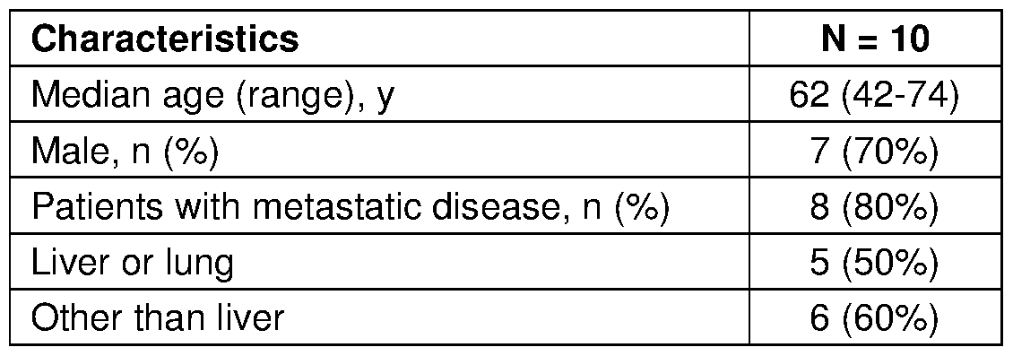
Claims
Priority Applications (9)
| Application Number | Priority Date | Filing Date | Title |
|---|---|---|---|
| CN201780030399.XA CN109154027A (en) | 2016-04-15 | 2017-04-14 | For monitoring and the method for the treatment of cancer |
| EP17735671.4A EP3443120A2 (en) | 2016-04-15 | 2017-04-14 | Methods for monitoring and treating cancer |
| CA3020718A CA3020718A1 (en) | 2016-04-15 | 2017-04-14 | Methods for monitoring and treating cancer |
| AU2017248766A AU2017248766A1 (en) | 2016-04-15 | 2017-04-14 | Methods for monitoring and treating cancer |
| JP2018553880A JP2019515670A (en) | 2016-04-15 | 2017-04-14 | Methods for monitoring and treating cancer |
| KR1020187032958A KR20190003957A (en) | 2016-04-15 | 2017-04-14 | Cancer monitoring and treatment methods |
| MX2018012493A MX2018012493A (en) | 2016-04-15 | 2017-04-14 | Methods for monitoring and treating cancer. |
| IL262223A IL262223A (en) | 2016-04-15 | 2018-10-09 | Methods for monitoring and treating cancer |
| US16/159,134 US20190038734A1 (en) | 2016-04-15 | 2018-10-12 | Methods for monitoring and treating cancer |
Applications Claiming Priority (2)
| Application Number | Priority Date | Filing Date | Title |
|---|---|---|---|
| US201662323280P | 2016-04-15 | 2016-04-15 | |
| US62/323,280 | 2016-04-15 |
Related Child Applications (1)
| Application Number | Title | Priority Date | Filing Date |
|---|---|---|---|
| US16/159,134 Continuation US20190038734A1 (en) | 2016-04-15 | 2018-10-12 | Methods for monitoring and treating cancer |
Publications (2)
| Publication Number | Publication Date |
|---|---|
| WO2017181079A2 true WO2017181079A2 (en) | 2017-10-19 |
| WO2017181079A3 WO2017181079A3 (en) | 2017-12-28 |
Family
ID=59285315
Family Applications (1)
| Application Number | Title | Priority Date | Filing Date |
|---|---|---|---|
| PCT/US2017/027725 WO2017181079A2 (en) | 2016-04-15 | 2017-04-14 | Methods for monitoring and treating cancer |
Country Status (10)
| Country | Link |
|---|---|
| US (1) | US20190038734A1 (en) |
| EP (1) | EP3443120A2 (en) |
| JP (1) | JP2019515670A (en) |
| KR (1) | KR20190003957A (en) |
| CN (1) | CN109154027A (en) |
| AU (1) | AU2017248766A1 (en) |
| CA (1) | CA3020718A1 (en) |
| IL (1) | IL262223A (en) |
| MX (1) | MX2018012493A (en) |
| WO (1) | WO2017181079A2 (en) |
Cited By (6)
| Publication number | Priority date | Publication date | Assignee | Title |
|---|---|---|---|---|
| US11211144B2 (en) | 2020-02-18 | 2021-12-28 | Tempus Labs, Inc. | Methods and systems for refining copy number variation in a liquid biopsy assay |
| US11211147B2 (en) | 2020-02-18 | 2021-12-28 | Tempus Labs, Inc. | Estimation of circulating tumor fraction using off-target reads of targeted-panel sequencing |
| US11410750B2 (en) | 2018-09-27 | 2022-08-09 | Grail, Llc | Methylation markers and targeted methylation probe panel |
| US11475981B2 (en) | 2020-02-18 | 2022-10-18 | Tempus Labs, Inc. | Methods and systems for dynamic variant thresholding in a liquid biopsy assay |
| US20230082122A1 (en) * | 2017-03-01 | 2023-03-16 | Genentech, Inc. | Diagnostic and therapeutic methods for cancer |
| US11780922B2 (en) | 2018-02-28 | 2023-10-10 | Ap Biosciences, Inc. | Bifunctional proteins combining checkpoint blockade for targeted therapy |
Families Citing this family (4)
| Publication number | Priority date | Publication date | Assignee | Title |
|---|---|---|---|---|
| KR102186571B1 (en) * | 2019-03-13 | 2020-12-03 | 울산대학교 산학협력단 | Relapse Prediction Method for patient with breast cancer Using immune response differential gene expression Model |
| KR102186570B1 (en) * | 2019-03-13 | 2020-12-03 | 울산대학교 산학협력단 | Complete Remission Prediction Method for patient with Triple negative breast cancer Using immune response differential gene expression prediction Model |
| KR20210052709A (en) * | 2019-10-30 | 2021-05-11 | 사회복지법인 삼성생명공익재단 | CXCL13 marker predictive of responsiveness to immunotherapy in a patient with lung cancer and use thereof |
| KR102396763B1 (en) * | 2020-04-09 | 2022-05-12 | 인하대학교 산학협력단 | Biomarker composition for predicting prognosis to therapy in a patient with triple-negative breast cancer comprising human umbilical vein endothelial cell derived cxcl11 |
Citations (152)
| Publication number | Priority date | Publication date | Assignee | Title |
|---|---|---|---|---|
| US3773919A (en) | 1969-10-23 | 1973-11-20 | Du Pont | Polylactide-drug mixtures |
| US4275149A (en) | 1978-11-24 | 1981-06-23 | Syva Company | Macromolecular environment control in specific receptor assays |
| US4318980A (en) | 1978-04-10 | 1982-03-09 | Miles Laboratories, Inc. | Heterogenous specific binding assay employing a cycling reactant as label |
| US4676980A (en) | 1985-09-23 | 1987-06-30 | The United States Of America As Represented By The Secretary Of The Department Of Health And Human Services | Target specific cross-linked heteroantibodies |
| US4683195A (en) | 1986-01-30 | 1987-07-28 | Cetus Corporation | Process for amplifying, detecting, and/or-cloning nucleic acid sequences |
| US4737456A (en) | 1985-05-09 | 1988-04-12 | Syntex (U.S.A.) Inc. | Reducing interference in ligand-receptor binding assays |
| US4816567A (en) | 1983-04-08 | 1989-03-28 | Genentech, Inc. | Recombinant immunoglobin preparations |
| WO1989006692A1 (en) | 1988-01-12 | 1989-07-27 | Genentech, Inc. | Method of treating tumor cells by inhibiting growth factor receptor function |
| US4943533A (en) | 1984-03-01 | 1990-07-24 | The Regents Of The University Of California | Hybrid cell lines that produce monoclonal antibodies to epidermal growth factor receptor |
| EP0404097A2 (en) | 1989-06-22 | 1990-12-27 | BEHRINGWERKE Aktiengesellschaft | Bispecific and oligospecific, mono- and oligovalent receptors, production and applications thereof |
| WO1991010741A1 (en) | 1990-01-12 | 1991-07-25 | Cell Genesys, Inc. | Generation of xenogeneic antibodies |
| WO1993001161A1 (en) | 1991-07-11 | 1993-01-21 | Pfizer Limited | Process for preparing sertraline intermediates |
| US5208020A (en) | 1989-10-25 | 1993-05-04 | Immunogen Inc. | Cytotoxic agents comprising maytansinoids and their therapeutic use |
| WO1993008829A1 (en) | 1991-11-04 | 1993-05-13 | The Regents Of The University Of California | Compositions that mediate killing of hiv-infected cells |
| US5212290A (en) | 1989-09-08 | 1993-05-18 | The Johns Hopkins University | Antibodies specific for type II mutant EGTR |
| WO1993016185A2 (en) | 1992-02-06 | 1993-08-19 | Creative Biomolecules, Inc. | Biosynthetic binding protein for cancer marker |
| WO1994010202A1 (en) | 1992-10-28 | 1994-05-11 | Genentech, Inc. | Vascular endothelial cell growth factor antagonists |
| WO1994011026A2 (en) | 1992-11-13 | 1994-05-26 | Idec Pharmaceuticals Corporation | Therapeutic application of chimeric and radiolabeled antibodies to human b lymphocyte restricted differentiation antigen for treatment of b cell lymphoma |
| WO1994029351A2 (en) | 1993-06-16 | 1994-12-22 | Celltech Limited | Antibodies |
| US5428130A (en) | 1989-02-23 | 1995-06-27 | Genentech, Inc. | Hybrid immunoglobulins |
| EP0659439A2 (en) | 1993-12-24 | 1995-06-28 | MERCK PATENT GmbH | Immunoconjugates |
| US5457105A (en) | 1992-01-20 | 1995-10-10 | Zeneca Limited | Quinazoline derivatives useful for treatment of neoplastic disease |
| WO1995027062A1 (en) | 1994-04-04 | 1995-10-12 | Genentech, Inc. | Agonist antibodies against the flk2/flt3 receptor and uses thereof |
| US5475001A (en) | 1993-07-19 | 1995-12-12 | Zeneca Limited | Quinazoline derivatives |
| WO1996003397A1 (en) | 1994-07-21 | 1996-02-08 | Akzo Nobel N.V. | Cyclic ketone peroxide formulations |
| US5500362A (en) | 1987-01-08 | 1996-03-19 | Xoma Corporation | Chimeric antibody with specificity to human B cell surface antigen |
| US5545807A (en) | 1988-10-12 | 1996-08-13 | The Babraham Institute | Production of antibodies from transgenic animals |
| US5545806A (en) | 1990-08-29 | 1996-08-13 | Genpharm International, Inc. | Ransgenic non-human animals for producing heterologous antibodies |
| EP0425235B1 (en) | 1989-10-25 | 1996-09-25 | Immunogen Inc | Cytotoxic agents comprising maytansinoids and their therapeutic use |
| WO1996030347A1 (en) | 1995-03-30 | 1996-10-03 | Pfizer Inc. | Quinazoline derivatives |
| WO1996030046A1 (en) | 1995-03-30 | 1996-10-03 | Genentech, Inc. | Vascular endothelial cell growth factor antagonists |
| US5569825A (en) | 1990-08-29 | 1996-10-29 | Genpharm International | Transgenic non-human animals capable of producing heterologous antibodies of various isotypes |
| WO1996033980A1 (en) | 1995-04-27 | 1996-10-31 | Zeneca Limited | Quinazoline derivatives |
| WO1996033735A1 (en) | 1995-04-27 | 1996-10-31 | Abgenix, Inc. | Human antibodies derived from immunized xenomice |
| WO1996034096A1 (en) | 1995-04-28 | 1996-10-31 | Abgenix, Inc. | Human antibodies derived from immunized xenomice |
| WO1996033978A1 (en) | 1995-04-27 | 1996-10-31 | Zeneca Limited | Quinazoline derivative |
| US5571894A (en) | 1991-02-05 | 1996-11-05 | Ciba-Geigy Corporation | Recombinant antibodies specific for a growth factor receptor |
| WO1996040210A1 (en) | 1995-06-07 | 1996-12-19 | Imclone Systems Incorporated | Antibody and antibody fragments for inhibiting the growth of tumors |
| US5587458A (en) | 1991-10-07 | 1996-12-24 | Aronex Pharmaceuticals, Inc. | Anti-erbB-2 antibodies, combinations thereof, and therapeutic and diagnostic uses thereof |
| US5624821A (en) | 1987-03-18 | 1997-04-29 | Scotgen Biopharmaceuticals Incorporated | Antibodies with altered effector functions |
| US5625126A (en) | 1990-08-29 | 1997-04-29 | Genpharm International, Inc. | Transgenic non-human animals for producing heterologous antibodies |
| US5633425A (en) | 1990-08-29 | 1997-05-27 | Genpharm International, Inc. | Transgenic non-human animals capable of producing heterologous antibodies |
| US5635483A (en) | 1992-12-03 | 1997-06-03 | Arizona Board Of Regents Acting On Behalf Of Arizona State University | Tumor inhibiting tetrapeptide bearing modified phenethyl amides |
| US5654307A (en) | 1994-01-25 | 1997-08-05 | Warner-Lambert Company | Bicyclic compounds capable of inhibiting tyrosine kinases of the epidermal growth factor receptor family |
| WO1997030087A1 (en) | 1996-02-16 | 1997-08-21 | Glaxo Group Limited | Preparation of glycosylated antibodies |
| US5661016A (en) | 1990-08-29 | 1997-08-26 | Genpharm International Inc. | Transgenic non-human animals capable of producing heterologous antibodies of various isotypes |
| WO1997038983A1 (en) | 1996-04-12 | 1997-10-23 | Warner-Lambert Company | Irreversible inhibitors of tyrosine kinases |
| US5712374A (en) | 1995-06-07 | 1998-01-27 | American Cyanamid Company | Method for the preparation of substantiallly monomeric calicheamicin derivative/carrier conjugates |
| US5714586A (en) | 1995-06-07 | 1998-02-03 | American Cyanamid Company | Methods for the preparation of monomeric calicheamicin derivative/carrier conjugates |
| US5731168A (en) | 1995-03-01 | 1998-03-24 | Genentech, Inc. | Method for making heteromultimeric polypeptides |
| WO1998014451A1 (en) | 1996-10-02 | 1998-04-09 | Novartis Ag | Fused pyrazole derivative and process for its preparation |
| US5739116A (en) | 1994-06-03 | 1998-04-14 | American Cyanamid Company | Enediyne derivatives useful for the synthesis of conjugates of methyltrithio antitumor agents |
| US5747498A (en) | 1996-05-28 | 1998-05-05 | Pfizer Inc. | Alkynyl and azido-substituted 4-anilinoquinazolines |
| US5750373A (en) | 1990-12-03 | 1998-05-12 | Genentech, Inc. | Enrichment method for variant proteins having altered binding properties, M13 phagemids, and growth hormone variants |
| US5760041A (en) | 1996-02-05 | 1998-06-02 | American Cyanamid Company | 4-aminoquinazoline EGFR Inhibitors |
| WO1998024893A2 (en) | 1996-12-03 | 1998-06-11 | Abgenix, Inc. | TRANSGENIC MAMMALS HAVING HUMAN IG LOCI INCLUDING PLURAL VH AND Vλ REGIONS AND ANTIBODIES PRODUCED THEREFROM |
| US5770710A (en) | 1987-10-30 | 1998-06-23 | American Cyanamid Company | Antitumor and antibacterial substituted disulfide derivatives prepared from compounds possessing a methlytrithio group |
| US5770701A (en) | 1987-10-30 | 1998-06-23 | American Cyanamid Company | Process for preparing targeted forms of methyltrithio antitumor agents |
| US5770429A (en) | 1990-08-29 | 1998-06-23 | Genpharm International, Inc. | Transgenic non-human animals capable of producing heterologous antibodies |
| US5780588A (en) | 1993-01-26 | 1998-07-14 | Arizona Board Of Regents | Elucidation and synthesis of selected pentapeptides |
| US5804396A (en) | 1994-10-12 | 1998-09-08 | Sugen, Inc. | Assay for agents active in proliferative disorders |
| WO1998043960A1 (en) | 1997-04-03 | 1998-10-08 | American Cyanamid Company | Substituted 3-cyano quinolines |
| US5821337A (en) | 1991-06-14 | 1998-10-13 | Genentech, Inc. | Immunoglobulin variants |
| WO1998045332A2 (en) | 1997-04-07 | 1998-10-15 | Genentech, Inc. | Humanized antibodies and methods for forming humanized antibodies |
| WO1998050433A2 (en) | 1997-05-05 | 1998-11-12 | Abgenix, Inc. | Human monoclonal antibodies to epidermal growth factor receptor |
| WO1998050038A1 (en) | 1997-05-06 | 1998-11-12 | American Cyanamid Company | Use of quinazoline compounds for the treatment of polycystic kidney disease |
| WO1998058964A1 (en) | 1997-06-24 | 1998-12-30 | Genentech, Inc. | Methods and compositions for galactosylated glycoproteins |
| US5866572A (en) | 1996-02-14 | 1999-02-02 | Zeneca Limited | Quinazoline derivatives |
| US5869046A (en) | 1995-04-14 | 1999-02-09 | Genentech, Inc. | Altered polypeptides with increased half-life |
| WO1999006378A1 (en) | 1997-07-29 | 1999-02-11 | Warner-Lambert Company | Irreversible inhibitors of tyrosine kinases |
| WO1999006396A1 (en) | 1997-07-29 | 1999-02-11 | Warner-Lambert Company | Irreversible bicyclic inhibitors of tyrosine kinases |
| WO1999009016A1 (en) | 1997-08-01 | 1999-02-25 | American Cyanamid Company | Substituted quinazoline derivatives and their use as tyrosine kinase inhibitors |
| US5891996A (en) | 1972-09-17 | 1999-04-06 | Centro De Inmunologia Molecular | Humanized and chimeric monoclonal antibodies that recognize epidermal growth factor receptor (EGF-R); diagnostic and therapeutic use |
| WO1999022764A1 (en) | 1997-10-31 | 1999-05-14 | Genentech, Inc. | Methods and compositions comprising glycoprotein glycoforms |
| WO1999024037A1 (en) | 1997-11-06 | 1999-05-20 | American Cyanamid Company | Use of quinazoline derivatives as tyrosine kinase inhibitors for treating colonic polyps |
| WO1999051642A1 (en) | 1998-04-02 | 1999-10-14 | Genentech, Inc. | Antibody variants and fragments thereof |
| US6002008A (en) | 1997-04-03 | 1999-12-14 | American Cyanamid Company | Substituted 3-cyano quinolines |
| US6054297A (en) | 1991-06-14 | 2000-04-25 | Genentech, Inc. | Humanized antibodies and methods for making them |
| US6075181A (en) | 1990-01-12 | 2000-06-13 | Abgenix, Inc. | Human antibodies derived from immunized xenomice |
| US6084095A (en) | 1994-01-25 | 2000-07-04 | Warner-Lambert Company | Substituted pyrido[3,2-d]pyrimidines capable of inhibiting tyrosine kinases of the epidermal growth factor receptor family |
| WO2000061739A1 (en) | 1999-04-09 | 2000-10-19 | Kyowa Hakko Kogyo Co., Ltd. | Method for controlling the activity of immunologically functional molecule |
| US6140332A (en) | 1995-07-06 | 2000-10-31 | Novartis Ag | Pyrrolopyrimidines and processes for the preparation thereof |
| US6150584A (en) | 1990-01-12 | 2000-11-21 | Abgenix, Inc. | Human antibodies derived from immunized xenomice |
| US6194551B1 (en) | 1998-04-02 | 2001-02-27 | Genentech, Inc. | Polypeptide variants |
| WO2001029246A1 (en) | 1999-10-19 | 2001-04-26 | Kyowa Hakko Kogyo Co., Ltd. | Process for producing polypeptide |
| US6248516B1 (en) | 1988-11-11 | 2001-06-19 | Medical Research Council | Single domain ligands, receptors comprising said ligands methods for their production, and use of said ligands and receptors |
| US6344455B1 (en) | 1998-11-19 | 2002-02-05 | Warner-Lambert Company | N-[4-(3-chloro-4-fluoro-phenylamino)-7-(3-morpholin-4-yl-propoxy)-quinazolin-6-yl]-acrylamide, and irreversible inhibitor of tyrosine kinases |
| WO2002031140A1 (en) | 2000-10-06 | 2002-04-18 | Kyowa Hakko Kogyo Co., Ltd. | Cells producing antibody compositions |
| US6391874B1 (en) | 1996-07-13 | 2002-05-21 | Smithkline Beecham Corporation | Fused heterocyclic compounds as protein tyrosine kinase inhibitors |
| US20020164328A1 (en) | 2000-10-06 | 2002-11-07 | Toyohide Shinkawa | Process for purifying antibody |
| WO2003011878A2 (en) | 2001-08-03 | 2003-02-13 | Glycart Biotechnology Ag | Antibody glycosylation variants having increased antibody-dependent cellular cytotoxicity |
| US20030115614A1 (en) | 2000-10-06 | 2003-06-19 | Yutaka Kanda | Antibody composition-producing cell |
| US6582959B2 (en) | 1991-03-29 | 2003-06-24 | Genentech, Inc. | Antibodies to vascular endothelial cell growth factor |
| US6596726B1 (en) | 1994-01-25 | 2003-07-22 | Warner Lambert Company | Tricyclic compounds capable of inhibiting tyrosine kinases of the epidermal growth factor receptor family |
| US6602684B1 (en) | 1998-04-20 | 2003-08-05 | Glycart Biotechnology Ag | Glycosylation engineering of antibodies for improving antibody-dependent cellular cytotoxicity |
| US20030157108A1 (en) | 2001-10-25 | 2003-08-21 | Genentech, Inc. | Glycoprotein compositions |
| US6630579B2 (en) | 1999-12-29 | 2003-10-07 | Immunogen Inc. | Cytotoxic agents comprising modified doxorubicins and daunorubicins and their therapeutic use |
| US20030190317A1 (en) | 1997-04-07 | 2003-10-09 | Genentech, Inc. | Anti-VEGF antibodies |
| WO2003085119A1 (en) | 2002-04-09 | 2003-10-16 | Kyowa Hakko Kogyo Co., Ltd. | METHOD OF ENHANCING ACTIVITY OF ANTIBODY COMPOSITION OF BINDING TO FcϜ RECEPTOR IIIa |
| WO2003085107A1 (en) | 2002-04-09 | 2003-10-16 | Kyowa Hakko Kogyo Co., Ltd. | Cells with modified genome |
| WO2003084570A1 (en) | 2002-04-09 | 2003-10-16 | Kyowa Hakko Kogyo Co., Ltd. | DRUG CONTAINING ANTIBODY COMPOSITION APPROPRIATE FOR PATIENT SUFFERING FROM FcϜRIIIa POLYMORPHISM |
| US20030206899A1 (en) | 1991-03-29 | 2003-11-06 | Genentech, Inc. | Vascular endothelial cell growth factor antagonists |
| US6703020B1 (en) | 1999-04-28 | 2004-03-09 | Board Of Regents, The University Of Texas System | Antibody conjugate methods for selectively inhibiting VEGF |
| US20040093621A1 (en) | 2001-12-25 | 2004-05-13 | Kyowa Hakko Kogyo Co., Ltd | Antibody composition which specifically binds to CD20 |
| US6737056B1 (en) | 1999-01-15 | 2004-05-18 | Genentech, Inc. | Polypeptide variants with altered effector function |
| US20040109865A1 (en) | 2002-04-09 | 2004-06-10 | Kyowa Hakko Kogyo Co., Ltd. | Antibody composition-containing medicament |
| US20040110282A1 (en) | 2002-04-09 | 2004-06-10 | Kyowa Hakko Kogyo Co., Ltd. | Cells in which activity of the protein involved in transportation of GDP-fucose is reduced or lost |
| US20040132140A1 (en) | 2002-04-09 | 2004-07-08 | Kyowa Hakko Kogyo Co., Ltd. | Production process for antibody composition |
| WO2004056312A2 (en) | 2002-12-16 | 2004-07-08 | Genentech, Inc. | Immunoglobulin variants and uses thereof |
| US20050014934A1 (en) | 2002-10-15 | 2005-01-20 | Hinton Paul R. | Alteration of FcRn binding affinities or serum half-lives of antibodies by mutagenesis |
| WO2005012359A2 (en) | 2003-08-01 | 2005-02-10 | Genentech, Inc. | Anti-vegf antibodies |
| US20050079574A1 (en) | 2003-01-16 | 2005-04-14 | Genentech, Inc. | Synthetic antibody phage libraries |
| WO2005035586A1 (en) | 2003-10-08 | 2005-04-21 | Kyowa Hakko Kogyo Co., Ltd. | Fused protein composition |
| WO2005035778A1 (en) | 2003-10-09 | 2005-04-21 | Kyowa Hakko Kogyo Co., Ltd. | PROCESS FOR PRODUCING ANTIBODY COMPOSITION BY USING RNA INHIBITING THE FUNCTION OF α1,6-FUCOSYLTRANSFERASE |
| US6884879B1 (en) | 1997-04-07 | 2005-04-26 | Genentech, Inc. | Anti-VEGF antibodies |
| US20050112126A1 (en) | 1997-04-07 | 2005-05-26 | Genentech, Inc. | Anti-VEGF antibodies |
| US20050119455A1 (en) | 2002-06-03 | 2005-06-02 | Genentech, Inc. | Synthetic antibody phage libraries |
| US20050123546A1 (en) | 2003-11-05 | 2005-06-09 | Glycart Biotechnology Ag | Antigen binding molecules with increased Fc receptor binding affinity and effector function |
| WO2005053742A1 (en) | 2003-12-04 | 2005-06-16 | Kyowa Hakko Kogyo Co., Ltd. | Medicine containing antibody composition |
| US20050186208A1 (en) | 2003-05-30 | 2005-08-25 | Genentech, Inc. | Treatment with anti-VEGF antibodies |
| WO2005100402A1 (en) | 2004-04-13 | 2005-10-27 | F.Hoffmann-La Roche Ag | Anti-p-selectin antibodies |
| US20050266000A1 (en) | 2004-04-09 | 2005-12-01 | Genentech, Inc. | Variable domain library and uses |
| US6982321B2 (en) | 1986-03-27 | 2006-01-03 | Medical Research Council | Altered antibodies |
| US20060009360A1 (en) | 2004-06-25 | 2006-01-12 | Robert Pifer | New adjuvant composition |
| US20060025576A1 (en) | 2000-04-11 | 2006-02-02 | Genentech, Inc. | Multivalent antibodies and uses therefor |
| WO2006029879A2 (en) | 2004-09-17 | 2006-03-23 | F.Hoffmann-La Roche Ag | Anti-ox40l antibodies |
| US7041870B2 (en) | 2000-11-30 | 2006-05-09 | Medarex, Inc. | Transgenic transchromosomal rodents for making human antibodies |
| US7087409B2 (en) | 1997-12-05 | 2006-08-08 | The Scripps Research Institute | Humanization of murine antibody |
| WO2006121168A1 (en) | 2005-05-09 | 2006-11-16 | Ono Pharmaceutical Co., Ltd. | Human monoclonal antibodies to programmed death 1(pd-1) and methods for treating cancer using anti-pd-1 antibodies alone or in combination with other immunotherapeutics |
| WO2007005874A2 (en) | 2005-07-01 | 2007-01-11 | Medarex, Inc. | Human monoclonal antibodies to programmed death ligand 1 (pd-l1) |
| US7189826B2 (en) | 1997-11-24 | 2007-03-13 | Institute For Human Genetics And Biochemistry | Monoclonal human natural antibodies |
| US20070061900A1 (en) | 2000-10-31 | 2007-03-15 | Murphy Andrew J | Methods of modifying eukaryotic cells |
| US20070117126A1 (en) | 1999-12-15 | 2007-05-24 | Genentech, Inc. | Shotgun scanning |
| US20070160598A1 (en) | 2005-11-07 | 2007-07-12 | Dennis Mark S | Binding polypeptides with diversified and consensus vh/vl hypervariable sequences |
| US20070237764A1 (en) | 2005-12-02 | 2007-10-11 | Genentech, Inc. | Binding polypeptides with restricted diversity sequences |
| US20070292936A1 (en) | 2006-05-09 | 2007-12-20 | Genentech, Inc. | Binding polypeptides with optimized scaffolds |
| US7371826B2 (en) | 1999-01-15 | 2008-05-13 | Genentech, Inc. | Polypeptide variants with altered effector function |
| WO2008077546A1 (en) | 2006-12-22 | 2008-07-03 | F. Hoffmann-La Roche Ag | Antibodies against insulin-like growth factor i receptor and uses thereof |
| US20090002360A1 (en) | 2007-05-25 | 2009-01-01 | Innolux Display Corp. | Liquid crystal display device and method for driving same |
| US7498298B2 (en) | 2003-11-06 | 2009-03-03 | Seattle Genetics, Inc. | Monomethylvaline compounds capable of conjugation to ligands |
| US7521541B2 (en) | 2004-09-23 | 2009-04-21 | Genetech Inc. | Cysteine engineered antibodies and conjugates |
| US7527791B2 (en) | 2004-03-31 | 2009-05-05 | Genentech, Inc. | Humanized anti-TGF-beta antibodies |
| WO2009089004A1 (en) | 2008-01-07 | 2009-07-16 | Amgen Inc. | Method for making antibody fc-heterodimeric molecules using electrostatic steering effects |
| WO2009101611A1 (en) | 2008-02-11 | 2009-08-20 | Curetech Ltd. | Monoclonal antibodies for tumor treatment |
| WO2009114335A2 (en) | 2008-03-12 | 2009-09-17 | Merck & Co., Inc. | Pd-1 binding proteins |
| WO2010027827A2 (en) | 2008-08-25 | 2010-03-11 | Amplimmune, Inc. | Targeted costimulatory polypeptides and methods of use to treat cancer |
| WO2010077634A1 (en) | 2008-12-09 | 2010-07-08 | Genentech, Inc. | Anti-pd-l1 antibodies and their use to enhance t-cell function |
| WO2011066389A1 (en) | 2009-11-24 | 2011-06-03 | Medimmmune, Limited | Targeted binding agents against b7-h1 |
| WO2011066342A2 (en) | 2009-11-24 | 2011-06-03 | Amplimmune, Inc. | Simultaneous inhibition of pd-l1/pd-l2 |
| US20110236388A1 (en) | 2010-03-26 | 2011-09-29 | Monika Baehner | Bispecific, bivalent anti-vegf/anti-ang-2 antibodies |
| US8219149B2 (en) | 2005-06-29 | 2012-07-10 | Nokia Corporation | Mobile communication terminal |
| WO2013019906A1 (en) | 2011-08-01 | 2013-02-07 | Genentech, Inc. | Methods of treating cancer using pd-1 axis binding antagonists and mek inhibitors |
Family Cites Families (6)
| Publication number | Priority date | Publication date | Assignee | Title |
|---|---|---|---|---|
| CA2766403A1 (en) * | 2009-07-13 | 2011-01-20 | Genentech, Inc. | Diagnostic methods and compositions for treatment of cancer |
| ES2705950T3 (en) * | 2011-06-03 | 2019-03-27 | Eisai R&D Man Co Ltd | Biomarkers to predict and assess the responsiveness of subjects with thyroid and kidney cancer to lenvatinib compounds |
| MX356802B (en) * | 2012-01-13 | 2018-06-13 | Genentech Inc | Biological markers for identifying patients for treatment with vegf antagonists. |
| CN105102631A (en) * | 2012-12-03 | 2015-11-25 | 阿尔玛克诊断有限公司 | Molecular diagnostic test for cancer |
| CN105264380B (en) * | 2013-05-14 | 2017-09-05 | 卫材R&D管理有限公司 | Pleasure is cut down for the biological marker of Buddhist nun's compound response for predicting and evaluating carcinoma of endometrium subject |
| EP3443350B1 (en) * | 2016-04-15 | 2020-12-09 | H. Hoffnabb-La Roche Ag | Methods for monitoring and treating cancer |
-
2017
- 2017-04-14 EP EP17735671.4A patent/EP3443120A2/en not_active Withdrawn
- 2017-04-14 JP JP2018553880A patent/JP2019515670A/en active Pending
- 2017-04-14 WO PCT/US2017/027725 patent/WO2017181079A2/en active Application Filing
- 2017-04-14 CN CN201780030399.XA patent/CN109154027A/en active Pending
- 2017-04-14 AU AU2017248766A patent/AU2017248766A1/en not_active Abandoned
- 2017-04-14 MX MX2018012493A patent/MX2018012493A/en unknown
- 2017-04-14 KR KR1020187032958A patent/KR20190003957A/en not_active Application Discontinuation
- 2017-04-14 CA CA3020718A patent/CA3020718A1/en not_active Abandoned
-
2018
- 2018-10-09 IL IL262223A patent/IL262223A/en unknown
- 2018-10-12 US US16/159,134 patent/US20190038734A1/en not_active Abandoned
Patent Citations (176)
| Publication number | Priority date | Publication date | Assignee | Title |
|---|---|---|---|---|
| US3773919A (en) | 1969-10-23 | 1973-11-20 | Du Pont | Polylactide-drug mixtures |
| US5891996A (en) | 1972-09-17 | 1999-04-06 | Centro De Inmunologia Molecular | Humanized and chimeric monoclonal antibodies that recognize epidermal growth factor receptor (EGF-R); diagnostic and therapeutic use |
| US4318980A (en) | 1978-04-10 | 1982-03-09 | Miles Laboratories, Inc. | Heterogenous specific binding assay employing a cycling reactant as label |
| US4275149A (en) | 1978-11-24 | 1981-06-23 | Syva Company | Macromolecular environment control in specific receptor assays |
| US4816567A (en) | 1983-04-08 | 1989-03-28 | Genentech, Inc. | Recombinant immunoglobin preparations |
| US4943533A (en) | 1984-03-01 | 1990-07-24 | The Regents Of The University Of California | Hybrid cell lines that produce monoclonal antibodies to epidermal growth factor receptor |
| US4737456A (en) | 1985-05-09 | 1988-04-12 | Syntex (U.S.A.) Inc. | Reducing interference in ligand-receptor binding assays |
| US4676980A (en) | 1985-09-23 | 1987-06-30 | The United States Of America As Represented By The Secretary Of The Department Of Health And Human Services | Target specific cross-linked heteroantibodies |
| US4683195A (en) | 1986-01-30 | 1987-07-28 | Cetus Corporation | Process for amplifying, detecting, and/or-cloning nucleic acid sequences |
| US4683195B1 (en) | 1986-01-30 | 1990-11-27 | Cetus Corp | |
| US6982321B2 (en) | 1986-03-27 | 2006-01-03 | Medical Research Council | Altered antibodies |
| US5500362A (en) | 1987-01-08 | 1996-03-19 | Xoma Corporation | Chimeric antibody with specificity to human B cell surface antigen |
| US5648260A (en) | 1987-03-18 | 1997-07-15 | Scotgen Biopharmaceuticals Incorporated | DNA encoding antibodies with altered effector functions |
| US5624821A (en) | 1987-03-18 | 1997-04-29 | Scotgen Biopharmaceuticals Incorporated | Antibodies with altered effector functions |
| US5770710A (en) | 1987-10-30 | 1998-06-23 | American Cyanamid Company | Antitumor and antibacterial substituted disulfide derivatives prepared from compounds possessing a methlytrithio group |
| US5770701A (en) | 1987-10-30 | 1998-06-23 | American Cyanamid Company | Process for preparing targeted forms of methyltrithio antitumor agents |
| WO1989006692A1 (en) | 1988-01-12 | 1989-07-27 | Genentech, Inc. | Method of treating tumor cells by inhibiting growth factor receptor function |
| US5545807A (en) | 1988-10-12 | 1996-08-13 | The Babraham Institute | Production of antibodies from transgenic animals |
| US6248516B1 (en) | 1988-11-11 | 2001-06-19 | Medical Research Council | Single domain ligands, receptors comprising said ligands methods for their production, and use of said ligands and receptors |
| US5428130A (en) | 1989-02-23 | 1995-06-27 | Genentech, Inc. | Hybrid immunoglobulins |
| EP0404097A2 (en) | 1989-06-22 | 1990-12-27 | BEHRINGWERKE Aktiengesellschaft | Bispecific and oligospecific, mono- and oligovalent receptors, production and applications thereof |
| US5212290A (en) | 1989-09-08 | 1993-05-18 | The Johns Hopkins University | Antibodies specific for type II mutant EGTR |
| US5208020A (en) | 1989-10-25 | 1993-05-04 | Immunogen Inc. | Cytotoxic agents comprising maytansinoids and their therapeutic use |
| US5416064A (en) | 1989-10-25 | 1995-05-16 | Immunogen, Inc. | Cytotoxic agents comprising maytansinoids and their therapeutic use |
| EP0425235B1 (en) | 1989-10-25 | 1996-09-25 | Immunogen Inc | Cytotoxic agents comprising maytansinoids and their therapeutic use |
| US6150584A (en) | 1990-01-12 | 2000-11-21 | Abgenix, Inc. | Human antibodies derived from immunized xenomice |
| WO1991010741A1 (en) | 1990-01-12 | 1991-07-25 | Cell Genesys, Inc. | Generation of xenogeneic antibodies |
| US6075181A (en) | 1990-01-12 | 2000-06-13 | Abgenix, Inc. | Human antibodies derived from immunized xenomice |
| US5661016A (en) | 1990-08-29 | 1997-08-26 | Genpharm International Inc. | Transgenic non-human animals capable of producing heterologous antibodies of various isotypes |
| US5633425A (en) | 1990-08-29 | 1997-05-27 | Genpharm International, Inc. | Transgenic non-human animals capable of producing heterologous antibodies |
| US5625126A (en) | 1990-08-29 | 1997-04-29 | Genpharm International, Inc. | Transgenic non-human animals for producing heterologous antibodies |
| US5545806A (en) | 1990-08-29 | 1996-08-13 | Genpharm International, Inc. | Ransgenic non-human animals for producing heterologous antibodies |
| US5770429A (en) | 1990-08-29 | 1998-06-23 | Genpharm International, Inc. | Transgenic non-human animals capable of producing heterologous antibodies |
| US5569825A (en) | 1990-08-29 | 1996-10-29 | Genpharm International | Transgenic non-human animals capable of producing heterologous antibodies of various isotypes |
| US5750373A (en) | 1990-12-03 | 1998-05-12 | Genentech, Inc. | Enrichment method for variant proteins having altered binding properties, M13 phagemids, and growth hormone variants |
| US5571894A (en) | 1991-02-05 | 1996-11-05 | Ciba-Geigy Corporation | Recombinant antibodies specific for a growth factor receptor |
| US20030206899A1 (en) | 1991-03-29 | 2003-11-06 | Genentech, Inc. | Vascular endothelial cell growth factor antagonists |
| US6582959B2 (en) | 1991-03-29 | 2003-06-24 | Genentech, Inc. | Antibodies to vascular endothelial cell growth factor |
| US20030203409A1 (en) | 1991-03-29 | 2003-10-30 | Genentech, Inc. | Antibodies to vascular endothelial cell growth factor |
| US6054297A (en) | 1991-06-14 | 2000-04-25 | Genentech, Inc. | Humanized antibodies and methods for making them |
| US5821337A (en) | 1991-06-14 | 1998-10-13 | Genentech, Inc. | Immunoglobulin variants |
| WO1993001161A1 (en) | 1991-07-11 | 1993-01-21 | Pfizer Limited | Process for preparing sertraline intermediates |
| US5587458A (en) | 1991-10-07 | 1996-12-24 | Aronex Pharmaceuticals, Inc. | Anti-erbB-2 antibodies, combinations thereof, and therapeutic and diagnostic uses thereof |
| WO1993008829A1 (en) | 1991-11-04 | 1993-05-13 | The Regents Of The University Of California | Compositions that mediate killing of hiv-infected cells |
| US5616582A (en) | 1992-01-20 | 1997-04-01 | Zeneca Limited | Quinazoline derivatives as anti-proliferative agents |
| US5457105A (en) | 1992-01-20 | 1995-10-10 | Zeneca Limited | Quinazoline derivatives useful for treatment of neoplastic disease |
| WO1993016185A2 (en) | 1992-02-06 | 1993-08-19 | Creative Biomolecules, Inc. | Biosynthetic binding protein for cancer marker |
| WO1994010202A1 (en) | 1992-10-28 | 1994-05-11 | Genentech, Inc. | Vascular endothelial cell growth factor antagonists |
| EP0666868B1 (en) | 1992-10-28 | 2002-04-03 | Genentech, Inc. | Use of anti-VEGF antibodies for the treatment of cancer |
| WO1994011026A2 (en) | 1992-11-13 | 1994-05-26 | Idec Pharmaceuticals Corporation | Therapeutic application of chimeric and radiolabeled antibodies to human b lymphocyte restricted differentiation antigen for treatment of b cell lymphoma |
| US5635483A (en) | 1992-12-03 | 1997-06-03 | Arizona Board Of Regents Acting On Behalf Of Arizona State University | Tumor inhibiting tetrapeptide bearing modified phenethyl amides |
| US5780588A (en) | 1993-01-26 | 1998-07-14 | Arizona Board Of Regents | Elucidation and synthesis of selected pentapeptides |
| WO1994029351A2 (en) | 1993-06-16 | 1994-12-22 | Celltech Limited | Antibodies |
| US5475001A (en) | 1993-07-19 | 1995-12-12 | Zeneca Limited | Quinazoline derivatives |
| EP0659439A2 (en) | 1993-12-24 | 1995-06-28 | MERCK PATENT GmbH | Immunoconjugates |
| US6084095A (en) | 1994-01-25 | 2000-07-04 | Warner-Lambert Company | Substituted pyrido[3,2-d]pyrimidines capable of inhibiting tyrosine kinases of the epidermal growth factor receptor family |
| US6455534B2 (en) | 1994-01-25 | 2002-09-24 | Warner-Lambert Company | Bicyclic compounds capable of inhibiting tyrosine kinases of the epidermal growth factor receptor family |
| US5654307A (en) | 1994-01-25 | 1997-08-05 | Warner-Lambert Company | Bicyclic compounds capable of inhibiting tyrosine kinases of the epidermal growth factor receptor family |
| US6521620B1 (en) | 1994-01-25 | 2003-02-18 | Warner-Lambert Company | Bicyclic compounds capable of inhibiting tyrosine kinases of the epidermal growth factor receptor family |
| US6713484B2 (en) | 1994-01-25 | 2004-03-30 | Warner-Lambert Company | Bicyclic compounds capable of inhibiting tyrosine kinases of the epidermal growth factor receptor family |
| US6596726B1 (en) | 1994-01-25 | 2003-07-22 | Warner Lambert Company | Tricyclic compounds capable of inhibiting tyrosine kinases of the epidermal growth factor receptor family |
| US6265410B1 (en) | 1994-01-25 | 2001-07-24 | Warner-Lambert Company | Bicyclic compounds capable of inhibiting tyrosine kinases of the epidermal growth factor receptor family |
| US5679683A (en) | 1994-01-25 | 1997-10-21 | Warner-Lambert Company | Tricyclic compounds capable of inhibiting tyrosine kinases of the epidermal growth factor receptor family |
| WO1995027062A1 (en) | 1994-04-04 | 1995-10-12 | Genentech, Inc. | Agonist antibodies against the flk2/flt3 receptor and uses thereof |
| US5877296A (en) | 1994-06-03 | 1999-03-02 | American Cyanamid Company | Process for preparing conjugates of methyltrithio antitumor agents |
| US5773001A (en) | 1994-06-03 | 1998-06-30 | American Cyanamid Company | Conjugates of methyltrithio antitumor agents and intermediates for their synthesis |
| US5739116A (en) | 1994-06-03 | 1998-04-14 | American Cyanamid Company | Enediyne derivatives useful for the synthesis of conjugates of methyltrithio antitumor agents |
| US5767285A (en) | 1994-06-03 | 1998-06-16 | American Cyanamid Company | Linkers useful for the synthesis of conjugates of methyltrithio antitumor agents |
| WO1996003397A1 (en) | 1994-07-21 | 1996-02-08 | Akzo Nobel N.V. | Cyclic ketone peroxide formulations |
| US5804396A (en) | 1994-10-12 | 1998-09-08 | Sugen, Inc. | Assay for agents active in proliferative disorders |
| US5731168A (en) | 1995-03-01 | 1998-03-24 | Genentech, Inc. | Method for making heteromultimeric polypeptides |
| WO1996030347A1 (en) | 1995-03-30 | 1996-10-03 | Pfizer Inc. | Quinazoline derivatives |
| WO1996030046A1 (en) | 1995-03-30 | 1996-10-03 | Genentech, Inc. | Vascular endothelial cell growth factor antagonists |
| US5869046A (en) | 1995-04-14 | 1999-02-09 | Genentech, Inc. | Altered polypeptides with increased half-life |
| WO1996033978A1 (en) | 1995-04-27 | 1996-10-31 | Zeneca Limited | Quinazoline derivative |
| WO1996033980A1 (en) | 1995-04-27 | 1996-10-31 | Zeneca Limited | Quinazoline derivatives |
| WO1996033735A1 (en) | 1995-04-27 | 1996-10-31 | Abgenix, Inc. | Human antibodies derived from immunized xenomice |
| US5770599A (en) | 1995-04-27 | 1998-06-23 | Zeneca Limited | Quinazoline derivatives |
| WO1996034096A1 (en) | 1995-04-28 | 1996-10-31 | Abgenix, Inc. | Human antibodies derived from immunized xenomice |
| WO1996040210A1 (en) | 1995-06-07 | 1996-12-19 | Imclone Systems Incorporated | Antibody and antibody fragments for inhibiting the growth of tumors |
| US5712374A (en) | 1995-06-07 | 1998-01-27 | American Cyanamid Company | Method for the preparation of substantiallly monomeric calicheamicin derivative/carrier conjugates |
| US5714586A (en) | 1995-06-07 | 1998-02-03 | American Cyanamid Company | Methods for the preparation of monomeric calicheamicin derivative/carrier conjugates |
| US6140332A (en) | 1995-07-06 | 2000-10-31 | Novartis Ag | Pyrrolopyrimidines and processes for the preparation thereof |
| US5760041A (en) | 1996-02-05 | 1998-06-02 | American Cyanamid Company | 4-aminoquinazoline EGFR Inhibitors |
| US5866572A (en) | 1996-02-14 | 1999-02-02 | Zeneca Limited | Quinazoline derivatives |
| US6399602B1 (en) | 1996-02-14 | 2002-06-04 | Zeneca Limited | Quinazoline derivatives |
| WO1997030087A1 (en) | 1996-02-16 | 1997-08-21 | Glaxo Group Limited | Preparation of glycosylated antibodies |
| WO1997038983A1 (en) | 1996-04-12 | 1997-10-23 | Warner-Lambert Company | Irreversible inhibitors of tyrosine kinases |
| US6344459B1 (en) | 1996-04-12 | 2002-02-05 | Warner-Lambert Company | Irreversible inhibitors of tyrosine kinases |
| US6602863B1 (en) | 1996-04-12 | 2003-08-05 | Warner-Lambert Company | Irreversible inhibitors of tyrosine kinases |
| US5747498A (en) | 1996-05-28 | 1998-05-05 | Pfizer Inc. | Alkynyl and azido-substituted 4-anilinoquinazolines |
| US6391874B1 (en) | 1996-07-13 | 2002-05-21 | Smithkline Beecham Corporation | Fused heterocyclic compounds as protein tyrosine kinase inhibitors |
| WO1998014451A1 (en) | 1996-10-02 | 1998-04-09 | Novartis Ag | Fused pyrazole derivative and process for its preparation |
| WO1998024893A2 (en) | 1996-12-03 | 1998-06-11 | Abgenix, Inc. | TRANSGENIC MAMMALS HAVING HUMAN IG LOCI INCLUDING PLURAL VH AND Vλ REGIONS AND ANTIBODIES PRODUCED THEREFROM |
| US6002008A (en) | 1997-04-03 | 1999-12-14 | American Cyanamid Company | Substituted 3-cyano quinolines |
| WO1998043960A1 (en) | 1997-04-03 | 1998-10-08 | American Cyanamid Company | Substituted 3-cyano quinolines |
| US20050112126A1 (en) | 1997-04-07 | 2005-05-26 | Genentech, Inc. | Anti-VEGF antibodies |
| WO1998045332A2 (en) | 1997-04-07 | 1998-10-15 | Genentech, Inc. | Humanized antibodies and methods for forming humanized antibodies |
| US6884879B1 (en) | 1997-04-07 | 2005-04-26 | Genentech, Inc. | Anti-VEGF antibodies |
| US7060269B1 (en) | 1997-04-07 | 2006-06-13 | Genentech, Inc. | Anti-VEGF antibodies |
| US20030190317A1 (en) | 1997-04-07 | 2003-10-09 | Genentech, Inc. | Anti-VEGF antibodies |
| WO1998050433A2 (en) | 1997-05-05 | 1998-11-12 | Abgenix, Inc. | Human monoclonal antibodies to epidermal growth factor receptor |
| US6235883B1 (en) | 1997-05-05 | 2001-05-22 | Abgenix, Inc. | Human monoclonal antibodies to epidermal growth factor receptor |
| WO1998050038A1 (en) | 1997-05-06 | 1998-11-12 | American Cyanamid Company | Use of quinazoline compounds for the treatment of polycystic kidney disease |
| WO1998058964A1 (en) | 1997-06-24 | 1998-12-30 | Genentech, Inc. | Methods and compositions for galactosylated glycoproteins |
| WO1999006378A1 (en) | 1997-07-29 | 1999-02-11 | Warner-Lambert Company | Irreversible inhibitors of tyrosine kinases |
| WO1999006396A1 (en) | 1997-07-29 | 1999-02-11 | Warner-Lambert Company | Irreversible bicyclic inhibitors of tyrosine kinases |
| WO1999009016A1 (en) | 1997-08-01 | 1999-02-25 | American Cyanamid Company | Substituted quinazoline derivatives and their use as tyrosine kinase inhibitors |
| WO1999022764A1 (en) | 1997-10-31 | 1999-05-14 | Genentech, Inc. | Methods and compositions comprising glycoprotein glycoforms |
| WO1999024037A1 (en) | 1997-11-06 | 1999-05-20 | American Cyanamid Company | Use of quinazoline derivatives as tyrosine kinase inhibitors for treating colonic polyps |
| US7189826B2 (en) | 1997-11-24 | 2007-03-13 | Institute For Human Genetics And Biochemistry | Monoclonal human natural antibodies |
| US7087409B2 (en) | 1997-12-05 | 2006-08-08 | The Scripps Research Institute | Humanization of murine antibody |
| US6194551B1 (en) | 1998-04-02 | 2001-02-27 | Genentech, Inc. | Polypeptide variants |
| WO1999051642A1 (en) | 1998-04-02 | 1999-10-14 | Genentech, Inc. | Antibody variants and fragments thereof |
| US6602684B1 (en) | 1998-04-20 | 2003-08-05 | Glycart Biotechnology Ag | Glycosylation engineering of antibodies for improving antibody-dependent cellular cytotoxicity |
| US6344455B1 (en) | 1998-11-19 | 2002-02-05 | Warner-Lambert Company | N-[4-(3-chloro-4-fluoro-phenylamino)-7-(3-morpholin-4-yl-propoxy)-quinazolin-6-yl]-acrylamide, and irreversible inhibitor of tyrosine kinases |
| US7371826B2 (en) | 1999-01-15 | 2008-05-13 | Genentech, Inc. | Polypeptide variants with altered effector function |
| US7332581B2 (en) | 1999-01-15 | 2008-02-19 | Genentech, Inc. | Polypeptide variants with altered effector function |
| US6737056B1 (en) | 1999-01-15 | 2004-05-18 | Genentech, Inc. | Polypeptide variants with altered effector function |
| WO2000061739A1 (en) | 1999-04-09 | 2000-10-19 | Kyowa Hakko Kogyo Co., Ltd. | Method for controlling the activity of immunologically functional molecule |
| US6703020B1 (en) | 1999-04-28 | 2004-03-09 | Board Of Regents, The University Of Texas System | Antibody conjugate methods for selectively inhibiting VEGF |
| WO2001029246A1 (en) | 1999-10-19 | 2001-04-26 | Kyowa Hakko Kogyo Co., Ltd. | Process for producing polypeptide |
| US20070117126A1 (en) | 1999-12-15 | 2007-05-24 | Genentech, Inc. | Shotgun scanning |
| US6630579B2 (en) | 1999-12-29 | 2003-10-07 | Immunogen Inc. | Cytotoxic agents comprising modified doxorubicins and daunorubicins and their therapeutic use |
| US20060025576A1 (en) | 2000-04-11 | 2006-02-02 | Genentech, Inc. | Multivalent antibodies and uses therefor |
| US20020164328A1 (en) | 2000-10-06 | 2002-11-07 | Toyohide Shinkawa | Process for purifying antibody |
| US20030115614A1 (en) | 2000-10-06 | 2003-06-19 | Yutaka Kanda | Antibody composition-producing cell |
| WO2002031140A1 (en) | 2000-10-06 | 2002-04-18 | Kyowa Hakko Kogyo Co., Ltd. | Cells producing antibody compositions |
| US20070061900A1 (en) | 2000-10-31 | 2007-03-15 | Murphy Andrew J | Methods of modifying eukaryotic cells |
| US7041870B2 (en) | 2000-11-30 | 2006-05-09 | Medarex, Inc. | Transgenic transchromosomal rodents for making human antibodies |
| WO2003011878A2 (en) | 2001-08-03 | 2003-02-13 | Glycart Biotechnology Ag | Antibody glycosylation variants having increased antibody-dependent cellular cytotoxicity |
| US20030157108A1 (en) | 2001-10-25 | 2003-08-21 | Genentech, Inc. | Glycoprotein compositions |
| US20040093621A1 (en) | 2001-12-25 | 2004-05-13 | Kyowa Hakko Kogyo Co., Ltd | Antibody composition which specifically binds to CD20 |
| US20040132140A1 (en) | 2002-04-09 | 2004-07-08 | Kyowa Hakko Kogyo Co., Ltd. | Production process for antibody composition |
| WO2003085107A1 (en) | 2002-04-09 | 2003-10-16 | Kyowa Hakko Kogyo Co., Ltd. | Cells with modified genome |
| WO2003085119A1 (en) | 2002-04-09 | 2003-10-16 | Kyowa Hakko Kogyo Co., Ltd. | METHOD OF ENHANCING ACTIVITY OF ANTIBODY COMPOSITION OF BINDING TO FcϜ RECEPTOR IIIa |
| WO2003084570A1 (en) | 2002-04-09 | 2003-10-16 | Kyowa Hakko Kogyo Co., Ltd. | DRUG CONTAINING ANTIBODY COMPOSITION APPROPRIATE FOR PATIENT SUFFERING FROM FcϜRIIIa POLYMORPHISM |
| US20040110704A1 (en) | 2002-04-09 | 2004-06-10 | Kyowa Hakko Kogyo Co., Ltd. | Cells of which genome is modified |
| US20040109865A1 (en) | 2002-04-09 | 2004-06-10 | Kyowa Hakko Kogyo Co., Ltd. | Antibody composition-containing medicament |
| US20040110282A1 (en) | 2002-04-09 | 2004-06-10 | Kyowa Hakko Kogyo Co., Ltd. | Cells in which activity of the protein involved in transportation of GDP-fucose is reduced or lost |
| US20050119455A1 (en) | 2002-06-03 | 2005-06-02 | Genentech, Inc. | Synthetic antibody phage libraries |
| US20050014934A1 (en) | 2002-10-15 | 2005-01-20 | Hinton Paul R. | Alteration of FcRn binding affinities or serum half-lives of antibodies by mutagenesis |
| WO2004056312A2 (en) | 2002-12-16 | 2004-07-08 | Genentech, Inc. | Immunoglobulin variants and uses thereof |
| US20050079574A1 (en) | 2003-01-16 | 2005-04-14 | Genentech, Inc. | Synthetic antibody phage libraries |
| US20050186208A1 (en) | 2003-05-30 | 2005-08-25 | Genentech, Inc. | Treatment with anti-VEGF antibodies |
| WO2005012359A2 (en) | 2003-08-01 | 2005-02-10 | Genentech, Inc. | Anti-vegf antibodies |
| WO2005035586A1 (en) | 2003-10-08 | 2005-04-21 | Kyowa Hakko Kogyo Co., Ltd. | Fused protein composition |
| WO2005035778A1 (en) | 2003-10-09 | 2005-04-21 | Kyowa Hakko Kogyo Co., Ltd. | PROCESS FOR PRODUCING ANTIBODY COMPOSITION BY USING RNA INHIBITING THE FUNCTION OF α1,6-FUCOSYLTRANSFERASE |
| US20050123546A1 (en) | 2003-11-05 | 2005-06-09 | Glycart Biotechnology Ag | Antigen binding molecules with increased Fc receptor binding affinity and effector function |
| US7498298B2 (en) | 2003-11-06 | 2009-03-03 | Seattle Genetics, Inc. | Monomethylvaline compounds capable of conjugation to ligands |
| WO2005053742A1 (en) | 2003-12-04 | 2005-06-16 | Kyowa Hakko Kogyo Co., Ltd. | Medicine containing antibody composition |
| US7527791B2 (en) | 2004-03-31 | 2009-05-05 | Genentech, Inc. | Humanized anti-TGF-beta antibodies |
| US20050266000A1 (en) | 2004-04-09 | 2005-12-01 | Genentech, Inc. | Variable domain library and uses |
| WO2005100402A1 (en) | 2004-04-13 | 2005-10-27 | F.Hoffmann-La Roche Ag | Anti-p-selectin antibodies |
| US20060009360A1 (en) | 2004-06-25 | 2006-01-12 | Robert Pifer | New adjuvant composition |
| WO2006029879A2 (en) | 2004-09-17 | 2006-03-23 | F.Hoffmann-La Roche Ag | Anti-ox40l antibodies |
| US7521541B2 (en) | 2004-09-23 | 2009-04-21 | Genetech Inc. | Cysteine engineered antibodies and conjugates |
| WO2006121168A1 (en) | 2005-05-09 | 2006-11-16 | Ono Pharmaceutical Co., Ltd. | Human monoclonal antibodies to programmed death 1(pd-1) and methods for treating cancer using anti-pd-1 antibodies alone or in combination with other immunotherapeutics |
| US8219149B2 (en) | 2005-06-29 | 2012-07-10 | Nokia Corporation | Mobile communication terminal |
| WO2007005874A2 (en) | 2005-07-01 | 2007-01-11 | Medarex, Inc. | Human monoclonal antibodies to programmed death ligand 1 (pd-l1) |
| US20070160598A1 (en) | 2005-11-07 | 2007-07-12 | Dennis Mark S | Binding polypeptides with diversified and consensus vh/vl hypervariable sequences |
| US20070237764A1 (en) | 2005-12-02 | 2007-10-11 | Genentech, Inc. | Binding polypeptides with restricted diversity sequences |
| US20070292936A1 (en) | 2006-05-09 | 2007-12-20 | Genentech, Inc. | Binding polypeptides with optimized scaffolds |
| WO2008077546A1 (en) | 2006-12-22 | 2008-07-03 | F. Hoffmann-La Roche Ag | Antibodies against insulin-like growth factor i receptor and uses thereof |
| US20090002360A1 (en) | 2007-05-25 | 2009-01-01 | Innolux Display Corp. | Liquid crystal display device and method for driving same |
| WO2009089004A1 (en) | 2008-01-07 | 2009-07-16 | Amgen Inc. | Method for making antibody fc-heterodimeric molecules using electrostatic steering effects |
| WO2009101611A1 (en) | 2008-02-11 | 2009-08-20 | Curetech Ltd. | Monoclonal antibodies for tumor treatment |
| WO2009114335A2 (en) | 2008-03-12 | 2009-09-17 | Merck & Co., Inc. | Pd-1 binding proteins |
| WO2010027827A2 (en) | 2008-08-25 | 2010-03-11 | Amplimmune, Inc. | Targeted costimulatory polypeptides and methods of use to treat cancer |
| WO2010077634A1 (en) | 2008-12-09 | 2010-07-08 | Genentech, Inc. | Anti-pd-l1 antibodies and their use to enhance t-cell function |
| US8217149B2 (en) | 2008-12-09 | 2012-07-10 | Genentech, Inc. | Anti-PD-L1 antibodies, compositions and articles of manufacture |
| WO2011066389A1 (en) | 2009-11-24 | 2011-06-03 | Medimmmune, Limited | Targeted binding agents against b7-h1 |
| WO2011066342A2 (en) | 2009-11-24 | 2011-06-03 | Amplimmune, Inc. | Simultaneous inhibition of pd-l1/pd-l2 |
| US20130034559A1 (en) | 2009-11-24 | 2013-02-07 | Medlmmune Limited | Targeted Binding Agents Against B7-H1 |
| US20110236388A1 (en) | 2010-03-26 | 2011-09-29 | Monika Baehner | Bispecific, bivalent anti-vegf/anti-ang-2 antibodies |
| WO2013019906A1 (en) | 2011-08-01 | 2013-02-07 | Genentech, Inc. | Methods of treating cancer using pd-1 axis binding antagonists and mek inhibitors |
Non-Patent Citations (143)
| Title |
|---|
| A. GENNARO: "Remington's Pharmaceutical Sciences, 18th ed.", 1990, MACK PUBLISHING CO. |
| ALMAGRO; FRANSSON, FRONT. BIOSCI., vol. 13, 2008, pages 1619 - 1633 |
| ANGEW CHEM. INTL. ED. ENGL., vol. 33, 1994, pages 183 - 186 |
| AUSUBEL ET AL.: "Current Protocols In Molecular Biology", 1995, article "Units 2 (Northern Blotting), 4 (Southern Blotting), 15 (Immunoblotting) and 18 (PCR Analysis)" |
| AVIS ET AL.: "Pharmaceutical Dosage Forms: Parenteral Medications", 1993, DEKKER |
| BACA ET AL., J. BIOL. CHEM., vol. 272, 1997, pages 10678 - 10684 |
| BOERNER ET AL., J. IMMUNOL., vol. 147, 1991, pages 86 |
| BORCHARDT ET AL.: "Directed Drug Delivery", 1985, HUMANA PRESS, article STELLA ET AL.: "Prodrugs: A Chemical Approach to Targeted Drug Delivery", pages: 247 - 267 |
| BRENNAN ET AL., SCIENCE, vol. 229, 1985, pages 81 |
| BRODEUR ET AL.: "Monoclonal Antibody Production Techniques and Applications", 1987, MARCEL DEKKER, INC., pages: 51 - 63 |
| BRUGGEMANN ET AL., J. EXP. MED., vol. 166, 1987, pages 1351 - 1361 |
| BRUGGEMANN ET AL., YEAR IN IMMUNOL, vol. 7, 1993, pages 33 |
| CARTER ET AL., PROC. NATL. ACAD. SCI. USA, vol. 89, 1992, pages 4285 |
| CHARI ET AL., CANCER RES., vol. 52, 1992, pages 127 - 131 |
| CHEN ET AL., J. MOL. BIOL., vol. 293, 1999, pages 865 - 881 |
| CHOTHIA; LESK, J. MOL. BIOL., vol. 196, 1987, pages 901 - 917 |
| CHOWDHURY, METHODS MOL. BIOL., vol. 207, 2008, pages 179 - 196 |
| CLACKSON ET AL., NATURE, vol. 352, 1991, pages 624 - 628 |
| CLYNES ET AL., PROC. NATL. ACAD. SCI. USA, vol. 95, 1998, pages 652 - 656 |
| CRAGG ET AL., BLOOD, vol. 101, 2003, pages 1045 - 1052 |
| CRAGG ET AL., BLOOD, vol. 103, 2004, pages 2738 - 2743 |
| CRONIN ET AL., AM. J. PATHOL, vol. 164, no. 1, 2004, pages 35 - 42 |
| CUNNINGHAM; WELLS, SCIENCE, vol. 244, 1989, pages 1081 - 1085 |
| DALL'ACQUA ET AL., METHODS, vol. 36, 2005, pages 43 - 60 |
| DUBOWCHIK ET AL., BIOORG. & MED. CHEM. LETTERS, vol. 12, 2002, pages 1529 - 1532 |
| DUNCAN; WINTER, NATURE, vol. 322, 1988, pages 738 - 40 |
| ERLICH: "PCR Technology", 1989, STOCKTON PRESS |
| FELLOUSE, PROC. NATL. ACAD. SCI. USA, vol. 101, no. 34, 2004, pages 12467 - 12472 |
| FERRARA, MOL. BIOL. CELL, vol. 21, 2010, pages 687 |
| FERRARA; ALITALO, NATURE MEDICINE, vol. 5, no. 12, 1999, pages 1359 - 1364 |
| FISHWILD ET AL., NATURE BIOTECHNOL., vol. 14, 1996, pages 845 - 851 |
| GARRARD ET AL., GENE, vol. 128, 1993, pages 103 |
| GASPARINI ET AL., NAT. CLIN. PRACT. ONCOL., vol. 2, 2005, pages 562 - 577 |
| GAZZANO-SANTORO ET AL., J. IMMUNOL. METHODS, vol. 202, 1996, pages 163 |
| GHOSH ET AL., MOL. CELL. BIOL., vol. 33, 2013, pages 2718 - 2731 |
| GILMAN ET AL.: "The Pharmacological Bases of Therapeutics, 8th ed.", 1990, PERGAMON PRESS |
| GRIFFITHS ET AL., EMBO J, vol. 12, 1993, pages 725 - 734 |
| GRUBER ET AL., J. IMMUNOL., vol. 152, 1994, pages 5368 |
| GUYER ET AL., J. IMMUNOL., vol. 117, 1976, pages 587 |
| HAMERS-CASTERMAN ET AL., NATURE, vol. 363, 1993, pages 446 - 448 |
| HAMMERLING ET AL.: "Monoclonal Antibodies and T cell Hybridomas", 1981, ELSEVIER, pages: 563 - 681 |
| HARLOW ET AL.: "Antibodies: A Laboratory Manual, 2nd ed.", 1988, COLD SPRING HARBOR LABORATORY PRESS |
| HELLSTROM, I ET AL., PROC. NATL. ACAD. SCI. USA, vol. 82, 1985, pages 1499 - 1502 |
| HELLSTROM, I. ET AL., PROC. NATL. ACAD. SCI. USA, vol. 83, 1986, pages 7059 - 7063 |
| HINMAN ET AL., CANCER RES., vol. 53, 1993, pages 3336 - 3342 |
| HODI ET AL., CANCER IMMUNOL. RES., vol. 2, 2014, pages 632 - 642 |
| HOLLINGE ET AL., PROC. NATL. ACAD. SCI. USA, vol. 90, 1993, pages 6444 - 6448 |
| HOLLINGER ET AL., PROC. NATL. ACAD. SCI. USA, vol. 90, 1993, pages 6444 - 6448 |
| HONGO ET AL., HYBRIDOMA, vol. 14, no. 3, 1995, pages 253 - 260 |
| HOOGENBOOM ET AL.: "Methods in Molecular Biology", vol. 178, 2001, HUMAN PRESS, pages: 1 - 37 |
| HOOGENBOOM; WINTER, J. MOL. BIOL., vol. 227, 1992, pages 381 - 388 |
| HOUCK ET AL., MOL. ENDOCRIN., vol. 5, 1991, pages 1806 |
| HUDSON ET AL., NAT. MED, vol. 9, 2003, pages 129 - 134 |
| IDUSOGIE ET AL., J. IMMUNOL., vol. 164, 2000, pages 4178 - 4184 |
| JAKOBOVITS ET AL., NATURE, vol. 362, 1993, pages 255 - 258 |
| JAKOBOVITS ET AL., PROC. NATL. ACAD. SCI. USA, vol. 90, 1993, pages 2551 |
| JEFFREY ET AL., BIOORGANIC & MED. CHEM. LETTERS, vol. 16, 2006, pages 358 - 362 |
| JOHNS ET AL., J. BIOL. CHEM., vol. 279, no. 29, 2004, pages 30375 - 30384 |
| JOHNSON; WU: "Methods in Molecular Biology", vol. 248, 2003, HUMAN PRESS, pages: 1 - 25 |
| JONES ET AL., NATURE, vol. 321, 1986, pages 522 - 525 |
| KABAT ET AL.: "Sequences of Immunological Interest", 1991, NATIONAL INSTITUTES OF HEALTH |
| KABAT ET AL.: "Sequences of Proteins of Immunological Interest, 5th ed.", 1991, NATIONAL INSTITUTES OF HEALTH |
| KABAT ET AL.: "Sequences of Proteins of Immunological Interest, 5th ed.", vol. 1-3, 1991, NIH PUBLICATION 91-3242 |
| KAM ET AL., PROC. NATL. ACAD. SCI. USA, vol. 102, 2005, pages 11600 - 11605 |
| KANDA, Y. ET AL., BIOTECHNOL. BIOENG, vol. 94, no. 4, 2006, pages 680 - 688 |
| KASHMIRI ET AL., METHODS, vol. 36, 2005, pages 25 - 34 |
| KIM ET AL., J. IMMUNOL., vol. 24, 1994, pages 249 |
| KING ET AL., J. MED. CHEM., vol. 45, 2002, pages 4336 - 4343 |
| KLAGSBRUN; D'AMORE, ANNU. REV. PHYSIOL.,, vol. 53, 1991, pages 217 - 39 |
| KLIMKA ET AL., BR. J. CANCER, vol. 83, 2000, pages 252 - 260 |
| KLINGER ET AL., PLOS ONE, vol. 8, 2013, pages e74231 |
| KNAPPEK ET AL., J. MOL. BIOL, vol. 296, 1999, pages 57 - 86 |
| KOHLER; MILSTEIN, NATURE, vol. 256, 1975, pages 495 - 97 |
| KOSTELNY ET AL., J. IMMUNOL., vol. 148, no. 5, 1992, pages 1547 - 1553 |
| KOZBOR, J. IMMUNOL., vol. 133, 1984, pages 3001 |
| KRATZ ET AL., CURRENT MED. CHEM., vol. 13, 2006, pages 477 - 523 |
| LAMAGNA ET AL., J. LEUKOC. BIOL, vol. 80, 2006, pages 705 - 713 |
| LEE ET AL., J. IMMUNOL. METHODS, vol. 284, no. 1-2, 2004, pages 119 - 132 |
| LEE ET AL., J. MOL. BIOL., vol. 340, no. 5, 2004, pages 1073 - 1093 |
| LEUNG ET AL., SCIENCE, vol. 246, 1989, pages 1306 |
| LI ET AL., PROC. NATL. ACAD. SCI. USA, vol. 103, 2006, pages 3557 - 3562 |
| LIEBERMAN ET AL.: "Pharmaceutical Dosage Forms: Disperse Systems", 1990, DEKKER |
| LIEBERMAN ET AL.: "Pharmaceutical Dosage Forms: Tablets", 1990, DEKKER |
| LODE ET AL., CANCER RES., vol. 58, 1998, pages 2925 - 2928 |
| LONBERG ET AL., INTERN. REV. IMMUNOL, vol. 13, 1995, pages 65 - 93 |
| LONBERG ET AL., NATURE, vol. 368, 1994, pages 856 - 859 |
| LONBERG, CURR. OPIN. IMMUNOL, vol. 20, 2008, pages 450 - 459 |
| LONBERG, NAT. BIOTECH, vol. 23, 2005, pages 1117 - 1125 |
| MA ET AL., CANCER CELL, vol. 5, 2004, pages 607 - 616 |
| MARKS ET AL., BIO/TECHNOLOGY, vol. 10, 1992, pages 779 - 783 |
| MARKS ET AL., J. MOL. BIOL., vol. 222, 1992, pages 581 - 597 |
| MARKS; BRADBURY: "Methods in Molecular Biology", vol. 248, 2003, HUMAN PRESS, pages: 161 - 175 |
| MCCAFFERTY ET AL., NATURE, vol. 348, 1990, pages 552 - 554 |
| MENDELSOHN AND ISRAEL: "The Molecular Basis of Cancer", 1995, WB SAUNDERS, article MURAKAMI ET AL.: "Chapter 1 - Cell cycle regulation, oncogenes, and antineoplastic drugs", pages: 13 |
| MILSTEIN; CUELLO, NATURE, vol. 305, 1983, pages 537 |
| MORRISON ET AL., PROC. NATL. ACAD. SCI. USA, vol. 81, 1984, pages 6851 - 6855 |
| MORRISON, NATURE, vol. 368, 1994, pages 812 - 813 |
| MOTZ ET AL., NAT. MED., vol. 20, 2014, pages 607 - 615 |
| MULLIS ET AL., COLD SPRING HARBOR SYMP. QUANT. BIOL., vol. 51, 1987, pages 263 |
| NAGY ET AL., PROC. NATL. ACAD. SCI. USA, vol. 97, 2000, pages 829 - 834 |
| NEUBERGER, NATURE BIOTECHNOL., vol. 14, 1996, pages 826 |
| NI, XIANDAI MIANYIXUE, vol. 26, no. 4, 2006, pages 265 - 268 |
| NISHIMURA ET AL., J. IMMUNOL., vol. 168, 2002, pages 6173 - 6180 |
| OKAZAKI ET AL., J. MOL. BIOL., vol. 336, 2004, pages 1239 - 1249 |
| OSBOURN ET AL., METHODS, vol. 36, 2005, pages 61 - 68 |
| OSOL, A.: "Remington's Pharmaceutical Sciences, 16th ed.", 1980 |
| PADLAN, MOT. IMMUNOL, vol. 28, 1991, pages 489 - 498 |
| PETKOVA ET AL., INT'L. IMMUNOL., vol. 18, no. 12, 2006, pages 1759 - 1769 |
| POPKOV ET AL., JOURNAL OF IMMUNOLOGICAL METHODS, vol. 288, 2004, pages 149 - 164 |
| PRESTA ET AL., CANCER RES., vol. 57, 1997, pages 4593 - 4599 |
| PRESTA ET AL., J. IMMUNOL., vol. 151, 1993, pages 2623 |
| PRESTA, CURR. OP. STRUCT. BIOL., vol. 2, 1992, pages 593 - 596 |
| QUEEN ET AL., PROC. NATL. ACAD. SCI. USA, vol. 86, 1989, pages 10029 - 10033 |
| RAVETCH; KINET, ANNU. REV. IMMUNOL, vol. 9, 1991, pages 457 - 492 |
| RIECHMANN ET AL., NATURE, vol. 332, 1988, pages 323 - 329 |
| RIPKA ET AL., ARCH. BIOCHEM. BIOPHYS., vol. 249, 1986, pages 533 - 545 |
| ROSENBURG AND MOORE: "The Pharmacology of Monoclonal Antibodies", vol. 113, 1994, SPRINGER-VERLAG, article PLUCKTHUN, pages: 269 - 315 |
| ROSOK ET AL., J. BIOL. CHEM., vol. 271, 1996, pages 22611 - 22618 |
| SATO, INT. J. CLIN. ONCOL., 2003, pages 200 - 206 |
| SCHLEIFMAN ET AL., PLOS ONE, vol. 8, 2014, pages e74231 |
| SHERIFF ET AL., NATURE STRUCT. BIOL, vol. 3, 1996, pages 733 - 736 |
| SHIELDS ET AL., J. BIOL. CHEM., vol. 9, no. 2, 2001, pages 6591 - 6604 |
| SIDHU ET AL., J. MOL. BIOL., vol. 338, no. 2, 2004, pages 299 - 310 |
| SIMS ET AL., J. IMMUNOL., vol. 151, 1993, pages 2296 |
| STRAGLIOTTO ET AL., EUR. J. CANCER, vol. 32A, 1996, pages 636 - 640 |
| STREIT; DETMAR, ONCOGENE, vol. 22, 2003, pages 3172 - 3179 |
| SZUKIEWICZ ET AL., MEDIATORS INFLAMM., vol. 437, 2013, pages 576 |
| TONINI ET AL., ONCOGENE, vol. 22, 2003, pages 6549 - 6556 |
| TORGOV ET AL., BIOCONJ. CHEM, vol. 16, 2005, pages 717 - 721 |
| TRAUNECKER ET AL., EMBO J., vol. 10, 1991, pages 3655 |
| TUTT ET AL., J. IMMUNOL., vol. 147, 1991, pages 60 |
| UMEHARA ET AL., ARTERIOSCLER. THROMB. VASE. BIOL., vol. 24, 2004, pages 34 - 40 |
| VAN DIJK; VAN DE WINKEL, CURR. OPIN. PHARMACOL., vol. 5, 2001, pages 368 - 74 |
| VITETTA ET AL., SCIENCE, vol. 238, 1987, pages 1098 |
| VOLLMERS; BRANDLEIN, HISTOLOGY AND HISTOPATHOLOGY, vol. 20, no. 3, 2005, pages 927 - 937 |
| VOLLMERS; BRANDLEIN, METHODS AND FINDINGS IN EXPERIMENTAL AND CLINICAL PHARMACOLOGY, vol. 27, no. 3, 2005, pages 185 - 91 |
| WALTERS: "Dermatological and Transdermal Formulations (Drugs and the Pharmaceutical Sciences", vol. 119, 2002, MARCEL DEKKER |
| WILMAN: "Prodrugs in Cancer Chemotherapy", BIOCHEMICAL SOCIETY TRANSACTIONS, vol. 14, 1986, pages 375 - 382 |
| WINTER ET AL., ANN. REV. IMMUNOL., vol. 12, 1994, pages 433 - 455 |
| WRIGHT ET AL., TIBTECH, vol. 15, 1997, pages 26 - 32 |
| XU ET AL., IMMUNITY, vol. 13, 2000, pages 37 - 45 |
| YAMADA ET AL., CANCER SCI., vol. 104, 2013, pages 14 - 21 |
| YAMANE-OHNUKI ET AL., BIOTECH. BIOENG., vol. 87, 2004, pages 614 |
Cited By (10)
| Publication number | Priority date | Publication date | Assignee | Title |
|---|---|---|---|---|
| US20230082122A1 (en) * | 2017-03-01 | 2023-03-16 | Genentech, Inc. | Diagnostic and therapeutic methods for cancer |
| US11780922B2 (en) | 2018-02-28 | 2023-10-10 | Ap Biosciences, Inc. | Bifunctional proteins combining checkpoint blockade for targeted therapy |
| EP3758735B1 (en) * | 2018-02-28 | 2023-12-13 | AP Biosciences, Inc. | Bifunctional proteins combining checkpoint blockade for targeted therapy |
| US11410750B2 (en) | 2018-09-27 | 2022-08-09 | Grail, Llc | Methylation markers and targeted methylation probe panel |
| US11685958B2 (en) | 2018-09-27 | 2023-06-27 | Grail, Llc | Methylation markers and targeted methylation probe panel |
| US11725251B2 (en) | 2018-09-27 | 2023-08-15 | Grail, Llc | Methylation markers and targeted methylation probe panel |
| US11795513B2 (en) | 2018-09-27 | 2023-10-24 | Grail, Llc | Methylation markers and targeted methylation probe panel |
| US11211144B2 (en) | 2020-02-18 | 2021-12-28 | Tempus Labs, Inc. | Methods and systems for refining copy number variation in a liquid biopsy assay |
| US11211147B2 (en) | 2020-02-18 | 2021-12-28 | Tempus Labs, Inc. | Estimation of circulating tumor fraction using off-target reads of targeted-panel sequencing |
| US11475981B2 (en) | 2020-02-18 | 2022-10-18 | Tempus Labs, Inc. | Methods and systems for dynamic variant thresholding in a liquid biopsy assay |
Also Published As
| Publication number | Publication date |
|---|---|
| US20190038734A1 (en) | 2019-02-07 |
| AU2017248766A1 (en) | 2018-11-01 |
| IL262223A (en) | 2018-11-29 |
| KR20190003957A (en) | 2019-01-10 |
| MX2018012493A (en) | 2019-06-06 |
| CA3020718A1 (en) | 2017-10-19 |
| CN109154027A (en) | 2019-01-04 |
| WO2017181079A3 (en) | 2017-12-28 |
| JP2019515670A (en) | 2019-06-13 |
| EP3443120A2 (en) | 2019-02-20 |
Similar Documents
| Publication | Publication Date | Title |
|---|---|---|
| US20230340613A1 (en) | Methods for monitoring and treating cancer | |
| US11402382B2 (en) | Diagnostic and therapeutic methods for cancer | |
| US20220073623A1 (en) | Therapeutic and diagnostic methods for cancer | |
| US11535671B2 (en) | Therapeutic and diagnostic methods for cancer | |
| AU2017339517B2 (en) | Therapeutic and diagnostic methods for cancer | |
| US20190038734A1 (en) | Methods for monitoring and treating cancer | |
| US20210253710A1 (en) | Diagnostic and therapeutic methods for kidney cancer | |
| US20240060135A1 (en) | Therapeutic and diagnostic methods for cancer |
Legal Events
| Date | Code | Title | Description |
|---|---|---|---|
| ENP | Entry into the national phase |
Ref document number: 3020718 Country of ref document: CA |
|
| ENP | Entry into the national phase |
Ref document number: 2018553880 Country of ref document: JP Kind code of ref document: A |
|
| NENP | Non-entry into the national phase |
Ref country code: DE |
|
| ENP | Entry into the national phase |
Ref document number: 2017248766 Country of ref document: AU Date of ref document: 20170414 Kind code of ref document: A |
|
| ENP | Entry into the national phase |
Ref document number: 20187032958 Country of ref document: KR Kind code of ref document: A |
|
| WWE | Wipo information: entry into national phase |
Ref document number: 2017735671 Country of ref document: EP |
|
| ENP | Entry into the national phase |
Ref document number: 2017735671 Country of ref document: EP Effective date: 20181115 |
|
| 121 | Ep: the epo has been informed by wipo that ep was designated in this application |
Ref document number: 17735671 Country of ref document: EP Kind code of ref document: A2 |