US7439346B2 - Nucleic acids arrays and methods of use therefor - Google Patents
Nucleic acids arrays and methods of use therefor Download PDFInfo
- Publication number
- US7439346B2 US7439346B2 US10/847,149 US84714904A US7439346B2 US 7439346 B2 US7439346 B2 US 7439346B2 US 84714904 A US84714904 A US 84714904A US 7439346 B2 US7439346 B2 US 7439346B2
- Authority
- US
- United States
- Prior art keywords
- chromosome
- nucleic acids
- syndrome
- nucleic acid
- surface according
- Prior art date
- Legal status (The legal status is an assumption and is not a legal conclusion. Google has not performed a legal analysis and makes no representation as to the accuracy of the status listed.)
- Expired - Fee Related, expires
Links
Images


Classifications
-
- C—CHEMISTRY; METALLURGY
- C07—ORGANIC CHEMISTRY
- C07H—SUGARS; DERIVATIVES THEREOF; NUCLEOSIDES; NUCLEOTIDES; NUCLEIC ACIDS
- C07H21/00—Compounds containing two or more mononucleotide units having separate phosphate or polyphosphate groups linked by saccharide radicals of nucleoside groups, e.g. nucleic acids
-
- C—CHEMISTRY; METALLURGY
- C40—COMBINATORIAL TECHNOLOGY
- C40B—COMBINATORIAL CHEMISTRY; LIBRARIES, e.g. CHEMICAL LIBRARIES
- C40B20/00—Methods specially adapted for identifying library members
- C40B20/04—Identifying library members by means of a tag, label, or other readable or detectable entity associated with the library members, e.g. decoding processes
-
- C—CHEMISTRY; METALLURGY
- C40—COMBINATORIAL TECHNOLOGY
- C40B—COMBINATORIAL CHEMISTRY; LIBRARIES, e.g. CHEMICAL LIBRARIES
- C40B30/00—Methods of screening libraries
- C40B30/04—Methods of screening libraries by measuring the ability to specifically bind a target molecule, e.g. antibody-antigen binding, receptor-ligand binding
-
- C—CHEMISTRY; METALLURGY
- C40—COMBINATORIAL TECHNOLOGY
- C40B—COMBINATORIAL CHEMISTRY; LIBRARIES, e.g. CHEMICAL LIBRARIES
- C40B40/00—Libraries per se, e.g. arrays, mixtures
- C40B40/04—Libraries containing only organic compounds
- C40B40/06—Libraries containing nucleotides or polynucleotides, or derivatives thereof
- C40B40/08—Libraries containing RNA or DNA which encodes proteins, e.g. gene libraries
-
- C—CHEMISTRY; METALLURGY
- C40—COMBINATORIAL TECHNOLOGY
- C40B—COMBINATORIAL CHEMISTRY; LIBRARIES, e.g. CHEMICAL LIBRARIES
- C40B60/00—Apparatus specially adapted for use in combinatorial chemistry or with libraries
- C40B60/10—Apparatus specially adapted for use in combinatorial chemistry or with libraries for identifying library members
Definitions
- This invention provides sets of nucleic acids, and articles of manufacture that are surfaces having multiple arrays, and methods for the detection of chromosomal abnormalities, such as chromosomal aneuploidies, amplifications, deletions, and the like, and a diagnosis or prognosis of syndromes associated with a contiguous gene abnormality.
- Articles of manufacture are provided that have multiple arrays, each array providing identical blocks having a set of cloned nucleic acids selected as associated with a chromosomal disorder, and having another set of cloned nucleic acids that are selected as not associated with a known disorder.
- Genomic DNA microarray based comparative genomic hybridization has the potential to perform faster, more efficiently and cheaper than traditional CGH methods, which rely on comparative hybridization on individual metaphase chromosomes.
- Array-based CGH uses immobilized nucleic acids arranged as an array on a biochip or a microarray platform.
- the so-called array or chip CGH approach can provide DNA sequence copy number information across the entire genome in a single, timely, cost-effective and sensitive procedure.
- the resolution of chip CGH is primarily dependent upon the number, size and map positions of the DNA elements within the array.
- Bacterial artificial chromosomes, or BACs can each accommodate on average about 150 kilobases (kb) of cloned genomic DNA, and are often used in the production of the array.
- Array CGH uses genomic DNA from cells of a series of samples to be compared, for example, a test sample and a reference sample (e.g., a sample from cells ideally free of known chromosomal aberrations).
- the two samples are labeled with different fluorescent dyes, and are mixed and co-hybridized to immobilized nucleic acids, e.g., BACs, or other clones that contain a set of cloned genomic DNA fragments that collectively include a pre-determined portion of a genome or an entire genome.
- the resulting co-hybridization produces a fluorescently labeled array, and the extent of fluorescence of each of the dyes on each spot reflects competitive hybridization of sequences in the test and reference genomic DNAs to the homologous sequences within the immobilized nucleic acids.
- the copy number ratio of homologous sequences in the test and reference genomic DNA samples should be directly proportional to the ratio of their respective fluorescent signal intensities at discrete BACs within the array.
- the versatility of the approach allows detection of constitutional variations in DNA copy number in clinical cytogenetic samples such as amniotic samples, chorionic villus samples (CVS), blood samples and tissue biopsies. It also allows detection of somatically acquired genomic changes in tumorigenically altered cells, for example, from bone marrow, blood or solid tumor samples.
- a feature of the invention is a surface for identifying a chromosomal disorder in a sample taken from a subject, the surface having a plurality of non-contiguous microarrays, such that each microarray comprises a plurality of cloned genomic nucleic acids immobilized on the surface at discrete and known spots, and each microarray comprises a first set of spots having nucleic acids associated with the chromosomal disorder and a second set of spots having control nucleic acids for the chromosome, the control nucleic acids not being associated with any chromosomal disorder on the chromosome.
- a related embodiment of the combination further has a cover for at least one of the plurality of microarrays. In this combination, the cover functions to separate fluid above at least one microarray from fluid above other microarrays of the plurality.
- the surface is planar, and the cover is planar or arcuate in cross section.
- the surface material is selected from the group consisting of a metal, silicon, a polymer plastic, paper, ceramic, quartz, gallium arsenide, metal, metalloid, cellulose, celluose acetate, nitrocellulose, and a glass.
- a glass microscope slide is exemplary for this material.
- examplary materials are nylon, polycarbonate, polyethylene, polystyrene, teflon, polypropylene, poly(4-methylbuene), polystyrene/latex, polymethacrylate, poly(ethylene terephthalate), rayon, polyvinylbutyrate, and polyvinylidene difluoride.
- the surface in one embodiment has nucleic acids associated with a plurality of chromosomes.
- the plurality of chromosomes comprises the genome of the subject, although a portion of the genome is also within the scope of the claims. Accordingly, the plurality of chromosomes can comprise autosomes. Alternatively or additionally, the plurality of chromosomes comprises at least one sex chromome. Thus in one embodiment, the plurality of chromosomes further comprises an X chromosome and a Y chromosome. In an alternate embodiment the surface has nucleic acids associated with a single chromosome.
- the surface of the microarray comprises a plurality of cloned genomic nucleic acids associated with the chromosomal disorder and a plurality of cloned genomic nucleic acids that are not associated with a known chromosomal disorder, i.e., a plurality of control portions of the chromosome.
- the plurality of cloned nucleic acids associated with the chromosomal disorder are located on a chromosome that is pre-selected by the user of the surface provided herein. Either or both of the chromosome of interest, or the disorder of interest, can be preselected.
- the plurality of cloned nucleic acids associated with the chromosomal disorder are located on a plurality of chromosomes, for example, a subset of chromosomes in the genome of the subject, or all of the chromosomes of the subject. Accordingly, both the plurality of control (i.e., backbone) portions and the plurality of disorder-associated portions are distributed among the plurality of chromosomes of the subject.
- the microarray further contains a plurality of cloned nucleic acids associated with a plurality of chromosomal disorders.
- the plurality chromosomal disorders is at least about 5 chromosomal disorders, or is at least about 10 chromosomal disorders, or is at least about 50 chromosomal disorders, or is at least about 100 or 200 chromosomal disorders.
- the number of chromosomal disorders is less than about 1,000, for example, the microarray contains about 5 to about 1,000 chromosomal disorders, or about 10 to about 300 chromosomal disorders, or about 20 to about 200 chromosomal disorders, or about 40 to about 100 chromosomal disorders.
- An embodiment of the surface has microarrays having nucleic acids associated with each of about 40 chromosomal disorders. This is a useful number of inherited disorders, since a large majority of inherited chromosomal disorders falls within this group of disorders, and such a surface is valuable for diagnosis and prognosis of a set of most commonly inherited chromosomal disorders.
- the microarrays herein may further have at least one calibration spot.
- the calibration spot includes a mixture of the plurality of cloned genomic nucleic acid not associated with known disorders, and so is a positive control for binding nucleic acids in a reference sample, and also in a test sample.
- a calibration spot has cloned genomic nucleic acid not associated with known disorders, and also located on the chromosome closely linked to centromere.
- Exemplary calibration spots of this embodiment can have centromere-linked nucleic acid for any one, or any plurality or all of the chromosomes of a subject organism.
- An alternative calibration spot has a nucleic acid from an unrelated source, for example, has DNA from a bacterium, an amphibian, or a piscean.
- Such calibration spots are used with calibration samples, including bacterial, amphibian or piscean nucleic acid, to monitor hybridization efficiency.
- the calibration samples have a known amount of labeled nucleic acid capable of annealing to, or hybridization with, the calibration spot, hence the efficiency of hybridization of the calibration sample to the calibration spot is useful as a positive control, and for adjustment of the photomultiplier for reading data from the arrays.
- the surface in another embodiment has at least one fluid barrier located between at least two of the microarrays, such that the barrier functions to separate hybridization fluid, which is a thin layer of fluid above each microarray, from fluid above other microarrays of the plurality on the surface.
- a barrier can be located between each of two microarrays; alternatively, two barriers can be located between each microarray in a linear series of three microarrays.
- the surface barrier is in one embodiment an elevated structure of hydrophobic composition contiguous with the surface; alternatively, the barrier is a hydrophobic strip printed on the surface.
- An example of the elevated structure is a glass barrier; alternatively, the elevated structure is a hydrophobic polymer barrier.
- Exemplary hydrophobic strip are strips printed from solutions or suspensions of polyethylene, silicone, paraffin, or Teflon®. Conditions of the application of strips, such as temperature of the applied material, nature of solvent, printing apparatus, are adjusted for each material.
- the invention features in other embodiments a set of cloned genomic nucleic acids comprising portions of nucleotide sequences of at least one chromosome of a subject, the set including a first subset of cloned nucleic acids associated with at least one chromosomal disorder, and a second subset of control cloned nucleic acids not associated with known chromosomal disorders.
- the set has a plurality of chromosomal disorders, and the cloned nucleic acids are known to be located at sites on a plurality of chromosomes, for example, autosomes, and/or sex chromosomes.
- the subject can be a mammal, although the set is useful for any nucleic acid bearing life form such as a virus, a bacterium, a fungus, a protozoan, an alga, a multicellular invertebrate, a cold-blooded vertebrate such as a fish or amphibian, or any bird or reptile.
- the subject is a human, however the method is also useful for mammals that are rodents, carnivores, ungulates, equines, bovines, caprines, farm animals, zoo animals, agricultural animals, and agricultural plants such as corn, wheat, sorghum, rice and soy.
- An embodiment of the invention is a compilation or set of nucleic acids, or an array containing this set, or a surface having multiples of arrays, the set having a plurality of chromosomal disorders each having a substantial frequency in a human population.
- a substantial frequency is at least one in about 10 5 births, is at least one in about 10 4 births, is at least one in about 10 3 births, or is at least one in about 10 2 births.
- Examples of chromosomal disorders are inherited diseases due to chromosomal deletions, insertions, inversions, translocations, and duplications.
- chromosomal disorders are associated with a variety of cancers
- the sets of cloned genomic nucleic acids can be associated with particular cancers, such as cancers of the prostate, the skin, lymphomas and leukemias, breast, pancreatic, liver, and brain, and lung.
- the set can contain a subset of cloned nucleic acids commonly associated with various hazardous chemical or physical exposures, to be used for detection of such exposure in a sample obtained from a subject.
- the sample can be any tissue or bodily fluid containing a source of a nucleic acid. Commonly used are cheek swabs and blood samples, however any tissue or fluid having a cellular component is a source of nucleic acid for the uses and methods herein.
- chromosomal disorders are generally associated with or are located near to ends of chromosome or telomeres, and that portions of a chromosome closely linked to centromeres are more commonly free of chromosomal disorders. Closely linked means that the portion of the chromosome is located between the tip of the chromosome and the centromere, for example, and closer to the centromer than to the tip, a distance which varies as measured either in Mb or in CM according to the length of the chromosome. In a long chromosome such as chromosome 1, a centromere-linked portion can be within 10, 20, 50 or even 100 or 150 Mb from the centromere.
- a centromere-linked portion can be within 10, 20, 30 or 40 Mb from the centromere.
- an embodiment of the invention herein in addition to providing a set of nucleic acids associated with chromosomal disorders or a “first subset”, also provides a second subset which are “backbone” or control cloned nucleic acids, i.e., portions of one or more chromosomes that are closely linked to the centromere (previously referred to as a kinetochore in plants), i.e., the set of cloned nucleic acids generally features a subset of control cloned nucleic acids not associated with known chromosomal disorders that are portions of the chromosome as closely linked to the centromere as possible.
- the set can be a set of cloned nucleic acids associated with chromosomal disorders such as: 1p36; Adrenal Hypoplasia Congenita; Alagille Syndrome; Angelman Syndrome; Azospermia Factor A; Azospermia Factor B; Azospermia Factor C; Bruton Agammaglobulinemia Tyrosine Kinase; Beckwith-Wiedemann Syndrome; Charcot-Marie Tooth 1A; Cri-du-chat Syndrome; DiGeorge 1/VCF Syndrome; DiGeorge 2 (10p13); Down Syndrome; Duchenne Muscular Dystrophy; Fragile X syndrome; Glycerol Kinase Deficiency; Greig Syndrome (GLI3), Hereditary Neuropathy with Liability to Pressure Palsies; Hypoparathyroidism, Sensorineural Deafness and Renal Dysplasia; Kallman Syndrome; Langer-Giedion Syndrome (Ext1 and TrpsI); Miller-Dieker Syndrome; Potock
- the set of chromosomal disorders is at least one selected from the group of: 1p36; Adrenal Hypoplasia Congenita; Alagille Syndrome; Angelman Syndrome; Azospermia Factor A; Azospermia Factor B; Azospermia Factor C; Bruton Agammaglobulinemia Tyrosine Kinase; Beckwith-Wiedemann Syndrome; Charcot-Marie Tooth 1A; Cri-du-chat Syndrome; DiGeorge 1/VCF Syndrome; DiGeorge 2 (10p13); Down Syndrome; Duchenne Muscular Dystrophy; Fragile X syndrome; Glycerol Kinase Deficiency; Greig Syndrome (GLI3), Hereditary Neuropathy with Liability to Pressure Palsies; Hypoparathyroidism, Sensorineural Deafness and Renal Dysplasia; Kallman Syndrome; Langer-Giedion Syndrome (Ext1 and TrpsI); Miller-Dieker Syndrome; Potocki-Shaffer Syndrome (
- the set of chromosomal disorders provided herein is related to cancer, and includes cloned nucleic acids associated with tumors or proliferative disorders, for example, from chromosome 8, in particular from the long arm of chromosome 8, in particular the telomeric end of the long arm of chromosome 8, and from chromosomes 9 and 22, in particular, a translocation involving chromosomes 9 and 22 associated with leukemias and forming the oncogene abl.
- Similar sets or compilations of nucleic acids are envisioned for diagnosing and prognosing exposure to hazardous materials and/or to radiation in the environment.
- arrays, or surfaces including arrays such as surfaces having multiple copies of arrays can be used in conjunction with tester cells, for example, cells of a cell culture that is used to monitor the environment for mutagenic chemical agents and ionizing radiation.
- Also featured herein is a method of detecting a chromosomal disorder in nucleic acid samples, the method including: providing a substrate including a surface having a plurality of non-contiguous arrays, each array comprising a plurality of cloned genomic nucleic acids immobilized on the surface at discrete and known spots; contacting a first array with a first solution comprising a detectably labeled first nucleic acid mixture under conditions allowing hybridization between nucleic acids in the first array to nucleci acids in the first mixture, and contacting a second array with a second solution comprising a detectably labeled second nucleic acid mixture under the conditions allowing hybridization, such that the first and second solutions are not in contact; and analyzing amounts of detectable label associated with each spot in the first and second arrays, thereby analyzing the samples to detect the chromosomal disorder in the nucleic acid samples.
- This method is also a method of performing a plurality of nucleic acid hybridizations on a single surface
- the first and second nucleic acid mixtures include nucleic acid sequences from a test subject and a reference subject.
- the first nucleic acid mixture has test subject nucleic acid detectably labeled with a first fluorescent dye and reference nucleic acid detectably labeled with a second fluorescent dye
- the second nucleic acid mixture has test subject nucleic acid detectably labeled with the second fluorescent dye and reference nucleic acid detectably labeled with the first fluorescent dye.
- the reference subject is a member of the same species from the test subject and does not carry known chromosomal disorders. Alternatively, the reference subject is a member of a different species as the test subject.
- the second nucleic acid is at least one cloned portion of a chromosome.
- the cloned portion is at least one BAC clone, or is a plurality of BAC clones.
- the plurality of cloned genomic nucleic acids immobilized in each array includes at least one cloned portion of the genome associated with a chromosomal disorder on a chromosome, and at least one cloned portion of the genome not associated with a chromosomal disorder on the chromosome.
- the first and second solutions further comprise a viscosity increasing solute. The method further includes, after contacting the first array and the second array with the solutions, disposing a cover on each of the first and second solutions.
- a feature of the present invention is a method of analyzing genomic nucleic acid of a subject for the presence of a chroosomal disorder, the method comprising: contacting a surface comprising a surface having a first microarray and a second microarray with a first nucleic acid mixture and a second nucleic acid mixture, respectively, the microarrays being non-contiguous on the surface, wherein each microarray comprises a plurality of cloned nucleic acids immobilized on the surface at discrete and known spots; wherein the first mixture comprises test subject nucleic acids linked to a first detectable label and reference nucleic acids linked to a second detectable label, and the first mixture is applied to the first microarray; and the second mixture comprises reference nucleic acids linked to the first detectable label and test nucleic acids linked to the second detectable label, wherein the first mixture and the second mixture are separately contacted to the first and second microarrays, respectively; hybridizing the solution nucleic acids to the microarray nucleic
- the method further involves, after contacting the surface with each of the first mixture and the second mixture, separately applying a cover to each of the first mixture and the second mixture.
- the first and second mixtures further comprise a viscosity-increasing solute.
- the viscosity-increasing solute is selected from glycerol, polyethylene glycol, albumin, gelatin, and dextran, or any other macromolecule capable of increasing viscosity at low concentration, without affecting rate of nucleic acid hybridization.
- analyzing amounts further is measuring the first and second labels bound to spots in the microarrays using an automated scanner.
- analyzing amounts further is measuring labels bound to spots in the microarrays using a laser scanner.
- using the automated scanner is using a CCD.
- the first and second microarrays comprise a plurality of cloned portions of the genome associated with a chromosomal disorder on a chromosome, and a plurality of cloned portions of the genome not associated with a chromosomal disorder on the chromosome.
- the first and second microarrays further comprise at least one calibration spot.
- the calibration spot comprises a mixture of the plurality of cloned genomic nucleic acid from portions of chromosomes not associated with known disorders.
- the calibration spot comprises cloned genomic nucleic acid from a portion of chromosomes not associated with known disorders and further having locations on the chromosomes closely linked to a centromere.
- the calibration spot comprises nucleic acids from a species different than the test subject, and wherein positive control samples comprise predetermined quantities of complementary nucleic acid sequences capable of hybridizing to the calibration spots, and analyzing the positive control data to determine copy number of a gene in the test sample.
- kits comprising a surface having a multiplicity of micro-arrays as described herein, a container, and instructions for use.
- the kit can further include nucleic acid of a reference subject.
- the kit can further include a first detectable label and a second detectable label.
- the stringent hybridization conditions comprise post-hybridization washing conditions comprising: pre-warming the following hybridization solutions at 50° C. in individual Petri dishes: 2 ⁇ SSC, 50% deionized formamide, 2 ⁇ SSC, 0.1% NP-40, 0.2 ⁇ SSC; soaking the array (e.g., a slide) in 2 ⁇ SSC, 0.5% SDS briefly at room temperature (RT), or alternatively, just 2 ⁇ SSC can be used; transferring the array (slide) to the pre-warmed 2 ⁇ SSC, 50% formamide; washing the slides by incubating in the shaking incubator at 50° C. for 20 minutes.
- the post-hybridization washing conditions comprise repeating the wash using a pre-warmed 2 ⁇ SSC, 0.1% NP-40. In one aspect, the post-hybridization washing conditions comprise repeating the wash using a pre-warmed 0.2 ⁇ SSC for 10 minutes. In one aspect, the post-hybridization washing conditions comprise rinsing the slides with distilled deionized water. In one aspect, this last wash does not exceed 10 seconds. In one aspect, the arrays (slides) are immediately dried under forced air.
- FIG. 1 is drawing of human chromosome 1, with the ideogram showing p, the short arm, at the top and q, the long arm, at the bottom, and the centromere as a constriction.
- loci for map positions of chromosomal disorders for each chromosome are indicated, as are loci not associated with known chromosomal disorders (leftward pointing arrows).
- the identifiers of BAC clones carrying nucleic acid sequences for each locus are shown in the first column of the table in the figure.
- the second column indicates the cytological position on the short or long arm.
- the third column indicates the position on the chromosome in megabases, from the p terminus at the top.
- FIG. 2 is a drawing of human chromosome 2, with arms, loci, positions and clones marked as indicated for FIG. 1 .
- FIG. 3 is a drawing of human chromosome 3, with arms, loci, positions and clones marked as indicated for FIG. 1 .
- FIG. 4 is a drawing of human chromosome 4, with arms, loci, positions and clones marked as indicated for FIG. 1 .
- FIG. 5 is a drawing of human chromosome 5, with arms, loci, positions and clones marked as indicated for FIG. 1 .
- FIG. 6 is a drawing of human chromosome 6, with arms, loci, positions and clones marked as indicated for FIG. 1 .
- FIG. 7 is a drawing of human chromosome 7, with arms, loci, positions and clones marked as indicated for FIG. 1 .
- FIG. 8 is a drawing of human chromosome 8, with arms, loci, positions and clones marked as indicated for FIG. 1 .
- FIG. 9 is a drawing of human chromosome 9, with arms, loci, positions and clones marked as indicated for FIG. 1 .
- FIG. 10 is a drawing of human chromosome 10, with arms, loci, positions and clones marked as indicated for FIG. 1 .
- FIG. 11 is a drawing of human chromosome 11, with arms, loci, positions and clones marked as indicated for FIG. 1 .
- FIG. 12 is a drawing of human chromosome 22, with arms, loci, positions and clones marked as indicated for FIG. 1 .
- FIG. 13 is a drawing of human chromosome 13, with arms, loci, positions and clones marked as indicated for FIG. 1 .
- FIG. 14 is a drawing of human chromosome 14, with arms, loci, positions and clones marked as indicated for FIG. 1 .
- FIG. 15 is a drawing of human chromosome 15, with arms, loci, positions and clones marked as indicated for FIG. 1 .
- FIG. 16 is a drawing of human chromosome 16, with arms, loci, positions and clones marked as indicated for FIG. 1 .
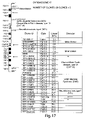
- FIG. 17 is a drawing of human chromosome 17, with arms, loci, positions and clones marked as indicated for FIG. 1 .
- FIG. 18 is a drawing of human chromosome 18, with arms, loci, positions and clones marked as indicated for FIG. 1 .
- FIG. 19 is a drawing of human chromosome 19, with arms, loci, positions and clones marked as indicated for FIG. 1 .
- FIG. 20 is a drawing of human chromosome 20, with arms, loci, positions and clones marked as indicated for FIG. 1 .
- FIG. 21 is a drawing of human chromosome 12, with arms, loci, positions and clones marked as indicated for FIG. 1 .
- FIG. 22 is a drawing of human chromosome 22, with arms, loci, positions and clones marked as indicated for FIG. 1 .
- FIG. 23A is a drawing of human chromosome X, with arms, loci, positions and clones marked as indicated for FIG. 1 .
- FIG. 23B is a table related to the drawing of human chromosome X in FIG. 23A .
- FIG. 24 is a drawing of human chromosome Y, with arms, loci, positions and clones marked as indicated for FIG. 1 .
- FIGS. 25A-25F shows a representation of human chromosomes 1-6 obtained by multiple label reversal (“dye swap”) analysis, of a Wolf-Hirschhorn patient as the test sample, and a reference sample of a subject not known to have any chromosomal disorders, the test sample and reference sample each labeled with a fluorescent dye, and then mixed and hybridized to an array of telomere-linked markers associated with chromosomal disorders, for each chromosome.
- telomere-linked markers associated with chromosomal disorders for each chromosome.
- each of the two sets of dye-swap data can indicate insertion or deletion of genetic material, as shown here and in FIGS. 26A-26F .
- FIGS. 26A-26F are representation of human chromosomes obtained by multiple dye swap analysis, using telomere-linked cloned loci associated with chromosomal disorders, for each chromosome, and control loci that are not associated with known chromosomal disorders. Other determinations are as shown in FIGS. 25A-25F .
- Novel compilations, or sets e.g., clone sets
- libraries or collections of nucleic acids and articles of manufacture which are surfaces, i.e., arrays
- application PCT/US02/33044 WO 03/091426A1 published 6 Nov. 2003, the entire contents of which are incorporated herein by reference.
- these compilations, or sets, libraries or collections, of nucleic acids and arrays are used in the detection of a chromosomal disorder or abnormality, such as a chromosomal aneuploidy (an abnormality involving a chromosome number that is not an exact multiple of the haploid number). They can also be used in the diagnosis or prognosis of a syndrome associated with a contiguous gene abnormality.
- the invention in one embodiment provides compilations, or sets, libraries or collections, of nucleic acids and arrays and methods for the detection of a chromosomal abnormality or a diagnosis or prognosis of a syndrome associated with a contiguous gene abnormality.
- These or sets of nucleic acids and/or arrays can be used for routine or directed genetic screening of embryos, fetuses, children or adults.
- These sets of nucleic acids and/or arrays can be used to aid in the diagnosis or prognosis of a syndrome, particularly when it is suspected that a patient may have symptoms associated with one or more chromosomal abnormalities, but those symptoms are not definitively diagnostic. Screening of individuals before symptoms appear will allow preventative or prophylactic treatment regimes.
- the invention provides methods for selecting genomic fragments, or clone sets (including, e.g., libraries, collections or compilations of fragments or clones), that are effective as hybridization targets in the detection of chromosomal disorder abnormalities, such as aneuploidies (i.e., abnormalities involving a chromosome number that is not an exact multiple of the haploid number), amplification, deletions and the like.
- chromosomal disorder abnormalities such as aneuploidies (i.e., abnormalities involving a chromosome number that is not an exact multiple of the haploid number), amplification, deletions and the like.
- these libraries, collections or compilations of genomic fragments or clones are immobilized on articles of manufacture, e.g., arrays.
- articles of manufacture e.g., arrays, comprising these libraries, collections or compilations of genomic fragments or clones (e.g., clone sets) are used to perform comparative genomic hybridization (CGH) to detect chromosomal aneuploidies.
- CGH comparative genomic hybridization
- the selection process comprises choosing a clone containing a specific region of the chromosome that hybridizes only to a single locus.
- the selection process can also comprise selection of chromosome fragments (e.g., a plurality of clones) each containing a portion of the genome containing at least 15% unique sequences, i.e., sequences that are not present in the other regions of the genome.
- Choice of a plurality of clones each of which contains a non-overlapping portion of closely linked portions of a chromosome increases extent of resolution of the technique.
- the article of manufacture is a surface having one or a multiple of arrays or microarrays, each array comprising a plurality of as few as about 10 and up to at least about 2500 chromosome fragments (e.g., clones) selected by this method, for example, as described below.
- Each array of about 10, 40, 125, 250 or more clones is provided in a plurality, i.e., multiple copies which are at least two non-contiguous copies.
- the genomic clones can be BAC, PAC, MAC, plasmids, recombinant viruses or phagemids and/or cosmids and the like.
- the selected chromosome fragments are cross-linked (immobilized) to a solid surface, e.g., an article of manufacture such as an array.
- the selected chromosome fragments are immobilized as described in U.S. Pat. No. 6,048,695, the methods therein producing covalent linkage of the nucleic acids to the surface, while minimizing non-specific binding of labeled sample nucleic acid as might otherwise arise using a derivatized surface.
- the article of manufacture can be an array comprising the selected chromosome fragments (e.g., clones) immobilized on a surface, for example, a glass slide.
- the slide is hybridized with fluorescently labeled test and control target DNA.
- the libraries, collections or compilations of fragments or clones of the invention comprise from about 5 up to about 1,000 or 2,500, for example, about 40 different chromosome fragments (e.g., clones) selected by this method, for example, as described below. Further, each fragment is present in at least one copy in the array, for example, is present in two copies or three copies.
- the articles of manufacture e.g., plurality arrays, each comprises a plurality of nucleic acids segments immobilized on a surface, for example, as an array, or “biochip.”
- each segment can be immobilized onto a discrete and known area, or “spot,” on the array.
- Each “spot” comprises a segment of genomic nucleic acid associated with a chromosomal abnormality, a contiguous gene abnormality, a genetically linked disease or a syndrome.
- there may be many nucleic acids molecules immobilized on a particular spot there is only one specie or representation of a genomic nucleic acid segment associated with a chromosomal abnormality per spot.
- a subset of the spots of the array of the invention includes a plurality of genomic nucleic acid segments, each associated with a chromosomal abnormality. Another subset of the spots are genomic nucleic acids not associated with any known chromosomal abnormalities. In alternative embodiments, as noted above, varying subpopulations of array spots comprise such genomic nucleic acid segments. In certain embodiments, a set of spots includes nucleic acid segments that serve as positive and/or negative controls. In one aspect, the test samples comprise calibration spots, for example, test and or reference samples are “spiked” with known types and amounts of nucleic acids, such as heterologous nucleic acids, to serve as positive and negative controls.
- kits comprising the compilations, or sets, libraries or collections of the invention, and/or arrays of the invention.
- the compilations, or sets, libraries or collections of the invention, and/or arrays of the invention comprise, or consist of at least one, or, all, of the clones as set forth in Tables and Figures herein.
- the kits can include instructions for use of the compilations, or sets, libraries or collections, of nucleic acids and/or arrays and practicing the methods of the invention, and, for the convenience of the practitioner, materials for extracting genomic DNA from a sample and preparing that DNA, including labeling of the genomic nucleic acid.
- kits can also include labeled “wild type” or reference genomic nucleic acid, e.g., human genomic nucleic acid that is provided to serve as a “wild type,” i.e., genomic nucleic acid from a subject known not to have any or substantially having no known chromosomal abnormalities and/or any known contiguous gene abnormalities.
- wild type genomic nucleic acid comprises a substantially complete genome; which is useful if the practitioner is performing a comparative genomic hybridization (CGH).
- CGH comparative genomic hybridization
- the reference nucleic acid can be a mixture of genomic nucleic acids from several subjects known not to have chromosomal abnormalities or disorders.
- BAC clones containing insert DNA are stored in 25% glycerol, and are kept frozen, for example, at minus 80° C. (stored in a ⁇ 80° C. freezer).
- array or “microarray” or “DNA array” or “nucleic acid array” or “chip” or “biochip” as used herein is a plurality of target elements, each target element comprising a defined amount of one or more biological molecules, e.g., genomic nucleic acid segments, immobilized on a defined location on a substrate surface; as described in further detail, below.
- a surface has a plurality, or multiple copies of the array in a non-contiguous arrangement, so that a plurality of hybridizations can be conducted on the surface.
- aryl-substituted 4,4-difluoro-4-bora-3a, 4a-diaza-s-indacene dye as used herein includes all “boron dipyrromethene difluoride fluorophore” or “BODIPY” dyes and “dipyrrometheneboron difluoride dyes” (see, e.g., U.S. Pat. No. 4,774,339), or equivalents, are a class of fluorescent dyes commonly used to label nucleic acids for their detection when used in hybridization reactions; see, e.g., Chen (2000) J. Org Chem. 65:2900-2906: Chen (2000) J. Biochem. Biophys. Methods 42:137-151. See also U.S. Pat. Nos. 6,060,324; 5,994,063; 5,614,386; 5,248,782; 5,227,487; 5,187,288.
- cyanine 5 or “Cy5TM” and “cyanine 3” or “Cy3TM” refer to fluorescent cyanine dyes produced by Amersham Pharmacia Biotech (Piscataway, N.J.) (Amersham Life Sciences, Arlington Heights, IL.), as described in detail, below, or equivalents. See U.S. Pat. Nos. 6,027,709; 5,714,386; 5,268,486; 5,151,507; 5,047,519. These dyes are typically incorporated into nucleic acids in the form of 5-amino-propargyl-2′-deoxycytidine 5′-triphosphate coupled to Cy5TM or Cy3TM.
- fluorescent dye and “fluorescent label” as used herein includes all known fluors, including rhodamine dyes (e.g., tetramethylrhodamine, dibenzorhodamine, see, e.g., U.S. Pat. No. 6,051,719); fluorescein dyes; “BODIPY” dyes and equivalents (e.g., dipyrrometheneboron difluoride dyes, see, e.g., U.S. Pat. No. 5,274,113); derivatives of 1-[isoindolyl]methylene-isoindole (see, e.g., U.S. Pat. No. 5,433,896); and all equivalents. See also U.S. Pat. Nos. 6,028,190; 5,188,934.
- rhodamine dyes e.g., tetramethylrhodamine, dibenzorhodamine, see, e.g., U.
- hybridizing specifically to and “specific hybridization” and “selectively hybridize to,” as used herein refer to the binding, duplexing, or hybridizing of a nucleic acid molecule preferentially to a particular nucleotide sequence under stringent conditions.
- stringent conditions refers to conditions under which one nucleic acid will hybridize preferentially to second sequence (e.g., a sample genomic nucleic acid hybridizing to an immobilized nucleic acid probe in an array), and to a lesser extent to, or not at all to, other sequences.
- stringent hybridization and “stringent hybridization wash conditions” in the context of nucleic acid hybridization are sequence dependent, and are different under different environmental parameters.
- Stringent hybridization conditions as used herein can include, e.g., hybridization in a buffer comprising 50% formamide, 5 ⁇ SSC, and 1% SDS at 42° C., or hybridization in a buffer comprising 5 ⁇ SSC and 1% SDS at 65° C., both with a wash of 0.2 ⁇ SSC and 0.1% SDS at 65° C.
- Exemplary stringent hybridization conditions can also include a hybridization in a buffer of 40% formamide, 1 M NaCl, and 1% SDS at 37° C., and a wash in 1 ⁇ SSC at 45° C.
- a hybridization in a buffer of 40% formamide, 1 M NaCl, and 1% SDS at 37° C. and a wash in 1 ⁇ SSC at 45° C.
- Those of ordinary skill will readily recognize that alternative but comparable hybridization and wash conditions can be utilized to provide conditions of similar stringency.
- wash conditions can include, e.g.: a salt concentration of about 0.02 molar at pH 7 and a temperature of at least about 50° C. or about 55° C. to about 60° C.; or, a salt concentration of about 0.15 M NaCl and a temperature of at least about 72° C. for at least about 15 minutes; or, a salt concentration of about 0.2 ⁇ SSC at a temperature of at least about 50° C. or about 55° C. to about 60° C.
- the hybridization complex is washed twice with a solution with a salt concentration of about 2 ⁇ SSC containing 0.1% SDS at room temperature for 15 minutes and then washed twice by 0.1 ⁇ SSC containing 0.1% SDS at 68° C. for 15 minutes; or, equivalent conditions.
- Stringent conditions for washing can also be, e.g., 0.2 ⁇ SSC/0.1% SDS at 42° C. See Sambrook, Ausubel, or Tijssen (cited herein) for detailed descriptions of equivalent hybridization and wash conditions and for reagents and buffers, e.g., SSC buffers and equivalent reagents and conditions.
- labeled with a detectable composition refers to a nucleic acid comprising a detectable composition, i.e., a label, as described in detail, below.
- the label can also be another biological molecule, as a nucleic acid, e.g., a nucleic acid in the form of a stem-loop structure as a “molecular beacon,” as described below. This includes incorporation of labeled bases (or, bases which can bind to a detectable label) into the nucleic acid by, e.g., nick translation, random primer extension, amplification with degenerate primers, and the like.
- the label can be detectable by any means, e.g., visual, spectroscopic, photochemical, biochemical, immunochemical, physical or chemical means.
- suitable fluorescent materials include umbelliferone, fluorescein, fluorescein isothiocyanate, rhodamine, dichlorotriazinylamine fluorescein, dansyl chloride or phycoerythrin; an example of a luminescent material includes luminol; examples of bioluminescent materials include luciferase, luciferin, and aequorin.
- nucleic acid refers to a deoxyribonucleotide or ribonucleotide in either single- or double-stranded form.
- the term encompasses nucleic acids containing known analogues of natural nucleotides.
- the term also encompasses nucleic-acid-like structures with synthetic backbones.
- DNA backbone analogues provided by the invention include phosphodiester, phosphorothioate, phosphorodithioate, methylphosphonate, phosphoramidate, alkyl phosphotriester, sulfamate, 3′-thioacetal, methylene(methylimino), 3′-N-carbamate, morpholino carbarniate, and peptide nucleic acids (PNAs); see Oligonucleotides and Analogues, a Practical Approach, edited by F. Eckstein, IRL Press at Oxford University Press (1991); Antisense Strategies, Annals of the New York Academy of Sciences, Volume 600, Eds. Baserga and Denhardt (NYAS 1992); Milligan (1993) J. Med. Chem.
- PNAs contain non-ionic backbones, such as N-(2-aminoethyl) glycine units. Phosphorothioate linkages are described, e.g., by U.S. Pat. Nos. 6,031,092; 6,001,982; 5,684,148; see also, WO 97/03211; WO 96/39154; Mata (1997) Toxicol. Appl. Pharmacol. 144:189-197.
- Other synthetic backbones encompassed by the term include methyl-phosphonate linkages or alternating methylphosphonate and phosphodiester linkages (see, e.g., U.S. Pat. No.
- nucleic acid is used interchangeably with gene, DNA, RNA, cDNA, mRNA, oligonucleotide primer, probe and amplification product.
- genomic DNA or “genomic nucleic acid” includes nucleic acid isolated from a nucleus of one or more cells, and, includes nucleic acid derived from (e.g., isolated from, amplified from, cloned from, synthetic versions of) genomic DNA.
- genomic DNA can be from any source, as discussed in detail, below.
- wild type genomic nucleic acid means a sample of genomic nucleic acid having no known or substantially no known contiguous gene abnormalities.
- sample comprising a nucleic acid refers to a sample comprising a DNA or an RNA, or nucleic acid representative of DNA or RNA isolated from a natural source, in a form suitable for hybridization (e.g., as a soluble aqueous solution) to another nucleic acid or polypeptide or combination thereof (e.g., immobilized probes).
- the nucleic acid may be isolated, cloned or amplified; it may be, e.g., genomic DNA, episomal DNA, mitochondrial DNA, mRNA, or cDNA; it may be a genomic segment that includes, e.g., particular promoters, enhancers, coding sequences, and the like; it may also include restriction fragments, cDNA libraries or fragments thereof, etc.
- the nucleic acid sample may be extracted from particular cells, tissues or body fluids, or, can be from cell cultures, including cell lines, or from preserved tissue sample, as described in detail, below.
- a computer/processor can be a conventional general-purpose digital computer, e.g., a personal “workstation” computer, including conventional elements such as microprocessor and data transfer bus.
- the computer/processor can further include any form of memory elements, such as dynamic random access memory, flash memory or the like, or mass storage such as magnetic disc optional storage.
- Making and using the compilations, or sets, libraries or collections of the invention and/or arrays of the invention, and practicing the methods of the invention may involve the isolation, synthesis, cloning, amplification, labeling and hybridization (e.g., CGH) of nucleic acids.
- the compilations, or sets, libraries or collections of the invention comprise nucleic acid segments. These nucleic acid segments and/or immobilized nucleic acid on the array can be representative of genomic DNA, including defined parts of, or entire, chromosomes, or entire genomes. Comparative genomic hybridization (CGH) reactions, see, e.g., U.S. Pat. Nos. 5,830,645; 5,976,790, are discussed in further detail, below.
- Nucleic acid samples, the compilations, or sets, libraries or collections of the invention (comprising nucleic acid segments), and, in some aspects, immobilized nucleic acids can be labeled with a detectable moiety, e.g., a fluorescent dye(s) or equivalent.
- a detectable moiety e.g., a fluorescent dye(s) or equivalent.
- a first sample can be labeled with a fluor and a second sample labeled with a second dye (e.g., Cy3TM and Cy5TM).
- each sample nucleic acid is labeled with at least one different detectable moiety, e.g., different fluorescent dyes, than those used to label the other samples of nucleic acids.
- the nucleic acids may be amplified using standard techniques such as PCR. Amplification can also be used to subclone or label the nucleic acid prior to the hybridization. The sample and/or the immobilized nucleic acid can be labeled, as described herein.
- the sample or the probe on the array an be produced from and collectively can be representative of a source of nucleic acids from one or more particular (pre-selected) portions of, e.g., a collection of polymerase chain reaction (PCR) amplification products, substantially an entire chromosome or a chromosome fragment, or substantially an entire genome, e.g., as a collection of clones, e.g., BACs, PACs, YACs, and the like (see below).
- PCR polymerase chain reaction
- the array-immbolilized nucleic acid or genomic nucleic acid sample may be processed in some manner, e.g., by blocking or removal of repetitive nucleic acids or by enrichment with selected nucleic acids.
- samples are applied to the immobilized probes (e.g., on the array) and, after hybridization and washing, the location (e.g., spots on the array) and amount of each dye are read.
- the compilations, or sets, libraries or collections, of nucleic acids or plurality of immobilized nucleic acid segments can be representative of any segment of genomic nucleic acid associated with a chromosomal abnormality, a contiguous gene abnormality, a genetically linked disease or a syndrome; including, e.g., part of or all of a chromosome or genome.
- each “spot” on the array has a known sequence, e.g., a known segment of genome or other sequence.
- the invention can be practiced in conjunction with any method or protocol or device known in the art, which are well described in the scientific and patent literature.
- RNA, cDNA, genomic DNA, vectors, viruses or hybrids thereof may be isolated from a variety of sources, genetically engineered, amplified, and/or expressed/generated recombinantly. Any recombinant expression system can be used, including, in addition to bacterial cells, e.g., mammalian, yeast, insect or plant cell expression systems.
- these nucleic acids can be synthesized in vitro by well-known chemical synthesis techniques, as described in, e.g., Carruthers (1982) Cold Spring Harbor Symp. Quant. Biol. 47:411-418; Adams (1983) J. Am. Chem. Soc. 105:661; Belousov (1997) Nucleic Acids Res. 25:3440-3444; Frenkel (1995) Free Radic. Biol. Med. 19:373-380; Blommers (1994) Biochemistry 33:7886-7896; Narang (1979) Meth. Enzymol. 68:90; Brown (1979) Meth. Enzymol. 68:109; Beaucage (1981) Tetra. Lett. 22:1859; U.S.
- Double stranded DNA fragments may then be obtained either by synthesizing the complementary strand and annealing the strands together under appropriate conditions, or by adding the complementary strand using DNA polymerase with a primer sequence.
- nucleic acids such as, e.g., subcloning, labeling probes (e.g., random-primer labeling using Klenow polymerase, nick translation, amplification), sequencing, hybridization, G-banding, CGH, SKY, FISH and the like are well described in the scientific and patent literature, see, e.g., Sambrook, ed., M OLECULAR C LONING: A L ABORATORY M ANUAL (2 ND ED .), Vols. 1-3, Cold Spring Harbor Laboratory, (1989); C URRENT P ROTOCOLS IN M OLECULAR B IOLOGY , Ausubel, ed.
- the compilations, or sets, libraries or collections of the invention, and arrays of the invention comprise nucleic acids, e.g., genomic nucleic acid segments.
- the nucleic acids used in the arrays, compilations and methods of the invention e.g., those immobilized onto arrays or used as samples, can be obtained and manipulated by cloning into various vehicles. If necessary, genomic nucleic acid samples can be screened and re-cloned or amplified from any source of genomic DNA.
- genomic nucleic acid used in the methods of the invention include genomic DNA, e.g., genomic libraries, contained in mammalian and human artificial chromosomes, satellite artificial chromosomes, yeast artificial chromosomes, bacterial artificial chromosomes, P1 artificial chromosomes, recombinant vectors and viruses, plasmids, and the like.
- genomic libraries e.g., genomic libraries, contained in mammalian and human artificial chromosomes, satellite artificial chromosomes, yeast artificial chromosomes, bacterial artificial chromosomes, P1 artificial chromosomes, recombinant vectors and viruses, plasmids, and the like.
- MACs Mammalian artificial chromosomes
- HAC human artificial chromosomes
- Kb kilobase
- Satellite artificial chromosomes or, satellite DNA-based artificial chromosomes (SATACs) are, e.g., described in Warburton (1997) Nature 386:553-555; Roush (1997) Science 276:38-39; Rosenfeld (1997) Nat. Genet. 15:333-335).
- SATACs can be made by induced de novo chromosome formation in cells of different mammalian species; see, e.g., Hadlaczky (2001) Curr. Opin. Mol. Ther. 3:125-132; Csonka (2000) J. Cell Sci. 113 ( Pt 18):3207-3216.
- Yeast artificial chromosomes can also be used and typically contain inserts ranging in size from 80 to 700 kb.
- YACs have been used for many years for the stable propagation of genomic fragments of up to one million base pairs in size; see, e.g., U.S. Pat. Nos. 5,776,745; 5,981,175; Feingold (1990) Proc. Natl. Acad. Sci. USA 87:8637-8641; Tucker (1997) Gene 199:25-30; Adam (1997) Plant J.11:1349-1358; Zeschnigk (1999) Nucleic Acids Res. 27:21.
- Bacterial artificial chromosomes are vectors that can contain cloned inserted DNA of length 120 kb or greater, see, e.g., U.S. Pat. Nos. 5,874,259; 6,277,621; 6,183,957.
- BACs are based on E. coli F factor plasmid cloning vehicle systems, and are simple to manipulate and purify in microgram quantities. Because BAC plasmids are maintained in vivo at one to two copies per E.
- P1 artificial chromosomes PACs
- bacteriophage P1-derived vectors are, e.g., described in Woon (1998) Genomics 50:306-316; Boren (1996) Genome Res. 6:1123-1130; Vietnamese (1994) Nature Genet. 6:84-89; Reid (1997) Genomics 43:366-375; Nothwang (1997) Genomics 41:370-378; Kern (1997) Biotechniques 23:120-124).
- P1 is a bacteriophage that infects E. coli that can contain 75 to 100 kb DNA inserts (see, e.g., Mejia (1997) Genome Res 7:179-186; Vietnamesenou (1994) Nat Genet 6:84-89).
- PACs are screened in much the same way as lambda libraries. See also Ashworth (1995) Analytical Biochem. 224:564-571; Gingrich (1996) Genomics 32:65-74.
- cloning vehicles can also be used, for example, recombinant viruses; cosmids, plasmids or cDNAs; see, e.g., U.S. Pat. Nos. 5,501,979; 5,288,641; 5,266,489.
- vectors can include marker genes, such as, e.g., luciferase and green fluorescent protein genes (see, e.g., Baker (1997) Nucleic Acids Res 25:1950-1956). Sequences, inserts, clones, vectors and the like can be isolated from natural sources, obtained from such sources as ATCC or GenBank libraries or commercial sources, or prepared by synthetic or recombinant methods.
- marker genes such as, e.g., luciferase and green fluorescent protein genes (see, e.g., Baker (1997) Nucleic Acids Res 25:1950-1956).
- Sequences, inserts, clones, vectors and the like can be isolated from natural sources, obtained from such sources as ATCC or GenBank libraries or commercial sources, or prepared by synthetic or recombinant methods.
- Amplification using oligonucleotide primers can be used to generate or manipulate, e.g., subclone, nucleic acids of the compilations, or sets, libraries or collections of the invention, nucleic acids used in the arrays of the invention and for practicing the methods of the invention, or to incorporate label into immobilized or sample nucleic acids, or to detect or measure levels of nucleic acids hybridized to an array, and the like.
- Amplification, typically with degenerate primers is also useful for incorporating detectable probes (e.g., Cy5TM- or Cy3TM-cytosine conjugates) into nucleic acids representative of test or control genomic DNA to be used to hybridize to immobilized genomic DNA.
- Amplification can be used to quantify the amount of nucleic acid is in a sample, see, e.g., U.S. Pat. No. 6,294,338.
- the skilled artisan can select and design suitable oligonucleotide amplification primers.
- Amplification methods are also well known in the art, and include, e.g., polymerase chain reaction, PCR (PCR PROTOCOLS, A GUIDE TO METHODS AND APPLICATIONS, ed. Innis, Academic Press, N.Y. (1990) and PCR STRATEGIES (1995), ed.
- LCR ligase chain reaction
- transcription amplification see, e.g., Kwoh (1989) Proc. Natl. Acad. Sci. USA 86:1173
- self-sustained sequence replication see, e.g., Guatelli (1990) Proc. Natl. Acad. Sci. USA 87:1874)
- Q Beta replicase amplification see, e.g., Smith (1997) J. Clin. Microbiol.
- samples of nucleic acid are hybridized to the compilations, or sets, libraries or collections of the invention or arrays of the invention, including immobilized nucleic acids.
- the hybridization and/or wash conditions are carried out under moderate to stringent conditions.
- the invention provides methods for selecting a genomic nucleic acid segment for use as a hybridization target in a hybridization reaction, e.g., a comparative genomic hybridization (CGH) reaction, for the detection of a chromosomal abnormality comprising, inter alia, selecting a chromosomal segment that hybridizes to a single locus under stringent conditions.
- Exemplary hybridization conditions including stringent hybridization conditions, are set forth below.
- Stringent hybridization and wash conditions can be selected to be about 5° C. lower than the thermal melting point (T m ) for the specific sequence at a defined ionic strength and pH.
- T m is the temperature (under defined ionic strength and pH) at which 50% of the target sequence hybridizes to a perfectly matched probe.
- Very stringent conditions are selected to be equal to the T m for a particular probe.
- Exemplary stringent hybridization conditions for hybridization of complementary nucleic acids which have more than 100 complementary residues on an array can comprise 42° C. using standard hybridization solutions (see, e.g., Sambrook), with the hybridization being carried out overnight.
- Exemplary highly stringent wash conditions can also comprise 0.15 M NaCl at 72° C. for about 15 minutes.
- Exemplary stringent wash conditions can also comprise a 0.2 ⁇ SSC wash at 65° C. for 15 minutes (see, e.g., Sambrook).
- a high stringency wash is preceded by a medium or low stringency wash to remove background probe signal.
- An exemplary medium stringency wash for a duplex of, e.g., more than 100 nucleotides comprises 1 ⁇ SSC at 45° C. for 15 minutes.
- An exemplary low stringency wash for a duplex of, e.g., more than 100 nucleotides can comprise 4 ⁇ to 6 ⁇ SSC at 40° C. for 15 minutes.
- the fluorescent dyes Cy3TM and Cy5TM are used to differentially label nucleic acid fragments from two samples, e.g., nucleic acid generated from a control (e.g., “wild type” or reference nucleic acid), to be compared to a test cell or tissue sample (sample nucleic acid).
- a control e.g., “wild type” or reference nucleic acid
- sample nucleic acid e.g., wild type or reference nucleic acid
- both the reference and sample nucleic acids are labeled with each of Cy3TM and Cy5TM, and two mixtures are made for a “dye swap” analysis.
- An embodiment of the methods and surfaces herein is that in providing at least two copies of an array on a single surface, both mixtures of nucleic acids needed to perform the dye swap can be hybridized to the same surface simultaneously.
- a variety of different techniques can be used alone or in combination to maintain separate fluids for the multiple hybridizations so that each sample is restricted to one array on the surface.
- a cover can be used; other techniques include use of a barrier which is a hydrophobic strip, or a raised barrier above the plane of the surface; and a viscosity-increasing solute can be used to decrease fluidity sufficiently to maintain the position of the fluid above the array.
- a cover can hold a predetermined volume of the fluid above the microarray by surface tension, for example, a cover slip, or can physically impede movement of the fluid.
- Cy5TM or fluors or other oxidation-sensitive compounds
- antioxidants and free radical scavengers can be used in hybridization mixes, the hybridization and/or the wash solutions.
- Cy5TM signals are dramatically increased and longer hybridization times are possible.
- the methods of the invention are carried out in a controlled, unsaturated humidity environment, and, the compilations, or sets, libraries or collections, of nucleic acids or arrays of the invention can further comprise apparatus or devices capable of controlling humidity.
- Controlling humidity is one parameter that can be manipulated to increase hybridization sensitivity.
- hybridization can be carried out in a controlled, unsaturated humidity environment; hybridization efficiency is significantly improved if the humidity is not saturated. The hybridization efficiency can be improved if the humidity is dynamically controlled, i.e., if the humidity changes during hybridization.
- Array devices comprising housings and controls that allow the operator to control the humidity during pre-hybridization, hybridization, wash and/or detection stages can be used.
- the device can have detection, control and memory components to allow pre-programming of the humidity (and temperature and other parameters) during the entire procedural cycle, including pre-hybridization, hybridization, wash and detection steps.
- the methods of the invention can incorporate hybridization conditions comprising temperature fluctuations and, the compilations, or sets, libraries or collections, of nucleic acids or arrays of the invention can further comprise apparatus or devices capable of controlling temperature, e.g., an oven.
- Hybridization has much better efficiency in a changing temperature environment as compared to conditions where the temperature is set precisely or at relatively constant level (e.g., plus or minus a couple of degrees, as with most commercial ovens).
- Reaction chamber temperatures can be fluctuatingly modified by, e.g., an oven, or other device capable of creating changing temperatures.
- the methods of the invention can comprise hybridization conditions comprising osmotic fluctuations, and, the compilations, or sets, libraries or collections, of nucleic acids or arrays of the invention can further comprise apparatus or devices capable of controlling osmotic conditions, e.g., generate a e.g., a solute gradient.
- Hybridization efficiency i.e., time to equilibrium
- a hybridization environment that comprises changing hyper-/hypo-tonicity, e.g., a solute gradient.
- a solute gradient is created in a device. For example, a low salt hybridization solution is placed on one side of the array hybridization chamber and a higher salt buffer is placed on the other side to generate a solute gradient in the chamber.
- the compilations, or sets, libraries or collections of nucleic acids, the immobilized and/or sample nucleic acids can be cloned, labeled or immobilized in a variety of lengths.
- the genomic nucleic acid segments can have a length smaller than about 200 bases.
- Use of labeled genomic DNA limited to this small size significantly improves the resolution of the molecular profile analysis, e.g., in array-based CGH.
- use of such small fragments allows for significant suppression of repetitive sequences and other unwanted, “background” cross-hybridization on the immobilized nucleic acid. Suppression of repetitive sequence hybridization greatly increases the reliability of the detection of copy number differences (e.g., amplifications or deletions) or detection of unique sequences.
- the resultant fragment lengths can be modified by, e.g., treatment with DNase. Adjusting the ratio of DNase to DNA polymerase in a nick translation reaction changes the length of the digestion product. Standard nick translation kits typically generate 300 to 600 base pair fragments. If desired, the labeled nucleic acid can be further fragmented to segments below 200 bases, down to as low as about 25 to 30 bases, random enzymatic digestion of the DNA is carried out, using, e.g., a DNA endonucleases, e.g., DNase (see, e.g., Herrera (1994) J. Mol. Biol. 236:405-411; Suck (1994) J. Mol. Recognit.
- a DNA endonucleases e.g., DNase
- Fragment size can be evaluated by a variety of techniques, including, e.g., sizing electrophoresis, as by Siles (1997) J. Chromatogr. A. 771:319-329, that analyzed DNA fragmentation using a dynamic size-sieving polymer solution in a capillary electrophoresis. Fragment sizes can also be determined by, e.g., matrix-assisted laser desorption/ionization time-of-flight mass spectrometry, see, e.g., Chiu (2000) Nucleic Acids Res. 28:E31.
- the invention provides compilations, or sets, libraries or collections of nucleic acids and arrays and methods for the detection of a chromosomal abnormality or for the diagnosis or prognosis of a syndrome associated with a contiguous gene abnormality.
- Any set or combination of genomic nucleic acid segments associated with a chromosomal abnormality, a contiguous gene abnormality, a genetically linked disease or a syndrome can be used in making and using the compilations, or sets, libraries or collections, of nucleic acids or arrays and practicing the methods of the invention, including genomic nucleic acid segments described herein and genomic nucleic acid segments or other nucleic acids not specifically exemplified herein.
- the compilations, or sets, libraries or collections of nucleic acids and/or arrays of the invention can comprise, or consist of, at least one, or all of the clones set forth in Table 1.
- the compilations, or sets, libraries or collections, of nucleic acids or arrays and methods of the invention also can comprise genomic nucleic acid segments set forth in the literature, see, e.g., Charles R. Scriver, et al., (2000) “The Metabolic and Molecular Bases of Inherited Disease,” 8 th edition, New York, McGraw-Hill; Pat Gilbert (2000) “The A-Z Reference Book of Syndromes and Inherited Disorders: A Manual for Health, Social and Education Workers” 3 Ed edition, Stanley Thornes Pub Ltd.; Suzanne B. Cassidy, et al. (Ed), (2001) “Management of Genetic Syndromes,” Wiley-Liss.
- the compilations, or sets, libraries or collections of the invention and/or arrays and methods of the invention can be used for the differential diagnosis of genetically linked diseases or syndromes, formulating appropriate treatment plans and estimating a prognosis.
- the methods of the invention can be used in situations where the causality, diagnosis, or prognosis (e.g., severity, metastatic potential) of a pathology or condition is associated with one or more genetic defects, e.g., a syndrome caused by a contiguous chromosomal disorder or defect.
- chromosomal disorder means one due to mutations in the genomic nucleic acid, primarily DNA, that are due to amplifications, deletions, inversions, translocations, and generally fall into the category of aneuploidy or deviation of the correct dosage of a chromosome or a portion of a chromosome.
- Chromosomal disorders can be “congenital” or inherited, i.e., present at or before birth, or can be somatic, i.e., associated with a disease such as cancer or exposure to hazardous materials or radiation. Somatic chromosomal disorders are likely to be characterized as mosaic, i.e., affecting a portion of cells of a tissue or organism.
- determining the presence of a contiguous gene defect can be helpful in predicting or diagnosing and the prognosis of the cancer, classifying a cancer or formulating a treatment plan or prognosis.
- metastasis suppressor genes on human chromosomes for cutaneous melanoma have been located on, e.g., 7q21-22, 7q31.2-32, 8p21-12, 10q11-22, 11p13-11.2, 12p11-q13, 12q24-ter, and 17pter-q23 (see, e.g., Goldberg (2000) Am. J. Hum. Genet.
- Chromosomal abnormalities are common in prostate cancer, including but not limited to, trisomy, hyperdiploidy and aneusomy of chromosomes 7 and 17 (Cui et al., Cancer Genet Cytogenet 107: 51, 19998), amplifications of 6p, 7q, 8q, 9q, and 16q (van Dekken et al., Lab Invest.
- the methods and arrays of the invention can be used for predicting, diagnosing and the prognosis of cancers; chromosomal damage from exposure to hazardous chemical and physical agents and certain inherited conditions.
- certain chromosomal disorders are correlated with less severe or more severe prognostic outcomes. It is contemplated that as arrays herein become widely standard and data is accumulated for a variety of cancers, additional correlations are made between chromosomal disorders and associated diagnoses and prognoses.
- a list below of inherited chromosomal disorders is exemplary of this type of disorder and not further limiting, nor are the methods, compilations, arrays, and surfaces herein limited to inherited disorders.
- the compilations, or sets, libraries or collections, of nucleic acids or arrays of the invention comprise a segment of genomic nucleic acid comprising chromosome 1, locus 1p;36, and the syndrome detected is 1p Deletion Syndrome.
- Patients with karyotypic abnormalities resulting in monosomy for a portion of 1p36.3 can have microcephaly, mental retardation, prominent forehead, deep-set eyes, depressed nasal bridge, flat midface, relative prognathism, and abnormal ears. See, e.g., Reish (1995) Am. J. Med. Genet. 59(4):467-475.
- the compilations, or sets, libraries or collections, of nucleic acids or arrays of the invention comprise a segment of genomic nucleic acid comprising chromosome 3, locus 3p25-pter, and the syndrome detected is 3p Deletion Syndrome.
- Chromosome 3p deletions are thought to be involved in the pathogenesis of sporadic endocrine pancreatic tumors (EPTs); also, von Hippel-Lindau's disease (VHL gene at 3p25.5) has been associated with EPTs.
- Chromosome 3p deletion is frequently involved in solid human tumors. See, e.g., Barghorn (2001) J. Pathol. 194(4):451-458. Allele loss in some regions of chromosome 3p has been detected in primary breast tumors. See, e.g., Maitra (2001) Am. J. Pathol. 159(1):119-130.
- the compilations, or sets, libraries or collections, of nucleic acids or arrays of the invention comprise a segment of genomic nucleic acid comprising chromosome 3, locus 3p21-pter, and the syndrome detected is 3p Duplication Syndrome.
- a partial trisomy of chromosome 3P an inverted duplication 3p22-->3pter (dup(3)(pter-->p26::p22(p26::p26-->ter)), was found to be associated with psychomotor retardation and slight dysmorphism.
- C syndrome a multiple congenital anomaly/mental retardation (MCA/MR) syndrome, was found to be associated with a duplication of 3p. See, e.g., McGaughran (2000) Am. J. Med. Genet. 94(4):311-315.
- the compilations, or sets, libraries or collections, of nucleic acids or arrays of the invention comprise a segment of genomic nucleic acid comprising chromosome 4, locus 4p16.3, and the syndrome detected is Wolf-Hirschhorn Syndrome.
- Wolf-Hirschhorn syndrome (WHS) is a well-known congenital malformation syndrome caused by deletion of the short arm of chromosome 4 (4p-). Most cases occur de novo and are of paternal origin. WHS children have severe developmental disabilities. The phenotype of adult WHS is in general similar to that of childhood WHS. Growth retardation, microcephaly and mental retardation are the rule in both adults and children. Facial dysmorphism also remains similar.
- the compilations, or sets, libraries or collections, of nucleic acids or arrays of the invention comprise a segment of genomic nucleic acid comprising chromosome 4, locus 4p15.2-16.1, and the syndrome detected is 4p Duplication Syndrome.
- Duplications of the distal half of 4p give rise to the partial trisomy 4 syndrome, characterized by a “boxer” nose configuration and deep-set eyes. These signs are usually observed even in cases of small terminal duplications.
- a “tandem” duplication of4p16.1p16.3 has been detected in association with a subtle deletion of 4p16.3pter on the same chromosome in a patient with the WHS phenotype. See, e.g., Zollino (1999) Am. J. Med. Genet. 82(5):371-375.
- the compilations, or sets, libraries or collections, of nucleic acids or arrays of the invention comprise a segment of genomic nucleic acid comprising chromosome 5, locus 5p 15.2-pter, and the syndrome detected is Cri du Chat Syndrome. Most patients with cri-du-chat syndrome have a de novo deletion of the short arm of chromosome 5 (5p). Patients show phenotypic and cytogenetic variability.
- deletions include: terminal-46,XX,del(5) (pter - - - p15.2:); interstitial-46,XX,del(5) (pter - - - p15.2::p13.3 - - - qter);46,XX,der(5)t(5;11) (p15;q25)mat.
- the compilations, or sets, libraries or collections, of nucleic acids or arrays of the invention comprise a segment of genomic nucleic acid comprising chromosome 7, locus 7p13.3, and the syndrome detected is Miller-Dieker Syndrome.
- Trisomy 5p and Miller-Dieker syndromes frequently are the result of unbalanced segregations of reciprocal translocations of chromosomes 5 and 17 with other autosomes.
- Miller-Dieker Syndrome has been associated with a breakpoint in chromosome 17p13.
- Miller-Dieker syndrome patients can present with mental retardation, postnatal growth deficiency, generalized muscular hypotonia, seizures, microcephaly, cortical atrophy, partial agenesis of corpus callosum, cerebral ventriculomegaly, facial anomalies. See, e.g., Mutchinick (1999) Am. J. Med. Genet. 85(2):99-104; Pollin (1999) Am. J. Med. Genet. 85(4):369-375.
- the compilations, or sets, libraries or collections, of nucleic acids or arrays of the invention comprise a segment of genomic nucleic acid comprising chromosome 7, locus 7q11.23, and the syndrome detected is William's Syndrome.
- Williams syndrome is typically due to a contiguous gene deletion at 7q11.23, and has been associated with a distinctive facial appearance, cardiac abnormalities, infantile hypercalcemia, and growth and developmental retardation, including mild to severe mental retardation.
- Williams syndrome was seen in a karyotype having microdeletions at 7q11.23 and 7q36 and additional chromosomal material at 7q36. See, e.g., Donnai (2000) Am. J. Med. Genet. 97(2):164-171; Wouters (2001) Am. J. Med. Genet. 102(3):261-265.
- LGS Langer-Giedion Syndrome
- TRPS II TRPS II
- the compilations, or sets, libraries or collections, of nucleic acids or arrays of the invention comprise a segment of genomic nucleic acid comprising chromosome 8, locus 8q24.1, and the syndrome detected is Langer-Giedion Syndrome (LGS) or tricho-rhino-phalangeal syndrome type II (TRPS II).
- LGS Langer-Giedion Syndrome
- TRPS II tricho-rhino-phalangeal syndrome type II
- TRPS Trichorhinophalangeal Syndrome
- the compilations, or sets, libraries or collections, of nucleic acids or arrays of the invention comprise a segment of genomic nucleic acid comprising chromosome 8, locus 8q24.1, and the syndrome detected is Trichorhinophalangeal Syndrome (TRPS) or TRPS I.
- TRPS I individuals typically have dysmorphic features and severe short stature.
- TRPS comprises a distinctive combination of hair, facial and bony abnormalities with variable expression. The absence of generalized shortness of all phalanges, metacarpals and metatarsals distinguish it from TRPS III, and absence of exostosis and mental retardation rule out TRPS II. See, e.g., George (1998) J. Eur. Acad. Dermatol. Venereol. 11(1):66-68; Naselli (1998) Pediatr. Radiol. 28(11):851-855.
- the compilations, or sets, libraries or collections, of nucleic acids or arrays of the invention comprise a segment of genomic nucleic acid comprising chromosome 9, locus 9p, e.g., locus 9p22-pter, and the syndrome detected is 9p Deletion Syndrome.
- This syndrome has been associated with de novo deletions in the short arm of chromosome 9.
- Patients can have developmental delay/mental retardation, seizures and learning disabilities.
- Mental retardation can be of variable degrees and there can be a marked deficit in visuo-praxic and visuo-spatial skills associated with memory disturbance. See, e g., Chilosi (2001) Am. J. Med. Genet. 100(2):138-144.
- the compilations, or sets, libraries or collections, of nucleic acids or arrays of the invention comprise a segment of genomic nucleic acid comprising chromosome 10, locus 10p13-p14, and the syndrome detected is DiGeorge Syndrome II.
- This syndrome is characterized by neural-crest-related developmental defects. Partial monosomy 10p is a rare chromosomal condition and a significant proportion of patients show features of DiGeorge syndrome (DGS) and velocardiofacial syndrome (VCFS).
- DRS DiGeorge syndrome
- the compilations, or sets, libraries or collections, of nucleic acids or arrays of the invention comprise a segment of genomic nucleic acid comprising chromosome 11, locus 11p13, and the syndrome detected is WAGR Syndrome.
- the Wilms' tumor-aniridia-genital anomalies-mental retardation (WAGR) syndrome is associated with an increased risk for developing Wilms' tumor.
- WAGR Wilms' tumor, aniridia, genital anomalies, and mental retardation
- syndrome anomalies have been associated with balanced reciprocal 7;11 translocation and an 11p13 breakpoint. See, e.g., Crolla (1997) J. Med. Genet. 34(3):207-212; Ariel (1996) Pediatr. Pathol. Lab. Med. 16(6):1013-1021.
- the compilations, or sets, libraries or collections, of nucleic acids or arrays of the invention comprise a segment of genomic nucleic acid comprising chromosome 11, locus 11p15.5, and the syndrome detected is Beckwith-Wiedemann Syndrome.
- Beckwith-Wiedemann syndrome (BWS) is an imprinting disorder characterized by somatic overgrowth, congenital malformations, and predisposition to childhood tumors.
- Chromosome 11p15.5 have been reported to have an imprinted gene cluster of 1 Mb, which has been implicated in a wide variety of malignancies and BWS. See, e.g., Li (2001) Genomics 74(3):370-376; Horike (2000) Hum. Mol. Genet. 9(14):2075-2083.
- the compilations, or sets, libraries or collections, of nucleic acids or arrays of the invention comprise a segment of genomic nucleic acid comprising chromosome 11, locus 11p11.2, and the syndrome detected is Potocki-Shaffer Syndrome (Multiple Exostoses II Locus). Potocki-Shaffer Syndrome is caused by a proximal deletion in the short arm of chromosome 11. Patients having the syndrome can have oval defects of the parietal bones (parietal foramina). See, e.g., Wu (2000) Am. J. Hum. Genet. 67(5):1327-1332.
- the compilations, or sets, libraries or collections, of nucleic acids or arrays of the invention comprise a segment of genomic nucleic acid comprising chromosome 15, locus 15q12 or 15q13, and the syndrome detected is Angelman Syndrome. It has been reported to be caused by the haploinsufficiency of the 15q11-q13 region, and, de novo deletions of chromosome 15q11-q13. It has also been reported that Angelman syndrome can be caused by genetic abnormalities affecting the maternal copy of chromosome region 15q12.
- PWS Prader-Willi Syndrome
- the compilations, or sets, libraries or collections, of nucleic acids or arrays of the invention comprise a segment of genomic nucleic acid comprising chromosome 15, locus 15q12, and the syndrome detected is Prader-Willi Syndrome (PWS).
- PWS is a neuroendocrine disorder reported to be due to: a large paternally derived chromosome deletion of 15q11q13, to maternal uniparental disomy (UPD), or imprinting mutation (IC).
- Severe learning disabilities e.g., attention-deficit hyperactivity disorder
- dyslexia dyslexia
- excessive daytime sleepiness are common symptoms in PWS. See, e.g., Manni (2001) Clin. Neurophysiol. 112(5):800-805; Fernandez-Novoa (2001) Rev. Neurol. 32(10):935-938.
- the compilations, or sets, libraries or collections, of nucleic acids or arrays of the invention comprise a segment of genomic nucleic acid comprising chromosome 16, locus distal 16p13.3, and the syndrome detected is Rubinstein-Taybi Syndrome (RTS).
- RTS is a malformation syndrome characterized by facial abnormalities, broad thumbs, broad big toes, and mental retardation.
- microdeletions, translocafions, and inversions involving chromosome band 16p13.3 can be detected.
- Immunodeficiency can be a prominent feature of this syndrome and may predispose these patients to recurrent infections. See, e.g., Petrij (2000) J. Med. Genet. 37(3):168-176; Villella (2000) Arch. Dis. Child. 83(4):360-361.
- the compilations, or sets, libraries or collections, of nucleic acids or arrays of the invention comprise a segment of genomic nucleic acid comprising chromosome 17, locus 17p12, and the syndrome detected is Charcot-Marie-Tooth Disease Type 1A(CMT-1A).
- Charcot-Marie-Tooth neuropathy type 1 (CMT1) is a genetically heterogeneous group of chronic demyelinating polyneuropathies with loci mapping to chromosome 17 (CMT1A), chromosome 1 (CMT1B) and to another unknown autosome (CMT1C).
- CMT1A accounts for 70-90% of cases of Charcot-Marie-Tooth Disease Type 1 and is most frequently caused by the tandem duplication of a 1.4-Mb genomic fragment on chromosome 17p12. Locus 17p12 is also associated with the peripheral neuropathies, such as hereditary neuropathy with liability to pressure palsies (HNPP) (see below). Some analyses have suggested that the syndrome is associated with de novo 17p11.2 duplication, paternal in origin, arising from unequal crossing over due to homologous recombination between flanking repeat gene clusters.
- X-linked dominant Charcot-Marie-Tooth (CMTX) disease is a motor and sensory neuropathy caused by mutations in the connexin 32 (CX32) gene. See, e.g., Badano (2001) Clin. Chem. 47(5):838-843; Potocki (2000) Nat. Genet. 24(1):84-87.
- HNPP Hereditary Neuropathy
- the compilations, or sets, libraries or collections, of nucleic acids or arrays of the invention comprise a segment of genomic nucleic acid comprising chromosome 17, locus 17p12, and the syndrome detected is Hereditary Neuropathy with Liability to Pressure Palsies (HNPP).
- HNPP Hereditary Neuropathy with Liability to Pressure Palsies
- HNPP is an autosomal dominant disorder that results in a recurrent, episodic demyelinating neuropathy. It also can be characterized by reversible episodes of sensorimotor deficits after neural compression injuries.
- tomaculous neuropathy HNPP is further characterized ultrastructurally by multiple focal thickenings (tomacula) of peripheral myelin and has an autosomal dominant inheritance.
- HNPP is associated with a 1.5-Mb deletion in chromosome 17p11.2-12 and results from reduced expression of the PMP22 gene. See, e.g., Mersiyanova (2000) Hum. Mutat.15(4):340-347; Chance (2001) Phys. Med. Rehabil. Clin. N. Am. 12(2):277-291; Lane (2001) J. Hand Surg. [Am] 26(4):670-674.
- the compilations, or sets, libraries or collections, of nucleic acids or arrays of the invention comprise a segment of genomic nucleic acid comprising chromosome 17, locus 17p13.3, and the syndrome detected is Miller-Dieker Syndrome/Isolated Lissencephaly.
- the Miller-Dieker syndrome (type I lissencephaly) is a neuronal migration disorder that is associated with microdeletions in the short arm of chromosome 17, at locus 17p13.3. For example, one patient was found to have a de novo balanced translocation with breakpoint at 8p11.23 and 17p13.3.
- neurofibromatosis type I is an autosomal dominant condition associated with mutations in the long arm of chromosome 17, and characterized by neurofibromas, cafe-au-lait spots and axillary freckling. See, e.g., King (2000) Acta Neuropathol. (Berl) 99(4):425-427; Hyundai (1998) Brain Dev. 20(3):190-192.
- the compilations, or sets, libraries or collections, of nucleic acids or arrays of the invention comprise a segment of genomic nucleic acid comprising chromosome 17, locus 17p11.2, and the syndrome detected is Smith-Magenis Syndrome (SMS).
- SMS is a clinically recognizable syndrome comprising multiple congenital anomalies and mental retardation. Its symptoms can include facial anomalies, brachydactyly, severe mental retardation, and self-injuring behavior. SMS is associated with a microdeletion (an interstitial deletion) of the short arm of chromosome 17, locus 17p11.2.
- a patient with a del(l 7)(p11.2p12) karyotype displayed symptoms of both SMS and Joubert syndrome (JS), the later characterized by cerebellar vermis hypoplasia, hypotonia, ataxic gait, developmental delay, and abnormal respiratory pattern.
- JS Joubert syndrome
- a prenatal case of SMS found dysmorphic facial features, tetralogy of Fallot, a thymic duct remnant, pancreatic islet cell hyperplasia, and abnormal lung fissuring. See, e.g., Juyal (1996) Am. J. Hum. Genet. 58(5):998-1007; Natacci (2000) Am. J. Med. Genet. 95(5):467-472; Thomas (2000) Fetal Diagn. Ther.
- SMS patients have a phase shift of their circadian rhythm of melatonin with a paradoxical diurnal secretion of the hormone. See, e.g., De Leersnyder (2001) J. Med. Genet. 38(9):586-590; De Leersnyder (2001) J. Pediatr. 139(1):111-116; Smith (1998) Am. J. Med. Genet. 81(2):186-191.
- Alagille Syndrome ABS
- the compilations, or sets, libraries or collections, of nucleic acids or arrays of the invention comprise a segment of genomic nucleic acid comprising chromosome 20, locus 20p11.2-p12, and the syndrome detected is Alagille Syndrome (AGS), also known as arteriohepatic dysplasia.
- AGS Alagille Syndrome
- Patients can have a deletion in chromosome 20p, with 20p11.23-p12.2 as the area of minimal overlap.
- AGS case had a paracentric inversion (PAI) of chromosome 20p12.2p13.
- Locus 20p11.2-p12 encodes a ligand for the Notch1 transmembrane receptor, which plays a key role in cell-to-cell signaling during differentiation. See, e.g., Yuan (1997) Acta Paediatr. Jpn 39(6):647-652; Hol (1995) Hum. Genet. 95(6):687-690.
- the compilations, or sets, libraries or collections, of nucleic acids or arrays of the invention comprise a segment of genomic nucleic acid comprising chromosome 22, locus 22q11.2, and the syndrome detected is Digeorge/Velocardiofacial Syndrome (VCFS).
- VCFS can result from a microdeletion on chromosome 22, locus 22q11.2.
- VCFS is associated with a broad clinical spectrum characterized by multiple congenital malformations, including cleft palate and cardiac anomalies, that frequently overlaps the DiGeorge syndrome. Estimates suggest that the 22q11.2 deletion occurs in approximately 1 in 4000 live births. Clinical studies indicate that more than 30% of children with VCFS will develop schizophrenia.
- Velofacial hypoplasia (Sedlackova syndrome) and velocardiofacial (Shprintzen) syndrome are also both associated with del 22q11.2. See, e.g., Eliez (2001) Am. J. Psychiatry 158(3):447-453; Fokstuen (2001) Eur. J. Pediatr. 160(1):54-57; Duke (2000) Arch. Otolaryngol. Head Neck Surg. 126(9):1141-1145.
- the compilations, or sets, libraries or collections, of nucleic acids or arrays of the invention comprise a segment of genomic nucleic acid comprising chromosome X, locus Xp21, and the syndrome detected is Adrenal Hypoplasia Congenita (AHC).
- AHC patients have a deletion on the short arm of the X chromosome, locus p21.1 to p22. 1.
- AHC is a developmental disorder of the human adrenal cortex and has been proposed to be caused by deletion or mutation of the DAX-1 gene within locus p21.1 to p22.1; DAX-1 is a member of the nuclear hormone receptor superfamily.
- the Xp21 syndrome should be considered in any infant with adrenal insufficiency.
- the compilations, or sets, libraries or collections, of nucleic acids or arrays of the invention comprise a segment of genomic nucleic acid comprising chromosome X, locus Xp21, and the syndrome detected is Duchenne/Becker Muscular Dystrophy.
- Cardiac abnormalities, cardiomyopathy and skeletal muscle weakness have been described in female carriers of the Xp21 (Duchenne and Becker) muscular dystrophies.
- Duchenne and Becker dystrophies have been associated with the absence or altered expression of dystrophin in cardiac and skeletal muscles. They are frequently complicated by cardiac hypertrophy and dilated cardiomyopathy. See, e.g., Grain (2001) Neuromuscul. Disord. 11(2):186-191; Crilley (2000) J. Am. Coll. Cardiol. 36(6):1953-1958, and the Glycerol kinase deficiency (GKD) discussion, below.
- GKD Glycerol kinase deficiency
- GKD Glycerol Kinase Deficiency
- the compilations, or sets, libraries or collections, of nucleic acids or arrays of the invention comprise a segment of genomic nucleic acid comprising chromosome X, locus Xp21, and the syndrome detected is Glycerol Kinase Deficiency (GKD).
- Glycerol kinase deficiency is an X-linked recessive disorder having a deletion on the short arm of the X chromosome, locus p21.1 to p22.1. There are two types. an isolated form and a complex form.
- the clinical and biochemical phenotype of isolated GKD may vary from a life-threatening childhood metabolic crisis to asymptomatic adult ‘pseudohypertriglyceridaemia’, resulting from hyperglycerolaemia.
- the complex GKD is an Xp21 contiguous gene syndrome involving the glycerol kinase locus together with the adrenal hypoplasia congenita (AHC) or Duchenne muscular dystrophy (DMD) loci or both.
- Complex GKD patients can have an “hourglass” appearance of the middle of the face; hypertelorism; rounded palpebral fissures; esotropia; wide, flattened earlobes; and a downtumed mouth. See, e.g., Sjarif (2000) J. Inherit. Metab. Dis. 23(6):529-547; Scheuerle (1995) J. Pediatr. 126(5 Pt 1):764-767.
- the compilations, or sets, libraries or collections, of nucleic acids or arrays of the invention comprise a segment of genomic nucleic acid comprising chromosome X, locus Xp22, and the syndrome detected is Pelizaeus-Merzbacher Disease (PMD).
- PMD is an X-linked recessive dysmyelinating disorder of the central nervous system.
- Most patients have point mutations in exons of the proteolipid protein (PLP1) gene or duplication of a genomic region that includes the PLP1 gene, on locus Xp22, on the short arm of the X chromosome. See, e.g., Hobson (2001) Hum. Mutat. 17(2):152; Hodes (2000) Am. J. Hum. Genet. 67(1):14-22; Inoue (1999) Ann. Neurol. 45(5):624-632.
- PLP1 proteolipid protein
- the compilations, or sets, libraries or collections, of nucleic acids or arrays of the invention comprise a segment of genomic nucleic acid comprising chromosome X, locus Xp22.3, and the syndrome detected comprises steroid sulfatase deficiency.
- X chromosome deletions in the Xp22.3 region can result in steroid sulfatase deficiency and X-linked ichthyosis.
- an interstitial deletion in Xp22.3 involved the Kallmann (KAL) gene, the steroid sulfatase (STS) gene and a putative mental retardation locus (MRX).
- KAL Kallmann
- STS steroid sulfatase
- MRX putative mental retardation locus
- X-linked ichthyosis is an inborn error of metabolism due to steroid sulfatase (STS) deficiency.
- X-linked ichthyosis is a disorder of keratinization characterized by a generalized desquamation of large, adherent, dark brown scales.
- Extracutaneous manifestations include corneal opacity and cryptorchidism. See, e.g., Weissortel (1998) Clin. Genet. 54(1):45-51; Santolaya-Forgas (1997) Fetal Diagn. Ther. 12(1):36-39; Valdes-Flores (2001) Am. J. Med. Genet. 102(2):146-148.
- the compilations, or sets, libraries or collections, of nucleic acids or arrays of the invention comprise a segment of genomic nucleic acid comprising chromosome Y, locus SRY locus/Yp, and the syndrome detected comprises abnormalities of the SRY (sex-determining region on the Y chromosome) locus.
- SRY has been identified at band Yp11.31p11.32 in normal XY males and in woman with XY gonadal dysgenesis.
- SRY signals have also been identified on Xp22 in one XX male.
- Ullrich-Turner syndrome has been associated with Y fragments and gonadoblastomas.
- DSS Sex Reversal
- the compilations, or sets, libraries or collections, of nucleic acids or arrays of the invention comprise a segment of genomic nucleic acid comprising chromosome X, locus Xp21, and the syndrome detected is Sex Reversal (DSS).
- the Xp21 locus contains the gene Ahch, also known as Dax1. Ahch encodes a transcription factor that has been implicated in sex determination and gonadal differentiation. Mutations in human AHC cause X-linked, adrenal hypoplasia congenita (AHC) and hypogonadotropic hypogonadism (HH). Studies have found Xp duplications in patients with sex reversal, with female or ambiguous genitalia occurring in spite of an intact Yp or SRY gene.
- the compilations, or sets, libraries or collections, of nucleic acids or arrays of the invention comprise a segment of genomic nucleic acid comprising chromosome X, locus Xp22.3, and the syndrome detected is Kallman's Disease or Kallmann's syndrome (KS).
- KS is characterized by hypogonadotrophic hypogonadism in association with anosmia or hyposmia.
- KS can be associated with X-linked ichthyosis (XLI) in a contiguous gene syndrome comprising a genetic defect in the Xp22.3 region.
- KS has also been associated with olfactory neuroblastoma. See, e.g., Maya-Nunez (1999) Clin. Endocrinol. (Oxf) 50(2):157-162; Zappia (1992) J. Otolaryngol. 21(1):16-19.
- the compilations, or sets, libraries or collections, of nucleic acids or arrays of the invention comprise a segment of genomic nucleic acid comprising chromosome 17, locus 17p11.2, and the syndrome detected is 17p11.2 Duplication Syndrome.
- Duplication of locus 17p11.2 may be associated with Birt-Hogg-Dube syndrome (BHD), an autosomal dominant neoplasia syndrome characterized mainly by benign skin tumors (e.g., benign tumors of the hair follicle), and to a lesser extent, renal tumors, lung cysts, and spontaneous pneumothorax.
- BHD Birt-Hogg-Dube syndrome
- the gene for BHD may associated with renal neoplasia and for lung and hair-follicle developmental defects. See, e.g., Schmidt (2001) Am. J. Hum. Genet. 69(4):876-82; Khoo (2001) Oncogene 20(37):5239-5242.
- the compilations, or sets, libraries or collections, of nucleic acids or arrays of the invention comprise a segment of genomic nucleic acid comprising chromosome 16, pericentromeric region, and the syndrome detected is idiopathic epilepsy and paroxysmal dyskinesia.
- This is a homogeneous syndrome of autosomal dominant infantile convulsions and paroxysmal (dystonic) choreoathetosis (ICCA).
- ICCA autosomal dominant infantile convulsions and paroxysmal choreoathetosis
- Use of the arrays and methods of the invention may be particularly useful because motor manifestations of epilepsy and of paroxysmal dyskinesia may be difficult to differentiate clinically. See, e.g., Guerrini (2001) Epilepsia 42 Suppl 3:36-41.
- the compilations, or sets, libraries or collections, of nucleic acids or arrays of the invention comprise a segment of genomic nucleic acid comprising chromosome 13, locus 13q22, and the syndrome detected is Hirschsprung disease, including Hirschsprung disease type 2, and Waardenburg syndrome.
- Hirschsprung disease is a developmental disorder resulting from the arrest of the craniocaudal migration of enteric neurons from the neural crest along gastrointestinal segments of variable length.
- Waardenburg-Shah syndrome is an auditory pigmentary disorder.
- Hirschsprung disease, malrotation, isochromia, a profound sensorineural hearing loss, and several other anomalies were found in an infant with an interstitial deletion of 13q, see, e.g., Shanske (2001) Am. J. Med. Genet. 102(3):231-236.
- the compilations, or sets, libraries or collections, of nucleic acids or arrays of the invention comprise a segment of genomic nucleic acid comprising 8, locus 8q13.3, and the syndrome detected is branchio-oto-renal (BOR) syndrome.
- Branchio-oto-renal (BOR) syndrome is an autosomal dominant disorder involving hearing loss, branchial defects, ear pits and renal abnormalities.
- the arrays and methods of the invention can be used to distinguish it from oto-facio-cervical (OFC) syndrome, which is clinically similar to BOR syndrome, with clinical features in addition to those of BOR syndrome. See, e.g., Rickard (2001) Hum. Genet. 108(5):398-403.
- the compilations, or sets, libraries or collections, of nucleic acids or arrays of the invention comprise a segment of genomic nucleic acid comprising chromosome 1, locus 7p 11.2, and the syndrome detected is Smith-Magenis syndrome (SMS).
- Smith-Magenis syndrome is a multiple congenital anomaly/mental retardation (MCA/MR) syndrome link to a contiguous-gene deletion syndrome, involving chromosome 1 7p 11.2,whose incidence is estimated to be 1:25,000 live births. SMS is characterized by a specific physical, behavioral and developmental pattern. The main clinical features consist of a broad flat midface with brachycefaly, broad nasal bridge, brachydactily, speech delay, hoarse deep voice and peripheral neuropathy. See, e.g., Di Cicco (2001 Int. J. Pediatr. Otorhinolaryngol. 59(2):147-150.
- the compilations, or sets, libraries or collections, of nucleic acids or arrays of the invention comprise a segment of genomic nucleic acid comprising chromosome X, locus Xp22.3, and the syndrome detected is Leri-Weill syndrome.
- Leri-Weill syndrome is characterized by short stature (SHOX), chondrodysplasia punctata (ARSE), bilateral Madelung deformity and mental retardation. See, e.g., Spranger (1999) Am. J. Med. Genet. 83(5):367-371.
- Chromosome abnormalities are common causes of congenital malformations and spontaneous abortions. They include structural abnormalities, polyploidy, trisomy, and mosaicism. Very few autosomal trisomies survive to birth, the three most common being those for chromosome 13, 18 and 21 giving rise to the syndromes named Patau, Edward's and Down's respectively (see, e.g., Moore (2000) Eur. J. Hum. Genet. 8:223-228).
- the arrays methods of the invention are used to diagnose Patau Syndrome, Edward's Syndrome and Down's Syndrome. See, e.g., Djalali (2000) Prenat. Diagn. 20:934-935.
- Table 1 shows a set of exemplary chromosomal disorders that can be diagnosed by the compilations, or sets, libraries or collections, of nucleic acids, arrays and methods of the invention:
- the sets of nucleic acids, arrays and methods of the invention can also be used to detect aneuploidy of chromosomes 13, 18, 21, X, and Y from genomic DNA from newborn uncultured blood samples (see, e.g., Jalal (1997) Mayo Clin. Proc. 72:705-710). Chromosomal abnormalities have been reported to occur in approximately 1%-2% of viable pregnancies studied by chorionic villus sampling at 9-11 weeks of gestation. See, e.g., Harrison (1993) Hum. Genet. 92:353-358.
- the compilations, or sets, libraries or collections, of nucleic acids, arrays and methods of the invention are used for preimplantation genetic diagnosis and the diagnosis of chromosomal abnormalities and structural abnormalities in oocytes and embryos. See, e.g., Fung (2001) J. Histochem. Cytochem. 49:797-798.
- the compilations, or sets, libraries or collections, of nucleic acids, arrays and methods of the invention are used with chorionic villus sampling (CVS) and fetal karyotyping. See, e.g., Sanz (2001) Fetal Diagn. Ther. 16:95-97.
- sets of nucleic acids of the invention, and/or the arrays and methods of the invention incorporate array-based comparative genomic hybridization (CGH) reactions to detect chromosomal abnormalities, e.g., contiguous gene abnormalities, in cell populations, such as tissue, e.g., biopsy or body fluid samples.
- CGH is a molecular cytogenetics approach that can be used to detect regions in a genome undergoing quantitative changes, e.g., gains or losses of sequence or copy numbers. Analysis of genomes of tumor cells can detect a region or regions of anomaly under going gains and/or losses.
- CGH reactions compare the genetic composition of test versus controls samples; e.g., whether a test sample of genomic DNA (e.g., from a cell population suspected of having one or more subpopulations comprising different, or cumulative, genetic defects) has amplified or deleted or mutated segments, as compared to a “negative” control, e.g., “normal” or “wild type” genotype, or “positive” control, e.g., a known cancer cell or a cell with a known defect, e.g., a translocation or deletion or amplification or the like.
- a test sample of genomic DNA e.g., from a cell population suspected of having one or more subpopulations comprising different, or cumulative, genetic defects
- a “negative” control e.g., “normal” or “wild type” genotype
- positive control e.g., a known cancer cell or a cell with a known defect, e.g., a translocation or deletion or amplification or the like.
- inventions provide articles of manufacture, such as arrays, comprising the nucleic acid compilations, or sets, libraries or collections of the invention.
- invention provides an article of manufacture comprising, or consisting of, at least one, or all, of the nucleic acid segments described in Table 1.
- Making and using the compilations, or sets, libraries or collections, of nucleic acids, arrays and practicing the methods of the present invention can incorporate any known “array,” also referred to as a “microarray” or “DNA array” or “nucleic acid array” or “biochip,” or variation thereof.
- Arrays are generically a plurality of “target elements,” or “spots,” each target element comprising a defined amount of one or more biological molecules, e.g., polypeptides, nucleic acid molecules, or probes, immobilized on a defined location on a substrate surface.
- the immobilized biological molecules are contacted with a sample for specific binding, e.g., hybridization, between molecules in the sample and the array.
- Immobilized nucleic acids can contain sequences from specific messages (e.g., as cDNA libraries) or genes (e.g., genomic libraries), including, e.g., substantially all or a subsection of a chromosome or substantially all of a genome, including a human genome.
- target elements can contain reference sequences, such as positive and negative controls, and the like.
- the target elements of the arrays may be arranged on the substrate surface at different sizes and different densities. Different target elements of the arrays can have the same molecular species, but, at different amounts, densities, sizes, labeled or unlabeled, and the like. The target element sizes and densities will depend upon a number of factors, such as the nature of the label (the immobilized molecule can also be labeled), the substrate support (it is solid, semi-solid, fibrous, capillary or porous), and the like.
- Each target element may comprise substantially the same nucleic acid sequences, or, a mixture of nucleic acids of different lengths and/or sequences.
- a target element may contain more than one copy of a cloned piece of DNA, and each copy may be broken into fragments of different lengths, as described herein.
- the length and complexity of the nucleic acid fixed onto the array surface is not critical to the invention.
- the array can comprise nucleic acids immobilized on any substrate, e.g., a solid surface (e.g., nitrocellulose, glass, quartz, fused silica, plastics and the like). See, e.g., U.S. Pat. No. 6,063,338 describing multi-well platforms comprising cycloolefin polymers if fluorescence is to be measured.
- Arrays used in the methods of the invention can comprise housing comprising components for controlling humidity and temperature during the hybridization and wash reactions.
- the present invention can use any known array, e.g., GeneChipsTM, Affymetrix, Santa Clara, Calif.; SPECTRALCHIPTM Mouse BAC Arrays, SPECTRALCHIPTM Human BAC Arrays and Custom Arrays of Spectral Genomics, Houston, Tex., and their accompanying manufacturer's instructions.
- GeneChipsTM GeneChipsTM, Affymetrix, Santa Clara, Calif.
- SPECTRALCHIPTM Mouse BAC Arrays SPECTRALCHIPTM Human BAC Arrays and Custom Arrays of Spectral Genomics, Houston, Tex., and their accompanying manufacturer's instructions.
- the compilations, or sets, libraries or collections, of nucleic acids of the invention, and the articles of manufacture, such as arrays, of the invention can comprise one, several or all of the human genomic nucleic acid segments set forth below in FIGS. 1-24 .
- These clones are identified by RP, GS, CTC or other CT clone names; the descriptors for clones are in Nature 409:953-958 (2001), “Integration of cytogenetic landmarks into the draft sequence of the human genome.”
- a second column gives the cytological position of each as a marker on either the short arm, p, or long arm, q, of the chromosome indicated, e.g.
- 8p23.3 is at the p arm telomere of chromosome 8, as shown in the ideogram of the chromosome in FIG. 8 .
- the numbers in the third column of each Fig. indicate the linear position of the cloned nucleic acid segment on the chromosome in megabases (Mb). These figures show clones that represent all 24 human chromosomes at a resolution of about or less than about 1 Mb resolution.
- the resolution is determined in part by the number of different cloned portions in a given length of a chromosome selected to be spotted on the array, and the nature of the cloned portions, i.e., whether they are overlapping or non-overlapping, and if non-overlapping, the extent of the gap between the cloned portions.
- the nucleic acid compilations, or sets, libraries or collections of the invention can be immobilized (directly or indirectly, covalently or by other means) to any substrate surface.
- the arrays of the invention can incorporate any substrate surface, e.g., a substrate means.
- the substrate surfaces can be of a rigid, semi-rigid or flexible material.
- the substrate surfaces can be flat or planar, be shaped as wells, raised regions, etched trenches, pores, beads, filaments, or the like.
- Substrates can be of any material upon which a nucleic acid (e.g., a “capture probe”) can be directly or indirectly bound.
- suitable materials can include paper, glass (see, e.g., U.S. Pat. No.
- Reactive functional groups can be, e.g., hydroxyl, carboxyl, amino groups or the like.
- Silane e.g., mono- and dihydroxyalkylsilanes, aminoalkyltrialkoxysilanes, 3-aminopropyl-triethoxysilane, 3-aminopropyltrimethoxysilane
- Silane can provide a hydroxyl functional group for reaction with an amine functional group.
- Multi-array surfaces provided herein have on each surface a plurality of copies of the micro-array, i.e., micro-arrays (arrays) of biological molecules, for example, nucleic acids.
- the term “multi-array surface” or “surface” as used herein means an article of manufacture having a plurality of micro-arrays applied to a side or a face of a substrate.
- the micro-arrays are printed or spotted or otherwise deposited on the face of the substrate, in an arrangement such that the micro-arrays are non-contiguous, i.e., the arrays are distal from eachother on the surface, or are not in contact, compared to the size of each array and the spacings of the spots within each array.
- a multi-array surface having a plurality of arrays is desirable for the following procedures: hybridizations are conducted in duplicate or triplicate on a single surface. Previous to the present invention, duplicate or triplicate or even a greater number of replicated spots have been described that are present on a single surface, however all spots were exposed to hybridization of a single hybridization mixture.
- the hybridization mixture is a solution that typically contains a nucleic acid sample from a test subject labeled with a fluorescent dye, or a mixture of two different samples of nucleic acids of different origins, each labeled with the same or a different dye.
- the hybridization mixture is formed prior to hybridization with the spots of the array on the surface, for example, the mixture includes nucleic acids from test subject labeled with a first fluorescent dye and nucleic acids that are a reference sample, labeled with a different dye.
- the reference sample can be nucleic acids from a normal individual of the same species as the test subject, or can be nucleic acids of a different species, or nucleic acids from a single BAC clone or from a mixture of BAC clones.
- NCBI maintains a human BAC resource, which provides genome-wide information concerning large-insert clones that integrate cytogenetic, radiation-hybrid, linkage, and sequence maps of the genome. See www.ncbi.nlm.nih.gov/genome/cyto/hrbc.shtml.
- label swap it is often desirable, in analyzing such data, to perform the hybridization in two different formats that reverse the fluorescent labels, what is commonly described as a “label reversal”, “label swap” or “dye swap” analysis.
- a dye swap analysis at least two nucleic acid samples are to be compared, and at least two mixtures are made.
- a first label such as a first fluorescent dye is used to identify the reference nucleic acid probe
- a second label such as a second fluorescent dye is used to identify the test sample, and after labeling each, the mixture is made.
- the labels are reversed, i.e., a second mixture is made in which the reference nucleic acid probe carries the second dye and the test sample carries the first dye.
- Each of the two mixtures provides a reference for the purpose of plotting amounts of hybridization of each solution nucleic acid, reference and test sample, to each of the immobilized cloned nucleic acids.
- the results are plotted as a function of the linear position of each of the cloned immobilized nucleic acids on a chromosome.
- a representation is made of a portion or of an entire chromosome, or of a plurality of chromosomes, or of a complete set of chromosomes (autosomes with or without sex chromosomes), i.e., of the entire genome.
- Results obtained from analyzing both sets of data are combined to reveal changes that would otherwise be undetectable if label reversal was not used. This is because small fluctuations from a ratio of 1.0 become statistically significant when the dye swap data are plotted together, which might not be significant if only a single mixture was used.
- a dye swap analysis was performed with two mixtures, the first being a mixture of the test nucleic acid labeled with the first dye and mixed with the reference nucleic acid labeled with the second dye, and the second being the test nucleic acid labeled with the second dye mixed with the reference nucleic acid labeled with the first dye, the two mixtues then being analyzed using two different surfaces.
- the use of separate hybridizations on different surfaces can be a source of variability, e.g., in efficiency of binding of spots to each surface, hybridization due to variability in conditions, minor variations in concentration of each nucleic acid, variation in concentrations, different efficiencies in elution of non-specifically bound materials due to minor variations in washing procedures or solutions, at the time of hybridization to each separate surface, or variations in photomultiplier settings in a scanner used to visualize and evaluate the array, after hybridization to each separate surface.
- the present surfaces provided herein address this problem in the prior common usage by having multi-arrays, which are a plurality of arrays on a single surface.
- two arrays are located at distal ends of a planar substrate such as a standard glass microscope slide, however alternative shapes and sizes of substrates, and shapes and sizes of arrays, are within the scope of surfaces, kits and methods envisioned herein.
- a substrate may be a one inch by 3 inch microscope slide, and may have a plurality of arrays such as two arrays, one at either end, or four arrays in a linear arrangement.
- a larger substrate such as a square slide may have four arrays, one in each corner, or nine arrays with three arrays on each side and one in the middle.
- barriers to maintain separation of fluids deposited on each array during hybridization may be used, the barriers being placed between each of the arrays, in addition to embodiments of the surface in the absence of barriers, as described herein.
- the barriers are physical “dykes” or “dams” having a height above the plane of the substrate face or surface, and such barriers include raised portions of the substrate as manufactured, or as added subsequently.
- the barriers may be hydrophobic materials that are printed on the substrate to produce a “strip” which can prevent the flow of an aqueous solution from one array to another.
- the barriers can be added before or after printing or depositing the micro-arrays, to produce the multi-array surfaces.
- the barriers are comprised of a material that is not soluble in aqueous solution, and the material hydrophobic.
- exemplary hydrophobic materials for barrier construction or printing include: polyethylene, silicone, paraffin, and Teflon®.
- Hybridization using the “multi-array surfaces” having multiple arrays on a single surface of a single substrate is conducted by adding the hybridization mixture to the array and protecting the hybridization mixture with a cover to prevent loss of volume of solution by evaporation, and to confine each hybridization of a particular sample or mixture of samples, labeled with one or more dyes as described above, to the appropriate micro-array.
- a pre-determined amount of hybridization mixture is deposited above the array, such that addition of a cover, for example placed directly on the fluid, yields a resulting thin layer of fluid above the array in which the sample nucleic acids can hybridize to complementary sequences within the array.
- Hybridization for each array on the surface is conducted under a separate cover.
- Conditions for hybridization can be modified, for example, the hybridization solution can be altered, to assure fluid separation of the multiple hybridizations on the surface.
- viscosity of the hybridization fluid may be increased to reduce fluidity by adding one or more solutes that do not interact with the nucleic acids during the hybridization.
- Exemplary solutes include small molecules that are viscuous liquids such as glycerol, and polymers of small molecules such as sugars which is exemplified by dextrans and starches such as corn starch, polymers of amino acids which are synthetic polypeptides or naturally occurring proteins such as albumins and gelatins, and synthetic polymers, for example, polyethylene glycol, or polyacrylamide or agarose, each at a concentration sufficient to increase viscosity without significantly affecting mobility of the solute nucleic acids for interaction and hybridization (annealing to form a double stranded complex) to the immobilized nucleic acids.
- the viscosity increasing solute may be chemically modified to improve its properties, for example, to render it resistant to digestion by extracellular enzymes of bacteria and fungi. Solutions for hybridization may be stored with antibiotic or growth inhibiting materials to retard spoilage during storage.
- a multi-array surface having four or nine arrays can be used to analyze multiple samples, for example, a plurality of members of a nuclear family, or multiple siblings and a proband carrying a chromosomal disorder, which can now be analyzed together on a single substrate having multiple micro-arrays, using separate hybridizations.
- any multiple number of subjects can be analyzed simultaneously on a single substrate, or any one subject can be analyzed in mixtures of different reference samples.
- Different reference samples can be different species, different known mutations, or different predeteremined single BAC or mixtures of BAC clones.
- calibration spots that act as positive controls for hybridization of a sample, and that are located within an array have been described (see, U.S. patent application 2003-0186250-A1, published Oct. 2, 2003, and incorporated herein in its entirety by reference).
- calibration spots may include a subset of cloned nucleic acids, for example, those clones of the human genome carrying sequences not known by any published references to be associated with a chromosomal disorder or disease. These clones are indicated in FIGS.
- non-reactive or backbone clones which act as positive hybridization controls, i.e., provide portions of the chromosome for any chromosome of interest that will hybridize to nucleic acid from a test subject.
- non-reactive means that the nucleic acid generally hybridizes to a full extent to a genomic nucleic acid from any test subject, i.e., and is “non-reactive” because it does not give a false “positive” diagnosis of a chromosomal disorder. Because the non-reactive or backbone clones are positive controls for hybridization, they are therefore expected to be non-reactive with a test sample for detection of a chromosomal disorder.
- a calibration spot may be a mixture of nucleic acids from backbone clones, for any one chromosome, or for all of the chromosomes in the human genome or genome of any other organism.
- An exemplary calibration spot may comprise a mixture of nucleic acids from backbone clones, for example, from about 10, from about 20, from about 40, or from about 80 backbone or non-reactive clones.
- An exemplary calibration spot contains 72 non-reactive backbone clones, selected to represent each of the set of autosomes and sex chromosomes.
- An alternative calibration spot contains nucleic acid from an unrelated heterologous species, such as a fish or amphibian, for purposes of standardizing hybridization, in which case an internal control carrying a recognizable label can be added or “spiked” into each hybridization mixture.
- Representation of each chromosome is made by calculating ratios of labels in each of the two double dye-labeled hybridizations (dye swap) and relative amounts are plotted graphically as a function of distance of each cloned chromosomal portion from the p terminus conventionally shown on the left.
- one of the two double labeled materials is plotted in a consistent line format (e.g., double line), and the other in a different color line format (e.g., solid line), such that deletion of a portion of nucleic acid in a test subject is displayed using a double line above the 1.0 ratio line (see FIGS. 25A-25F and FIGS. 26A-26F ), and an insertion such as an amplification is plotted as solid line above the 1.0 ratio line.
- a calibration spot that is a mixture of exclusively centromere-linked nucleic acids, or substantially having centromere-linked nucleic acids, provides a strong positive control that is useful for one exemplary calibration spot as described herein.
- arrays provided herein as shown in drawings and examples herein include cloned nucleic acids from portions of each chromosome that are not associated with any known chromosomal disorders, so that representations of a chromosome of a test subject's DNA is facilitated, and a chromosomal disorder on a given chromosome is more readily distinguished from normal portions of that chromosome.
- nucleic acids associated with a detectable label can be made and used and incorporated into the compositions of the invention.
- the detectable label can be incorporated into, associated with or conjugated to a nucleic acid. Any detectable moiety can be used.
- the association with the detectable moiety can be covalent or non-covalent.
- the array-immobilized nucleic acids and sample nucleic acids are differentially detectable, e.g., they have different labels and emit difference signals.
- Useful labels include, e.g., 32 P, 35 S, 3 H, 14 C, 125 I, 131 I; fluorescent dyes (e.g., Cy5TM, Cy3TM, FITC, rhodamine, lanthanide phosphors, Texas red), electron-dense reagents (e.g. gold), enzymes, e.g., as commonly used in an ELISA (e.g., horseradish peroxidase, beta-galactosidase, luciferase, alkaline phosphatase), colorimetric labels (e.g. colloidal gold), magnetic labels (e.g.
- DynabeadsTM biotin, dioxigenin, or haptens and proteins for which antisera or monoclonal antibodies are available.
- the label can be directly incorporated into the nucleic acid to be detected, or it can be attached to a probe or antibody that hybridizes or binds to the target.
- a peptide can be made detectable by incorporating (e.g., into a nucleoside base) predetermined polypeptide epitopes recognized by a secondary reporter (e.g., leucine zipper pair sequences, binding sites for secondary antibodies, transcriptional activator polypeptide, metal binding domains, epitope tags).
- Label can be attached by spacer arms of various lengths to reduce potential steric hindrance or impact on other useful or desired properties.
- fluors can be paired together; for example, one fluor labeling the control (e.g., the “nucleic acid of “known, or normal, karyotype”) and another fluor the test nucleic acid (e.g., from a chorionic villus sample or a cancer cell sample).
- the control e.g., the “nucleic acid of “known, or normal, karyotype”
- the test nucleic acid e.g., from a chorionic villus sample or a cancer cell sample.
- Exemplary pairs are: rhodamine and fluorescein (see, e.g., DeRisi (1996) Nature Genetics 14:458-460); lissamine-conjugated nucleic acid analogs and fluorescein-conjugated nucleotide analogs (see, e.g., Shalon (1996) supra); Spectrum RedTM and Spectrum GreenTM (Vysis, Downers Grove, IL.); Cy3TM and Cy5TM. Cy3TM and Cy5TM can be used together; both are fluorescent cyanine dyes produced by Amersham Life Sciences (Arlington Heights, IL.).
- Cyanine and related dyes are particularly strongly light-absorbing and highly luminescent, see, e.g., U.S. Pat. Nos. 4,337,063; 4,404,289; 6,048,982.
- fluorescent nucleotide analogs can be used, see, e.g., Jameson (1997) Methods Enzymol. 278:363-390; Zhu (1994) Nucleic Acids Res. 22:3418-3422.
- U.S. Pat. Nos. 5,652,099 and 6,268,132 also describe nucleoside analogs for incorporation into nucleic acids, e.g., DNA and/or RNA, or oligonucleotides, via either enzymatic or chemical synthesis to produce fluorescent oligonucleotides.
- U.S. Pat. No. 5,135,717 describes phthalocyanine and tetrabenztriazaporphyrin reagents for use as fluorescent labels.
- Detectable moieties can be incorporated into sample genomic nucleic acid and, if desired, any member of the compilation of nucleic acids or array-immobilized nucleic acids, by covalent or non-covalent means, e.g., by transcription, such as by random-primer labeling using Klenow polymerase, or “nick translation,” or, amplification, or equivalent.
- a nucleoside base is conjugated to a detectable moiety, such as a fluorescent dye, e.g., Cy3TM or Cy5TM, and then incorporated into a sample genomic nucleic acid.
- Samples of genomic DNA can be incorporated with Cy3TM- or Cy5TM-dCTP conjugates mixed with unlabeled dCTP.
- Cy5TM is typically excited by the 633 nm line of HeNe laser, and emission is collected at 680 nm. See also, e.g., Bartosiewicz (2000) Archives of Biochem. Biophysics 376:66-73; Schena (1996) Proc. Natl. Acad. Sci. USA 93:10614-10619; Pinkel (1998) Nature Genetics 20:207-211; Pollack (1999) Nature Genetics 23:41-46.
- modified nucleotides synthesized by coupling allylamine-dUTP to the succinimidyl-ester derivatives of the fluorescent dyes or haptenes are used; this method allows custom preparation of most common fluorescent nucleotides, see, e.g., Henegariu (2000) Nat. Biotechnol. 18:345-348.
- labeling with a detectable composition also can include a nucleic acid attached to another biological molecule, such as a nucleic acid, e.g., a nucleic acid in the form of a stem-loop structure as a “molecular beacon” or an “aptamer beacon.”
- a nucleic acid e.g., a nucleic acid in the form of a stem-loop structure as a “molecular beacon” or an “aptamer beacon.”
- Molecular beacons as detectable moieties are well known in the art; for example, Sokol (1998) Proc. Natl. Acad. Sci.
- molecular beacon reporter oligodeoxynucleotides with matched fluorescent donor and acceptor chromophores on their 5′ and 3′ ends.
- the molecular beacon remains in a stem-loop conformation where fluorescence resonance energy transfer prevents signal emission.
- the stem-loop structure opens increasing the physical distance between the donor and acceptor moieties thereby reducing fluorescence resonance energy transfer and allowing a detectable signal to be emitted when the beacon is excited by light of the appropriate wavelength.
- Aptamer beacons are similar to molecular beacons; see, e.g., Hamaguchi (2001) Anal. Biochem. 294:126-131; Poddar (2001) Mol. Cell. Probes 15:161-167; Kaboev (2000) Nucleic Acids Res. 28:E94. Aptamer beacons can adopt two or more conformations, one of which allows ligand binding. A fluorescence-quenching pair is used to report changes in conformation induced by ligand binding. See also, e.g., Yamamoto (2000) Genes Cells 5:389-396; Smimov (2000) Biochemistry 39:1462-1468.
- the invention can be practiced using any apparatus or methods to detect “detectable labels” of a sample nucleic acid, a member of the compilation of nucleic acids, or an array-immobilized nucleic acid, or, any apparatus or methods to detect nucleic acids specifically hybridized to each other.
- devices and methods for the simultaneous detection of multiple fluorophores are used; they are well known in the art, see, e.g., U.S. Pat. Nos. 5,539,517; 6,049,380; 6,054,279; 6,055,325; 6,294,331.
- any known device or method, or variation thereof, can be used or adapted to practice the methods of the invention, including array reading or “scanning” devices, such as scanning and analyzing multicolor fluorescence images; see, e.g., U.S. Pat. Nos. 6,294,331; 6,261,776; 6,252,664; 6,191,425; 6,143,495; 6,140,044; 6,066,459; 5,943,129; 5,922,617; 5,880,473; 5,846,708; 5,790,727; and, the patents cited in the discussion of arrays, herein. See also published U.S. patent applications Nos. 20010018514; 20010007747; published international patent applications Nos. WO0146467 A; WO9960163 A; WO0009650 A; WO0026412 A; WO0042222 A; WO0047600 A; WO0101144 A.
- a spectrograph can image an emission spectrum onto a two-dimensional array of light detectors; a full spectrally resolved image of the array is thus obtained.
- Photophysics of the fluorophore e.g., fluorescence quantum yield and photodestruction yield, and the sensitivity of the detector are read time parameters for an oligonucleotide array. With sufficient laser power and use of Cy5TM and/or Cy3TM, which have lower photodestruction yields an array can be read in less than 5 seconds.
- CCDs Charge-coupled devices, or CCDs, are used in microarray scanning systems, including practicing the methods of the invention.
- CCDs used in the methods of the invention can scan and analyze multicolor fluorescence images.
- Color discrimination can also be based on 3-color CCD video images; these can be performed by measuring hue values. Hue values are introduced to specify colors numerically. Calculation is based on intensities of red, green and blue light (RGB) as recorded by the separate channels of the camera. The formulation used for transforming the RGB values into hue, however, simplifies the data and does not make reference to the true physical properties of light.
- spectral imaging can be used; it analyzes light as the intensity per wavelength, which is the only quantity by which to describe the color of light correctly.
- spectral imaging can provide spatial data, because it contains spectral information for every pixel in the image.
- a spectral image can be made using brightfield microscopy, see, e.g., U.S. Pat. No. 6,294,331.
- the methods of the invention further comprise data analysis, which can include the steps of determining, e.g., fluorescent intensity as a function of substrate position, removing “outliers” (data deviating from a predetermined statistical distribution), or calculating the relative binding affinity of the targets from the remaining data.
- the resulting data can be displayed as an image with color in each region varying according to the light emission or binding affinity between targets and probes. See, e.g., U.S. Pat. Nos. 5,324,633; 5,863,504; 6,045,996.
- the invention can also incorporate a device for detecting a labeled marker on a sample located on a support, see, e.g., U.S. Pat. No. 5,578,832.
- the invention provides methods of detecting a genetic mosaicism in any sample comprising nucleic acid, such as a cell population or tissue or fluid sample, by performing an array-based comparative genomic hybridization (CGH).
- the nucleic acid can be derived from (e.g., isolated from, amplified from, cloned from) genomic DNA.
- the genomic DNA can be from any source.
- the cell, tissue or fluid sample from which the nucleic acid sample is prepared is taken from a patient suspected of having a pathology or a condition associated with genetic defects.
- the causality, diagnosis or prognosis of the pathology or condition may be associated with genetic defects, e.g., with genomic nucleic acid base substitutions, amplifications, deletions and/or translocations.
- the cell, tissue or fluid can be from, e.g., amniotic samples, chorionic villus samples (CVS), serum, blood, chord blood or urine samples, CSF or bone marrow aspirations, fecal samples, saliva, tears, tissue and surgical biopsies, needle or punch biopsies, and the like.
- a “clinical sample” derived from a patient includes frozen sections or paraffin sections taken for histological purposes.
- the sample can also be derived from supernatants (of cell cultures), lysates of cells, cells from tissue culture in which it may be desirable to detect levels of mosaicisms, including chromosomal abnormalities and copy numbers.
- DNA is prepared in one embodiment by a modified alkaline lysis protocol (see, e.g., Sambrook), however, any protocol for purifying DNA is within the scope of the method. The DNA is labeled, as described below.
- the DNA is then chemically modified as described by U.S. Pat. No. 6,048,695.
- the modified DNA is then dissolved in proper buffer and printed directly on clean glass surfaces as described by U.S. Pat. No. 6,048,695. Usually multiple spots are printed for each clone.
- a standard random priming method is used to label genomic DNA before its attachment to the array, see, e.g., Sambrook.
- Sample nucleic acid is also similarly labeled.
- Cy3TM or Cy5TM labeled nucleotides are supplemented together with corresponding unlabeled nucleotides at a molar ratio ranging from 0.0 to about 6 (unlabeled nucleotide to labeled nucleotides).
- Labeling is carried out at 37° C. for 2 to 10 hours. After labeling the reaction mix is heated up to 95° C. to 100° C. for 3 to 5 minutes to inactivate the polymerase and denature the newly generated, labeled “probe” nucleic acid from the template.
- the heated sample is then chilled on ice for 5 minutes.
- “Calibrated” DNase (DNA endonuclease) enzyme is added to fragment the labeled template (generated by random priming).
- “Trace” amounts of DNase is added (final concentration was 0.2 to 2 ng/ml; incubation time 15 to 30 minutes) to digest/fragment the labeled nucleic acid to segments of about 30 to about 100 bases in size.
- DNA is fragmented by sonication.
- Nucleic acids are alternatively fragmented by sonication.
- Sonication protocols include establishing standard power levels and times of sonication, to obtain fragments of desired length.
- the examples set forth herein provide exemplary methods for pretreating nucleic acid samples and hybridizing these samples to arrays.
- This exemplary hybridization protocol can be used to determine if a nucleic acid segment, such as a genomic clone, is within the scope of the invention (e.g., is a member of a compilation, library, clone set of the invention).
- Random prime labeling of large sized DNA samples can be more efficient if the DNA sample is first digested or otherwise treated to produce smaller fragments of more uniform size and mobility in solution. For every test sample to be analyzed, four digests of genomic DNA were performed: two of the test sample and two of an appropriate reference or control sample.
- the objective in this step is to label the test and reference samples with both Cy-3 and Cy-5 to facilitate the co-hybridization between the Cy-3 labeled test and Cy-5 labeled reference samples, and conversely the Cy-5 labeled test and Cy-3 labeled reference samples.
- Coplin jars can be used in the post-hybridization washes, it is recommended to wash each slide in individual Petri dishes in a shaking platform incubator.
- FIG. 1 is seen an ideogram of chromsome 1.
- On the right side of the ideogram and in the table are listed three BAC clones carrying nucleic acid sequences corresponding to a syndrom known as 1p36, and six clones associated with a syndrome known as 1q44, which are named for known chromosomal disorders that are associated with known areas of chromosome 1.
- chromosome 1 Also shown are nine clones carrying portions of chromosome 1 that are not associated with known chromosomal disorders (leftward pointing arrows). Further, some of these clones are closely linked to the centromere of chromosome 1, which is indicated by the constriction in the ideogram between 1p11 and 1q11. Microarrays were prepared on glass slides having DNA from the listed BAC clones in FIG. 1 that contain these loci on chromosome 1, and the slides further had two copies of each of the microarrays.
- FIG. 2 In FIG. 2 is seen an ideogram of chromsome 2. On the right side of the ideogram and in the table are listed two BAC clones carrying nucleic acid sequences corresponding to a syndrom known as 2p25.3, and four clones associated with a syndrome known as 2q37.3, which are named known chromosomal disorders that are associated with known areas of chromosome 2.
- chromosome 2 also shown are seven clones carrying portions of chromosome 2 that are not associated with known chromosomal disorders (leftward pointing arrows). Further, some of these clones are closely linked to the centromere of chromosome 2, which is indicated by the constriction in the ideogram between 2p11 and 2q11.
- Microarrays were prepared on glass slides having DNA from the listed BAC clones in FIG. 2 that contain these loci on chromosome 2, and the slides further had two copies of each of the microarrays.
- FIG. 3 is seen an ideogram of chromsome 3.
- BAC clones carrying nucleic acid sequences corresponding to a syndrome known as 3p26, and five clones associated with a syndrome known as 3q25, and two clones associated with 3q29, which are named for known chromosomal disorders that are associated with known areas of chromosome 3.
- chromosome 3 carrying portions of chromosome 3 that are not associated with known chromosomal disorders (leftward pointing arrows). Further, some of these clones are closely linked to the centromere of chromosome 3, which is indicated by the constriction in the ideogram between 3p11 and 3q11.
- Microarrays were prepared on glass slides having DNA from the listed BAC clones in FIG. 3 that contain these loci on chromosome 3, and the slides further had two copies of each of the microarrays.
- FIG. 4 is seen an ideogram of chromsome 4.
- BAC clones carrying nucleic acid sequences corresponding to locus 4p16 some of which are associated with a syndrome known as Wolf-Hirschhom syndrome, and sev clones associated with a syndrome known as 3q25, and two clones associated with 4q35, which are named for known chromosomal disorders that are associated with known areas of chromosome 4.
- chromosome 4 also shown are seven clones carrying portions of chromosome 4 that are not associated with known chromosomal disorders (leftward pointing arrows). Further, some of these clones are closely linked to the centromere of chromosome 4, which is indicated by the constriction in the ideogram between 4p11 and 4q11. Microarrays were prepared on glass slides having DNA from the listed BAC clones in FIG. 4 that contain these loci on chromosome 4, and the slides further had two copies of each of the microarrays.
- FIG. 5 is seen an ideogram of chromsome 5.
- BAC clones carrying nucleic acid sequences corresponding to locus 5p15, some of which are associated with a syndrome known as Cri-du-Chat syndrome, and seven clones associated with a syndrome at locus 5q35 known as Sotos Syndrom+C92s, which are known chromosomal disorders that are associated with known areas of chromosome 5.
- chromosome 5 that are not associated with known chromosomal disorders (leftward pointing arrows). Further, some of these clones are closely linked to the centromere of chromosome 5, which is indicated by the constriction in the ideogram between 5p11 and 5q11.
- Microarrays were prepared on glass slides having DNA from the listed BAC clones in FIG. 5 that contain these loci on chromosome 5, and the slides further had two copies of each of the microarrays.
- FIG. 6 In FIG. 6 is seen an ideogram of chromsome 6. On the right side of the ideogram and in the table are listed ten BAC clones carrying nucleic acid sequences corresponding to locus 6p25, which are associated with a syndrome, and three clones associated with a syndrome at locus 6q27, which are known chromosomal disorders that are associated with known areas of chromosome 6.
- chromosome 6 Also shown are four clones carrying portions of chromosome 6 that are not associated with known chromosomal disorders (leftward pointing arrows). Further, some of these clones are closely linked to the centromere of chromosome 6, which is indicated by the constriction in the ideogram between 6p11 and 6q11. Microarrays were prepared on glass slides having DNA from the listed BAC clones in FIG. 6 that contain these loci on chromosome 6, and the slides further had two copies of each of the microarrays.
- FIG. 7 is seen an ideogram of chromsome 7.
- five BAC clones carrying nucleic acid sequences corresponding to locus 7p22.3 and 7p21 three of which are associated with a syndrome known as Saethre-Chotzen Syndrom, and three clones associated with a syndrome at locus 7p13 known as Greig cephelopolysyndactyly syndrome, and 13 clones associated with 7q11.23 associated with Williams-Beuren syndrom, and four clones associated with locus 7q36.3, which are known chromosomal disorders that are associated with known areas of chromosome 7.
- chromosome 7 Also shown are four clones carrying portions of chromosome 7 that are not associated with known chromosomal disorders (leftward pointing arrows). Further, one of these clones are closely linked to the centromere of chromosome 7, which is indicated by the constriction in the ideogram between 7p11 and 7q11. Microarrays were prepared on glass slides having DNA from the listed BAC clones in FIG. 7 that contain these loci on chromosome 7, and the slides further had two copies of each of the microarrays.
- FIG. 8 is seen an ideogram of chromsome 8.
- BAC clones carrying nucleic acid sequences corresponding to locus 8p22, which are associated with a syndrome known as Kabuki syndrome, and four clones associated with a syndrome at locus 8q24.12 associated with Trichorhinophalangeal Syndrome type 1 (TRPS1), and three clones associated with 8q24.12 associated with Exotoses, multiple, type 1 (EXT1) syndrome, and three clones associated with locus 8q24.3, which are known chromosomal disorders that are associated with known areas of chromosome 8.
- TRPS1 Trichorhinophalangeal Syndrome type 1
- EXT1 multiple, type 1
- locus 8q24.3 which are known chromosomal disorders that are associated with known areas of chromosome 8.
- chromosome 8 Also shown are seven clones carrying portions of chromosome 8 that are not associated with known chromosomal disorders (leftward pointing arrows). Further, some of these clones are closely linked to the centromere of chromosome 8, which is indicated by the constriction in the ideogram between 8p11 and 8q11. Microarrays were prepared on glass slides having DNA from the listed BAC clones in FIG. 8 that contain these loci on chromosome 8, and the slides further had two copies of each of the microarrays.
- FIG. 9 is seen an ideogram of chromsome 9.
- On the right side of the ideogram and in the table are listed three BAC clones carrying nucleic acid sequences corresponding to locus 9p24.3, and three clones associated with a syndrome at locus 9q34.3, which are known chromosomal disorders that are associated with known areas of chromosome 9.
- chromosome 9 Also shown are five clones carrying portions of chromosome 9 that are not associated with known chromosomal disorders (leftward pointing arrows). Further, some of these clones are closely linked to the centromere of chromosome 9, which is indicated by the constriction in the ideogram between 9p11 and 9q11. Microarrays were prepared on glass slides having DNA from the listed BAC clones in FIG. 9 that contain these loci on chromosome 9, and the slides further had two copies of each of the microarrays.
- FIG. 10 is seen an ideogram of chromsome 10.
- DGS2 or DGCR2 DiGeorge Syndrome/velocardiofacial Syndrome complex-2
- NEBL Nebulette
- NEBL Nebulette
- 10q24 associated
- chromosome 10 carrying portions of chromosome 10 that are not associated with known chromosomal disorders (leftward pointing arrows). Further, two of these clones are closely linked to the centromere of chromosome 10, which is indicated by the constriction in the ideogram between 10p11 and 10q11.
- Microarrays were prepared on glass slides having DNA from the listed BAC clones in FIG. 10 that contain these loci on chromosome 10, and the slides further had two copies of each of the microarrays.
- FIG. 11 is seen an ideogram of chromsome 11.
- On the right side of the ideogram and in the table are listed four BAC clones carrying nucleic acid sequences corresponding to locus 11p15.5, two of which are associated with Beckwith-Wiedemann Syndrome (BWS), and one clone associated with WAGR syndrome at locus 11p13, and one clone associated with Wilm's Tumor syndrome at locus 11p13, and four clones associated with a syndrome at locus 11p11.2 known as Potocki-Shaffer Syndrome (PSS), and three clones at locus 11q25, which are known chromosomal disorders that are associated with known areas of chromosome 11.
- BWS Beckwith-Wiedemann Syndrome
- PSS Potocki-Shaffer Syndrome
- chromosome 11 Also shown are four clones carrying portions of chromosome 11 that are not associated with known chromosomal disorders (leftward pointing arrows). Further, one of these clones isclosely linked to the centromere of chromosome 11, which is indicated by the constriction in the ideogram between 11p11 and 11q11.
- Microarrays were prepared on glass slides having DNA from the listed BAC clones in FIG. 11 that contain these loci on chromosome 11, and the slides further had two copies of each of the microarrays.
- FIG. 12 In FIG. 12 is seen an ideogram of chromsome 12. On the right side of the ideogram and in the table are listed four BAC clones carrying nucleic acid sequences corresponding to portions of the chromosome between loci 12p13.33 and 12p13.32, and three clones at locus 12q24.33, which are known chromosomal disorders that are associated with known areas of chromosome 12.
- chromosome 12 Also shown are four clones carrying portions of chromosome 12 that are not associated with known chromosomal disorders (leftward pointing arrows). Further, one of these clones is closely linked to the centromere of chromosome 12, which is indicated by the constriction in the ideogram between 12p11 and 12q11. Microarrays were prepared on glass slides having DNA from the listed BAC clones in FIG. 12 that contain these loci on chromosome 12, and the slides further had two copies of each of the microarrays.
- FIG. 13 is seen an ideogram of chromsome 13.
- two BAC clones carrying nucleic acid sequences corresponding to portions of the chromosome at locus 13q14 and associated with retinoblastoma 1 (RB 1), and four clones associated with loci between 13q32 and 13q34, which are known chromosomal disorders that are associated with known areas of chromosome 13.
- chromosome 13 Also shown are two clones carrying portions of chromosome 13 that are not associated with known chromosomal disorders (leftward pointing arrows). Further, one of these clones is closely linked to the centromere of chromosome 13, which is indicated by the constriction in the ideogram between 13p11 and 13q11. Microarrays were prepared on glass slides having DNA from the listed BAC clones in FIG. 13 that contain these loci on chromosome 13, and the slides further had two copies of each of the microarrays.
- FIG. 14 is seen an ideogram of chromsome 14.
- On the right side of the ideogram and in the table are listed two BAC clones carrying nucleic acid sequences corresponding to portions of the chromosome at locus 14q24.3 and associated with known chromosomal disorders that are associated with known areas of chromosome 14.
- chromosome 14 Also shown are four clones carrying portions of chromosome 14 that are not associated with known chromosomal disorders (leftward pointing arrows). Further, one of these clones is closely linked to the centromere of chromosome 14, which is indicated by the constriction in the ideogram between 14p11 and 14q11. Microarrays were prepared on glass slides having DNA from the listed BAC clones in FIG. 14 that contain these loci on chromosome 14, and the slides further had two copies of each of the microarrays.
- FIG. 15 is seen an ideogram of chromsome 15.
- BAC clones carrying nucleic acid sequences corresponding to portions of the chromosome at locus 15p11.2 and associated with known chromosomal disorder Prader-Willi Syndrome (PWS)/Angelman Region (AR), and three clones at locus 15p14, and one clone at locus 15q11.2 and associated with known chromosomal disorder Prader-Willi Syndrome (PWS)/Angelman Region (AR), and one additional clone at 15q14, and three clones at 15q34, that are associated with known areas of chromosome 14.
- PWS chromosomal disorder
- AR Angelman Region
- Microarrays were prepared on glass slides having DNA from the listed BAC clones in FIG. 15 that contain these loci on chromosome 15, and the slides further had two copies of each of the microarrays.
- FIG. 16 is seen an ideogram of chromsome 16.
- two BAC clones carrying nucleic acid sequences corresponding to portions of the chromosome at locus 16p13.3 are listed on the right side of the ideogram and in the table, and three more clones associated with this locus and also associated with known chromosomal disorder Tuberous sclerosis-2 (TSC2)/Polycystic kidney disease adult type (PKD1), and four clones at locus 16p13.3 associated with Rubinstein-Taybi Syndrome (RTS), and three clones carrying portions of DNA from between loci 16q24.2 and 16q24.3, associated with known chromosomal disorders associated with known areas of chromosome 16.
- TSC2 Tuberous sclerosis-2
- PTD1 Polycystic kidney disease adult type
- RTS Rubinstein-Taybi Syndrome
- Microarrays were prepared on glass slides having DNA from the listed BAC clones in FIG. 16 that contain these loci on chromosome 16, and the slides further had two copies of each of the microarrays.
- FIG. 17 is seen an ideogram of chromsome 17.
- BAC clones carrying nucleic acid sequences corresponding to portions of the chromosome at locus 17p13.3, four of which associated with known chromosomal disorder Miller-Dieker Syndrome, and 16.clones at locus 17p11.2, seven of which are associated with Charcot-Marie-Tooth disease, type 1A (CMT1A), and six clones of which are associated with Smith-Magenis Syndrome (SMS).
- CMT1A Charcot-Marie-Tooth disease
- SMS Smith-Magenis Syndrome
- the ideogram and table further show three additional clones at 17q11.2 associated with Neurofibromatosis, type 1 (NF 1), and fove clones at 17q25.3, that are associated with known areas of chromosome 14 and known chromosomal disorders.
- Microarrays were prepared on glass slides having DNA from the listed BAC clones in FIG. 17 that contain these loci on chromosome 17, and the slides further had two copies of each of the microarrays.
- FIG. 18 is seen an ideogram of chromsome 18.
- BAC clones carrying nucleic acid sequences corresponding to portions of the chromosome between loci 18p11.3 and 18p11.2, one clone at locus 18q12, and five clones between loci 18q21 and 18q23, which are associated with known areas of chromosome 18 and known chromosomal disorders.
- Microarrays were prepared on glass slides having DNA from the listed BAC clones in FIG. 18 that contain these loci on chromosome 18, and the slides further had two copies of each of the microarrays.
- FIG. 19 is seen an ideogram of chromsome 19.
- two BAC clones carrying nucleic acid sequences corresponding to portions of the chromosome at locus 19p13.3, and two clones for portions of the chromosome between loci 19q13.4 and 19q13.43, which are associated with known areas of chromosome 19 and known chromosomal disorders.
- Microarrays were prepared on glass slides having DNA from the listed BAC clones in FIG. 19 that contain these loci on chromosome 19, and the slides further had two copies of each of the microarrays.
- FIG. 20 is seen an ideogram of chromsome 20.
- two BAC clones carrying nucleic acid sequences corresponding to portions of the chromosome at locus 20p13 are listed on the right side of the ideogram and in the table.
- two BAC clones carrying nucleic acid sequences corresponding to portions of the chromosome at locus 20p13 are listed on the right side of the ideogram and in the table.
- two BAC clones carrying nucleic acid sequences corresponding to portions of the chromosome at locus 20p13 are listed
- Alagille Syndrome Alagille Syndrome
- Microarrays were prepared on glass slides having DNA from the listed BAC clones in FIG. 20 that contain these loci on chromosome 20, and the slides further had two copies of each of the microarrays.
- FIG. 21 is seen an ideogram of chromsome 21.
- BAC clones carrying nucleic acid sequences corresponding to portions of the chromosome between loci 21q21 and 21q22.3, one of which is associated with Down Syndrome Critical Region Gene 1 (DSCR1), and which are associated with known areas of chromosome 21 and known chromosomal disorders, particularly Down Syndrome.
- DSCR1 Down Syndrome Critical Region Gene 1
- chromosome 21 that is not associated with known chromosomal disorders (leftward pointing arrows), and which is closely linked to the centromere of chromosome 21.
- Microarrays were prepared on glass slides having DNA from the listed BAC clones in FIG. 21 that contain these loci on chromosome 21, and the slides further had two copies of each of the microarrays.
- FIG. 22 is seen an ideogram of chromsome 22.
- eleven BAC clones carrying nucleic acid sequences corresponding to portions of the chromosome between loci 22q11.2 and 22q13.33, one of which is associated with DiGeorge Syndrome Velocardiofacial Syndrome (DGS/VCFS), one of which is associated with DiGeorge Syndrome Velocardiofacial Syndrome (DGS/VCFS)/T-box 1 (TBX1), and five of which are associated with DiGeorge Syndrome (DGS).
- DDS/VCFS DiGeorge Syndrome Velocardiofacial Syndrome
- TBX1 DiGeorge Syndrome Velocardiofacial Syndrome
- TBX1 DiGeorge Syndrome Velocardiofacial Syndrome
- four clones contain portions of chromosome 22 at locus 22q13.33 and are associated with chromosomal disorders at this site.
- Microarrays were prepared on glass slides having DNA from the listed BAC clones in FIG. 22 that contain these loci on chromosome 22, and the slides further had two copies of each of the microarrays.
- FIG. 23A is seen an ideogram of chromsome X.
- On the right side of the ideogram and in the table of FIG. 23B are listed 59 BAC clones carrying nucleic acid sequences corresponding to portions of the chromosome between loci Xp22.33 and Xq27.3.
- chromosomal disorders including: Placental steroid sulfatase deficiency (STS) at locus Xp22.32; Kallmann Syndrome (KAL) at locus Xp22.32; MLS syndrome deletion at loci Xp22.22 and locus Xp22.32; Glycerol Kinase deficiency at locus Xp21.3; Glycerol Kinase deficiency/Adrenal hypoplasia, congenital (AHC) also at locus Xp21.3; Duchenne Muscular Dystrophy (DMD) from locus Xp21.3 to Xp21.2; Pelizaeus-Merzbacher Disease (PMD) at locus Xq22.2; and Fragile X mental retardafion-1 (FMR1) at locus Xq27.3.
- STS Placental steroid sulfatase deficiency
- KAL Kallmann Syndrome
- MLS syndrome deletion at
- Microarrays were prepared on glass slides having DNA from the listed BAC clones in FIGS. 23A-23B that contain these loci on chromosome X, and the slides further had two copies of each of the microarrays.
- FIG. 24 is seen an ideogram of chromsome Y.
- BAC clones carrying nucleic acid sequences corresponding to portions of the Y chromosome and associated with chromosomal disorders, most of which have a phenotype affecting gonadal development (associated with locus Yq3 or Yq21) or sperm formation (Azoospermia factors 1, 2, and c, associated with locus Yq11).
- Microarrays were prepared on glass slides having DNA from the listed BAC clones in FIG. 24 that contain these loci on chromosome Y, and the slides further had two copies of each of the microarrays.
- FIGS. 25A-25F are representation of exemplary human chromosomes 1-6, using data obtained from dye swap analysis using the multiple single surface technique as described herein.
- the p terminus is on the left, and the q terminus on the right.
- a line parallel to the abscissa having a ratio of 1.0 indicates equal quantities of binding of test sample and reference sample nucleic acids to the spot having the cloned nucleic acid.
- Comparison of the two sets of dye-swap data at each point is used to produce the two lines. Deviation from a ratio of 1.0 for both lines is associated with insertion or deletion of genetic material.
- the p terminus of chromosome 4 shown in FIGS. 25A-25F was found to have a substantial deviation from a ratio of 1.0.
- a typical software analysis indicates open squares connected by a double line above the 1.0 ratio for a chromosomal disorder that indicates a deletion of material.
- the sample nucleic acid is from a patient having Wolf-Hirschhorn Syndrome.
- FIGS. 26A-26F form a representation of human chromosomes 1-6. obtained by multiple array format dye swap analysis, using both telomere-linked cloned loci associated with chromosomal disorders, for each chromosome, and control loci that are not associated with known chromosomal disorders, including control loci that are linked to the centromeres for each of the chromosomes.
- the test sample is from the same patient as the data shown in Example 26.
- chromosome 4 For chromosome 4, it can be seen that the deletion is confined to the p terminus, and that the remainder of the chromosome appears to have the normal complement of sequence material compared to the normal reference nucleic acid sample.
Abstract
Description
| TABLE 1 |
| Chromosome Loci Profiles of Contiguous Gene Syndromes |
| Chromosome | |||
| | Locus | Syndrome | |
| 1 | 1p36 | |
|
| 3 | 3p25 - pter | |
|
| 3 | 3p21 - pter | |
|
| 4 | 4p16.3 | Wolf- |
|
| 4 | 4p15.2 - 16.1 | |
|
| 5 | 5p15.2 - pter | Cri du |
|
| 7 | 7p13.3 | Miller- |
|
| 7 | 7p11.23 | William's |
|
| 8 | 8q24.1 | Langer-Giedion Syndrome (LGS) | |
| 8 | 8q24.1 | Trichorhinophalangeal Syndrome (TRPS) | |
| 9 | 9p, usually | 9p Deletion Syndrome | |
| 9p22 - |
|||
| 10 | 10p13p14 | |
|
| 11 | | WAGR Syndrome | |
| 11 | 11p15.5 | Beckwith- |
|
| 11 | 11p11.2 | Potocki-Shaffer Syndrome (Multiple | |
| Exostoses II Locus) | |||
| 15 | 15q12 | |
|
| 15 | 15q12 | Prader- |
|
| 16 | Distal 16p13.3 | Rubinstein- |
|
| 17 | 17p12 | Charcot-Marie- |
|
| 1A(CMT-1A) | |||
| 17 | 17p12 | Hereditary Neuropathy with Liability to | |
| Pressure |
|||
| 17 | 17p13.3 | Miller-Dieker Syndrome/Isolated | |
| |
|||
| 17 | 17p11.2 | Smith- |
|
| 20 | 20p11.2p12 | |
|
| 22 | 22q11.2 (also | Digeoege/Velocardiofacial Syndrome | |
| see 1-p13p14) | |||
| X | Xp21 | Adrenal Hypoplasia Congenita (AHC) | |
| X | Xp21 | Duchenne/Becker Muscular Dystrophy | |
| X | Xp21 | Glycerol Kinase Deficiency | |
| X | Xp22 | Pelizaeus-Merzbacher Disease | |
| X | Xp22.3 | Steroid Sulfatase Deficiency | |
| Y | SRY locus/Yp | Abnormalities of the SRY locus | |
-
- 1. Restriction enzyme digest of genomic DNA: on ice, pipet the following into an autoclaved microcentrifuge tube:
- DNA× μl for 1 μg
-
React 3 10×Buffer 5 μl -
Eco R1 2 μl (20units) - Water (orange vial) μl to a final volume of 50 μl
- 2. After addition of the enzyme and DNA, mix briefly by vortexing and recollect samples by brief centrifugation.
- 3. Incubate samples overnight (16 hours) at 37° C.
- 4. Determine the completion of the reaction by removing a 5 μl aliquot from the reaction mix, and analyzing the aliquot by agarose gel electrophoresis (0.8% agarose).
- If the digestion is complete, stop the reaction by incubating in a heating block at 72° C. for 10 minutes. It is recommended to fill the wells of the heating block with water approximately 15 minutes before denaturing the samples so that the tubes are in contact with water at 72° C.
- 5. Re-purify the digested DNA sample (either by phenol/chloroform extraction/EtOH precipitation or a suitable commercially available ‘post-enzyme digestion/PCR clean-up kit’ such as Zymo Research's DNA Clean and Concentrator TM-5 Cat No. D4005). Note: It is recommended to requantifying the DNA samples at this juncture to ensure that equitable amounts of the test and reference samples will be labeled in the following step.
- At least 500 ng of digested DNA of each sample were used for labeling.
- Genomic DNA samples adequately digested with a four base pair (4-bp) cutter restriction enzyme, such as EcoR1, should produce a relatively homogenous smear extending from 20 kb to approximately 600 bp.
Differential Labeling of DNA with Cy3-dCTP and Cy5-dCTP
-
- 1. To the re-purified DNA samples, add sterile water to bring the total volume to 25 μl. Then add 20 μl of 2.5× random primer/reaction buffer mix (e.g., from Gibco/BRL's BIOPRIME™ labeling kit).
- 2. Mix the samples well and then boil for 5 minutes.
- 3. Immediately place the samples on ice and allow to sit for 5 minutes.
- 4. On ice, add 2.5 μl of SPECTRAL LABELING BUFFER, for use with SPECTRAL CHIP™ (Spectral Genomics, Houston Tex.) to each sample.
- 5. Add 1.5 μl Cy5-dCTP or Cy3-dCTP to the respective test and reference DNA samples (1 mM stocks).
- 6. Finally, add 1 μl Klenow Fragment (from the Gibco/BRL BIOPRIME™ labeling kit) to the samples, mix the sample well by tapping, and re-collect by brief centrifugation.
- 7. Incubate the sample at 37° C for 1½-2 hours. Place the samples on ice and determine the probe size distribution by removing a 5 μl aliquot from the reaction mix, and analyzing the aliquot by agarose gel electrophoresis (0.8% agarose). Note: Optimally, the majority of the probe should range in size between 100-500 bp.
- 8. Stop the reaction by adding 5 μl 0.5 M EDTA pH8.0 and incubating in a heating block at 72° C. for 10 minutes. Place the samples on ice. The samples can now be used to proceed with hybridization or can be stored at −20° C. until required.
- Optimally the majority of the probe should range in size between 100-500 bp.
Hybridizing Labeled DNA to the Array
-
- 1. Combine the Cy3-labeled test DNA sample with the Cy5-labeled reference sample and, conversely, the Cy5-labeled test DNA sample with the Cy3-labeled reference sample. Add 45 μl of SPECTRAL HYBRIDIZATION BUFFER I, for use with SPECTRAL CHIP™ (Spectral Genomics, Houston Tex.) to each of the two tubes.
- 2. Precipitate the two samples by adding 11.3 μl of 5MNaCl and 101 μl of room temperature isopropanol. Mix the samples well and incubate in the dark at room temperature for 10-15 minutes.
- 3. Centrifuge the samples at full speed (10,000 g) for 10 minutes.
- 4. Aspirate the supernatant, avoiding the pellet. Note: The pellets should have a purplish hue, indicating that there are expected amounts of Cy3 and Cy5 labeled DNA. Too pink or too blue a sample, suggests that the corresponding genomic DNA was not appropriately labeled.
- 5. Rinse the pellets with 500 μl of 70% ethanol and allow the pellets to air-dry briefly in the dark at room temperature.
- 6. Add 10 μl of sterile water (orange vial) to the pellets. Let stand at room temperature for 5 minutes and then thoroughly resuspend. After ensuring that the pellets are completely resuspended, add 30 μl of SPECTRAL HYBRIDIZATION BUFFER II, for use with SPECTRAL CHIP™ (Spectral Genomics, Houston Tex.) and mix well by repeated pipetting.
- 7. Denature the samples by incubating in a water bath at 72° C. for 10 minutes. Note: Alternatively, the sample can be denatured in a heating block set at 72° C. We recommend filling the wells of the heating block with water approximately 15 minutes before denaturing the samples so that the tubes are in contact with water at 72° C.
- 8. After the denaturation of the samples, immediately place the tubes on ice for 5 minutes.
- 9. Incubate the samples at 37° C. for 30 minutes.
- 10. Pipette the sample onto the center of the array and cover with a 22×60 cover slip to spread it out. Note: It is imperative that the entire array is covered and that air bubbles are avoided.
- 11. Place the slide in a hybridization chamber. If a microarray hybridization chamber is used, then add 10 μl of 2×SSC, 50% formamide to either side of the chamber. (H2O works just as well).
- 12. Close the chamber and wrap with aluminum foil. Put the chambers in a Kapak Pouch with wet paper and heat seal the bag. Put the bag in a 37° C. incubator for 16 hours. Note: We recommend using a shaking platform incubator to facilitate and maintain even distribution of the probe on the slide.
Post Hybridization Washes
-
- 1. Pre-warm the following solutions at 50° C. in individual Petri dishes:
- 2×SSC, 50% deionized Formamide
- 2×SSC, 0.1% NP-40
- 0.2×SSC
- 2. Soak the slide in 2×SSC, 0.5% SDS briefly at room temperature and gently slide off the cover slip using a pair of clean forceps. Avoid peeling off the cover slip by force. (Alternatively, 2×SSC can be used)
- 3. Using a pair of forceps, transfer the slide to pre-warmed 2×SSC, 50% Formamide. Wash the slides by incubating in the shaking incubator at 50° C. for 20 minutes.
- 4.
Repeat step 3 using pre-warmed 2×SSC, 0.1% NP-40. - 5.
Repeat step 3 using pre-warmed 0.2×SSC for 10 minutes. - 6. Briefly rinse the slides with distilled deionized water. This last wash greatly reduces background fluorescence but should not exceed 10 seconds.
- 7. Immediately dry the slides under forced air. Do not air dry the slides. The slides are now ready for scanning.
Claims (30)
Priority Applications (2)
| Application Number | Priority Date | Filing Date | Title |
|---|---|---|---|
| US10/847,149 US7439346B2 (en) | 2001-10-12 | 2004-05-17 | Nucleic acids arrays and methods of use therefor |
| US12/210,317 US20090075841A1 (en) | 2002-10-15 | 2008-09-15 | Nucleic acids arrays and methods of use therefor |
Applications Claiming Priority (4)
| Application Number | Priority Date | Filing Date | Title |
|---|---|---|---|
| US32903001P | 2001-10-12 | 2001-10-12 | |
| US10/273,399 US7335470B2 (en) | 2001-10-12 | 2002-10-15 | Compilations of nucleic acids and arrays and methods of using them |
| US47121603P | 2003-05-16 | 2003-05-16 | |
| US10/847,149 US7439346B2 (en) | 2001-10-12 | 2004-05-17 | Nucleic acids arrays and methods of use therefor |
Related Parent Applications (1)
| Application Number | Title | Priority Date | Filing Date |
|---|---|---|---|
| US10/273,399 Continuation-In-Part US7335470B2 (en) | 2001-10-12 | 2002-10-15 | Compilations of nucleic acids and arrays and methods of using them |
Related Child Applications (1)
| Application Number | Title | Priority Date | Filing Date |
|---|---|---|---|
| US12/210,317 Continuation US20090075841A1 (en) | 2002-10-15 | 2008-09-15 | Nucleic acids arrays and methods of use therefor |
Publications (2)
| Publication Number | Publication Date |
|---|---|
| US20050059041A1 US20050059041A1 (en) | 2005-03-17 |
| US7439346B2 true US7439346B2 (en) | 2008-10-21 |
Family
ID=46302071
Family Applications (1)
| Application Number | Title | Priority Date | Filing Date |
|---|---|---|---|
| US10/847,149 Expired - Fee Related US7439346B2 (en) | 2001-10-12 | 2004-05-17 | Nucleic acids arrays and methods of use therefor |
Country Status (1)
| Country | Link |
|---|---|
| US (1) | US7439346B2 (en) |
Cited By (16)
| Publication number | Priority date | Publication date | Assignee | Title |
|---|---|---|---|---|
| WO2009121152A2 (en) | 2008-04-03 | 2009-10-08 | Katholieke Universiteit Leuven | Gene signatures |
| US20100160717A1 (en) * | 2008-10-03 | 2010-06-24 | Scott Jr Richard T | In vitro fertilization |
| US20100206316A1 (en) * | 2009-01-21 | 2010-08-19 | Scott Jr Richard T | Method for determining chromosomal defects in an ivf embryo |
| US20100317916A1 (en) * | 2009-06-12 | 2010-12-16 | Scott Jr Richard T | Method for relative quantitation of chromosomal DNA copy number in single or few cells |
| US7994296B2 (en) * | 2002-03-27 | 2011-08-09 | Perkinelmer Health Sciences, Inc. | Arrays, computer program products and methods for in silico array-based comparative binding assays |
| CN102439167A (en) * | 2008-06-20 | 2012-05-02 | 加的夫大学学院咨询有限公司 | Method for determining DNA copy number by competitive pcr |
| WO2012072685A1 (en) | 2010-12-02 | 2012-06-07 | Katholieke Universiteit Leuven, K.U.Leuven R&D | Irak-related interventions and diagnosis |
| KR101266250B1 (en) | 2011-08-30 | 2013-06-07 | (주) 엠지메드 | Microarray and kit for diagnosing Langer-Giedion syndrome |
| KR101280817B1 (en) | 2011-08-30 | 2013-07-02 | (주) 엠지메드 | Microarray and kit for diagnosing angelman syndrome |
| KR101280818B1 (en) * | 2011-08-30 | 2013-07-02 | (주) 엠지메드 | Microarray and kit for diagnosing glycerol kinase deficiency |
| KR101335297B1 (en) * | 2011-08-30 | 2013-12-05 | (주) 엠지메드 | Microarray and kit for diagnosing Duchenne muscular dystrophy |
| US8774488B2 (en) | 2010-03-11 | 2014-07-08 | Cellscape Corporation | Method and device for identification of nucleated red blood cells from a maternal blood sample |
| WO2015127517A1 (en) | 2014-02-27 | 2015-09-03 | Katholieke Universiteit Leuven | Oxidative stress and cardiovascular disease events |
| WO2016094970A1 (en) | 2014-12-19 | 2016-06-23 | Vrije Universiteit Brussel | In vitro maturation of a mammalian cumulus oocyte complex |
| US10704097B2 (en) | 2014-02-27 | 2020-07-07 | Katholieke Universiteit Leuven | Oxidative stress and cardiovascular disease events |
| US10988801B2 (en) | 2010-06-07 | 2021-04-27 | Esoterix Genetic Laboratories, Llc | Enumeration of nucleic acids |
Families Citing this family (7)
| Publication number | Priority date | Publication date | Assignee | Title |
|---|---|---|---|---|
| WO2007044642A2 (en) * | 2005-10-06 | 2007-04-19 | President And Fellows Of Harvard College And Children's Medical Center Corporation | Device and method for combined microfluidic-micromagnetic separation of material in continuous flow |
| WO2007070482A2 (en) * | 2005-12-14 | 2007-06-21 | Xueliang Xia | Microarray-based preimplantation genetic diagnosis of chromosomal abnormalities |
| US8180572B2 (en) * | 2007-10-25 | 2012-05-15 | Canon U.S. Life Sciences, Inc. | High-resolution melting analysis |
| US8318413B2 (en) * | 2007-12-11 | 2012-11-27 | The University Of Medicine And Dentistry Of New Jersey | Amniopunch and uses thereof |
| WO2010123594A2 (en) | 2009-01-15 | 2010-10-28 | Children's Medical Center Corporation | Device for filtration of fluids there through and accompanying method |
| US9582309B2 (en) | 2014-12-09 | 2017-02-28 | Vmware, Inc. | Allocating cost of disk usage to a linked clone virtual machine based on a parameter of usage |
| US9710296B2 (en) | 2014-12-09 | 2017-07-18 | Vmware, Inc. | Allocating cost of disk usage to a linked clone virtual machine |
Citations (95)
| Publication number | Priority date | Publication date | Assignee | Title |
|---|---|---|---|---|
| US4337063A (en) | 1979-03-01 | 1982-06-29 | Fuji Photo Film Co., Ltd. | Competitive immunoassay using spectral sensitizer label |
| US4355153A (en) | 1980-11-19 | 1982-10-19 | Societa' Italiana Resine S.I.R. S.P.A. | Process for the polymerization of formaldehyde |
| US4404289A (en) | 1980-09-02 | 1983-09-13 | Fuji Photo Film Co., Ltd. | Method for immunochemical measurement of trace components |
| US4458066A (en) | 1980-02-29 | 1984-07-03 | University Patents, Inc. | Process for preparing polynucleotides |
| US4652613A (en) | 1985-12-16 | 1987-03-24 | Celanese Corporation | Novel elastomer/oxymethylene polymer blends containing polymeric compatibilizing agents |
| US4683202A (en) | 1985-03-28 | 1987-07-28 | Cetus Corporation | Process for amplifying nucleic acid sequences |
| US4683195A (en) | 1986-01-30 | 1987-07-28 | Cetus Corporation | Process for amplifying, detecting, and/or-cloning nucleic acid sequences |
| US4774339A (en) | 1987-08-10 | 1988-09-27 | Molecular Probes, Inc. | Chemically reactive dipyrrometheneboron difluoride dyes |
| US4806631A (en) | 1985-09-30 | 1989-02-21 | Miles Inc. | Immobilization of nucleic acids on solvolyzed nylon supports |
| US4818681A (en) | 1985-02-22 | 1989-04-04 | Molecular Diagnostics, Inc. | Fast and specific immobilization of nucleic acids to solid supports |
| US4826790A (en) | 1984-02-09 | 1989-05-02 | Ecc International Limited | Production of porous mullite bodies |
| US4937858A (en) | 1987-08-17 | 1990-06-26 | Telenorma Telefonbau Und Normalzeit Gmbh | Telephone handset with electro-acoustic converters electrically connected by a cord |
| US4937188A (en) | 1986-04-15 | 1990-06-26 | Northeastern University | Enzyme activity amplification method for increasing assay sensitivity |
| US5008220A (en) | 1988-02-15 | 1991-04-16 | Ecc International Limited | Biological support |
| US5024933A (en) | 1988-05-10 | 1991-06-18 | Enzo Biochem, Inc. | Method and kit for sample adherence to test substrate |
| US5047519A (en) | 1986-07-02 | 1991-09-10 | E. I. Du Pont De Nemours And Company | Alkynylamino-nucleotides |
| US5055429A (en) | 1987-11-27 | 1991-10-08 | Ecc International Limited | Porous inorganic material |
| US5068269A (en) | 1989-09-02 | 1991-11-26 | Akzo N.V. | Cellulosic membranes |
| US5135717A (en) | 1986-12-24 | 1992-08-04 | British Technology Group Usa Inc. | Tetrabenztriazaporphyrin reagents and kits containing the same |
| US5143854A (en) | 1989-06-07 | 1992-09-01 | Affymax Technologies N.V. | Large scale photolithographic solid phase synthesis of polypeptides and receptor binding screening thereof |
| US5151507A (en) | 1986-07-02 | 1992-09-29 | E. I. Du Pont De Nemours And Company | Alkynylamino-nucleotides |
| US5187288A (en) | 1991-05-22 | 1993-02-16 | Molecular Probes, Inc. | Ethenyl-substituted dipyrrometheneboron difluoride dyes and their synthesis |
| US5188934A (en) | 1989-11-14 | 1993-02-23 | Applied Biosystems, Inc. | 4,7-dichlorofluorescein dyes as molecular probes |
| US5190864A (en) | 1986-04-15 | 1993-03-02 | Northeastern University | Enzyme amplification by using free enzyme to release enzyme from an immobilized enzyme material |
| US5215882A (en) | 1989-11-30 | 1993-06-01 | Ortho Diagnostic Systems, Inc. | Method of immobilizing nucleic acid on a solid surface for use in nucleic acid hybridization assays |
| US5227487A (en) | 1990-04-16 | 1993-07-13 | Molecular Probes, Inc. | Certain tricyclic and pentacyclic-hetero nitrogen rhodol dyes |
| US5248782A (en) | 1990-12-18 | 1993-09-28 | Molecular Probes, Inc. | Long wavelength heteroaryl-substituted dipyrrometheneboron difluoride dyes |
| US5266489A (en) | 1990-03-12 | 1993-11-30 | Rhone Merieux | Recombinant herpesviruses, in particular for the production of vaccines, process for preparing them, plasmids produced during this process and vaccines obtained |
| US5268486A (en) | 1986-04-18 | 1993-12-07 | Carnegie-Mellon Unversity | Method for labeling and detecting materials employing arylsulfonate cyanine dyes |
| US5274113A (en) | 1991-11-01 | 1993-12-28 | Molecular Probes, Inc. | Long wavelength chemically reactive dipyrrometheneboron difluoride dyes and conjugates |
| US5288625A (en) | 1991-09-13 | 1994-02-22 | Biologic Research Center Of The Hungarian Academy Of Sciences | Mammalian artificial chromosomes |
| US5288641A (en) | 1984-06-04 | 1994-02-22 | Arch Development Corporation | Herpes Simplex virus as a vector |
| US5324633A (en) | 1991-11-22 | 1994-06-28 | Affymax Technologies N.V. | Method and apparatus for measuring binding affinity |
| US5434049A (en) | 1992-02-28 | 1995-07-18 | Hitachi, Ltd. | Separation of polynucleotides using supports having a plurality of electrode-containing cells |
| US5433896A (en) | 1994-05-20 | 1995-07-18 | Molecular Probes, Inc. | Dibenzopyrrometheneboron difluoride dyes |
| US5472842A (en) | 1993-10-06 | 1995-12-05 | The Regents Of The University Of California | Detection of amplified or deleted chromosomal regions |
| US5501979A (en) | 1989-02-01 | 1996-03-26 | The General Hospital Corporation | Herpes simplex virus type I expression vector |
| US5514785A (en) | 1990-05-11 | 1996-05-07 | Becton Dickinson And Company | Solid supports for nucleic acid hybridization assays |
| US5532226A (en) | 1991-07-18 | 1996-07-02 | Ortho Pharmaceutical Corporation | Trifluoromethybenzylphosphonates useful in treating osteoporosis |
| US5539517A (en) | 1993-07-22 | 1996-07-23 | Numetrix Ltd. | Method for simultaneously measuring the spectral intensity as a function of wavelength of all the pixels of a two dimensional scene |
| US5554744A (en) | 1994-09-23 | 1996-09-10 | Hybridon, Inc. | Method for loading solid supports for nucleic acid synthesis |
| US5556752A (en) | 1994-10-24 | 1996-09-17 | Affymetrix, Inc. | Surface-bound, unimolecular, double-stranded DNA |
| US5578832A (en) | 1994-09-02 | 1996-11-26 | Affymetrix, Inc. | Method and apparatus for imaging a sample on a device |
| US5601982A (en) | 1995-02-07 | 1997-02-11 | Sargent; Jeannine P. | Method and apparatus for determining the sequence of polynucleotides |
| US5610287A (en) | 1993-12-06 | 1997-03-11 | Molecular Tool, Inc. | Method for immobilizing nucleic acid molecules |
| US5614386A (en) | 1995-06-23 | 1997-03-25 | Baylor College Of Medicine | Alternative dye-labeled primers for automated DNA sequencing |
| US5630932A (en) | 1995-09-06 | 1997-05-20 | Molecular Imaging Corporation | Tip etching system and method for etching platinum-containing wire |
| US5632957A (en) | 1993-11-01 | 1997-05-27 | Nanogen | Molecular biological diagnostic systems including electrodes |
| US5635351A (en) | 1995-03-14 | 1997-06-03 | The Regents Of The University Of California | Genetic gain and loss in gliomas |
| US5637687A (en) | 1993-08-31 | 1997-06-10 | Wiggins; James C. | Methods and compositions for isolating nucleic acids |
| US5641630A (en) | 1985-06-13 | 1997-06-24 | Amgen Inc. | Method and kit for performing nucleic acid hybridization assays |
| US5652099A (en) | 1992-02-12 | 1997-07-29 | Conrad; Michael J. | Probes comprising fluorescent nucleosides and uses thereof |
| US5665549A (en) | 1992-03-04 | 1997-09-09 | The Regents Of The University Of California | Comparative genomic hybridization (CGH) |
| US5684148A (en) | 1988-05-26 | 1997-11-04 | Competitive Technologies, Inc. | Nucleoside thiophosphoramidites |
| US5700637A (en) | 1988-05-03 | 1997-12-23 | Isis Innovation Limited | Apparatus and method for analyzing polynucleotide sequences and method of generating oligonucleotide arrays |
| US5714386A (en) | 1996-01-11 | 1998-02-03 | Board Of Trustees Of The Leland Stanford Junior University | Cy7-allophycocyanin conjugates for use in multiplex fluorescence detection assays |
| US5721118A (en) | 1995-10-31 | 1998-02-24 | The Regents Of The University Of California, San Diego | Mammalian artificial chromosomes and methods of using same |
| US5721098A (en) | 1986-01-16 | 1998-02-24 | The Regents Of The University Of California | Comparative genomic hybridization |
| US5744305A (en) | 1989-06-07 | 1998-04-28 | Affymetrix, Inc. | Arrays of materials attached to a substrate |
| US5770456A (en) | 1989-06-07 | 1998-06-23 | Affymetrix, Inc. | Cyclic nucleic acid and polypeptide arrays |
| US5776745A (en) | 1993-07-23 | 1998-07-07 | The Johns Hopkins University | Recombinationally targeted cloning in yeast artificial chromosomes |
| US5790727A (en) | 1997-02-05 | 1998-08-04 | Brookhaven Science Associates Llc | Laser illumination of multiple capillaries that form a waveguide |
| US5795557A (en) | 1995-07-07 | 1998-08-18 | Universite Claude Bernard | Process for the preparation of monolithic silica aerogels |
| US5800992A (en) | 1989-06-07 | 1998-09-01 | Fodor; Stephen P.A. | Method of detecting nucleic acids |
| US5807522A (en) | 1994-06-17 | 1998-09-15 | The Board Of Trustees Of The Leland Stanford Junior University | Methods for fabricating microarrays of biological samples |
| US5830645A (en) | 1994-12-09 | 1998-11-03 | The Regents Of The University Of California | Comparative fluorescence hybridization to nucleic acid arrays |
| US5843767A (en) | 1993-10-28 | 1998-12-01 | Houston Advanced Research Center | Microfabricated, flowthrough porous apparatus for discrete detection of binding reactions |
| US5846708A (en) | 1991-11-19 | 1998-12-08 | Massachusetts Institiute Of Technology | Optical and electrical methods and apparatus for molecule detection |
| US5856174A (en) | 1995-06-29 | 1999-01-05 | Affymetrix, Inc. | Integrated nucleic acid diagnostic device |
| US5856097A (en) | 1992-03-04 | 1999-01-05 | The Regents Of The University Of California | Comparative genomic hybridization (CGH) |
| US5863504A (en) | 1995-03-16 | 1999-01-26 | Bio-Rad Laboratories, Inc. | Fluorescence imaging instrument utilizing fish |
| US5874259A (en) | 1997-11-21 | 1999-02-23 | Wisconsin Alumni Research Foundation | Conditionally amplifiable BAC vector |
| US5880473A (en) | 1997-07-28 | 1999-03-09 | Applied Imaging, Inc. | Multifluor-fluorescence in-situ hybridization (M-FISH) imaging techniques using multiple multiband filters with image registration |
| US5922617A (en) | 1997-11-12 | 1999-07-13 | Functional Genetics, Inc. | Rapid screening assay methods and devices |
| US5925525A (en) | 1989-06-07 | 1999-07-20 | Affymetrix, Inc. | Method of identifying nucleotide differences |
| US5939261A (en) | 1997-06-24 | 1999-08-17 | Sarnoff Corporation | Method for capturing a nucleic acid |
| US5943129A (en) | 1997-08-07 | 1999-08-24 | Cambridge Research & Instrumentation Inc. | Fluorescence imaging system |
| US5959098A (en) | 1996-04-17 | 1999-09-28 | Affymetrix, Inc. | Substrate preparation process |
| US5962674A (en) | 1995-06-01 | 1999-10-05 | Hybridon, Inc. | Synthesis of oligonucleotides containing alkylphosphonate internucleoside linkages |
| US5965452A (en) | 1996-07-09 | 1999-10-12 | Nanogen, Inc. | Multiplexed active biologic array |
| US5981175A (en) | 1993-01-07 | 1999-11-09 | Genpharm Internation, Inc. | Methods for producing recombinant mammalian cells harboring a yeast artificial chromosome |
| US6001982A (en) | 1993-07-29 | 1999-12-14 | Isis Pharmaceuticals, Inc. | Synthesis of oligonucleotides |
| US6013440A (en) | 1996-03-11 | 2000-01-11 | Affymetrix, Inc. | Nucleic acid affinity columns |
| US6022963A (en) | 1995-12-15 | 2000-02-08 | Affymetrix, Inc. | Synthesis of oligonucleotide arrays using photocleavable protecting groups |
| US6024872A (en) | 1997-07-01 | 2000-02-15 | Zenon Evironmental Inc. | Method of making a dope comprising hydrophilized PVDF and α-alumina, and a membrane made therefrom |
| US6025155A (en) | 1996-04-10 | 2000-02-15 | Chromos Molecular Systems, Inc. | Artificial chromosomes, uses thereof and methods for preparing artificial chromosomes |
| US6027709A (en) | 1997-01-10 | 2000-02-22 | Li-Cor Inc. | Fluorescent cyanine dyes |
| US6028190A (en) | 1994-02-01 | 2000-02-22 | The Regents Of The University Of California | Probes labeled with energy transfer coupled dyes |
| US6031092A (en) | 1995-10-20 | 2000-02-29 | Mc Gill University | Preparation of phosphorothioate oligomers |
| US6040138A (en) | 1995-09-15 | 2000-03-21 | Affymetrix, Inc. | Expression monitoring by hybridization to high density oligonucleotide arrays |
| US6045996A (en) | 1993-10-26 | 2000-04-04 | Affymetrix, Inc. | Hybridization assays on oligonucleotide arrays |
| US6048695A (en) | 1998-05-04 | 2000-04-11 | Baylor College Of Medicine | Chemically modified nucleic acids and methods for coupling nucleic acids to solid support |
| US6048982A (en) | 1986-04-18 | 2000-04-11 | Carnegie Mellon University | Cyanine dyes as labeling reagents for detection of biological and other materials by luminescence methods |
| US6048457A (en) | 1997-02-26 | 2000-04-11 | Millipore Corporation | Cast membrane structures for sample preparation |
| US6049380A (en) | 1997-11-12 | 2000-04-11 | Regents Of The University Of California | Single molecule identification using selected fluorescence characteristics |
Family Cites Families (29)
| Publication number | Priority date | Publication date | Assignee | Title |
|---|---|---|---|---|
| US6054270A (en) * | 1988-05-03 | 2000-04-25 | Oxford Gene Technology Limited | Analying polynucleotide sequences |
| US6379895B1 (en) * | 1989-06-07 | 2002-04-30 | Affymetrix, Inc. | Photolithographic and other means for manufacturing arrays |
| US5936731A (en) * | 1991-02-22 | 1999-08-10 | Applied Spectral Imaging Ltd. | Method for simultaneous detection of multiple fluorophores for in situ hybridization and chromosome painting |
| US5817462A (en) * | 1995-02-21 | 1998-10-06 | Applied Spectral Imaging | Method for simultaneous detection of multiple fluorophores for in situ hybridization and multicolor chromosome painting and banding |
| US6468742B2 (en) * | 1993-11-01 | 2002-10-22 | Nanogen, Inc. | Methods for determination of single nucleic acid polymorphisms using bioelectronic microchip |
| DE4344726C2 (en) * | 1993-12-27 | 1997-09-25 | Deutsches Krebsforsch | Method for the detection of unbalanced genetic material of a species or for the detection of gene expression in cells of a species |
| EP0880598A4 (en) * | 1996-01-23 | 2005-02-23 | Affymetrix Inc | Nucleic acid analysis techniques |
| US20010018514A1 (en) * | 1998-07-31 | 2001-08-30 | Mcgall Glenn H. | Nucleic acid labeling compounds |
| US5790557A (en) * | 1996-09-13 | 1998-08-04 | Electronics And Telecommunications Research Institute | Apparatus for implementing the function of a virtual container-11 and a tributary unit group-2 in a synchronous digital hierarchy |
| JPH10227740A (en) * | 1997-02-18 | 1998-08-25 | Hitachi Ltd | Multicolor fluorescence detection electrophoresis analyzer |
| US5846726A (en) * | 1997-05-13 | 1998-12-08 | Becton, Dickinson And Company | Detection of nucleic acids by fluorescence quenching |
| US6063338A (en) * | 1997-06-02 | 2000-05-16 | Aurora Biosciences Corporation | Low background multi-well plates and platforms for spectroscopic measurements |
| US6096817A (en) * | 1997-06-26 | 2000-08-01 | E. I. Du Pont De Nemours And Company | Mixtures of polyimides and elastomers |
| US6826296B2 (en) * | 1997-07-25 | 2004-11-30 | Affymetrix, Inc. | Method and system for providing a probe array chip design database |
| US6465178B2 (en) * | 1997-09-30 | 2002-10-15 | Surmodics, Inc. | Target molecule attachment to surfaces |
| US6060324A (en) * | 1997-11-12 | 2000-05-09 | Phytochem Technologies, Inc. | Fluorometric assay composition for measurement of antioxidant activity |
| US5936087A (en) * | 1997-11-25 | 1999-08-10 | The Perkin-Elmer Corporation | Dibenzorhodamine dyes |
| US6277621B1 (en) * | 1998-02-26 | 2001-08-21 | Medigene, Inc. | Artificial chromosome constructs containing foreign nucleic acid sequences |
| US6077673A (en) * | 1998-03-31 | 2000-06-20 | Clontech Laboratories, Inc. | Mouse arrays and kits comprising the same |
| US6183957B1 (en) * | 1998-04-16 | 2001-02-06 | Institut Pasteur | Method for isolating a polynucleotide of interest from the genome of a mycobacterium using a BAC-based DNA library application to the detection of mycobacteria |
| US6277489B1 (en) * | 1998-12-04 | 2001-08-21 | The Regents Of The University Of California | Support for high performance affinity chromatography and other uses |
| US6235504B1 (en) * | 1999-01-11 | 2001-05-22 | The Rockefeller University | Methods for identifying genomic equivalent markers and their use in quantitating cells and polynucleotide sequences therein |
| US6251601B1 (en) * | 1999-02-02 | 2001-06-26 | Vysis, Inc. | Simultaneous measurement of gene expression and genomic abnormalities using nucleic acid microarrays |
| US6277581B1 (en) * | 1999-03-01 | 2001-08-21 | Lankenau Medical Research Center | ODC allelic analysis method for assessing carcinogenic susceptibility |
| US6221653B1 (en) * | 1999-04-27 | 2001-04-24 | Agilent Technologies, Inc. | Method of performing array-based hybridization assays using thermal inkjet deposition of sample fluids |
| US6653151B2 (en) * | 1999-07-30 | 2003-11-25 | Large Scale Proteomics Corporation | Dry deposition of materials for microarrays using matrix displacement |
| US6252664B1 (en) * | 1999-10-15 | 2001-06-26 | Biocrystal Ltd. | Fluorescence filter cube for fluorescence detection and imaging |
| US6383749B2 (en) * | 1999-12-02 | 2002-05-07 | Clontech Laboratories, Inc. | Methods of labeling nucleic acids for use in array based hybridization assays |
| US20010008765A1 (en) * | 1999-12-06 | 2001-07-19 | Fuji Photo Film Co., Ltd. | DNA chip and reactive solid carrier |
-
2004
- 2004-05-17 US US10/847,149 patent/US7439346B2/en not_active Expired - Fee Related
Patent Citations (102)
| Publication number | Priority date | Publication date | Assignee | Title |
|---|---|---|---|---|
| US4337063A (en) | 1979-03-01 | 1982-06-29 | Fuji Photo Film Co., Ltd. | Competitive immunoassay using spectral sensitizer label |
| US4458066A (en) | 1980-02-29 | 1984-07-03 | University Patents, Inc. | Process for preparing polynucleotides |
| US4404289A (en) | 1980-09-02 | 1983-09-13 | Fuji Photo Film Co., Ltd. | Method for immunochemical measurement of trace components |
| US4355153A (en) | 1980-11-19 | 1982-10-19 | Societa' Italiana Resine S.I.R. S.P.A. | Process for the polymerization of formaldehyde |
| US4826789A (en) | 1984-02-09 | 1989-05-02 | Ecc International Limited | Porous inorganic materials |
| US4826790A (en) | 1984-02-09 | 1989-05-02 | Ecc International Limited | Production of porous mullite bodies |
| US4963436A (en) | 1984-02-09 | 1990-10-16 | Ecc International Limited | Coated porous mullite bodies |
| US5288641A (en) | 1984-06-04 | 1994-02-22 | Arch Development Corporation | Herpes Simplex virus as a vector |
| US4818681A (en) | 1985-02-22 | 1989-04-04 | Molecular Diagnostics, Inc. | Fast and specific immobilization of nucleic acids to solid supports |
| US4683202A (en) | 1985-03-28 | 1987-07-28 | Cetus Corporation | Process for amplifying nucleic acid sequences |
| US4683202B1 (en) | 1985-03-28 | 1990-11-27 | Cetus Corp | |
| US5641630A (en) | 1985-06-13 | 1997-06-24 | Amgen Inc. | Method and kit for performing nucleic acid hybridization assays |
| US4806631A (en) | 1985-09-30 | 1989-02-21 | Miles Inc. | Immobilization of nucleic acids on solvolyzed nylon supports |
| US4652613A (en) | 1985-12-16 | 1987-03-24 | Celanese Corporation | Novel elastomer/oxymethylene polymer blends containing polymeric compatibilizing agents |
| US5721098A (en) | 1986-01-16 | 1998-02-24 | The Regents Of The University Of California | Comparative genomic hybridization |
| US4683195A (en) | 1986-01-30 | 1987-07-28 | Cetus Corporation | Process for amplifying, detecting, and/or-cloning nucleic acid sequences |
| US4683195B1 (en) | 1986-01-30 | 1990-11-27 | Cetus Corp | |
| US4937188A (en) | 1986-04-15 | 1990-06-26 | Northeastern University | Enzyme activity amplification method for increasing assay sensitivity |
| US5190864A (en) | 1986-04-15 | 1993-03-02 | Northeastern University | Enzyme amplification by using free enzyme to release enzyme from an immobilized enzyme material |
| US5268486A (en) | 1986-04-18 | 1993-12-07 | Carnegie-Mellon Unversity | Method for labeling and detecting materials employing arylsulfonate cyanine dyes |
| US6048982A (en) | 1986-04-18 | 2000-04-11 | Carnegie Mellon University | Cyanine dyes as labeling reagents for detection of biological and other materials by luminescence methods |
| US5151507A (en) | 1986-07-02 | 1992-09-29 | E. I. Du Pont De Nemours And Company | Alkynylamino-nucleotides |
| US5047519A (en) | 1986-07-02 | 1991-09-10 | E. I. Du Pont De Nemours And Company | Alkynylamino-nucleotides |
| US5135717A (en) | 1986-12-24 | 1992-08-04 | British Technology Group Usa Inc. | Tetrabenztriazaporphyrin reagents and kits containing the same |
| US4774339A (en) | 1987-08-10 | 1988-09-27 | Molecular Probes, Inc. | Chemically reactive dipyrrometheneboron difluoride dyes |
| US4937858A (en) | 1987-08-17 | 1990-06-26 | Telenorma Telefonbau Und Normalzeit Gmbh | Telephone handset with electro-acoustic converters electrically connected by a cord |
| US5055429A (en) | 1987-11-27 | 1991-10-08 | Ecc International Limited | Porous inorganic material |
| US5008220A (en) | 1988-02-15 | 1991-04-16 | Ecc International Limited | Biological support |
| US5700637A (en) | 1988-05-03 | 1997-12-23 | Isis Innovation Limited | Apparatus and method for analyzing polynucleotide sequences and method of generating oligonucleotide arrays |
| US5024933A (en) | 1988-05-10 | 1991-06-18 | Enzo Biochem, Inc. | Method and kit for sample adherence to test substrate |
| US5684148A (en) | 1988-05-26 | 1997-11-04 | Competitive Technologies, Inc. | Nucleoside thiophosphoramidites |
| US5501979A (en) | 1989-02-01 | 1996-03-26 | The General Hospital Corporation | Herpes simplex virus type I expression vector |
| US5800992A (en) | 1989-06-07 | 1998-09-01 | Fodor; Stephen P.A. | Method of detecting nucleic acids |
| US5744305A (en) | 1989-06-07 | 1998-04-28 | Affymetrix, Inc. | Arrays of materials attached to a substrate |
| US5143854A (en) | 1989-06-07 | 1992-09-01 | Affymax Technologies N.V. | Large scale photolithographic solid phase synthesis of polypeptides and receptor binding screening thereof |
| US5770456A (en) | 1989-06-07 | 1998-06-23 | Affymetrix, Inc. | Cyclic nucleic acid and polypeptide arrays |
| US5925525A (en) | 1989-06-07 | 1999-07-20 | Affymetrix, Inc. | Method of identifying nucleotide differences |
| US5068269A (en) | 1989-09-02 | 1991-11-26 | Akzo N.V. | Cellulosic membranes |
| US5188934A (en) | 1989-11-14 | 1993-02-23 | Applied Biosystems, Inc. | 4,7-dichlorofluorescein dyes as molecular probes |
| US5215882A (en) | 1989-11-30 | 1993-06-01 | Ortho Diagnostic Systems, Inc. | Method of immobilizing nucleic acid on a solid surface for use in nucleic acid hybridization assays |
| US5266489A (en) | 1990-03-12 | 1993-11-30 | Rhone Merieux | Recombinant herpesviruses, in particular for the production of vaccines, process for preparing them, plasmids produced during this process and vaccines obtained |
| US5227487A (en) | 1990-04-16 | 1993-07-13 | Molecular Probes, Inc. | Certain tricyclic and pentacyclic-hetero nitrogen rhodol dyes |
| US5514785A (en) | 1990-05-11 | 1996-05-07 | Becton Dickinson And Company | Solid supports for nucleic acid hybridization assays |
| US5248782A (en) | 1990-12-18 | 1993-09-28 | Molecular Probes, Inc. | Long wavelength heteroaryl-substituted dipyrrometheneboron difluoride dyes |
| US5187288A (en) | 1991-05-22 | 1993-02-16 | Molecular Probes, Inc. | Ethenyl-substituted dipyrrometheneboron difluoride dyes and their synthesis |
| US5532226A (en) | 1991-07-18 | 1996-07-02 | Ortho Pharmaceutical Corporation | Trifluoromethybenzylphosphonates useful in treating osteoporosis |
| US5288625A (en) | 1991-09-13 | 1994-02-22 | Biologic Research Center Of The Hungarian Academy Of Sciences | Mammalian artificial chromosomes |
| US5274113A (en) | 1991-11-01 | 1993-12-28 | Molecular Probes, Inc. | Long wavelength chemically reactive dipyrrometheneboron difluoride dyes and conjugates |
| US5846708A (en) | 1991-11-19 | 1998-12-08 | Massachusetts Institiute Of Technology | Optical and electrical methods and apparatus for molecule detection |
| US5324633A (en) | 1991-11-22 | 1994-06-28 | Affymax Technologies N.V. | Method and apparatus for measuring binding affinity |
| US5652099A (en) | 1992-02-12 | 1997-07-29 | Conrad; Michael J. | Probes comprising fluorescent nucleosides and uses thereof |
| US5434049A (en) | 1992-02-28 | 1995-07-18 | Hitachi, Ltd. | Separation of polynucleotides using supports having a plurality of electrode-containing cells |
| US5856097A (en) | 1992-03-04 | 1999-01-05 | The Regents Of The University Of California | Comparative genomic hybridization (CGH) |
| US5976790A (en) | 1992-03-04 | 1999-11-02 | The Regents Of The University Of California | Comparative Genomic Hybridization (CGH) |
| US5665549A (en) | 1992-03-04 | 1997-09-09 | The Regents Of The University Of California | Comparative genomic hybridization (CGH) |
| US5965362A (en) | 1992-03-04 | 1999-10-12 | The Regents Of The University Of California | Comparative genomic hybridization (CGH) |
| US5981175A (en) | 1993-01-07 | 1999-11-09 | Genpharm Internation, Inc. | Methods for producing recombinant mammalian cells harboring a yeast artificial chromosome |
| US5539517A (en) | 1993-07-22 | 1996-07-23 | Numetrix Ltd. | Method for simultaneously measuring the spectral intensity as a function of wavelength of all the pixels of a two dimensional scene |
| US5776745A (en) | 1993-07-23 | 1998-07-07 | The Johns Hopkins University | Recombinationally targeted cloning in yeast artificial chromosomes |
| US6001982A (en) | 1993-07-29 | 1999-12-14 | Isis Pharmaceuticals, Inc. | Synthesis of oligonucleotides |
| US5637687A (en) | 1993-08-31 | 1997-06-10 | Wiggins; James C. | Methods and compositions for isolating nucleic acids |
| US5472842A (en) | 1993-10-06 | 1995-12-05 | The Regents Of The University Of California | Detection of amplified or deleted chromosomal regions |
| US6045996A (en) | 1993-10-26 | 2000-04-04 | Affymetrix, Inc. | Hybridization assays on oligonucleotide arrays |
| US5843767A (en) | 1993-10-28 | 1998-12-01 | Houston Advanced Research Center | Microfabricated, flowthrough porous apparatus for discrete detection of binding reactions |
| US5632957A (en) | 1993-11-01 | 1997-05-27 | Nanogen | Molecular biological diagnostic systems including electrodes |
| US5610287A (en) | 1993-12-06 | 1997-03-11 | Molecular Tool, Inc. | Method for immobilizing nucleic acid molecules |
| US6028190A (en) | 1994-02-01 | 2000-02-22 | The Regents Of The University Of California | Probes labeled with energy transfer coupled dyes |
| US5433896A (en) | 1994-05-20 | 1995-07-18 | Molecular Probes, Inc. | Dibenzopyrrometheneboron difluoride dyes |
| US6110426A (en) * | 1994-06-17 | 2000-08-29 | The Board Of Trustees Of The Leland Stanford Junior University | Methods for fabricating microarrays of biological samples |
| US5807522A (en) | 1994-06-17 | 1998-09-15 | The Board Of Trustees Of The Leland Stanford Junior University | Methods for fabricating microarrays of biological samples |
| US5578832A (en) | 1994-09-02 | 1996-11-26 | Affymetrix, Inc. | Method and apparatus for imaging a sample on a device |
| US5554744A (en) | 1994-09-23 | 1996-09-10 | Hybridon, Inc. | Method for loading solid supports for nucleic acid synthesis |
| US5556752A (en) | 1994-10-24 | 1996-09-17 | Affymetrix, Inc. | Surface-bound, unimolecular, double-stranded DNA |
| US5830645A (en) | 1994-12-09 | 1998-11-03 | The Regents Of The University Of California | Comparative fluorescence hybridization to nucleic acid arrays |
| US5601982A (en) | 1995-02-07 | 1997-02-11 | Sargent; Jeannine P. | Method and apparatus for determining the sequence of polynucleotides |
| US5635351A (en) | 1995-03-14 | 1997-06-03 | The Regents Of The University Of California | Genetic gain and loss in gliomas |
| US5863504A (en) | 1995-03-16 | 1999-01-26 | Bio-Rad Laboratories, Inc. | Fluorescence imaging instrument utilizing fish |
| US5962674A (en) | 1995-06-01 | 1999-10-05 | Hybridon, Inc. | Synthesis of oligonucleotides containing alkylphosphonate internucleoside linkages |
| US5614386A (en) | 1995-06-23 | 1997-03-25 | Baylor College Of Medicine | Alternative dye-labeled primers for automated DNA sequencing |
| US5856174A (en) | 1995-06-29 | 1999-01-05 | Affymetrix, Inc. | Integrated nucleic acid diagnostic device |
| US5795557A (en) | 1995-07-07 | 1998-08-18 | Universite Claude Bernard | Process for the preparation of monolithic silica aerogels |
| US5630932A (en) | 1995-09-06 | 1997-05-20 | Molecular Imaging Corporation | Tip etching system and method for etching platinum-containing wire |
| US6040138A (en) | 1995-09-15 | 2000-03-21 | Affymetrix, Inc. | Expression monitoring by hybridization to high density oligonucleotide arrays |
| US6031092A (en) | 1995-10-20 | 2000-02-29 | Mc Gill University | Preparation of phosphorothioate oligomers |
| US5721118A (en) | 1995-10-31 | 1998-02-24 | The Regents Of The University Of California, San Diego | Mammalian artificial chromosomes and methods of using same |
| US6022963A (en) | 1995-12-15 | 2000-02-08 | Affymetrix, Inc. | Synthesis of oligonucleotide arrays using photocleavable protecting groups |
| US5714386A (en) | 1996-01-11 | 1998-02-03 | Board Of Trustees Of The Leland Stanford Junior University | Cy7-allophycocyanin conjugates for use in multiplex fluorescence detection assays |
| US6013440A (en) | 1996-03-11 | 2000-01-11 | Affymetrix, Inc. | Nucleic acid affinity columns |
| US6025155A (en) | 1996-04-10 | 2000-02-15 | Chromos Molecular Systems, Inc. | Artificial chromosomes, uses thereof and methods for preparing artificial chromosomes |
| US5959098A (en) | 1996-04-17 | 1999-09-28 | Affymetrix, Inc. | Substrate preparation process |
| US5965452A (en) | 1996-07-09 | 1999-10-12 | Nanogen, Inc. | Multiplexed active biologic array |
| US6027709A (en) | 1997-01-10 | 2000-02-22 | Li-Cor Inc. | Fluorescent cyanine dyes |
| US5790727A (en) | 1997-02-05 | 1998-08-04 | Brookhaven Science Associates Llc | Laser illumination of multiple capillaries that form a waveguide |
| US6048457A (en) | 1997-02-26 | 2000-04-11 | Millipore Corporation | Cast membrane structures for sample preparation |
| US5939261A (en) | 1997-06-24 | 1999-08-17 | Sarnoff Corporation | Method for capturing a nucleic acid |
| US6024872A (en) | 1997-07-01 | 2000-02-15 | Zenon Evironmental Inc. | Method of making a dope comprising hydrophilized PVDF and α-alumina, and a membrane made therefrom |
| US5880473A (en) | 1997-07-28 | 1999-03-09 | Applied Imaging, Inc. | Multifluor-fluorescence in-situ hybridization (M-FISH) imaging techniques using multiple multiband filters with image registration |
| US5943129A (en) | 1997-08-07 | 1999-08-24 | Cambridge Research & Instrumentation Inc. | Fluorescence imaging system |
| US5922617A (en) | 1997-11-12 | 1999-07-13 | Functional Genetics, Inc. | Rapid screening assay methods and devices |
| US6049380A (en) | 1997-11-12 | 2000-04-11 | Regents Of The University Of California | Single molecule identification using selected fluorescence characteristics |
| US5874259A (en) | 1997-11-21 | 1999-02-23 | Wisconsin Alumni Research Foundation | Conditionally amplifiable BAC vector |
| US6048695A (en) | 1998-05-04 | 2000-04-11 | Baylor College Of Medicine | Chemically modified nucleic acids and methods for coupling nucleic acids to solid support |
Non-Patent Citations (100)
| Title |
|---|
| Adam, Plant J., 11:1349-1358, 1997. |
| Adams, J. Am. Chem. Soc., 105: 661 (1983). |
| Anderson et al, "Quantitative Fiber Hybridization," Nucleic and hybridization a practical approachIRL Press, pp. 98-99, 1985. |
| Ariel, Pediatr. Pathol. Lab. Med. 16(6):1013-1021, 1996. |
| Asakawa, Gene, 69-79, 1997. |
| Ascenzioni, Cancer Lett. 118:135-142, 1997. |
| Ashworth, Analytical Biochem. 224:564-571, 1995. |
| Barringer, Gene, 89:117, 1990. |
| Battaglia, Adv. Pediatr., 48:75-113, 2001. |
| Baumer, Hum. Genet, 105(6):598-602, 1999. |
| Beattie et al., "Hybridization of DNA Targets to Glass-Tethered Oligonucleotide Probes," Molecular Biotechnology, vol. 4:213-225, 1995. |
| Beaucage, Trtra. Lett. 22:1859, 1981. |
| Birch, Lett. Appl. Microbiol. 33:296-301, 2001. |
| Blommers, Biochemistry, 33:7886-7896, 1994. |
| Boren, Genome Res. 6:1123-1130, 1996. |
| Brown, Meth. Enzymol., 68:109, 1979. |
| Burg, Mol. Cell. Probes, 10:257-271, 1996. |
| Cao, Genome Res. 9:763-774, 1999. |
| Carruthers, Cold Spring Harbor Symp. Quant. Biol., 47:411-418, 1982. |
| Castellino Alexander M.., "When the Chips are Down," Genome Research, vol. 7:943-946, 1997. |
| Chen, J. Biochem. Biophys. Methods, 42:147-151, 2000. |
| Chilosi, Amer. J. Med. Genet., 100(2):138-144, 2001. |
| Chiu, Nucleic Acids Res., 28:E31, 2000. |
| Crolla, J. Med. Genet. 34(3):207-212, 1997. |
| Csonka, J. Cell Sci., 113 (Pt. 18):3207-3216, 2000. |
| Cui et al., Cancer Genet. Cytolgenet, 107:51, 1998. |
| Deininger, Anal. Biochem., 129:216-223, 1983. |
| DeRisi et al., "Genomics and array technology," Current Opinion Oncology, 11(1) Jan. 1999. |
| DeRisi et al., "Use of a cDNA microarray to analyze gene expression patterns in human cancer," Nature Genetics, 14:457-460, 1996. |
| Eisen et al., Methods Enzymol, 303: 179, 1999. |
| Emerson et al., "LXIII Cold Spring Harbor Symposium on Quantitative Biology: Mechanisms of Transcription," Biochemica et Biophysica Acta 1423: R45-R51, 1998. |
| Epstein, Trends Genet.17(10):S13-17, 2001. |
| Eugster, Amer. J. Med. Genet., 40(4):409-412, 1997. |
| Fan et al., "Parallel Genotyping of Human SNP's Using Generic High-density Oligonucleotide Tag Arrays," Research, vol. 10, No. 6:853-860, Jun. 2000. |
| Feingold, Proc. Nat'l. Acad.Sci. USA, 87:8637-8641, 1990. |
| Fitzgerald, Nucleic Acids Res., 20:3753-3762, 1992. |
| Frenkel, Free Radic. Biol. Med. 19:373-380, 1995. |
| George, J. Eur. Acad. Dermatol. Venereol. 11(1):66-68, 1998. |
| Greijer, J. Virol. Methods, 96:133-147, 2001. |
| Guatelli, Proc. Nat;l Acad. Sci., USA, 87:1874, 1990. |
| Guo et al., "Direct fluorescence analysis of genetic polymorphisms by hybridization with oligonucleotide arrays on glass supports," Nucleic Acids Res., vol. 22, No. 24:5456-5465, 1994. |
| Hacia et al, "Detection of heterozygous mutations in BGRCA1 using high density oligonucleotide arrays and two -colour fluorescence analysis," Nature Genetics, vol. 14: 441-447, Dec. 1996. |
| Herrera, J. Mol. Biol., 236:405-411, 1994. |
| Hilton, Genomics, 71(2):192-199, 2001. |
| Houldsworth et al., "Comparative Genomic Hybridization: An Overview," Amer. Journal of Pathology, vol. 145, No. 6, Dec. 1994. |
| Ichikawa, Asian J. Androl. 2(3): 167-171, 2000. |
| Ioannou, Nature Genet. 6:84-89, 1994. |
| Issa, J.P., "CpG-Island Methylation in Aging and Cancer," Curr. Top Microbiol. Immunol., 249:101-118, 2000. |
| Kant, J. Med. Genet., 34(7):569-572, 1997. |
| Kern et al., "Direct Hybridization of Large-Insert Genomic Clones on High-Density Gridded cDNA Filter Arrays," Bio Techniques, 23:120-124, Jul. 1997. |
| Kimmel et al, Methods Enzymol. 152:307-316 (1987). |
| Kobayashi, J. Craniomaxillofac. Sugy., 28(3):165-170, 2000. |
| Kuroiwa, Nat. Biotechnol. 18:1086-1090, 2000. |
| Kwoh, Proc. Nat;l Acad. Sci., USA, 86:1173, 1989. |
| Li, Genomics, 74(3):370-376, 2001. |
| Lockhart et al., "Expression monitoring by hybridization to high-density oligonucleotide arrays," Nature Biology, vol. 14: 1675-1680, Dec. 1996. |
| Marcelis, Genet. Couns,. 12:35-48, 2001. |
| Marshall et al., "DNA chips: An array of possibilities," Nature Biotechnology, vol. 16: 27-31, Jan. 1998. |
| Maskos et al., Oligonucleotide hybridizations on glass supports: a novel linker for oligonucleotide synthesis and hybridization properties of oligonucleotides synthesized in situ, Nucleic Acids Research, vol. 20, No. 7:1679-1684, Mar. 1992. |
| Mata, Toxicol. Appl. Pharmacol., 144:189-197, 1997. |
| Matsuyama et al., Aktuel Urol. 34:247, 2003. |
| Matsuyama et al., Prostrate, 54,103, 2003. |
| Mejia, Am. J. Hum. Genet., 69:315-326, 2001. |
| Mejia, Genome Res., 7:179-186, 1997. |
| Milligan, J. Med Chem., 36:1923-1937, 1993. |
| Molecular Biology Reagents/Protocols 1992, United States Biochemical Corporation, 1991, Cleveland, Ohio, pp. 218-219. * |
| Narang, Meth. Enzymol., 68:90, 1979. |
| Nardmann, Hum. Genet., 99(5):638-643, 1997. |
| Naselli, Pediatr. Radio. 28(11):851-855, 1998. |
| Oakeley E.J., "DNA methylation analysis: a review of current methodolgies," Pharmacology & Therapeutics, vol. 84, No. 3: 389-400, Dec. 1999. |
| Ordahl, Nucleic Acids Res., 3:2985-2999, 1976. |
| Pfeifer et al., "Mutation Hotspots and DNA Methylation," Curr. Top Microbiol. Immunol., 249:1-19. 2000. |
| Pogribny et al., "A Sensitive New Method for Rapid Detection of Abnormal Methylation Patterns in Global DNA and winin CpG Islands," Biochemical and Biophysical Res. Communications, 262:624-628, 1999. |
| Pollin, Amer. J. Med. Genet., 85(4):369-375, 1999. |
| Ramsay, Graham, "DNA chips: State-of-the-Art," Nature Biotechnology, vol. 16: 40-44, Jan. 1998. |
| Reish, Amer. J. Med. Genet., 59(4):467-475, 1995. |
| Rice et al., "Comparative Genomic Hybridization in Pediatric Acute Lymphoblastic Leukemia," Pediatric Hematology and Oncology, 17:141-147, 2000. |
| Robertson et al., "DNA methylation: past, present and future directions," Carcinogenesis, vol. 21, No. 3:461-467, Mar. 2000. |
| Rosenfeld, Nat. Genet. 15:333-335, 1997. |
| Roush, Science, 276:38-39, 1997. |
| Samstag, Antisense Nucleic Acid Drug Dev., 6:153-156, 1996. |
| Sapolsky et al., "High-throughput polymorphism screening and genotyping with high-density oligonucleotide arrays," Genetic Analysis Biomolecular Engineering, vol. 14., Nos. 5-6:187-192, Feb. 1999. |
| Schena et al., "Parallel human genome analysis: Microarray-based expression monitoring of 1000 genes," Proc. Nat'l Acad. Sci., vol. 93:10614-10619, Oct. 1996. |
| Schena et al., "Quantitative Monitoring of Gene Expression Patterns with a Complementary DNA Microarray," Science, vol. 270:467-470, Oct. 20, 1995. |
| Schena, Mark, "Genome analysis with gene expression microarrays," BioEssays, vol. 18, No. 5:427-431, Jan. 1996. |
| Shalon et al., A DNA Microarray System for Analyzing Complex DNA Samples Using Two-color Fluorescent Probe Hybridization,: Genome Research, 6:639-645, 1996. |
| Siles, J. Chromatogr. A., 771:319-329, 1997. |
| Smeets, Genet. Couns. 12(1):85-89, 2001. |
| Smith, J. Clin. Microbiol. 35:1477-1491, 1997. |
| Stewart D.J., "Making and Using DNA Microarrays: A Short Course at Cold Spring Harbor Laboratory," Genome Research, vol. 10 (1), 1-3. www. genome.org. |
| Strauss-Soukup, Biochemistry, 36:8692-8698, 1997. |
| Suck, J. Mol. Recognit. 7:65-70, 1994. |
| Suzuki et al., "Construction and evaluation of a porcine bacterial artificial chromosome library," Anim. Genet., 31(1) 8, (Feb. 2000). |
| Torrisi, Am. J. Med. Genet., 106(2):125-128, 2001. |
| Tucker, Gene, 199:25-30, 1997. |
| Wa'el Fi-Rital et al., "High-Resolution Deletion Mapping of Chromosome 14 in Stromal Tumors of the Gastrointestinal 14 in Stromal Tumors of the Gastrointestinal Tract Suggests Two Distinct Tumor Suppressor Loci," Genes, Chromosomes & Cancer, 27: 387-391, 2000. |
| Warburton, Nature, 386:553-555, 1997. |
| Wu, Genomics, 4:560, 1989. |
| Zeschnigk, Nucleic Acids Res., 27:21, 1999. |
| Zhao et al., "High-density cDMA filter analysis: a novel approach for large-scale, quantitative analysis of gene expression," Gene, 156(2): 207, Apr. 24, 1995. |
Cited By (16)
| Publication number | Priority date | Publication date | Assignee | Title |
|---|---|---|---|---|
| US7994296B2 (en) * | 2002-03-27 | 2011-08-09 | Perkinelmer Health Sciences, Inc. | Arrays, computer program products and methods for in silico array-based comparative binding assays |
| WO2009121152A2 (en) | 2008-04-03 | 2009-10-08 | Katholieke Universiteit Leuven | Gene signatures |
| CN102439167A (en) * | 2008-06-20 | 2012-05-02 | 加的夫大学学院咨询有限公司 | Method for determining DNA copy number by competitive pcr |
| US20100160717A1 (en) * | 2008-10-03 | 2010-06-24 | Scott Jr Richard T | In vitro fertilization |
| US20100206316A1 (en) * | 2009-01-21 | 2010-08-19 | Scott Jr Richard T | Method for determining chromosomal defects in an ivf embryo |
| US20100317916A1 (en) * | 2009-06-12 | 2010-12-16 | Scott Jr Richard T | Method for relative quantitation of chromosomal DNA copy number in single or few cells |
| US8774488B2 (en) | 2010-03-11 | 2014-07-08 | Cellscape Corporation | Method and device for identification of nucleated red blood cells from a maternal blood sample |
| US10988801B2 (en) | 2010-06-07 | 2021-04-27 | Esoterix Genetic Laboratories, Llc | Enumeration of nucleic acids |
| WO2012072685A1 (en) | 2010-12-02 | 2012-06-07 | Katholieke Universiteit Leuven, K.U.Leuven R&D | Irak-related interventions and diagnosis |
| KR101280817B1 (en) | 2011-08-30 | 2013-07-02 | (주) 엠지메드 | Microarray and kit for diagnosing angelman syndrome |
| KR101335297B1 (en) * | 2011-08-30 | 2013-12-05 | (주) 엠지메드 | Microarray and kit for diagnosing Duchenne muscular dystrophy |
| KR101280818B1 (en) * | 2011-08-30 | 2013-07-02 | (주) 엠지메드 | Microarray and kit for diagnosing glycerol kinase deficiency |
| KR101266250B1 (en) | 2011-08-30 | 2013-06-07 | (주) 엠지메드 | Microarray and kit for diagnosing Langer-Giedion syndrome |
| WO2015127517A1 (en) | 2014-02-27 | 2015-09-03 | Katholieke Universiteit Leuven | Oxidative stress and cardiovascular disease events |
| US10704097B2 (en) | 2014-02-27 | 2020-07-07 | Katholieke Universiteit Leuven | Oxidative stress and cardiovascular disease events |
| WO2016094970A1 (en) | 2014-12-19 | 2016-06-23 | Vrije Universiteit Brussel | In vitro maturation of a mammalian cumulus oocyte complex |
Also Published As
| Publication number | Publication date |
|---|---|
| US20050059041A1 (en) | 2005-03-17 |
Similar Documents
| Publication | Publication Date | Title |
|---|---|---|
| US7335470B2 (en) | Compilations of nucleic acids and arrays and methods of using them | |
| US7439346B2 (en) | Nucleic acids arrays and methods of use therefor | |
| US20210355530A1 (en) | Oligonucleotide Paints | |
| US7351529B2 (en) | Methods for detecting genetic mosaicisms using arrays | |
| US7994296B2 (en) | Arrays, computer program products and methods for in silico array-based comparative binding assays | |
| US7534567B2 (en) | Detection of nucleic acid sequence differences by comparative genomic hybridization | |
| US20070212689A1 (en) | Prenatal Diagnosis Using Cell-Free Fetal DNA in Amniotic Fluid | |
| AU2002330141A1 (en) | Methods for detecting genetic mosaicisms using arrays | |
| EP1846760B1 (en) | Rapid comparative genome hybridization | |
| US20090075841A1 (en) | Nucleic acids arrays and methods of use therefor | |
| US20060003342A1 (en) | Fetal RNA in amniotic fluid to determine gene expression in the developing fetus | |
| US20030124542A1 (en) | Methods for mapping the chromosomal loci of genes expressed by a cell | |
| Wolff | Fluorescence in situ hybridization (FISH) | |
| CA2534441A1 (en) | Arrays, methods and kits for preparation and use of syntenic genomic arrays for diagnostics and toxicology | |
| Class et al. | Inventors: Chao-Ting Wu (Brookline, MA, US) Chao-Ting Wu (Brookline, MA, US) George M. Church (Brookline, MA, US) Benjamin Richard Williams (Seattle, WA, US) Assignees: President and Fellows of Havard College |
Legal Events
| Date | Code | Title | Description |
|---|---|---|---|
| AS | Assignment |
Owner name: SPECTRAL GENOMICS, INC., TEXAS Free format text: ASSIGNMENT OF ASSIGNORS INTEREST;ASSIGNORS:JOHNSON, ROBERT C.;MOHAMMED, MANSOOR;KIM, JAE WEON;AND OTHERS;REEL/FRAME:015856/0142;SIGNING DATES FROM 20040924 TO 20041001 |
|
| AS | Assignment |
Owner name: BIOTEX FINANCE, LTD., TEXAS Free format text: SECURITY AGREEMENT;ASSIGNOR:SPECTRAL GENOMICS, INC.;REEL/FRAME:015788/0643 Effective date: 20041213 Owner name: TECHXAS II AFFILIATES FUND, L.P., TEXAS Free format text: SECURITY AGREEMENT;ASSIGNOR:SPECTRAL GENOMICS, INC.;REEL/FRAME:015788/0643 Effective date: 20041213 Owner name: BCM TECHNOLOGIES, INC., TEXAS Free format text: SECURITY AGREEMENT;ASSIGNOR:SPECTRAL GENOMICS, INC.;REEL/FRAME:015788/0643 Effective date: 20041213 Owner name: BURRILL AGBIO CAPITAL FUND, L.P., CALIFORNIA Free format text: SECURITY AGREEMENT;ASSIGNOR:SPECTRAL GENOMICS, INC.;REEL/FRAME:015788/0643 Effective date: 20041213 Owner name: BURRILL BIOTECHNOLOGY CAPITAL FUND L.P., CALIFORNI Free format text: SECURITY AGREEMENT;ASSIGNOR:SPECTRAL GENOMICS, INC.;REEL/FRAME:015788/0643 Effective date: 20041213 Owner name: TECHXAS FUND IIQ, L.P., TEXAS Free format text: SECURITY AGREEMENT;ASSIGNOR:SPECTRAL GENOMICS, INC.;REEL/FRAME:015788/0643 Effective date: 20041213 Owner name: TECHXAS FUND IIA, L.P., TEXAS Free format text: SECURITY AGREEMENT;ASSIGNOR:SPECTRAL GENOMICS, INC.;REEL/FRAME:015788/0643 Effective date: 20041213 |
|
| AS | Assignment |
Owner name: PERKINELMER LAS, INC., MASSACHUSETTS Free format text: ASSIGNMENT OF ASSIGNORS INTEREST;ASSIGNOR:SPECTRAL GENOMICS, INC.;REEL/FRAME:017586/0291 Effective date: 20060428 |
|
| AS | Assignment |
Owner name: BURRILL AGBIO CAPITAL FUND, L.P., CALIFORNIA Free format text: RELEASE OF SECURITY AGREEMENT;ASSIGNOR:SGI HOLDING CORP. (F/K/A SPECTRAL GENOMICS, INC.);REEL/FRAME:018420/0349 Effective date: 20060814 Owner name: BCM TECHNOLOGIES, INC., TEXAS Free format text: RELEASE OF SECURITY AGREEMENT;ASSIGNOR:SGI HOLDING CORP. (F/K/A SPECTRAL GENOMICS, INC.);REEL/FRAME:018420/0349 Effective date: 20060814 Owner name: BIOTEX FINANCE, LTD., TEXAS Free format text: RELEASE OF SECURITY AGREEMENT;ASSIGNOR:SGI HOLDING CORP. (F/K/A SPECTRAL GENOMICS, INC.);REEL/FRAME:018420/0349 Effective date: 20060814 Owner name: TECHXAS FUND IIQ, L.P., TEXAS Free format text: RELEASE OF SECURITY AGREEMENT;ASSIGNOR:SGI HOLDING CORP. (F/K/A SPECTRAL GENOMICS, INC.);REEL/FRAME:018420/0349 Effective date: 20060814 Owner name: TECHXAS II AFFILIATES FUND, L.P., TEXAS Free format text: RELEASE OF SECURITY AGREEMENT;ASSIGNOR:SGI HOLDING CORP. (F/K/A SPECTRAL GENOMICS, INC.);REEL/FRAME:018420/0349 Effective date: 20060814 Owner name: TECHXAS FUND IIA, L.P., TEXAS Free format text: RELEASE OF SECURITY AGREEMENT;ASSIGNOR:SGI HOLDING CORP. (F/K/A SPECTRAL GENOMICS, INC.);REEL/FRAME:018420/0349 Effective date: 20060814 Owner name: BURRILL BIOTECHNOLOGY CAPITAL FUND, L.P., CALIFORN Free format text: RELEASE OF SECURITY AGREEMENT;ASSIGNOR:SGI HOLDING CORP. (F/K/A SPECTRAL GENOMICS, INC.);REEL/FRAME:018420/0349 Effective date: 20060814 |
|
| FPAY | Fee payment |
Year of fee payment: 4 |
|
| REMI | Maintenance fee reminder mailed | ||
| LAPS | Lapse for failure to pay maintenance fees | ||
| STCH | Information on status: patent discontinuation |
Free format text: PATENT EXPIRED DUE TO NONPAYMENT OF MAINTENANCE FEES UNDER 37 CFR 1.362 |
|
| FP | Lapsed due to failure to pay maintenance fee |
Effective date: 20161021 |