WO2008096375A2 - Genetic variants contributing to risk of prostate cancer - Google Patents
Genetic variants contributing to risk of prostate cancer Download PDFInfo
- Publication number
- WO2008096375A2 WO2008096375A2 PCT/IS2008/000003 IS2008000003W WO2008096375A2 WO 2008096375 A2 WO2008096375 A2 WO 2008096375A2 IS 2008000003 W IS2008000003 W IS 2008000003W WO 2008096375 A2 WO2008096375 A2 WO 2008096375A2
- Authority
- WO
- WIPO (PCT)
- Prior art keywords
- seq
- allele
- prostate cancer
- markers
- marker
- Prior art date
Links
Classifications
-
- C—CHEMISTRY; METALLURGY
- C12—BIOCHEMISTRY; BEER; SPIRITS; WINE; VINEGAR; MICROBIOLOGY; ENZYMOLOGY; MUTATION OR GENETIC ENGINEERING
- C12Q—MEASURING OR TESTING PROCESSES INVOLVING ENZYMES, NUCLEIC ACIDS OR MICROORGANISMS; COMPOSITIONS OR TEST PAPERS THEREFOR; PROCESSES OF PREPARING SUCH COMPOSITIONS; CONDITION-RESPONSIVE CONTROL IN MICROBIOLOGICAL OR ENZYMOLOGICAL PROCESSES
- C12Q1/00—Measuring or testing processes involving enzymes, nucleic acids or microorganisms; Compositions therefor; Processes of preparing such compositions
- C12Q1/68—Measuring or testing processes involving enzymes, nucleic acids or microorganisms; Compositions therefor; Processes of preparing such compositions involving nucleic acids
- C12Q1/6876—Nucleic acid products used in the analysis of nucleic acids, e.g. primers or probes
- C12Q1/6883—Nucleic acid products used in the analysis of nucleic acids, e.g. primers or probes for diseases caused by alterations of genetic material
- C12Q1/6886—Nucleic acid products used in the analysis of nucleic acids, e.g. primers or probes for diseases caused by alterations of genetic material for cancer
-
- C—CHEMISTRY; METALLURGY
- C12—BIOCHEMISTRY; BEER; SPIRITS; WINE; VINEGAR; MICROBIOLOGY; ENZYMOLOGY; MUTATION OR GENETIC ENGINEERING
- C12Q—MEASURING OR TESTING PROCESSES INVOLVING ENZYMES, NUCLEIC ACIDS OR MICROORGANISMS; COMPOSITIONS OR TEST PAPERS THEREFOR; PROCESSES OF PREPARING SUCH COMPOSITIONS; CONDITION-RESPONSIVE CONTROL IN MICROBIOLOGICAL OR ENZYMOLOGICAL PROCESSES
- C12Q2600/00—Oligonucleotides characterized by their use
- C12Q2600/106—Pharmacogenomics, i.e. genetic variability in individual responses to drugs and drug metabolism
-
- C—CHEMISTRY; METALLURGY
- C12—BIOCHEMISTRY; BEER; SPIRITS; WINE; VINEGAR; MICROBIOLOGY; ENZYMOLOGY; MUTATION OR GENETIC ENGINEERING
- C12Q—MEASURING OR TESTING PROCESSES INVOLVING ENZYMES, NUCLEIC ACIDS OR MICROORGANISMS; COMPOSITIONS OR TEST PAPERS THEREFOR; PROCESSES OF PREPARING SUCH COMPOSITIONS; CONDITION-RESPONSIVE CONTROL IN MICROBIOLOGICAL OR ENZYMOLOGICAL PROCESSES
- C12Q2600/00—Oligonucleotides characterized by their use
- C12Q2600/156—Polymorphic or mutational markers
-
- C—CHEMISTRY; METALLURGY
- C12—BIOCHEMISTRY; BEER; SPIRITS; WINE; VINEGAR; MICROBIOLOGY; ENZYMOLOGY; MUTATION OR GENETIC ENGINEERING
- C12Q—MEASURING OR TESTING PROCESSES INVOLVING ENZYMES, NUCLEIC ACIDS OR MICROORGANISMS; COMPOSITIONS OR TEST PAPERS THEREFOR; PROCESSES OF PREPARING SUCH COMPOSITIONS; CONDITION-RESPONSIVE CONTROL IN MICROBIOLOGICAL OR ENZYMOLOGICAL PROCESSES
- C12Q2600/00—Oligonucleotides characterized by their use
- C12Q2600/172—Haplotypes
Definitions
- Cancer the uncontrolled growth of malignant cells, is a major health problem of the modem medical era and is one of the leading causes of death in developed countries. In the United States, one in four deaths is caused by cancer (Jemal, A. et al., CA Cancer J. CHn. 52:23-47 (2002)).
- Prostate cancer is the most frequently diagnosed noncutaneous malignancy among men in industrialized countries, and in the United States, 1 in 8 men will develop prostate cancer during his life (Simard, J. et al., Endocrinology 143(6): 2029-40 (2002)). Although environmental factors, such as dietary factors and lifestyle-related factors, contribute to the risk of prostate cancer, genetic factors have also been shown to play an important role.
- prostate cancer An average 40% reduction in life expectancy affects males with prostate cancer. If detected early, prior to metastasis and local spread beyond the capsule, prostate cancer can be cured (e.g., using surgery). However, if diagnosed after spread and metastasis from the prostate, prostate cancer is typically a fatal disease with low cure rates. While prostate-specific antigen (PSA)-based screening has aided early diagnosis of prostate cancer, it is neither highly sensitive nor specific (Punglia et.al., N Engl J Med. 349(4) ⁇ 33S- 42 (2003)). This means that a high percentage of false negative and false positive diagnoses are associated with the test. The consequences are both many instances of missed cancers and unnecessary follow-up biopsies for those without cancer.
- PSA prostate-specific antigen
- PSA testing also has difficulty with specificity and predicting prognosis.
- PSA levels can be abnormal in those without prostate cancer.
- benign prostatic hyperplasia BPH
- a variety of noncancer conditions may elevate serum PSA levels, including urinary retention, prostatitis, vigorous prostate massage and ejaculation.
- DRE Digital rectal examination
- Prostate cancer is a multifactorial disease with genetic and environmental components involved in its etiology. It is characterized by heterogeneous growth patterns that range from slow growing tumors to very rapid highly metastatic lesions. Although genetic factors are among the strongest epidemiological risk factors for prostate cancer, the search for genetic determinants involved in the disease has been challenging. Studies have revealed that linking candidate genetic markers to prostate cancer has been more difficult than identifying susceptibility genes for other cancers, such as breast, ovary and colon cancer.
- prostate cancer is often diagnosed at a late age thereby often making it difficult to obtain DNA samples from living affected individuals for more than one generation; the presence within high-risk pedigrees of phenocopies that are associated with a lack of distinguishing features between hereditary and sporadic forms; and the genetic heterogeneity of prostate cancer and the accompanying difficulty of developing appropriate statistical transmission models for this complex disease (Simard, J. et al., Endocrinology 143(6):2029-40 (2002)).
- RNASEL which encodes a widely expressed latent endoribonuclease that participates in an interferon-inducible RNA- decay pathway believed to degrade viral and cellular RNA, and has been linked to the HPC locus (Carpten, J. er a/., Nat. Genet 30: 181-84 (2002); Casey, G. et al., Nat. Genet. 32(4):581-83 (2002)). Mutations in RNASEL have been associated with increased susceptibility to prostate cancer.
- the macrophage-scavenger receptor 1 (MSRl) gene which is located at 8p22, has also been identified as a candidate prostate cancer-susceptibility gene (Xu, J. er a/., Nat. Genet. 32:321-25 (2002)).
- a mutant MSRl allele was detected in approximately 3% of men with nonhereditary prostate cancer but only 0.4% of unaffected men.
- not all subsequent reports have confirmed these initial findings (see, e.g., Lindmark, F. er a/., Prostate 59(2J: 132-40 (2004); Seppala, E. H. er a/., Clin. Cancer Res. 9(74J: 5252-56
- MSRl encodes subunits of a macrophage-scavenger receptor that is capable of binding a variety of ligands, including bacterial lipopolysaccharide and lipoteicholic acid, and oxidized high-density lipoprotein and low-density lipoprotein in serum (Nelson, W. G. et al., N. Engl. J. Med. 349(4) :366-81 (2003)).
- the ELAC2 gene on Chrl7p was the first prostate cancer susceptibility gene to be cloned in high risk prostate cancer families from Utah (Tavtigian, S.V., er a/., Nat. Genet. 27(2): 172-80 (2001)).
- a frameshift mutation (1641InsG) was found in one pedigree.
- the relative risk of prostate cancer in men carrying both Ser217Leu and Ala541Thr was found to be 2.37 in a cohort not selected on the basis of family history of prostate cancer (Rebbeck, T.R., er ah, Am. J.
- Polymorphic variants of genes involved in androgen action have also been implicated in increased risk of prostate cancer (Nelson, W.G. et al., N. Engl. J. Med. 349(4) :366-81 (2003)).
- AR androgen receptor
- CYP17 cytochrome P-450cl7
- SRD5A2 steroid-5- ⁇ - reductase type II
- the present invention relates to methods of determining an increased or decreased susceptibility to prostate cancer (e.g., aggressive prostate cancer), by evaluating certain markers that have been found to be associated with increased or decreased susceptibility of prostate cancer (e.g., aggressive prostate cancer).
- prostate cancer e.g., aggressive prostate cancer
- certain markers that have been found to be associated with increased or decreased susceptibility of prostate cancer (e.g., aggressive prostate cancer).
- Various applications based on the association of particular polymorphic markers to prostate cancer are described herein.
- the present invention relates to a determining a susceptibility to prostate cancer in a human individual, comprising determining the presence or absence of at least one allele of at least one polymorphic marker in a nucleic acid sample obtained from the individual, wherein the at least one polymorphic marker is selected from the polymorphic markers set forth in Tables 11 - 15, and markers in linkage disequilibrium therewith, and wherein the presence of the at least one allele is indicative of a susceptibility to prostate cancer.
- the invention in another aspect, relates to a method for determining a susceptibility to prostate cancer in a human individual, comprising determining the presence or absence of at least one allele of at least one polymorphic marker in a genotype dataset derived from the individual, wherein the at least one polymorphic marker is selected from the polymorphic markers set forth in Tables 11 - 15, and markers in linkage disequilibrium therewith, and wherein the presence of the at least one allele is indicative of a susceptibility to prostate cancer.
- the genotype dataset derived from the individual contains a set of genotypes that are based on an analysis of nucleic acid sample from that individual. In other words, the genotypes are characteristic of the individual.
- the at least one marker is selected from the group of markers listed in Tables 7-11, and markers in linkage disequilibrium therewith.
- the invention relates to a method of predicting aggressive prostate in a human individual who has been diagnosed with, or presents symptoms for, prostate cancer, by determining the presence or absence of at least one allele of at least one polymorphic marker in a nucleic acid sample obtained from the individual, or in a genotype dataset derived from the individual, wherein the at least one polymorphic marker is selected from the polymorphic markers set forth in Tables 11 - 15, and markers in linkage disequilibrium therewith, and wherein the presence of the at least one allele is indicative of aggressive prostate cancer.
- the at least one polymorphic marker is selected from the markers set forth in Table 11, and markers in linkage disequilibrium therewith.
- the at least one polymorphic marker is selected from rs2710646, and markers in linkage disequilibrium therewith.
- the at least one marker is selected from the group of markers listed in Tables 7-11. In another embodiment, the at least one marker is selected from the markers set forth in SEQ ID NO: 1-362. In another embodiment, the at least one marker is selected from the group of markers listed in Tables 1, 4a and 4b, and markers in linkage disequilibrium therewith.
- the at least one marker is selected from marker rs3923603 (SEQ ID NO: 1), rs4430796 (SEQ ID NO:2), rs7501939 (SEQ ID NO:3), rsl859962 (SEQ ID N0:4), D17S1350 (SEQ ID NO:5), rs5945572 (SEQ ID NO:6), rs5945605 (SEQ ID NO:7), rs2710646 (SEQ ID NO:8), rs3760511 (SEQ ID NO:56), rs7214479 (SEQ ID NO: 134), rs6501445 (SEQ ID NO: 146), rs983085 (SEQ ID NO: 150), rs5945605 (SEQ ID NO: 178) and rs721048 (SEQ ID NO: 344), and markers in linkage disequilibrium therewith.
- marker rs3923603 SEQ ID NO: 1
- rs4430796
- the at least one polymorphic marker is selected from rs2710646 (SEQ ID NO:8) and rs721048 (SEQ ID NO:344), and markers in linkage disequilibrium therewith. In one such embodiment, the at least one polymorphic marker is selected from the markers set forth in Table 11. In another embodiment, the at least one polymorphic marker is rs3923603 (SEQ ID NO: 1), or markers in linkage disequilibrium therewith. In one such embodiment, the at least one polymorphic marker is selected from the markers set forth in Table 7. In another embodiment, the at least one polymorphic marker is rs7501939 (SEQ ID NO: 3), or markers in linkage disequilibrium therewith.
- the at least one polymorphic marker is selected from the group of markers set forth in Table 8.
- the at least one polymorphic marker is rsl859962 (SEQ ID NO:4), or markers in linkage disequilibrium therewith.
- the at least one polymorphic marker is selected from the group of markers set forth in Table 9.
- the at least one polymorphic marker is rs5945572 (SEQ ID NO:6), or markers in linkage disequilibrium therewith .
- the at least one polymorphic marker is selected from the group of markers set forth in Table 10.
- the method further comprises assessing frequency of at least one haplotype in the individual, wherein the presence of the at least one haplotype is indicative of a susceptibility to prostate cancer.
- the at least one marker is selected from the group of markers listed in Table 7, and markers in linkage disequilibrium therewith. In another embodiment the at least one marker is selected from the group of markers listed in Table 8, and markers in linkage disequilibrium therewith. In another embodiment the at least one marker is selected from the group of markers listed in Table 9, and markers in linkage disequilibrium therewith. In another embodiment the at least one marker is selected from the group of markers listed in Table 10, and markers in linkage disequilibrium therewith. In another embodiment the at least one marker is selected from the group of markers listed in Table 11, and markers in linkage disequilibrium therewith.
- the present invention relates to a method of determining a susceptibility to prostate cancer in a human individual, comprising analyzing a nucleic acid sample obtained from the individual for the presence or absence at least one allele of at least one polymorphic marker associated with LD block C02, LD block C04a, the TCF2 gene, LD block C17b and LD block COXa, wherein the presence of the at least one allele is indicative of a susceptibility to prostate cancer.
- the at least marker is selected from the group of markers located within LD block C04a, the TCF2 gene, LD block C17b and/or LD block COXa.
- the at least one polymorphism is selected from rs3923603, rs7501939, rsl859962, rs5945572, rs2710646, rs3760511, rs4430796, rs7214479, rs6501455, rs983085, rs5945605, and rs721048, and wherein the presence of allele A in marker rs3923603, allele C in rs7501939, allele G in rsl859962, allele A in rs5945572, allele A in rs2710646, allele C in rs3760511, allele A in rs4430796, allele T in rs7214479, allele A in rs6501455, allele C in rs983085, allele T in rs5945605, or allele A in rs721048 is indicative of increased susceptibility to prostate cancer.
- the at least one polymorphic marker associated with susceptibility of prostate cancer is associated with the TCF2 gene.
- the marker is in linkage disequilibrium with the TCF2 gene.
- the at least one marker is in one embodiment selected from the group of markers listed in Table 13.
- the at least one marker is selected from the markers set forth in Table 4a, Table 4b and/or Table 8.
- the at least one marker is in another embodiment selected from markers rs7501939 (SEQ ID NO:3) and rs4430796 (SEQ ID NO:2), and markers in linkage disequilibrium therewith.
- the at least one polymorphic marker is selected from rs7501939 (SEQ ID NO:3) and rs4430796 (SEQ ID NO:2), and wherein the presence of allele C in marker rs7501939 or allele A in marker rs4430796 is indicative of increased susceptibility of prostate cancer.
- the at least one polymorphic marker is associated with LD block C04a, wherein the presence of the at least one allele is indicative of a susceptibility to prostate cancer.
- the marker is selected from the markers within LD block C04a as set forth in Table 12.
- the marker is selected from the group of markers within LD block C04a set forth in Table 7.
- the marker is marker rs3923603 (SEQ ID NO: 1), and markers in linkage disequilibrium therewith.
- the marker is marker rs3923603, and wherein the presence of allele 1 in rs3923603 is indicative of increased susceptibility of prostate cancer.
- the at least one polymorphic marker is associated with LD block C17b, wherein the presence of the at least one allele is indicative of a susceptibility to prostate cancer.
- the marker is selected from the markers within LD block C17b as set forth in Table 14.
- the marker is selected from the markers within LD block C17b set forth in Table 9.
- the at least one polymorphic marker is selected from markers rsl859962 (SEQ ID NO:4) and D17S1350 (SEQ ID NO:5), and markers in linkage disequilibrium therewith.
- the marker is selected from markers rsl859962 and
- the at least one haplotype is a haplotype comprising allele G at marker rsl7763769 and allele 0 or allele 2 at marker D17S1350.
- Another embodiment of the invention relates to to a method of determining a susceptibility to prostate cancer in an individual, comprising analyzing a nucleic acid sample obtained from the individual for the presence or absence at least one allele of at least one polymorphic marker associated with LD Block COXa, wherein the presence of the at least one allele is indicative of a susceptibility to prostate cancer.
- the at least one polymorphic marker is selected from markers within LD block COXa as set forth in Table 15.
- the at least one polymorphic marker is selected from markers within LD block COXa set forth in Table 10.
- the marker is marker rs5945572 (SEQ ID NO:6), and markers in linkage disequilibrium therewith.
- the presence of allele 1 in marker rs5945572 is indicative of increased susceptibility of prostate cancer.
- the at least one polymorphic marker is associated, with LD Block C02, wherein the presence of the at least one allele is indicative of a susceptibility to prostate cancer.
- the marker is selected from the markers within LD block C02 as set forth in Table 16.
- the marker is selected from the markers within LD block C02 set forth in Table 11.
- the at least one polymorphic marker is marker rs2710646 (SEQ ID NO:8) or marker rs721048 (SEQ ID NO: 344), and markers in linkage disequilibrium therewith.
- the marker is selected from markers rs2710646 or marker rs721048, and wherein the presence of allele A in marker rs2710646 or allele A in marker rs721048 is indicative of increased susceptibility of prostate cancer.
- the present invention relates to a method of diagnosing or determining a susceptibility of prostate cancer in an individual, the method comprising determining the identity of at least one allele of at least one polymorphic marker in a nucleic acid sample obtained from the individual, wherein the at least one marker is selected from the group of markers located within the TCF2 gene, and markers in linkage disequilibrium therewith, wherein the presence of the at least one allele is indicative of a susceptibility of prostate cancer.
- the at least one marker is selected from the markers set forth in Table 13. In another embodiment, the at least one marker is selected from the markers set forth in Table 6, and markers in linkage disequilibrium therewith. In another embodiment, the at least one marker is selected from marker rs7501939 (SEQ ID NO:3), rs3760511 (SEQ ID NO:56) and rs4430796 (SEQ ID NO:2), and markers in linkage disequilbrium therewith.
- Another aspect of the invention relates to a method of identification a marker for use in assessing susceptibility to prostate cancer, the method comprising identifying at least one polymorphism in linkage disequilibrium with at least one of the polymorphisms listed in Tables 7-11, and determining the genotype status of a sample of individuals diagnosed with, or having a susceptibility to, prostate cancer, and a control sample, wherein significant association to prostate cancer, or a susceptibility to prostate cancer, of at least one allele in the at least one polymorphism is indicative of the polymorphism being useful for assessing susceptibility to prostate cancer.
- linkage disequilibrium is characterized by numerical values of r 2 of greater than 0.2.
- Another aspect of the invention relates to a method of identification of a marker for use in assessing susceptibility to prostate cancer, the method comprising (a) identifying at least one polymorphic marker in linkage disequilibrium with at least one of the markers set forth in any of the Tables 7, 8, 9, 10 or 11 (SEQ ID NO: 1 - 362); (b) determining the genotype status of a sample of individuals diagnosed with, or having a susceptibility to, prostate cancer; and (c) determining the genotype status of a sample of control individuals; wherein a significant difference in frequency of at least one allele in at least one polymorphism in individuals diagnosed with, or having a susceptibility to, prostate cancer, as compared with the frequency of the at least one allele in the control sample is indicative of the at least one polymorphism being useful for assessing susceptibility to prostate cancer.
- an increase in frequency of the at least one allele in the at least one polymorphism in individuals diagnosed with, or having a susceptibility to, prostate cancer, as compared with the frequency of the at least one allele in the control sample is indicative of the at least one polymorphism being useful for assessing increased susceptibility to prostate cancer.
- a decrease in frequency of the at least one allele in the at least one polymorphism in individuals diagnosed with, or having a susceptibility to, prostate cancer, as compared with the frequency of the at least one allele in the control sample is indicative of the at least one polymorphism being useful for assessing decreased susceptibility to, or protection against, prostate cancer.
- a further aspect relates to a method of genotyping a nucleic acid sample obtained from a human individual at risk for, or diagnosed with, prostate cancer, comprising determining the presence or absence of at least one allele of at least one polymorphic marker in the sample, wherein the at least one marker is selected from the group consisting of the markers set forth in Tables 7 - 11 (SEQ ID NO: 1 - 362), and markers in linkage disequilibrium therewith, and wherein the presence or absence of the at least one allele of the at least one polymorphic marker is indicative of a susceptibility of prostate cancer.
- genotyping comprises amplifying a segment of a nucleic acid that comprises the at least one polymorphic marker by Polymerase Chain Reaction (PCR), using a nucleotide primer pair flanking the at least one polymorphic marker.
- genotyping is performed using a process selected from allele-specific probe hybridization, allele-specific primer extension, allele-specific amplification, nucleic acid sequencing, 5'-exonuclease digestion, molecular beacon assay, oligonucleotide ligation assay, size analysis, and single-stranded conformation analysis.
- the process comprises allele-specific probe hybridization.
- the process comprises DNA sequencing.
- the method comprises the steps of (1) contacting copies of the nucleic acid with a detection oligonucleotide probe and an enhancer oligonucleotide probe under conditions for specific hybridization of the oligonucleotide probe with the nucleic acid; wherein (a) the detection oligonucleotide probe is from 5-100 nucleotides in length and specifically hybridizes to a first segment of the nucleic acid whose nucleotide sequence is given by any one of SEQ ID NO: 1 - SEQ ID NO:362; (b)the detection oligonucleotide probe comprises a detectable label at its 3' terminus and a quenching moiety at its 5' terminus; (c) the enhancer oligonucleotide is from 5-100 nucleotides in length and is complementary to a second segment of the nucleotide sequence that is 5' relative to the oligonucleotide probe, such that the enhancer oligonucleo
- Another aspect of the invention relates to the use of an oligonucleotide probe in the manufacture of a reagent for diagnosing and/or assessing susceptibility to prostate cancer in a human individual, wherein the probe hybridizes to a segment of a nucleic acid whose nucleotide sequence is given by any one of SEQ ID NO: 1 - SEQ ID NO: 362, and wherein the probe is 15-500 nucleotides in length.
- a further aspect of the invention relates to a method of assessing an individual for probability of response to a therapeutic agent for preventing and/or ameliorating symptoms associated with prostate cancer, comprising: determining the presence or absence of at least one allele of at least one polymorphic marker in a nucleic acid sample obtained from the individual, wherein the at least one polymorphic marker is selected from the group consisting of the polymorphic markers set forth in Tables 7-11 (SEQ ID NO: 1 - 362), and markers in linkage disequilibrium therewith, wherein the presence of the at least one allele of the at least one marker is indicative of a probability of a positive response to the therapeutic agent.
- Yet another aspect relates to method of predicting prognosis of an individual diagnosed with, cancer, the method comprising determining the presence or absence of at least one allele of at least one polymorphic marker in a nucleic acid sample obtained from the individual, wherein the at least one polymorphic marker is selected from the group consisting of the polymorphic markers set forth in Tables 7 - 11, and markers in linkage disequilibrium therewith, wherein the presence of the at least one allele is indicative of a worse prognosis of the cancer in the individual.
- a further aspect of the invention relates to a method of monitoring progress of a treatment of an individual undergoing treatment for prostate cancer, the method comprising determining the presence or absence of at least one allele of at least one polymorphic marker in a nucleic acid sample obtained from the individual, wherein the at least one polymorphic marker is selected from the group consisting of the polymorphic markers listed in Tables 7 - 11 (SEQ ID NO: 1 - 362), and markers in linkage disequilibrium therewith, wherein the presence of the at least one allele is indicative of the treatment outcome of the individual.
- Certain embodiments of the methods of the invention further comprise assessing at least one biomarker in a sample from the individual.
- the sample is in certain embodiments a blood sample or a cancer biopsy sample.
- Other embodiments include a further step of analyzing a sample comprising genomic DNA from a human individual or a genotype dataset derived from a human individual for the presence or absence of at least one at-risk allele of at least one at-risk variant for prostate cancer not in linkage disequilibrium with any one of the markers set forth in Tables 7-11.
- the at least one at-risk variant for prostate cancer is selected from rsl0505483, rsl447295, rs6983267 and rsl0896450, and markers in linkage disequilibrium therewith.
- the presence of allele A in rsl0505483, allele A in rsl447295, allele G in rs6983267 and allele G in rsl0896450 is indicative of increased susceptibility of prostate cancer.
- the invention comprises determining the presence or absence of at least one allele of at least two polymorphic markers selected from the group of polymorphic markers set forth in Tables 7 - 11, and markers in linkage disequilibrium therewith, and wherein the presence of the at least one allele of the at least two polymorphic markers is indicative of an increased susceptibility to prostate cancer.
- the method comprises determining the presence of an at-risk allele of markers rsl0505483, rsl447295, rsl859962, rs2710646, rs4430796, rs5945572, and rs6983267.
- the method comprises determining the presence of an at-risk allele of markers rsl0505483, rsl447295, rsl859962, rs2710646, rs4430796, rs5945572, rs6983267 and rsl0896450.
- the at-risk allele is allele A in rsl0505483, allele A in rsl447295, allele G in rs6983267, allele G in rsl0896450, allele G in rsl859962, allele A in rs2710646, allele A in rs4430796, and allele A in rs5945572.
- Certain embodiments further comprise analyzing non-genetic information to make risk assessment, diagnosis, or prognosis of the individual.
- the non-genetic information is in some embodiments selected from age, gender, ethnicity, socioeconomic status, previous disease diagnosis, medical history of subject, family history of cancer, biochemical measurements, and clinical measurements.
- kits for assessing susceptibility to prostate cancer in a human individual comprises reagents necessary for selectively detecting at least one allele of at least one polymorphic marker in the genome of the individual, wherein the at least one polymorphic marker is selected from the group consisting of the polymorphic markers listed in Tables 12-16, and markers in linkage disequilibrium therewith, and wherein the presence of the at least one allele is indicative of a susceptibility to prostate cancer.
- the at least one polymorphic marker is selected from the markers set forth in Tables 7-11.
- the at least one polymorphic markers is selected from the group of markers associated with the TCF2 gene.
- the kit comprises reagents for selectively detecting at least one allele of at least one polymorphic marker in the genome of the individual, wherein the polymorphic marker is selected from the markers set forth in Tables 7 - 11 (SEQ ID NO: 1 - 362), and markers in linkage disequilibrium therewith, and wherein the presence of the at least one allele is indicative of a susceptibility to prostate cancer.
- the reagents comprise at least one contiguous oligonucleotide that hybridizes to a fragment of the genome of the individual comprising the at least one polymorphic marker, a buffer and a detectable label.
- the reagents comprise at least one pair of oligonucleotides that hybridize to opposite strands of a genomic nucleic acid segment obtained from the subject, wherein each oligonucleotide primer pair is designed to selectively amplify a fragment of the genome of the individual that includes one polymorphic marker, and wherein the fragment is at least 30 base pairs in size.
- the at least one oligonucleotide is completely complementary to the genome of the individual.
- the oligonucleotide is about 18 to about 50 nucleotides in length. In another embodiment, the oligonucleotide is 20-30 nucleotides in length.
- the kit comprises: (a) a detection oligonucleotide probe that is from 5-100 nucleotides in length; (b) an enhancer oligonucleotide probe that is from 5-100 nucleotides in length; and (c) an endonuclease enzyme; wherein the detection oligonucleotide probe specifically hybridizes to a first segment of the nucleic acid whose nucleotide sequence is given by any one of SEQ ID NO: 1 - SEQ ID NO:362; and wherein the detection oligonucleotide probe comprises a detectable label at its 3' terminus and a quenching moiety at its 5' terminus; wherein the enhancer oligonucleotide is from 5- 100 nucleotides in length and is complementary to a second segment of the nucleotide sequence that is 5' relative to the oligonucleotide probe, such that the enhancer oligonucleotide is located 3' relative to
- the kit can in another embodiment comprise at least one detection oligonucleotide probe that is from 5-100 nucleotides in length and specifically hybridizes (under stringent conditions) to all or a portion of the TCF2 gene, the LD block C02, the LD block C17b, the LD block COXa or the LD block C04a, and wherein at least one of said at least one oligonucleotide probes comprises a polymorphism selected from the group of polymorphisms listed in Tables 7-11, and polymorphisms in linkage disequilibrium therewith.
- kits comprising at least one reagent for determining the presence in a human nucleic acid of at least one at-risk allele of at least one marker in the TCF2 gene, the LD block C02, the LD block C17b, the LD block COXa or the LD block C04a, wherein the presence of the at least one at-risk allele correlates with an increased prevalence of prostate cancer in humans.
- the at least one reagent comprises at least one contiguous nucleotide sequence that is fully complementary to a region of human nucleic acid that comprises the at least one marker.
- the kit comprises at least one allele-specific nucleotide that differentially hybridizes to single-stranded human nucleic acid molecules that contain different alleles of the marker, wherein the marker is selected from the group of markers set forth in any of the Tables 7-11, and wherein the allele-specific oligonucleotide is from 15-200 nucleotides in size.
- a further method of the invention relates to detecting the presence of at least one allele of at least one polymorphic marker associated with prostate cancer, the method comprising a step of contacting at least one oligonucleotide probe that specifically hybridizes to an oligonucleotide sequence comprising said polymorphic marker with a test sample comprising genomic DNA from a human individual, and determining whether the at least one oligonucleotide probe hybridizes to the genomic DNA from the test sample.
- the oligonucleotide probe comprises a label.
- the label is a fluorescent label.
- the oligonucleotide probe further comprises a quencher.
- the method comprises contacting two oligonucleotide probes with the test sample, wherein at least one of the oligonucleotide probes contains a fluorescent label and a quencher.
- the oligonucleotide probes are in preferred embodiments from 15 to 100 nucleotides in size, such as from 18-50 nucleotides, such as 20-30 nucleotides in size.
- the presence of the at least one allele or haplotype is indicative of increased susceptibility to prostate cancer.
- the increased susceptibility is characterized by a relative risk of at least 1.1, including a relative risk of at least 1.15, a relative risk of at least 1.2, a relative risk of at least 1.25, a relative risk of at least 1.3, a relative risk of at least 1.4, a relative risk of at least 1.5, a relative risk of at least 1.6, a relative risk of at least 1.7, and a relative risk of at least 2.0.
- the presence of the at least one allele or haplotype is indicative of decreased susceptibility to prostate cancer.
- the decreased susceptibility is characterized by a relative risk of less than 0.9, including a relative risk of less than 0.85, a relative risk of less than 0.8, a relative risk of less than 0.75, a relative risk of less than 0.7, a relative risk of less than 0.6, and a relative risk of less than 0.5.
- the various aspects (i.e., uses, methods, kits, media, and apparatus) of the invention can, in a general sense, be reduced to practice using any one or a plurality of the markers described herein to be associated with a susceptibility of prostate cancer.
- the at least one marker to be assessed is selected from the group of markers listed in Tables 7-11, and markers in linkage disequilibrium therewith.
- the at least one marker is selected from the group of markers listed in Tables 7-11.
- the at least one marker is selected from the markers set forth in SEQ ID NO: 1-362.
- the at least one marker is selected from the group of markers listed in Tables 1, 4a and 4b, and markers in linkage disequilibrium therewith.
- the at least one marker is selected from marker rs3923603 (SEQ ID NO: 1), rs4430796 (SEQ ID NO:2), rs7501939 (SEQ ID NO:3), rsl859962 (SEQ ID NO:4), D17S1350 (SEQ ID NO:5), rs5945572 (SEQ ID NO:6), rs5945605 (SEQ ID NO:7), rs2710646 (SEQ ID NO:8), rs3760511 (SEQ ID NO:56), rs7214479 (SEQ ID NO: 134), rs6501445 (SEQ ID NO: 146), rs983085 (SEQ ID NO: 150), rs5945605 (SEQ ID NO: 178) and rs721048 (SEQ ID NO: 344), and markers in linkage disequilibrium therewith.
- marker rs3923603 SEQ ID NO: 1
- rs4430796 S
- the at least one polymorphic marker is selected from rs2710646 (SEQ ID NO:8) and rs721048 (SEQ ID NO:344), and markers in linkage disequilibrium therewith.
- the at least one polymorphic marker is selected from the markers set forth in Table 11.
- the at least one polymorphic marker is rs3923603 (SEQ ID NO:1), or markers in linkage disequilibrium therewith.
- the at least one polymorphic marker is selected from the markers set forth in Table 7.
- the at least one polymorphic marker is rs7501939 (SEQ ID NO: 3), or markers in linkage disequilibrium therewith.
- the at least one polymorphic marker is selected from the group of markers set forth in Table 8. In some further embodiments, the at least one polymorphic marker is rsl859962 (SEQ ID NO:4), or markers in linkage disequilibrium therewith. In certain such embodiments, the at least one polymorphic marker is selected from the group of markers set forth in Table 9. In other embodiments, the at least one polymorphic marker is rs5945572 (SEQ ID NO:6), or markers in linkage disequilibrium therewith . In particular embodiments, the at least one polymorphic marker is selected from the group of markers set forth in Table 10.
- the at least one marker is selected from the group of markers listed in Table 7, and markers in linkage disequilibrium therewith. In other embodiments, the at least one marker is selected from the group of markers listed in Table 8, and markers in linkage disequilibrium therewith. In other embodiments, the at least one marker is selected from the group of markers listed in Table 9, and markers in linkage disequilibrium therewith. In other embodiments, the at least one marker is selected from the group of markers listed in Table 10, and markers in linkage disequilibrium therewith. In other embodiments, the at least one marker is selected from the group of markers listed in Table 11, and markers in linkage disequilibrium therewith.
- the at least one marker is selected from markers associated with LD block C02, LD block C04a, the TCF2 gene, LD block C17b or LD block COXa, as described herein. In certain such embodiments, the at least marker is selected from the group of markers located within LD block C04a, the TCF2 gene, LD block C17b or LD block COXa.
- the prostate cancer phenotype applicable for assessment by the methods, uses and kits of the invention is in certain embodiments aggressive prostate cancer, as described further herein.
- the markers of the invention are in certain embodiments predictive of aggressive prostate cancer, as defined herein, in an individual.
- markers associated with LD block C02 are predictive of aggressive prostate cancer, such as markers selected from the markers set forth in Table 11, or markers in linkage disequilibrium therewith.
- the invention pertains individuals with specific age at onset of disease.
- age at onset of disease is the age at which first diagnosis of the disease is made.
- age at onset is the age of first symptoms of the disease, which may occur at an earlier age than the actual disease diagnosis.
- age at onset of prostate cancer is early - also sometimes called young onset disease.
- age at onset is before age 70.
- age at onset is before age 65.
- age at onset is before age 60.
- age at onset of disease is before age 55.
- linkage disequilibrium is characterized by particular cutoff values of the linkage disequilibrium measures r 2 and/or
- linkage disequilibrium is characterized by numerical values of r ⁇ of greater than 0.1.
- linkage disequilibrium is characterized by values of r 2 of greater than 0.2.
- linkage disequilibrium is characterized by values of I -2 Of greater than 0.3, 0.4, 0.5, 0.6, 0.7, 0.8, 0.9, 0.95 or 0.99.
- linkage disequilibrium is characterized by numerical values of
- are also contemplated and are also encompassed by the present invention.
- are characteristic of the linkage disequilibrium.
- linkage disequlibrium is characterized by values of r 2 of greater than 0.2 and/or
- FIG 1 shows the genomic structure of the TCF2 gene.
- the gene is characterized by the presence of several splice variants, varying in length from about 5kb (2 exons) to almost 60kb (9 exons) of genomic sequence.
- the positions of SNP markers are indicated by vertical bars.
- the present invention discloses polymorphic variants and haplotypes that have been found to be associated with prostate cancer.
- Particular alleles at polymorphic markers e.g., the markers of Tables 12-16, e.g., the markers of Tables 7-11, e.g., markers rs3923603 (SEQ ID NO: 1), rs4430796 (SEQ ID NO: 2), rs7501939 (SEQ ID NO: 3), rsl859962 (SEQ ID N0:4), D17S1350 (SEQ ID NO:5), rs5945572 (SEQ ID NO:6), rs 5945605 (SEQ ID NO:7), rs2710646 (SEQ ID NO:8), rs721048 (SEQ ID NO:344) and markers in linkage disequilibrium therewith) and haplotypes comprising such alleles have been found to be associated with prostate cancer.
- markers and haplotypes are useful for diagnostic purposes, as described in further detail herein. Further applications of the present invention includes methods for assessing response to prostate cancer therapeutic agents utilizing the polymorphic markers of the invention, as well as kits for assessing susceptibility of an individual to prostate cancer.
- the marker can comprise any allele of any variant type found in the genome, including single nucleotide polymorphisms (SNPs), microsatellites, insertions, deletions, duplications and translocations.
- an "allele” refers to the nucleotide sequence of a given locus (position) on a chromosome.
- a polymorphic marker allele thus refers to the composition ⁇ i.e., sequence) of the marker on a chromosome.
- a nucleotide position at which more than one sequence is possible in a population is referred to herein as a "polymorphic site”.
- a "Single Nucleotide Polymorphism” or "SNP” is a DNA sequence variation occurring when a single nucleotide at a specific location in the genome differs between members of a species or between paired chromosomes in an individual. Most SNP polymorphisms have two alleles. Each individual is in this instance either homozygous for one allele of the polymorphism ⁇ i.e. both chromosomal copies of the individual have the same nucleotide at the SNP location), or the individual is heterozygous ⁇ i.e. the two sister chromosomes of the individual contain different nucleotides).
- the SNP nomenclature as reported herein refers to the official Reference SNP (rs) ID identification tag as assigned to each unique SNP by the National Center for Biotechnological Information (NCBI).
- a “variant”, as described herein, refers to a segment of DNA that differs from the reference DNA.
- a “marker” or a “polymorphic marker”, as defined herein, is a variant. Alleles that differ from the reference are referred to as “variant” alleles.
- a "microsatellite” is a polymorphic marker that has multiple small repeats of bases that are 2-8 nucleotides in length (such as CA repeats) at a particular site, in which the number of repeat lengths varies in the general population.
- An “indel” is a common form of polymorphism comprising a small insertion or deletion that is typically only a few nucleotides long.
- haplotype refers to a segment of genomic DNA within one strand of DNA that is characterized by a specific combination of alleles arranged along the segment.
- a haplotype comprises one member of the pair of alleles for each polymorphic marker or locus.
- the haplotype can comprise two or more alleles, three or more alleles, four or more alleles, or five or more alleles.
- Haplotypes are described herein in the context of the marker name and the allele of the marker in that haplotype, e.g., "A rs3923603" refers to the A allele of marker rs3923603 being in the haplotype, and is equivalent to "rs3923603 allele A”.
- susceptibility refers to an individual (or group of individuals) being prone to developing a certain state ⁇ e.g., a certain trait, phenotype or disease), or being less able to resist a particular state than the average individual.
- the term encompasses both increased susceptibility and decreased susceptibility.
- particular alleles at polymorphic markers and/or haplotypes of the invention as described herein may be characteristic of increased susceptibility (Ae., increased risk) of prostate cancer, as characterized by a relative risk (RR) or odds ratio (OR) of greater than one for the particular allele or haplotype.
- the markers and/or haplotypes of the invention are characteristic of decreased susceptibility ⁇ i.e., decreased risk) of prostate cancer, as characterized by a relative risk of less than one.
- look-up table is a table that correlates one form of data to another form, or one or more forms of data to a predicted outcome to which the data is relevant, such as phenotype or trait.
- a look-up table can comprise a correlation between allelic data for at least one polymorphic marker and a particular trait or phenotype, such as a particular disease diagnosis, that an individual who comprises the particular allelic data is likely to display, or is more likely to display than individuals who do not comprise the particular allelic data.
- Look-up tables can be multidimensional, i.e. they can contain information about multiple alleles for single markers simultaneously, or the can contain information about multiple markers, and they may also comprise other factors, such as particulars about diseases diagnoses, racial information, biomarkers, biochemical measurements, therapeutic methods or drugs, etc.
- a "computer-readable medium” is an information storage medium that can be accessed by a computer using a commercially available or custom-made interface.
- exemplary compute-readable media include memory (e.g., RAM, ROM, flash memory, etc.), optical storage media (e.g., CD-ROM), magnetic storage media (e.g., computer hard drives, floppy disks, etc.), punch cards, or other commercially available media.
- Information may be transferred between a system of interest and a medium, between computers, or between computers and the computer-readable medium for storage or acess of stored information. Such transmission can be electrical, or by other available methods, such as IR links, wireless connections, etc.
- nucleic acid sample is a sample obtained from an individual that contains nucleic acid (DNA or RNA).
- the nucleic acid sample comprises genomic DNA.
- a nucleic acid sample can be obtained from any source that contains genomic DNA, including as a blood sample, sample of amniotic fluid, sample of cerebrospinal fluid, or tissue sample from skin, muscle, buccal or conjunctival mucosa, placenta, gastrointestinal tract or other organs.
- prostate cancer therapeutic agent refers to an agent that can be used to ameliorate or prevent symptoms associated with prostate cancer.
- prostate cancer-associated nucleic acid refers to a nucleic acid that has been found to be associated to prostate cancer. This includes, but is not limited to, the markers and haplotypes described herein and markers and haplotypes in strong linkage disequilibrium (LD) therewith.
- a prostate cancer- associated nucleic acid refers to an LD-block found to be associated with prostate cancer through at least one polymorphic marker located within the LD block or associated with the LD block.
- “Aggressive prostate cancer” refers to prostate cancer with combined Gleason grades of 7 or higher OR stage T3 or higher OR node positive OR metastasis positive disease OR death because of prostate cancer. Note that it is sufficient to have one of these criteria to be determined aggressive prostate cancer. These clinical parameters are well known surrogates for increased aggressiveness of the disease.
- LD block C04a refers to the Linkage Disequilibrium (LD) block on Chromosome 4 between positions 145,601,002 and 145,805,005 of NCBI (National Center for Biotechnology Information) Build 34.
- the position of the LD block in NCBI Build 35 is between positions 145,380,980 and 145,584,983, and in NCBI Build 36, the LD block is between positions 145,242,825 and 145,446,828. In all these sequence builds, the LD block spans 204,004 bp.
- LD block C17b refers to the Linkage Disequilibrium (LD) block on Chromosome 17 between positions 69,665,200 and 69,843,150 of NCBI (National Center for Biotechnology Information) Build 34.
- NCBI National Center for Biotechnology Information
- the position of the LD block in NCBI Build 35 and in NCBI Build 36 is between positions 66,579,672 and 66,757,622. In all these sequence builds, the LD block spans 177,951 bp.
- LD block COXa refers to the Linkage Disequilibrium (LD) block on Chromosome X between positions 50,084,494 and 50,695,908 of NCBI (National Center for Biotechnology Information) Build 34, spanning markers rs972635 and rs4986573.
- NCBI National Center for Biotechnology Information
- the LD Block is located between positions 51,000,724 and 51,612,138 in NCBI Build 35, while in NCBI Build 36, the LD block is located between positions 51,184,428 and 51,795,842.
- the block spans 611,414 bp in all these sequence builds.
- TCF2 refers to the human transcription factor 2 gene on chromosome 17ql2.
- Other names for this gene include Hepatocyte nuclear factor 1-beta (HNF-lbeta or HNF-IB), Variant hepatic nuclear factor 1 (VHNFl) and Homeoprotein (LFB3)).
- HNF-lbeta or HNF-IB Hepatocyte nuclear factor 1-beta
- VHNFl Variant hepatic nuclear factor 1
- LFB3 Homeoprotein
- the TCF2 gene is characterized by several splice variants, the longest of which stretches across a 64kb region, between positions 36,235,927 and 36,324,014 in NCBI Build 34, and between position 33,114,490 and 33,202,577 in NCBI Build 35.
- LD block C02 refers to the Linkage Disequilibrium
- LD LD block on Chromosome 2 between positions 62,767,002 and 63,881,002 of NCBI (National Center for Biotechnology Information) Build 34.
- NCBI National Center for Biotechnology Information
- the position of the LD block in NCBI Build 35 is between positions 62,704,119 and 63,818,119, and in NCBI Build 36, the LD block spans positions 62,645,972 and 63,759,972. In all these sequence builds, the LD block spans 1,114,000 bp.
- a genome-wide analysis for variants associated with prostate cancer revealed association of prostate cancer to five regions of the genome, on chromosome 2 (2pl5), chromosome 4 (4q31.21), chromosome 17 (17ql2 and 17q24.3) and chromosome X (XpIl.22). Particular markers have been found to be associated with an increased risk of prostate cancer in these four regions, as shown herein.
- marker rs3923603 (SEQ ID NO: 1) on chromosome 4q31.21 (also called rs3923603 A allele) was found to be associated with an increased risk of prostate cancer.
- the marker is located in a linkage disequilibrium region which we call LD block C04a between positions 145,601,002 and 145,805,005 bp on chromosome 4 (NCBI Build 34).
- markers rs7501939 2 allele (SEQ ID NO: 3) and rs4430796 A allele (SEQ ID NO: 2), residing within the TCF2 gene between positions 36,235,927 and 36,324,014 (NCBI Build 34) on chromosome 17 were found to be associated with prostate cancer. Furthermore, rsl859962 3 allele on chromosome 17q24.3 (SEQ ID N0:4) was found to be associated with an increased risk of prostate cancer.
- markers D17S1350 SEQ ID NO: 5; alleles 0 and 2 combined together
- rsl859962 G allele SEQ ID NO:4 refined the association signal and increases the risk and decreases the P value. Both these markers are located in what we call LD block C17b between positions 69,665,200 and 69,843,150 bp on chromosome 17 (NCBI Build 34).
- the marker rs5945572 A allele (SEQ ID NO:6) on chromosome XpIl.22 has been found to be associated with an increased risk of prostate cancer.
- the marker is located in what we call LD block COXa between positions 50,084,494 and 50,695,908 bp (NCBI Build 34) on chromosome X.
- Marker rs2710646 A allele (SEQ ID NO: 8) on chromosome 2 has also been found to be associated with increased risk of prostate cancer. This marker is located within a region on chromosome 2 with extensive linkage disequilibrium, between position 62,767,002 and 63,881,002 (NCBI Build 34), which we denote LD block C02.
- markers, and markers that are correlated with these markers are useful in the methods of the present invention.
- polymorphic markers at each locus either within or close to the LD blocks defined herein that are in strong LD with the SNP markers shown herein to be associated with prostate cancer (e.g., aggressive prostate cancer).
- SNPs or other polymorphic markers such as microsatellites or indels, as well as other correlated SNPs or other polymorphic markers, could therefore be used, alone or in combination, as surrogate markers for detecting the association to prostate cancer described herein.
- prostate cancer is a non-aggressive form that will not spread beyond the prostate and cause morbidity or mortality, and treatments of prostate cancer including prostatectomy, radiation, and chemotherapy all have side effects and significant cost, it would be valuable to have diagnostic markers, such as those described herein, that show greater risk for aggressive prostate cancer as compared to the less aggressive form(s).
- TCF2 Transcription Factor 2 Gene
- the present invention in one aspect relates to identification of a prostate cancer- associated gene encoding transcription factor 2 (official gene symbol is TCF2, but other names for this gene are: Hepatocyte nuclear factor 1-beta (HNF-lbeta) (HNF-IB), Variant hepatic nuclear factor 1 (VHNFl) and Homeoprotein (LFB3)).
- HNF-lbeta Hepatocyte nuclear factor 1-beta
- VHNFl Variant hepatic nuclear factor 1
- LLB3 Homeoprotein
- the TCF2 (HNFlbeta) is the only known gene that maps to the region on chromosome 17ql2 of the human genome within which an association to prostate cancer has been found.
- the underlying variation in markers or haplotypes associated with region and cancer may affect expression of the TCF2 gene. It is however also possible that the expression and/or function of the nearby genes, such as DDX52, APlGBPl, TBC1D3/TBC1D3B (PRC17), are affected by the variants found to be associated to prostate cancer, or variants in linkage disequilibrium therewith.
- such variation may affect RNA or protein stability or may have structural consequences, such that the region is more prone to somatic rearrangement in haplotype/allele carriers.
- the underlying variation could affect uncharacterized genes directly linked to the markers and/or haplotypes described herein, or could possibly also influence neighbouring genes that are not directly associated to the markers and/or haplotypes described herein.
- diagnostic assays are used to identify the presence of particular alleles at chromosomal regions 2pl5, 4q31.21, 17ql2, 17q24.3 and XpIl.23, in particular the regions defined by LD block C02, LD block C04a, the TCF2 gene, LD block C17b and LD block COXa.
- markers or SNPs identified using the methods described herein, can be used for diagnosis of an increased susceptibility to prostate cancer, and also for diagnosis of a decreased susceptibility to prostate cancer or for identification of an allele that is protective against prostate cancer.
- the diagnostic assays presented below can be used to identify the presence or absence of these particular alleles.
- the genomic sequence within populations is not identical when individuals are compared. Rather, the genome exhibits sequence variability between individuals at many locations in the genome. Such variations in sequence are commonly referred to as polymorphisms, and there are many such sites within each genome
- the human genome exhibits sequence variations which occur on average every 500 base pairs.
- the most common sequence variant consists of base variations at a single base position in the genome, and such sequence variants, or polymorphisms, are commonly called Single Nucleotide Polymorphisms ("SNPs"). These SNPs are believed to have occurred in a single mutational event, and therefore there are usually two possible alleles possible at each SNPsite; the original allele and the mutated allele.
- sequence variants Due to natural genetic drift and possibly also selective pressure, the original mutation has resulted in a polymorphism characterized by a particular frequency of its alleles in any given population.
- Many other types of sequence variants are found in the human genome, including microsatellites, insertions, deletions, inversions and copy number variations.
- a polymorphic microsatellite has multiple small repeats of bases (such as CA repeats, TG on the complimentary strand) at a particular site in which the number of repeat lengths varies in the general population.
- each version of the sequence with respect to the polymorphic site represents a specific allele of the polymorphic site.
- sequence variants can all be referred to as polymorphisms, occurring at specific polymorphic sites characteristic of the sequence variant in question.
- polymorphisms can comprise any number of specific alleles.
- the polymorphism is characterized by the presence of two or more alleles in any given population.
- the polymorphism is characterized by the presence of three or more alleles.
- the polymorphism is characterized by four or more alleles, five or more alleles, six or more alleles, seven or more alleles, nine or more alleles, or ten or more alleles. All such polymorphisms can be utilized in the methods and kits of the present invention, and are thus within the scope of the invention. In some instances, reference is made to different alleles at a polymorphic site without choosing a reference allele.
- a reference sequence can be referred to for a particular polymorphic site.
- the reference allele is sometimes referred to as the "wild-type” allele and it usually is chosen as either the first sequenced allele or as the allele from a "non-affected" individual (e.g., an individual that does not display a disease or abnormal phenotype).
- Alleles for SNP markers as referred to herein refer to the bases A, C, G or T as they occur at the polymorphic site in the SNP assay employed.
- the assay employed may either measure the percentage or ratio of the two bases possible, i.e. A and G.
- the percentage or ratio of the complementary bases T/C can be measured. Quantitatively (for example, in terms of relative risk), identical results would be obtained from measurement of either DNA strand (+ strand or - strand).
- Polymorphic sites can allow for differences in sequences based on substitutions, insertions or deletions.
- a polymorphic microsatellite has multiple small repeats of bases (such as CA repeats) at a particular site in which the number of repeat lengths varies in the general population.
- Each version of the sequence with respect to the polymorphic site represents a specific allele of the polymorphic site.
- a reference sequence is referred to for a particular sequence. Alleles that differ from the reference are referred to as "variant" alleles.
- the genomic DNA sequence from position 145,601,002 to position 145,805,005 bp on Chromosome 4 of NCBI Build 34 represents a reference sequence.
- Other reference sequences related to the present invention include the genomic DNA sequence from position 36,235,927 to position 36,324,014 bp on Chromosome 17 of NCBI Build 34 ("TCF2 gene”), the genomic DNA sequence from position 69,665,200 to position 69,843,150 bp on Chromosome 17 of NCBI Build 34 ("LD block C17b”), the genomic DNA sequence from position 50,084,494 to position 50,695,908 bp on Chromosome X of NCBI Build 34 ("LD block COXa”), and the genomic DNA sequence from position 62,767,002 to 63,881,002 bp on Chromosome 2 of NCBI Build 34 ("LD block C02").
- a variant sequence refers to a sequence that differs from the reference sequence but is otherwise substantially similar. Alleles at the polymorphic genetic markers that define the haplotypes described herein are variants. Additional variants can include changes that affect a polypeptide, e.g., a polypeptide encoded by the sequences represented by LD block C02, LD block C04a, the TCF2 gene sequence, LD block C17b, or and LD block COXa.
- Sequence differences when compared to a reference nucleotide sequence, can include the insertion or deletion of a single nucleotide, or of more than one nucleotide, resulting in a frame shift; the change of at least one nucleotide, resulting in a change in the encoded amino acid; the change of at least one nucleotide, resulting in the generation of a premature stop codon; the deletion of several nucleotides, resulting in- a deletion of one or more amino acids encoded by the nucleotides; the insertion of one or several nucleotides, such as by unequal recombination or gene conversion, resulting in an interruption of the coding sequence of a reading frame; duplication of all or a part of a sequence; transposition; or a rearrangement of a nucleotide sequence, as described in detail herein.
- sequence changes alter the polypeptide encoded by the nucleic acid.
- the change in the nucleic acid sequence causes a frame shift
- the frame shift can result in a change in the encoded amino acids, and/or can result in the generation of a premature stop codon, causing generation of a truncated polypeptide.
- a polymorphism associated with prostate cancer or a susceptibility to prostate cancer can be a synonymous change in one or more nucleotides ⁇ i.e., a change that does not result in a change in the amino acid sequence).
- Such a polymorphism can, for example, alter splice sites, affect the stability or transport of mRNA, or otherwise affect the transcription or translation of an encoded polypeptide.
- polypeptide encoded by the reference nucleotide sequence is the "reference” polypeptide with a particular reference amino acid sequence
- polypeptides encoded by variant alleles are referred to as "variant" polypeptides with variant amino acid sequences.
- a haplotype refers to a segment of DNA that is characterized by a specific combination of alleles arranged along the segment.
- a haplotype comprises one member of the pair of alleles for each polymorphic marker or locus .
- the haplotype can comprise two or more alleles, three or more alleles, four or more alleles, or five or more alleles, each allele corresponding to a specific polymorphic marker along the segment.
- Haplotypes can comprise a combination of various polymorphic markers, e.g., SNPs and microsatellites, having particular alleles at the polymorphic sites. The haplotypes thus comprise a combination of alleles at various genetic markers.
- Detecting specific polymorphic markers and/or haplotypes can be accomplished by methods known in the art for detecting sequences at polymorphic sites. For example, standard techniques for genotyping for the presence of SNPs and/or microsatellite markers can be used, such as fluorescence-based techniques (Chen, X. et al., Genome Res. 9(5): 492-98 (1999); Kutyavin et al., Nucleic Acid Res. 34:el28 (2006)), including PCR, LCR, Nested PCR and other techniques for nucleic acid amplification.
- SNP genotyping examples include, but are not limited to, TaqMan genotyping assays and SNPIex platforms (Applied Biosystems), mass spectrometry (e.g., MassARRAY system from Sequenom), minisequencing methods, real-time PCR, Bio-Plex system (BioRad), CEQ and SNPstream systems (Beckman), Molecular Inversion Probe array technology (e.g., Affymetrix GeneChip), and BeadArray Technologies (e.g., Illumina GoldenGate and Infinium assays).
- Applied Biosystems mass spectrometry
- MassARRAY system from Sequenom minisequencing methods
- real-time PCR real-time PCR
- Bio-Plex system BioRad
- CEQ and SNPstream systems Beckman
- Molecular Inversion Probe array technology e.g., Affymetrix GeneChip
- BeadArray Technologies e.g., Illumina GoldenGate and Infinium assays.
- an individual who is at an increased susceptibility ⁇ i.e., at risk) for prostate cancer is an individual in whom at least one specific allele at one or more polymorphic marker or haplotype conferring increased susceptibility for prostate cancer is identified ⁇ i.e., at-risk marker alleles or haplotypes).
- the at-risk marker or haplotype is one that confers a significant increased risk (or susceptibility) of prostate cancer.
- significance associated with a marker or haplotype is measured by a relative risk (RR).
- significance associated with a marker or haplotype is measured by an odds ratio (OR).
- the significance is measured by a percentage.
- a significant increased risk is measured as a risk (relative risk and/or odds ratio) of at least 1.2, including but not limited to: at least 1.2, at least 1.3, at least 1.4, at least 1.5, at least 1.6, at least 1.7, 1.8, at least 1.9, at least 2.0, at least 2.5, at least 3.0, at least 4.0, and at least 5.0.
- a risk (relative risk and/or odds ratio) of at least 1.2 is significant.
- a risk of at least 1.3 is significant.
- a risk of at least 1.4 is significant.
- a risk of at least 1.5 is significant.
- a significant increase in risk is at least 1.7 is significant.
- other cutoffs are also contemplated, e.g., at least 1.15, 1.25, 1.35, and so on, and such cutoffs are also within scope of the present invention.
- a significant increase in risk is at least about 20%, including but not limited to about 25%, 30%, 35%, 40%, 45%, 50%, 55%, 60%, 65%, 70%, 75%, 80%, 85%, 90%, 95%, 100%, 150%, 200%, 300%, and 500%.
- a significant increase in risk is at least 20%.
- a significant increase in risk is at least 30%, at least 40%, at least 50%, at least 60%, at least 70%, at least 80%, at least 90% and at least 100%.
- Other cutoffs or ranges as deemed suitable by the person skilled in the art to characterize the invention are however also contemplated, and those are also within scope of the present invention.
- a significant increase in risk is characterized by a p- value, such as a p-value of less than 0.05, less than 0.01, less than 0.001, less than 0.0001, less than 0.00001, less than 0.000001, less than 0.0000001, less than 0.00000001, or less than 0.000000001.
- An at-risk polymorphic marker or haplotype of the present invention is one where at least one allele of at least one marker or haplotype is more frequently present in an individual at risk for prostate cancer (affected), compared to the frequency of its presence in a healthy individual (control), and wherein the presence of the marker or haplotype is indicative of increased susceptibility to prostate cancer.
- the control group may in one embodiment be a population sample, i.e. a random sample from the general population.
- the control group is represented by a group of individuals who are disease-free, i.e. individuals who have not been diagnosed with prostate cancer. Such disease-free control may in one embodiment be characterized by the absence of one or more specific symptoms characteristic of prostate cancer.
- the disease-free control group is characterized by the absence of one or more disease-specific risk factors.
- risk factors are in one embodiment at least one environmental risk factor.
- Representative environmental factors are natural products, minerals or other chemicals which are known to affect, or contemplated to affect, the risk of developing the specific disease or trait.
- Other environmental risk factors are risk factors related to lifestyle, including but not limited to food and drink habits, geographical location of main habitat, and occupational risk factors.
- the risk factors comprise at least one additional genetic risk factor.
- a simple test for correlation would be a Fisher-exact test on a two by two table. Given a cohort of chromosomes the two by two table is constructed out of the number of chromosomes that include both of the markers or haplotypes, one of the markers or haplotypes but not the other and neither of the markers or haplotypes. Other statistical tests of association known to the skilled person are also contemplated and are also within scope of the invention.
- an individual who is at a decreased susceptibility ⁇ i.e., at a decreased risk) for prostate cancer is an individual in whom at least one specific allele at one or more polymorphic marker or haplotype conferring decreased susceptibility for prostate cancer is identified.
- the marker alleles and/or haplotypes conferring decreased risk are also said to be protective.
- the protective marker or haplotype is one that confers a significant decreased risk (or susceptibility) of prostate cancer.
- significant decreased risk is measured as a relative risk (or odds ratio) of less than 0.9, less than 0.8, less than 0.7, less than 0.6, less than 0.5, less than 0.4, less than 0.3, less than 0.2 and less than 0.1.
- significant decreased risk is less than 0.7. In another embodiment, significant decreased risk is less than 0.5. In yet another embodiment, significant decreased risk is less than 0.3. In another embodiment, the decrease in risk (or susceptibility) is at least 20%, including but not limited to at least 25%, at least 30%, at least 35%, at least 40%, at least 45%, at least 50%, at least 55%, at least 60%, at least 65%, at least 70%, at least 75%, at least 80%, at least 85%, at least 90%, at least 95% and at least 98%. In one particular embodiment, a significant decrease in risk is at least about 30%. In another embodiment, a significant decrease in risk is at least about 50%. In another embodiment, the decrease in risk is at least about 70%. Other cutoffs or ranges as deemed suitable by the person skilled in the art to characterize the invention are however also contemplated, and those are also within scope of the present invention.
- a genetic variant associated with a disease or a trait can be used alone to predict the risk of the disease for a given genotype.
- a biallelic marker such as a SNP
- Risk associated with variants at multiple loci can be used to estimate overall risk.
- For multiple SNP variants, there are k possible genotypes k 3" x 2"; where n is the number autosomal loci and p the number of gonosomal (sex chromosomal) loci.
- Overall risk assessment calculations usually assume that the relative risks of different genetic variants multiply, i.e.
- the overall risk (e.g., RR or OR) associated with a particular genotype combination is the product of the risk values for the genotype at each locus. If the risk presented is the relative risk for a person, or a specific genotype for a person, compared to a reference population with matched gender and ethnicity, then the combined risk - is the product of the locus specific risk values - and which also corresponds to an overall risk estimate compared with the population. If the risk for a person is based on a comparison to non-carriers of the at risk allele, then the combined risk corresponds to an estimate that compares the person with a given combination of genotypes at all loci to a group of individuals who do not carry risk variants at any of those loci.
- the risk presented is the relative risk for a person, or a specific genotype for a person, compared to a reference population with matched gender and ethnicity
- the combined risk - is the product of the locus specific risk values - and which also corresponds to an overall risk estimate
- the group of non-carriers of any at risk variant has the lowest estimated risk and has a combined risk, compared with itself (i.e., non-carriers) of 1.0, but has an overall risk, compare with the population, of less than 1.0. It should be noted that the group of non-carriers can potentially be very small, especially for large number of loci, and in that case, its relevance is correspondingly small.
- the multiplicative model is a parsimonious model that usually fits the data of complex traits reasonably well. Deviations from multiplicity have been rarely described in the context of common variants for common diseases, and if reported are usually only suggestive since very large sample sizes are usually required to be able to demonstrate statistical interactions between loci.
- Linkage Disequilibrium refers to a non-random assortment of two genetic elements. For example, if a particular genetic element (e.g., "alleles" of a polymorphic marker) occurs in a population at a frequency of 0.50 (50%) and another occurs at a frequency of 0.50 (50%), then the predicted occurrance of a person's having both elements is 0.25 (25%), assuming a random distribution of the elements. However, if it is discovered that the two elements occur together at a frequency higher than 0.25, then the elements are said to be in linkage disequilibrium since they tend to be inherited together at a higher rate than what their independent allele frequencies would predict.
- a particular genetic element e.g., "alleles" of a polymorphic marker
- LD is generally correlated with the frequency of recombination events between the two elements. Allele frequencies can be determined in a population by genotyping individuals in a population and determining the occurence of each allele in the population. For populations of diploids, e.g., human populations, individuals will typically have two alleles or allelic combinations for each genetic element (e.g., a marker, haplotype or gene).
- LD linkage disequilibrium
- r 2 sometimes denoted ⁇ 2
- is defined in such a way that it is equal to 1 if just two or three of the possible haplotypes are present, and it is ⁇ 1 if all four possible haplotypes are present.
- that is ⁇ 1 indicates that historical recombination may have occurred between two sites (recurrent mutation can also cause
- the measure r 2 represents the statistical correlation between two sites, and takes the value of 1 if only two haplotypes are present.
- the r 2 measure is arguably the most relevant measure for association mapping, because there is a simple inverse relationship between r 2 and the sample size required to detect association between susceptibility loci and SNPs. These measures are defined for pairs of sites, but for some applications a determination of how strong LD is across an entire region that contains many polymorphic sites might be desirable (e.g., testing whether the strength of LD differs significantly among loci or across populations, or whether there is more or less LD in a region than predicted under a particular model). Measuring LD across a region is not straightforward, but one approach is to use the measure r, which was developed in population genetics.
- a significant r 2 value can be at least 0.1 such as at least 0.1, 0.15, 0.2, 0.25, 0.3, 0.35, 0.4, 0.45, 0.5, 0.55, 0.6, 0.65, 0.7, 0.75, 0.8, 0.85, 0.9, 0.91, 0.92, 0.93, 0.94, 0.95, 0.96, 0.97, 0.98, or at lesat 0.99.
- the significant r 2 value can be at least 0.2.
- linkage disequilibrium as described herein refers to linkage disequilibrium characterized by values of
- linkage disequilibrium represents a correlation between alleles of distinct markers. It is measured by correlation coefficient or
- linkage disequilibrium is defined in terms of values for both the r 2 and
- a significant linkage disequilibrium is defined as r 2 > 0.1 and
- Linkage disequilibrium can be determined in a single human population, as defined herein, or it can be determined in a collection of samples comprising individuals from more than one human population.
- LD is determined in a sample from one or more of the HapMap populations (Caucasian, african, Japanese, Chinese), as defined (http://www.hapmap.org). In one such embodiment, LD is determined in the CEU population of the HapMap samples. In another embodiment, LD is determined in the YRI population. In yet another embodiment, LD is determined in samples from the Icelandic population.
- Genomic LD maps have been generated across the genome, and such LD maps have been proposed to serve as framework for mapping disease-genes (Risch, N. & Merkiangas, K, Science 273: 1516-1517 (1996); Maniatis, N., et al., Proc Natl Acad Sci USA 99:2228-2233 (2002); Reich, DE et al, Nature 411: 199-204 (2001)).
- blocks can be defined as regions of DNA that have limited haplotype diversity (see, e.g., Daly, M. et al., Nature Genet. 29:229-232 (2001); Patil, N. et al., Science 294: 1719-1723 (2001);
- Haplotype blocks can be used to map associations between phenotype and haplotype status, using single markers or haplotypes comprising a plurality of markers.
- the main haplotypes can be identified in each haplotype block, and then a set of "tagging" SNPs or markers (the smallest set of SNPs or markers needed to distinguish among the haplotypes) can then be identified.
- These tagging SNPs or markers can then be used in assessment of samples from groups of individuals, in order to identify association between phenotype and haplotype. If desired, neighboring haplotype blocks can be assessed concurrently, as there may also exist linkage disequilibrium among the haplotype blocks.
- markers used to detect association thus in a sense represent "tags" for a genomic region (i.e., a haplotype block or LD block) that is associating with a given disease or trait, and as such are useful for use in the methods and kits of the present invention.
- One or more causative (functional) variants or mutations may reside within the region found to be associating to the disease or trait.
- Such variants may confer a higher relative risk (RR) or odds ratio (OR) than observed for the tagging markers used to detect the association.
- the present invention thus refers to the markers used for detecting association to the disease, as described herein, as well as markers in linkage disequilibrium with the markers.
- markers that are in LD with the markers and/or haplotypes of the invention, as described herein may be used as surrogate markers.
- the surrogate markers have in one embodiment relative risk (RR) and/or odds ratio (OR) values smaller than for the markers or haplotypes initially found to be associating with the disease, as described herein.
- the surrogate markers have RR or OR values greater than those initially determined for the markers initially found to be associating with the disease, as described herein.
- An example of such an embodiment would be a rare, or relatively rare (such as ⁇ 10% allelic population frequency) variant in LD with a more common variant (> 10% population frequency) initially found to be associating with the disease, such as the variants described herein. Identifying and using such markers for detecting the association discovered by the inventors as described herein can be performed by routine methods well known to the person skilled in the art, and are therefore within the scope of the present invention.
- the frequencies of haplotypes in patient and control groups can be estimated using an expectation-maximization algorithm (Dempster A. et al., J. R. Stat Soc. B, 39: 1-38 (1977)). An implementation of this algorithm that can handle missing genotypes and uncertainty with the phase can be used. Under the null hypothesis, the patients and the controls are assumed to have identical frequencies. Using a likelihood approach, an alternative hypothesis is tested, where a candidate at-risk-haplotype, which can include the markers described herein, is allowed to have a higher frequency in patients than controls, while the ratios of the frequencies of other haplotypes are assumed to be the same in both groups. Likelihoods are maximized separately under both hypotheses and a corresponding 1-df likelihood ratio statistic is used to evaluate the statistical significance.
- markers and haplotypes within a linkage region For example, association of all possible combinations of genotyped markers is studied, provided those markers span a practical region.
- the combined patient and control groups can be randomly divided into two sets, equal in size to the original group of patients and controls.
- the marker and haplotype analysis is then repeated and the most significant p- value registered is determined.
- This randomization scheme can be repeated, for example, over 100 times to construct an empirical distribution of p-values.
- a p-value of ⁇ 0.05 is indicative of a significant marker and/or haplotype association.
- haplotype analysis involves using likelihood-based inference applied to NEsted MOdels (Gretarsdottir S., et al., Nat. Genet. 35: 131-38 (2003)).
- the method is implemented in the program NEMO, which allows for many polymorphic markers, SNPs and microsatellites.
- the method and software are specifically designed for case-control studies where the purpose is to identify haplotype groups that confer different risks. It is also a tool for studying LD structures.
- maximum likelihood estimates, likelihood ratios and p-values are calculated directly, with the aid of the EM algorithm, for the observed data treating it as a missing-data problem.
- the Fisher exact test can be used to calculate two-sided p-values for each individual allele. Usually, all p-values are presented unadjusted for multiple comparisons unless specifically indicated.
- the presented frequencies are allelic frequencies as opposed to carrier frequencies.
- first and second-degree relatives can be eliminated from the patient list.
- the test can be repeated for association correcting for any remaining relatedness among the patients, by extending a variance adjustment procedure described in Risch, N. & Teng, J.
- relative risk and the population attributable risk (PAR) can be calculated assuming a multiplicative model (haplotype relative risk model) (Terwilliger, J. D. & Ott, J., Hum. Hered. 42:337-46 (1992) and FaIk, CT. & Rubinstein, P, Ann. Hum. Genet. 51 (Pt 3):227-33 (1987)), i.e., that the risks of the two alleles/haplotypes a person carries multiply.
- a multiplicative model haplotype relative risk model
- haplotypes are independent, i.e., in Hardy-Weinberg equilibrium, within the affected population as well as within the control population. As a consequence, haplotype counts of the affecteds and controls each have multinomial distributions, but with different haplotype frequencies under the alternative hypothesis.
- risk( ⁇ ,)/risk(/j ; ) (fi/Pi)/(fj/Pj), where f and p denote, respectively, frequencies in the affected population and in the control population. While there is some power loss if the true model is not multiplicative, the loss tends to be mild except for extreme cases. Most importantly, p- values are always valid since they are computed with respect to null hypothesis.
- LD between pairs of markers can be calculated using the standard definition of D' and r 2 (Lewontin, R., Genetics 49:49-67 (1964); Hill, W. G. & Robertson, A. Theor. Appl. Genet. 22: 226-231 (1968)).
- D' and r 2 Lewontin, R., Genetics 49:49-67 (1964); Hill, W. G. & Robertson, A. Theor. Appl. Genet. 22: 226-231 (1968)).
- NEMO frequencies of the two marker allele combinations are estimated by maximum likelihood and deviation from linkage equilibrium is evaluated by a likelihood ratio test.
- the definitions of D' and r 2 are extended to include microsatellites by averaging over the values for all possible allele combination of the two markers weighted by the marginal allele probabilities.
- an absolute risk of developing a disease or trait defined as the chance of a person developing the specific disease or trait over a specified time-period.
- a woman's lifetime absolute risk of breast cancer is one in nine. That is to say, one woman in every nine will develop breast cancer at some point in their lives.
- Risk is typically measured by looking at very large numbers of people, rather than at a particular individual. Risk is often presented in terms of Absolute Risk (AR) and Relative Risk (RR).
- AR Absolute Risk
- RR Relative Risk
- Relative Risk is used to compare risks associating with two variants or the risks of two different groups of people. For example, it can be used to compare a group of people with a certain genotype with another group having a different genotype.
- a relative risk of 2 means that one group has twice the chance of developing a disease as the other group.
- certain polymorphic markers and haplotypes comprising such markers are found to be useful for risk assessment of prostate cancer.
- Risk assessment can involve the use of the markers for diagnosing a susceptibility to prostate cancer.
- Particular alleles of polymorphic markers are found more frequently in individuals with prostate cancer, than in individuals without diagnosis of prostate cancer. Therefore, these marker alleles have predictive value for detecting prostate cancer, or a susceptibility to prostate cancer, in an individual.
- Tagging markers within haplotype blocks or LD blocks comprising at-risk markers, as described herein, such as the markers of the present invention, can be used as surrogates for other markers and/or haplotypes within the haplotype block or LD block.
- Markers with values of r 2 equal to 1 are perfect surrogates for the at-risk variants, i.e. genotypes for one marker perfectly predicts genotypes for the other. Markers with smaller values of r 2 than 1 can also be surrogates for the at-risk variant, or alternatively represent variants with relative risk values as high as or possibly even higher than the at-risk variant.
- the at-risk variant identified may not be the functional variant itself, but is in this instance in linkage disequilibrium with the true functional variant.
- the present invention encompasses the assessment of such surrogate markers for the markers as disclosed herein.
- markers are annotated, mapped and listed in public databases, as well known to the skilled person, or can alternatively be readily identified by sequencing the region or a part of the region identified by the markers of the present invention in a group of individuals, and identify polymorphisms in the resulting group of sequences.
- the tagging or surrogate markers in LD with the at-risk variants detected also have predictive value for detecting association to prostate cancer, or a susceptibility to prostate cancer, in an individual.
- These tagging or surrogate markers that are in LD with the markers of the present invention can also include other markers that distinguish among haplotypes, as these similarly have predictive value for detecting susceptibility to prostate cancer.
- the present invention can in certain embodiments be practiced by assessing a sample comprising genomic DNA from an individual for the presence of variants described herein to be associated with prostate cancer. Such assessment includes steps of detecting the presence or absence of at least one allele of at least one polymorphic marker, using methods well known to the skilled person and further described herein, and based on the outcome of such assessment, determine whether the individual from whom the sample is derived is at increased or decreased risk (increased or decreased susceptibility) of prostate cancer. Alternatively, the invention can be practiced utilizing a dataset comprising information about the genotype status of at least one polymorphic marker described herein to be associated with prostate cancer (or markers in linkage disequilibrium with at least one marker shown herein to be associated with prostate cancer).
- a dataset containing information about such genetic status for example in the form of genotype counts at a certain polymorphic marker, or a plurality of markers ⁇ e.g., an indication of the presence or absence of certain at-risk alleles), or actual genotypes for one or more markers, can be queried for the presence or absence of certain at-risk alleles at certain polymorphic markers shown by the present inventors to be associated with prostate cancer.
- a positive result for a variant (e.g., marker allele) associated with prostate cancer, as shown herein, is indicative of the individual from which the dataset is derived is at increased susceptibility (increased risk) of prostate cancer.
- a polymorphic marker is correlated to prostate cancer by referencing genotype data for the polymorphic marker to a look-up table that comprises correlations between at least one allele of the polymorphism and prostate cancer.
- the table comprises a correlation for one polymorhpism.
- the table comprises a correlation for a plurality of polymorhpisms. In both scenarios, by referencing to a look-up table that gives an indication of a correlation between a marker and prostate cancer, a risk for prostate cancer, or a susceptibility to prostate cancer, can be identified in the individual from whom the sample is derived.
- the correlation is reported as a statistical measure.
- the statistical measure may be reported as a risk measure, such as a relative risk (RR), an absolute risk (AR) or an odds ratio (OR).
- the markers and haplotypes of the invention may be useful for risk assessment and diagnostic purposes, either alone or in combination.
- the association may have significant implications.
- relatively common variants may have significant contribution to the overall risk (Population Attributable Risk is high), or combination of markers can be used to define groups of individual who, based on the combined risk of the markers, is at significant combined risk of developing the disease
- a plurality of variants is used for overall risk assessment. These variants are in one embodiment selected from the variants as disclosed herein. Other embodiments include the use of the variants of the present invention in combination with other variants known to be useful for diagnosing a susceptibility to prostate cancer.
- the genotype status of a plurality of markers and/or haplotypes is determined in an individual, and the status of the individual compared with the population frequency of the associated variants, or the frequency of the variants in clinically healthy subjects, such as age-matched and sex-matched subjects.
- Methods known in the art such as combined or joint risk analyses, may subsequently be used to determine the overall risk conferred based on the genotype status at the multiple loci. Assessment of risk based on such analysis may subsequently be used in the methods, uses and kits of the invention, as described herein.
- the haplotype block structure of the human genome has the effect that a large number of variants (markers and/or haplotypes) in linkage disequilibrium with the variant originally associated with a disease or trait may be used as surrogate markers for assessing association to the disease or trait.
- the number of such surrogate markers will depend on factors such as the historical recombination rate in the region, the mutational frequency in the region [i.e., the number of polymorphic sites or markers in the region), and the extent of LD (size of the LD block) in the region.
- markers are usually located within the physical boundaries of the LD block or haplotype block in question as defined using the methods described herein, or by other methods known to the person skilled in the art.
- markers and haplotypes may in those cases be also used as surrogate markers and/or haplotypes for the markers and/or haplotypes physically residing within the haplotype block as defined.
- markers and haplotypes in LD may in those cases be also used as surrogate markers and/or haplotypes for the markers and/or haplotypes physically residing within the haplotype block as defined.
- markers and haplotypes of the present invention are also within the scope of the invention, even if they are physically located beyond the boundaries of the haplotype block as defined herein.
- the opposite allele to the allele found to be in excess in patients is found in decreased frequency in prostate cancer.
- These markers and haplotypes in LD and/or comprising such markers are thus protective for prostate cancer, i.e. they confer a decreased risk or susceptibility of individuals carrying these markers and/or haplotypes developing prostate cancer.
- Certain variants of the present invention comprise, in some cases, a combination of various genetic markers, e.g., SNPs and microsatellites. Detecting haplotypes can be accomplished by methods known in the art and/or described herein for detecting sequences at polymorphic sites. Furthermore, correlation between certain haplotypes or sets of markers and disease phenotype can be verified using standard techniques. A representative example of a simple test for correlation would be a Fisher-exact test on a two by two table.
- a marker allele or haplotype found to be associated with prostate cancer is one in which the marker allele or haplotype is more frequently present in an individual at risk for prostate cancer (affected), compared to the frequency of its presence in a healthy individual (control), wherein the presence of the marker allele or haplotype is indicative of prostate cancer or a susceptibility to prostate cancer.
- At-risk markers in linkage disequilibrium with one or more markers shown herein to be associated with prostate cancer are tagging markers that are more frequently present in an individual at risk for prostate cancer (affected), compared to the frequency of their presence in a healthy individual (control), wherein the presence of the tagging markers is indicative of increased susceptibility to prostate cancer.
- at-risk markers alleles i.e.
- markers found to be associated with prostate cancer are markers comprising one or more allele that is more frequently present in an individual at risk for prostate cancer, compared to the frequency of their presence in a healthy individual (control), wherein the presence of the markers is indicative of increased susceptibility to prostate cancer.
- the methods and kits of the invention can be utilized from samples containing genomic DNA from any source, i.e. any individual.
- the individual is a human individual.
- the individual can be an adult, child, or fetus.
- the nucleic acid source may be any sample comprising nucleic acid material, including biological samples, or a sample comprising nucleic acid material derived therefrom.
- the present invention also provides for assessing markers and/or haplotypes in individuals who are members of a target population.
- Such a target population is in one embodiment a population or group of individuals at risk of developing prostate cancer, based on other genetic factors, biomarkers, biophysical parameters (e.g., weight, BMD, blood pressure), or general health and/or lifestyle parameters ⁇ e.g., history of cancer, e.g., prostate cancer, or related diseases, previous diagnosis of prostate cancer, family history of prostate cancer).
- biomarkers e.g., weight, BMD, blood pressure
- general health and/or lifestyle parameters e.g., history of cancer, e.g., prostate cancer, or related diseases, previous diagnosis of prostate cancer, family history of prostate cancer.
- the invention provides for embodiments that include individuals from specific age subgroups, such as those over the age of 40, over age of 45, or over age of 50, 55, 60, 65, 70, 75, 80, or 85.
- Other embodiments of the invention pertain to other age groups, such as individuals aged less than 85, such as less than age 80, less than age 75, or less than age 70, 65, 60, 55, 50, 45, 40, 35, or age 30.
- Other embodiments relate to individuals with age at onset of the disease in any of the age ranges described in the above.
- the invention pertains to individuals with age at onset of prostate cancer of less than 65 years.
- the invention pertains to individuals with age at onset of less than 60 years.
- a range of ages may be relevant in certain embodiments, such as age at onset at more than age 45 but less than age 60. Other age ranges are however also contemplated, including all age ranges bracketed by the age values listed in the above.
- the invention furthermore relates to individuals of either sex, males or females.
- the Icelandic population is a Caucasian population of Northern European ancestry.
- the markers of the present invention found to be associated with prostate cancer are believed to show similar association in other human populations, as illustrated by the replication data shown in the Examples herein (Tables 1, 4a and 4b).
- Particular embodiments comprising individual human populations are thus also contemplated and within the scope of the invention.
- Such embodiments relate to human subjects that are from one or more human population including, but not limited to, Caucasian populations, European populations, American populations, Eurasian populations, Asian populations, Central/South Asian populations, East Asian populations, Middle Eastern populations, African populations, Hispanic populations, and Oceanian populations.
- European populations include, but are not limited to, Swedish, Norwegian, Finnish, Russian, Danish, Icelandic, Irish, Kelt, English, Scottish, Dutch, Belgian, French, German, Spanish, Portugues, Italian, Polish, Bulgarian, Slavic, Serbian, Laun, Chech, Greek and Vietnamese populations.
- the invention furthermore in other embodiments can be practiced in specific human populations that include Bantu, Mandenk, Yoruba, San, Mbuti Pygmy, Orcadian, Adygei, Russian, Sardinian, Tuscan, Mozabite, Bedouin, Druze, Vietnamese, Balochi, Brahui, Makrani, Sindhi, Pathan, Burusho, Hazara, Uygur, Kalash, Han, Dai, Daur, Hezhen, Lahu, Miao, Oroqen, She, Tujia, Tu, Xibo, Yi, Mongolan, Naxi, Cambodian, Japanese, Yakut, Melanesian, Papuan, Karitianan, Surui, Colmbian, Maya and Pima.
- the invention relates to populations that include black African ancestry such as populations comprising persons of African descent or lineage.
- Black African ancestry may be determined by self reporting as African-Americans, Afro-Americans, Black Americans, being a member of the black race or being a member of the negro race.
- African Americans or Black Americans are those persons living in North America and having origins in any of the black racial groups of Africa.
- self-reported persons of black African ancestry may have at least one parent of black African ancestry or at least one grandparent of black African ancestry.
- the invention relates to individuals of Caucasian origin.
- the racial contribution in individual subjects may also be determined by genetic analysis. Genetic analysis of ancestry may be carried out using unlinked microsatellite markers such as those set out in Smith et al. ⁇ Am J Hum Genet 74, 1001-13 (2004)).
- the invention relates to markers and/or haplotypes identified in specific populations, as described in the above.
- measures of linkage disequilibrium (LD) may give different results when applied to different populations. This is due to different population history of different human populations as well as differential selective pressures that may have led to differences in LD in specific genomic regions.
- certain markers e.g. SNP markers
- SNP markers are polymorphic in one population but not in another.
- the person skilled in the art will however apply the methods available and as tought herein to practice the present invention in any given human population. This may include assessment of polymorphic markers in the LD region of the present invention, so as to identify those markers that give strongest association within the specific population.
- the at-risk variants of the present invention may reside on different haplotype background and in different frequencies in various human populations.
- the invention can be practiced in any given human population.
- the variants described herein in general do not, by themselves, provide an absolute identification of individuals who will a priori develop prostate cancer.
- the variants described herein do however indicate increased and/or decreased likelihood that individuals carrying the at-risk or protective variants of the invention will ultimately develop prostate cancer.
- This information is however extremely valuable in itself, as outlined in more detail in the below, as it can be used to, for example, initiate preventive measures at an early stage, perform regular physical and/or mental exams to monitor the progress and/or appearance of symptoms, or to schedule exams at a regular interval to identify early symptoms, so as to be able to apply treatment at an early stage.
- the knowledge of a genetic variant that confers a risk of developing cancer offers the opportunity to apply a genetic-test to distinguish between individuals with increased risk of developing the disease ⁇ i.e. carriers of the at-risk variant) and those with decreased risk of developing the disease ⁇ i.e. carriers of the protective variant, or non-carriers of the at-risk variant).
- the core values of genetic testing, for individuals belonging to both of the above mentioned groups, are the possibilities of being able to diagnose the disease at an early stage and provide information to the clinician about prognosis/aggressiveness of the disease in order to be able to apply the most appropriate treatment.
- the application of a genetic test for prostate cancer can provide an opportunity for the detection of the disease at an earlier stage which may lead to the application of therapeutic measures at an earlier stage, and thus can minimize the deleterious effects of the symptoms and serious health consequences conferred by cancer.
- Some advantages of genetic tests for cancer include:
- the application of a genetic test for prostate cancer can provide an opportunity for the detection of the disease at an earlier stage which leads to higher cure rates, if found locally, and increases survival rates by minimizing regional and distant spread of the tumor.
- a genetic test will most likely increase the sensitivity and specificity of the already generally applied Prostate Specific Antigen (PSA) test and Digital Rectal Examination (DRE). This can lead to lower rates of false positives (thus minimize unnecessary procedures such as needle biopsies) and false negatives (thus increasing detection of occult disease and minimizing morbidity and mortality due to PCA).
- PSA Prostate Specific Antigen
- DRE Digital Rectal Examination
- Genetic testing can provide information about pre-diagnostic prognostic indicators and enable the identification of individuals at high or low risk for aggressive tumor types that can lead to modification in screening strategies. For example, an individual determined to be a carrier of a high risk allele for the development of aggressive prostate cancer will likely undergo more frequent PSA testing, examination and have a lower threshold for needle biopsy in the presence of an abnormal PSA value.
- identifying individuals that are carriers of high or low risk alleles for aggressive tumor types will lead to modification in treatment strategies. For example, if prostate cancer is diagnosed in an individual that is a carrier of an allele that confers increased risk of developing an aggressive form of prostate cancer, then the clinician would likely advise a more aggressive treatment strategy such as a prostatectomy instead of a less aggressive treatment strategy.
- Prostate Specific Antigen is a protein that is secreted by the epithelial cells of the prostate gland, including cancer cells. An elevated level in the blood indicates an abnormal condition of the prostate, either benign or malignant. PSA is used to detect potential problems in the prostate gland and to follow the progress of prostate cancer therapy. PSA levels above 4 ng/ml are indicative of the presence of prostate cancer (although as known in the art and described herein, the test is neither very specific nor sensitive).
- the method of the invention is performed in combination with (either prior to, concurrently or after) a PSA assay.
- a marker or haplotype in conjunction with the subject having a PSA level greater than 4 ng/ml, is indicative of a more aggressive prostate cancer and/or a worse prognosis.
- particular markers and haplotypes are associated with high Gleason ⁇ i.e., more aggressive) prostate cancer.
- the presence of a marker or haplotype, in a patient who has a normal PSA level ⁇ e.g., less than 4 ng/ml), is indicative of a high Gleason ⁇ i.e., more aggressive) prostate cancer and/or a worse prognosis.
- a "worse prognosis” or “bad prognosis” occurs when it is more likely that the cancer will grow beyond the boundaries of the prostate gland, metastasize, escape therapy and/or kill the host.
- the presence of a marker or haplotype is indicative of a predisposition to a somatic rearrangement [e.g., one or more of an amplification, a translocation, an insertion and/or deletion) in a tumor or its precursor.
- a somatic rearrangement itself may subsequently lead to a more aggressive form of prostate cancer ⁇ e.g., a higher histologic grade, as reflected by a higher Gleason score or higher stage at diagnosis, an increased progression of prostate cancer (e.g., to a higher stage), a worse outcome (e.g., in terms of morbidity, complications or death)).
- the Gleason grade is a widely used method for classifying prostate cancer tissue for the degree of loss of the normal glandular architecture (size, shape and differentiation of glands).
- a grade from 1-5 is assigned successively to each of the two most predominant tissue patterns present in the examined tissue sample and are added together to produce the total or combined Gleason grade (scale of 2-10). High numbers indicate poor differentiation and therefore more aggressive cancer.
- Aggressive prostate cancer is cancer that grows beyond the prostate, metastasizes and eventually kills the patient.
- one surrogate measure of aggressiveness is a high combined Gleason grade. The higher the grade on a scale of 2-10 the more likely it is that a patient has aggressive disease.
- stage is used to define the size and physical extent of a cancer (e.g., prostate cancer).
- TNM tumor size and invasiveness (e.g., the primary tumor in the prostate); N relates to nodal involvement (e.g., prostate cancer that has spread to lymph nodes); and M indicates the presence or absence of metastates (spread to a distant site).
- kits for assaying a sample from a subject to detect susceptibility to prostate cancer are also encompassed by the invention. Diagnostic and screening methods
- the present invention pertains to methods of diagnosing, or aiding in the diagnosis of, prostate cancer or a susceptibility to prostate cancer, by detecting particular alleles at genetic markers that appear more frequently in prostate cancer subjects or subjects who are susceptible to prostate cancer.
- the invention is a method of diagnosing a susceptibility to prostate cancer by detecting at least one allele of at least one polymorphic marker (e.g., the markers described herein).
- the present invention describes methods whereby detection of particular alleles of particular markers or haplotypes is indicative of a susceptibility to prostate cancer.
- prognostic or predictive assays can also be used to determine prophylactic treatment of a subject prior to the onset of symptoms of prostate cancer.
- the present invention pertains in some embodiments to methods of clinical applications of diagnosis, e.g., diagnosis performed by a medical professional. In other embodiments, the invention pertains to methods of diagnosis or determination of a susceptibility performed by a layman.
- genotyping technologies including high- throughput genotyping of SNP markers, such as Molecular Inversion Probe array technology ⁇ e.g., Affymetrix GeneChip), and BeadArray Technologies (e.g., Illumina GoldenGate and Infinium assays) have made it possible for individuals to have their own genome assessed for up to one million SNPs simultaneously, at relatively little cost.
- the resulting genotype information made available to the individual can be compared to information from the public litterature about disease or trait (e.g., prostate cancer) risk associated with various SNPs.
- the diagnostic application of disease-associated alleles as described herein can thus be performed either by the individual, through analysis of his/her genotype data, or by a health professional based on results of a clinical test.
- diagnosis or assessment of a susceptibility based on genetic risk can be made by health professionals, genetic counselors or by the layman, based on information about his/her genotype and publications on various risk factors.
- diagnosis or assessment of a susceptibility based on genetic risk can be made by health professionals, genetic counselors or by the layman, based on information about his/her genotype and publications on various risk factors.
- the term “diagnosing”, “diagnose a susceptibility” and “determine a susceptibility” is meant to refer to any available diagnostic method, including those mentioned above.
- the present invention pertains to methods of diagnosing, or aiding in the diagnosis of, a decreased susceptibility to prostate cancer, by detecting particular genetic marker alleles or haplotypes that appear less frequently in prostate cancer patients than in individual not diagnosed with prostate cancer or in the general population.
- particular marker alleles or haplotypes e.g. markers located within LD block C02, LD block C04a, TCF2, LD block C17b and LD block COXa, the markers and haplotypes as listed in Tables 12-16, and markers in linkage disequilibrium therewith, e.g., the markers as listed in Tables 7-11
- prostate cancer e.g., aggressive prostate cancer
- the marker allele or haplotype is one that confers a significant risk or susceptibility to prostate cancer.
- the invention relates to a method of diagnosing a susceptibility to prostate cancer in a human individual, the method comprising determining the presence or absence of at least one allele of at least one polymorphic marker in a nucleic acid sample obtained from the individual, wherein the at least one polymorphic marker is selected from the group consisting of the polymorphic markers located within LD block C02, LD block C04a, TCF2, LD block C17b and LD block COXa, the markers as listed in Tables 7-11, and markers in linkage disequilibrium therewith, and/or the markers as listed in Tables 11-15.
- the invention pertains to methods of diagnosing a susceptibility to prostate cancer in a human individual, by screening for at least one marker allele or haplotype, e.g. markers located within LD block C02, LD block C04a, TCF2, LD block C17b or LD block COXa, and markers in linkage disequilibrium therewith.
- the marker allele or haplotype is more frequently present in a subject having, or who is susceptible to, prostate cancer (affected), as compared to the frequency of its presence in a healthy subject (control, such as population controls).
- the invention in another embodiment, relates to a method of determining a susceptibility to prostate cancer in a human individual, the method comprising determining the presence or absence of at least one allele of at least one polymorphic marker in a nucleic acid sample obtained from the individual, wherein the at least one polymorphic marker is selected from the group consisting of markers rs3923603 (SEQ ID NO: 1), rs4430796 (SEQ ID NO: 2), rs7501939 (SEQ ID NO:3), rsl859962 (SEQ ID NO:4), D17S1350 (SEQ ID NO:5), rs5945572 (SEQ ID NO:6), rs5945605 (SEQ ID NO: 7), rs2710646 (SEQ ID NO:8), rs3760511 (SEQ ID NO: 56), rs7214479 (SEQ ID NO: 134), rs6501445 (SEQ ID NO: 146), rs983085 (S
- the significance of association of the at least one marker allele or haplotype is characterized by a p value ⁇ 0.05. In other embodiments, the significance of association is characterized by smaller p- values, such as ⁇ 0.01, ⁇ 0.001, ⁇ 0.0001, ⁇ 0.00001, ⁇ 0.000001, ⁇ 0.0000001, ⁇ 0.00000001 or ⁇ 0.000000001.
- the presence of the at least one marker allele or haplotype is indicative of a susceptibility to prostate cancer.
- These diagnostic methods involve detecting the presence or absence of at least one marker allele or haplotype that is associated with prostate cancer.
- the haplotypes described herein include combinations of alleles at various genetic markers (e.g., SNPs, microsatellites). The detection of the particular genetic marker alleles that make up the particular haplotypes can be performed by a variety of methods described herein and/or known in the art.
- genetic markers can be detected at the nucleic acid level (e.g., by direct nucleotide sequencing or by other means known to the skilled in the art) or at the amino acid level if the genetic marker affects the coding sequence of a protein encoded by a prostate cancer -associated nucleic acid (e.g., by protein sequencing or by immunoassays using antibodies that recognize such a protein).
- the marker alleles or haplotypes of the present invention correspond to fragments of a genomic DNA sequence associated with prostate cancer.
- Such fragments encompass the DNA sequence of the polymorphic marker or haplotype in question, but may also include DNA segments in strong LD (linkage disequilibrium) with the marker or haplotype (for example, as determined by a value of ⁇ greater than 0.2 and/or
- strong LD linkage disequilibrium
- diagnosis of a susceptibility to prostate cancer can be accomplished using hybridization methods, such as Southern analysis, Northern analysis, and/or in situ hybridizations (see Current Protocols in Molecular Biology, Ausubel, F. et a/., eds., John Wiley & Sons, including all supplements).
- a biological sample from a test subject or individual (a "test sample") of genomic DNA, RNA, or cDNA is obtained from a subject suspected of having, being susceptible to, or predisposed for prostate cancer (the "test subject").
- the subject can be an adult, child, or fetus.
- the test sample can be from any source that contains genomic DNA, such as a blood sample, sample of amniotic fluid, sample of cerebrospinal fluid, or tissue sample from skin, muscle, buccal or conjunctival mucosa, placenta, gastrointestinal tract or other organs.
- genomic DNA such as a blood sample, sample of amniotic fluid, sample of cerebrospinal fluid, or tissue sample from skin, muscle, buccal or conjunctival mucosa, placenta, gastrointestinal tract or other organs.
- a test sample of DNA from fetal cells or tissue can be obtained by appropriate methods, such as by amniocentesis or chorionic villus sampling.
- the DNA, RNA, or cDNA sample is then examined.
- the presence of a specific marker allele can be indicated by sequence-specific hybridization of a nucleic acid probe specific for the particular allele.
- a haplotype can be indicated by a single nucleic acid probe that is specific for the specific haplotype ⁇ i.e., hybridizes specifically to a DNA strand comprising the specific marker alleles characteristic of the haplotype).
- a sequence-specific probe can be directed to hybridize to genomic DNA, RNA, or cDNA.
- a "nucleic acid probe”, as used herein, can be a DNA probe or an RNA probe that hybridizes to a complementary sequence. One of skill in the art would know how to design such a probe so that sequence specific hybridization will occur only if a particular allele is present in a genomic sequence from a test sample.
- a hybridization sample is formed by contacting the test sample containing an prostate cancer -associated nucleic acid, such as a genomic DNA sample, with at least one nucleic acid probe.
- a probe for detecting mRNA or genomic DNA is a labeled nucleic acid probe that is capable of hybridizing to mRNA or genomic DNA sequences described herein.
- the nucleic acid probe can be, for example, a full-length nucleic acid molecule, or a portion thereof, such as an oligonucleotide of at least 15, 30, 50, 100, 250 or 500 nucleotides in length that is sufficient to specifically hybridize under stringent conditions to appropriate mRNA or genomic DNA.
- the nucleic acid probe can comprise all or a portion of the nucleotide sequence of LD block C02, LD block C04a, TCF2, LD block C17b and LD block COXa, as described herein, or all or a portion of the nucleotide sequences set forth in SEQ ID NO: 1 - 362 herein, optionally comprising at least one allele of a marker described herein, or at least one haplotype described herein (e.g., the markers and haplotypes as listed in Tables 7-11, and markers in linkage disequilibrium therewith), or the probe can be the complementary sequence of such a sequence.
- the nucleic acid probe is a portion of the nucleotide sequence of LD block C02, LD block C04a, TCF2, LD block C17b and LD block COXa, or all or a portion of the nucleotide sequences set forth in SEQ ID NO: 1 - 362, as described herein, optionally comprising at least one allele of a marker described herein (e.g., the markers and haplotypes as listed in Tables 7-11, and markers in linkage disequilibrium therewith), or at least one allele contained in the haplotypes described herein, or the probe can be the complementary sequence of such a sequence.
- a marker described herein e.g., the markers and haplotypes as listed in Tables 7-11, and markers in linkage disequilibrium therewith
- the probe can be the complementary sequence of such a sequence.
- Other suitable probes for use in the diagnostic assays of the invention are described herein.
- Hybridization can be performed by methods well known to the person skilled in the art (see, e.g., Current Protocols in Molecular Biology, Ausubel, F. et al., eds., John Wiley & Sons, including all supplements).
- hybridization refers to specific hybridization, i.e., hybridization with no mismatches (exact hybridization).
- the hybridization conditions for specific hybridization are high stringency.
- Specific hybridization if present, is detected using standard methods. If specific hybridization occurs between the nucleic acid probe and the prostate cancer-associated nucleic acid in the test sample, then the sample contains the allele that is complementary to the nucleotide that is present in the nucleic acid probe. The process can be repeated for other markers of the present invention, or markers that make up a haplotype of the present invention, or multiple probes can be used concurrently to detect more than one marker alleles at a time. It is also possible to design a single probe containing more than one marker alleles of a particular haplotype (e.g., a probe containing alleles complementary to 2, 3, 4, 5 or all of the markers that make up a particular haplotype).
- Detection of the particular markers of the haplotype in the sample is indicative that the source of the sample has the particular haplotype (e.g., a haplotype) and therefore is susceptible to prostate cancer.
- a method utilizing a detection oligonucleotide probe comprising a fluorescent moiety or group at its 3' terminus and a quencher at its 5' terminus, and an enhancer oligonucleotide is employed, as described by Kutyavin et al. (Nucleic Acid Res. 34:el28 (2006)).
- the fluorescent moiety can be Gig Harbor Green or Yakima Yellow, or other suitable fluorescent moieties.
- the detection probe is designed to hybridize to a short nucleotide sequence that includes the SNP polymorphism to be detected.
- the SNP is anywhere from the terminal residue to -6 residues from the 3' end of the detection probe.
- the enhancer is a short oligonucleotide probe which hybridizes to the DNA template 3' relative to the detection probe.
- the probes are designed such that a single nucleotide gap exists between the detection probe and the enhancer nucleotide probe when both are bound to the template. The gap creates a synthetic abasic site that is recognized by an endonuclease, such as Endonuclease IV.
- the enzyme cleaves the dye off the fully complementary detection probe, but cannot cleave a detection probe containing a mismatch.
- assessment of the presence of a particular allele defined by nucleotide sequence of the detection probe can be performed.
- the detection probe can be of any suitable size, although preferably the probe is relatively short. In one embodiment, the probe is from 5-100 nucleotides in length. In another embodiment, the probe is from 10-50 nucleotides in length, and in another embodiment, the probe is from 12-30 nucleotides in length. Other lengths of the probe are possible and within scope of the skill of the average person skilled in the art.
- the DNA template containing the SNP polymorphism is amplified by Polymerase Chain Reaction (PCR) prior to detection.
- PCR Polymerase Chain Reaction
- the amplified DNA serves as the template for the detection probe and the enhancer probe.
- modified bases including modified A and modified G.
- modified bases can be useful for adjusting the melting temperature of the nucleotide molecule (probe and/or primer) to the template DNA, for example for increasing the melting temperature in regions containing a low percentage of G or C bases, in which modified A with the capability of forming three hydrogen bonds to its complementary T can be used, or for decreasing the melting temperature in regions containing a high percentage of G or C bases, for example by using modified G bases that form only two hydrogen bonds to their complementary C base in a double stranded DNA molecule.
- modified bases are used in the design of the detection nucleotide probe. Any modified base known to the skilled person can be selected in these methods, and the selection of suitable bases is well within the scope of the skilled person based on the teachings herein and known bases available from commercial sources as known to the skilled person.
- Northern analysis (see Current Protocols in Molecular Biology, Ausubel, F. et a/., eds., John Wiley & Sons, supra) is used to identify the presence of a polymorphism associated with prostate cancer.
- a test sample of RNA is obtained from the subject by appropriate means.
- specific hybridization of a nucleic acid probe to RIMA from the subject is indicative of a particular allele complementary to the probe.
- nucleic acid probes see, for example, U.S. Patent Nos. 5,288,611 and 4,851,330
- a peptide nucleic acid (PNA) probe can be used in addition to, or instead of, a nucleic acid probe in the hybridization methods described herein.
- a PNA is a DNA mimic having a peptide-like, inorganic backbone, such as N-(2- aminoethyl)glycine units, with an organic base (A, G, C, T or U) attached to the glycine nitrogen via a methylene carbonyl linker (see, for example, Nielsen, P., et al., Bioconjug. Chem. 5:3-7 (1994)).
- the PNA probe can be designed to specifically hybridize to a molecule in a sample suspected of containing one or more of the marker alleles or haplotypes that are associated with prostate cancer. Hybridization of the PNA probe is thus diagnostic for prostate cancer or a susceptibility to prostate cancer.
- a test sample containing genomic DNA obtained from the subject is collected and the polymerase chain reaction (PCR) is used to amplify a fragment comprising one ore more markers or haplotypes of the present invention.
- PCR polymerase chain reaction
- identification of a particular marker allele or haplotype associated with prostate cancer can be accomplished using a variety of methods (e.g., sequence analysis, analysis by restriction digestion, specific hybridization, single stranded conformation polymorphism assays (SSCP), electrophoretic analysis, etc.).
- diagnosis is accomplished by expression analysis using quantitative PCR (kinetic thermal cycling).
- This technique can, for example, utilize commercially available technologies, such as TaqMan ® (Applied Biosystems, Foster City, CA), to allow the identification of polymorphisms and haplotypes.
- the technique can assess the presence of an alteration in the expression or composition of a polypeptide or splicing variant(s) that is encoded by a prostate cancer-associated nucleic acid. Further, the expression of the variant(s) can be quantified as physically or functionally different.
- restriction digestion in another method of the invention, analysis by restriction digestion can be used to detect a particular allele if the allele results in the creation or elimination of a restriction site relative to a reference sequence.
- Restriction fragment length polymorphism (RFLP) analysis can be conducted, e.g., as described in Current Protocols in Molecular Biology, supra. The digestion pattern of the relevant DNA fragment indicates the presence or absence of the particular allele in the sample.
- Sequence analysis can also be used to detect specific alleles at polymorphic sites associated with prostate cancer (e.g. the polymorphic markers and haplotypes of Tables 7 - 11, and markers in linkage disequilibrium therewith). Therefore, in one embodiment, determination of the presence or absence of a particular marker allelesor haplotypes comprises sequence analysis. For example, a test sample of DNA or RNA can be obtained from the test subject. PCR or other appropriate methods can be used to amplify a portion of a prostate cancer-associated nucleic acid, and the presence of a specific allele can then be detected directly by sequencing the polymorphic site (or multiple polymorphic sites) of the genomic DNA in the sample.
- arrays of oligonucleotide probes that are complementary to target nucleic acid sequence segments from a subject can be used to identify polymorphisms in a prostate cancer-associated nucleic acid (e.g. the polymorphic markers and haplotypes of Tables 7-11 and markers in linkage disequilibrium therewith).
- a prostate cancer-associated nucleic acid e.g. the polymorphic markers and haplotypes of Tables 7-11 and markers in linkage disequilibrium therewith.
- an oligonucleotide array can be used.
- Oligonucleotide arrays typically comprise a plurality of different oligonucleotide probes that are coupled to a surface of a substrate in different known locations. These oligonucleotide arrays, also described as "GenechipsTM,” have been generally described in the art (see, e.g., U.S. Patent No.
- nucleic acid analysis can be used to detect a particular allele at a polymorphic site associated with prostate cancer (e.g. the polymorphic markers and haplotypes of Tables 7 - 11, and markers in linkage disequilibrium therewith).
- Representative methods include, for example, direct manual sequencing (Church and Gilbert, Proc. Natl. Acad. Sci. USA, 81 : 1991-1995 (1988); Sanger, F., et al., Proc. Natl. Acad. Sci. USA, 74:5463-5467 (1977); Beavis, et al., U.S. Patent No. 5,288,644); automated fluorescent sequencing; single-stranded conformation polymorphism assays (SSCP); clamped denaturing gel electrophoresis (CDGE); denaturing gradient gel electrophoresis (DGGE) (Sheffield, V., et al., Proc. Natl. Acad. Sci.
- SSCP single-stranded conformation polymorphism assays
- CDGE clamped denaturing gel electrophoresis
- DGGE denaturing gradient gel electrophoresis
- diagnosis or determination of prostate cancer or a susceptibility to prostate cancer can be made by examining expression and/or composition of a polypeptide encoded by prostate cancer-associated nucleic acid in those instances where the genetic marker(s) or haplotype(s) of the present invention result in a change in the composition or expression of the polypeptide.
- diagnosis of a susceptibility to prostate cancer can be made by examining expression and/or composition of one of these polypeptides, or another polypeptide encoded by a prostate cancer- associated nucleic acid, in those instances where the genetic marker or haplotype of the present invention results in a change in the composition or expression of the polypeptide.
- haplotypes and markers of the present invention that show association to prostate cancer may play a role through their effect on one or more of these nearby genes.
- Possible mechanisms affecting these genes include, e.g., effects on transcription, effects on RNA splicing, alterations in relative amounts of alternative splice forms of mRNA, effects on RNA stability, effects on transport from the nucleus to cytoplasm, and effects on the efficiency and accuracy of translation.
- the variants (markers or haplotypes) of the invention showing association to prostate cancer affect the expression of a nearby gene. It is well known that regulatory element affecting gene expression may be located far away, even as far as tenths or even hundreds of kilobases away, from the promoter region of a gene. By assaying for the presence or absence of at least one allele of at least one polymorphic marker of the present invention, it is thus possible to assess the expression level of such nearby genes. It is thus contemplated that the detection of the markers or haplotypes of the present invention can be used for assessing expression for one or more of such genes.
- the gene is the TCF2 gene.
- a variety of methods can be used to make such a detection, including enzyme linked immunosorbent assays (ELISA), Western blots, immunoprecipitations and immunofluorescence.
- ELISA enzyme linked immunosorbent assays
- a test sample from a subject is assessed for the presence of an alteration in the expression and/or an alteration in composition of the polypeptide encoded by a prostate cancer-associated nucleic acid.
- An alteration in expression of a polypeptide encoded by a prostate cancer-associated nucleic acid can be, for example, an alteration in the quantitative polypeptide expression (i.e., the amount of polypeptide produced).
- An alteration in the composition of a polypeptide encoded by a prostate cancer-associated nucleic acid is an alteration in the qualitative polypeptide expression (e.g., expression of a mutant polypeptide or of a different splicing variant).
- diagnosis of a susceptibility to prostate cancer is made by detecting a particular splicing variant encoded by a prostate cancer-associated nucleic acid, or a particular pattern of splicing variants.
- An "alteration" in the polypeptide expression or composition refers to an alteration in expression or composition in a test sample, as compared to the expression or composition of polypeptide encoded by a prostate cancer-associated nucleic acid in a control sample.
- a control sample is a sample that corresponds to the test sample (e.g., is from the same type of cells), and is from a subject who is not affected by, and/or who does not have a susceptibility to, prostate cancer (e.g., a subject that does not possess a marker allele or haplotype as described herein).
- the presence of one or more different splicing variants in the test sample, or the presence of significantly different amounts of different splicing variants in the test sample, as compared with the control sample can be indicative of a susceptibility to prostate cancer.
- An alteration in the expression or composition of the polypeptide in the test sample, as compared with the control sample can be indicative of a specific allele in the instance where the allele alters a splice site relative to the reference in the control sample.
- Various means of examining expression or composition of a polypeptide encoded by a prostate cancer-associated nucleic acid can be used, including spectroscopy, colorimetry, electrophoresis, isoelectric focusing, and immunoassays (e.g., David et a/., U.S. Pat. No. 4,376,110) such as immunoblotting (see, e.g., Current Protocols in Molecular Biology, particularly chapter 10, supra).
- an antibody e.g., an antibody with a detectable label
- Antibodies can be polyclonal or monoclonal.
- An intact antibody, or a fragment thereof e.g., Fv, Fab, Fab', F(ab') 2
- the term "labeled", with regard to the probe or antibody is intended to encompass direct labeling of the probe or antibody by coupling (i.e., physically linking) a detectable substance to the probe or antibody, as well as indirect labeling of the probe or antibody by reactivity with another reagent that is directly labeled.
- indirect labeling examples include detection of a primary antibody using a labeled secondary antibody (e.g., a fluorescently-labeled secondary antibody) and end-labeling of a DNA probe with biotin such that it can be detected with fluorescently-labeled streptavidin.
- a labeled secondary antibody e.g., a fluorescently-labeled secondary antibody
- end-labeling of a DNA probe with biotin such that it can be detected with fluorescently-labeled streptavidin.
- the level or amount of polypeptide encoded by a prostate cancer-associated nucleic acid in a test sample is compared with the level or amount of the polypeptide encoded by a prostate cancer-associated nucleic acid in a control sample.
- a level or amount of the polypeptide in the test sample that is higher or lower than the level or amount of the polypeptide in the control sample, such that the difference is statistically significant is indicative of an alteration in the expression of the polypeptide encoded by the prostate cancer-associated nucleic acid, and is diagnostic for a particular allele or haplotype responsible for causing the difference in expression.
- composition of the polypeptide encoded by a prostate cancer-associated nucleic acid in a test sample is compared with the composition of the polypeptide encoded by a prostate cancer-associated nucleic acid in a control sample.
- both the level or amount and the composition of the polypeptide can be assessed in the test sample and in the control sample.
- the diagnosis of a susceptibility to prostate cancer is made by detecting at least one prostate cancer-associated marker allele or haplotype (e.g., the polymorphic markers and haplotypes of Tables 1-10 and markers in linkage disequilibrium therewith) in combination with an additional protein-based, RNA-based or DNA-based assay.
- the methods of the invention can also be used in combination with an analysis of a subject's family history and risk factors (e.g., environmental risk factors, lifestyle risk factors).
- Kits useful in the methods of diagnosis comprise components useful in any of the methods described herein, including for example, hybridization probes, restriction enzymes (e.g., for RFLP analysis), allele-specific oligonucleotides, antibodies that bind to an altered polypeptide encoded by prostate cancer-associated nucleic acid (e.g., antibodies that bind to a polypeptide encoded by a genomic segment comprising at least one polymorphic marker and/or haplotype of the present invention) or to a non-altered (native) polypeptide encoded by a prostate cancer-associated nucleic acid, means for amplification of a prostate cancer-associated nucleic acid, including amplification of markers associated with prostate cancer, as described herein, means for analyzing the nucleic acid sequence of prostate cancer-associated nucleic acid, means for analyzing the amino acid sequence of a polypeptide encoded by a prostate cancer-associated nucleic acid, etc.
- hybridization probes e.g., restriction enzymes (e.g.,
- kits can for example include necessary buffers, nucleic acid primers for amplifying nucleic acids of the invention (e.g., one or more of the polymorphic markers as described herein), and reagents for allele-specific detection of the fragments amplified using such primers and necessary enzymes (e.g., DNA polymerase). Additionally, kits can provide reagents for assays to be used in combination with the methods of the present invention, e.g., reagents for use with other prostate cancer diagnostic assays.
- nucleic acid primers for amplifying nucleic acids of the invention e.g., one or more of the polymorphic markers as described herein
- reagents for allele-specific detection of the fragments amplified using such primers and necessary enzymes e.g., DNA polymerase.
- kits can provide reagents for assays to be used in combination with the methods of the present invention, e.g., reagents for use with other prostate cancer diagnostic assays.
- the invention is a kit for assaying a sample from a subject to detect prostate cancer or a susceptibility to prostate cancer in a subject, wherein the kit comprises reagents necessary for selectively detecting at least one allele of at least one polymorphism of the present invention in the genome of the individual.
- the reagents comprise at least one contiguous oligonucleotide that hybridizes to a fragment of the genome of the individual comprising at least one polymorphism of the present invention (e.g., the polymorphic markers and haplotypes of Tables 7-11 and markers in linkage disequilibrium therewith).
- the reagents comprise at least one pair of oligonucleotides that hybridize to opposite strands of a genomic segment obtained from a subject, wherein each oligonucleotide primer pair is designed to selectively amplify a fragment of the genome of the individual that includes one polymorphism, wherein the polymorphism is selected from the group consisting of the polymorphisms as defined in and polymorphic markers in linkage disequilibrium therewith.
- the fragment is at least 20 base pairs in size.
- kits can be designed using portions of the nucleic acids flanking polymorphisms (e.g., SNPs or microsatellites) that are indicative of prostate cancer.
- the kit comprises one or more labeled nucleic acids capable of detecting one or more specific polymorphic markers or haplotypes associated with prostate cancer, and reagents for detection of the label.
- Suitable labels include, e.g., a radioisotope, a fluorescent label, an enzyme label, an enzyme co-factor label, a magnetic label, a spin label, an epitope label.
- the polymorphic marker or haplotype to be detected by the reagents of the kit comprises one or more markers, two or more markers, three or more markers, four or more markers or five or more markers selected from the group consisting of the markers set forth in any one of the Tables 7 - 15 herein.
- the marker or haplotype to be detected comprises at least one of the markers set forth in any of the Tables 7 - 11 herein.
- the marker or haplotype to be detected comprises at least one of the markers rs3923603 (SEQ ID
- the kit of the invention in one embodiment comprises at least one oligonucleotide probe that is from 5-100 nucleotides in length and specifically hybridizes (under stringent conditions) to all or a portion of the TCF2 gene, the LD block C02, the LD block C17b, the LD block COXa or the LD block C04a, and wherein at least one of said at least one oligonucleotide probes comprises a polymorphism selected from the group of polymorphisms listed in Tables 7-11, and polymorphisms in linkage disequilibrium therewith.
- the kit further comprises at least one oligonucleotide pair for amplifying a genomic fragment comprising at least one polymorphism as set forth in SEQ ID NO: 1-362, the segment being from 40-500 nucleotides in length.
- the oligonucleotide probe comprises a detectable label.
- the kit comprises two oligonucleotide probes, wherein one of said probes comprises at least one detectable label and a polymorphism as listed in any of the Tables 7-11.
- one of said probes comprises one detectable label, a quencher and a polymorphism as listed in any of the Tables 7-11.
- the invention in another embodiment, relates to a kit for assessing susceptibility to prostate cancer, the kit comprising at least one oligonucleotide, from 15 to 200 nucleotides in length, that specifically hybridizes to a nucleotide molecule comprising at least one polymorphic marker as set forth in any of the Tables 7-11, and polymorphic markers in linkage disequilibrium therewith.
- the kit for detecting the markers of the invention comprises a detection oligonucleotide probe, that hybridizes to a segment of template DNA containing a SNP polymorphisms to be detected, an enhancer oligonucleotide probe and an endonuclease.
- the detection oligonucleotide probe comprises a fluorescent moiety or group at its 3' terminus and a quencher at its 5' terminus, and an enhancer oligonucleotide, is employed, as described by Kutyavin et al. ⁇ Nucleic Acid Res. 34:el28 (2006)).
- the fluorescent moiety can be Gig Harbor Green or Yakima Yellow, or other suitable fluorescent moieties.
- the detection probe is designed to hybridize to a short nucleotide sequence that includes the SNP polymorphism to be detected.
- the SNP is anywhere from the terminal residue to -6 residues from the 3' end of the detection probe.
- the enhancer is a short oligonucleotide probe which hybridizes to the DNA template 3' relative to the detection probe.
- the probes are designed such that a single nucleotide gap exists between the detection probe and the enhancer nucleotide probe when both are bound to the template. The gap creates a synthetic abasic site that is recognized by an endonuclease, such as Endonuclease IV.
- the enzyme cleaves the dye off the fully complementary detection probe, but cannot cleave a detection probe containing a mismatch.
- assessment of the presence of a particular allele defined by nucleotide sequence of the detection probe can be performed.
- the detection probe can be of any suitable size, although preferably the probe is relatively short. In one embodiment, the probe is from 5-100 nucleotides in length. In another embodiment, the probe is from 10-50 nucleotides in length, and in another embodiment, the probe is from 12-30 nucleotides in length. Other lengths of the probe are possible and within scope of the skill of the average person skilled in the art.
- the DNA template containing the SNP polymorphism is amplified by Polymerase Chain Reaction (PCR) prior to detection, and primers for such amplification are included in the reagent kit.
- PCR Polymerase Chain Reaction
- the amplified DNA serves as the template for the detection probe and the enhancer probe.
- the DNA template is amplified by means of Whole Genome Amplification (WGA) methods, prior to assessment for the presence of specific polymorphic markers as described herein. Standard methods well known to the skilled person for performing WGA may be utilized, and are within scope of the invention.
- reagents for performing WGA are included in the reagent kit.
- modified bases including modified A and modified G.
- modified bases can be useful for adjusting the melting temperature of the nucleotide molecule (probe and/or primer) to the template DNA, for example for increasing the melting temperature in regions containing a low percentage of G or C bases, in which modified A with the capability of forming three hydrogen bonds to its complementary T can be used, or for decreasing the melting temperature in regions containing a high percentage of G or C bases, for example by using modified G bases that form only two hydrogen bonds to their complementary C base in a double stranded DNA molecule.
- modified bases are used in the design of the detection nucleotide probe. Any modified base known to the skilled person can be selected in these methods, and the selection of suitable bases is well within the scope of the skilled person based on the teachings herein and known bases available from commercial sources as known to the skilled person.
- the presence of the marker or haplotype is indicative of a susceptibility (increased susceptibility or decreased susceptibility) to prostate cancer.
- the presence of the marker or haplotype is indicative of response to a prostate cancer therapeutic agent.
- the presence of the marker or haplotype is indicative of prognosis of prostate cancer.
- the presence of the marker or haplotype is indicative of progress of treatment of prostate cancer. Such treatment may include intervention by surgery, medication or by other means (e.g., lifestyle changes).
- a pharmaceutical pack comprising a therapeutic agent and a set of instructions for administration of the therapeutic agent to humans diagnostically tested for one or more variants of the present invention, as disclosed herein.
- the therapeutic agent can be a small molecule drug, an antibody, a peptide, an antisense or RNAi molecule, or other therapeutic molecules.
- an individual identified as a carrier of at least one variant of the present invention is instructed to take a prescribed dose of the therapeutic agent.
- an individual identified as a homozygous carrier of at least one variant of the present invention is instructed to take a prescribed dose of the therapeutic agent.
- an individual identified as a non- carrier of at least one variant of the present invention is instructed to take a prescribed dose of the therapeutic agent.
- the kit further comprises a set of instructions for using the reagents comprising the kit.
- Variants of the present invention ⁇ e.g., the markers and/or haplotypes of the invention, e.g., the markers listed in Tables 7-11) can also be used to identify novel therapeutic targets for prostate cancer.
- genes containing, or in linkage disequilibrium with, variants (markers and/or haplotypes) associated with prostate cancer ⁇ e.g., TCF2), or their products, as well as genes or their products that are directly or indirectly regulated by or interact with these variant genes or their products can be targeted for the development of therapeutic agents to treat prostate cancer, or prevent or delay onset of symptoms associated with prostate cancer.
- Therapeutic agents may comprise one or more of, for example, small non-protein and non-nucleic acid molecules, proteins, peptides, protein fragments, nucleic acids (DNA, RNA), PNA (peptide nucleic acids), or their derivatives or mimetics which can modulate the function and/or levels of the target genes or their gene products.
- small non-protein and non-nucleic acid molecules proteins, peptides, protein fragments, nucleic acids (DNA, RNA), PNA (peptide nucleic acids), or their derivatives or mimetics which can modulate the function and/or levels of the target genes or their gene products.
- nucleic acids and/or variants of the invention may be used as antisense constructs to control gene expression in cells, tissues or organs.
- the methodology associated with antisense techniques is well known to the skilled artisan, and is described and reviewed in Antisense Drug Technology: Principles, Strategies, and Applications, Crooke, ed., Marcel Dekker Inc., New York (2001).
- a ⁇ tisense nucleic acid molecules are designed to be complementary to a region of mRNA expressed by a gene, so that the antisense molecule hybridizes to the mRNA, thus blocking translation of the mRNA into protein.
- antisense oligonucleotide Several classes of antisense oligonucleotide are known to those skilled in the art, including cleavers and blockers.
- the former bind to target RNA sites, activate intracellular nucleases ⁇ e.g., RnaseH or Rnase L), that cleave the target RNA.
- Blockers bind to target RNA, inhibit protein translation by steric hindrance of the ribosomes. Examples of blockers include nucleic acids, morpholino compounds, locked nucleic acids and methylphosphonates (Thompson, Drug Discovery Today, 7:912-917 (2002)).
- Antisense oligonucleotides are useful directly as therapeutic agents, and are also useful for determining and validating gene function, for example by gene knock-out or gene knock-down experiments. Antisense technology is further described in Lavery et al., Curr. Opin. Drug Discov. Devel. 6:561-569 (2003), Stephens et al., Curr. Opin. MoI. Then 5:118-122 (2003), Kurreck, Eur. J. Biochem. 270:1628-44 (2003), Dias et al., MoI. Cancer Ter. 1:347-55 (2002), Chen, Methods MoI. Med. 75:621- 636 (2003), Wang et al., Curr. Cancer Drug Targets 1:177-96 (2001), and Bennett, Antisense Nucleic Acid Drug.Dev. 12:215-24 (2002)
- the variants described herein can be used for the selection and design of antisense reagents that are specific for particular variants. Using information about the variants described herein, antisense oligonucleotides or other antisense molecules that specifically target mRNA molecules that contain one or more variants of the invention can be designed. In this manner, expression of mRNA molecules that contain one or more variant of the present invention (markers and/or haplotypes) can be inhibited or blocked.
- the antisense molecules are designed to specifically bind a particular allelic form ⁇ i.e., one or several variants (alleles and/or haplotypes)) of the target nucleic acid, thereby inhibiting translation of a product originating from this specific allele or haplotype, but which do not bind other or alternate variants at the specific polymorphic sites of the target nucleic acid molecule.
- antisense molecules can be used to inactivate mRNA so as to inhibit gene expression, and thus protein expression, the molecules can be used to treat a disease or disorder, such as prostate cancer.
- the methodology can involve cleavage by means of ribozymes containing nucleotide sequences complementary to one or more regions in the mRNA that attenuate the ability of the mRNA to be translated.
- mRNA regions include, for example, protein-coding regions, in particular protein-coding regions corresponding to catalytic activity, sustrate and/or ligand binding sites, or other functional domains of a protein.
- RNAi RNA interference
- RNA interference also called gene silencing, is based on using double-stranded RNA molecules (dsRNA) to turn off specific genes.
- dsRNA double-stranded RNA molecules
- siRNA small interfering RNA
- the siRNA molecules are typically about 20, 21, 22 or 23 nucleotides in length.
- one aspect of the invention relates to isolated nucleic acid molecules, and the use of those molecules for RNA interference, i.e. as small interfering RNA molecules (siRNA).
- the isolated nucleic acid molecules are 18-26 nucleotides in length, preferably 19-25 nucleotides in length, more preferably 20-24 nucleotides in length, and more preferably 21, 22 or 23 nucleotides in length.
- RNAi-mediated gene silencing originates in endogenously encoded primary microRNA (pri-miRNA) transcripts, which are processed in the cell to generate precursor miRNA (pre-miRNA). These miRNA molecules are exported from the nucleus to the cytoplasm, where they undergo processing to generate mature miRNA molecules (miRNA), which direct translational inhibition by recognizing target sites in the 3' untranslated regions of mRNAs, and subsequent mRNA degradation by processing P- bodies (reviewed in Kim & Rossi, Nature Rev. Genet. 8: 173-204 (2007)).
- RNAi Clinical applications of RNAi include the incorporation of synthetic siRNA duplexes, which preferably are approximately 20-23 nucleotides in size, and preferably have 3' overlaps of 2 nucleotides. Knockdown of gene expression is established by sequence- specific design for the target mRNA. Several commercial sites for optimal design and synthesis of such molecules are known to those skilled in the art.
- siRNA molecules typically 25-30 nucleotides in length, preferably about 27 nucleotides
- shRNAs small hairpin RNAs
- siRNAs and shRNAs are substrates for in vivo processing, and in some cases provide more potent gene-silencing than shorter designs (Kim et al., Nature Biotechnol. 23:222-226 (2005); Siolas et al., Nature Biotechnol. 23:227-231 (2005)).
- siRNAs provide for transient silencing of gene expression, because their intracellular concentration is diluted by subsequent cell divisions.
- shRNAs mediate long-term, stable knockdown of target transcrips, for as long as transcription of the shRNA takes place (Marques et al., Nature Biotechnol. 23:559-565 (2006); Brummelkamp et al., Science 296: 550-553 (2002)).
- RNAi molecules including siRNA, miRNA and shRNA
- the variants of the present invention can be used to design RNAi reagents that recognize specific nucleic acid molecules comprising specific alleles and/or haplotypes [e.g., the alleles and/or haplotypes of the present invention), while not recognizing nucleic acid molecules comprising other alleles or haplotypes.
- RNAi reagents can thus recognize and destroy the target nucleic acid molecules.
- RNAi reagents can be useful as therapeutic agents (i.e., for turning off disease-associated genes or disease-associated gene variants), but may also be useful for characterizing and validating gene function (e.g., by gene knock-out or gene knock-down experiments).
- RNAi may be performed by a range of methodologies known to those skilled in the art. Methods utilizing non-viral delivery include cholesterol, stable nucleic acid-lipid particle (SNALP), heavy-chain antibody fragment (Fab), aptamers and nanoparticles. Viral delivery methods include use of lentivirus, adenovirus and adeno- associated virus.
- the siRNA molecules are in some embodiments chemically modified to increase their stability. This can include modifications at the 2' position of the ribose, including 2'-O-methylpurines and 2'-fluoropyrimidines, which provide resistance to Rnase activity. Other chemical modifications are possible and known to those skilled in the art.
- a genetic defect leading to increased predisposition or risk for development of a disease, including prostate cancer, or a defect causing the disease may be corrected permanently by administering to a subject carrying the defect a nucleic acid fragment that incorporates a repair sequence that supplies the normal/wild-type nucleotide(s) at the site of the genetic defect.
- site-specific repair sequence may concompass an RNA/DNA oligonucleotide that operates to promote endogenous repair of a subject's genomic DNA.
- the administration of the repair sequence may be performed by an appropriate vehicle, such as a complex with polyethelenimine, encapsulated in anionic liposomes, a viral vector such as an adenovirus vector, or other pharmaceutical compositions suitable for promoting intracellular uptake of the adminstered nucleic acid.
- an appropriate vehicle such as a complex with polyethelenimine, encapsulated in anionic liposomes, a viral vector such as an adenovirus vector, or other pharmaceutical compositions suitable for promoting intracellular uptake of the adminstered nucleic acid.
- the genetic defect may then be overcome, since the chimeric oligonucleotides induce the incorporation of the normal sequence into the genome of the subject, leading to expression of the normal/wild-type gene product.
- the replacement is propagated, thus rendering a permanent repair and alleviation of the symptoms associated with the disease or condition.
- the present invention provides methods for identifying compounds or agents that can be used to treat prostate cancer.
- the variants of the invention are useful as targets for the identification and/or development of therapeutic agents.
- Such methods may include assaying the ability of an agent or compound to modulate the activity and/or expression of a nucleic acid that includes at least one of the variants (markers and/or haplotypes) of the present invention, or the encoded product of the nucleic acid. This in turn can be used to identify agents or compounds that inhibit or alter the undesired activity or expression of the encoded nucleic acid product.
- Assays for performing such experiments can be performed in cell-based systems or in cell-free systems, as known to the skilled person.
- Cell-based systems include cells naturally expressing the nucleic acid molecules of interest, or recombinant cells that have been genetically modified so as to express a certain desired nucleic acid molecule.
- Variant gene expression in a patient can be assessed by expression of a variant- containing nucleic acid sequence (for example, a gene containing at least one variant of the present invention, which can be transcribed into RNA containing the at least one variant, and in turn translated into protein), or by altered expression of a normal/wild-type nucleic acid sequence due to variants affecting the level or pattern of expression of the normal transcripts, for example variants in the regulatory or control region of the gene.
- Assays for gene expression include direct nucleic acid assays (mRNA), assays for expressed protein levels, or assays of collateral compounds involved in a pathway, for example a signal pathway.
- mRNA direct nucleic acid assays
- assays for expressed protein levels or assays of collateral compounds involved in a pathway, for example a signal pathway.
- the expression of genes that are up- or down- regulated in response to the signal pathway can also be assayed.
- One embodiment includes operably linking a reporter gene, such as lucifer
- Modulators of gene expression can in one embodiment be identified when a cell is contacted with a candidate compound or agent, and the expression of mRNA is determined. The expression level of mRNA in the presence of the candidate compound or agent is compared to the expression level in the absence of the compound or agent. Based on this comparison, candidate compounds or agents for treating prostate cancer can be identified as those modulating the gene expression of the variant gene.
- candidate compounds or agents for treating prostate cancer can be identified as those modulating the gene expression of the variant gene.
- expression of mRNA or the encoded protein is statistically significantly greater in the presence of the candidate compound or agent than in its absence, then the candidate compound or agent is identified as a stimulator or up-regulator of expression of the nucleic acid.
- nucleic acid expression or protein level is statistically significantly less in the presence of the candidate compound or agent than in its absence, then the candidate compound is identified as an inhibitor or down-regulator of the nucleic acid expression.
- the invention further provides methods of treatment using a compound identified through drug (compound and/or agent) screening as a gene modulator ⁇ i.e. stimulator and/or inhibitor of gene expression).
- a particular therapy e.g., a thereapeutic agent or therapeutic method.
- Pharmacogenomics addresses the issue of how genetic variations ⁇ e.g., the variants (markers and/or haplotypes) of the present invention) affect drug response, due to altered drug disposition and/or abnormal or altered action of the drug .
- the basis of the differential response may be genetically determined in part.
- Clinical outcomes due to genetic variations affecting drug response may result in toxicity of the drug in certain individuals (e.g., carriers or non- carriers of the genetic variants of the present invention), or therapeutic failure of the drug.
- the variants of the present invention may determine the manner in which a therapeutic agent and/or method acts on the body, or the way in which the body metabolizes the therapeutic agent.
- the therapeutic agent is in a preferred embodiment a therapeutic agent for prostate cancer.
- the therapeutic agent is an agent for hormonal therapy, such as an antiandrogen (e.g., flutamide, bicalutamide, nilutamide, cyproterone acetate), an andrenal androgen blocker such as ketoconazole or aminoglutethimide, a GnRH antagonist such as abarelix, a GnRH agonist such as leuprolide, goserelin, triptorelin, or buserelin, or an agent for chemotherapy, such as docetaxel, or a bisphosphonate such as zoledronic acid.
- an antiandrogen e.g., flutamide, bicalutamide, nilutamide, cyproterone acetate
- the presence of a particular allele at a polymorphic site or haplotype is indicative of a different, e.g. a different response rate, to a particular treatment modality.
- a patient diagnosed with prostate cancer, and carrying a certain allele at a polymorphic or haplotype of the present invention would respond better to, or worse to, a specific therapeutic, drug and/or other therapy used to treat the disease.
- Common treatment modalities for prostate cancer include surgery (prostatectomy), radiation therapy, cryosurgery, and high intensity focused ultrasound (HIFU).
- the presence or absence of the marker allele or haplotype could aid in deciding what treatment should be used for a the patient.
- the presence of a marker or haplotype of the present invention may be assessed ⁇ e.g., through testing DNA derived from a blood sample, as described herein). If the patient is positive for a marker allele or haplotype at (that is, at least one specific allele of the marker, or haplotype, is present), then the physician recommends one particular therapy, while if the patient is negative for the at least one allele of a marker, or a haplotype, then a different course of therapy may be recommended (which may include recommending that no immediate therapy, other than serial monitoring for progression of the disease, be performed).
- the patient's carrier status could be used to help determine whether a particular treatment modality should be administered.
- the value lies within the possibilities of being able to diagnose the disease at an early stage, to select the most appropriate treatment, and provide information to the clinician about prognosis/aggressiveness of the disease in order to be able to apply the most appropriate treatment.
- the present invention also relates to methods of monitoring progress or effectiveness of a treatment for prostate cancer, including medication, surgery (prostatectomy), radiation therapy, cryosurgery, and high intensity focused ultrasound (HIFU).
- This can be done based on the genotype and/or haplotype status of the markers and haplotypes of the present invention, i.e., by assessing the absence or presence of at least one allele of at least one polymorphic marker as disclosed herein, or by monitoring expression of genes that are associated with the variants (markers and haplotypes) of the present invention.
- the risk gene mRNA or the encoded polypeptide can be measured in a tissue sample [e.g., a peripheral blood sample, or a biopsy sample).
- Expression levels and/or mRNA levels can thus be determined before and during treatment to monitor its effectiveness.
- the genotype and/or haplotype status of at least one risk variant for prostate cancer, as presented herein, is determined before and during treatment to monitor its effectiveness.
- biological networks or metabolic pathways related to the markers and haplotypes of the present invention can be monitored by determining mRNA and/or polypeptide levels. This can be done for example, by monitoring expression levels or polypeptides for several genes belonging to the network and/or pathway, in samples taken before and during treatment. Alternatively, metabolites belonging to the biological network or metabolic pathway can be determined before and during treatment. Effectiveness of the treatment is determined by comparing observed changes in expression levels/metabolite levels during treatment to corresponding data from healthy subjects.
- the markers of the present invention can be used to increase power and effectiveness of clinical trials. Thus, individuals who are carriers of at least one at-risk variant of the present invention, i.e.
- individuals who are carriers of at least one allele of at least one polymorphic marker conferring increased risk of developing prostate cancer may be more likely to respond to a particular treatment modality.
- individuals who carry at-risk variants for gene(s) in a pathway and/or metabolic network for which a particular treatment ⁇ e.g., small molecule drug) is targeting are more likely to be responders to the treatment.
- individuals who carry at-risk variants for a gene, which expression and/or function is altered by the at-risk variant are more likely to be responders to a treatment modality targeting that gene, its expression or its gene product.
- This application can improve the safety of clinical trials, but can also enhance the chance that a clinical trial will demonstrate statistically significant efficacy, which may be limited to a certain sub-group of the population.
- one possible outcome of such a trial is that carriers of certain genetic variants, e.g., the markers and haplotypes of the present invention, are statistically significantly likely to show positive response to the therapeutic agent, i.e. experience alleviation of symptoms associated with prostate cancer when taking the therapeutic agent or drug as prescribed.
- markers and haplotypes of the present invention can be used for targeting the selection of pharmaceutical agents for specific individuals.
- Personalized selection of treatment modalities, lifestyle changes or combination of the two can be realized by the utilization of the at-risk variants of the present invention.
- the knowledge of an individual's status for particular markers of the present invention can be useful for selection of treatment options that target genes or gene products affected by the at-risk variants of the invention.
- Certain combinations of variants may be suitable for one selection of treatment options, while other gene variant combinations may target other treatment options.
- Such combination of variant may include one variant, two variants, three variants, or four or more variants, as needed to determine with clinically reliable accuracy the selection of treatment module.
- the present invention also relates to computer-implemented functions using the polymorphic markers and haplotypes described herein to be associated with prostate cancer. Such functions can be useful for storing, manipulating or otherwise analyzing genotype data that is useful in the methods of the invention.
- One such aspect relates to computer-readable media.
- such medium has capabilities of storing (i) identifer information for at least one polymorphic marker or a haplotye; (U) an indicator of the frequency of at least one allele of said at least one marker, or the frequency of a haplotype, in individuals with prostate cancer; and an indicator of the frequency of at least one allele of said at least one marker, or the frequency of a haplotype, in a reference population.
- the reference population can be a disease-free population of individuals.
- the reference population is a random sample from the general population, and is thus representativ of the population at large.
- the frequency indicator may be a calculated frequency, a count of alleles and/or haplotype copies, or normalized or otherwise manipulated values of the actual frequencies that are suitable for the particular medium.
- Additional information about the individual can be stored on the medium, such as ancestry information, information about sex, physical attributes or characteristics (including height and weight), biochemical measurements (such as blood pressure, blood lipid levels, history of previous disease diagnosis (such as cancer, e.g., prostate cancer), family history of cancer, e.g., prostate cancer), or other useful information that is desirable to store or manipulate in the context of the genotype status of a particular individual.
- ancestry information such as information about sex, physical attributes or characteristics (including height and weight), biochemical measurements (such as blood pressure, blood lipid levels, history of previous disease diagnosis (such as cancer, e.g., prostate cancer), family history of cancer, e.g., prostate cancer), or other useful information that is desirable to store or manipulate in the context of the genotype status of a particular individual.
- the invention furthermore relates to an apparatus that is suitable for determination or manipulation of genetic data useful for determining a susceptibility to prostate cancer in a human individual.
- an apparatus can include a computer-readable memory, a routine for manipulating data stored on the computer-readable memory, and a routine for generating an output that includes a measure of the genetic data.
- measure can include values such as allelic or haplotype frequencies, genotype counts, sex, age, phenotype information, values for odds ratio (OR) or relative risk (RR), population attributable risk (PAR), or other useful information that is either a direct statistic of the original genotype data or based on calculations based on the genetic data.
- markers and haplotypes shown herein to be associated with increased susceptibility (e.g., increased risk) of prostate cancer are in certain embodiments useful for interpretation and/or analysis of genotype data.
- an identification of an at-risk allele for prostate cancer, as shown herein, or an allele at a polymorphic marker in LD with any one of the markers shown herein to be associated with prostate cancer is indicative of the individual from whom the genotype data originates is at increased risk of prostate cancer.
- genotype data is provided for at least one polymorphic marker shown herein to be associated with prostate cancer, or a marker in linkage disequilibrium therewith.
- the genotype data is subsequently made available to the individual from whom the data originates, for example via a user interface accessable over the internet, together with an interpretation of the genotype data, e.g., in the form of a risk measure (such as an absolute risk (AR), risk ratio (RR) or odds ratio (OR)) for the disease (e.g., prostate cancer), based on the known association between the at least one marker and prostate cancer, as shown herein.
- a risk measure such as an absolute risk (AR), risk ratio (RR) or odds ratio (OR)
- AR absolute risk
- RR risk ratio
- OR odds ratio
- at- risk markers identified in a genotype dataset derived from an individual are assessed and results from the assessment of the risk conferred by the presence of such at-risk varians in the dataset are made available to the individual, for example via a secure web interface, or by other communication means.
- results of such risk assessment can be reported in numeric form (e.g., by risk values, such as absolute risk, relative risk, and/or an odds ratio, or by a percentage increase in risk compared with a reference), by graphical means, or by other means suitable to illustrate the risk to the individual from whom the genotype data is derived.
- the results of risk assessment is made available to a third party, e.g., a physician, other healthcare worker or genetic counselor.
- markers and haplotypes of the invention can all be practiced with the markers and haplotypes of the invention that have in more detail been described herein in general terms as being useful for assessing susceptibility to prostate cancer.
- these applications can in certain embodiments be reduced to practice using any one, or a plurality of, markers located within, or in linkage disequilibrium with LD block C02, LD block C04a, the TCF2 gene, LD block C17b and/or LD block COXa, as defined herein.
- the at least one marker is selected from the markers set forth in Tables 7-11, and markers in linkage disequilibrium therewith.
- the at least one marker is selected from the markers set forth in tables 11 - 16.
- the at least one marker is selected from marker rs3923603 (SEQ ID NO: 1), rs4430796 (SEQ ID NO: 2), rs7501939 (SEQ ID NO: 3), rsl859962 (SEQ ID NO:4), D17S1350 (SEQ ID NO:5), rs5945572 (SEQ ID NO:6), rs5945605 (SEQ ID NO:7), rs2710646 (SEQ ID NO:8), rs3760511 (SEQ ID NO:56), rs7214479 (SEQ ID NO: 134), rs6501445 (SEQ ID NO: 146), rs983085 (SEQ ID NO: 150), rs5945605 (SEQ ID NO: 178) and rs721048 (SEQ ID NO: 344), optionally including markers in linkage disequilibrium therewith.
- marker rs3923603 SEQ ID NO: 1
- the at least one polymorphic marker is selected from rs2710646 (SEQ ID NO:8) and rs721048 (SEQ ID NO:344), and markers in linkage disequilibrium therewith. In one such embodiment, the at least one marker is selected from the markers set forth in Table 11. In another embodiment, the at least one polymorphic marker is rs3923603 (SEQ ID NO: 1), or markers in linkage disequilibrium therewith. In one embodiment, the at least one polymorphic marker is selected from the markers set forth in Table 7. In another embodiment, the at least one polymorphic marker is rs7501939 (SEQ ID NO:3), or markers in linkage disequilibrium therewith.
- the at least one polymorphic marker is selected from the group of markers set forth in Table 8.
- the at least one polymorphic marker is rsl859962 (SEQ ID N0:4), or markers in linkage disequilibrium therewith.
- the at least one polymorphic marker is selected from the group of markers set forth in Table 9.
- the at least one polymorphic marker is rs5945572 (SEQ ID NO:6), or markers in linkage disequilibrium therewith.
- the at least one polymorphic marker is selected from the group of markers set forth in Table 10.
- the at least one polymorphic marker is selected from markers associated with the TCF2 gene by values of the linkage disequilibrium measure r 2 of greater than 0.2. In one such embodiment, the at least one polymorphic marker is selected from the markers set forth in Table 13.
- the presence of any one of allele A in marker rs3923603, allele C in rs7501939, allele G in rsl859962, allele A in rs5945572, allele A in rs2710646, allele C in rs3760511, allele A in rs4430796, allele T in rs7214479, allele A in rs6501455, allele C in rs983085, allele T in rs5945605, or allele A in rs721048 is indicative of increased susceptibility to prostate cancer.
- nucleic acids and polypeptides described herein can be used in any methods or kits as described herein.
- An "isolated" nucleic acid molecule is one that is separated from nucleic acids that normally flank the gene or nucleotide sequence (as in genomic sequences) and/or has been completely or partially purified from other transcribed sequences (e.g., as in an RNA library).
- an isolated nucleic acid of the invention can be substantially isolated with respect to the complex cellular milieu in which it naturally occurs, or culture medium when produced by recombinant techniques, or chemical precursors or other chemicals when chemically synthesized.
- the isolated material will form part of a composition (for example, a crude extract containing other substances), buffer system or reagent mix.
- the material can be purified to essential homogeneity, for example as determined by polyacrylamide gel electrophoresis (PAGE) or column chromatography (e.g., HPLC).
- An isolated nucleic acid molecule of the invention can comprise at least about 50%, at least about 80% or at least about 90% (on a molar basis) of all macromolecular species present.
- genomic DNA the term "isolated" also can refer to nucleic acid molecules that are separated from the chromosome with which the genomic DNA is naturally associated.
- the isolated nucleic acid molecule can contain less than about 250 kb, 200 kb, 150 kb, 100 kb, 75 kb, 50 kb, 25 kb, 10 kb, 5 kb, 4 kb, 3 kb, 2 kb, 1 kb, 0.5 kb or 0.1 kb of the nucleotides that flank the nucleic acid molecule in the genomic DNA of the cell from which the nucleic acid molecule is derived.
- nucleic acid molecule can be fused to other coding or regulatory sequences and still be considered isolated.
- recombinant DNA contained in a vector is included in the definition of "isolated " as used herein.
- isolated nucleic acid molecules include recombinant DNA molecules in heterologous host cells or heterologous organisms, as well as partially or substantially purified DNA molecules in solution.
- isolated nucleic acid molecules also encompass in vivo and in vitro RNA transcripts of the DNA molecules of the present invention.
- An isolated nucleic acid molecule or nucleotide sequence can include a nucleic acid molecule or nucleotide sequence that is synthesized chemically or by recombinant means.
- Such isolated nucleotide sequences are useful, for example, in the manufacture of the encoded polypeptide, as probes for isolating homologous sequences (e.g., from other mammalian species), for gene mapping (e.g., by in situ hybridization with chromosomes), or for detecting expression of the gene in tissue (e.g., human tissue), such as by Northern blot analysis or other hybridization techniques.
- homologous sequences e.g., from other mammalian species
- gene mapping e.g., by in situ hybridization with chromosomes
- tissue e.g., human tissue
- the invention also pertains to nucleic acid molecules that hybridize under high stringency hybridization conditions, such as for selective hybridization, to a nucleotide sequence described herein (e.g., nucleic acid molecules that specifically hybridize to a nucleotide sequence containing a polymorphic site associated with a haplotype described herein).
- the invention includes variants that hybridize under high stringency hybridization and wash conditions (e.g., for selective hybridization) to a nucleotide sequence that comprises the nucleotide sequence of any one of SEQ ID NO: 1- 362, as set forth herein.
- the invention includes variants that hybridize under high stringency hybridization and wash conditions (e.g., for selective hybridization) to a nucleotide sequence that comprises the nucleotide sequence of LD block C02, LD block C04a, the nucleotide sequence encoding the TCF2 gene or a fragment thereof, LD block C17b, or LD block COXa (or a nucleotide sequence comprising the complement of any one of SEQ ID NO: 1-362, the nucleotide sequence of LD block C02, LD block C04a, the nucleotide sequence encoding the TCF2 gene or a fragment thereof, LD block C17b, or LD block COXa).
- a nucleotide sequence comprising the complement of any one of SEQ ID NO: 1-362, the nucleotide sequence of LD block C02, LD block C04a, the nucleotide sequence encoding the TCF2 gene or a fragment thereof, LD block C17b
- the variant comprises a nucleotide sequence containing at least one polymorphic marker or haplotype of the present invention (e.g., the markers and haplotypes disclosed in any one of Tables 7-11, and markers in linkage disequilibrium therewith).
- nucleic acid molecules can be detected and/or isolated by allele- or sequence- specific hybridization (e.g., under high stringency conditions). Stringency conditions and methods for nucleic acid hybridizations are explained on pages 2.10.1-2.10.16 and pages 6.3.1-6.3.6 in Current Protocols in Molecular Biology (Ausubel, F. et ah, "Current Protocols in Molecular Biology", John Wiley & Sons, (1998)), and Kraus, M. and Aaronson, S., Methods Enzymol., 200:546-556 (1991), the entire teachings of which are incorporated by reference herein.
- the length of a sequence aligned for comparison purposes is at least 30%, at least 40%, at least 50%, at least 60%, at least 70%, at least 80%, at least 90%, or at least 95%, of the length of the reference sequence.
- the percent identity between two amino acid sequences can be accomplished using the GAP program in the GCG software package (Accelrys, Cambridge, UK).
- the present invention also provides isolated nucleic acid molecules that contain a fragment or portion that hybridizes under highly stringent conditions to a nucleic acid that comprises, or consists of, the nucleotide sequence of any one of SEQ ID NO: 1-362, LD block C02, LD block C04a, the nucleotide sequence encoding the TCF2 gene or a fragment thereof, LD block C17b, or LD block COXa (or a nucleotide sequence comprising the complement of the nucleotide sequence of any one of SEQ ID NO: 1-362, LD block C02, LD block C04a, the nucleotide sequence encoding the TCF2 gene or a fragment thereof, LD block C17b, or LD block COXa), wherein the nucleotide sequence comprises at least one polymorphic allele contained in the haplotypes (e.g., haplotypes) described herein.
- the nucleic acid fragments of the invention are at least about 15, at least about
- probes or primers are oligonucleotides that hybridize in a base-specific manner to a complementary strand of a nucleic acid molecule.
- probes and primers include polypeptide nucleic acids (PNA), as described in Nielsen, P. etal., Science 254:1497-1500 (1991).
- a probe or primer comprises a region of nucleotide sequence that hybridizes to at least about 15, typically about 20-25, and in certain embodiments about 40, 50 or 75, consecutive nucleotides of a nucleic acid molecule comprising a contiguous nucleotide sequence from any one of SEQ ID NO: 1-362, LD block C02, LD block C04a, the nucleotide sequence encoding the TCF2 gene or a fragment thereof, LD block Cl 7b, or LD block COXa (or a nucleotide sequence comprising the complement of the nucleotide sequence of any one of SEQ ID NO: 1-362, LD block C02, LD block C04a, the nucleotide sequence encoding the TCF2 gene or a fragment thereof, LD block C17b, or LD block COXa) and may in one embodiment comprise at least one allele contained in one or more markers and haplotypes described herein (e.g., the markers and haplotypes of Table
- a probe or primer can comprise 100 or fewer nucleotides; for example, in certain embodiments from 6 to 50 nucleotides, or, for example, from 12 to 30 nucleotides.
- the probe or primer is at least 70% identical, at least 80% identical, at least 85% identical, at least 90% identical, or at least 95% identical, to the contiguous nucleotide sequence or to the complement of the contiguous nucleotide sequence.
- the probe or primer is capable of selectively hybridizing to the contiguous nucleotide sequence or to the complement of the contiguous nucleotide sequence.
- the probe or primer further comprises a label, e.g., a radioisotope, a fluorescent label, an enzyme label, an enzyme co-factor label, a magnetic label, a spin label, an epitope label.
- the nucleic acid molecules of the invention can be identified and isolated using standard molecular biology techniques well known to the skilled person.
- the amplified DNA can be labeled (e.g., radiolabeled) and used as a probe for screening a cDNA library derived from human cells.
- the cDNA can be derived from mRNA and contained in a suitable vector.
- Corresponding clones can be isolated, DNA can obtained following in vivo excision, and the cloned insert can be sequenced in either or both orientations by art-recognized methods to identify the correct reading frame encoding a polypeptide of the appropriate molecular weight. Using these or similar methods, the polypeptide and the DNA encoding the polypeptide can be isolated, sequenced and further characterized.
- the isolated nucleic acid sequences of the invention can be used as molecular weight markers on Southern gels, and as chromosome markers that are labeled to map related gene positions.
- the nucleic acid sequences can also be used to compare with endogenous DNA sequences in patients to identify prostate cancer or a susceptibility to prostate cancer, and as probes, such as to hybridize and discover related DNA sequences or to subtract out known sequences from a sample (e.g., subtractive hybridization).
- the nucleic acid sequences can further be used to derive primers for genetic fingerprinting, to raise anti-polypeptide antibodies using immunization techniques, and/or as an antigen to raise anti-DNA antibodies or elicit immune responses.
- Antibodies that specifically bind one form of the gene product of any gene associated with the variants shown herein to be associated with increased risk of prostate cancer (e.g., TCF2), but not to the other form of the gene product, are also provided. Antibodies are also provided which bind a portion of either the variant or the reference gene product that contains the polymorphic site or sites.
- the term "antibody” as used herein refers to immunoglobulin molecules and immunologically active portions of immunoglobulin molecules, i.e., molecules that contain antigen-binding sites that specifically bind an antigen.
- a molecule that specifically binds to a polypeptide of the invention is a molecule that binds to that polypeptide or a fragment thereof, but does not substantially bind other molecules in a sample, e.g., a biological sample, which naturally contains the polypeptide.
- immunologically active portions of immunoglobulin molecules include F(ab) and F(ab') 2 fragments which can be generated by treating the antibody with an enzyme such as pepsin.
- the invention provides polyclonal and monoclonal antibodies that bind to a polypeptide of the invention.
- monoclonal antibody or “monoclonal antibody composition”, as used herein, refers to a population of antibody molecules that contain only one species of an antigen binding site capable of immunoreacting with a particular epitope of a polypeptide of the invention.
- a monoclonal antibody composition thus typically displays a single binding affinity for a particular polypeptide of the invention with which it immunoreacts.
- Polyclonal antibodies can be prepared as described above by immunizing a suitable subject with a desired immunogen, e.g., polypeptide of the invention or a fragment thereof.
- a desired immunogen e.g., polypeptide of the invention or a fragment thereof.
- the antibody titer in the immunized subject can be monitored over time by standard techniques, such as with an enzyme linked immunosorbent assay (ELISA) using immobilized polypeptide.
- ELISA enzyme linked immunosorbent assay
- the antibody molecules directed against the polypeptide can be isolated from the mammal (e.g., from the blood) and further purified by well-known techniques, such as protein A chromatography to obtain the IgG fraction.
- antibody-producing cells can be obtained from the subject and used to prepare monoclonal antibodies by standard techniques, such as the hybridoma technique originally described by Kohler and Milstein, Nature 256:495-497 (1975), the human B cell hybridoma technique (Kozbor et al., Immunol. Today 4: 72 (1983)), the EBV-hybridoma technique (Cole et ah, Monoclonal Antibodies and Cancer Therapy, Alan R. Liss,1985, Inc., pp. 77- 96) or trioma techniques.
- standard techniques such as the hybridoma technique originally described by Kohler and Milstein, Nature 256:495-497 (1975), the human B cell hybridoma technique (Kozbor et al., Immunol. Today 4: 72 (1983)), the EBV-hybridoma technique (Cole et ah, Monoclonal Antibodies and Cancer Therapy, Alan R. Liss,1985, Inc., pp. 77-
- hybridomas The technology for producing hybridomas is well known (see generally Current Protocols in Immunology (1994) Coligan et al., (eds.) John Wiley & Sons, Inc., New York, NY). Briefly, an immortal cell line (typically a myeloma) is fused to lymphocytes (typically splenocytes) from a mammal immunized with an immunogen as described above, and the culture supernatants of the resulting hybridoma cells are screened to identify a hybridoma producing a monoclonal antibody that binds a polypeptide of the invention.
- lymphocytes typically splenocytes
- a monoclonal antibody to a polypeptide of the invention can be identified and isolated by screening a recombinant combinatorial immunoglobulin library ⁇ e.g., an antibody phage display library) with the polypeptide to thereby isolate immunoglobulin library members that bind the polypeptide.
- Kits for generating and screening phage display libraries are commercially available (e.g., the Pharmacia Recombinant Phage Antibody System, Catalog No. 27-9400- 01; and the Stratagene SutfZAPTM Phage Display Kit, Catalog No. 240612).
- examples of methods and reagents particularly amenable for use in generating and screening antibody display library can be found in, for example, U.S. Patent No. 5,223,409; PCT Publication No. WO 92/18619; PCT Publication No. WO 91/17271; PCT Publication No. WO 92/20791; PCT Publication No. WO 92/15679; PCT Publication No. WO 93/01288; PCT Publication No. WO 92/01047; PCT Publication No. WO 92/09690; PCT Publication No. WO 90/02809; Fuchs et al., Bio/Technology 9: 1370-1372 (1991); Hay et a/.. Hum. Antibod. Hybridomas 3:81-85 (1992); Huse et al., Science 246: 1275-1281 (1989); and Griffiths et al., EMBO J. 12:725-734 (1993).
- recombinant antibodies such as chimeric and humanized monoclonal antibodies, comprising both human and non-human portions, which can be made using standard recombinant DNA techniques, are within the scope of the invention.
- chimeric and humanized monoclonal antibodies can be produced by recombinant DNA techniques known in the art.
- antibodies of the invention can be used to isolate a polypeptide of the invention by standard techniques, such as affinity chromatography or immunoprecipitation.
- a polypeptide-specific antibody can facilitate the purification of natural polypeptide from cells and of recombinantly produced polypeptide expressed in host cells.
- an antibody specific for a polypeptide of the invention can be used to detect the polypeptide (e.g., in a cellular lysate, cell supernatant, or tissue sample) in order to evaluate the abundance and pattern of expression of the polypeptide.
- Antibodies can be used diagnostically to monitor protein levels in tissue as part of a clinical testing procedure, e.g., to, for example, determine the efficacy of a given treatment regimen.
- the antibody can be coupled to a detectable substance to facilitate its detection. Examples of detectable substances include various enzymes, prosthetic groups, fluorescent materials, luminescent materials, bioluminescent materials, and radioactive materials.
- suitable enzymes include horseradish peroxidase, alkaline phosphatase, beta- galactosidase, or acetylcholinesterase;
- suitable prosthetic group complexes include streptavidin/biotin and avidin/biotin;
- suitable fluorescent materials include umbelliferone, fluorescein, fluorescein isothiocyanate, rhodamine, dichlorotriazinylamine fluorescein, dansyl chloride or phycoerythrin;
- an example of a luminescent material includes luminol;
- examples of bioluminescent materials include luciferase, luciferin, and aequorin, and examples of suitable radioactive material include 125 I, 131 I, 35 S or 3 H.
- Antibodies may also be useful in pharmacogenomic analysis.
- antibodies against variant proteins encoded by nucleic acids according to the invention such as variant proteins that are encoded by nucleic acids that contain at least one polymorpic marker of the invention, can be used to identify individuals that require modified treatment modalities.
- Antibodies can furthermore be useful for assessing expression of variant proteins in disease states, such as in active stages of a disease, or in an individual with a predisposition to a disease related to the function of the protein, in particular prostate cancer.
- Antibodies specific for a variant protein of the present invention that is encoded by a nucleic acid that comprises at least one polymorphic marker or haplotype as described herein can be used to screen for the presence of the variant protein, for example to screen for a predisposition to prostate cancer as indicated by the presence of the variant protein.
- Antibodies can be used in other methods. Thus, antibodies are useful as diagnostic tools for evaluating proteins, such as variant proteins of the invention, in conjunction with analysis by electrophoretic mobility, isoelectric point, tryptic or other protease digest, or for use in other physical assays known to those skilled in the art. Antibodies may also be used in tissue typing. In one such embodiment, a specific variant protein has been correlated with expression in a specific tissue type, and antibodies specific for the variant protein can then be used to identify the specific tissue type.
- Subcellular localization of proteins can also be determined using antibodies, and can be applied to assess aberrant subcellular localization of the protein in cells in various tissues. Such use can be applied in genetic testing, but also in monitoring a particular treatment modality. In the case where treatment is aimed at correcting the expression level or presence of the variant protein or aberrrant tissue distribution or developmental expression of the variant protein, antibodies specific for the variant protein or fragments thereof can be used to monitor therapeutic efficacy.
- Antibodies are further useful for inhibiting variant protein function, for example by blocking the binding of a variant protein to a binding molecule or partner. Such uses can also be applied in a therapeutic context in which treatment involves inhibiting a variant protein's function.
- An antibody can be for example be used to block or competitively inhibit binding, thereby modulating (i.e., agonizing or antagonizing) the activity of the protein.
- I Antibodies can be prepared against specific protein fragments containing sites required for specific function or against an intact protein that is associated with a cell or cell membrane.
- an antibody may be linked with an additional therapeutic payload, such as radionuclide, an enzyme, an immunogenic epitope, or a cytotoxic agent, including bacterial toxins (diphteria or plant toxins, such as ricin).
- an additional therapeutic payload such as radionuclide, an enzyme, an immunogenic epitope, or a cytotoxic agent, including bacterial toxins (diphteria or plant toxins, such as ricin).
- bacterial toxins diphteria or plant toxins, such as ricin.
- kits for using antibodies in the methods described herein includes, but is not limited to, kits for detecting the presence of a variant protein in a test sample.
- kits for detecting the presence of a variant protein in a test sample comprises antibodies such as a labelled or labelable antibody and a compound or agent for detecting variant proteins in a biological sample, means for determining the amount or the presence and/or absence of variant protein in the sample, and means for comparing the amount of variant protein in the sample with a standard, as well as instructions for use of the kit.
- NEMO NEsted MOdels
- LD linkage disequilibrium
- the maximum likelihood estimates, likelihood ratios and P-values are computed with the aid of the EM-algorithm directly for the observed data, and hence the loss of information due to the uncertainty with phase and missing genotypes is automatically captured by the likelihood ratios, and under most situations, large sample theory can be used to reliably determine statistical significance.
- the relative risk (RR) of an allele or a haplotype i.e., the risk of an allele compared to all other alleles of the same marker, is calculated assuming the multiplicative model (Terwilliger, J. D. & Ott, J. A haplotype-based 'haplotype relative risk' approach to detecting allelic associations. Hum. Hered. 42, 337-46 (1992) and FaIk, CT.
- haplotype analysis it may be useful to group haplotypes together and test the group as a whole for association to the disease. This is possible to do with NEMO.
- a model is defined by a partition of the set of all possible haplotypes, where haplotypes in the same group are assumed to confer the same risk while haplotypes in different groups can confer different risks.
- a null hypothesis and an alternative hypothesis are said to be nested when the latter corresponds to a finer partition than the former.
- NEMO provides complete flexibility in the partition of the haplotype space. In this way, it is possible to test multiple haplotypes jointly for association and to test if different haplotypes confer different risk.
- haplotypes that can be constructed out of the dense set of markers genotyped over the whole genome are very large and even though the number of haplotypes that are actually observed in the patient and control cohort is much smaller, testing all of those haplotypes for association to the disease is a daunting task. It should be noted that we do not restrict our analysis to haplotypes constructed from a set of consecutive markers, as some markers may be very mutable and might split up an otherwise well conserved haplotype constructed out of surrounding markers Results
- Example 1 and Example 2 we have identified five loci that confer an increased risk for particular cancers (e.g., prostate cancer (e.g., aggressive prostate cancer)). In all cases, the loci were identified in a genomewide scan in Icelandic prostate cancer material. Following-up genotyping was performed in other cohorts to verify the signals. The five loci are located on three different chromosomes: One locus is on chromosome 2 (2p 15), one locus is on chromosome 4 (4q31.21), two loci are on chromosome 17 (17ql2 and 17q24.3) and one locus is on chromosome X (XpIl.22).
- cancers e.g., prostate cancer (e.g., aggressive prostate cancer)
- the five loci are located on three different chromosomes: One locus is on chromosome 2 (2p 15), one locus is on chromosome 4 (4q31.21), two loci are on chromosome 17 (17ql2 and 17q24.3) and one loc
- Table 1 shows data for the association of marker rs2710646 to prostate cancer.
- the rs2710646 marker is located within a region denoted herein as LD block C02, between positions 62,767,002 and 63,881,002 on chromosome 2.
- the 1 allele of marker rs3923603 on chromosome 4q31.21 (also called rs3923603 1 allele) was found to be associated with an increased risk of prostate cancer.
- the marker is located in what we call LD block C04a between positions 145601002 and 145805005 bp (NCBI Build 34); the location of the marker is indicated in Table 2.
- the original finding has been replicated in cohorts from Holland, Spain and the US (European Americans from Chicago), thus providing further support for the significance of the association, with overall p-value of 1.3xlO "s (Table 1). Locus COXa on chromosome Xp 11.22
- markers S5945572 and rs5945605 on chromosome XpIl.22 have been found to be associated with an increased risk of prostate cancer.
- the markers are located in what we call LD block COXa between positions 50033978 and 50259000 bp (NCBI Build 34), and location of the markers is indicated in Table 2.
- the original finding has been replicated in cohorts from Holland, Spain and the US (European Americans from Chicago), thus providing further support for the significance of the association, with overall p-value of 1.3xlO "5 (Table 1).
- Table 2 shows the genomic location of anchor SNP and microsatellite markers from the five chromosomal regions (loci) discussed herein (see SEQ ID NO: 1-7). Location of the markers is given with respect to NCBI Build34 of the human genome assembly. The relative position of the markers in basepair position (Build 34 NCBI) is indicated. Further discussion of Chromosome 17 loci are found in Example 2 herein.
- Chromosome 4q31.21 rs3923603 (SEQ ID NO:1 ) 145688956
- Chromosome 17q12 rs4430796 (SEQ ID NO:2) 36293590
- Chromosome 17q12 rs7501939 (SEQ ID NO:3) 36296706
- Chromosome 17q24.3 rs1859962 (SEQ ID NO:4) 69705876
- Chromosome Xp11 .22 rs5945572 (SEQ ID NO:6) 50146489
- Chromosome Xp11 .22 rs5945605 (SEQ ID NO:7) 50107288
- Chromosome 2p15 rs2710646 (SEQ ID NO:8) 63109413
- Prostate cancer is the most common non-dermatological cancer of males worldwide, and the second leading cause of cancer-related death in men from western industrialized countries 1 .
- Firmly established risk factors for this type of malignancy are age, ethnicity and family history. In addition diet, lifestyle, and circulating androgens may have impact on the risk.
- the variants on 8q24 are the only common sequence variants reported so far that account for substantial proportion of the cases 2"5 .
- SNPs on chromosome 17q having the lowest P values ( ⁇ 5x lO '4 ) and ranking from 68 to 100 among the most significantly associated SNPs in our genome-wide analysis, were selected for further analysis (Fig. 1). These SNPs mapped to two distinct regions on chromosome 17q that are both within a region with LOD scores ranging approximately between 1 and 2 but outside the proposed 10 cM candidate gene region reported in a recent linkage analysis 10 .
- One locus was on 17ql2 (rs7501939 and rs3760511), encompassing the 5' end of the TCF2 (HNFl ⁇ ) gene, where the linkage disequilibrium (LD) is weak (based on the Utah CEPH (CEU) HapMap data set).
- the second locus is in a gene poor area on 17q24.3 (rsl859962, rs7214479, rs6501455 and rs983085) where all four SNPs fall within a strong LD-block (based on the CEU HapMap data set).
- the two loci are separated by approximately 33 Mb and no LD was observed between the two of them.
- CGEMS Cancer Genetic Markers of Susceptibility study group
- the SNPs rs7501939 and rs4430796 on 17ql2 are located in the first and second intron of the TCF2 gene, respectively, which encodes a transcription factor playing an important role in embryonic development of the kidney, pancreas and liver.
- genetic variants in TCF2 have not previously been implicated in the risk of prostate cancer but germline mutations have been identified in renal carcinoma 14 and epigenetic inactivation by methylation has been found in ovarian cancer and various cancer cell lines 15 .
- RNA expression analysis demonstrated expression of TCF2 in both normal and tumor prostate tissue (data not shown).
- TCF2 mutations More than 50 different exonic TCF2 mutations have been reported in individuals with renal cysts, maturity-onset diabetes of the young, type 5 (MODY5), pancreatic atrophy, and genital tract abnormalities 16 ' 17 .
- MODY5 type 5
- pancreatic atrophy pancreatic atrophy
- genital tract abnormalities 16 ' 17 We sequenced all 9 exons of TCF2 in 200 Icelandic prostate cancer cases and 200 controls without detecting any mutations explaining our association signal (data not shown).
- T2D type 2 diabetes mellitus
- the more distal SNP, rsl859962, on chromosome 17q24.3 is in a 177.5 kb LD-block spanning from 66,579 Mb to 66,757 Mb, (NCBI Build 35), based on the CEU HapMap group.
- One mRNA (BC039327) and several unspliced ESTs have been localized to this region, but no known genes (www.genome.ucsc.edu, May 2004 Assembly).
- the closest telomeric gene located ⁇ 900 kb away from the LD-block, is SOX9 a SRY related (Sex determining Region Y) transcription factor known to be important for sex determination 23 .
- SOX9 is expressed in normal prostate basal cells, and is probably involved in regulating androgen receptor expression in prostate cells 24 .
- Various rearrangement breakpoints have been described that affect the expression of SOX9 and lead to a skeletal malformation syndrome called campomelic dysplasia (CD) 25 .
- One such translocation breakpoint has been described within the LD-block containing the rsl859962 22 .
- SOX9crel a potential c/s-acting regulatory element for SOX9 leading to altered expression of SOX9 26 .
- SOX9crel a potential c/s-acting regulatory element for SOX9 leading to altered expression of SOX9 26 .
- Overexpression of SOX9 has been shown to suppress growth and tumorigenicity of a prostate tumor cell line 27 , suggesting that SOX9 might act as a tumor suppressor gene.
- Men diagnosed with prostate cancer were identified based on a nationwide list from the Icelandic Cancer Registry (ICR) that contained all 3,886 Icelandic prostate cancer patients diagnosed from January 1, 1955, to December 31, 2005.
- the Icelandic prostate cancer sample collection included 1,615 patients (diagnosed from December 1974 to December 2005) who were recruited from November 2000 until June 2006 out of the 1,968 affected individuals who were alive during the study period (a participation rate of about 82%).
- a total of 1,541 patients were included in a genome wide SNP genotyping effort, using the Infinium II assay method and the Sentrix HumanHap300 BeadChip (Illumina, San Diego, CA, USA). Thereof, 1,501 (97%) were successfully genotyped according to our quality control criteria and used in the present case-control association analysis.
- the mean age at diagnosis for the consenting patients was 71 years (median 71 years) and the range was from 40 to 96 years, while the mean age at diagnosis was 73 years for all prostate cancer patients in the ICR.
- the median time from diagnosis to blood sampling was 2 years (range 0 to 26 years).
- the 11,290 controls (5,010 males and 6,280 females) used in this study consisted of 758 controls randomly selected from the Icelandic genealogical database and 10,532 individuals from other ongoing genome-wide association studies at deCODE. Specifically, around 1,400 from studies on T2D; about 1,600 from studies on breast cancer; 1,800 from studies on myocardial infarction, and studies on colon cancer, anxiety, addiction, schizophrenia and infectious diseases provided around 700 - 1,000 controls each.
- the controls had a mean age of 66 years (median 67) and the range was from 22 to 102 years (see Amundadottir ef a/. 2 for a more detailed description of the Icelandic study population).
- the male controls were absent from the nationwide list of prostate cancer patients according to the ICR.
- the total number of Dutch prostate cancer cases in this study was 1,013 of which 999 (98%) were successfully genotyped.
- the Dutch study population was comprised of two recruitment-sets of prostate cancer cases; Group-A was comprised of 390 hospital-based cases recruited from January 1999 to June 2006 at the Urology Outpatient Clinic of the Radboud University Nijmegen Medical Centre (RUNMC); Group-B consisted of 623 cases recruited from June 2006 to December 2006 through a population-based cancer registry held by the Comprehensive Cancer Centre IKO. Both groups were of self-reported European descent.
- the average age at diagnosis for patients in Group-A was 63 years (median 63 years) and the range was from 43 to 83 years.
- the average age at diagnosis for patients in Group-B was 65 years (median 66 years) and the range was from 43 to 75 years.
- the 1,466 control individuals were cancer free and were matched for age with the cases. They were recruited within a project entitled "The Nijmegen Biomedical Study", in the Netherlands. This is a population-based survey conducted by the Department of Epidemiology and Biostatistics and the Department of Clinical Chemistry of the RUNMC, in which 9,371 individuals participated from a total of 22,500 age and sex stratified, randomly selected inhabitants of Nijmegen. Control individuals from the Nijmegen Biomedical Study were invited to participate in a study on gene-environment interactions in multifactorial diseases, such as cancer.
- the Chicago study population consisted of 557 prostate cancer cases of which 537 (96%) were successfully genotyped. The cases were recruited from the Pathology Core of Northwestern University's Prostate Cancer Specialized Program of Research Excellence (SPORE) from May 2002 to September 2006. The average age at diagnosis for the patients was 60 years (median 59 years) and the range was from 39 to 87 years.
- SPORE Prostate Cancer Specialized Program of Research Excellence
- Winckler etal 20 report the results for allele A. However, in the main text we provide their corresponding results for the other one (allele G of rs757210) because that allele was the one positively correlated to allele C of rs7501939 reported by us.
- Results presented are based on adjusting the chi-square statistics by dividing each of them by 1.098.
- a Aggressive is determined here as cancers with Gleason scores of 7 or higher AND/OR Stage of T3 or higher AND/OR node positive disease AND/OR metastatic disease.
- CM rs4430796 (A) 0.517 0.526 0.97 (0.91-1.03) 0.29 ⁇ The Netherlands (367/915) rs7501939 (C) 0.563 0.579 0.94 (0.79-1.11 ) 0.46 rs4430796 (A) 0.494 0.506 0.95 (0.79-1.14) 0.58
- the following therefore contains tables and markers lists that are within the scope of the invention.
- the tables thus list markers that are in LD, as determined by values of r 2 in the HapMap CEU population, of greater than 0.2.
- the tables furthermore list all markers within the LD blocks defined by the present invention.
- the markers that have been genotyped and assessed by the HapMap do not represent a full coverage of all relevant markers in the regions defined by the invention as being associating with Prostate Cancer. It is thus possible that other markers, such as the markers listed in the below, can be suitably used to assess susceptibility to Prostate Cancer, as described herein.
- the LD structure was derived from our own analysis of the samples described herein. Regions characterized by high r 2 and/or
- markers residing within the LD block regions as defined herein and not listed in Tables 7-11 are also associated with the markers disclosed herein to be associated with prostate cancer (e.g., the markers of Table 1, Table 4a and Table 4b), since such markers are in linkage disequilibrium with the anchor markers listed in Table 2.
- Tables 12-16 provides a list of publically known SNP markers in the four regions discussed herein (Ae., LD block C02, LD block C04a, the TCF gene, LD block C17b and LD block COX), based on the public SNP database dbSNP 125, and locations are according to NCBI Build 34). Table 12. Public SNPs from dbSNP Build125 in the LD block C04a region
- patterns of LD in the genome are usually not "perfect". In practical terms, this means that linkage disequilibrium between individual markers within blocks, or between entire blocks, is known to occasionally extend beyond the boundaries of LD blocks as defined.
- Such extended pattern of LD may comprise single markers in neighbouring or even possibly distant regions, or several markers in nearby LD blocks. In such cases, the LD blocks themselves are not "perfect” in the sense that even though the local LD pattern is strongest within the LD block as defined, weaker pattern of LD in these instances is observed beyond the LD block.
- Such extended pattern of LD is also within scope of the present invention, as markers displaying such LD with the markers of the invention can also be used as surrogate markers in the methods of the invention.
- chromosome 2pl5, 4q31.21, 17ql2, 17q24.3 and XpIl.22 have been demonstrated to play a role in cancer (e.g., prostate cancer (e.g., aggressive prostate cancer).
- cancer e.g., prostate cancer (e.g., aggressive prostate cancer).
- Particular markers and/or haplotypes at each locus, including the TCF2 gene on Chromosome 17ql2, are present at a higher than expected frequency in subjects having prostate cancer.
- genetic susceptibility assays e.g., a diagnostic screening test
- the TCF2 (HNFlbeta) is the only known gene within the genomic region on chromosome 17ql2 of the human genome that is found to be associated to prostate cancer.
- the underlying variation in markers or. haplotypes associated with region and cancer may affect expression of the TCF2 gene. It is however also conceivable that the effect extends to, or is mainly limited to, nearby genes, such as DDX52, APlGBPl,
- TBC1D3/TBC1D3B PRC17
- PRC17 TBC1D3/TBC1D3B
- such variation may affect RNA or protein stability or may have structural consequences, such that the region is more prone to somatic rearrangement in haplotype/allele carriers.
- the underlying variation could affect uncharacterized genes directly linked to the markers and/or haplotypes described herein, or could influence neighbouring genes not directly linked to the markers and/or haplotypes described herein.
- EHBPl EH domain-binding protein 1, also called KIAA0903
- OTXl homolog of orthodenticle (drosophila)
- BC093752 LOC51057.
- EHBPl has been found to be expressed in all tissues and specific brain regions examined except lung, pancreas, and spleen, in which expression was found to be low.
- OTX is a homeobox family gene related to a gene expressed in drosophila head termed Orthodenticle. 1 OTX transcription factors bind with high affinity to TAATCC/T elements on DNA.
- rs721048 A which are close to 31% and 4% of the general population, have a genotypic OR of 1.22 and 1.49 of being diagnosed with aggressive prostate cancer compared to the non-carriers, respectively.
- rs721048 A like rs5945572 A, did not show a stronger association in patients with younger age at onset of prostate cancer.
- Both of the variants, rs5945572 on XpIl and rs2710646/rs721048 on 2pl5, are located within regions characterized by extended linkage disequilibrium (LD), based on the CEU HapMap results.
- LD extended linkage disequilibrium
- the LD-block spans a region from about 51.001 Mb to 51.612 Mb, or 611 kb (NCBI Build 35).
- the 2pl5 LD-region is even larger, or about 1.1 Mb (62.709 Mb to 63.782 Mb; NCBI Build 35) and contains several genes.
- Both of the 2pl5 SNPs, rs2710646 and rs721048, are located within one of the introns of the EWSPJ gene.
- GSPT2 is related to GSPTl 1 a gene encoding a GTP-binding protein that plays an essential role at the Gl- to S-phase transition of the cell cycle.
- MAGEDl has been implicated in programmed cell death through a JNK- and c- ⁇ / ⁇ -dependent mitochondrial pathway 7 ' 8 .
- the genes closest to the SNP rs5945572 are: NUDTlO and NUDTIl 1 along with a single exon transcript LOC340602 with unknown function (www.genome.ucsc.edu, May 2004 Assembly).
- NUDTlO and 11 belong to a subgroup of phosphohydrolases that preferentially hydrolyze diphosphoinositol polyphosphates (DIPPS) 9 . It has been proposed that members of this protein superfamily may be involved in vesicle trafficking, stress responses, DNA repair and apoptosis 10 '".
- RT- PCR analysis of various cDNA libraries revealed detectable expression of GSPT2, MAGEDl, LOC340602, NUDTlO and 11 in several different tissue libraries, including those derived from the prostate (data not shown). Which one of these genes, if any, confers the risk identified by the association reported here remains to be shown.
- a Aggressive prostate cancer is defined here as: Gleason >7 and/or T3 or higher and/or node positive and/or metastatic disease b
- Results presented for Iceland were adjusted for relatedness using a method of genomic control c
- d Results presented for Rochester were adjusted for relatedness by applying a pedigree correction factor.
- the reported control frequency was the average, unweighted control frequency of the individual populations, while the OR and the P value were estimated using the Mantel-Haenszel model.
- Table 19 LD-characteristics and adjusted association results for markers correlated with markers rs5945572 and rs2710646
- Chromosome Marker-1 Marker-2 D' r 2 Pvalue 3
- P values and LD-characteristics (D' and r 4 ) from an analysis on 1 ,500 and 800 Icelandic patients and controls, respectively.
- aThe P-value is for the association of Marker-1 to prostate cancer and has been adjusted for Marker-2 (rs5945572 or rs2710646).
Abstract
Description


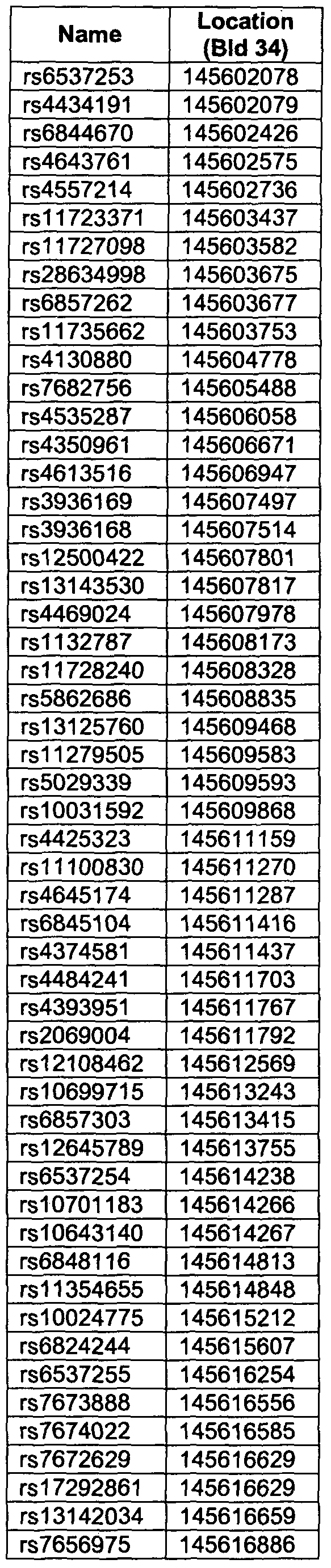


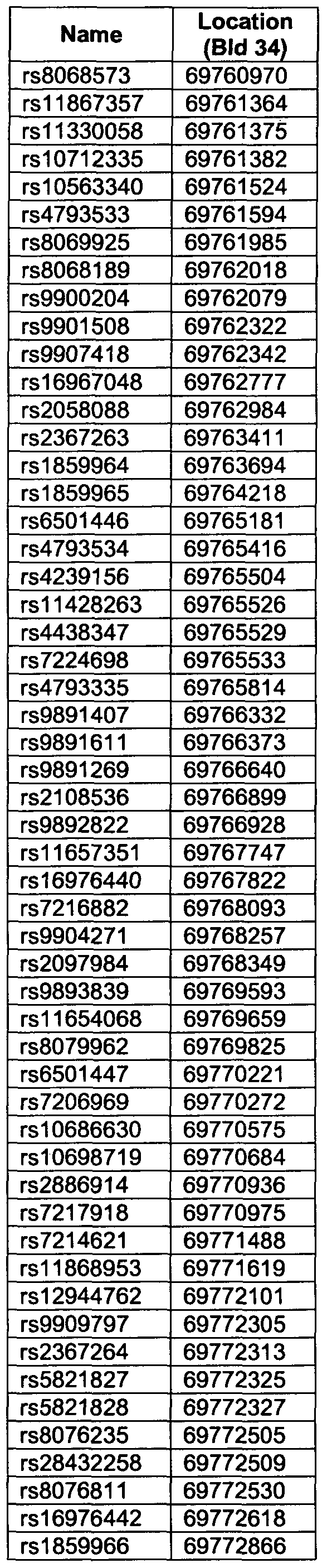


Claims
Priority Applications (11)
| Application Number | Priority Date | Filing Date | Title |
|---|---|---|---|
| US12/442,171 US8865400B2 (en) | 2007-02-07 | 2008-02-07 | Genetic variants contributing to risk of prostate cancer |
| JP2009548801A JP2010520745A (en) | 2007-02-07 | 2008-02-07 | Genetic variants that contribute to prostate cancer risk |
| AU2008212117A AU2008212117B2 (en) | 2007-02-07 | 2008-02-07 | Genetic variants contributing to risk of prostate cancer |
| CA002677994A CA2677994A1 (en) | 2007-02-07 | 2008-02-07 | Genetic variants contributing to risk of prostate cancer |
| CN2008800107105A CN102317470A (en) | 2007-02-07 | 2008-02-07 | Genetic variants contributing to risk of prostate cancer |
| BRPI0807236A BRPI0807236A2 (en) | 2007-02-07 | 2008-02-07 | methods for determining prostate cancer susceptibility in a human individual, identifying a marker for use in prostate cancer susceptibility assessment, and for genotyping a nucleic acid sample obtained from a human individual at risk of, or diagnosed with, cancer to assess a human individual for likelihood of response to a therapeutic agent to prevent and / or ameliorate symptoms associated with prostate cancer, to predict prognosis of an individual diagnosed with cancer, and to monitor progress in treatment of an individual undergoing treatment. for prostate cancer, kit for assessing susceptibility to prostate cancer in a human individual, use of a computer readable oligonucleotide probe, and apparatus for determining a genetic indicator for prostate cancer in a human individual. |
| IN5722DEN2009 IN2009DN05722A (en) | 2007-02-07 | 2008-02-07 | |
| EP08710289.3A EP2115171B1 (en) | 2007-02-07 | 2008-02-07 | Genetic variants contributing to risk of prostate cancer |
| NZ579401A NZ579401A (en) | 2007-02-07 | 2008-02-07 | Genetic variants contributing to risk of prostate cancer |
| MX2009008461A MX2009008461A (en) | 2007-02-07 | 2008-02-07 | Genetic variants contributing to risk of prostate cancer. |
| IL200197A IL200197A (en) | 2007-02-07 | 2009-08-02 | Method for assessing risk of prostate cancer by determining genetic variants |
Applications Claiming Priority (4)
| Application Number | Priority Date | Filing Date | Title |
|---|---|---|---|
| IS8604 | 2007-02-07 | ||
| IS8604 | 2007-02-07 | ||
| IS8654 | 2007-06-22 | ||
| IS8654 | 2007-06-22 |
Publications (2)
| Publication Number | Publication Date |
|---|---|
| WO2008096375A2 true WO2008096375A2 (en) | 2008-08-14 |
| WO2008096375A3 WO2008096375A3 (en) | 2008-12-18 |
Family
ID=39650503
Family Applications (1)
| Application Number | Title | Priority Date | Filing Date |
|---|---|---|---|
| PCT/IS2008/000003 WO2008096375A2 (en) | 2007-02-07 | 2008-02-07 | Genetic variants contributing to risk of prostate cancer |
Country Status (13)
| Country | Link |
|---|---|
| US (1) | US8865400B2 (en) |
| EP (2) | EP2115171B1 (en) |
| JP (1) | JP2010520745A (en) |
| CN (1) | CN102317470A (en) |
| AU (1) | AU2008212117B2 (en) |
| BR (1) | BRPI0807236A2 (en) |
| CA (1) | CA2677994A1 (en) |
| IL (1) | IL200197A (en) |
| IN (1) | IN2009DN05722A (en) |
| MX (1) | MX2009008461A (en) |
| NZ (1) | NZ579401A (en) |
| SG (1) | SG193026A1 (en) |
| WO (1) | WO2008096375A2 (en) |
Cited By (5)
| Publication number | Priority date | Publication date | Assignee | Title |
|---|---|---|---|---|
| WO2012031207A2 (en) | 2010-09-03 | 2012-03-08 | Wake Forest University Health Sciences | Methods and compositions for correlating genetic markers with prostate cancer risk |
| WO2013037118A1 (en) * | 2011-09-16 | 2013-03-21 | 上海长海医院 | Prostate cancer biomarkers, therapeutic targets and uses thereof |
| US8697360B2 (en) | 2007-11-30 | 2014-04-15 | Decode Genetics Ehf. | Genetic variants on CHR 11Q and 6Q as markers for prostate and colorectal cancer predisposition |
| US9534256B2 (en) | 2011-01-06 | 2017-01-03 | Wake Forest University Health Sciences | Methods and compositions for correlating genetic markers with risk of aggressive prostate cancer |
| RU2669809C2 (en) * | 2012-11-20 | 2018-10-16 | Пхадиа Аб | Prognostic method for individuals with prostate cancer |
Families Citing this family (11)
| Publication number | Priority date | Publication date | Assignee | Title |
|---|---|---|---|---|
| WO2009085196A1 (en) * | 2007-12-21 | 2009-07-09 | Wake Forest University Health Sciences | Methods and compositions for correlating genetic markers with prostate cancer risk |
| EP2577538A1 (en) | 2010-05-25 | 2013-04-10 | The Regents of the University of California | Bambam: parallel comparative analysis of high-throughput sequencing data |
| WO2012029080A1 (en) * | 2010-08-30 | 2012-03-08 | Decode Genetics Ehf | Sequence variants associated with prostate specific antigen levels |
| CN103060433B (en) * | 2012-10-19 | 2014-12-03 | 卫生部北京医院 | Kit and method for predicating susceptibility to prostate cancer |
| KR101944927B1 (en) | 2016-03-24 | 2019-02-07 | 서울대학교산학협력단 | Single Nucleotide Polymorphisms Associated With Korean Prostate Cancer And Development Of Genetic Risk Score Using Thereof |
| WO2017164699A1 (en) * | 2016-03-24 | 2017-09-28 | 서울대학교병원 (분사무소) | Prostate cancer-related single nucleotide polymorphism and development of genetic risk score by using same |
| KR102068666B1 (en) * | 2019-02-28 | 2020-02-11 | 주식회사 프로카젠 | Ethnic-specific Prostate Cancer Genetic Risk Score Calculating Device, Calculating Method and Recording Medium thereof |
| KR102068667B1 (en) * | 2019-02-28 | 2020-01-21 | 주식회사 프로카젠 | Prostate Cancer Genetic Risk Score Calculating Device, Calculating Method and Recording Medium thereof |
| KR102371655B1 (en) * | 2019-02-28 | 2022-03-08 | 주식회사 프로카젠 | Device, Method of Calculating Prostate Cancer Genetic Risk Score Based on Individual Weights for each Genetic Variation and Recording Medium thereof |
| KR102371654B1 (en) * | 2019-02-28 | 2022-03-08 | 주식회사 프로카젠 | Device, Calculating Method of Calculating Ethnic-specific Prostate Cancer Genetic Risk Score Considering the Predictive Power According to the Number of Genetic Variations and Recording Medium thereof |
| CN110596060B (en) * | 2019-09-12 | 2020-08-21 | 江南大学 | Construction method and application of fluorescence sensor in spectral analysis for detecting prostate specific antigen |
Citations (8)
| Publication number | Priority date | Publication date | Assignee | Title |
|---|---|---|---|---|
| US4851330A (en) | 1983-01-10 | 1989-07-25 | Kohne David E | Method for detection, identification and quantitation of non-viral organisms |
| WO1990015070A1 (en) | 1989-06-07 | 1990-12-13 | Affymax Technologies N.V. | Very large scale immobilized peptide synthesis |
| WO1992010092A1 (en) | 1990-12-06 | 1992-06-25 | Affymax Technologies N.V. | Very large scale immobilized polymer synthesis |
| US5288611A (en) | 1983-01-10 | 1994-02-22 | Gen-Probe Incorporated | Method for detecting, identifying, and quantitating organisms and viruses |
| US5384261A (en) | 1991-11-22 | 1995-01-24 | Affymax Technologies N.V. | Very large scale immobilized polymer synthesis using mechanically directed flow paths |
| US5424186A (en) | 1989-06-07 | 1995-06-13 | Affymax Technologies N.V. | Very large scale immobilized polymer synthesis |
| US5837832A (en) | 1993-06-25 | 1998-11-17 | Affymetrix, Inc. | Arrays of nucleic acid probes on biological chips |
| US5858659A (en) | 1995-11-29 | 1999-01-12 | Affymetrix, Inc. | Polymorphism detection |
Family Cites Families (30)
| Publication number | Priority date | Publication date | Assignee | Title |
|---|---|---|---|---|
| US4376110A (en) | 1980-08-04 | 1983-03-08 | Hybritech, Incorporated | Immunometric assays using monoclonal antibodies |
| US6054270A (en) | 1988-05-03 | 2000-04-25 | Oxford Gene Technology Limited | Analying polynucleotide sequences |
| US5700637A (en) | 1988-05-03 | 1997-12-23 | Isis Innovation Limited | Apparatus and method for analyzing polynucleotide sequences and method of generating oligonucleotide arrays |
| US5223409A (en) | 1988-09-02 | 1993-06-29 | Protein Engineering Corp. | Directed evolution of novel binding proteins |
| AU4308689A (en) | 1988-09-02 | 1990-04-02 | Protein Engineering Corporation | Generation and selection of recombinant varied binding proteins |
| US5744101A (en) | 1989-06-07 | 1998-04-28 | Affymax Technologies N.V. | Photolabile nucleoside protecting groups |
| US5288644A (en) | 1990-04-04 | 1994-02-22 | The Rockefeller University | Instrument and method for the sequencing of genome |
| US5427908A (en) | 1990-05-01 | 1995-06-27 | Affymax Technologies N.V. | Recombinant library screening methods |
| GB9015198D0 (en) | 1990-07-10 | 1990-08-29 | Brien Caroline J O | Binding substance |
| WO1992020791A1 (en) | 1990-07-10 | 1992-11-26 | Cambridge Antibody Technology Limited | Methods for producing members of specific binding pairs |
| CA2095633C (en) | 1990-12-03 | 2003-02-04 | Lisa J. Garrard | Enrichment method for variant proteins with altered binding properties |
| CA2105300C (en) | 1991-03-01 | 2008-12-23 | Robert C. Ladner | Process for the development of binding mini-proteins |
| DK1471142T3 (en) | 1991-04-10 | 2009-03-09 | Scripps Research Inst | Heterodimeric receptor libraries using phagemids |
| DE4122599C2 (en) | 1991-07-08 | 1993-11-11 | Deutsches Krebsforsch | Phagemid for screening antibodies |
| EP0695941B1 (en) | 1994-06-08 | 2002-07-31 | Affymetrix, Inc. | Method and apparatus for packaging a chip |
| US6287850B1 (en) | 1995-06-07 | 2001-09-11 | Affymetrix, Inc. | Bioarray chip reaction apparatus and its manufacture |
| US6300063B1 (en) | 1995-11-29 | 2001-10-09 | Affymetrix, Inc. | Polymorphism detection |
| EP0912761A4 (en) | 1996-05-29 | 2004-06-09 | Cornell Res Foundation Inc | Detection of nucleic acid sequence differences using coupled ligase detection and polymerase chain reactions |
| US5925519A (en) * | 1996-06-03 | 1999-07-20 | The Regents Of The University Of California | Genetic alterations associated with prostate cancer |
| AU773006B2 (en) | 1998-06-05 | 2004-05-13 | Genset S.A. | Polymorphic markers of prostate carcinoma tumor antigen-1 (PCTA-1) |
| US6355623B2 (en) | 1998-09-24 | 2002-03-12 | Hopital-Sainte-Justine | Method of treating IBD/Crohn's disease and related conditions wherein drug metabolite levels in host blood cells determine subsequent dosage |
| JP2002529065A (en) * | 1998-11-06 | 2002-09-10 | ミリアド・ジェネティックス・インコーポレイテッド | Chromosome 17q linkage-prostate cancer susceptibility gene |
| US6429027B1 (en) | 1998-12-28 | 2002-08-06 | Illumina, Inc. | Composite arrays utilizing microspheres |
| EP1585433A4 (en) | 2002-09-25 | 2008-01-23 | Ge Healthcare Sv Corp | Detection methods |
| US20050136407A1 (en) | 2003-12-19 | 2005-06-23 | Cramer Scott D. | Genetic polymorphisms in the prostate-specific antigen gene promoter |
| US20050255507A1 (en) | 2004-02-17 | 2005-11-17 | Jenkins Robert B | Cytogenetically determined diagnosis and prognosis of proliferative disorders |
| WO2006063285A2 (en) * | 2004-12-10 | 2006-06-15 | Sequenom, Inc. | Methods for identifying risk of breast cancer or prostate cancer and treatments thereof |
| JP5227167B2 (en) | 2005-05-18 | 2013-07-03 | デコード・ジェネティクス・イーエイチエフ | Mutants at CHR8Q24.21 pose cancer risk |
| SG175680A1 (en) | 2006-10-27 | 2011-11-28 | Decode Genetics Ehf | Cancer susceptibility variants on chr8q24.21 |
| US8697360B2 (en) | 2007-11-30 | 2014-04-15 | Decode Genetics Ehf. | Genetic variants on CHR 11Q and 6Q as markers for prostate and colorectal cancer predisposition |
-
2008
- 2008-02-07 JP JP2009548801A patent/JP2010520745A/en not_active Ceased
- 2008-02-07 IN IN5722DEN2009 patent/IN2009DN05722A/en unknown
- 2008-02-07 AU AU2008212117A patent/AU2008212117B2/en not_active Ceased
- 2008-02-07 BR BRPI0807236A patent/BRPI0807236A2/en not_active IP Right Cessation
- 2008-02-07 CN CN2008800107105A patent/CN102317470A/en active Pending
- 2008-02-07 EP EP08710289.3A patent/EP2115171B1/en active Active
- 2008-02-07 US US12/442,171 patent/US8865400B2/en not_active Expired - Fee Related
- 2008-02-07 MX MX2009008461A patent/MX2009008461A/en active IP Right Grant
- 2008-02-07 NZ NZ579401A patent/NZ579401A/en not_active IP Right Cessation
- 2008-02-07 EP EP12160294.0A patent/EP2522743A3/en not_active Withdrawn
- 2008-02-07 WO PCT/IS2008/000003 patent/WO2008096375A2/en active Application Filing
- 2008-02-07 SG SG2012008371A patent/SG193026A1/en unknown
- 2008-02-07 CA CA002677994A patent/CA2677994A1/en not_active Abandoned
-
2009
- 2009-08-02 IL IL200197A patent/IL200197A/en not_active IP Right Cessation
Patent Citations (9)
| Publication number | Priority date | Publication date | Assignee | Title |
|---|---|---|---|---|
| US4851330A (en) | 1983-01-10 | 1989-07-25 | Kohne David E | Method for detection, identification and quantitation of non-viral organisms |
| US5288611A (en) | 1983-01-10 | 1994-02-22 | Gen-Probe Incorporated | Method for detecting, identifying, and quantitating organisms and viruses |
| WO1990015070A1 (en) | 1989-06-07 | 1990-12-13 | Affymax Technologies N.V. | Very large scale immobilized peptide synthesis |
| US5143854A (en) | 1989-06-07 | 1992-09-01 | Affymax Technologies N.V. | Large scale photolithographic solid phase synthesis of polypeptides and receptor binding screening thereof |
| US5424186A (en) | 1989-06-07 | 1995-06-13 | Affymax Technologies N.V. | Very large scale immobilized polymer synthesis |
| WO1992010092A1 (en) | 1990-12-06 | 1992-06-25 | Affymax Technologies N.V. | Very large scale immobilized polymer synthesis |
| US5384261A (en) | 1991-11-22 | 1995-01-24 | Affymax Technologies N.V. | Very large scale immobilized polymer synthesis using mechanically directed flow paths |
| US5837832A (en) | 1993-06-25 | 1998-11-17 | Affymetrix, Inc. | Arrays of nucleic acid probes on biological chips |
| US5858659A (en) | 1995-11-29 | 1999-01-12 | Affymetrix, Inc. | Polymorphism detection |
Non-Patent Citations (19)
| Title |
|---|
| "Current Protocols in Molecular Biology", JOHN WILEY & SONS |
| AMUNDADOTTIR, L.T. ET AL., NAT GENET., vol. 38, 2006, pages 652 - 58 |
| CHURCH; GILBERT, PROC. NATL. ACAD. SCI. USA, vol. 81, 1988, pages 1991 - 1995 |
| FALK, C.T.; RUBINSTEIN, P.: "Haplotype relative risks: an easy reliable way to construct a proper control sample for risk calculations", ANN. HUM. GENET., vol. 51, 1987, pages 227 - 33 |
| FODOR, S. ET AL., SCIENCE, vol. 251, 1991, pages 767 - 773 |
| GRANT, S.F. ET AL., NAT GENET., vol. 38, 2006, pages 320 - 23 |
| GRETARSD6TTIR ET AL., NAT GENET., vol. 35, no. 2, October 2003 (2003-10-01), pages 131 - 8 |
| GRIFFITHS ET AL., EMBO J., vol. 12, 1993, pages 725 - 734 |
| GUDMUNDSSON, J. ET AL., NAT GENET., vol. 39, 2007, pages 631 - 37 |
| HELGADOTTIR, A. ET AL., SCIENCE, vol. 316, 2007, pages 1491 - 93 |
| HILL, W.G.; A. ROBERTSON, THEOR. APPL. GENET., vol. 22, 1968, pages 226 - 231 |
| HUM. ANTIBOD. HYBRIDOMAS, vol. 3, 1992, pages 81 - 85 |
| HUSE ET AL., SCIENCE, vol. 246, 1989, pages 1275 - 1281 |
| LEWONTIN, R., GENETICS, vol. 49, 1964, pages 49 - 67 |
| NIELSEN, P. ET AL., BIOCONJUG. CHEM., vol. 5, 1994, pages 3 - 7 |
| SMITH ET AL., AM J HUM GENET, vol. 74, 2004, pages 1001 - 13 |
| STACEY, S.N. ET AL., NAT GENET., 27 May 2007 (2007-05-27) |
| STEINTHORSDOTTIR, V. ET AL., NAT GENET., vol. 39, 2007, pages 770 - 75 |
| TERWILLIGER, J.D.; OTT, J.: "A haplotype-based 'haplotype relative risk' approach to detecting allelic associations", HUM. HERED., vol. 42, 1992, pages 337 - 46 |
Cited By (12)
| Publication number | Priority date | Publication date | Assignee | Title |
|---|---|---|---|---|
| US8697360B2 (en) | 2007-11-30 | 2014-04-15 | Decode Genetics Ehf. | Genetic variants on CHR 11Q and 6Q as markers for prostate and colorectal cancer predisposition |
| WO2012031207A2 (en) | 2010-09-03 | 2012-03-08 | Wake Forest University Health Sciences | Methods and compositions for correlating genetic markers with prostate cancer risk |
| EP2611943A2 (en) * | 2010-09-03 | 2013-07-10 | Wake Forest University Health Sciences | Methods and compositions for correlating genetic markers with prostate cancer risk |
| EP2611943A4 (en) * | 2010-09-03 | 2014-01-08 | Univ Wake Forest Health Sciences | Methods and compositions for correlating genetic markers with prostate cancer risk |
| US9732389B2 (en) | 2010-09-03 | 2017-08-15 | Wake Forest University Health Sciences | Methods and compositions for correlating genetic markers with prostate cancer risk |
| US10443105B2 (en) | 2010-09-03 | 2019-10-15 | Wake Forest University Health Sciences | Methods and compositions for correlating genetic markers with prostate cancer risk |
| US11421282B2 (en) | 2010-09-03 | 2022-08-23 | Wake Forest University Health Sciences | Methods and compositions for correlating genetic markers with prostate cancer risk |
| US9534256B2 (en) | 2011-01-06 | 2017-01-03 | Wake Forest University Health Sciences | Methods and compositions for correlating genetic markers with risk of aggressive prostate cancer |
| WO2013037118A1 (en) * | 2011-09-16 | 2013-03-21 | 上海长海医院 | Prostate cancer biomarkers, therapeutic targets and uses thereof |
| CN103797120A (en) * | 2011-09-16 | 2014-05-14 | 上海长海医院 | Prostate cancer biomarkers, therapeutic targets and uses thereof |
| RU2669809C2 (en) * | 2012-11-20 | 2018-10-16 | Пхадиа Аб | Prognostic method for individuals with prostate cancer |
| US10428385B2 (en) | 2012-11-20 | 2019-10-01 | Phadia Ab | Prognostic method for individuals with prostate cancer |
Also Published As
| Publication number | Publication date |
|---|---|
| AU2008212117B2 (en) | 2014-09-11 |
| MX2009008461A (en) | 2009-08-17 |
| CN102317470A (en) | 2012-01-11 |
| EP2522743A2 (en) | 2012-11-14 |
| IL200197A0 (en) | 2010-04-29 |
| EP2115171A2 (en) | 2009-11-11 |
| IL200197A (en) | 2014-07-31 |
| US8865400B2 (en) | 2014-10-21 |
| SG193026A1 (en) | 2013-09-30 |
| CA2677994A1 (en) | 2008-08-14 |
| JP2010520745A (en) | 2010-06-17 |
| AU2008212117A1 (en) | 2008-08-14 |
| US20100041037A1 (en) | 2010-02-18 |
| BRPI0807236A2 (en) | 2019-09-24 |
| EP2115171B1 (en) | 2015-01-21 |
| NZ579401A (en) | 2012-03-30 |
| EP2522743A3 (en) | 2013-08-07 |
| IN2009DN05722A (en) | 2015-07-24 |
| WO2008096375A3 (en) | 2008-12-18 |
Similar Documents
| Publication | Publication Date | Title |
|---|---|---|
| AU2008212117B2 (en) | Genetic variants contributing to risk of prostate cancer | |
| AU2007310412B2 (en) | Cancer susceptibility variants on Chr8q24.21 | |
| EP2535425A1 (en) | Variantes génétiques sur les chr 10q26 utilisées comme marqueurs dans l'évaluation, le diagnostic, le pronostic et le traitement d'un risque de cancer du sein | |
| US20110117545A1 (en) | Genetic variants on chr2 and chr16 as markers for use in breast cancer risk assessment, diagnosis, prognosis and treatment | |
| WO2010004591A2 (en) | Genetic variants for breast cancer risk assessment | |
| WO2010128530A1 (en) | Genetic variants contributing to risk of prostate cancer | |
| EP2099937A2 (en) | Genetic susceptibility variants of type 2 diabetes mellitus | |
| US20140248615A1 (en) | Genetic variants on chr 11q and 6q as markers for prostate and colorectal cancer predisposition |
Legal Events
| Date | Code | Title | Description |
|---|---|---|---|
| WWE | Wipo information: entry into national phase |
Ref document number: 200880010710.5 Country of ref document: CN |
|
| 121 | Ep: the epo has been informed by wipo that ep was designated in this application |
Ref document number: 08710289 Country of ref document: EP Kind code of ref document: A2 |
|
| WWE | Wipo information: entry into national phase |
Ref document number: 12442171 Country of ref document: US |
|
| WWE | Wipo information: entry into national phase |
Ref document number: 200197 Country of ref document: IL |
|
| ENP | Entry into the national phase |
Ref document number: 2009548801 Country of ref document: JP Kind code of ref document: A |
|
| WWE | Wipo information: entry into national phase |
Ref document number: 2677994 Country of ref document: CA Ref document number: MX/A/2009/008461 Country of ref document: MX |
|
| NENP | Non-entry into the national phase |
Ref country code: DE |
|
| WWE | Wipo information: entry into national phase |
Ref document number: 2008212117 Country of ref document: AU |
|
| WWE | Wipo information: entry into national phase |
Ref document number: 2008710289 Country of ref document: EP |
|
| WWE | Wipo information: entry into national phase |
Ref document number: 579401 Country of ref document: NZ |
|
| WWE | Wipo information: entry into national phase |
Ref document number: 5722/DELNP/2009 Country of ref document: IN |
|
| ENP | Entry into the national phase |
Ref document number: 2008212117 Country of ref document: AU Date of ref document: 20080207 Kind code of ref document: A |
|
| ENP | Entry into the national phase |
Ref document number: PI0807236 Country of ref document: BR Kind code of ref document: A2 Effective date: 20090806 |