WO2011099664A1 - Probe for hpv genotype diagnosis and analysis method thereof - Google Patents
Probe for hpv genotype diagnosis and analysis method thereof Download PDFInfo
- Publication number
- WO2011099664A1 WO2011099664A1 PCT/KR2010/000914 KR2010000914W WO2011099664A1 WO 2011099664 A1 WO2011099664 A1 WO 2011099664A1 KR 2010000914 W KR2010000914 W KR 2010000914W WO 2011099664 A1 WO2011099664 A1 WO 2011099664A1
- Authority
- WO
- WIPO (PCT)
- Prior art keywords
- hpv
- pcr
- probe
- present
- primer
- Prior art date
Links
Images


Classifications
-
- C—CHEMISTRY; METALLURGY
- C12—BIOCHEMISTRY; BEER; SPIRITS; WINE; VINEGAR; MICROBIOLOGY; ENZYMOLOGY; MUTATION OR GENETIC ENGINEERING
- C12Q—MEASURING OR TESTING PROCESSES INVOLVING ENZYMES, NUCLEIC ACIDS OR MICROORGANISMS; COMPOSITIONS OR TEST PAPERS THEREFOR; PROCESSES OF PREPARING SUCH COMPOSITIONS; CONDITION-RESPONSIVE CONTROL IN MICROBIOLOGICAL OR ENZYMOLOGICAL PROCESSES
- C12Q1/00—Measuring or testing processes involving enzymes, nucleic acids or microorganisms; Compositions therefor; Processes of preparing such compositions
- C12Q1/70—Measuring or testing processes involving enzymes, nucleic acids or microorganisms; Compositions therefor; Processes of preparing such compositions involving virus or bacteriophage
- C12Q1/701—Specific hybridization probes
- C12Q1/708—Specific hybridization probes for papilloma
Landscapes
- Chemical & Material Sciences (AREA)
- Life Sciences & Earth Sciences (AREA)
- Health & Medical Sciences (AREA)
- Organic Chemistry (AREA)
- Zoology (AREA)
- Engineering & Computer Science (AREA)
- Immunology (AREA)
- Wood Science & Technology (AREA)
- Proteomics, Peptides & Aminoacids (AREA)
- Virology (AREA)
- Biotechnology (AREA)
- Microbiology (AREA)
- Molecular Biology (AREA)
- Physics & Mathematics (AREA)
- Biophysics (AREA)
- Analytical Chemistry (AREA)
- Biochemistry (AREA)
- Bioinformatics & Cheminformatics (AREA)
- General Engineering & Computer Science (AREA)
- General Health & Medical Sciences (AREA)
- Genetics & Genomics (AREA)
- Measuring Or Testing Involving Enzymes Or Micro-Organisms (AREA)
Abstract
The present invention relates to a probe for HPV genotype diagnosis and to an analysis method thereof.
Description
본 발명은 HPV 유전자형 진단용 프로브 및 그 분석방법에 관한 것이다.The present invention relates to a HPV genotype diagnostic probe and an analysis method thereof.
전 세계적으로 자궁경부암 환자의 99.7%에서 HR (High Risk)-HPV DNA가 존재하며 이러한 HR-HPV 감염의 존속이 침윤성 자궁경부암, 자궁경부 전암으로의 전이를 일으키는 것으로 알려져 있다. 자궁경부암의 경우 전 세계적으로는 매년 50만명, 우리나라의 경우 6000명의 새로운 환자가 발생하고 있으며 50%는 치명적인 결과를 보인다. 이러한 자궁경부 종양으로 진행하는 과정에 인두유종 바이러스 (Human Papilloma Virus, HPV)가 중요한 원인으로 알려지면서 자궁암의 조기 진단에 HPV DNA의 존재 유무를 이용하고 있다 (Bernard, H. U., I. E. Calleja-Macias, and S. T. Dunn. 2006. Int J Cancer 118:1071-6). HPV는 약 7.9 kb의 이중나선 구조의 DNA 바이러스로 papovavirus에 속하는 것으로 100개 이상의 subtype이 존재하고 있으며 이중 40개의 subtype은 생식기를 통해 감염 되는 것으로 알려져 있다. 또한 HPV 바이러스는 10개의 바이러스성 단백질을 지니고 있는데 바이러스의 캡시드를 구성하는 구조 단백질 합성에 관여하는 2개의 early 유전자 산물인 L1, L2와 바이러스의 감염 의 활성과 잠복을 결정하는 바이러스의 복제에 관여하는 단백질의 합성을 조절하는 8개의 late 유전자 산물인 E1, E2, E3, E4, E5, E6, E7, E8을 발현한다. 특히 E6와 E7은 전암 단백질을 암호화 하고 있고, 암과 연관이 있는 부분으로 알려져 있으며 (Sedman, S. A., 등. 1991. J Virol 65:4860-6), 여러 보고에 의하여 HPV 바이러스가 여성의 자궁경부암을 유발하고, 여러 가지 악성 종양과 밀접한 관계가 있는 것으로 알려져 있다 (Godfroid, E., 등. 1998. J Virol Methods 75:69-81).High risk (HPV) -HPV DNA is present in 99.7% of cervical cancer patients worldwide, and the persistence of such HR-HPV infection is known to cause metastasis to invasive cervical cancer and cervical precancerous cancer. Cervical cancer causes 500,000 cases worldwide and 6,000 new cases in Korea each year, with 50% fatal outcomes. Human Papilloma Virus (HPV) is known to be an important cause of the progression of cervical tumors, and HPV DNA is used for early diagnosis of cervical cancer (Bernard, HU, IE Calleja-Macias, and ST Dunn. 2006. Int J Cancer 118: 1071-6). HPV is a double-stranded DNA virus of about 7.9 kb, belonging to papovavirus, which contains more than 100 subtypes, of which 40 are known to infect the genitals. The HPV virus also contains 10 viral proteins that are involved in the replication of the virus, which determines the activity and latency of L1 and L2, two early gene products involved in the synthesis of structural proteins that make up the capsid of the virus, and the viral infection. It expresses eight late gene products, E1, E2, E3, E4, E5, E6, E7, and E8, which regulate protein synthesis. In particular, E6 and E7 encode precancerous proteins and are known to be associated with cancer (Sedman, SA, et al. 1991. J Virol 65: 4860-6). And is closely associated with various malignancies (Godfroid, E., et al. 1998. J Virol Methods 75: 69-81).
여성의 자궁경부암은 전 세계적으로 유방암에 이어 두 번째로 높은 발병율을 나타내고 있고, 우리나라에서도 매년 1,000명 이상의 사망자가 보이고 있어 자궁경부암의 조기 진단을 위한 노력이 매우 중요하게 여겨지고 있다 (Wui, J. H., 등. 2006. Journal of Gynecologic oncology and Coloscopy 17:39-4711). Cervical cancer in women is the second highest incidence in the world after breast cancer, with more than 1,000 deaths annually in Korea, making efforts for early diagnosis of cervical cancer very important (Wui, JH, etc.). 2006. Journal of Gynecologic oncology and Coloscopy 17: 39-4711.
현재 HPV의 유전자형은 100가지 이상이 알려져 있고 (Likes, W. M., 및 J. Itano. 2003. Clin J Oncol Nurs 7:271-6), 이 중에서 사람에게 질병을 일으킬 수 있는 유전자형은 30가지 정도로 알려져 있다. 그 유전자형은 고위험군 (16, 18, 31, 33, 35, 39, 45, 51, 52, 56, 58, 59, 68, 73, 82) 과 저위험군 (6, 11, 40, 42, 43, 44, 54, 61, 70, 72, 81), 잠재위험군(34, 57, 83)으로 분류하고 있으며 (Munoz, N., 등. 2003. N Engl J Med 348:518-27), 각각의 유전자형을 지닌 HPV가 병소의 위치와 병변의 진행정도에 따라 유전자형 특이적으로 발견됨으로써 HPV 감염의 생물학적 다양성을 인지하게 되었다.There are currently over 100 known genotypes of HPV (Likes, WM, and J. Itano. 2003. Clin J Oncol Nurs 7: 271-6), of which about 30 are known to cause disease in humans. . The genotypes were high risk (16, 18, 31, 33, 35, 39, 45, 51, 52, 56, 58, 59, 68, 73, 82) and low risk (6, 11, 40, 42, 43, 44). , 54, 61, 70, 72, 81), and potential risk groups (34, 57, 83) (Munoz, N., et al. 2003. N Engl J Med 348: 518-27). HPV was found to be genotype-specific depending on the location of the lesion and the progression of the lesion, thereby recognizing the biological diversity of HPV infection.
이러한 HPV의 감염여부를 진단하기 위하여 가장 많이 쓰이는 방법은 자궁세포진 검사(Papnicolaou Smear, Pap) 방법으로서 현재도 자궁경부암의 조직학적 진단은 이 검사법에 의해 진단하고 있으나 이 검사법은 검사자의 경험과 숙련도에 많은 의존을 해야 하고, 그 때문에 정확도가 떨어진다는 단점을 가지고 있다(Kurman, R. J., D. E. Henson, A. L. Herbst, K. L. Noller, and M. H. Schiffman. 1994. JAMA 271:1866-9). 뿐만 아니라, 질확대경을 이용한 검사 방법은 자궁세포진 검사보다 정확한 결과를 얻을 수는 있지만, 숙련된 기술자와 고가의 장비가 필요하며, HPV의 유전자형을 구별할 수 없다는 단점이 있으며 위 두 방법을 통해서는 악성화 위험도가 다른 HPV 유전형을 분류할 수 없다는 단점이 있다. The most commonly used method for diagnosing HPV infection is Papnicolaou Smear (Pap) method. Currently, histological diagnosis of cervical cancer is diagnosed by this test. It has a lot of dependence and has the disadvantage of poor accuracy (Kurman, RJ, DE Henson, AL Herbst, KL Noller, and MH Schiffman. 1994. JAMA 271: 1866-9). In addition, the vaginal magnification test is more accurate than the Pap smear, but requires a skilled technician and expensive equipment, and the HPV genotype cannot be distinguished. The disadvantage is that the risk of malignancy cannot be classified between different HPV genotypes.
상기한 단점을 보완하기 위하여 PCR, HC, DNA chip등을 이용하여 HPV 특이적 DNA 증폭을 통해 자궁경부암 전암 단계를 진단하고 유전자형을 확인하는 방법들의 연구가 계속 진행되고 있는 실정이다.In order to make up for the above drawbacks, studies of methods for diagnosing cervical cancer precancerous stage and confirming genotype through HPV-specific DNA amplification using PCR, HC, DNA chip, etc. are being conducted.
본 발명은 상기의 문제점을 해결하고, 상기의 필요성에 의하여 안출된 것으로서 본 발명의 목적은 기존의 DNA chip 보다 HPV에 대한 특이도와 민감도가 좋은 프로브를 제공하는 것이다.The present invention solves the above problems, and the object of the present invention is to provide a probe having a specificity and sensitivity for HPV better than the conventional DNA chip.
본 발명의 다른 목적은 상기 프로브를 이용한 HPV 유전자형 분석방법을 제공하는 것이다.Another object of the present invention to provide a method for analyzing HPV genotype using the probe.
상기의 목적을 달성하기 위하여 본 발명은 상기의 목적을 달성하기 위하여 본 발명은 서열번호 5 내지 서열번호 28의 염기서열로 구성된 군으로부터 선택된 하나 이상의 HPV 진단용 올리고뉴크레오타이드 프로브를 제공한다.In order to achieve the above object, the present invention provides at least one HPV diagnostic oligonucleotide probe selected from the group consisting of the nucleotide sequences of SEQ ID NO: 5 to SEQ ID NO: 28.
또한 본 발명은 상기의 본 발명의 프로브 및 프라이머를 포함하는 HPV 진단용 키트를 제공한다.In another aspect, the present invention provides a kit for diagnosing HPV comprising the probe and primer of the present invention.
본 발명에 있어서, 상기 프라이머는 서열번호 1 내지 4에 기재된 프라이머인 것이 바람직하나 이에 한정되는 것은 아니다.In the present invention, the primer is preferably a primer described in SEQ ID NOs: 1 to 4, but is not limited thereto.
본 발명의 일 실시예에 있어서, 상기 키트는 PCR 산물을 탐지하기 위한 표지수단을 더욱 포함하는 것이 바람직하다.In one embodiment of the invention, the kit preferably further comprises a label means for detecting the PCR product.
또한 본 발명은 a) 상기 본 발명의 프로브를 고체 서포트에 부착하는 단계;The present invention also comprises the steps of: a) attaching the probe of the present invention to a solid support;
b) 상기 프로브에 증폭된 HPV PCR 산물을 교잡하는 단계; 및b) hybridizing the amplified HPV PCR product to the probe; And
c) 상기 교잡된 산물을 검출하는 단계를 포함하는 HPV 유전자형 확인 방법을 제공한다.c) providing a method for identifying HPV genotype comprising detecting the hybridized product.
본 발명에 있어서, 상기 고체 서포트는 막, 슬라이드, 또는 웰 플레이트인 것이 바람직하나 이에 한정되는 것은 아니다.In the present invention, the solid support is preferably a membrane, a slide, or a well plate, but is not limited thereto.
본 발명의 일 실시예에 있어서, 상기 검출단계는 발광 또는 발색반응을 이용하여 검출하는 것이 바람직하다.In one embodiment of the present invention, the detecting step is preferably detected using a luminescence or color reaction.
본 발명은 검체로부터 핵산을 추출하고, PCR 증폭을 수행하였다. 세포로부터 핵산을 추출하는 방법 및 PCR 방법은 Sambrook 등의 Molecular cloning(1989)에 기재된 방법과 같은 당업계에 주지된 방법을 통하여 수행하였다.In the present invention, nucleic acid was extracted from a sample and PCR amplification was performed. Methods of extracting nucleic acids from cells and PCR methods were performed through methods well known in the art, such as those described in Molecular cloning (1989) by Sambrook et al.
본 발명의 핵산 증폭 방법은 프라이머를 사용하였다. 일반적으로 프라이머는 약 10에서 25bp 길이의 올리고뉴크레오타이드가 바람직하나 더 긴 것도 가능하며, 프라이머는 단일 가닥이 바람직하나 이중 또는 단일 가닥 형태로 존재할 수도 있으며, 본 발명에서 사용한 특정 프라이머는 본 발명의 실시예에 기재하였다.The nucleic acid amplification method of the present invention used a primer. Generally, the primer is preferably about 10 to 25 bp in length oligonucleotide, but may be longer. The primer is preferably single stranded but may be present in double or single stranded form. It described in the Example.
본 발명의 프라이머는 통상의 올리고뉴크레오타이드 합성법에 의하여 제조되었으며, 그러한 합성 방법은 미국특허 제 4,659,774, 4,816,571, 5,141,813, 5,264,566, 4,959,463, 5,428,148, 5,554,744, 5,574,146, 및 5,602,244 등에 개재된다.The primers of the present invention were prepared by conventional oligonucleotide synthesis methods, and such synthesis methods are disclosed in US Pat.
본 발명의 프라이머와 같은 서열들은 샘플로부터 유래한 DNA와 같은 상보적인 서열들과 선택적으로 이중 나선을 형성하는 데 사용될 수 있다. 당업자는 타겟 서열에 대한 프라이머의 선택성을 변화시키기 위하여 교잡 조건을 변경할 수 있다.Sequences such as primers of the invention can be used to form a double helix optionally with complementary sequences, such as DNA derived from a sample. One skilled in the art can alter the hybridization conditions to change the selectivity of the primer for the target sequence.
일부 실시예에서는 교잡을 결정하기 위하여 표지와 같은 적당한 수단과 결합하여 본 발명의 한정된 서열의 핵산을 채택할 수 있다. 적당한 표지수단은 형광, 방사성동위원소 효소 또는 다른 리간드를 포함하는 다양한 표지수단이 가능하다.In some embodiments, nucleic acids of defined sequences of the invention may be employed in combination with appropriate means such as labels to determine hybridization. Suitable labeling means are various labeling means including fluorescence, radioisotope enzymes or other ligands.
다양한 주형 의존적인 과정들이 주어진 세포 샘플에 존재하는 특정 유전자 산물을 증폭하는데 가능하다. 가장공지된 증폭 방법 중 하나가 중합효소 연쇄반응(PCR)이다 이 방법은 미국특허 제 4,683,202 및 4,800,159 등에 기재된다.Various template dependent processes are possible to amplify specific gene products present in a given cell sample. One of the most known amplification methods is polymerase chain reaction (PCR), which is described in US Pat. Nos. 4,683,202 and 4,800,159 and the like.
일반적으로 한 단계 또는 또 다른 단계에서 증폭산물을 주형 또는 초과 프라이머 또는 다른 증폭산물로부터 분리하는 것이 바람직하다. 예를 들어 증폭 산물은 Sambrook 등의 Molecular cloning(1989)에 기재된 표준 방법을 아가로스, 아가로스-아크릴아마이드 또는 PAGE를 사용하여 분리될 수 있다.It is generally desirable to separate the amplification product from the template or excess primer or other amplification product in one or another step. For example, the amplification products can be separated using agarose, agarose-acrylamide or PAGE using the standard method described in Molecular cloning (1989) by Sambrook et al.
또 흡착, 분배, 이온교환 등과 같은 크로마토그래피도 효과적인 분리방법으로 채택될 수 있다. 이들 분리방법들은 대량의 샘플을 가공하기 위하여 임상 셋팅에서 기능하도록 채택될 수 있으며, 수천개의 샘플을 한번에 분리하거나 검출할 수 있는 새로운 방법인 FMAT (fluorometric microvolume assay technique), chemiluminescence,sequence detection systems (Applied Biosystems) 및 질량 분광법 등이 있다.Chromatography, such as adsorption, partitioning and ion exchange, can also be employed as an effective separation method. These separations can be adapted to function in clinical settings to process large volumes of samples, and new methods for separating or detecting thousands of samples at once, FMAT (fluorometric microvolume assay technique), chemiluminescence, and sequence detection systems (Applied) Biosystems) and mass spectroscopy.
본 발명에 따른 핵산은 검출되고 정량화될 것이다. 일 실시예에서 그 검출은 시각적 수단에 의하여 수행될 수 있다. 전형적인 시각적 수단 방법은 ethidium bromide로 젤을 염색하고, UV 광 하에서 밴드들을 시각화하는 것이다. 또 만약 증폭 산물이 방사선 또는 형광으로 표지된 뉴크레오타이드이면 분리된 증폭산물은 삽입된 방사선 동위원소 표지의 방사성 신티그램 촬영 또는 형광 검출을 수행한다.Nucleic acids according to the invention will be detected and quantified. In one embodiment the detection can be performed by visual means. Typical visual means method is to dye the gel with ethidium bromide and visualize the bands under UV light. If the amplification product is a nucleotide that is labeled with radiation or fluorescence, the isolated amplification product performs radiosynthesis or fluorescence detection of the inserted radioisotope label.
본 발명은 상기 방법을 수행하기 위한 키트를 포함한다. 비한정적인 실시예에서 본 발명의 키트는 프라이머들,증폭을 위한 효소 및 부가적인 시약을 포함할 수 있다. 따라서 본 키트들은 적당한 용기 수단들에 이들 하나 이상의 시약들을 포함할 것이다. 그 키트들은 또한 핵산을 분리하고 증폭 산물을 정제하는 시약들을 포함할 수도 있다. 키트들의 구성성분은 수용성 매체 또는 동결건조 형태로 포장될 수 있고, 키트의 적당한 용기 수단은 구성성분이 들어갈 수 있는 적어도 하나의 바이알, 시험관, 플라스크, 병 등을 포함한다. 키트에 하나 이상의 구성요소가 존재한다면 부가적인 구성요소들이 따로 위치될 수 있는 제2, 제3 등의 다른 부가적인 용기를 포함할 수 있다. 그러나 여러 구성요소의 조합이 하나의 용기에 포함될 수도 있다.The present invention includes a kit for carrying out the method. In a non-limiting example, the kit of the present invention may include primers, enzymes for amplification and additional reagents. Thus, the kits will include these one or more reagents in suitable container means. The kits may also include reagents for isolating nucleic acids and purifying amplification products. The components of the kits may be packaged in an aqueous medium or lyophilized form, and suitable container means of the kit include at least one vial, test tube, flask, bottle, etc. into which the components may be placed. If one or more components are present in the kit, they may include other additional containers, such as second, third, etc., where the additional components may be located separately. However, a combination of components may be included in one container.
본 발명에서 사용된 nested PCR 방법은 통상 Two-tube nested PCR을 의미하는데, 'Two-tube nested PCR'은 보통 시료의 DNA양이 매우 제한되어 있어 한번의 PCR로는 검출할 수 없는 경우에 두 번째 PCR을 보다 내측의 primer를 이용하여 시행하는 방법으로, 원하는 부분이 잘 증폭되기 위해서는 4개의 primers가 부착해야 하고, outer primers를 이용한 첫 번째 중합효소 연쇄반응을 실시하고 여기서 나온 산물로 두 번째 증폭을 할 때에는 다시 새로운 시약을 첨가하기 때문에 반응이 원활하게 일어날 수 있으며, 한 쌍의 primers가 비교적 적은 횟수의 반응을 하여 기존의 PCR에 비해 우수한 민감도와 특이성이 높일 수 있는 장점을 가지고 있지만 tube를 바꾸어 두 번의 증폭과정을 거치면서 오염의 문제가 종종 발생하기도 하는데, 이러한 문제를 해결하고자 하나의 tube에 2쌍의 primer를 같이 넣어 one-tube nested PCR을 시도하여, 오염의 문제를 줄이는 동시에 민감도 부분에서도 기존의 Two-tube PCR보다 좋은 결과가 나타남을 확인할 수 있었다(실시예 8 참조).The nested PCR method used in the present invention generally means a two-tube nested PCR. 'Two-tube nested PCR' usually has a very limited amount of DNA in a sample, so the second PCR cannot be detected by a single PCR. In order to perform the amplification using the inner primer, four primers should be attached in order to amplify the desired part well.The first polymerase chain reaction using the outer primers is performed and the second amplification is performed with the product. In this case, the reaction can occur smoothly because new reagents are added again, and a pair of primers have a relatively small number of reactions, which has an advantage of increasing sensitivity and specificity compared to conventional PCR. The problem of contamination often occurs during the amplification process. To solve this problem, two pairs of primers are used in one tube. The attempt to put a one-tube nested PCR, could simultaneously reduce the problem of pollution in the sensitive area is better than the existing Two-tube PCR check it appears (see Example 8).
본 발명의 HPV 유전자형 확인방법은 HPV L1 유전자를 PCR 증폭하고 미리 그 부위에 각 HPV type 특이적으로 결합할 수 있는 프로브를 제작, 부착한 막에 증폭된 DNA를 ECL-Southern hybridization 을 이용한 역 교잡 혼성성화를 이용한 방법으로 최종 목적은 기존의 DNA chip 보다 특이도와 민감도가 좋은 probe 개발과 좀 더 경제적인 방법을 개발하고자 한다.In the HPV genotyping method of the present invention, PCR amplification of HPV L1 gene, a probe capable of specifically binding to each HPV type in its region, and reverse hybridization hybridization using ECL-Southern hybridization of amplified DNA on a membrane attached thereto As a method using torch, the end purpose is to develop a probe with better specificity and sensitivity than a conventional DNA chip and to develop a more economic method.
특히 본 발명에서는 2쌍의 primer를 같은 tube 안에 넣어서 증폭시키는 one-tube nested PCR을 이용하여 HPV의 검출률을 관찰하고 PCR의 결과를 다른 진단방법들과 비교함으로써 새로운 HPV의 진단 방법을 제공하고자한다.In particular, the present invention is to provide a new method for detecting HPV by observing the detection rate of HPV using one-tube nested PCR amplification by putting two pairs of primers in the same tube and comparing the results of PCR with other diagnostic methods.
본 발명의 구성 및 도면 등에서 알 수 있는 바와 같이, 본 발명의 HPV 유전자형 확인방법은 HPV L1 유전자를 PCR 증폭하고 미리 그 부위에 각 HPV type 특이적으로 결합할 수 있는 프로브를 제작, 부착한 막에 증폭된 DNA를 ECL-Southern hybridization 을 이용한 역 교잡 혼성성화를 이용한 방법으로 기존의 DNA chip 보다 특이도와 민감도가 좋고, 더 경제적인 방법임을 알 수 있었다.As can be seen from the configuration and drawings of the present invention, the HPV genotype identification method of the present invention by PCR amplification of the HPV L1 gene, and in advance to prepare a probe that can specifically bind to each HPV type specific to the site, the membrane By using reverse hybridization hybridization using ECL-Southern hybridization, the amplified DNA was more specific, more sensitive, and more economical than conventional DNA chips.
도 1은 본 발명에서 언급한 Reverse Blot Hybridization의 기본 원리에 대한 설명도이다. 즉 ① 아민기를 표지한 프로브를 멤브레인에 결합시킴.②타겟 유전자를 biotin이 라벨된 프라이머로 증폭한 후, membrane에 결합된 프로브와 반응시킴. ③ Streptavidin-conjugated alkaline phosphatase와 반응시킴. ④ Alkaline1 is an explanatory diagram for the basic principle of Reverse Blot Hybridization mentioned in the present invention. That is, ① the amine-labeled probe is bound to the membrane. ② the target gene is amplified with a biotin-labeled primer and then reacted with the probe bound to the membrane. ③ Reacts with Streptavidin-conjugated alkaline phosphatase. ④ Alkaline
phosphatase의 기질을 처리하여 프로브와 증폭된 타켓유전자의 결함을 검출함.Detects defects in probes and amplified target genes by treating phosphatase substrates.
도 2는 확보된 ATCC 클론 중 고위험군에 속하는 HPV 타입 35, 43, 56, 16, 18과 저위험군 HPV 타입 6. 11, 44 클론으로부터 HPV L1 유전자를 PCR 증폭하여 membrane에 결합된 프로브와 Reverse Blot Hybridization 반응을 시켜 발광으로 검출하는 것을 보여주는 사진.Figure 2 shows the PCR and amplification of the HPV L1 gene from the high- risk HPV type 35, 43, 56, 16, 18 and the low-risk HPV type 6. 11, 44 clones of the ATCC clones obtained and reverse blot hybridization Photo showing the reaction detected by luminescence.
도 3은 Capture®2 High-Risk HPV DNA Test™ (HC2) 양성 검체의 REBA 결과를 나타낸 사진이다. 즉 Digene (HC2) 양성검체로부터 전체 44개 검체중 31개의 검체에서 PCR양성이 나왔고, 31개 검체를 REBA(Reverse Blot Hybridization 발광실험)를 수행한 결과 16, 18, 31, 33, 35, 39, 56, 58의 single type으로 나온 19검체와 16/18, 16/58, 52/58, 59/68의 double type으로 나온 4검체, 16/51/58, 16/56/58, 16/51/58의 triple type으로 나온 3검체, 총 26개의 검체에서 HPV type을 보여주는 사진이다.Figure 3 is a photograph showing the REBA results of Capture ® 2 High-Risk HPV DNA Test ™ (HC2) positive sample. In other words, PCR positive results were obtained from 31 specimens out of 44 specimens from the Digene (HC2) positive specimens, and 31 specimens were subjected to REBA (Reverse Blot Hybridization Luminescence Experiment), resulting in 16, 18, 31, 33, 35, 39, 19 specimens of single type of 56, 58 and 4 specimens of double type of 16/18, 16/58, 52/58, 59/68, 16/51/58, 16/56/58, 16/51 / Three samples from the triple type of 58, showing the HPV type in a total of 26 samples.
도 4는 임상샘플의 REBA 결과 (발색실험)를 나타낸 사진이다. 즉 18개의 임상샘플을 이용하여 membrane strip을 가지고 발색실험을 진행한 결과로 16, 18, 53, 56, 58, 59, 70, 81의 single type으로 나온 12검체, 58/70, 31/53, 66/43의 double type으로 나온 3검체, 16/53/56의 triple type으로 나온 1검체, 그리고 113번 샘플처럼 HPV 양성으로 나온 1검체, 총 17개의 임상샘플에서는 HPV 각 타입별로 분리된 것을 확인할 수 있었으며, 131번 샘플의 경우는 PCR이 안되었고 이에 따른 REBA 발색테스트에서도 아무것도 나오지 않음을 보여주는 사진임.Figure 4 is a photograph showing the REBA results (color development experiments) of clinical samples. That is, as a result of color development experiment with membrane strip using 18 clinical samples, 12 specimens of single type of 16, 18, 53, 56, 58, 59, 70, 81, 58/70, 31/53, Three samples from the 66/43 double type, one from the 16/53/56 triple type, and one sample from the HPV-positive sample, such as sample 113, were isolated from each of the 17 types of HPV. In case of sample 131, PCR was not performed and accordingly, the picture showed that nothing appeared in REBA color development test.
도 5-7은 임상샘플의 REBA 결과 (발색실험-88)에 대한 sequencing결과를 나타낸 그림이다. 즉 도 5는 도 4의 발색결과에서 나온 임상샘플 88번을 가지고 GP6 primer를 이용하여 sequencing을 실시하였고 chromas 프로그램을 이용하여 각각의 염기서열이 나열되어 sequencing결과가 나온 사진이고, 도 6과 7은 도 5에서 나온 염기서열을 가지고 http://blast.ncbi.nlm.nih.gov/Blast.cgi 사이트에서 blast search 하여 나온 결과를 보여주는 사진임.5-7 shows sequencing results for REBA results (coloring experiment-88) of clinical samples. 5 is a sequencing using GP6 primer with clinical sample No. 88 from the color development result of FIG. 4, and the sequencing results are shown by listing each base sequence using chromas program. FIGS. 6 and 7 It is a photograph showing the result of blast search from http://blast.ncbi.nlm.nih.gov/Blast.cgi site with the nucleotide sequence shown in FIG.
도 8은 Plasmid DNA 클론의 REBA 결과 (발광실험)를 나타낸 사진이다. 즉 확보된 각 타입별 임상샘플을 가지고 positive control로 이용하기 위하여 cloning을 실시하였으며 cloning을 실시하여 확보된 plasmid DNA 클론을 가지고 REBA(발광실험)을 수행한 사진임.Figure 8 is a photograph showing the REBA results (luminescence experiment) of Plasmid DNA clone. That is, cloning was performed to use positive control with clinical samples of each type, and REBA (luminescence experiment) was performed with plasmid DNA clone obtained by cloning.
도 9는 Plasmid DNA 클론의 REBA 결과 (발색실험)를 나타낸 사진이다. 즉 확보된 각 타입별 임상샘플을 가지고 positive control로 이용하기 위하여 cloning을 실시하였으며 cloning을 실시하여 확보된 plasmid DNA 클론을 가지고 REBA(발색실험)을 수행한 사진임.
Figure 9 is a photograph showing the REBA results (color development experiment) of Plasmid DNA clone. In other words, cloning was performed to use positive control with clinical samples of each type, and REBA (coloring experiment) was performed with plasmid DNA clone obtained by cloning .
도 10과 도 11은 각각 two-tube nested PCR과 one-tube nested PCR의 결과를 나타내는 사진이다.10 and 11 are photographs showing the results of two-tube nested PCR and one-tube nested PCR, respectively.
이하 비한정적인 실시예를 통하여 본 발명을 더욱 상세하게 설명한다. 단 하기 실시예는 본 발명을 예시하기 위하여 기재된 것으로서 본 발명의 범위는 하기 실시예에 의하여 제한되는 것으로 해석되지 아니한다.Hereinafter, the present invention will be described in more detail with reference to non-limiting examples. However, the following examples are set forth to illustrate the present invention and the scope of the present invention is not to be construed as being limited by the following examples.
실시예 1: HPV 양성 클론 확보Example 1: Obtaining HPV Positive Clones
ATCC로부터 HPV genotype 중 8개에 해당하는 플라스미드를 확보하였으며 특히 pHPV-16(Cat. No.45133)을 전체 실험의 양성 대조군으로 사용하였다. ATCC로부터 확보한 플라스미드는 HPV-6b (Cat. No.45150), HPV-11 (Cat. No.45151), HPV-18 (Cat. No.45152), HPV-35 clone 2A (Cat. No.40330), HPV-43 Clone 2A (Cat. No.40338), HPV-44 Clone 2 (Cat. No.40353), HPV-56 Clone 2C (Cat. No.40549)이며, 이들 모두 E. coli에 형질전환 된 상태이기 때문에 배양 후 플라스미드 DNA를 추출 (GeneAll, Seoul, Korea)하여 중합효소연쇄증폭반응 (Polymerase Chain Reaction, PCR)의 주형으로 사용하였다. Eight plasmids corresponding to eight of the HPV genotypes were obtained from ATCC, and in particular, pHPV-16 (Cat. No. 45133) was used as a positive control for the entire experiment. Plasmids obtained from ATCC are HPV-6b (Cat.No.45150), HPV-11 (Cat.No.45151), HPV-18 (Cat.No.45152), HPV-35 clone 2A (Cat.No.40330 ), HPV-43 Clone 2A (Cat.No.40338), HPV-44 Clone 2 (Cat.No.40353), HPV-56 Clone 2C (Cat.No.40549), all of which are transformed into E. coli After culturing, plasmid DNA was extracted (GeneAll, Seoul, Korea) and used as a template for polymerase chain reaction (PCR).
실시예 2:HPV 임상적 검체의 확보 Example 2: Obtaining HPV Clinical Specimens
본 발명에 사용된 임상검체는 2005년 11월부터 6개월간 강서 미즈메디 병원에서 이미 Hybrid Capture 2 High-Risk HPV DNA Test™ (HC2, Digene, Gaithersburg, USA)로 검사를 받은 환자 137명 중 양성 고위험군 (High Risk, HR)으로 결과를 얻은 환자 44명을 대상으로 본 발명의 실험군으로 사용하였다. 이들 임상검체의 경우 PCR 효율성을 증폭시키기 위해 DNA가 들어있는 용액을 중화시키기 위해 0.45N HCl을 동량 넣어 섞어준 후 이를 주형으로 사용하였다. 또한 본 연구의 신뢰도 검증을 위하여 대한민국 미즈메디 병원의 환자로부터 분리된 임상학적 검체 중 sequencing으로 genotyping이 끝난 HPV type 고위험군 16, 31, 33, 39, 51, 56, 58과 저위험군 6, 11을 확보하였고 HPV type 42, 45는 바이오메트릭스를 통해서, 고위험군 HPV type 52, 53, 59, 68과 저위험군 HPV type 70, 72, 81, 84, 87은 전남대병원과 원주기독병원을 통해서 확보하였다.The clinical sample used in the present invention is already in Hybrid Capture in Gangseo MizMedi Hospital for 6 months from November 2005. 2 out of 137 patients tested by the High-Risk HPV DNA Test ™ (HC2, Digene, Gaithersburg, USA) were used in the experimental group of 44 patients with positive high risk (HR) results. . For these clinical samples, 0.45N HCl was added in an equal amount to neutralize the solution containing DNA to amplify PCR efficiency, and then used as a template. In order to verify the reliability of this study, HPV type high risk group 16, 31, 33, 39, 51, 56, 58 and low risk group 6, 11 which were genotyping by sequencing among clinical samples isolated from patients at MizMedi Hospital in Korea were obtained. HPV types 42 and 45 were obtained through biometrics, while high- risk HPV types 52, 53, 59, 68 and low- risk HPV types 70, 72, 81, 84, and 87 were obtained from Chonnam National University and Wonju-German Hospital.
실시예 3: PCR 반응 Example 3: PCR Reaction
PCR 반응은 베타-globin PCR를 먼저 수행하도록 하여 DNA산물에 inhibitor 가 있는지 없는지를 확인한 후에 진행하였다. 반응조건으로는 94℃의 변성 (denaturation) 온도에서 5분간 예비 변성시킨 후, 94℃에서 30초 변성, 55℃에서 30초 결합(annealing), 72℃에서 30초 신장(extension)을 총 35회 반복하고 마지막으로 72℃에서 7분간 최종 신장 반응을 하였다.The PCR reaction was performed after the beta-globin PCR was performed to confirm that there was no inhibitor in the DNA product. The reaction conditions were preliminarily denatured at a denaturation temperature of 94 ° C. for 5 minutes, followed by 30 seconds of denaturation at 94 ° C., 30 seconds of annealing at 55 ° C., and 30 seconds of extension at 72 ° C. for a total of 35 times. The final elongation reaction was repeated for 7 minutes at 72 ° C.
반응 후 PCR이 증폭된 산물의(양성검체) DNA를 가지고 One-tube nested PCR을 실시하였다. 200㎕ PCR용 tube에 20 mM Tris-HCl (pH8.0), 100 mM KCl, 0.1 mM EDTA, 1 mM DTT, 0.5% Tween-20, 0.5% Nonidet P-40과 각각의 10 mM dNTP, 25 mM MgCl2, 각각의 10 pmole primer과 1 unit의 Taq polymerase를 사용하였으며 총 반응 양은 20㎕로 실시하였다. After the reaction, one-tube nested PCR was performed using DNA of amplified product (positive sample). 20 mM Tris-HCl (pH8.0), 100 mM KCl, 0.1 mM EDTA, 1 mM DTT, 0.5% Tween-20, 0.5% Nonidet P-40 and 10 mM dNTP, 25 mM MgCl2, Each 10 pmole primer and 1 unitTaq Polymerase was used and the total reaction amount was 20 μl.
반응 조건은 94℃의 변성 (denaturation) 온도에서 5분간 예비 변성 시킨 후, 94℃에서 30초 변성, 55℃에서 30초 결합(annealing)로 15cycle을 먼저 반응시킨 후, 다시 94℃에서 30초 변성, 52℃에서 30초 결합(annealing)로 45 cycle을 반응시키고 마지막에 72℃에서 10분간 최종 신장 반응을 하였다. 이 때 사용한 primer는 MY11/09-GP5-1/6+를 사용하며 이로 인해 150bp의 증폭 산물을 얻을 수 있다. 각각의 primer의 염기서열은 표 1에 표기하였다. The reaction conditions were premodified for 5 minutes at a denaturation temperature of 94 ° C., followed by 30 cycles of degeneration at 94 ° C. and 15 cycles of reaction at 55 ° C. for 30 seconds, followed by 30 seconds of denaturation at 94 ° C. , 45 cycles were reacted by annealing at 52 ° C for 30 seconds, and finally the final elongation reaction was carried out at 72 ° C for 10 minutes. In this case, the primer used is MY11 / 09-GP5-1 / 6 +, and thus amplification products of 150bp can be obtained. The base sequence of each primer is shown in Table 1.
표 1 
Table 1
표 1은 프라이머 서열을 나타낸다. 표 1에서 MY11, MY09, GP5-1 및 GP6+ 서열은 각각 서열번호 1 내지 4에 기재하였다.Table 1 shows the primer sequences. In Table 1 the MY11, MY09, GP5-1 and GP6 + sequences are set forth in SEQ ID NOs: 1-4, respectively.
표 2 
TABLE 2
표 2는 PCR조건을 나타낸다.Table 2 shows the PCR conditions.
실시예 4: Probe로 사용할 oligonucleotide와 probe가 부착된 막의 제작Example 4: Preparation of a membrane with an oligonucleotide and a probe to be used as a probe
HPV type 특이적 probe의 oligonucleotide의 염기서열은 표 3과 같다.The oligonucleotide sequences of the HPV type specific probes are shown in Table 3.
Probe로 사용할 oligonucleotide는 막의 음성전위를 띄는 카르복실기와 결합시키기 위해 양성전위를 갖도록 5'말단에는 아민기를 첨가하도록 바이오니아(대한민국)에 제작을 의뢰하였으며 준비된 probe를 막에 부착하기 위하여 0.5 M NaHCO3용액에 전체 probe 농도가 100 pmole이(각타입별로 농도를 조절하여) 되도록 희석하였고, 1.6 g/10 ml EDAC로 10분간 반응시킨 Miniblotter-MN45 (Immunetics, Cambridge, MA)를 이용하여 각각의 슬롯에 probe를 로딩한 후 실온에서 1시간 동안 반응하였다. 이 후 100 mM NaOH로 9분간 반응시킨 후, 최종적으로 100 ml의 2X SSPE/0.1% (w/v) SDS 용액으로 60℃에서 5분간 반응시켜 비 결합 probe를 제거하였다.The oligonucleotide to be used as a probe was commissioned to Bioneer (Korea) to add an amine group at the 5 'end so as to have a positive potential to bind to the negative carboxyl group of the membrane. The oligonucleotide was prepared in 0.5 M NaHCO 3 solution to attach the prepared probe to the membrane. The total probe concentration was diluted to 100 pmole (adjusting the concentration for each type), and the probe was placed in each slot using Miniblotter-MN45 (Immunetics, Cambridge, MA) reacted for 10 minutes with 1.6 g / 10 ml EDAC. After loading it was reacted for 1 hour at room temperature. Thereafter, the reaction was performed with 100 mM NaOH for 9 minutes, and finally, non-binding probe was removed by reacting with 100 ml of 2X SSPE / 0.1% (w / v) SDS solution at 60 ° C. for 5 minutes.
표 3 
TABLE 3
실시예 5: Reverse Blot Hybridization (REBA)의 발광과 발색 실험 Example 5 Luminescence and Color Development of Reverse Blot Hybridization (REBA)
역 교잡 혼성화 반응 수행을 위하여는 probe에 상보적으로 결합 가능한 염기서열을 가지고 있는 PCR 산물을 얻기 위해 MY11/09-GP5-1/GP6+ primer를 5'-biotin (바이오틴)으로 표지하여 제작하였다. 이 primer로 얻은 150 bp의 PCR 산물 중 20 ㎕를 HPV 특이적 probe들이 부착된 막에 miniblotter를 이용하여 혼성화 하였다. 즉, 증폭 확인된 PCR 산물 15 ㎕를 DS(0.2N NaOH, 0.2mM EDTA)와 동량 섞은 후 5분간 실온에 방치하여 단일가닥으로 만든 후 115 ㎕의 2X SSPE/0.1% (w/v) SDS로 희석하였다. PCR 산물을 막에 혼성화하기 전에, 막을 활성화시키기 위해 100 ml 2X SSPE/0.1% (w/v) SDS 용액에 넣고 실온에서 5분 동안 반응시킨 다음, 막을 miniblotter 상에 놓인 지지체 큐션 위에 올려놓았다. 슬롯 내 여분의 습기를 흡입장치를 통해 제거하고 probe들이 붙은 방향과 수직이 되게 하여 PCR 산물을 침적하였다. Oligonucleotide probe가 부착되어 있는 방향은 잉크를 이용하여 혼성화 전에 막에 표시하였다. PCR 산물이 침적된 슬롯 주변의 빈 슬롯은 2X SSPE/0.1% (w/v) SDS로 채워서 교차흐름을 방지하였다. 이것을 바닥이 고른 50℃에서 30분 동안 혼성화 형성 반응을 진행하였고 이후 흡입장치를 이용하여 남아있는 시료를 miniblotter에서 모두 제거한 후 막을 100 ml 2X SSPE/0.5% SDS용액으로 50℃에서 10분간 2회 반복 세척하였다. In order to perform the reverse hybridization reaction, MY11 / 09-GP5-1 / GP6 + primers were labeled with 5'-biotin (biotin) to obtain PCR products having nucleotide sequences complementary to the probe. 20 μl of the 150 bp PCR product obtained from this primer was hybridized to a membrane to which HPV-specific probes were attached using a miniblotter. That is, 15 μl of the amplified PCR product was mixed with DS (0.2N NaOH, 0.2 mM EDTA) in the same amount and left at room temperature for 5 minutes to make single strand, followed by 115 μl of 2X SSPE / 0.1% (w / v) SDS. Diluted. Prior to hybridization of the PCR product to the membrane, it was placed in 100 ml 2X SSPE / 0.1% (w / v) SDS solution to activate the membrane and allowed to react for 5 minutes at room temperature, and then the membrane was placed on a support cushion placed on a miniblotter. The excess product in the slot was removed through the suction device and the PCR product was deposited by being perpendicular to the direction in which the probes were attached. The orientation to which the oligonucleotide probe is attached was indicated on the membrane before hybridization using ink. Empty slots around the slots in which PCR products were deposited were filled with 2 × SSPE / 0.1% (w / v) SDS to prevent crossflow. This was followed by hybridization formation reaction at 50 ° C. for 30 minutes, and then using a suction device to remove all remaining samples from the miniblotter, and then repeating the membrane twice for 10 minutes at 50 ° C. with 100 ml 2X SSPE / 0.5% SDS solution. Washed.
막을 rolling bottle에 넣고, streptavidine-alkaline phosphatase conjugate (AP conjugate, Roche Applied Science, Germany)를 2X SSPE/0.5% SDS 적당량에 1:2000 (v/v)으로 희석하여 잘 섞은 후, 실온에서 30분간 반응시켰다. 이 후 막을 2X SSPE/0.5% SDS 용액 100 ml으로 실온에서 두 번 세척한 후, 2X SSPE 용액 100 ml 으로 두 번 세척하였다. 화학형광 검출을 위해 막에 10 ml의 CDP-Star TM detection reagent (Amersham pharmacia biotech., Buckinghamshire, England)에 넣고 4분간 반응시킨 후 Hyper sensitivity film에 20분간 노출시켜 결과를 확인하였다 (도 1).The membrane is placed in a rolling bottle, the streptavidine-alkaline phosphatase conjugate (AP conjugate, Roche Applied Science, Germany) is diluted 1: 2000 (v / v) in a suitable amount of 2X SSPE / 0.5% SDS and mixed well, followed by reaction at room temperature for 30 minutes. I was. The membrane was then washed twice with 100 ml of 2X SSPE / 0.5% SDS solution at room temperature and then twice with 100 ml of 2X SSPE solution. For chemifluorescence detection, the membrane was placed in 10 ml of CDP-Star TM detection reagent (Amersham pharmacia biotech., Buckinghamshire, England) and reacted for 4 minutes, and then exposed to a hyper sensitivity film for 20 minutes to confirm the result (FIG. 1).
발색반응을 위해서는 AP conjugate 반응이 끝난 막을 TBS (50 mM Tis-HCl, 150 mM NaCl, pH 7.5)로 1분간 2회 세척하고 발색용 기질용액인 NBT/BCIP [Nitro Blue Tertrazolium Chloride and 5-Bromo-4-Chloro-3-indolyl Phosphate, Toluidine salt in 67% DMSO (v/v), Roche Applied Science, Germany)]를 TBS (100 mM Tis-HCl, 100 mM NaCl, 50 mM MgCl2, pH 9.5)에 1:50 (v/v)으로 희석하여 실온에서 5분 동안 반응시키면서 원하는 정도의 발색이 끝나면 발색 용액을 완전히 제거하고 3차 증류수를 첨가하여 반응을 정지시켰다. For the color reaction, the membranes with AP conjugate reaction were washed twice with TBS (50 mM Tis-HCl, 150 mM NaCl, pH 7.5) for 1 minute and NBT / BCIP (Nitro Blue Tertrazolium Chloride and 5-Bromo- 4-Chloro-3-indolyl Phosphate, Toluidine salt in 67% DMSO (v / v), Roche Applied Science, Germany) was added to TBS (100 mM Tis-HCl, 100 mM NaCl, 50 mM MgCl 2 , pH 9.5). After diluting to 1:50 (v / v) and reacting for 5 minutes at room temperature, the color development solution was completely removed and tertiary distilled water was added to stop the reaction.
실시예 6: 임상적 검체의 plasmid DNA 클로닝Example 6: Plasmid DNA Cloning of Clinical Specimens
클론으로 보유하고 있지 않은 HPV 유전자형과 바이오메트릭스에서 얻은 HPV 유전자형 42, 45와 역 교잡 혼성화 방법을 통하여 typing이 된 검체를 최종적으로 검증하기 위하여 MY11/09-GP5-1/6 primer로 PCR 한 후 TOPO TA Cloning kit를 이용하여 클로닝을 수행하였다. 양성 클론은 코스모에 sequencing을 의뢰하여 분석한 후 유전자 형이 정확한지를 분석한 후 최종 HPV type의 클론으로 확보하였다. HPV genotypes not cloned and cloned with HPV genotypes 42, 45 obtained from biometrics and reverse hybridization method to be finally tested for PCR with MY11 / 09-GP5-1 / 6 primers and then TOPO TA Cloning Cloning was performed using the kit. Positive clones were analyzed by requesting sequencing to Cosmo and analyzed for genotypes.
실시예 7: DNA 칩과의 민감도 비교Example 7: Sensitivity Comparison with DNA Chips
HPV의 임상 샘플은 전남대 의대(광주, 대한민국)으로부터 제공받고. 모든 HPV 테스팅은 DNA 칩에 의하여 수반되었다. Clinical samples of HPV were provided by Chonnam National University Medical School (Gwangju, South Korea). All HPV testing was accompanied by DNA chips.
한편 DNA 칩과의 민감도 비교를 위한 본 발명의 HPV 유전자 HPV 추출, PCR 및 reverse blot hybridization 분석은 상기 본 발명의 실시예에 기재된 방법에 의하여 수행되었다.Meanwhile, the HPV gene HPV extraction, PCR and reverse blot hybridization analysis of the present invention for comparing the sensitivity with the DNA chip were performed by the method described in the embodiment of the present invention.
실시예 8:본 발명의 one-tube PCR과 기존의 two-tube PCR의 비교Example 8 Comparison of One-Tube PCR of the Present Invention with Existing Two-tube PCR
HPV에 대한 two-tube nested PCR은 대한민국 특허공개번호 10-2009-0129639의 실시예 3 등에 자세하게 기재되었다. 요약하면 먼저 outer primer(MY11/09 primer)를 이용하여 45 cycle로 PCR을 한 다음 여기서 나온 산물을 가지고 inner primer (GP5/6 primer)를 이용하여 다시 45 cycle로 PCR을 진행한다. 이때 나온 PCR의 민감도가 100ag이 나온 것을 확인할 수 있었다.(도 10) 첫 번째 중합효소 연쇄반응을 실시하고 여기서 나온 산물로 두 번째 증폭을 할 때에는 다시 새로운 시약을 첨가하기 때문에 반응이 원활하게 일어날 수 있으며, 한 쌍의 primers가 비교적 적은 횟수의 반응을 하여 기존의 PCR에 비해 우수한 민감도와 특이성이 높일 수 있는 장점을 가지고 있지만 tube를 바꾸어 두 번의 증폭과정을 거치면서 오염의 문제가 종종 발생하기도 하는위험성이 있었다. Two-tube nested PCR for HPV is described in detail in Example 3 of Korean Patent Publication No. 10-2009-0129639. In summary, PCR is first performed with 45 cycles using the outer primer (MY11 / 09 primer), and then PCR is performed again with 45 cycles using the inner primer (GP5 / 6 primer). At this time, the sensitivity of the PCR was found to be 100ag (Fig. 10). When the first polymerase chain reaction was performed and the second amplification was performed with the product, a new reagent was added again so that the reaction could occur smoothly. Although a pair of primers have a relatively small number of reactions, it has the advantage of superior sensitivity and specificity compared to conventional PCR, but the risk of contamination often occurs during two amplification processes by changing the tube. There was this.
이러한 문제를 해결하고자 본 발명은 상기 실시예 3에서 같이, 하나의 tube에 2쌍의 primer(MY11/9-GP5-1/6)를 같이 넣어 먼저 55℃에서 15cylce로 증폭을 시킨다음 온도를 낮춰(52℃) nested PCR을 시도하여, 오염의 문제를 줄이는 동시에 민감도 부분에서도 기존의 Two-tube PCR에 영향을 미치지 않는 결과(도 11)가 나타나 one-tube nested PCR 조건을 확립하였다 In order to solve this problem, in the present invention, as in Example 3, two pairs of primers (MY11 / 9-GP5-1 / 6) are put together in one tube and amplified by 15 cylce at 55 ° C. and then lowered in temperature. Attempted to nested PCR (52 ℃), to reduce the problem of contamination and at the same time the sensitivity of the existing two-tube PCR results did not affect the results (Fig. 11) to establish the one-tube nested PCR conditions
이하 상기 실시예의 결과를 설명한다.The results of the above embodiment will be described below.
1) ATCC 클론의 HPV typing (도 2)1) HPV typing of ATCC clones (FIG. 2)
ATCC로부터 확보한 클론을 PCR을 수행한 결과 HPV type 43을 제외하고 모두 PCR 산물이 증폭되었고, 이 산물을 본 발명의 HPV 특이적 probe에 역 교잡 혼성화 방법을 통해 그 유전자형을 재 검증할 수 있었다. 그러나 HPV type 43의 경우 비특이적 결합을 보였다. 이 실험은 독립적으로 각각 2개의 플라스미드를 추출하여 PCR을 수행한 산물을 사용하였으나 type 44의 경우에는 실험상의 문제로 1개의 플라스미드만 추출하여 얻은 결과이다.PCR was performed on clones obtained from ATCC, except for HPV type 43, and PCR products were amplified, and the product could be re-generated by reverse hybridization hybridization to the HPV-specific probe of the present invention. However, HPV type 43 showed nonspecific binding. In this experiment, two plasmids were independently extracted and PCR was used. However, in the case of type 44, only one plasmid was extracted due to experimental problems.
2) 역 교잡 혼성화 방법을 통한 Digene양성 검체와 이미 sequencing으로 genotype이 확인된 임상검체의 검증2) Verification of Digene positive samples by reverse hybridization method and clinical samples with genotype already identified by sequencing
미즈메디 병원의 Digene (by HC2) 양성 검체로부터 고위험군 결과를 얻은 검체의 경우 MY11/09 primer를 사용한 첫 번째 PCR에서는 450bp의 PCR 산물 증폭된 것과 안된 것이 존재였으나 모든 첫 번째 PCR 산물을 nested primer인 GP5-1/6+로 PCR을 수행한 결과, 전체 44개 검체 중 31개의 검체에서 PCR 양성이 나왔고, 31개 검체를 Reverse Blot Hybridization (REBA, 도 3) 결과 26개의 검체에서 HPV type을 확인할 수 있었다. 이때 양성대조군으로 ATCC로부터 획득한 HPV type 56 클론을 사용하였다. In the case of high-risk results from the Digene (by HC2) positive sample from MizMedi Hospital, the first PCR using MY11 / 09 primer was amplified by 450 bp PCR product and the other, but all the first PCR products were nested primer GP5- As a result of PCR by 1/6 +, 31 samples out of 44 samples were positive for PCR, and 31 samples were HPV type confirmed by Reverse Blot Hybridization (REBA, FIG. 3). At this time, HPV type 56 clone obtained from ATCC was used as a positive control.
[규칙 제91조에 의한 정정 08.04.2010]
표 4 
[Revision under Rule 91 08.04.2010]
Table 4
표 4
Table 4
이들 HPV type의 분포를 살펴 보면 단독 감염의 경우 총 19개 중 HPV type 16이 31.5%, HPV type 58이 21%를 차지하고 있으며 다른 임상검체의 전체적인 genotype의 결과는 표 4에 표기하였다. 특히 역 교잡 혼성성화 (REBA)를 통해 type이 결정된 검체 26개 중 7개의 검체는 중복 감염된 것으로 추정하였으며 이들 모두 자궁경부암으로 발전할 가능성이 높은 고위험군들이 중복 감염된 것으로 추정된다(표 4). 또한 임상검체의 일부를 발색방법으로 테스트를 실시하였으며(도 4), 그 중 일부 샘플을 sequencing으로 확인한 결과 동일하게 나옴을 알 수 있었다(도 5-7)In the distribution of these HPV types, HPV type 16 accounted for 31.5% and HPV type 58 accounted for 21% of the total 19 infections. The results of the genotypes of other clinical specimens are shown in Table 4. In particular, seven out of 26 specimens determined by reverse hybridization hybridization (REBA) were duplicated, and all of them were considered to have high-risk groups that were more likely to develop cervical cancer (Table 4). In addition, some of the clinical specimens were tested by the color development method (FIG. 4), and some of them were confirmed by sequencing, which showed the same (FIGS. 5-7).
표 5
Table 5
| No. | REBA 결과 | Sequencing 결과 | 유사도 |
| 72 | 59 | 59 | 86% |
| 73 | 58 | 58 | 89% |
| 74 | 16, 53, 59 | 16, 54 | 71% |
| 75 | 56 | 56 | 94% |
| 76 | 16 | 16 | 100% |
| 78 | 16, 53, 56 | 56 | 87% |
| 81 | 18 | 18 | 98% |
| 82 | 31, 53 | 31 | 77% |
| 84 | 53 | 53 | 91% |
| 88 | 58 | 58 | 97% |
| 92 | 58 | 58 | 95% |
| 102 | 16, 53, 56 | 16 | 94% |
| 104 | 16, 70 | 16 | 92% |
| 113 | HPV (+) | 62 | 94% |
| 116 | 70, 58 | 70 | 97% |
| 119 | 81 | 81 | 95% |
| 132 | 58 | 58 | 97% |
| No. | REBA results | | Similarity | |
| 72 | 59 | 59 | 86% | |
| 73 | 58 | 58 | 89% | |
| 74 | 16, 53, 59 | 16, 54 | 71% | |
| 75 | 56 | 56 | 94% | |
| 76 | 16 | 16 | 100% | |
| 78 | 16, 53, 56 | 56 | 87% | |
| 81 | 18 | 18 | 98% | |
| 82 | 31, 53 | 31 | 77% | |
| 84 | 53 | 53 | 91% | |
| 88 | 58 | 58 | 97% | |
| 92 | 58 | 58 | 95% | |
| 102 | 16, 53, 56 | 16 | 94% | |
| 104 | 16, 70 | 16 | 92% | |
| 113 | HPV (+) | 62 | 94% | |
| 116 | 70, 58 | 70 | 97% | |
| 119 | 81 | 81 | 95% | |
| 132 | 58 | 58 | 97% |
표 5는 임상검체에서 REBA결과와 sequencing 결과의 비교한 표이다. Table 5 shows a comparison of REBA and sequencing results in clinical specimens.
3) Plasmid DNA 클론의 확보3) Acquisition of Plasmid DNA Clones
위에 기술한 검체들을 최종적으로 genotyping을 하기 위해서 본 발명에서는 HPV L1 유전자를 MY11/09-GP5-1/6 primer로 증폭한 산물을 플라스미드 DNA 클론을 확보하고자 하였다. 임상검체에서 분리된 DNA 중 HPV type 31, 33, 35, 39, 45, 51, 52, 53, 56, 58, 66, 59, 68, 42, 43, 70, 72, 81, 84, 87의 클론을 확보하였다. 이들 플라스미드 DNA 클론들의 REBA결과는 도 4에 제시한 바와 같이 본 발명에서 사용한 probe와 실험방법 (REBA)으로 타 집단에서 수행한 결과와 동일함을 알 수 있었으며 (도 8) 상기한 발광 실험에서 도출된 결과를 통해 발색반응을 수행하여 동일한 결과는 얻었다 (도 9). In order to finally genotyping the above-described samples, the present invention was to obtain a plasmid DNA clone of the product amplified by the HPV L1 gene with MY11 / 09-GP5-1 / 6 primer. Clones of HPV type 31, 33, 35, 39, 45, 51, 52, 53, 56, 58, 66, 59, 68, 42, 43, 70, 72, 81, 84, 87 of DNA isolated from clinical specimens Secured. The REBA results of these plasmid DNA clones were shown to be the same as those performed in other populations using the probe and test method (REBA) used in the present invention as shown in FIG. 4 (FIG. 8). The same result was obtained by performing the color reaction through the obtained results (FIG. 9).
4) DNA 칩과의 민감도 비교4) Sensitivity Comparison with DNA Chip
하기 표 6에서 알 수 있는 바와 같이, DNA 칩과 본 발명의 방법의 민감도를 비교하면 DNA 칩은 약 94%의 민감도를 가지지만 본 발명은 100%의 민감도를 나타내었다. 또한 sequencing비교결과를 확인해 보면 REBA결과가 더 정확하게 나온것을 확인할 수 있었다. 즉 하기 표 6에서는 HPV(-)샘플이 DNA chip에서는 4개 샘플이 나왔으나(다른 말로, 4개 샘플에 대해서는 판독이 안 되었으나) REBA 결과에서는 HPV Universal probe에 붙어 HPV 양성샘플로 판독을 했고 이를 시퀀싱 한 결과 REBA안에 있는 25종의 type외 other type으로 나온것을 확인할 수 있었기 때문에 REBA법으로는 70개 모두를 판독했으나 DNA chip에서는 66개(94.29%)만 판독이 되어 REBA법이 DNA chip 보다는 좋은 결과가 나온 것이 확인되었다.As can be seen in Table 6, when comparing the sensitivity of the DNA chip and the method of the present invention, the DNA chip had a sensitivity of about 94%, but the present invention showed a sensitivity of 100%. In addition, the results of the sequencing comparison showed that the REBA results were more accurate. That is, in Table 6 below, four samples of HPV (-) samples appeared in the DNA chip (in other words, four samples could not be read), but in the REBA results, HPV positive probes were attached to the HPV Universal probe. As a result of sequencing, it was found out that it came out of 25 types in REBA and other types. Therefore, all 70 were read by REBA method, but only 66 (94.29%) were read by DNA chip, so REBA method was better than DNA chip. The results were confirmed.
또한 DNA chip을 이용할 경우에는 고도의 기술과 scanner, calibration에 따른 고가의 장비가 필요하나 본 발명의 REBA는 특별한 장비가 필요없이 실험을 진행할 수 있기 때문에 적은 비용으로도 우수한 결과를 낼 수 있는 장점이 있다. In addition, the use of DNA chip requires expensive equipment according to advanced technology, scanner and calibration, but the REBA of the present invention can perform experiments without the need for special equipment. have.
[규칙 제91조에 의한 정정 08.04.2010]
표 6 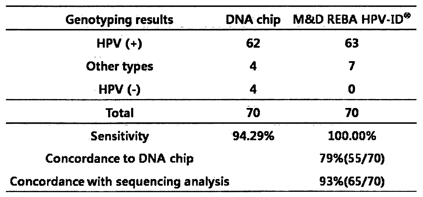
[Revision under Rule 91 08.04.2010]
Table 6
표 6
Table 6
Claims (8)
- 서열번호 5 내지 서열번호 28의 염기서열로 이루어진 HPV 진단용 올리고뉴크레오타이드 프로브 조성물.HPV diagnostic oligonucleotide probe composition consisting of the nucleotide sequence of SEQ ID NO: 5 to SEQ ID NO: 28.
- 제 1항의 프로브 조성물 및 프라이머를 포함하는 HPV 진단용 키트.HPV diagnostic kit comprising the probe composition of claim 1 and a primer.
- 제 2항에 있어서, 상기 프라이머는 서열번호 1 내지 4에 기재된 프라이머인 것을 특징으로 하는 HPV 진단용 키트.The diagnostic kit for HPV according to claim 2, wherein the primers are primers set forth in SEQ ID NOs: 1-4.
- 제 2항 또는 제 3항에 있어서, 상기 키트는 PCR 산물을 탐지하기 위한 표지수단을 더욱 포함하는 것을 특징으로 하는 HPV 진단용 키트.The kit for diagnosing HPV according to claim 2 or 3, wherein the kit further comprises labeling means for detecting a PCR product.
- a) 제 1항의 프로브를 고체 서포트에 부착하는 단계;a) attaching the probe of claim 1 to a solid support;b) 상기 프로브에 증폭된 HPV PCR 산물을 교잡하는 단계; 및b) hybridizing the amplified HPV PCR product to the probe; Andc) 상기 교잡된 산물을 검출하는 단계를 포함하는 HPV 유전자형 확인 방법.c) HPV genotyping method comprising the step of detecting the hybridized product.
- 제 5항에 있어서, 상기 고체 서포트는 막, 슬라이드, 또는 웰 플레이트인 것을 특징으로 하는 HPV 유전자형 확인 방법.6. The method of claim 5, wherein said solid support is a membrane, slide, or well plate.
- 제 5항에 있어서, 상기 PCR 산물은 서열번호 1 내지 4에 기재된 프라이머를 통하여 증폭되는 것을 특징으로 하는 HPV 유전자형 확인 방법.The method of claim 5, wherein the PCR product is HPV genotype identification method characterized in that the amplification through the primers described in SEQ ID NOS: 1-4.
- 제 5항에 있어서, 상기 검출단계는 발광 또는 발색반응을 이용하여 검출하는것을 특징으로 하는 HPV 유전자형 확인 방법.The method of claim 5, wherein the detecting step is HPV genotyping method, characterized in that the detection using a luminescence or color reaction.
Priority Applications (3)
| Application Number | Priority Date | Filing Date | Title |
|---|---|---|---|
| PCT/KR2010/000914 WO2011099664A1 (en) | 2010-02-12 | 2010-02-12 | Probe for hpv genotype diagnosis and analysis method thereof |
| US13/577,948 US20130078614A1 (en) | 2010-02-12 | 2010-02-12 | Probe for hpv genotype diagnosis and analysis method thereof |
| EP10845835.7A EP2535411A4 (en) | 2010-02-12 | 2010-02-12 | Probe for hpv genotype diagnosis and analysis method thereof |
Applications Claiming Priority (1)
| Application Number | Priority Date | Filing Date | Title |
|---|---|---|---|
| PCT/KR2010/000914 WO2011099664A1 (en) | 2010-02-12 | 2010-02-12 | Probe for hpv genotype diagnosis and analysis method thereof |
Publications (1)
| Publication Number | Publication Date |
|---|---|
| WO2011099664A1 true WO2011099664A1 (en) | 2011-08-18 |
Family
ID=44367921
Family Applications (1)
| Application Number | Title | Priority Date | Filing Date |
|---|---|---|---|
| PCT/KR2010/000914 WO2011099664A1 (en) | 2010-02-12 | 2010-02-12 | Probe for hpv genotype diagnosis and analysis method thereof |
Country Status (3)
| Country | Link |
|---|---|
| US (1) | US20130078614A1 (en) |
| EP (1) | EP2535411A4 (en) |
| WO (1) | WO2011099664A1 (en) |
Citations (17)
| Publication number | Priority date | Publication date | Assignee | Title |
|---|---|---|---|---|
| US4659774A (en) | 1985-11-01 | 1987-04-21 | American Hoechst Corporation | Support for solid-phase oligonucleotide synthesis |
| US4683202A (en) | 1985-03-28 | 1987-07-28 | Cetus Corporation | Process for amplifying nucleic acid sequences |
| US4800159A (en) | 1986-02-07 | 1989-01-24 | Cetus Corporation | Process for amplifying, detecting, and/or cloning nucleic acid sequences |
| US4816571A (en) | 1987-06-04 | 1989-03-28 | Applied Biosystems, Inc. | Chemical capping by phosphitylation during oligonucleotide synthesis |
| US4959463A (en) | 1985-10-15 | 1990-09-25 | Genentech, Inc. | Intermediates |
| US5141813A (en) | 1989-08-28 | 1992-08-25 | Clontech Laboratories, Inc. | Multifunctional controlled pore glass reagent for solid phase oligonucleotide synthesis |
| US5428148A (en) | 1992-04-24 | 1995-06-27 | Beckman Instruments, Inc. | N4 - acylated cytidinyl compounds useful in oligonucleotide synthesis |
| US5554744A (en) | 1994-09-23 | 1996-09-10 | Hybridon, Inc. | Method for loading solid supports for nucleic acid synthesis |
| US5574146A (en) | 1994-08-30 | 1996-11-12 | Beckman Instruments, Inc. | Oligonucleotide synthesis with substituted aryl carboxylic acids as activators |
| US5602244A (en) | 1988-05-26 | 1997-02-11 | Competitive Technologies, Inc. | Polynucleotide phosphorodithioate compounds |
| KR100452163B1 (en) * | 2001-09-14 | 2004-10-12 | 주식회사 바이오메드랩 | Genotyping kit for diagnosis of human papilloma virus infection |
| KR100527285B1 (en) * | 2004-01-16 | 2005-11-09 | 학교법인고려중앙학원 | A cysteine protease gene and a promoter which are expressed specifically in rice anther, a production method of male sterile rice using supression of the gene expression |
| KR100663992B1 (en) * | 2004-07-05 | 2007-01-02 | (주)바이오메드랩 | The method selecting highly specific probes for HPV genotype analysis and the probes thereof |
| US20080155715A1 (en) * | 2002-05-20 | 2008-06-26 | National Institute Of Agrobiological Sciences | Stress-Responsive Root-Specific Genes |
| KR20080072043A (en) * | 2005-12-08 | 2008-08-05 | 가부시끼가이샤 도시바 | Method of detecting human papilloma virus by using nucleic acid amplification method and nucleic acid chain-immobilized carrier |
| KR20090042650A (en) * | 2007-10-26 | 2009-04-30 | 주식회사 마이진 | Dna chip, kit for detecting genotype of human papillomavirus and detection method using the same |
| US7527948B2 (en) * | 2003-09-25 | 2009-05-05 | Third Wave Technologies, Inc. | Detection of HPV |
Family Cites Families (10)
| Publication number | Priority date | Publication date | Assignee | Title |
|---|---|---|---|---|
| US5639871A (en) * | 1988-09-09 | 1997-06-17 | Roche Molecular Systems, Inc. | Detection of human papillomavirus by the polymerase chain reaction |
| US5447839A (en) * | 1988-09-09 | 1995-09-05 | Hoffmann-La Roche Inc. | Detection of human papillomavirus by the polymerase chain reaction |
| US6921636B1 (en) * | 1991-09-04 | 2005-07-26 | Metrigen, Inc. | Method and apparatus for conducting an array of chemical reactions on a support surface |
| TWI229132B (en) * | 2001-05-04 | 2005-03-11 | King Car Food Ind Co Ltd | Method and detector for identifying subtypes of human papilloma viruses |
| KR100633525B1 (en) * | 2004-10-04 | 2006-10-16 | 굿젠 주식회사 | Probe Of Human Papillomavirus Oligonucleotide Microarray And Genotyping Kit Comprising The Same And Genotyping Method For Human Papillomavirus Using The Same |
| GB0516145D0 (en) * | 2005-08-05 | 2005-09-14 | Genomica Sau | In vitro diagnostic kit for identification of human papillomavirus in clinical samples |
| US7910719B2 (en) * | 2005-12-08 | 2011-03-22 | Kabushiki Kaisha Toshiba | Method of detecting human papilloma virus by using nucleic acid amplification method and nucleic acid chain-immobilized carrier |
| EP2057180A4 (en) * | 2006-08-11 | 2010-10-20 | Chu Sainte Justine | Oligonucleotides for discriminating related nucleic acid sequences |
| CN101464456B (en) * | 2007-12-20 | 2013-06-12 | 北京金菩嘉医疗科技有限公司 | Method for virus detection by surface plasma resonance technology and chip used in the same |
| KR101018407B1 (en) * | 2008-06-13 | 2011-03-02 | 엠앤디 (주) | Probe for detecting HPV and Genotyping method thereof |
-
2010
- 2010-02-12 WO PCT/KR2010/000914 patent/WO2011099664A1/en active Application Filing
- 2010-02-12 US US13/577,948 patent/US20130078614A1/en not_active Abandoned
- 2010-02-12 EP EP10845835.7A patent/EP2535411A4/en not_active Withdrawn
Patent Citations (19)
| Publication number | Priority date | Publication date | Assignee | Title |
|---|---|---|---|---|
| US4683202A (en) | 1985-03-28 | 1987-07-28 | Cetus Corporation | Process for amplifying nucleic acid sequences |
| US4683202B1 (en) | 1985-03-28 | 1990-11-27 | Cetus Corp | |
| US4959463A (en) | 1985-10-15 | 1990-09-25 | Genentech, Inc. | Intermediates |
| US5264566A (en) | 1985-10-15 | 1993-11-23 | Genentech, Inc. | Method for in vitro oligonucleotide synthesis using H-phosphonates |
| US4659774A (en) | 1985-11-01 | 1987-04-21 | American Hoechst Corporation | Support for solid-phase oligonucleotide synthesis |
| US4800159A (en) | 1986-02-07 | 1989-01-24 | Cetus Corporation | Process for amplifying, detecting, and/or cloning nucleic acid sequences |
| US4816571A (en) | 1987-06-04 | 1989-03-28 | Applied Biosystems, Inc. | Chemical capping by phosphitylation during oligonucleotide synthesis |
| US5602244A (en) | 1988-05-26 | 1997-02-11 | Competitive Technologies, Inc. | Polynucleotide phosphorodithioate compounds |
| US5141813A (en) | 1989-08-28 | 1992-08-25 | Clontech Laboratories, Inc. | Multifunctional controlled pore glass reagent for solid phase oligonucleotide synthesis |
| US5428148A (en) | 1992-04-24 | 1995-06-27 | Beckman Instruments, Inc. | N4 - acylated cytidinyl compounds useful in oligonucleotide synthesis |
| US5574146A (en) | 1994-08-30 | 1996-11-12 | Beckman Instruments, Inc. | Oligonucleotide synthesis with substituted aryl carboxylic acids as activators |
| US5554744A (en) | 1994-09-23 | 1996-09-10 | Hybridon, Inc. | Method for loading solid supports for nucleic acid synthesis |
| KR100452163B1 (en) * | 2001-09-14 | 2004-10-12 | 주식회사 바이오메드랩 | Genotyping kit for diagnosis of human papilloma virus infection |
| US20080155715A1 (en) * | 2002-05-20 | 2008-06-26 | National Institute Of Agrobiological Sciences | Stress-Responsive Root-Specific Genes |
| US7527948B2 (en) * | 2003-09-25 | 2009-05-05 | Third Wave Technologies, Inc. | Detection of HPV |
| KR100527285B1 (en) * | 2004-01-16 | 2005-11-09 | 학교법인고려중앙학원 | A cysteine protease gene and a promoter which are expressed specifically in rice anther, a production method of male sterile rice using supression of the gene expression |
| KR100663992B1 (en) * | 2004-07-05 | 2007-01-02 | (주)바이오메드랩 | The method selecting highly specific probes for HPV genotype analysis and the probes thereof |
| KR20080072043A (en) * | 2005-12-08 | 2008-08-05 | 가부시끼가이샤 도시바 | Method of detecting human papilloma virus by using nucleic acid amplification method and nucleic acid chain-immobilized carrier |
| KR20090042650A (en) * | 2007-10-26 | 2009-04-30 | 주식회사 마이진 | Dna chip, kit for detecting genotype of human papillomavirus and detection method using the same |
Non-Patent Citations (9)
| Title |
|---|
| BERNARD, H. U.; I. E. CALLEJA-MACIAS; S. T. DUNN., INT J CANCER, vol. 118, 2006, pages 1071 - 6 |
| GODFROID, E., J VIROL METHODS, vol. 75, 1998, pages 69 - 81 |
| KURMAN, R. J.; D. E. HENSON; A. L. HERBST; K. L. NOLLER; M. H. SCHIFFMAN, JAMA, vol. 271, 1994, pages 1866 - 9 |
| LIKES, W. M.; J. ITANO., CLIN J ONCOL NURS, vol. 7, 2003, pages 271 - 6 |
| MUNOZ, N., N ENGL J MED, vol. 348, 2003, pages 518 - 27 |
| SAMBROOK, MOLECULAR CLONING, 1989 |
| SEDMAN, S. A., J VIROL, vol. 65, 1991, pages 4860 - 6 |
| See also references of EP2535411A4 * |
| WUI, J. H, JOURNAL OF GYNECOLOGIC ONCOLOGY AND COLOSCOPY, vol. 17, 2006, pages 39 - 4711 |
Also Published As
| Publication number | Publication date |
|---|---|
| US20130078614A1 (en) | 2013-03-28 |
| EP2535411A4 (en) | 2013-07-03 |
| EP2535411A1 (en) | 2012-12-19 |
Similar Documents
| Publication | Publication Date | Title |
|---|---|---|
| Gheit et al. | Development of a sensitive and specific multiplex PCR method combined with DNA microarray primer extension to detect Betapapillomavirus types | |
| US20090029346A1 (en) | Detection of human papilloma virus | |
| EP0477972B1 (en) | Nucleotide sequences useful as type-specific probes, PCR primers and LCR probes for the amplification and detection of human papilloma virus, and related kits and methods | |
| US8895247B2 (en) | Method for detection of human papillomavirus (HPV) type | |
| Doorn et al. | Molecular detection and genotyping of human papillomavirus | |
| Williamson et al. | Detection of genital human papillomaviruses by polymerase chain reaction amplification with degenerate nested primers | |
| KR101018407B1 (en) | Probe for detecting HPV and Genotyping method thereof | |
| WO2013162109A1 (en) | Polymerase chain reaction primer and probe for detecting hepatitis b virus, and detection kit and method using same | |
| EP1546413B1 (en) | Method and kit for quantitative and qualitative determination of human papillomavirus | |
| KR101152840B1 (en) | A probe for identifying hpv genotype and a method for identifying hpv genotype | |
| KR101401940B1 (en) | Kit for analyzing high-risk HPV gene and method for analyzing the same | |
| CN105695627A (en) | Specific primer and probe combination and reagent kit for fluorescent quantitative PCR (polymerase chain reaction) detection on 15 types of HPV (human papillomavirus) | |
| CN113215325B (en) | Reaction system, method and kit for detecting multiple HPV subtypes by two-dimensional PCR single tube closed tube | |
| WO2011099664A1 (en) | Probe for hpv genotype diagnosis and analysis method thereof | |
| WO2020218772A1 (en) | Hpv detection kit and hpv detection method using same | |
| Clavel et al. | DNA-EIA to detect high and low risk HPV genotypes in cervical lesions with E6/E7 primer mediated multiplex PCR. | |
| US8741568B2 (en) | Detection of human papillomavirus | |
| WO2013168832A1 (en) | Analysis kit for high-risk human papillomavirus genes, and analysis method therefor | |
| Wu et al. | Detection of five types of HPV genotypes causing Anogenital warts (Condyloma Acuminatum) using PCR-Tm analysis technology | |
| KR101331170B1 (en) | A probe for identifying hpv genotype and a method for identifying hpv genotype | |
| KR100979271B1 (en) | Dna chip, kit for detecting genotype of human papillomavirus and detection method using the same | |
| EP2362916B1 (en) | Hpv types and variants associated with cervical cancer and the uses thereof | |
| KR20180007432A (en) | Multianalytical HPV Genotyping Luminex kit and HPV gene Detection Method Thereof | |
| WO2015008885A1 (en) | Method for diagnosing cervical cancer and diagnostic kit therefor | |
| WO2013015609A2 (en) | Method and kit for detecting human papilloma virus |
Legal Events
| Date | Code | Title | Description |
|---|---|---|---|
| 121 | Ep: the epo has been informed by wipo that ep was designated in this application |
Ref document number: 10845835 Country of ref document: EP Kind code of ref document: A1 |
|
| NENP | Non-entry into the national phase |
Ref country code: DE |
|
| WWE | Wipo information: entry into national phase |
Ref document number: 2010845835 Country of ref document: EP |
|
| WWE | Wipo information: entry into national phase |
Ref document number: 13577948 Country of ref document: US |