WO2012069593A2 - Fusion enzymes - Google Patents
Fusion enzymes Download PDFInfo
- Publication number
- WO2012069593A2 WO2012069593A2 PCT/EP2011/070956 EP2011070956W WO2012069593A2 WO 2012069593 A2 WO2012069593 A2 WO 2012069593A2 EP 2011070956 W EP2011070956 W EP 2011070956W WO 2012069593 A2 WO2012069593 A2 WO 2012069593A2
- Authority
- WO
- WIPO (PCT)
- Prior art keywords
- acetylglucosaminyltransferase
- host cell
- glycan
- seq
- catalytic domain
- Prior art date
Links
Classifications
-
- C—CHEMISTRY; METALLURGY
- C12—BIOCHEMISTRY; BEER; SPIRITS; WINE; VINEGAR; MICROBIOLOGY; ENZYMOLOGY; MUTATION OR GENETIC ENGINEERING
- C12N—MICROORGANISMS OR ENZYMES; COMPOSITIONS THEREOF; PROPAGATING, PRESERVING, OR MAINTAINING MICROORGANISMS; MUTATION OR GENETIC ENGINEERING; CULTURE MEDIA
- C12N9/00—Enzymes; Proenzymes; Compositions thereof; Processes for preparing, activating, inhibiting, separating or purifying enzymes
- C12N9/10—Transferases (2.)
- C12N9/1048—Glycosyltransferases (2.4)
- C12N9/1051—Hexosyltransferases (2.4.1)
-
- C—CHEMISTRY; METALLURGY
- C12—BIOCHEMISTRY; BEER; SPIRITS; WINE; VINEGAR; MICROBIOLOGY; ENZYMOLOGY; MUTATION OR GENETIC ENGINEERING
- C12N—MICROORGANISMS OR ENZYMES; COMPOSITIONS THEREOF; PROPAGATING, PRESERVING, OR MAINTAINING MICROORGANISMS; MUTATION OR GENETIC ENGINEERING; CULTURE MEDIA
- C12N1/00—Microorganisms, e.g. protozoa; Compositions thereof; Processes of propagating, maintaining or preserving microorganisms or compositions thereof; Processes of preparing or isolating a composition containing a microorganism; Culture media therefor
- C12N1/14—Fungi; Culture media therefor
-
- C—CHEMISTRY; METALLURGY
- C12—BIOCHEMISTRY; BEER; SPIRITS; WINE; VINEGAR; MICROBIOLOGY; ENZYMOLOGY; MUTATION OR GENETIC ENGINEERING
- C12N—MICROORGANISMS OR ENZYMES; COMPOSITIONS THEREOF; PROPAGATING, PRESERVING, OR MAINTAINING MICROORGANISMS; MUTATION OR GENETIC ENGINEERING; CULTURE MEDIA
- C12N15/00—Mutation or genetic engineering; DNA or RNA concerning genetic engineering, vectors, e.g. plasmids, or their isolation, preparation or purification; Use of hosts therefor
- C12N15/09—Recombinant DNA-technology
- C12N15/63—Introduction of foreign genetic material using vectors; Vectors; Use of hosts therefor; Regulation of expression
- C12N15/79—Vectors or expression systems specially adapted for eukaryotic hosts
- C12N15/80—Vectors or expression systems specially adapted for eukaryotic hosts for fungi
-
- C—CHEMISTRY; METALLURGY
- C12—BIOCHEMISTRY; BEER; SPIRITS; WINE; VINEGAR; MICROBIOLOGY; ENZYMOLOGY; MUTATION OR GENETIC ENGINEERING
- C12N—MICROORGANISMS OR ENZYMES; COMPOSITIONS THEREOF; PROPAGATING, PRESERVING, OR MAINTAINING MICROORGANISMS; MUTATION OR GENETIC ENGINEERING; CULTURE MEDIA
- C12N5/00—Undifferentiated human, animal or plant cells, e.g. cell lines; Tissues; Cultivation or maintenance thereof; Culture media therefor
- C12N5/10—Cells modified by introduction of foreign genetic material
-
- C—CHEMISTRY; METALLURGY
- C12—BIOCHEMISTRY; BEER; SPIRITS; WINE; VINEGAR; MICROBIOLOGY; ENZYMOLOGY; MUTATION OR GENETIC ENGINEERING
- C12P—FERMENTATION OR ENZYME-USING PROCESSES TO SYNTHESISE A DESIRED CHEMICAL COMPOUND OR COMPOSITION OR TO SEPARATE OPTICAL ISOMERS FROM A RACEMIC MIXTURE
- C12P19/00—Preparation of compounds containing saccharide radicals
- C12P19/18—Preparation of compounds containing saccharide radicals produced by the action of a glycosyl transferase, e.g. alpha-, beta- or gamma-cyclodextrins
-
- C—CHEMISTRY; METALLURGY
- C12—BIOCHEMISTRY; BEER; SPIRITS; WINE; VINEGAR; MICROBIOLOGY; ENZYMOLOGY; MUTATION OR GENETIC ENGINEERING
- C12P—FERMENTATION OR ENZYME-USING PROCESSES TO SYNTHESISE A DESIRED CHEMICAL COMPOUND OR COMPOSITION OR TO SEPARATE OPTICAL ISOMERS FROM A RACEMIC MIXTURE
- C12P21/00—Preparation of peptides or proteins
- C12P21/005—Glycopeptides, glycoproteins
-
- C—CHEMISTRY; METALLURGY
- C12—BIOCHEMISTRY; BEER; SPIRITS; WINE; VINEGAR; MICROBIOLOGY; ENZYMOLOGY; MUTATION OR GENETIC ENGINEERING
- C12Y—ENZYMES
- C12Y204/00—Glycosyltransferases (2.4)
- C12Y204/01—Hexosyltransferases (2.4.1)
- C12Y204/01101—Alpha-1,3-mannosyl-glycoprotein 2-beta-N-acetylglucosaminyltransferase (2.4.1.101)
-
- C—CHEMISTRY; METALLURGY
- C12—BIOCHEMISTRY; BEER; SPIRITS; WINE; VINEGAR; MICROBIOLOGY; ENZYMOLOGY; MUTATION OR GENETIC ENGINEERING
- C12Y—ENZYMES
- C12Y204/00—Glycosyltransferases (2.4)
- C12Y204/01—Hexosyltransferases (2.4.1)
- C12Y204/01143—Alpha-1,6-mannosyl-glycoprotein 2-beta-N-acetylglucosaminyltransferase (2.4.1.143)
-
- C—CHEMISTRY; METALLURGY
- C07—ORGANIC CHEMISTRY
- C07K—PEPTIDES
- C07K2319/00—Fusion polypeptide
- C07K2319/01—Fusion polypeptide containing a localisation/targetting motif
- C07K2319/03—Fusion polypeptide containing a localisation/targetting motif containing a transmembrane segment
-
- Y—GENERAL TAGGING OF NEW TECHNOLOGICAL DEVELOPMENTS; GENERAL TAGGING OF CROSS-SECTIONAL TECHNOLOGIES SPANNING OVER SEVERAL SECTIONS OF THE IPC; TECHNICAL SUBJECTS COVERED BY FORMER USPC CROSS-REFERENCE ART COLLECTIONS [XRACs] AND DIGESTS
- Y02—TECHNOLOGIES OR APPLICATIONS FOR MITIGATION OR ADAPTATION AGAINST CLIMATE CHANGE
- Y02P—CLIMATE CHANGE MITIGATION TECHNOLOGIES IN THE PRODUCTION OR PROCESSING OF GOODS
- Y02P20/00—Technologies relating to chemical industry
- Y02P20/50—Improvements relating to the production of bulk chemicals
- Y02P20/52—Improvements relating to the production of bulk chemicals using catalysts, e.g. selective catalysts
Definitions
- the present disclosure relates to compositions and methods useful for the production of N-glycans.
- Posttranslational modification of proteins is often necessary for proper protein folding and function.
- a common protein modification is the addition of oligosaccharides (glycans) to nascent polypeptides in the endoplasmic reticulum to form glycoproteins, a process known as glycosylation.
- N-glycosylation is of particular importance in the production of recombinant proteins used for therapeutic purposes. Because standard prokaryotic expression systems lack the proper machinery necessary for such
- yeast and fungal cells are attractive options for expressing proteins as they can be easily grown at a large scale in simple media, which allows low production costs.
- tools are available to manipulate the relatively simple genetic makeup of yeast and fungal cells as well as more complex eukaryotic cells such as mammalian or insect cells (De Pourcq et al., Appl Microbiol Biotechnol, 87(5): 1617-31).
- Man8GlcNAc2 Man8GlcNAc2
- yeast additional mannose subunits are added to Man8GlcNAc2 by mannosyltransferases and mannan polymerases to yield high-mannose type N-glycans.
- mannose sugars are removed from the human Man8GlcNAc2 to yield
- Mns II mannosidase II
- GnTII N- acetylglucosaminyltransferase II
- compositions including recombinant proteins having N- acetylglucosaminyltransferase activity. Further described herein are methods of producing complex N-glycans and methods of producing Man3GlcNAc2 glycans.
- one aspect includes recombinant proteins having N- acetylglucosaminyltransferase activity, where the recombinant proteins catalyze the transfer of N-acetylglucosamine to a terminal Mana3 residue and catalyze the transfer of N-acetylglucosamine to a terminal Mana6 residue of an acceptor glycan, and where the recombinant protein contains catalytic domains from at least two different enzymes.
- the acceptor glycan is attached to a molecule selected from an amino acid, a peptide, or a polypeptide.
- the molecule is a heterologous polypeptide.
- the acceptor glycan is Man3.
- the recombinant protein is a fusion protein containing an N-acetylglucosaminyltransferase I catalytic domain and an N- acetylglucosaminyltransferase II catalytic domain.
- the N- acetylglucosaminyltransferase I catalytic domain and the N- acetylglucosaminyltransferase II catalytic domain are from human enzymes.
- the N-acetylglucosaminyltransferase I catalytic domain includes a sequence that is at least 70%, at least 75%, at least 80%, at least 85%, at least 90%, at least 95%, at least 96%, at least 97%, at least 98%, at least 99%, or 100% identical to amino acid residues 105-445 of SEQ ID NO: 1.
- the N-acetylglucosaminyltransferase II catalytic domain includes a sequence that is at least 70%, at least 75%, at least 80%, at least 85%, at least 90%, at least 95%, at least 96%, at least 97%, at least 98%, at least 99%, or 100% identical amino acid residues 30-447 of SEQ ID NO: 21.
- the N- acetylglucosaminyltransferase I catalytic domain is N-terminal to the N- acetylglucosaminyltransferase II catalytic domain.
- the N-acetylglucosaminyltransferase II catalytic domain is N-terminal to the N-acetylglucosaminyltransferase I catalytic domain.
- the recombinant proteins further contain a spacer in between the N- acetylglucosaminyltransferase I catalytic domain and the N- acetylglucosaminyltransferase II catalytic domain.
- the spacer contains sequence from a stem domain.
- the spacer is at least 5, at least 10, at least 15, at least 20, at least 30, at least 40, or at least 50 amino acids in length.
- the spacer contains a sequence that is selected from SEQ ID NO: 118, SEQ ID NO: 120, SEQ ID NO: 122, and SEQ ID NO: 124. In certain embodiments, the spacer contains a sequence that is selected from SEQ ID NO: 118, SEQ ID NO: 120, and SEQ ID NO: 124. In certain embodiments, the spacer contains the sequence of SEQ ID NO: 120 or SEQ ID NO: 124. In certain embodiments, the spacer contains the sequence of SEQ ID NO: 124.
- the recombinant proteins further contain a targeting peptide linked to the N-terminal end of the catalytic domains.
- the targeting peptide contains a stem domain.
- the stem domain is from an N- acetylglucosaminyltransferase I enzyme or an N-acetylglucosaminyltransferase II enzyme.
- the N-acetylglucosaminyltransferase I enzyme and the N-acetylglucosaminyltransferase II enzyme are human enzymes.
- the stem domain is from a protein selected from a mannosidase, a mannosyltransferase, a glycosyltransferase, a Type 2 Golgi protein, MNN2, MNN4, MNN6, MNN9, MNN10, MNS1, KRE2, VAN1, or OCHl .
- the protein is from an organism selected from
- the targeting peptide is a Kre2 targeting peptide.
- the targeting peptide contains a transmembrane domain. In certain embodiments that may be combined with the preceding embodiments, the targeting peptide further contains a transmembrane domain linked to the N-terminal end of the stem domain. In certain embodiments that may be combined with the preceding embodiments, the transmembrane domain is from an N-acetylglucosaminyltransferase I enzyme or an N- acetylglucosaminyltransferase II enzyme. In certain embodiments, the N- acetylglucosaminyltransferase I enzyme and the N-acetylglucosaminyltransferase II enzyme are human enzymes.
- the transmembrane domain is from a protein selected from a mannosidase, a mannosyltransferase, a glycosyltransferase, a Type 2 Golgi protein, MNN2, MNN4, MNN6, MNN9, MNN10, MNS1, KRE2, VAN1, or OCHl .
- the protein is from an organism selected from Acremonium, Aspergillus, Aureobasidium, Cryptococcus, Chrysosporium, Chrysosporium lucknowense,
- the targeting peptide contains a cytoplasmic domain. In certain embodiments that may be combined with the preceding embodiments, the targeting peptide further contains a cytoplasmic domain linked to the N-terminal end of the stem domain.
- the targeting peptide further contains a cytoplasmic domain linked to the N-terminal end of the transmembrane domain.
- the cytoplasmic domain is from an N- acetylglucosaminyltransferase I enzyme or an N-acetylglucosaminyltransferase II enzyme.
- the N-acetylglucosaminyltransferase I enzyme and the N-acetylglucosaminyltransferase II enzyme are human enzymes.
- the cytoplasmic domain is from a protein selected from a mannosidase, a mannosyltransferase, a glycosyltransferase, a Type 2 Golgi protein, MNN2, MNN4, MNN6, MNN9, MNN10, MNS1, KRE2, VAN1, or OCHl .
- the protein is from an organism selected from
- Another aspect includes recombinant proteins containing a human N- acetylglucosaminyltransferase II catalytic domain and a human N- acetylglucosaminyltransferase I catalytic domain where the N- acetylglucosaminyltransferase II catalytic domain is located N-terminal to the N- acetylglucosaminyltransferase I catalytic domain, a spacer sequence containing sequence from a human N-acetylglucosaminyltransferase I stem domain located in between the catalytic domains, and a targeting peptide located N-terminal to the N- acetylglucosaminyltransferase II catalytic domain where the targeting peptide contains a cytoplasmic domain, a transmembrane domain, and a stem domain from human N- acetylglucosaminyl
- Another aspect includes a recombinant protein containing a sequence that is at least 70%, at least 75%, at least 80%>, at least 85%>, at least 90%, at least 95%, at least 96%, at least 97%, at least 98%, at least 99%, or 100% identical to SEQ ID NO: 95.
- Another aspect includes recombinant proteins containing N- acetylglucosaminyltransferase II catalytic domain and a N-acetylglucosaminyltransferase I catalytic domain, where the N-acetylglucosaminyltransferase II catalytic domain is located N-terminal to the N-acetylglucosaminyltransferase I catalytic domain; a spacer located in between the catalytic domains, where the spacer contains a sequence selected from SEQ ID NO: 118, SEQ ID NO: 120, SEQ ID NO: 122, and SEQ ID NO: 124; and a targeting peptide located N-terminal to the N-acetylglucosaminyltransferase II catalytic domain where the targeting peptide contains a cytoplasmic domain, a transmembrane domain, and a stem domain from human N-acetylgluco
- the spacer contains a sequence that is selected from SEQ ID NO: 118, SEQ ID NO: 120, and SEQ ID NO: 124. In certain embodiments, the spacer contains the sequence of SEQ ID NO: 120 or SEQ ID NO: 124. In certain embodiments, the spacer contains the sequence of SEQ ID NO: 124.
- Another aspect includes isolated polynucleotides encoding the recombinant protein of any of the preceding embodiments.
- Another aspect includes expression vectors containing the isolated polynucleotide of the preceding embodiment operably linked to a promoter.
- the promoter is a constitutive promoter.
- the promoter is an inducible promoter.
- the promoter is from a gene selected from gpdA, cbhl, Aspergillus oryzae TAKA amylase, Rhizomucor miehei aspartic proteinase, Aspergillus niger neutral alpha-amylase,
- Aspergillus niger acid stable alpha-amylase Aspergillus niger glucoamylase (glaA), Aspergillus awamori glaA, Rhizomucor miehei lipase, Aspergillus oryzae alkaline protease, Aspergillus oryzae triose phosphate isomerase, Aspergillus nidulans
- acetamidase Aspergillus oryzae acetamidase, Fusarium oxysporum trypsin-like protease, fungal endo a-L-arabinase (abnA), fungal a-L-arabinofuranosidase A (abfA), fungal a-L- arabinofuranosidase B (abfB), fungal xylanase (xlnA), fungal phytase, fungal ATP- synthetase, fungal subunit 9 (oliC), fungal triose phosphate isomerase (tpi), fungal alcohol dehydrogenase (adhA), fungal a-amylase (amy), fungal amyloglucosidase (glaA), fungal acetamidase (amdS), fungal glyceraldehyde-3 -phosphate dehydrogenase (gpd), yeast alcohol dehydrogena
- Another aspect includes methods of producing the recombinant protein of any the preceding embodiments, including the steps of introducing an isolated polynucleotide that encodes the recombinant protein into a host cell, and culturing the host cell such that the recombinant protein is expressed.
- the methods further include a step of purifying the recombinant protein from the host cell.
- the host cell is a fungal cell.
- the fungal cell is selected from yeast or filamentous fungus.
- Another aspect includes methods of producing a complex N-glycan including the steps of providing a host cell, where the host cell contains a polynucleotide encoding a fusion protein containing an N-acetylglucosaminyltransferase I catalytic domain and an N-acetylglucosaminyltransferase II catalytic domain, and culturing the host cell such that the fusion protein is expressed, where the fusion protein catalyzes the transfer of N- acetylglucosamine to a terminal Mana3 residue and N-acetylglucosamine to a terminal Mana6 residue of an acceptor glycan to produce a complex N-glycan.
- the complex N-glycan is attached to a molecule selected from an amino acid, a peptide, or a polypeptide.
- the molecule is a heterologous polypeptide.
- the acceptor glycan is Man3 .
- the complex N-glycan is
- the host cell is a eukaryotic cell. In certain embodiments that may be combined with the preceding embodiments, the host cell is a fungal cell. In certain embodiments, the fungal cell is a yeast cell selected from S. cerevisiae, K. lactis, P. pastoris, H. polymorpha, C. albicans, Schizosaccharomyces, or Yarrowia.
- the fungal cell is a filamentous fungal cell selected from Trichoderma sp., Acremonium, Aspergillus, Aureobasidium, Cryptococcus, Chrysosporium, Chrysosporium lucknowense, Filibasidium, Fusarium, Gibberella, Magnaporthe, Mucor, Myceliophthora, Myrothecium, Neocallimastix, Neurospora, Paecilomyces, Penicillium, Piromyces, Schizophyllum, Talaromyces, Thermoascus, Thielavia, or Tolypocladium.
- Trichoderma sp. Acremonium, Aspergillus, Aureobasidium, Cryptococcus, Chrysosporium, Chrysosporium lucknowense, Filibasidium, Fusarium, Gibberella, Magnaporthe, Mucor, Myceliophthora, Myrothecium, Neocallimastix, Neurospor
- the host cell further contains a polynucleotide encoding a UDP- GlcNAc transporter.
- the host cell has a reduced level of activity of a dolichyl-P- Man:Man(5)GlcNAc(2)-PP-dolichyl mannosyltransferase compared to the level of activity in a wild-type host cell.
- the host cell has a reduced level of expression of an alg3 gene compared to the level of expression in a wild-type host cell.
- the alg3 gene is deleted from the host cell.
- the host cell has a reduced level of activity of an a- 1,6-mannosyl transferase compared to the level of activity in a wild-type host cell. In certain embodiments, the host cell has a reduced level of expression of an ochl gene compared to the level of expression in a wild-type host cell. In certain embodiments, the ochl gene is deleted from the host cell. In certain
- the host cell further contains a polynucleotide encoding an a-l,2-mannosidase.
- the host cell further contains a polynucleotide encoding a P-l,4-galactosyltransferase. In certain embodiments that may be combined with the preceding embodiments, the host cell further contains a polynucleotide encoding a sialyltransferase. In certain embodiments that may be combined with the preceding embodiments, the host cell is a Trichoderma cell that has a reduced level of activity of a dolichyl-P-Man:Man(5)GlcNAc(2)-PP- dolichyl mannosyltransferase compared to the level of activity in a wild-type
- the host cell is a yeast or fungal cell that has a reduced level of activity of a dolichyl-P-Man:Man(5)GlcNAc(2)-PP-dolichyl mannosyltransferase and a reduced level of activity of an alpha- 1,6-mannosyl transferase compared to the levels of activity in a wild-type yeast cell and further contains a polynucleotide encoding an a-1,2- mannosidase.
- Another aspect includes methods of producing a complex N-glycan including the steps of providing a Trichoderma host cell, where the host cell has a reduced level of expression of an algS gene compared to the level of expression in a wild-type host cell and contains a first polynucleotide encoding an N-acetylglucosaminyltransferase I catalytic domain and a second polynucleotide encoding an N- acetylglucosaminyltransferase II catalytic domain, and culturing the host cell to produce a complex N-glycan.
- Another aspect includes methods of producing a complex N-glycan including the steps of incubating a fusion protein containing an N-acetylglucosaminyltransferase I catalytic domain and an N-acetylglucosaminyltransferase II catalytic domain, an acceptor glycan, and an N-acetylglucosamine donor together in a buffer, where the fusion protein catalyzes the transfer of N-acetylglucosamine to a terminal Mana3 residue and N- acetylglucosamine to a terminal Mana6 residue of an acceptor glycan to produce a complex N-glycan.
- the acceptor glycan is attached to a molecule selected from an amino acid, a peptide, or a polypeptide.
- the molecule is a heterologous polypeptide.
- the acceptor glycan is Man3.
- the N-acetylglucosamine donor is a UDP-GlcNAc transporter.
- Another aspect includes filamentous fungal cells containing a mutation of alg3 and Man3GlcNAc2, where the Man3GlcNAc2 includes at least 50%, at least 60%, at least 70%, at least 80%, at least 90%, or 100% (mol %) of neutral N-glycans secreted by the cells.
- the neutral N-glycans may be attached to a molecule selected from the group consisting of an amino acid, a peptide, and a polypeptide.
- the mutation of alg3 is a deletion of alg3.
- the cell is a Trichoderma reesei cell.
- the filamentous fungal cell further contains a first polynucleotide encoding an N- acetylglucosaminyltransferase I catalytic domain and a second polynucleotide encoding an N-acetylglucosaminyltransferase II catalytic domain.
- the filamentous fungal cell further contains a polynucleotide encoding a fusion protein containing an N- acetylglucosaminyltransferase I catalytic domain and an N-acetylglucosaminyltransferase II catalytic domain.
- Another aspect includes methods of producing a Man3GlcNAc2 glycan in a host cell including the steps of providing a host cell with a reduced level of activity of a mannosyltransferase compared to the level of activity in a wild-type host cell and culturing the host cell to produce a Man3GlcNAc2 glycan, where the Man3GlcNAc2 glycan includes at least 50%, at least 60%, at least 70%, at least 80%, at least 90%, or 100%) (mol %) of the neutral N-glycans secreted by the host cell.
- the neutral N- glycans may be attached to a molecule selected from an amino acid, a peptide, and a polypeptide.
- the Man3GlcNAc2 glycan is attached to a heterologous polypeptide.
- the mannosyltransferase is a dolichyl-P-Man:Man(5)GlcNAc(2)- PP-dolichyl mannosyltransferase.
- the host cell has a reduced level of expression of an alg3 gene compared to the level of expression in a wild-type host cell.
- the alg3 gene is deleted from the host cell.
- the host cell is a Trichoderma cell.
- the level of activity of alpha- 1,6-mannosyltransferase in the host cell is reduced compared to the level of activity in a wild-type host cell.
- the host cell contains an endogenous polynucleotide encoding an a-l,2-mannosidase.
- Another aspect includes a filamentous fungal cell having a reduced level of expression of an alg3 gene compared to the level of expression in a wild-type filamentous fungal cell, where the filamentous fungal cell contains a recombinant protein of any of the preceding embodiments.
- the alg3 gene contains a mutation.
- the recombinant protein has N-acetylglucosaminyltransferase activity, where the recombinant protein catalyzes the transfer of N-acetyl glucosamine to a terminal Mana3 residue and catalyzes the transfer of N-acetyl glucosamine to a terminal Mana6 residue of an acceptor glycan, and where the recombinant protein is a fusion protein containing an N-acetylglucosaminyltransferase I catalytic domain and an N- acetylglucosaminyltransferase II catalytic domain.
- the mutation of the alg3 gene is a deletion of the alg3 gene.
- the fusion protein is encoded by a
- the promoter is an inducible promoter.
- the inducible promoter is the cbhl promoter. In certain embodiments that may be combined with the preceding
- the filamentous fungal cell further contains a polynucleotide encoding a UDP-GlcNAc transporter.
- the filamentous fungal has a reduced level of activity of an a- 1,6-mannosyl transferase compared to the level of activity in a wild-type filamentous fungal cell.
- the filamentous fungal has a reduced level of expression of an ochl gene compared to the level of expression in a wild-type filamentous fungal cell.
- the filamentous fungal cell further contains a polynucleotide encoding an a-l,2-mannosidase.
- the filamentous fungal cell further contains a polynucleotide encoding a P-l,4-galactosyltransf erase. In certain embodiments that may be combined with the preceding embodiments, the filamentous fungal cell further contains a polynucleotide encoding a sialyltransferase. In certain embodiments that may be combined with the preceding embodiments, the filamentous fungal cell is selected from Trichoderma sp., Acremonium, Aspergillus, Aureobasidium, Cryptococcus,
- Chrysosporium Chrysosporium lucknowense, Filibasidium, Fusarium, Gibberella, Magnaporthe, Mucor, Myceliophthora, Myrothecium, Neocallimastix, Neurospora, Paecilomyces, Penicillium, Piromyces, Schizophyllum, Talaromyces, Thermoascus, Thielavia, and Tolypocladium.
- Fig. 1 shows mass spectrometric neutral N-glycan profiles of average
- Fig. 2 shows fragmentation analysis of monophosphorylated Man7Gn2. Only one example structure of monophosphorylated Man7Gn2 is shown.
- Fig. 3 shows mass spectrometric acidic glycan profiles of T. reesei strains M44, M81, M84, M109, MHO, M131, M132, M133, M134, and M124.
- Fig. 4 shows neutral (a) and acidic (b) N-glycan profiles of T. reesei strain M44 cultured in a fermentor for 131.4 hours (fed batch).
- Fig. 5 shows mass spectrometric neutral (a) and acidic (b) N-glycan profiles of T. reesei culture medium.
- Fig. 6 shows a membrane blot of T. reesei M44 secreted proteins.
- Fig. 7 shows an example of analyzed protein bands of T. reesei M44 cultivated in a fermentor.
- the glycosylation of proteins did not differ significantly from average glycosylation in T. reesei.
- the spectrum was focused to the minor base line signals, and the major signal of the spectrum was not quantitative in comparison to other signals.
- Fig. 8 shows a Southern blot of DNA from the parental strain and from Alg3 knockout strains with an alg3 probe.
- Fig. 9 A shows a restriction enzyme map of a section of the pTTv38 construct with sizes of predicted restriction products.
- Figure 9B shows a Southern blot of genomic DNA from the parental strain and the Alg3 knockout strains digested with EcoRI + Pvul (E + P) or Kpnl + Nhel (K + N). The control DNA was pTTv38 plasmid DNA digested with Not! The blot was probed with an AmdS probe.
- Fig. 10 shows MALDI analysis of neutral N-glycans.
- Part A shows the parental strain Ml 24.
- Part B shows the Alg3 knockout 4 A.
- Squares represent N- acetylglucosamine, and circles represent mannose, except for the one labeled glucose.
- Fig. 11 shows fragmentation analysis of Man3Gn2 from the 4A Alg3 knockout strain.
- Fig. 12 shows fragmentation analysis of Hex5Gn2 from Alg3 knockout strain 4A (part A) and parental strain M124 (part B). The signal marked with a box exists only as an isomer from the Alg3 knockout strain.
- Fig. 13 shows neutral N-glycans from Alg3 knockout strain 4 A after a- mannosidase digestion.
- Fig. 14 shows the separation of two major glycans from the Alg3 knockout strain by liquid chromatography.
- Fig. 15 shows proton NMR spectra of Hex3HexNAc2 (part A) and
- Hex6HexNAc2 (part B) fractions were collected at 40°C using a Varian Unity INOVA 600 MHz spectrometer equipped with a cryoprobe.
- Fig. 16 shows the acidic fraction of parental strain M124 (part A) and Alg3 knockout strain 4 A (B). N-glycans with two phosphate units are marked with an asterisk.
- Fig. 17 shows neutral N-glycans from supernatant of T. reesei Alg3 knockout strain 4A that was cultured in a flask for 5 days.
- Fig. 18 shows neutral N-glycans from supernatant of T. reesei Alg3 knockout strain 4 A that was cultured in a fermentor for 10 days.
- Fig. 19 shows a MALDI spectrum of GnTI reaction mixture. GnTI has converted 54% of the acceptor to the product with one additional HexNAc.
- Fig. 20 shows Western blot analysis of GnTII expression. Samples were run in 12% SDS-PAGE gel and blotted on nitrocellulose membrane. Histidine-tagged GnTII was detected on the membrane using mouse a-HIS monoclonal antibodies. Numbers shown on the left are the sizes of molecular weight marker proteins (kDa).
- Fig. 21 shows a MALDI spectrum of GnTII reaction mixture. 83% of the acceptor (m/z 913.340) was converted to product (m/z 1136.433).
- Fig. 22 shows GnTI activity observed for the GnTI/GnTII fusion protein.
- Fig. 23 shows the N-glycans present in GnTI/GnTII T. reesei transformants obtained by targeting to the alg3 locus.
- Fig. 24 shows a MALDI spectrum of the purified reaction mixture from the enzyme activity test of the GnTII/GnTI fusion protein.
- Fig. 25 shows a spectrum of the pi-2,3,4,6-N-acetylglucosaminidase reaction mixture.
- Fig. 26 shows a MALDI spectrum of pi-4GalT reaction mixture.
- Fig. 27 shows diagrams of observed N-glycans from supernatant proteins of T. reesei M127 pTTvl 10 transformants (gnt II/I in alg3 locus) on days 3 (A), 5 (B) and 7 (C and D).
- the clone 17A produced the most GO on day 7.
- E Mass spectrum of neutral N- glycans of supernatant proteins from T. reesei strain M127 GnT II/I transformant clone 17A cultivated for 7 days in shake flasks. Signals marked with asterisks originated from the culture medium.
- Fig. 28 shows neutral N-glycans of rituximab from T. reesei M202 GnT II/I transformant clones (A) 9A-1 and (B) 31A-1, both cultivated with soybean trypsin inhibitor, and (C) mass spectrum of neutral N-glycans of rituximab purified from T. reesei strain M202 GnT II/I transformant clone 9A-1 cultivated for 5 days in shake flasks in the presence of soybean trypsin inhibitor.
- Fig. 29 shows MALDI spectra of spacer modified GnTII/GnTI fusion reaction mixtures.
- Part (A) shows a reaction mixture of GnTII/GnTI with 3xG4S spacer modification. 36% of the acceptor has been converted to product with two additional HexNAcs.
- Part (B) shows a reaction mixture of GnTII/GnTI with 2xG4S spacer modification. 38% of the acceptor has been converted to product with two additional HexNAcs.
- Calculated m/z values for [M+Na]+ -signals of GnTI product, Hex3HexNAc2 (calc. m/z 933.318), was not detected in either spectra because all of the GnTI product was converted directly to Hex3HexNAc3, (calc. m/z 1136.318).
- Fig. 30 shows Western blots of GnTII/I spacer variant cell pellets(A), and supernatants (B).
- Fig. 31 shows GnT activities of wild-type GnTII/I and spacer variants from supernatants and expressed in the presence of protease inhibitors after day 3 (A) expression phases and day 4 (B) expression phases.
- Fig. 32 shows GnT activities of GnTII/I fusion protein (with wild type spacer) in supernatant, cells and lysate. GnTI and GnTII products have been added together
- Fig. 33 shows GnT activities of GnTII/I wild-type and spacer variants in (A) supernatants, (B) cells, and (C) ly sates.
- Fig. 34 shows example spectra of neutral N-glycans of parental strain M124 and GnTl transformants on day 5. Signal with Gn addition (m/z 1460) is marked with an arrow. (pTTvl l with cbhl promoter, pTTvl3 with gpdA promoter).
- Fig. 35 shows the amounts of Man5 and GnlMan5 in four positive GNT1 transformants on days 3 and 5. Quantitation was carried out against internal calibrant (Hex2HexNAc4, 2 pmol).
- Fig. 36 shows example spectra of phosphorylated N-glycans of parental Ml 24 strain and GnTl transformants with internal calibrant (NeuAcHex4HexNAc2, 0.5 pmol.). GnTl products are marked with an arrow.
- Fig. 37 shows diagrams of neutral N-glycans of different GnTII strains/clones from day 5.
- Part (A) show the pTTv 140 clone.
- Part (B) shows the pTTv 142 clone.
- Part (C) shows the pTTvl43 clone.
- Part (D) shows the pTTvl41 clone.
- Fig. 38 shows an example of neutral N-glycans of different GnTII strains/clones and the parental strain M198 from days 3, 5, and 7.
- Part (A) shows clone 1-117A.
- Part (B) shows clone
- Part (C) shows clone 30A.
- Part (D) shows parental stain M198.
- Fig. 39 shows the membrane of separated proteins of T. reesei strain M198 and GnTII clone 3-17A.
- the 50 kDA protein is marked with an arrow.
- Fig. 40 shows column diagrams of total secreted proteins versus individual secreted protein(s) of parental strain M198 (A) and the GnTII clone 3-17A (B).
- Fig. 41 shows a column diagram of fermentor cultured GnTII strain M329 from day 3 to day 7, and shake flask culture of strain M329 from day 5.
- Fig. 42 shows a multiple amino acid sequence alignment of T. reesei ALG3 and ALG3 homologs.
- the present invention relates to recombinant proteins having N- acetylglucosaminyltransferase activity where the recombinant protein catalyzes the transfer of N-acetyl glucosamine (GlcNAc) to a terminal Mana3 residue and catalyzes the transfer of N-acetyl glucosamine to a terminal Mana6 residue of an acceptor glycan, and where the recombinant protein contains catalytic domains from at least two different enzymes.
- GlcNAc N-acetyl glucosamine
- the recombinant proteins of the invention include two catalytic domains, where one catalytic domain has N-acetylglucosaminyltransferase I (GnTI) activity (e.g., reacts with a terminal Mana3 residue), and the other catalytic domain has N-acetylglucosaminyltransferase II (GnTII) activity (e.g., reacts with a terminal Mana6 residue).
- GnTI N-acetylglucosaminyltransferase I
- GnTII N-acetylglucosaminyltransferase II
- the recombinant proteins of the present invention catalyze reactions that occur essentially sequentially.
- the recombinant proteins of the present invention may catalyze the transfer of GlcNAc to a terminal Mana3 -residue, first, and then catalyze the transfer of GlcNAc to a terminal Mana6-residueof an acceptor glycan.
- the essentially sequential reactions are at least 10 fold, at least 20 fold, at least 30 fold, at least 40 fold, at least 50 fold, at least 60 fold, at least 70 fold, at least 80 fold, at least 90 fold, or at least 100 fold, more effective than the two reactions in the reversed order.
- a sequential reaction means that essentially or absolutely no GlcNAc can be transferred to the terminal Manoc6-residue if GlcNAc has not yet been transferred to the terminal Man oc3 -residue.
- the acceptor glycan contains a GlcNAcP2Manoc3 -branch.
- the recombinant proteins react specifically with both Mana3 and Mana6 residues, optionally in branched acceptor glycans but not substantially or absolutely with other Manoc-structures, e.g. Manoc-monosaccharide conjugates, with Manocbenzyl and/or ManocSer/Thr-peptide.
- the non-substantial reactivity is preferably below 10%, below 8%, below 6%, below 4%, below 2%, below 1 %, or below 0.1 % of the Vmax with 0.1 mM acceptor glycan concentrations of reactions with terminal Mana3 and Mana6 residues.
- the recombinant proteins have substantially similar reactivities with the terminal Mana3 (preferably as GnTI reaction) and the terminal Mana6 residue (preferably as GnTII reaction) of the acceptor glycan.
- the terminal Mana3 preferably as GnTI reaction
- the terminal Mana6 residue preferably as GnTII reaction
- neither catalytic activity has more than a 10-fold, 5-fold, 3-fold or 2-fold difference in reaction effectiveness compared to the other catalytic activity under the same conditions.
- the transfer of GlcNAc to the terminal Manoc3 and Manoc6 cause a conversion of at least 10 %, at least 25 %, at least 50 %, at least 70 %, at least 90%, or at least 95 % of Man3 glycan to a glycan with two terminal GlcNAcs.
- the effectiveness of the reaction can be measured by in vitro or in vivo assays as described in the examples disclosed herein.
- the effectiveness of the GlcNAc transfer reactions can be measured essentially as described in the Examples or as maximal reaction rate Vmax with 0.1 mM acceptor concentrations and saturating donor concentrations.
- the effectiveness of the reaction is measured with a Man3 acceptor glycan attached to an amino acid, a peptide, or a polypeptide.
- the present disclosure further relates to methods of producing a complex N- glycan, including the steps of providing a host cell, where the host cell contains a nucleic acid encoding a fusion protein containing an N-acetylglucosaminyltransferase I catalytic domain and an N-acetylglucosaminyltransferase II catalytic domain, and culturing the host cell such that the fusion protein is expressed, where the fusion protein catalyzes the transfer of N-acetyl glucosamine to a terminal Mana3 residue and N-acetyl glucosamine to a terminal Mana6 residue of an acceptor glycan to produce a complex N-glycan.
- the present invention also relates to a filamentous fungal cell having a reduced level of expression of an alg3 gene compared to the level of expression in a wild-type filamentous fungal cell, where the filamentous fungal cell contains a recombinant protein of the invention.
- recombinant protein refers to any protein that has been produced from a recombinant nucleic acid.
- Recombinant nucleic acid refers to a polymer of nucleic acids where at least one of the following is true: (a) the sequence of nucleic acids is foreign to ⁇ i.e., not naturally found in) a given host cell; (b) the sequence may be naturally found in a given host cell, but is present in an unnatural ⁇ e.g., greater than expected) amount or expressed at a level that is more or less than the natural level of expression; or (c) the sequence of nucleic acids includes two or more subsequences that are not found in the same relationship to each other in nature.
- a recombinant nucleic acid sequence will have two or more sequences from unrelated genes arranged to make a new functional nucleic acid.
- a recombinant nucleic acid sequence will contain a promoter sequence and a gene-encoding sequence that are not naturally found adjacent to one another.
- N-acetylglucosaminyltransferase activity refers to the activity of an enzyme that transfers an N-acetylglucosaminyl residue (GlcNAc) to an acceptor glycan.
- enzymes having this activity are N-acetylglucosaminyltransferases (GlcNAc transferases).
- GlcNAc transferases are eukaryotic.
- the GlcNAc transferases are mammalian enzymes forming a ⁇ - linkage from the 1 -position of a GlcNAc-residue to the terminal mannose residues.
- the GlcNAc transferases are 2-N-acetylglucosaminyltransferases transferring 2-linked GlcNAc-residue(s) to the 2-position terminal mannose residues of glycans, in particular to an N-linked glycan.
- the 2-GlcNAc transferases are enzymes having GnTI activity and GnTII activity. GnTI activity transfers a GlcNAc residue to a Manoc3 branch.
- the Mana3 branch may be a Man oc3 (It- Man oc6)ManP-branch of on N-linked glycan core structure, such as Man3GlcNAc2 or Man3 or Man5GlcNAc2 or Man5.
- GnTI enzymes may be mammalian enzymes, plant enzymes, or lower eukaryotic enzymes.
- GnTII activity transfers a GlcNAc residue to a Manoc6-branch such as a Manoc6(GlcNAcP2Manoc3)ManP-branch of an N-linked glycan core structure.
- An example of such a Manoc6-branch is GlcNAclMan3GlcNAc2.
- N-acetylglucosamine refers to an N-acetylglucosamine residue (GlcNAc).
- GlcNAc may be part of a glycan structure. The amine group is on position 2, has a D-configuration, and has a pyranose structure as a residue. It may be alternatively named 2-acetamido-2-deoxy-D-glucopyranose (D-GlcpNAc). GlcNAc may also be a free reducing monosaccharide (i.e., not part of glycan).
- Man refers to a mannose residue.
- a “terminal Mana3” or a “terminal Mana6” refers to a mannose that is not substituted to the non-reducing end terminal residue by another monosaccharide residue or residues.
- glycan refers to an oligosaccharide chain that can be linked to a carrier such as an amino acid, peptide, polypeptide, lipid or a reducing end conjugate.
- the invention relates to N-linked glycans conjugated to a polypeptide N-glycosylation site such as -Asn-Xxx-Ser/Thr- by N-linkage to side-chain amide nitrogen of asparagine residue (Asn), where Xxx is any amino acid residue except Pro.
- the invention may further relate to glycans as part of dolichol-phospho- oligosaccharide (Dol-P-P-OS) precursor lipid structures, which are precursors of N-linked glycans in the endoplasmic reticulum of eukaryotic cells.
- the precursor oligosaccharides are linked from their reducing end to two phosphate residues on the dolichol lipid.
- oc3-mannosyl transferase Alg3 modifies the Dol-P-P-oligosaccharide precursor of N-glycans.
- the glycan structures described herein are terminal glycan structures, where the non-reducing residues are not modified by other monosaccharide residue or residues.
- glycoprotein refers to a peptide or polypeptide attached to a glycan.
- the glycan may be attached to the peptide or polypeptide in a cotranslational or posttranslational modification.
- glycolipid refers to a lipid attached to a glycan and includes glyceroglycolipids, glycosphingolipids, and glycosylphosphatidylinositols.
- glycolipid and carbohydrate nomenclature is essentially according to recommendations by the IUPAC-IUB
- Gal galactose
- Glc glucose
- GlcNAc N-acetylglucosamine
- GalNAc N- acetylgalactosamine
- Man Mannose
- Neu5 Ac are of the D-configuration, Fuc of the L-configuration, and all the monosaccharide units in the pyranose form (D-Galp, D-Glcp, D-GlcpNAc, D-GalpNAc, D-Manp, L-Fucp, D-Neup5Ac).
- the amine group is as defined for natural galactose and glucosamines on the 2-position of GalNAc or GlcNAc.
- Glycosidic linkages are shown partly in shorter and partly in longer nomenclature, the linkages of the sialic acid SA/Neu5X-residues oc3 and oc6 mean the same as oc2-3 and oc2- 6, respectively, and for hexose monosaccharide residues ocl-3, ocl-6, ⁇ 1-2, ⁇ 1-3, ⁇ 1-4, and ⁇ 1-6 can be shortened as oc3, oc6, ⁇ 2, ⁇ 3, ⁇ 4, and ⁇ 6, respectively.
- Lactosamine refers to type II N-acetyllactosamine, Ga ⁇ 4GlcNAc, and/or type I N-acetyllactosamine.
- Ga ⁇ 3GlcNAc and sialic acid SA refer to N-acetylneuraminic acid (Neu5Ac), N- glycolylneuraminic acid (Neu5Gc), or any other natural sialic acid including derivatives of Neu5X.
- Sialic acid is referred to as NeuNX or Neu5X, where preferably X is Ac or Gc. Occasionally Neu5 Ac/Gc/X may be referred to as NeuNAc/NeuNGc/NeuNX.
- the invention herein relates to recombinant proteins having N- acetylglucosaminyltransferase activity, where the recombinant proteins catalyze the transfer of N-acetylglucosamine to a terminal Mana3 residue and catalyze the transfer of N-acetylglucosamine to a terminal Mana6 residue of an acceptor glycan.
- Recombinant proteins of the invention may include, without limitation, full length proteins having N- acetylglucosaminyltransferase activity, fragments of proteins having N- acetylglucosaminyltransferase activity, catalytic domains having N- acetylglucosaminyltransferase activity, and fusion proteins having N- acetylglucosaminyltransferase activity.
- a single recombinant protein of the invention has the capability to catalyze both transfers of N-acetylglucosamines. The transfer of N- acetylglucosamine to a terminal Mana3 residue may occur before or after the transfer of N-acetylglucosamine to a terminal Mana6 residue. Alternatively, the transfers may occur simultaneously.
- the acceptor glycan may be attached to a molecule such as an amino acid, a peptide, or a polypeptide.
- the amino acid is an asparagine residue.
- the asparagine residue may be in aminoglycosidic linkage from the side-chain amide (a biologic mammalian polypeptide N-glycan linkage structure) and may be part of a peptide chain such as a dipeptide, an oligopeptide, or a polypeptide.
- the glycan may be a reducing end derivative such as an N-, 0-, or C-linked, preferably glycosidic, derivative of the reducing GlcNAc or Man, such as a spacer or terminal organic residue with a certain glycan linked structure selected from the group of an amino acid, alkyl, heteroalkyl, acyl, alkyloxy, aryl, arylalkyl, or heteroarylalkyl.
- the spacer may be further linked to a polyvalent carrier or a solid phase.
- alkyl-containing structures include methyl, ethyl, propyl, and C4-C26 alkyls, lipids such as glycerolipids, phospholipids, dolichol-phospholipids and ceramides and derivatives.
- the reducing end may also be derivatized by reductive amination to a secondary amine linkage or a derivative structure.
- Certain carriers include biopoly- or oligomers such as (poly)peptides, poly(saccharides) such as dextran, cellulose, amylose, or glycosaminoglycans, and other organic polymers or oligomers such as plastics including polyethylene, polypropylene, polyamides (e.g., nylon or polystyrene), polyacrylamide, and polylactic acids, dendrimers such as PAMAM, Starburst or Starfish dendrimers, or polylysine, and polyalkyl glycols such as polyethylene glycol (PEG).
- biopoly- or oligomers such as (poly)peptides, poly(saccharides) such as dextran, cellulose, amylose, or glycosaminoglycans
- plastics including polyethylene, polypropylene, polyamides (e.g., nylon or polystyrene), polyacrylamide, and polylactic acids, dendrimers such as PAMAM, Starburst
- Solid phases may include microtiter wells, silica particles, glass, metal (including steel, gold, and silver), polymer beads such as polystyrene or resin beads, polylactic acid beads, polysaccharide beads or organic spacers containing magnetic beads.
- the acceptor glycan is attached to a heterologous polypeptide.
- a "peptide” and a “polypeptide” are amino acid sequences including a plurality of consecutive polymerized amino acid residues.
- peptides are those molecules including up to 50 amino acid residues, and polypeptides include more than 50 amino acid residues.
- the peptide or polypeptide may include modified amino acid residues, naturally occurring amino acid residues not encoded by a codon, and non-naturally occurring amino acid residues.
- protein may refer to a peptide or a polypeptide of any size.
- heterologous polypeptide refers to a polypeptide that is not naturally found in a given host cell or is not endogenous to a given host cell.
- the heterologous polypeptide is a therapeutic protein.
- Therapeutic proteins may include monoclonal antibodies, erythropoietins, interferons, growth hormones, enzymes, or blood-clotting factors.
- the acceptor glycan may be attached to a therapeutic protein such as rituximab.
- the structure of the acceptor glycan has the following formula, [Ri] y Man 3([R 2 ] z Man 6)Man ⁇ 4GlcNAc(Fuc x) n [ 4GlcNAc] m ⁇ q , where q, y, z, n and m are 0 or 1; x is linkage position 3 or 6, of optional fucose residue; Rl is GlcNAc, preferably GlcNAc 2; and R2 is a branched structure Manoc3(Manoc6), with the provision that when z is 1, then y is 0, and when z is 0, then y is 0 or 1.
- ( ) defines a branch in the regular N-glycan core structure, either present or absent.
- [ ] and ⁇ ⁇ define a part of the glycan structure either present or absent in a linear sequence.
- the structure is a Man3 glycan
- the structure is a GlcNAcMan3 glycan
- the glycan is a Man5 glycan.
- the acceptor glycan may be beta-glycosidically linked to an Asn residue, preferably from the reducing end GlcNAc.
- the acceptor glycan is a polypeptide linked N-glycan, where m and q are 1, and the acceptor structure contains a derivative of [Ri] y Man 3([R 2 ] z Man 6)Man 4GlcNAc(Fuc x) n 4GlcNAc.
- Optional derivatives include substitutions by monosaccharide residues such as GlcNAc or xylose.
- the acceptor glycan may be Man3, GlcNAcMan3, or Man5.
- the acceptor glycan is Man3 or GlcNAcMan3.
- Man3 is a trimannosyl glycan comprising at least one of Manoc3 or Manoc6 residues and is preferably a branched oligosaccharide, such as Mana3(Manoc6)Man.
- Other certain Man3 oligosaccharides are Mana3(Manoc6)ManP, Mana3(Manoc6)ManP4GlcNAc, and polypeptide-linked
- the glycan can contain Fuc, Xyl or GlcNAc in Man and/or GlcNAc residues, such as Man 3(Man 6)Man 4GlcNAc 4(Fuc x) n GlcNAc, where x is 3 or 6 and n is 0 or 1, also described by a monosaccharide composition formula indicating the terminal mannose structure and reducing end composition as Man3GlcNAc2 (n is 0) and Man3GlcNAc2Fuc (n is 1).
- the Man3 structure is a Mana3(Mana6)ManP4GlcNAcP4(Fucoc6) n GlcNAc.
- the polypeptide-linked GlcNAcMan3 structure is
- GlcNAc 2Man 3(Man 6)Man 4GlcNAc 4(Fuc 6) n GlcNAc also described by a monosaccharide composition formula GlcNAcMan3GlcNAc2 (n is 0) and
- the polypeptide-linked Man5 structure is Man 3 ⁇ Man 3(Man 6)Man 6 ⁇ Man 4GlcNAc 4(Fuc 6) n GlcNAc, where ⁇ ⁇ and ( ) indicate a branch and n is 0 or 1, also described by a monosaccharide composition formula Man5GlcNAc2 (n is 0) and Man5GlcNAc2Fuc (n is 1).
- the certain Man3 glycans have structures according to the following formula, Mana3(Mana6)ManP4GlcNAc(Fucax) n p4GlcNAc, where n is 0 or 1, indicating presence or absence of part of the molecule, where x is 3 or 6, and where ( ) defines a branch in the structure.
- the acceptor glycan is Man3
- the recombinant protein catalyzes the transfer of N-acetyl glucosamine to the terminal Mana3 and Manoc6 of Man3, thus resulting in GlcNAc2Man3
- GlcNAcMan5 for example, by mannosidase II
- the recombinant protein catalyzes the transfer of N-acetylglucosamine to the terminal Mana6, thus resulting in GlcNAc2Man3 (which has the structure GlcNAcp2Mancc3 (GlcNAcp2Mancc6)Manp4GlcNAcp4(Fucax) n GlcNAc, where n is 0 or 1, also referred to as GO if attached to an antibody).
- the recombinant proteins of the invention are fusion proteins containing an N-acetylglucosaminyltransferase I catalytic domain and an N- acetylglucosaminyltransferase II catalytic domain.
- fusion protein refers to any protein or polypeptide containing a protein or polypeptide linked to heterologous amino acids.
- An N-acetylglucosaminyltransferase I catalytic domain is any portion of an N- acetylglucosaminyltransferase I enzyme that is capable of catalyzing this reaction.
- N-acetylglucosaminyltransferase I enzymes from various organisms are listed in SEQ ID NOs: 1-19. Additional GnTI enzymes are listed in the CAZy database in the glycosyltransferase family 13 (cazy.org/GT13_all). Enzymatically characterized species includes thaliana AAR78757.1 (US6 653 459), C. elegans AAD03023.1 (Chen S. et al J. Biol.Chem 1999;274(l):288-97), D. melanogaster
- AAF57454.1 (Sarkar & Schachter Biol Chem. 2001 Feb;382(2):209-17); C. griseus AAC52872.1 (Puthalakath H. et al J. Biol.Chem 1996 271(44):27818-22); H. sapiens AAA52563.1 (Kumar R. et al Proc Natl Acad Sci U S A. 1990 Dec;87(24):9948-52); M. auratus AAD04130.1 (Opat As et al Biochem J. 1998 Dec 15;336 (Pt 3):593-8),
- the N-acetylglucosaminyltransferase I catalytic domain is from the human N-acetylglucosaminyltransf erase I enzyme (SEQ ID NO: 1), or variants thereof.
- the N-acetylglucosaminyltransferase I catalytic domain contains a sequence that is at least 70%, at least 75%, at least 80%, at least 85%, at least 90%, at least 95%, at least 96%, at least 97%, at least 98%, at least 99%, or 100% identical to amino acid residues 84-445 of SEQ ID NO: 1.
- a shorter sequence can be used as a catalytic domain (e.g.
- GnTI catalytic domain amino acid residues from about amino acid 30 to 445 of the human enzyme or any C-terminal stem domain starting between amino acid residue 30 to 105 and continuing to about amino acid 445 of the human enzyme, or corresponding homologous sequence of another GnTI or a catalytically active variant or mutant thereof.
- the catalytic domain may include N-terminal parts of the enzyme such as all or part of the stem domain, the transmembrane domain, or the cytoplasmic domain.
- cytoplasmic is used to refer to a part of a protein that interacts with the cytoplasm of a cell.
- An N-acetylglucosaminyltransferase II catalytic domain is any portion of an N- acetylglucosaminyltransferase II enzyme that is capable of catalyzing this reaction.
- N-acetylglucosaminyltransferase II enzymes from various organisms are listed in SEQ ID NOs: 20-33.
- the N- acetylglucosaminyltransferase II catalytic domain is from the human N- acetylglucosaminyltransferase II enzyme (SEQ ID NO: 20).
- Additional GnTII species are listed in the CAZy database in the glycosyltransferase family 16 (cazy.org/GT16_all). Enzymatically characterized species include GnTII of C. elegans, D.
- the N-acetylglucosaminyltransferase II catalytic domain contains a sequence that is at least 70%, at least 75%, at least 80%, at least 85%, at least 90%, at least 95%, at least 96%, at least 97%, at least 98%, at least 99%, or 100% identical to amino acid residues from about 30 to about 447 of SEQ ID NO: 21.
- the catalytic domain may include N-terminal parts of the enzyme such as all or part of the stem domain, the transmembrane domain, or the cytoplasmic domain.
- the N-acetylglucosaminyltransferase I catalytic domain is N-terminal to the N-acetylglucosaminyltransferase II catalytic domain.
- the N-acetylglucosaminyltransferase II catalytic domain is N-terminal to the N-acetylglucosaminyltransferase I catalytic domain.
- N-terminal refers to the positioning of a set of amino acid residues closer to the end of a polypeptide that is terminated by an amino acid with a free amine group (-NH 2 ) compared to a reference set of amino acid residues.
- the recombinant protein contains a spacer in between the N-acetylglucosaminyltransferase I catalytic domain and the N- acetylglucosaminyltransferase II catalytic domain.
- spacer refers to any number of consecutive amino acids of any sequence separating the N- acetylglucosaminyltransferase I catalytic domain and the N- acetylglucosaminyltransferase II catalytic domain such that the spacer has no effect on the enzymatic function of the catalytic domains.
- the spacer is at least 5, at least 10, at least 15, at least 20, at least 30, at least 40, or at least 50 amino acids in length.
- the spacer contains sequence from a stem domain.
- stem domain refers to a protein domain, or a fragment thereof, which is located adjacent to the transmembrane domain of a native enzyme, such as a glycosyltransferase or a glycosyl hydrolase, and optionally targets the enzyme to or assists in retention of the enzyme in the ER/Golgi.
- Stem domains generally start with the first amino acid following the hydrophobic transmembrane domain and end at the catalytic domain.
- Exemplary stem domains include, but are not limited to, the stem domain of human GnTI, amino acid residues from about 30 to about 83 or from about 30 to about 105 for the human GnTII, or amino acid residues from about 26 to about 106 or from about 26 to about 83 for the T. reesei KRE2.
- the spacer includes amino acids 30-83 of the human GnTI sequence (SEQ ID NO: 34).
- the spacer may include any of the sequences listed in SEQ ID NOs: 35-38.
- suitable spacers include, without limitation, the flexible spacer 3XG4S (SEQ ID NO: 118), the flexible spacer 2XG4S (SEQ ID NO: 120), the spacer for the T. reesei CBHI (SEQ ID NO: 122); and the spacer for the T. reesei ECTV cellulase (SEQ ID NO: 124).
- the length of the spacer is about the same as the length of a stem domain of GnTI .
- the length is about 74 amino acid residues, plus or minus about 37 amino acids.
- the spacer length is about 30 amino acids to about 110 amino acids, or from about 35 amino acids to about 100 amino acids, or as exemplified in the examples described herein, plus or minus 2, 3, 4, or 5 amino acids.
- the spacer length corresponds to a truncated stem domain of GnTI, for example, start from amino acid 25 to amino acid 104, or between amino acid 30 to amino acid 101, to the end of the GnTI stem domain.
- the spacer may include a part of the stem domain of human GnTI, which may start from an amino acid positioned between amino acid 70 to amino acid 87 (according to numbering in SEQ ID NO: 34), or between amino acid 76 and amino acid 104, or beginning from amino acid 30, 35, 40, 45, 50, 60, 70, 73, 74, 75, 76, 80, 83, 84, 85, 86, 87, 100, 101, 102, 103, or 104, to the end of the human GnTI stem domain.
- the spacer may include a heterologous spacer peptide, which may include a fungal spacer peptide and/or a repetitive oligomer spacer peptide.
- the spacer is an elongated peptide without specific conformation and contains amino acid residues allowing high flexibility (e.g., Gly and Ala), hydroplicity (e.g., Ser and Thr), and optionally Pro to prevent conformation.
- the spacer may be glycosylated.
- the spacer is O-glycosylated including fungal O- mannosylation.
- the spacer is an endogenous fungal, filamentous fungal, or Trichoderma spacer peptide, such as a spacer that naturally separates protein domains.
- the spacer may be derived from a secreted or cellulolytic enzyme of a fungus such as a filamentous fungus (e.g., T.
- the natural fungal spacer may contain dimeric or oligomeric proline and/or glycine and/or serine and/or threonine, and/or multiple amino acid residues selected from Ser, Thr, Gly, Pro or Ala or any combinations thereof.
- the spacer is a repeating oligomer containing a monomer with 1-10 or 1-5 amino acid residues selected from Ser, Thr, Gly, Pro or Ala and optionally a charged amino acid residue selected from negatively charged residues Glu or Asp or positively charged residues Lys or Arg. In certain embodiments the charged residue is negatively charged.
- the monomer contains dimeric or oligomeric amino acid residues, and/or multiple single amino acid residues selected from Ser, Thr, Gly, Pro and Ala.
- the oligomer contains a monomer of a dimer or oligomer of glycine and a single residue selected from the Ser, Thr, Gly, Pro and Ala.
- the single residue is Ser or Thr.
- the residue is Ser.
- the sequence of the repeating spacer is ⁇ (Yyy) n Xxx ⁇ m where n is 2 to 10, m is 2 to 10, and Xxx and Yyy are selected from Ser, Thr, Gly, Pro and Ala, with the proviso that Xxx and Yyy are not the same amino acid residue.
- the repeating spacer is ⁇ (Gly) n Xxx ⁇ m where n is 2 to 10, m is 2 to 10, and Xxx is selected from Ser, Thr, Gly, Pro and Ala.
- Xxx is Ser or Thr.
- Xxx is Ser.
- recombinant proteins of the invention include a targeting peptide linked to the catalytic domains.
- the term "linked” as used herein means that two polymers of amino acid residues in the case of a polypeptide or two polymers of nucleotides in the case of a polynucleotide are either coupled directly adjacent to each other or are within the same polypeptide or polynucleotide but are separated by intervening amino acid residues or nucleotides.
- a “targeting peptide”, as used herein, refers to any number of consecutive amino acid residues of the recombinant protein that are capable of localizing the recombinant protein to the endoplasmic reticulum (ER) or Golgi apparatus (Golgi) within the host cell.
- the targeting peptide may be N-terminal or C-terminal to the catalytic domains. In certain embodiments, the targeting peptide is N- terminal to the catalytic domains.
- the targeting peptide provides binding to an ER or Golgi component, such as to a mannosidase II enzyme. In other embodiments, the targeting peptide provides direct binding to the ER or Golgi membrane.
- Components of the targeting peptide may come from any enzyme that normally resides in the ER or Golgi apparatus.
- Such enzymes include mannosidases, mannosyltransferases, glycosyltransferases, Type 2 Golgi proteins, and MNN2, MNN4, MNN6, MNN9, MNN10, MNS1, KRE2, VAN1, and OCH1 enzymes.
- Such enzymes may come from a yeast or fungal species such as those of Acremonium, Aspergillus, Aureobasidium, Cryptococcus, Chrysosporium, Chrysosporium lucknowense,
- the targeting peptide comes from the same enzyme and organism as one of the catalytic domains of the recombinant protein.
- the targeting peptide of the recombinant protein is from the human GnTII enzyme.
- the targeting peptide may come from a different enzyme and/or organism as the catalytic domains of the recombinant protein.
- Examples of various targeting peptides for use in targeting proteins to the ER or Golgi that may be used for targeting recombinant proteins of the invention include:
- Kre2/Mntl N-terminal peptide fused to galactosyltransferase (Schwientek, JBC 1996, 3398), UDEL for localization of mannosidase to ER of yeast cells to produce Man5 (Chiba, JBC 1998, 26298-304; Callewaert, FEBS Lett 2001, 173-178), OCH1 targeting peptide fused to GnTI catalytic domain (Yoshida et al, Glycobiology 1999, 53-8), yeast N-terminal peptide of Mnsl fused to a2-mannosidase (Martinet et al, Biotech Lett 1998, 1171), N-terminal portion of Kre2 linked to catalytic domain of GnTI or p4GalT
- the targeting peptide is the Kre2/Mntl(i.e., Kre2) targeting peptide having the amino acid sequence of SEQ ID NO: 115 or SEQ ID NO: 116.
- sequences that may be used for targeting peptides include the sequences listed in Table 1 below.
- Table 1 Targeting peptides. Putative transmembrane domains are underlined. In KRE2, the stem domain enabling Golgi localization is underlined and double- underlined. Otherl and Other02 are putative mannosylation-related proteins.
- MNS1 alternatives MLVVGRPRLVRN IILTLAILSIWHLGL SRTPTSASALVSASVSASSEWSRLERLMNRGAPLTPYPDSNSSFDW estExt_GeneWise S L SAIPFRYPPHNTTHLPPRHKQPPLPRIQHRFGPESPAAAKERIKRLKA Plus.CJ 60228 SEQ ID NO: 75 SEQ ID NO: 76 VKQVFLRAWQAYKGYAWKQDALLPISGGGREQFSGWAATLVDALDT
- VAN1 MMPRHHSSGFSN VGIAVVVILVLVL QPRSVASLISLGILSGYDDLKLETVRYYDLSNVQGTARGWEREERILL estExt_GeneWise GYPRADTFEISPH WFG CVPLRDAEQHLPMFFSHLKNFTYPHNLIDLAFLVSDSKDHTLESLTEH Plus.C_400029 RFQPRATLPPHRK SEQ ID NO: 81 LEAIQADPDPKQPYGEISIIEKDFGQKVNQDVESRHGFAAQASRRKLM
- MSLSRSPSPVPG ILLPLIIICTIVAYYG THEAPGFVHWWRRISMGGGGEKFVIILGANVGGGVMEWKGAREWAI fgenesh5_pg.C_s GGWSSPGLNINS SEQ ID NO: 93 ERDSVRNKRKYATRWGYDLEIVDMKTKKRYAHEWRESWEKVDFIRA caffold_5000342 GRSSPSNAAGSS AMRKYPKAEWFWWLDLNTYVMEPSYSLQRHLFNHLDRHVYRDINVF
- Uncharacterized sequences may be tested for use as targeting peptides by expressing proteins in the glycosylation pathway in a host cell, where one of the proteins contains the uncharacterized sequence as the sole targeting peptide, and measuring the glycans produced in view of the cytoplasmic localization of glycan biosynthesis (e.g. as in Schwientek JBC 1996 3398), or by expressing a fluorescent reporter protein fused with the targeting peptide, and analyzing the localization of the protein in the Golgi by
- the targeting peptide may include a stem domain.
- the stem domain is from an N-acetylglucosaminyltransferase I enzyme or an N- acetylglucosaminyltransferase II enzyme.
- the stem domain is from a human N-acetylglucosaminyltransferase I enzyme or a human N- acetylglucosaminyltransferase II enzyme.
- the sequence corresponding to the stem domain from human N-acetylglucosaminyltransferase I enzyme is SEQ ID NO: 34.
- the sequence corresponding to the stem domain from human N-acetylglucosaminyltransferase II enzyme is residues 30-85 of SEQ ID NO: 20.
- the targeting peptide may include a transmembrane domain.
- transmembrane domain refers to any sequence of amino acid residues that is thermodynamically stable in a membrane as a three-dimensional structure.
- the targeting peptide also includes a stem domain
- transmembrane domain is N-terminal to the stem domain.
- the transmembrane domain is from an N-acetylglucosaminyltransferase I enzyme or an N- acetylglucosaminyltransferase II enzyme.
- the transmembrane domain is from a human N-acetylglucosaminyltransferase I enzyme or a human N-acetylglucosaminyltransferase II enzyme.
- the sequence corresponding to the transmembrane domain from human N-acetylglucosaminyltransferase I enzyme is residues 7-29 of SEQ ID NO: 1.
- the sequence corresponding to the transmembrane domain from human N-acetylglucosaminyltransferase II enzyme is residues 10-29 of SEQ ID NO: 20.
- the targeting peptide may include a cytoplasmic domain.
- cytoplasmic domain refers to an amino acid sequence that is thermodynamically stable in a cytoplasmic environment as a three-dimensional structure.
- the targeting peptide also includes a stem domain
- the cytoplasmic domain is N-terminal to the stem domain.
- the targeting peptide also includes a transmembrane domain
- the cytoplasmic domain is N-terminal to the transmembrane domain.
- the cytoplasmic domain is from an N- acetylglucosaminyltransferase I enzyme or an N-acetylglucosaminyltransferase II enzyme.
- the cytoplasmic domain is from a human N- acetylglucosaminyltransferase I enzyme or a human N-acetylglucosaminyltransferase II enzyme.
- the sequence corresponding to the cytoplasmic domain from human N- acetylglucosaminyltransferase I enzyme is residues 1-6 of SEQ ID NO: 1.
- the sequence corresponding to the cytoplasmic domain from human N-acetylglucosaminyltransferase II enzyme is residues 1-9 of SEQ ID NO: 20.
- the recombinant protein contains a human GnTII catalytic domain N-terminal to a human GnTI catalytic domain with a spacer sequence containing human GnTI stem domain sequence in between the catalytic domains.
- the recombinant protein also includes a targeting peptide N-terminal to the GnTII catalytic domain with cytoplasmic, transmembrane, and stem domains from human GnTII.
- sequence of the recombinant protein in this embodiment is at least 70%, at least 75%, at least 80%, at least 85%, at least 90%, at least 95%, at least 96%, at least 97%, at least 98%, at least 99%, or 100% identical to SEQ ID NO: 95, and the sequence of a possible cDNA encoding the recombinant protein of this embodiment is SEQ ID NO: 96.
- the recombinant protein contains a human GnTII catalytic domain N-terminal to a human GnTI catalytic domain with a spacer sequence.
- the spacer sequence may include, without limitation, a sequence that is at least 70%, at least 75%, at least 80%, at least 85%, at least 90%, at least 95%, at least 96%, at least 97%, at least 98%, at least 99%, or 100% identical to SEQ ID NOs: 118, 120, 122, or 124.
- the recombinant protein also includes a targeting peptide N- terminal to the GnTII catalytic domain with cytoplasmic, transmembrane, and stem domains from human GnTII.
- the sequence of the recombinant protein is at least 70%, at least 75%, at least 80%, at least 85%, at least 90%, at least 95%, at least 96%, at least 97%, at least 98%, at least 99%, or 100% identical to a sequence selected from SEQ ID NOs: 119, 121, 123, and 125.
- the sequence of a possible cDNA encoding the recombinant protein of SEQ ID NO: 119 is SEQ ID NO: 141.
- the sequence of a possible cDNA encoding the recombinant protein of SEQ ID NO: 121 is SEQ ID NO: 139.
- sequence of a possible cDNA encoding the recombinant protein of SEQ ID NO: 123 is SEQ ID NO: 143.
- sequence of a possible cDNA encoding the recombinant protein of SEQ ID NO: 125 is SEQ ID NO: 145.
- Another aspect of the invention includes isolated polynucleotides encoding the recombinant proteins of the invention.
- polynucleotide As used herein, the terms “polynucleotide,” “nucleic acid sequence,” “sequence of nucleic acids,” and variations thereof shall be generic to polydeoxyribonucleotides (containing 2-deoxy-D-ribose), to
- polyribonucleotides containing D-ribose
- any other type of polynucleotide that is an N-glycoside of a purine or pyrimidine base and to other polymers containing non- nucleotidic backbones, provided that the polymers contain nucleobases in a configuration that allows for base pairing and base stacking, as found in DNA and RNA.
- these terms include known types of nucleic acid sequence modifications, for example, substitution of one or more of the naturally-occurring nucleotides with an analog; inter- nucleotide modifications, such as, for example, those with uncharged linkages (e.g., methyl phosphonates, phosphotriesters, phosphorami dates, carbamates, etc.), with negatively charged linkages (e.g., phosphorothioates, phosphorodithioates, etc.), and with positively charged linkages (e.g., aminoalkylphosphoramidates,
- aminoalkylphosphotriesters those containing pendant moieties, such as, for example, proteins (including nucleases, toxins, antibodies, signal peptides, poly-L-lysine, etc.); those with intercalators (e.g., acridine, psoralen, etc.); and those containing chelators (e.g., metals, radioactive metals, boron, oxidative metals, etc.).
- proteins including nucleases, toxins, antibodies, signal peptides, poly-L-lysine, etc.
- intercalators e.g., acridine, psoralen, etc.
- chelators e.g., metals, radioactive metals, boron, oxidative metals, etc.
- Sequences of the isolated polynucleotides are prepared by any suitable method known to those of ordinary skill in the art, including, for example, direct chemical synthesis or cloning.
- formation of a polymer of nucleic acids typically involves sequential addition of 3'-blocked and 5'-blocked nucleotide monomers to the terminal 5'-hydroxyl group of a growing nucleotide chain, where each addition is effected by nucleophilic attack of the terminal 5'-hydroxyl group of the growing chain on the 3'-position of the added monomer, which is typically a phosphorus derivative, such as a phosphotriester, phosphoramidite, or the like.
- the desired sequences may be isolated from natural sources by splitting DNA using appropriate restriction enzymes, separating the fragments using gel electrophoresis, and thereafter, recovering the desired nucleic acid sequence from the gel via techniques known to those of ordinary skill in the art, such as utilization of polymerase chain reactions (PCR; e.g., U.S. Pat. No. 4,683,195).
- PCR polymerase chain reactions
- Each polynucleotide of the invention can be incorporated into an expression vector.
- "Expression vector” or “vector” refers to a compound and/or composition that transduces, transforms, or infects a host cell, thereby causing the cell to express nucleic acids and/or proteins other than those native to the cell, or in a manner not native to the cell.
- An "expression vector” contains a sequence of nucleic acids (ordinarily RNA or DNA) to be expressed by the host cell.
- the expression vector also comprises materials to aid in achieving entry of the nucleic acid into the host cell, such as a virus, liposome, protein coating, or the like.
- the expression vectors contemplated for use in the present invention include those into which a nucleic acid sequence can be inserted, along with any certain or required operational elements. Further, the expression vector must be one that can be transferred into a host cell and replicated therein. Certain expression vectors are plasmids, particularly those with restriction sites that have been well documented and that contain the operational elements certain or required for transcription of the nucleic acid sequence. Such plasmids, as well as other expression vectors, are well known to those of ordinary skill in the art.
- incorporación of the individual polynucleotides may be accomplished through known methods that include, for example, the use of restriction enzymes (such as BamHI, EcoRI, Hhal, Xhol, Xmal, and so forth) to cleave specific sites in the expression vector, e.g., plasmid.
- restriction enzymes such as BamHI, EcoRI, Hhal, Xhol, Xmal, and so forth
- the restriction enzyme produces single-stranded ends that may be annealed to a polynucleotide having, or synthesized to have, a terminus with a sequence
- each of the desired polynucleotides can be initially generated in a separate PCR. Thereafter, specific primers are designed such that the ends of the PCR products contain complementary sequences. When the PCR products are mixed, denatured, and reannealed, the strands having the matching sequences at their 3' ends overlap and can act as primers for each other. Extension of this overlap by DNA polymerase produces a molecule in which the original sequences are "spliced" together. In this way, a series of individual polynucleotides may be "spliced” together and subsequently transduced into a host cell simultaneously. Thus, expression of each of the plurality of polynucleotides is affected.
- a typical expression vector contains the desired polynucleotide preceded by one or more regulatory regions, along with a ribosome binding site, e.g., a nucleotide sequence that is 3-9 nucleotides in length and located 3-11 nucleotides upstream of the initiation codon in E. coli. See Shine and Dalgarno (1975) Nature 254(5495):34-38 and Steitz (1979) Biological Regulation and Development (ed.
- operably linked refers to a configuration in which a control sequence is placed at an appropriate position relative to the coding sequence of a DNA sequence or polynucleotide such that the control sequence directs the expression of a polypeptide.
- Regulatory regions include, for example, those regions that contain a promoter and an operator.
- a promoter is operably linked to the desired polynucleotide or portion of a polynucleotide encoding a polypeptide, thereby initiating transcription of the polynucleotide, or portion of the polynucleotide encoding a polypeptide, via an RNA polymerase enzyme.
- An operator is a sequence of nucleic acids adjacent to the promoter, which contains a protein-binding domain where a repressor protein can bind. In the absence of a repressor protein, transcription initiates through the promoter.
- the repressor protein specific to the protein-binding domain of the operator binds to the operator, thereby inhibiting transcription. In this way, control of transcription is accomplished, based upon the particular regulatory regions used and the presence or absence of the corresponding repressor protein.
- lactose promoters Lad repressor protein changes conformation when contacted with lactose, thereby preventing the Lad repressor protein from binding to the operator
- tryptophan promoters when complexed with tryptophan, TrpR repressor protein has a conformation that binds the operator; in the absence of tryptophan, the TrpR repressor protein has a conformation that does not bind to the operator.
- tac promoter see de Boer et al., (1983) Proc Natl Acad Sci USA 80(l):21-25.
- these and other regulatory regions may be used in the present invention, and the invention is not limited in this respect.
- promoters for linkage to the isolated polynucleotides encoding the recombinant proteins of the invention include promoters from the following genes: gpdA, cbhl, Aspergillus oryzae TAKA amylase, Rhizomucor miehei aspartic proteinase, Aspergillus niger neutral alpha-amylase, Aspergillus niger acid stable alpha- amylase, Aspergillus niger glucoamylase (glaA), Aspergillus awamori glaA, Rhizomucor miehei lipase, Aspergillus oryzae alkaline protease, Aspergillus oryzae triose phosphate isomerase, Aspergillus nidulans acetamidase, Aspergillus oryzae acetamidase, Fusarium oxysporum trypsin-like proteas
- isolated polynucleotides encoding the recombinant proteins of the invention are operably linked to a constitutive promoter.
- isolated polynucleotides encoding the recombinant proteins of the invention are operably linked to an inducible promoter.
- the inducible promoter is from a cbhl gene.
- plasmids such as pSClOl, pBR322, pBBRlMCS-3, pUR, pEX, pMRlOO, pCR4, pBAD24, pUC19; bacteriophages, such as Ml 3 phage and ⁇ phage.
- expression vectors may only be suitable for particular host cells.
- One of ordinary skill in the art can readily determine through routine experimentation whether any particular expression vector is suited for any given host cell.
- the expression vector can be introduced into the host cell, which is then monitored for viability and expression of the sequences contained in the vector.
- a host cell as used herein refers to a living biological cell that can be transformed via insertion of recombinant DNA or RNA. Such recombinant DNA or RNA can be in an expression vector.
- a host cell as described herein may be a prokaryotic organism (e.g., an organism of the kingdom eubacteria) or a eukaryotic cell.
- a prokaryotic cell lacks a membrane-bound nucleus, while a eukaryotic cell has a membrane-bound nucleus.
- host cells used for production of the recombinant proteins of the invention are fungal cells such as yeast or filamentous fungi. In other embodiments, the host cells are mammalian cells. Such cells may be human or non-human.
- Another aspect of the invention includes methods of producing the
- the method includes the steps of introducing an isolated polynucleotide that encodes the recombinant protein into a host cell, and culturing the host cell such that the recombinant protein is expressed.
- the method may also include a step of purifying the recombinant protein from the host cell.
- Methods of producing the recombinant proteins of the invention may include the introduction or transfer of expression vectors containing the recombinant
- polynucleotides of the invention into the host cell.
- Such methods for transferring expression vectors into host cells are well known to those of ordinary skill in the art.
- one method for transforming E. coli with an expression vector involves a calcium chloride treatment where the expression vector is introduced via a calcium precipitate.
- Other salts e.g., calcium phosphate, may also be used following a similar procedure.
- electroporation i.e., the application of current to increase the permeability of cells to nucleic acid sequences
- microinjection of the nucleic acid sequences provides the ability to transfect host cells.
- Other means, such as lipid complexes, liposomes, and dendrimers may also be employed. Those of ordinary skill in the art can transfect a host cell with a desired sequence using these or other methods.
- the vector may be an autonomously replicating vector, i.e., a vector which exists as an extrachromosomal entity, the replication of which is independent of chromosomal replication, e.g., a plasmid, an extrachromosomal element, a
- the vector may contain any means for assuring self-replication.
- the vector may be one which, when introduced into the host, is integrated into the genome and replicated together with the
- chromosome(s) into which it has been integrated.
- a single vector or plasmid or two or more vectors or plasmids which together contain the total DNA to be introduced into the genome of the host, or a transposon may be used.
- the vectors may contain one or more selectable markers which permit easy selection of transformed hosts.
- a selectable marker is a gene, the product of which provides, for example, biocide or viral resistance, resistance to heavy metals, prototrophy to auxotrophs, and the like. Selection of bacterial cells may be based upon antimicrobial resistance that has been conferred by genes such as the amp, gpt, neo, and hyg genes.
- Suitable markers for yeast hosts are, for example, ADE2, HIS3, LEU2, LYS2, MET3, TRP1, and URA3.
- Selectable markers for use in a filamentous fungal host include, but are not limited to, amdS (acetamidase), argB (ornithine
- carbamoyltransferase carbamoyltransferase
- bar phosphinothricin acetyltransferase
- hph hygromycin phosphotransferase
- niaD nitrate reductase
- pyrG orotidine 5'-phosphate
- Aspergillus is the amdS and pyrG genes of Aspergillus nidulans or Aspergillus oryzae and the bar gene of Streptomyces
- the vectors may contain an element(s) that permits integration of the vector into the host's genome or autonomous replication of the vector in the cell independent of the genome.
- the vector may rely on the gene's sequence or any other element of the vector for integration of the vector into the genome by homologous or nonhomologous recombination.
- the vector may contain additional nucleotide sequences for directing integration by homologous recombination into the genome of the host. The additional nucleotide sequences enable the vector to be integrated into the host genome at a precise location(s) in the chromosome(s).
- the integrational elements may contain a sufficient number of nucleic acids, such as 100 to 10,000 base pairs, preferably 400 to 10,000 base pairs, and most preferably 800 to 10,000 base pairs, which are highly homologous with the corresponding target sequence to enhance the probability of homologous recombination.
- the integrational elements may be any sequence that is homologous with the target sequence in the genome of the host.
- the integrational elements may be non-encoding or encoding nucleotide sequences.
- the vector may be integrated into the genome of the host by non-homologous recombination.
- the vector may further comprise an origin of replication enabling the vector to replicate autonomously in the host in question.
- the origin of replication may be any plasmid replicator mediating autonomous replication which functions in a cell.
- the term "origin of replication" or “plasmid replicator” is defined herein as a sequence that enables a plasmid or vector to replicate in vivo.
- origins of replication for use in a yeast host are the 2 micron origin of replication, ARS1, ARS4, the combination of ARS1 and CEN3, and the combination of ARS4 and CEN6.
- origins of replication useful in a filamentous fungal cell are AMA1 and ANSI (Gems et al., 1991; Cullen et al., 1987; WO 00/24883). Isolation of the AMAl gene and construction of plasmids or vectors comprising the gene can be accomplished according to the methods disclosed in WO 00/24883.
- transformation procedures may be found, for example, in Jeremiah D. Read, et al., Applied and Environmental Microbiology, Aug. 2007, p. 5088- 5096, for Kluyveromyces, in Osvaldo Delgado, et al., FEMS Microbiology Letters 132, 1995, 23-26, for Zymomonas, in US 7,501,275 for Pichia stipitis, and in WO
- More than one copy of a gene may be inserted into the host to increase production of the gene product.
- An increase in the copy number of the gene can be obtained by integrating at least one additional copy of the gene into the host genome or by including an amplifiable selectable marker gene with the nucleotide sequence where cells containing amplified copies of the selectable marker gene, and thereby additional copies of the gene, can be selected for by cultivating the cells in the presence of the appropriate selectable agent.
- the host cell is transformed with at least one expression vector.
- the vector will contain all of the nucleic acid sequences necessary.
- Methods of the invention may include culturing the host cell such that recombinant nucleic acids in the cell are expressed.
- this process entails culturing the cells in a suitable medium.
- cells are grown at 35°C in appropriate media.
- Certain growth media in the present invention include, for example, common commercially-prepared media such as Luria-Bertani (LB) broth, Sabouraud Dextrose (SD) broth or Yeast medium (YM) broth.
- LB Luria-Bertani
- SD Sabouraud Dextrose
- YM Yeast medium
- Other defined or synthetic growth media may also be used and the appropriate medium for growth of the particular host cell will be known by someone skilled in the art of microbiology or fermentation science. Temperature ranges and other conditions suitable for growth are known in the art (see, e.g., Bailey and Ollis 1986).
- the recombinant proteins carry additional sequence tags to facilitate purification.
- markers include epitope tags and protein tags.
- epitope tags include c-myc, hemagglutinin (HA), polyhistidine (6x-HIS), GLU-GLU, and DYKDDDDK (FLAG) (SEQ ID NO: 117) epitope tags.
- Epitope tags can be added to peptides by a number of established methods. DNA sequences of epitope tags can be inserted into recombinant protein coding sequences as oligonucleotides or through primers used in PCR amplification. As an alternative, peptide-coding sequences can be cloned into specific vectors that create fusions with epitope tags; for example, pRSET vectors (Invitrogen Corp., San Diego, Calif).
- Non-limiting examples of protein tags include glutathione-S-transferase (GST), green fluorescent protein (GFP), and maltose binding protein (MBP). Protein tags are attached to peptides or polypeptides by several well-known methods.
- the coding sequence of a polypeptide or peptide can be cloned into a vector that creates a fusion between the polypeptide or peptide and a protein tag of interest.
- Suitable vectors include, without limitation, the exemplary plasmids, pGEX (Amersham Pharmacia Biotech, Inc., Piscataway, N.J.), pEGFP (CLONTECH Laboratories, Inc., Palo Alto, Calif), and pMAL.TM. (New England BioLabs, Inc., Beverly, Mass.).
- the epitope or protein-tagged polypeptide or peptide can be purified from a crude lysate of the host cell by chromatography on an appropriate solid-phase matrix. In some cases, it may be preferable to remove the epitope or protein tag (i.e., via protease cleavage) following purification.
- Another aspect of the invention includes methods of producing a complex N- glycan, including the steps of providing a host cell, where the host cell contains a polynucleotide encoding a fusion protein comprising an N-acetylglucosaminyltransferase I catalytic domain and an N-acetylglucosaminyltransferase II catalytic domain and culturing the host cell such that the fusion protein is expressed, where the fusion protein catalyzes the transfer of N-acetyl glucosamine to a terminal Mana3 residue and N- acetylglucosamine to a terminal Mana6 residue of an acceptor glycan to produce a complex N-glycan.
- this aspect includes methods of producing human-like N-glycans in a Trichoderma cell.
- complex N-glycan refers to an N-glycan comprising a terminal GlcNAc 2 Man 3 structure.
- the complex N-glycan includes any glycan having the formula
- the complex N-glycan includes GlcNAc 2Man 3(GlcNAc 2] w Man 6)Man ⁇ 4GlcNAc (Fuc x) n [4GlcNAc] m ⁇ P; where n, m, and p are 0 or 1, indicating presence or absence of part of the molecule, with the provision that when m is 0, then n is 0 (fucose is a branch linked to the GlcNAc), where x is 3 or 6, where ( ) defines a branch in the structure, where [ ] defines a part of the glycan structure either present or absent in a linear sequence, and where z and w are 0 or 1.
- w and z are 1.
- the complex N-glycan includes GlcNAc 2Man 3(GlcNAc 2Man 6)Man 4GlcNAc 4GlcNAc,
- the complex N- glycans are fungal non-fucosylated GlcNAcMan3, GlcNAc2Man3, and or Man3
- the method of producing a complex N-glycan will generate a mixture of different glycans.
- the complex N-glycan may constitute at least 1%, at least 3%, at least 5%, at least 10%, at least 15%>, at least 20%>, at least 25%>, at least 50%), or at least 75%> or more of such a glycan mixture.
- the acceptor glycan, and thus the complex N-glycan may be attached to a molecule such as an amino acid, a peptide, or a polypeptide.
- the amino acid derivative is an asparagine residue.
- the asparagine residue may be in aminoglycosidic linkage from the side-chain amide (a biologic mammalian polypeptide N-glycan linkage structure) and may be part of a peptide chain such as a dipeptide, an oligopeptide, or a polypeptide.
- the glycan may be a reducing end derivative such as an N-, 0-, or C-linked, preferably glycosidic, derivative of the reducing GlcNAc or Man, such as a spacer or terminal organic residue with a certain glycan linked structure selected from the group of an amino acid, alkyl, heteroalkyl, acyl, alkyloxy, aryl, arylalkyl, and heteroarylalkyl.
- the spacer may be further linked to a polyvalent carrier or a solid phase.
- alkyl-containing structures include methyl, ethyl, propyl, and C4-C26 alkyls, lipids such as glycerolipids, phospholipids, dolichol-phospholipids and ceramides and derivatives.
- the reducing end may also be derivatized by reductive amination to a secondary amine linkage or a derivative structure.
- Certain carriers include biopoly- or oligomers such as (poly)peptides, poly(saccharides) such as dextran, cellulose, amylose, or glycosaminoglycans, and other organic polymers or oligomers such as plastics including polyethylene, polypropylene, polyamides (e.g., nylon or
- Solid phases may include microtiter wells, silica particles, glass, metal (including steel, gold and silver), polymer beads such as polystyrene or resin beads, polylactic acid beads, polysaccharide beads or organic spacers containing magnetic beads.
- the acceptor glycan is attached to a heterologous polypeptide.
- the heterologous polypeptide is a therapeutic protein.
- Therapeutic proteins may include monoclonal antibodies, erythropoietins, interferons, growth hormones, enzymes, or blood-clotting factors and may be useful in the treatment of humans or animals.
- the acceptor glycan may be attached to a therapeutic protein such as rituximab.
- the acceptor glycan may be any of the acceptor glycans described in the section entitled, "Recombinant Proteins of the Invention.”
- the acceptor glycan may be Man5.
- a Man5 expressing T. reesei strain is transformed with a GnTII/GnTI fusion enzyme using random integration or by targeted integration to a known site known not to affect Man5 glycosylation.
- Strains that produce GlcNAcMan5 are selected.
- the selected strains are further transformed with a catalytic domain of a mannosidase II-type mannosidase capable of cleaving Man5 structures to generate GlcNAcMan3.
- mannosidase II-type enzymes belong to glycoside hydrolase family 38 (cazy.org/GH38_all.html). Characterized enzymes include enzymes listed in
- Especially useful enzymes are Golgi-type enzymes that cleaving glycoproteins, such as those of subfamily a-mannosidase II
- Man2Al;ManA2 examples include human enzyme AAC50302, D. melanogaster enzyme (Van den Elsen J.M. et al (2001) EMBO J. 20: 3008-3017), those with the 3D structure according to PDB-reference 1HTY, and others referenced with the catalytic domain in PDB.
- the catalytic domain of the mannosidase is typically fused with an N-terminal targeting peptide or expressed with endogenous animal or plant Golgi targeting structures of animal or plant mannosidase II enzymes. After transformation with the catalytic domain of a mannosidase II-type mannosidase, a strain effectively producing GlcNAc2Man3 is selected.
- the methods of producing a complex N-glycan include a first step of providing a host cell.
- Any prokaryotic or eukaryotic host cell may be used in the present invention so long as it remains viable after being transformed with a sequence of nucleic acids.
- the host cell is not adversely affected by the transduction of the necessary nucleic acid sequences, the subsequent expression of recombinant proteins, or the resulting intermediates.
- Suitable eukaryotic cells include, but are not limited to, fungal, plant, insect or mammalian cells.
- the host is a fungal strain.
- "Fungi” as used herein includes the phyla Ascomycota, Basidiomycota, Chytridiomycota, and Zygomycota (as defined by Hawksworth et al., In, Ainsworth and Bisby's Dictionary of The Fungi, 8th edition, 1995, CAB International, University Press, Cambridge, UK) as well as the Oomycota (as cited in Hawksworth et al., 1995, supra, page 171) and all mitosporic fungi (Hawksworth et al., 1995, supra).
- the fungal host is a yeast strain.
- yeast as used herein includes ascosporogenous yeast (Endomycetales), basidiosporogenous yeast, and yeast belonging to the Fungi Imperfecti (Blastomycetes). Since the classification of yeast may change in the future, for the purposes of this invention, yeast shall be defined as described in Biology and Activities of Yeast (Skinner, F. A., Passmore, S. M., and Davenport, R. R., eds, Soc. App. Bacteriol. Symposium Series No. 9, 1980).
- the yeast host is a Candida, Hansenula,
- Kluyveromyces Pichia, Saccharomyces, Schizosaccharomyces, or Yarrowia strain.
- the yeast host is Saccharomyces cerevisiae
- the fungal host cell is a filamentous fungal strain.
- "Filamentous fungi” include all filamentous forms of the subdivision Eumycota and Oomycota (as defined by Hawksworth et al., 1995, supra).
- the filamentous fungi are generally characterized by a mycelial wall composed of chitin, cellulose, glucan, chitosan, mannan, and other complex polysaccharides.
- Vegetative growth is by hyphal elongation and carbon catabolism is obligately aerobic.
- vegetative growth by yeasts such as Saccharomyces cerevisiae is by budding of a unicellular thallus and carbon catabolism may be fermentative.
- the filamentous fungal host cell may be, for example, an Acremonium, Aspergillus, Fusarium, Humicola, Mucor, Myceliophthora, Neurospora, Penicillium, Scytalidium, Thielavia, Tolypocladium, or Trichoderma strain.
- the filamentous fungal host cell is a Trichoderma sp., Acremonium, Aspergillus, Aureobasidium, Cryptococcus, Chrysosporium, Chrysosporium lucknowense, Filibasidium, Fusarium, Gibberella, Magnaporthe, Mucor,
- the host cell is a mammalian cell. Such cells may be human or non-human.
- the host cell is prokaryotic, and in certain embodiments, the prokaryotes are E. coli, Bacillus subtilis, Zymomonas mobilis,
- Clostridium sp. Clostridium phytofermentans, Clostridium thermocellum, Clostridium beijerinckii, Clostridium acetobutylicum Moorella thermoaceticd)
- the prokaryotic host cells are Carboxydocella sp., Coryne bacterium glutamicum, Enterobacteriaceae, Erwinia chrysanthemi, Lactobacillus sp., Pediococcus acidilactici, Rhodopseudomonas capsulata, Streptococcus lactis, Vibrio furnissii, Vibrio furnissii Ml, Caldicellulosiruptor saccharolyticus, or Xanthomonas campestris.
- the prokaryotic host cells are Carboxydocella sp., Coryne bacterium glutamicum, Enterobacteriaceae, Erwinia chrysanthemi, Lactobacillus sp., Pediococcus acidilactici, Rhodopseudomonas capsulata, Streptococcus lactis, Vibrio furnissii,
- the host cells are cyanobacteria.
- Additional examples of bacterial host cells include, without limitation, those species assigned to the Escherichia, Enterobacter, Azotobacter, Erwinia, Bacillus, Pseudomonas, Klebsiella, Proteus, Salmonella, Serratia, Shigella, Rhizobia, Vitreoscilla, Synechococcus, Synechocystis, and Paracoccus taxonomical classes.
- the methods include a step of culturing the host cell such that the fusion protein is expressed.
- this process entails culturing the cells in a suitable medium.
- cells are grown at 35°C in appropriate media.
- Certain growth media in the present invention include, for example, common commercially-prepared media such as Luria- Bertani (LB) broth, Sabouraud Dextrose (SD) broth or Yeast medium (YM) broth.
- LB Luria- Bertani
- SD Sabouraud Dextrose
- YM Yeast medium
- Other defined or synthetic growth media may also be used and the appropriate medium for growth of the particular host cell will be known by someone skilled in the art of microbiology or fermentation science.
- the pH of cell culture is between 3.5 and 7.5, between 4.0 and 7.0, between 4.5 and 6.5, between 5 and 5.5, or at 5.5.
- the host cells used in the methods of producing a complex N-glycan contain a polynucleotide encoding any of the recombinant proteins of the invention as described in the section entitled "Recombinant Proteins of the Invention.”
- the host cell contains a polynucleotide encoding a fusion protein comprising an N- acetylglucosaminyltransferase I catalytic domain and an N-acetylglucosaminyltransferase II catalytic domain, where the fusion protein catalyzes the transfer of N- acetylglucosamine to a terminal Mana3 residue and N-acetylglucosamine to a terminal Mana6 residue of an acceptor glycan to produce a complex N-glycan.
- the host cell contains a polynucleotide encoding a UDP-GlcNAc transporter.
- the polynucleotide encoding the UDP-GlcNAc transporter may be endogenous (i.e., naturally present) in the host cell, or it may be heterologous to the host cell.
- the host cell contains a polynucleotide encoding a a- 1,2-mannosidase.
- the polynucleotide encoding the a-l,2-mannosidase may be endogenous in the host cell, or it may be heterologous to the host cell.
- polynucleotides are especially useful for a host cell expressing high-mannose glycans transferred from the Golgi to the ER without effective exo-a-2-mannosidase cleavage.
- the a-l,2-mannosidase may be a mannosidase I type enzyme belonging to the glycoside hydrolase family 47 (cazy.org/GH47_all.html).
- the a-1,2- mannosidase is an enzyme listed at malariay.org/GH47_characterized.html.
- the a-l,2-mannosidase may be an ER-type enzyme that cleaves glycoproteins such as enzymes in the subfamily of ER a-mannosidase I EC 3.2.1.113 enzymes.
- Such enzymes include human ⁇ -2-mannosidase IB (AAC26169), a combination of mammalian ER mannosidases, or a filamentous fungal enzyme such as a- 1,2- mannosidase (MDS1) (T. reesei AAF34579; Maras M et al J Biotech. 77, 2000, 255).
- MDS1 a- 1,2- mannosidase
- the catalytic domain of the mannosidase is typically fused with a targeting peptide, such as HDEL, KDEL, or part of an ER or early Golgi protein, or expressed with an endogenous ER targeting structures of an animal or plant mannosidase I enzyme.
- the host cell contains a polynucleotide encoding a galactosyltransferase.
- Galactosyltransferases transfer ⁇ -linked galactosyl residues to terminal N-acetylglucosaminyl residue.
- the galactosyltransferase is a ⁇ -4-galactosyltransferase.
- ⁇ -4-galactosyltransferases belong to the CAZy glycosyltransferase family 7 (cazy.org/GT7_all.html) and include ⁇ - ⁇ - acetylglucosaminyl-glycopeptide ⁇ -l,4-galactosyltransferase (EC 2.4.1.38), which is also known as N-acetylactosamine synthase (EC 2.4.1.90).
- Useful subfamilies include ⁇ 4- GalTl, ⁇ 4-GalT-II, -III, -IV, -V, and -VI, such as mammalian or human ⁇ 4-GalTI or ⁇ 4 ⁇ - ⁇ , -III, -IV, -V, and -VI or any combinations thereof.
- ⁇ 4- ⁇ 1, ⁇ 4- ⁇ , or ⁇ 4-GalTIII are especially useful for galactosylation of terminal 01 ⁇ 2-8 ⁇ 8 on N-glycans such as GlcNAcMan3, GlcNAc2Man3, or GlcNAcMan5 (Guo S. et al.
- the three-dimensional structure of the catalytic region is known (e.g. (2006) J.Mol.Biol. 357: 1619-1633), and the structure has been represented in the PDB database with code 2FYD.
- the CAZy database includes examples of certain enzymes. Characterized enzymes are also listed in the CAZy database at
- ⁇ 4GalT enzymes examples include ⁇ 4GalTl, e.g. bovine Bos taurus enzyme AAA30534.1 (Shaper N.L. et al Proc. Natl. Acad. Sci. U.S.A. 83 (6), 1573-1577 (1986)), human enzyme (Guo S. et al. Glycobiology 2001, 11 :813-20), and M musculus enzyme AAA37297 (Shaper, N.L. et al. 1998 J. Biol. Chem.
- ⁇ 4GalTl e.g. bovine Bos taurus enzyme AAA30534.1 (Shaper N.L. et al Proc. Natl. Acad. Sci. U.S.A. 83 (6), 1573-1577 (1986)
- human enzyme Guo S. et al. Glycobiology 2001, 11 :813-20
- M musculus enzyme AAA37297 Shaper, N.L. et al
- ⁇ 4 ⁇ enzymes such as human ⁇ 4 ⁇ BAA75819.1, Chinese hamster Cricetulus griseus AAM77195, Mus musculus enzyme BAA34385, and Japanese Medaka fish Oryzias latipes BAH36754; and 4GalTIII enzymes such as human P4GalTIII BAA75820.1, Chinese hamster Cricetulus griseus AAM77196 and Mus musculus enzyme AAF22221.
- the galactosyltransferase may be expressed in the cytoplasm of the host cell.
- a heterologous targeting peptide such as a Kre2 peptide described in Schwientek J.Biol. Chem 1996 3398, may be used.
- Promoters that may be used for expression of the galactosyltransferase include constitutive promoters such as gpd, promoters of endogenous glycosylation enzymes and glycosyltransferases such as
- mannosyltransferases that synthesize N-glycans in the Golgi or ER, and inducible promoters of high-yield endogenous proteins such as the cbhl promoter.
- the host cell contains a polynucleotide encoding a galactosyltransferase
- the host cell also contains a
- polynucleotide encoding a UDP-Gal and/or UDP-Gal transporter.
- the host cell contains a polynucleotide encoding a galactosyltransferase
- lactose may be used as the carbon source instead of glucose when culturing the host cell.
- the culture medium may be between pH 4.5 and 7.0 or between 5.0 and 6.5.
- a divalent cation such as Mn2+, Ca2+ or Mg2+ may be added to the cell culture medium.
- the host cell contains a polynucleotide encoding a sialyltransf erase.
- a sialyltransferase transfers oc3- or oc6-linked sialic acid, such as Neu5 Ac, to the terminal Gal of galactosylated complex glycans. Examples of suitable sialyltransferases can be found in the glycosylation protein family 29
- Useful oc3- or oc6-sialyltransferases include ⁇ -galactoside a-2,6- sialyl transferase (EC 2.4.99.1) with a certain subfamily ST6Gal-I, and N- acetylactosaminide a-2,3-sialyltransferase (EC 2.4.99.6) with possible cross-reactivity with ⁇ -galactoside a-2,3 -sialyltransferase (EC 2.4.99.4).
- Useful subtypes of oc3- sialyl transferases include ST3Gal-III and ST3Gal-IV. Certain enzymatically
- the polynucleotide encoding the oc3- or oc6-linked sialyltransf erase may be endogenous to the host cell, or it may be heterologous to the host cell. Sialylation in the host cell may require expression of enzymes synthesizing the donor CMP-sialic acid such as CMP-Neu5Ac, especially in fungal, plant, nematode/parasite, or insect cells.
- the host cell may have increased or reduced levels of activity of various endogenous enzymes.
- a reduced level of activity may be provided by inhibiting the activity of the endogenous enzyme with an inhibitor, an antibody, or the like.
- the host cell is genetically modified in ways to increase or reduce activity of various endogenous enzymes.
- Genetically modified refers to any recombinant DNA or RNA method used to create a prokaryotic or eukaryotic host cell that expresses a polypeptide at elevated levels, at lowered levels, or in a mutated form. In other words, the host cell has been transfected, transformed, or transduced with a recombinant polynucleotide molecule, and thereby been altered so as to cause the cell to alter expression of a desired protein.
- Genetic modifications which result in a decrease in gene expression, in the function of the gene, or in the function of the gene product (i.e., the protein encoded by the gene) can be referred to as inactivation (complete or partial), deletion, interruption, blockage, silencing, or down-regulation, or attenuation of expression of a gene.
- a genetic modification in a gene which results in a decrease in the function of the protein encoded by such gene can be the result of a complete deletion of the gene (i.e., the gene does not exist, and therefore the protein does not exist), a mutation in the gene which results in incomplete or no translation of the protein (e.g., the protein is not expressed), or a mutation in the gene which decreases or abolishes the natural function of the protein (e.g., a protein is expressed which has decreased or no enzymatic activity or action).
- reference to decreasing the action of proteins discussed herein generally refers to any genetic modification in the host cell in question, which results in decreased expression and/or functionality (biological activity) of the proteins and includes decreased activity of the proteins (e.g., decreased catalysis), increased inhibition or degradation of the proteins as well as a reduction or elimination of expression of the proteins.
- the action or activity of a protein of the present invention can be decreased by blocking or reducing the production of the protein, reducing protein action, or inhibiting the action of the protein. Combinations of some of these modifications are also possible. Blocking or reducing the production of a protein can include placing the gene encoding the protein under the control of a promoter that requires the presence of an inducing compound in the growth medium.
- Blocking or reducing the action of a protein could also include using an excision technology approach similar to that described in U.S. Pat. No. 4,743,546. To use this approach, the gene encoding the protein of interest is cloned between specific genetic sequences that allow specific, controlled excision of the gene from the genome. Excision could be prompted by, for example, a shift in the cultivation temperature of the culture, as in U.S. Pat. No.
- an increase or a decrease in a given characteristic of a mutant or modified protein is made with reference to the same characteristic of a wild-type (i.e., normal, not modified) protein that is derived from the same organism (from the same source or parent sequence), which is measured or established under the same or equivalent conditions.
- a characteristic of a genetically modified host cell e.g., expression and/or biological activity of a protein, or production of a product
- a wild-type host cell of the same species and preferably the same strain, under the same or equivalent conditions.
- Such conditions include the assay or culture conditions (e.g., medium components, temperature, pH, etc.) under which the activity of the protein (e.g., expression or biological activity) or other characteristic of the host cell is measured, as well as the type of assay used, the host cell that is evaluated, etc.
- equivalent conditions are conditions (e.g., culture conditions) which are similar, but not necessarily identical (e.g., some conservative changes in conditions can be tolerated), and which do not substantially change the effect on cell growth or enzyme expression or biological activity as compared to a comparison made under the same conditions.
- a genetically modified host cell that has a genetic modification that increases or decreases the activity of a given protein (e.g., an enzyme) has an increase or decrease, respectively, in the activity or action (e.g., expression, production and/or biological activity) of the protein, as compared to the activity of the wild-type protein in a wild-type host cell, of at least about 5%, and more preferably at least about 10%, and more preferably at least about 15%, and more preferably at least about 20%, and more preferably at least about 25%, and more preferably at least about 30%, and more preferably at least about 35%, and more preferably at least about 40%, and more preferably at least about 45%, and more preferably at least about 50%, and more preferably at least about 55%, and more preferably at least about 60%, and more preferably at least about 65%, and more preferably at least about 70%, and more preferably at least about 75%, and more preferably at least about 80%, and more preferably at least about 85%, and more
- a genetically modified host cell that has a genetic modification that increases or decreases the activity of a given protein (e.g., an enzyme) has an increase or decrease, respectively, in the activity or action (e.g., expression, production and/or biological activity) of the protein, as compared to the activity of the wild-type protein in a wild-type host cell, of at least about 2-fold, and more preferably at least about 5-fold, and more preferably at least about 10-fold, and more preferably about 20-fold, and more preferably at least about 30-fold, and more preferably at least about 40-fold, and more preferably at least about 50-fold, and more preferably at least about 75-fold, and more preferably at least about 100-fold, and more preferably at least about 125-fold, and more preferably at least about 150-fold, or any whole integer increment starting from at least about 2-fold (e.g., 3-fold, 4-fold, 5-fold, 6-fold, etc.).
- a given protein e.g., an enzyme
- the host cell has a reduced level of activity of a dolichyl-P-Man:Man(5)GlcNAc(2)-PP-dolichyl mannosyltransferase compared to the level of activity in a wild-type host cell.
- Dolichyl-P-Man:Man(5)GlcNAc(2)-PP-dolichyl mannosyltransferase (EC 2.4.1.130) transfers an alpha-D-mannosyl residue from dolichyl-phosphate D-mannose into a membrane lipid-linked oligosaccharide.
- the dolichyl-P-Man:Man(5)GlcNAc(2)-PP-dolichyl mannosyltransferase enzyme is encoded by an alg3 gene.
- the host cell has a reduced level of expression of an algS gene compared to the level of expression in a wild-type host cell.
- the alg3 gene is deleted from the host cell.
- the host cell has a reduced level of activity of a alpha- 1,6-mannosyl transferase compared to the level of activity in a wild-type host cell.
- Alpha- 1,6-mannosyl transferase (EC 2.4.1.232) transfers an alpha-D-mannosyl residue from GDP-mannose into a protein-linked oligosaccharide, forming an elongation initiating alpha-(l->6)-D-mannosyl-D-mannose linkage in the Golgi apparathus.
- the alpha- 1,6-mannosyltransferase enzyme is encoded by an ochl gene.
- the host cell has a reduced level of expression of an ochl gene compared to the level of expression in a wild-type host cell.
- the ochl gene is deleted from the host cell.
- the host cell has a reduced level of protease activity.
- genes encoding various proteases are deleted from the host cell. These genes include, for example, genes encoding proteases such as pepl (pepA in Aspergillus) and cellulolytic enzymes, such as cellobiohydrolasel (cbhl).
- the host cell may have a reduced level of activity of proteins involved in non-homologous end joining (NHEJ) in order to enhance the efficiency of homologous recombination.
- genes encoding these proteins are deleted from the host cell.
- the genes and their homologues include, but are not limited to, Ku70, Ku80, Lig4, Rad50, Xrs2, Sir4, Lifl, or Neil as described in, for example, Ninomiya et al. 2004, Ishibashi et al. 2006, Villalba et al. 2008, and Mizutani et al. 2008.
- the host cell is a Trichoderma cell that has a reduced level of activity of a dolichyl-P- Man:Man(5)GlcNAc(2)-PP-dolichyl mannosyltransferase compared to the level of activity in a wild-type Trichoderma cell.
- the host cell is a yeast cell that has a reduced level of activity of a dolichyl-P- Man:Man(5)GlcNAc(2)-PP-dolichyl mannosyltransferase and a reduced level of activity of an alpha- 1,6-mannosyltransferase compared to the levels of activity in a wild-type yeast cell and further comprises a polynucleotide encoding a a-l,2-mannosidase.
- the invention provides a method of producing a complex N- glycan, including a step of incubating a fusion protein comprising an N- acetylglucosaminyltransferase I catalytic domain and an N-acetylglucosaminyltransferase II catalytic domain, an acceptor glycan, and an N-acetylglucosamine donor together in a buffer, where the fusion protein catalyzes the transfer of N-acetylglucosamine to a terminal Mana3 residue and N-acetylglucosamine to a terminal Mana6 residue of an acceptor glycan to produce a complex N-glycan.
- the acceptor glycan is attached to an amino acid, a peptide, or a polypeptide. In certain embodiments the acceptor glycan is attached to a heterologous polypeptide. In certain embodiments, the acceptor glycan is Man 3 . In certain embodiments the N-acetylglucosamine donor is a UDP-GlcNAc transporter. Typically the buffer contains a divalent cation such as Mn 2+ , Ca 2+ , or Mg 2+ at concentrations of 1 ⁇ to 100 mM, 100 ⁇ to 50 mM, or 0.1 mM to 25 mM.
- a divalent cation such as Mn 2+ , Ca 2+ , or Mg 2+ at concentrations of 1 ⁇ to 100 mM, 100 ⁇ to 50 mM, or 0.1 mM to 25 mM.
- the N-acetylglucosamine donor is typically used in molar excess, such as 1.1- 100 fold excess with regard to the reactive acceptor sites on the acceptor glycan.
- the concentration of the acceptor glycan is typically between 1 ⁇ to 100 mM, 100 ⁇ to 50 mM, or 1 to 25 mM. Where the acceptor glycan is attached to a polypeptide, the concentration ranges are typically at the lower end because of higher molecular weights.
- the concentrations of the components of the reaction may be adjusted based on their solubilities in the buffer.
- the amount of enzyme activity (units) may be adjusted to allow an effective reaction within a reasonable reaction time. A reasonable reaction time is typically from a few minutes to several days. In certain embodiments the reaction time will be from about 0.5 hours to one day or from 1 to 6 hours.
- Useful buffers include buffers suitable for the fusion protein such as TRIS, HEPES, MOPS in pH ranges of about 5 to 8.5, 5.5. to 8.0, or 6.0 and 7.5. Typically concentrations of TRIS, HEPES, or MOPS buffers will be between 5 to 150 mM, between 10-100 mM, or 10-60 mM adjusted to maintain the pH.
- the reaction may be optimized by adding salt such as NaCl at 10-200 mM and/or an enzyme stabilizing but not glycosylatable protein (e.g., a pure non-glycosylated or non-acceptor glycan containing albumin.
- the in vitro reaction is adjusted to be performed in cell culture medium. Phosphate buffers may be used to reduce reaction speed.
- the present invention provides filamentous fungal cells containing a mutation of alg3 and Man3GlcNAc2, where the Man3GlcNAc2 includes at least 50%, at least 60%, at least 70%, at least 80%, at least 90%, or 100% (mol %) of neutral N-glycans secreted by the cells.
- the neutral N-glycans may be attached to an amino acid, a peptide, or a polypeptide.
- the alg3 gene may be mutated by any means known in the art, such as point mutations or deletion of the entire alg3 gene.
- the function of the alg3 protein is reduced or eliminated by the mutation of alg3.
- the filamentous fungal cell may be an Acremonium, Aspergillus, Aureobasidium,
- Cryptococcus Chrysosporium, Chrysosporium lucknowense, Filibasidium, Fusarium, Gibberella, Humicola, Magnaporthe, Mucor, Myceliophthora, Myrothecium,
- the filamentous fungal cell is a T reesei cell. In certain embodiments, the filamentous fungal cell further contains one or more polynucleotides encoding any of the recombinant proteins of the invention.
- the filamentous fungal cell may further contain a first polynucleotide encoding an N- acetylglucosaminyltransferase I catalytic domain and a second polynucleotide encoding an N-acetylglucosaminyltransferase II catalytic domain.
- the filamentous fungal cell may further contain a polynucleotide encoding a fusion protein including an N-acetylglucosaminyltransferase I catalytic domain and an N- acetylglucosaminyltransferase II catalytic domain.
- the present invention provides methods of producing a Man 3 GlcNAc 2 glycan in a host cell, including the steps of providing a host cell with a reduced level of activity of a mannosyltransferase compared to the level of activity in a wild-type host cell, and culturing the host cell to produce a Man 3 GlcNAc 2 glycan, where the Man 3 GlcNAc 2 glycan makes up at least 50%, at least 60%, at least 70%, at least 80%, at least 90%, or 100% (mol %) of the neutral N-glycans secreted by the host cell.
- the Man 3 GlcNAc 2 glycan may be attached to a molecule such as an amino acid, a peptide, or a polypeptide.
- the amino acid is an asparagine residue.
- the asparagine residue may be in aminoglycosidic linkage from the side-chain amide (a biologic mammalian protein N-glycan linkage structure) and may be part of a peptide chain such as a dipeptide, an oligopeptide, or a polypeptide.
- the glycan may be a reducing end derivative such as an N-, 0-, or C-linked, preferably glycosidic, derivative of the reducing GlcNAc or Man, such as a spacer or terminal organic residue with a certain glycan-linked structure selected from the group of an amino acid, alkyl, heteroalkyl, acyl, alkyloxy, aryl, arylalkyl, and heteroarylalkyl.
- the spacer may be further linked to a polyvalent carrier or a solid phase.
- alkyl- containing structures include methyl, ethyl, propyl, and C4-C26 alkyls, lipids such as glycerolipids, phospholipids, dolichol-phospholipids and ceramides and derivatives.
- the reducing end may also be derivatized by reductive amination to a secondary amine linkage or a derivative structure.
- Certain carriers include biopoly- or oligomers such as (poly)peptides, poly(saccharides) such as dextran, cellulose, amylose, or
- glycosaminoglycans and other organic polymers or oligomers such as plastics including polyethylene, polypropylene, polyamides (e.g., nylon or polystyrene), polyacrylamide, and polylactic acids, dendrimers such as PAMAM, Starburst or Starfish dendrimers, or polylysine, and polyalkyl glycols such as polyethylene glycol (PEG).
- Solid phases may include microtiter wells, silica particles, glass, metal including steel, gold and silver, polymer beads such as polystyrene or resin beads, polylactic acid beads, polysaccharide beads or organic spacers containing magnetic beads.
- the Man 3 GlcNAc 2 glycan is attached to a
- heterologous polypeptide is a therapeutic protein.
- Therapeutic proteins may include monoclonal antibodies, erythropoietins, interferons, growth hormones, enzymes, or blood-clotting factors and may be useful in the treatment of humans or animals.
- the Man 3 GlcNAc 2 glycan may be attached to a therapeutic protein such as rituximab.
- the Man 3 GlcNAc 2 glycan will be further modified to become a complex glycan. Such modification may take place in vivo in the host cell or by in vitro methods.
- the mannosyltransferase is a dolichyl-P- Man:Man(5)GlcNAc(2)-PP-dolichyl mannosyltransferase.
- the dolichyl-P- Man:Man(5)GlcNAc(2)-PP-dolichyl mannosyltransferase enzyme is encoded by an alg3 gene.
- the host cell has a reduced level of expression of an alg3 gene compared to the level of expression in a wild-type host cell.
- the alg3 gene is deleted from the host cell.
- SEQ ID NOs: 97 and 98 provide the nucleic acid and amino acid sequences of the algS gene in T. reesei, respectively.
- the level of activity of alpha- 1,6-mannosyltransf erase in the host cell is not reduced compared to the level of activity in a wild-type host cell.
- the alpha- 1,6-mannosyltransferase enzyme is encoded by an ochl gene.
- the host cell contains an endogenous polynucleotide encoding an a- 1,2-mannosidase.
- the host cell is a Trichoderma cell, and in certain embodiments, the host cell is a Trichoderma reesei cell.
- the present invention provides filamentous fungal cells having a reduced level of expression of an alg3 gene of the invention, compared to the level of expression of the alg3 gene in a wild-type filamentous fungal cell, where the filamentous fungal cell also contains any of the recombinant proteins of the invention as described in the section entitled "Recombinant Proteins of the Invention.”
- the filamentous fungal cell further contains a polynucleotide encoding a fusion protein including an N-acetylglucosaminyltransferase I catalytic domain and an N-acetylglucosaminyltransferase II catalytic domain.
- the expression of the fusion protein may be controlled by a promoter that is operably linked to the polynucleotide.
- the promoter may be a constitutive promoter or an inducible promoter.
- the promoter is an inducible promoter, such as the cbhl inducible promoter.
- the present invention provides filamentous fungal cells having a reduced level of expression of an alg3 gene of the invention, compared to the level of expression of the alg3 gene in a wild-type filamentous fungal cell, where the filamentous fungal cell also contains a first polynucleotide encoding a recombinant N- acetylglucosaminyltransferase I catalytic domain and a second polynucleotide encoding a recombinant N-acetylglucosaminyltransferase II catalytic domain.
- the expression of the recombinant N-acetylglucosaminyltransferase I catalytic domain is controlled by a promoter that is operably linked to the first polynucleotide and the expression of the recombinant N-acetylglucosaminyltransferase II catalytic domain is controlled by a promoter that is operably linked to the second polynucleotide.
- the promoter may be a constitutive promoter or an inducible promoter.
- the promoter is an inducible promoter, such as the cbhl inducible promoter.
- a single polynucleotide may encode both the recombinant N-acetylglucosaminyltransferase I catalytic domain and the recombinant N- acetylglucosaminyltransferase II catalytic domain such that they are expressed as separate polypeptides.
- the polynucleotide may contain an internal ribosome entry site that allows for the separate translation of each catalytic domain from the polynucleotide.
- the expression of the recombinant N- acetylglucosaminyltransferase I catalytic domain is controlled by a promoter that is operably linked to the portion of the polynucleotide that encodes the N- acetylglucosaminyltransferase I catalytic domain and the expression of the recombinant N-acetylglucosaminyltransferase II catalytic domain is controlled by a promoter that is operably linked to the portion of the polynucleotide that encodes the N- acetylglucosaminyltransferase II catalytic domain.
- the promoter may be a constitutive promoter or an inducible promoter. In certain preferred embodiments, the promoter is an inducible promoter, such as the cbhl inducible promoter.
- N-acetylglucosaminyltransferase I catalytic domain is any portion of an N-acetylglucosaminyltransferase I enzyme that is capable of catalyzing this reaction.
- Amino acid sequences for N-acetylglucosaminyltransferase I enzymes from various organisms are listed in SEQ ID NOs: 1-19. Additional GnTI enzymes are listed in the CAZy database in the glycosyltransferase family 13 ( malariay.org/GT13_all).
- Enzymatically characterized species includes thaliana
- AAR78757.1 (US6 653 459), C. elegans AAD03023.1 (Chen S. et al J. Biol.Chem 1999;274(l):288-97), D. melanogaster AAF57454.1 (Sarkar & Schachter Biol Chem. 2001 Feb;382(2):209-17); C. griseus AAC52872.1 (Puthalakath H. et al J. Biol.Chem 1996 271(44):27818-22); H. sapiens AAA52563.1 (Kumar R. et al Proc Natl Acad Sci U S A. 1990 Dec;87(24):9948-52); M.
- the Protein Data Bank (PDB) structures for GnTI are 1F08, 1F09, 1FOA, 2AM3, 2AM4, 2AM5, and 2APC.
- the N- acetylglucosaminyltransferase I catalytic domain is from the human N- acetylglucosaminyltransferase I enzyme (SEQ ID NO: 1), or variants thereof.
- the N-acetylglucosaminyltransferase I catalytic domain contains a sequence that is at least 70%, at least 75%, at least 80%, at least 85%, at least 90%, at least 95%, at least 96%, at least 97%, at least 98%, at least 99%, or 100% identical to amino acid residues 84-445 of SEQ ID NO: 1.
- a shorter sequence can be used as a catalytic domain (e.g. amino acid residues 105-445 of the human enzyme or amino acid residues 107-447 of the rabbit enzyme; Sarkar et al. (1998) Glycoconjugate J 15: 193-197).
- GnTI catalytic domain Additional sequences that can be used as the GnTI catalytic domain include amino acid residues from about amino acid 30 to 445 of the human enzyme or any C-terminal stem domain starting between amino acid residue 30 to 105 and continuing to about amino acid 445 of the human enzyme, or corresponding homologous sequence of another GnTI or a catalytically active variant or mutant thereof.
- the catalytic domain may include N-terminal parts of the enzyme such as all or part of the stem domain, the transmembrane domain, or the cytoplasmic domain.
- N-acetylglucosaminyltransferase II catalytic domain is any portion of an N-acetylglucosaminyltransferase II enzyme that is capable of catalyzing this reaction.
- Amino acid sequences for N-acetylglucosaminyltransferase II enzymes from various organisms are listed in SEQ ID NOs: 20-33. In certain
- the N-acetylglucosaminyltransferase II catalytic domain is from the human N-acetylglucosaminyltransf erase II enzyme (SEQ ID NO: 20), or variants thereof. Additional GnTII species are listed in the CAZy database in the glycosyltransferase family 16 (cazy.org/GT16_all). Enzymatically characterized species include GnTII of C. elegans, D. melanogaster, Homo sapiens, Rattus norvegigus, Sus scrofa
- the N- acetylglucosaminyltransferase II catalytic domain contains a sequence that is at least 70%, at least 75%, at least 80%, at least 85%, at least 90%, at least 95%, at least 96%, at least 97%), at least 98%>, at least 99%, or 100% identical to amino acid residues from about 30 to about 447 of SEQ ID NO: 21.
- the catalytic domain may include N-terminal parts of the enzyme such as all or part of the stem domain, the transmembrane domain, or the cytoplasmic domain.
- the fusion protein may further contain a spacer in between the N- acetylglucosaminyltransferase I catalytic domain and the N- acetylglucosaminyltransferase II catalytic domain. Any of the spacers of the invention as described in the section entitled "Spacers" may be used. In certain preferred embodiments,
- the spacer is an EGIV spacer, a 2xG4S spacer, a 3xG4S spacer, or a CBHI spacer. In other embodiments, the spacer contains a sequence from a stem domain.
- N-acetylglucosaminyltransferase I and/or N- acetylglucosaminyltransferase II catalytic domain is typically fused with a targeting peptide or a part of an ER or early Golgi protein, or expressed with an endogenous ER targeting structures of an animal or plant N-acetylglucosaminyltransferase enzyme.
- the N-acetylglucosaminyltransferase I and/or N- acetylglucosaminyltransferase II catalytic domain contains any of the targeting peptides of the invention as described in the section entitled "Targeting peptides.”
- the targeting peptide is linked to the N-terminal end of the catalytic domain.
- the targeting peptide contains any of the stem domains of the invention as described in the section entitled "Targeting peptides.”
- the targeting peptide is a Kre2 targeting peptide.
- the targeting peptide further contains a transmembrane domain linked to the N-terminal end of the stem domain or a cytoplasmic domain linked to the N-terminal end of the stem domain. In embodiments where the targeting peptide further contains a transmembrane domain, the targeting peptide may further contain a cytoplasmic domain linked to the N-terminal end of the transmembrane domain.
- the level of expression of an alg3 gene of the invention may be reduced by any suitable method known in the art, including, without limitation, mutating the alg3 gene.
- the alg3 may be mutated by, for example, point mutations or deletion of the entire alg3 gene.
- the function of the alg3 protein is reduced or eliminated by the mutation of alg3.
- the alg3 gene encodes a dolichyl-P-Man:Man(5)GlcNAc(2)-PP- dolichyl alpha-l,3-mannosyltransferase..
- a dolichyl-P- Man:Man(5)GlcNAc(2)-PP-dolichyl mannosyltransferase of the invention transfers an alpha-D-mannosyl residue from dolichyl-phosphate D-mannose into a membrane lipid- linked oligosaccharide.
- the filamentous fungal cell may contain a
- polynucleotide encoding a UDP-GlcNAc transporter may be endogenous (i.e., naturally present) in the filamentous fungal cell, or it may be heterologous to the filamentous fungal cell.
- the filamentous fungal cell may also contain a polynucleotide encoding a a-l,2-mannosidase of the invention as described in the section entitled "Host Cells.”
- the polynucleotide encoding the a-l,2-mannosidase may be endogenous in the filamentous fungal cell, or it may be heterologous to the filamentous fungal cell.
- These polynucleotides are especially useful for a filamentous fungal cell expressing high-mannose glycans transferred from the Golgi to the ER without effective exo-a-2-mannosidase cleavage.
- the catalytic domain of the mannosidase is typically fused with a targeting peptide, such as HDEL, KDEL, or part of an ER or early Golgi protein, or expressed with an endogenous ER targeting structures of an animal or plant mannosidase I enzyme.
- a targeting peptide such as HDEL, KDEL, or part of an ER or early Golgi protein, or expressed with an endogenous ER targeting structures of an animal or plant mannosidase I enzyme.
- the filamentous fungal cell may also contain a polynucleotide encoding a galactosyltransferase of the invention as described in the section entitled "Host Cells.”
- Galactosyltransferases transfer ⁇ -linked galactosyl residues to terminal N-acetylglucosaminyl residue.
- the filamentous fungal cell may also contain a polynucleotide encoding a galactosyltransferase of the invention as described in the section entitled "Host Cells.”
- Galactosyltransferases transfer ⁇ -linked galactosyl residues to terminal N-acetylglucosaminyl residue.
- the filamentous fungal cell may also contain a polynucleotide encoding a galactosyltransferase of the invention as described in the section entitled "Host Cells.”
- Galactosyltransferases transfer ⁇ -linked galact
- galactosyltransferase is a ⁇ -4-galactosyltransferase.
- the galactosyltransferase may be expressed in the cytoplasm of the filamentous fungal.
- a heterologous targeting peptide such as a Kre2 peptide described in Schwientek J.Biol. Chem 1996 3398, may be used.
- Promoters that may be used for expression of the galactosyltransferase include constitutive promoters such as gpd, promoters of endogenous glycosylation enzymes and glycosyltransferases such as mannosyltransferases that synthesize N-glycans in the Golgi or ER, and inducible promoters of high-yield endogenous proteins such as the cbhl promoter.
- the host cell contains a polynucleotide encoding a galactosyltransferase
- the host cell also contains a polynucleotide encoding a UDP-Gal and/or UDP-Gal transporter.
- the filamentous fungal cell contains a polynucleotide encoding a galactosyltransferase
- lactose may be used as the carbon source instead of glucose when culturing the filamentous fungal cell.
- the culture medium may be between pH 4.5 and 7.0 or between 5.0 and 6.5.
- a divalent cation such as Mn2+, Ca2+ or Mg2+ may be added to the cell culture medium.
- the filamentous fungal cell may also contain a polynucleotide encoding a sialyltransferase of the invention as described in the section entitled "Host Cells.”
- a sialyltransferase transfers oc3- or oc6-linked sialic acid, such as Neu5 Ac, to the terminal Gal of galactosylated complex glycans.
- the polynucleotide encoding the oc3- or oc6-linked sialyltransferase may be endogenous to the filamentous fungal cell, or it may be heterologous to the filamentous fungal cell.
- Sialylation in the filamentous fungal cell may require expression of enzymes synthesizing the donor CMP- sialic acid such as CMP-Neu5Ac, especially in fungal, plant, nematode/parasite, or insect cells.
- the filamentous fungal cell may have increased or reduced levels of activity of various additional endogenous enzymes.
- a reduced level of activity may be provided by inhibiting the activity of the endogenous enzyme with an inhibitor, an antibody, or the like.
- the filamentous fungal cell is genetically modified in ways to increase or reduce activity of one or more endogenous enzymes.
- filamentous fungal cell has a reduced level of activity of a alpha- 1,6- mannosyltransferase compared to the level of activity in a wild-type filamentous fungal cell.
- Alpha-l,6-mannosyltransferase (EC 2.4.1.232) in the Golgi apparatus transfers an elongation initiating alpha-D-mannosyl residue from GDP-mannose into a protein-linked N-glycan oligosaccharide, forming an alpha-(l->6)-D-mannosyl-D-mannose linkage.
- the alpha- 1,6-mannosyltransferase enzyme is encoded by an ochl gene.
- the filamentous fungal cell has a reduced level of expression of an ochl gene compared to the level of expression in a wild-type filamentous fungal cell.
- the ochl gene is deleted from the filamentous fungal cell.
- the filamentous fungal cell may be, for example, an Acremonium, Aspergillus, Aureobasidium, Cryptococcus, Chrysosporium, Chrysosporium lucknowense,
- the filamentous fungal cell is a T. reesei cell.
- the present invention provides a composition, e.g., a pharmaceutical composition, containing one or more complex N-glycans attached to a heterologous molecule produced by the methods of the invention, formulated together with a pharmaceutically acceptable carrier.
- Pharmaceutical compositions of the invention also can be administered in combination therapy, i.e., combined with other agents.
- the combination therapy can include an complex N-glycans attached to a heterologous molecule according to the present invention combined with at least one other therapeutic agent.
- pharmaceutically acceptable carrier includes any and all solvents, dispersion media, coatings, antibacterial and antifungal agents, isotonic and absorption delaying agents, and the like that are physiologically compatible.
- the carrier is suitable for intravenous, intramuscular, subcutaneous, parenteral, spinal or epidermal administration (e.g., by injection or infusion).
- the active compound i.e., the complex N-glycan attached to a heterologous molecule according to the invention, may be coated in a material to protect the compound from the action of acids and other natural conditions that may inactivate the compound.
- compositions of the invention may include one or more pharmaceutically acceptable salts.
- a “pharmaceutically acceptable salt” refers to a salt that retains the desired biological activity of the parent compound and does not impart any undesired toxicological effects (see e.g., Berge, S.M., et al. (1977) J. Pharm. Sci. 66: 1-19). Examples of such salts include acid addition salts and base addition salts.
- Acid addition salts include those derived from nontoxic inorganic acids, such as hydrochloric, nitric, phosphoric, sulfuric, hydrobromic, hydroiodic, phosphorous and the like, as well as from nontoxic organic acids such as aliphatic mono- and dicarboxylic acids, phenyl- substituted alkanoic acids, hydroxy alkanoic acids, aromatic acids, aliphatic and aromatic sulfonic acids and the like.
- nontoxic inorganic acids such as hydrochloric, nitric, phosphoric, sulfuric, hydrobromic, hydroiodic, phosphorous and the like
- nontoxic organic acids such as aliphatic mono- and dicarboxylic acids, phenyl- substituted alkanoic acids, hydroxy alkanoic acids, aromatic acids, aliphatic and aromatic sulfonic acids and the like.
- Base addition salts include those derived from alkaline earth metals, such as sodium, potassium, magnesium, calcium and the like, as well as from nontoxic organic amines, such as ⁇ , ⁇ '-dibenzylethylenediamine, N-methylglucamine, chloroprocaine, choline, diethanolamine, ethylenediamine, procaine and the like.
- a pharmaceutical composition of the invention also may include a
- antioxidants examples include: (1) water soluble antioxidants, such as ascorbic acid, cysteine hydrochloride, sodium bisulfate, sodium metabisulfite, sodium sulfite and the like; (2) oil- soluble antioxidants, such as ascorbyl palmitate, butylated hydroxyanisole (BHA), butylated hydroxytoluene (BHT), lecithin, propyl gallate, alpha-tocopherol, and the like; and (3) metal chelating agents, such as citric acid, ethylenediamine tetraacetic acid (EDTA), sorbitol, tartaric acid, phosphoric acid, and the like.
- water soluble antioxidants such as ascorbic acid, cysteine hydrochloride, sodium bisulfate, sodium metabisulfite, sodium sulfite and the like
- oil- soluble antioxidants such as ascorbyl palmitate, butylated hydroxyanisole (BHA), butylated hydroxytoluene (BHT
- aqueous and nonaqueous carriers examples include water, ethanol, polyols (such as glycerol, propylene glycol, polyethylene glycol, and the like), and suitable mixtures thereof, vegetable oils, such as olive oil, and injectable organic esters, such as ethyl oleate.
- polyols such as glycerol, propylene glycol, polyethylene glycol, and the like
- vegetable oils such as olive oil
- injectable organic esters such as ethyl oleate.
- Proper fluidity can be maintained, for example, by the use of coating materials, such as lecithin, by the maintenance of the required particle size in the case of
- microorganisms may be ensured both by sterilization procedures, and by the inclusion of various antibacterial and antifungal agents, for example, paraben, chlorobutanol, phenol sorbic acid, and the like. It may also be desirable to include isotonic agents, such as sugars, sodium chloride, and the like into the compositions. In addition, prolonged absorption of the injectable pharmaceutical form may be brought about by the inclusion of agents which delay absorption such as aluminum monostearate and gelatin.
- Pharmaceutically acceptable carriers include sterile aqueous solutions or dispersions and sterile powders for the extemporaneous preparation of sterile injectable solutions or dispersion.
- sterile aqueous solutions or dispersions and sterile powders for the extemporaneous preparation of sterile injectable solutions or dispersion.
- the use of such media and agents for pharmaceutically active substances is known in the art. Except insofar as any conventional media or agent is incompatible with the active compound, use thereof in the pharmaceutical compositions of the invention is contemplated. Supplementary active compounds can also be incorporated into the compositions.
- compositions typically must be sterile and stable under the conditions of manufacture and storage.
- the composition can be formulated as a solution, microemulsion, liposome, or other ordered structure suitable to high drug concentration.
- the carrier can be a solvent or dispersion medium containing, for example, water, ethanol, polyol (for example, glycerol, propylene glycol, and liquid polyethylene glycol, and the like), and suitable mixtures thereof.
- the proper fluidity can be maintained, for example, by the use of a coating such as lecithin, by the maintenance of the required particle size in the case of dispersion and by the use of surfactants.
- isotonic agents for example, sugars, polyalcohols such as mannitol, sorbitol, or sodium chloride in the composition.
- Prolonged absorption of the injectable compositions can be brought about by including in the composition an agent that delays absorption, for example, monostearate salts and gelatin.
- Sterile injectable solutions can be prepared by incorporating the active compound in the required amount in an appropriate solvent with one or a combination of ingredients enumerated above, as required, followed by sterilization microfiltration.
- dispersions are prepared by incorporating the active compound into a sterile vehicle that contains a basic dispersion medium and the required other ingredients from those enumerated above.
- the certain methods of preparation are vacuum drying and fireeze- drying (lyophilization) that yield a powder of the active ingredient plus any additional desired ingredient from a previously sterile-filtered solution thereof.
- the amount of active ingredient which can be combined with a carrier material to produce a single dosage form will vary depending upon the subject being treated, and the particular mode of administration.
- the amount of active ingredient which can be combined with a carrier material to produce a single dosage form will generally be that amount of the composition which produces a therapeutic effect. Generally, out of one hundred percent, this amount will range from about 0.01 percent to about ninety-nine percent of active ingredient, preferably from about 0.1 percent to about 70 percent, most preferably from about 1 percent to about 30 percent of active ingredient in combination with a pharmaceutically acceptable carrier.
- Dosage regimens are adjusted to provide the optimum desired response (e.g., a therapeutic response). For example, a single bolus may be administered, several divided doses may be administered over time or the dose may be proportionally reduced or increased as indicated by the exigencies of the therapeutic situation. It is especially advantageous to formulate parenteral compositions in dosage unit form for ease of administration and uniformity of dosage.
- Dosage unit form as used herein refers to physically discrete units suited as unitary dosages for the subjects to be treated; each unit contains a predetermined quantity of active compound calculated to produce the desired therapeutic effect in association with the required pharmaceutical carrier.
- the dosage ranges from about 0.0001 to 100 mg/kg, and more usually 0.01 to 5 mg/kg, of the host body weight.
- dosages can be 0.3 mg/kg body weight, 1 mg/kg body weight, 3 mg/kg body weight, 5 mg/kg body weight or 10 mg/kg body weight or within the range of 1-10 mg/kg.
- An exemplary treatment regime entails administration once per week, once every two weeks, once every three weeks, once every four weeks, once a month, once every 3 months or once every three to 6 months.
- Certain dosage regimens for a complex N-glycan attached to a heterologous antibody include 1 mg/kg body weight or 3 mg/kg body weight via intravenous administration, with the antibody being given using one of the following dosing schedules: (i) every four weeks for six dosages, then every three months; (ii) every three weeks; (iii) 3 mg/kg body weight once followed by 1 mg/kg body weight every three weeks.
- a complex N-glycan attached to a heterologous molecule according to the invention can be administered as a sustained release formulation, in which case less frequent administration is required.
- Dosage and frequency vary depending on the half-life of the administered substance in the patient.
- human antibodies show the longest half life, followed by humanized antibodies, chimeric antibodies, and nonhuman antibodies.
- the dosage and frequency of administration can vary depending on whether the treatment is prophylactic or therapeutic.
- prophylactic applications a relatively low dosage is administered at relatively infrequent intervals over a long period of time. Some patients continue to receive treatment for the rest of their lives.
- a relatively high dosage at relatively short intervals is sometimes required until progression of the disease is reduced or terminated, and preferably until the patient shows partial or complete amelioration of symptoms of disease. Thereafter, the patient can be administered a prophylactic regime.
- Actual dosage levels of the active ingredients in the pharmaceutical compositions of the present invention may be varied so as to obtain an amount of the active ingredient which is effective to achieve the desired therapeutic response for a particular patient, composition, and mode of administration, without being toxic to the patient.
- the selected dosage level will depend upon a variety of pharmacokinetic factors including the activity of the particular compositions of the present invention employed, or the ester, salt or amide thereof, the route of administration, the time of administration, the rate of excretion of the particular compound being employed, the duration of the treatment, other drugs, compounds and/or materials used in combination with the particular compositions employed, the age, sex, weight, condition, general health and prior medical history of the patient being treated, and like factors well known in the medical arts.
- a "therapeutically effective dosage" of immunoglobulin of the invention preferably results in a decrease in severity of disease symptoms, an increase in frequency and duration of disease symptom-free periods, or a prevention of impairment or disability due to the disease affliction.
- a "therapeutically effective dosage” preferably inhibits cell growth or tumor growth by at least about 20%, more preferably by at least about 40%, even more preferably by at least about 60%, and still more preferably by at least about 80% relative to untreated subjects.
- the ability of a compound to inhibit tumor growth can be evaluated in an animal model system predictive of efficacy in human tumors.
- this property of a composition can be evaluated by examining the ability of the compound to inhibit, such inhibition in vitro by assays known to the skilled practitioner.
- a therapeutically effective amount of a therapeutic compound can decrease tumor size, or otherwise ameliorate symptoms in a subject.
- One of ordinary skill in the art would be able to determine such amounts based on such factors as the subject's size, the severity of the subject's symptoms, and the particular composition or route of administration selected.
- a composition of the present invention can be administered via one or more routes of administration using one or more of a variety of methods known in the art. As will be appreciated by the skilled artisan, the route and/or mode of administration will vary depending upon the desired results. Certain routes of administration for binding moieties of the invention include intravenous, intramuscular, intradermal, intraperitoneal, subcutaneous, spinal or other parenteral routes of administration, for example by injection or infusion.
- parenteral administration means modes of administration other than enteral and topical administration, usually by injection, and includes, without limitation, intravenous, intramuscular, intraarterial, intrathecal, intracapsular, intraorbital, intracardiac, intradermal, intraperitoneal, transtracheal, subcutaneous, subcuticular, intraarticular, subcapsular, subarachnoid, intraspinal, epidural and intrasternal injection and infusion.
- a complex N-glycan attached to a heterologous molecule according to the invention can be administered via a nonparenteral route, such as a topical, epidermal or mucosal route of administration, for example, intranasally, orally, vaginally, rectally, sublingually or topically.
- the active compounds can be prepared with carriers that will protect the compound against rapid release, such as a controlled release formulation, including implants, transdermal patches, and microencapsulated delivery systems.
- Biodegradable, biocompatible polymers can be used, such as ethylene vinyl acetate, polyanhydrides, polyglycolic acid, collagen, polyorthoesters, and polylactic acid.
- Therapeutic compositions can be administered with medical devices known in the art.
- a therapeutic composition of the invention can be administered with a needleless hypodermic injection device, such as the devices disclosed in U.S. Patent Nos. 5,399,163; 5,383,851; 5,312,335; 5,064,413; 4,941,880; 4,790,824; or 4,596,556.
- a needleless hypodermic injection device such as the devices disclosed in U.S. Patent Nos. 5,399,163; 5,383,851; 5,312,335; 5,064,413; 4,941,880; 4,790,824; or 4,596,556.
- Examples of well-known implants and modules useful in the present invention include: U.S. Patent No. 4,487,603, which discloses an implantable micro-infusion pump for dispensing medication at a controlled rate; U.S. Patent No. 4,486, 194, which discloses a therapeutic device for administering medicants through the skin; U.S.
- Patent No. 4,447,233 which discloses a medication infusion pump for delivering medication at a precise infusion rate
- U.S. Patent No. 4,447,224 which discloses a variable flow implantable infusion apparatus for continuous drug delivery
- U.S. Patent No. 4,439, 196 which discloses an osmotic drug delivery system having multi-chamber compartments
- U.S. Patent No. 4,475, 196 which discloses an osmotic drug delivery system.
- the use of the complex N-glycan attached to a heterologous molecule according to the invention is for the treatment of any disease that may be treated with therapeutic antibodies.
- the aim of this example was to identify optimal T. reesei strains for glycoengineering.
- An optimal strain produces high amounts of Man5 N-glycans and low amounts of acidic glycans.
- T. reesei strains including M44 (VTT-D-00775; Selinheimo et al., FEBSJ. 2006, 273(18): 4322-35), M81, M84, M109, MHO, M131, M132, M133, M134 and M124 (a mus53-deleted strain of M44) were analyzed. Each of the ten strains was grown in shake flask cultures. Samples were taken at three different time points: 3 days, 5 days, and 7 days. Both supernatants (secreted proteins) and cell pellets were collected and stored frozen at -20°C until glycan analysis was conducted.
- N-glycans were isolated from secreted proteins from the indicated time points followed by matrix-assisted laser deso tion/ionization-time-of-flight (MALDI-TOF) glycan profiling.
- MALDI-TOF matrix-assisted laser deso tion/ionization-time-of-flight
- Strain M44 was also subjected to batch and fed-batch fermentor cultivation in order to assess the difference on glycan profile between shake flask and fermentor culture.
- samples from three different time points were analyzed for a total of 12 samples (6 neutral and 6 acidic fractions).
- culture medium was analyzed.
- MALDI-TOF mass spectrometry was performed with a Bruker Ultraflex TOF/TOF instrument (Bruker Daltonics, Germany). Neutral N-glycans were detected in positive ion reflector mode as [M + Na] + ions, and acidic N-glycans were detected in negative ion linear mode as [M-H] " ions. The relative molar abundance of neutral N- glycan components was assigned based on their relative signal intensities in the spectra. The resulting glycan signals in the presented glycan profiles were normalized to 100% to allow comparison between samples.
- Proteins from a fermentor-cultured sample were separated with SDS-PAGE and blotted to a PVDF membrane.
- the protein bands of interest were excised, and N- glycans were liberated by enzymatic release with PNGase F.
- the desired Man5 structure can be observed as a [M+Na] + signal at m/z value of 1257.4 in the mass spectra presented in Figure 1.
- the neutral glycome of the analyzed T. reesei strains were found to have either Man5 or Man8 as the main neutral glycan species (H5N2 and H8N2 in Table 2).
- Table 2 The percentage of different neutral N-glycan signals of analyzed T. reesei strains.
- MS/MS analysis Mass spectrometric fragmentation of glycans was performed using Bruker Ultraflex TOF/TOF in MS/MS analysis mode ( Figure 2). Because the glycans were not permethylated, definitive structural assignment based on the MS/MS data could not be obtained.
- Table 3 The percentage of different acidic N-glycan signals of analyzed T. reesei strains.
- Strain M44 was cultivated in a fermentor in order to find out if different culture conditions can cause changes in its glycan profile. N-glycan analysis was performed for samples cultured in a fermentor (Batch; 41 : 10, 88:45 and 112:50 hours, and Fed batch; 45:50, 131 :40 and 217:20 hours) and compared to that of shake flask culture. Neutral and acidic N-glycans of secreted proteins of T. reesei strain M44 cultured in fermentor are shown in Figure 4. Comparison between the N-glycan percentages from flask and fermentor cultures is presented below in Table 4. Table 4: The percentage of N-glycan signals of T. reesei strain M44 cultured in flask and in fermentor.
- FIG. 5a shows neutral N-glycan analysis in which no N-glycans were observed. Only minor signals of hexose oligomers, most likely derived from the plant material used in the medium, were visible above the baseline. In Figure 5b (acidic glycans), no signals corresponding to N-glycans were observed.
- the glycan profile of strains was a bit more favorable for glycoengineering when cultivated in a fermentor compared to in shake flasks.
- the glycosylation of individual proteins from fermentor-cultured samples didn't differ significantly from average glycosylation.
- All analyzed proteins contained Man5 as a main glycoform. This observation suggested that all secreted proteins go through similar glycan processing. Thus it appeared that the majority of secreted proteins were glycosylated similarly by the T. reesei host cells, which is not always the case with mammalian cells.
- N-glycan The phosphorylation of N-glycan is not generally desired for glycoengineering because the terminal phosphate residue is not present in regular therapeutic proteins, including antibodies. Some exceptions to this rule are a few specialized proteins used for lysosomal glycosylation storage disorders. Phosphorylation of N-glycans may be protein- specific in fungi. In animals, mannose phosphorylation is a conserved lysosomal targeting signal.
- Phosphorylation is more common when T. reesei is cultivated in low pH values, as is the case in flask cultures, which may be related to low pH stress and mycelia breakage (Stals et al., 2004, Glycobiology 14:713-724).
- Acidic N-glycans all phosphorylated, were observed in shake flask culture samples.
- the amount of acidic N- glycans in fermentor samples may have been below the detection limit, or, because of higher pH there may have been no significant phosphorylation of glycans.
- the proportion of acidic N-glycans to the total amount of N-glycans could not be verified with the method used in this study due to the different ionization efficiencies between neutral and acidic glycan species.
- N-glycans were released by N- glycanase from 10 ⁇ g of T. reesei secreted protein cultured in batch and fed batch fermentor. Protein concentration was measured using a Bradford -based method with BSA as a standard. One pmol of standard molecule NeuAcHex4HexNAc2 was added to acidic N-glycans samples prior to MALDI-TOF analysis.
- Amounts of major glycoforms were 0.9 pmol/10 ⁇ g of secreted protein of batch culture, 0.6 pmol/10 ⁇ g of secreted protein of fed batch culture, and 160 pmol/10 ⁇ g of secreted protein of flask culture when the pH of the culture was allowed to drop.
- the amount of neutral N-glycans was measured using 10 pmol of standard glycan Hex2HexNAc4 added to neutral N-glycan samples, prior to MALDI-TOF analysis.
- the amount of major glycoform Hex5HexNAc2 was 87 pmol/10 ⁇ g of secreted protein in batch and fed-batch cultures and 145 pmol/10 ⁇ g of secreted protein in flask culture.
- the proportion of acidic N-glycans to total amount of N- glycans was 1% in batch culture, 0.7% in fed-batch culture and 52% in flask culture. Quantitation was based only on signal intensity comparison using MALDI-TOF data.
- N-glycans were also larger in acidic fraction. This may have been due to phospho-mannosylation reactions in which phosphorus with one hexose unit is attached to a glycan backbone. Some diphosphorylated structures were seen in acidic spectra. This explanation is in agreement with the previously published data on phosphorylated glycans found in T. reesei (Stals et al. 2004, Glycobiology 14:725-737). When cultured in a fermentor, the proportion of acidic N-glycans was very low, below the detection limit.
- N-glycan spectra of T. reesei culture media did not reveal contamination of the T. reesei N-glycome with glycans derived from plant material containing medium.
- the possible presence of glucose, including H8N2 as a minor component in Man5-producing strains was considered.
- the enrichment of H8N2 could have indicated partial glucosidase deficiency.
- Strain M44 contained almost no phosphorylated glycans. Leaking acidic glycans observed in neutral glycan fraction as signals at m/z 1521 and m/z 1683 were observed in samples from strains M131, M109, M132 and M124, which indicated higher phosphorylation levels and the presence of potential phosphodi ester structures.
- Man5Gn2 structure and low-level production of acidic (phosphorylated) N-glycans had over 80% of Man5 under pH-controlled shake flask culture conditions. The best strains also had reduced production of di-phosphorylated glycans and/or larger phosphorylated structures (see Table 3).
- the gene encoding the ALG3 mannosyltransferase was identified in the Trichoderma reesei genome sequence.
- a disruption construct was designed to insert the acetamidase selection marker between 1000 bp 5' and 3' flanking region fragments of the alg3 gene.
- the flanking region fragments were amplified by PCR, and the construct was made by homologous recombination cloning in Saccharomyces cerevisiae.
- the disruption cassette was released from its backbone vector by digestion and transformed into the T reesei strain M124. Transformants were selected on acetamidase medium and screened by PCR with a forward primer outside the 5' flanking region fragment of the construct and the reverse primer inside the AmdS selection marker.
- the major glycoform in parental strain M124 was Man5Gn2. In all Alg3 knockout strains the major glycoform was Man3 ( Figure 10). No Man3 was found in the parental strain M124. In different Alg3 knockout strains the amount of Man3 ranged between 49.7% - 55.2% in the shake-flask cultures allowing pH drop. Hex6Gn2 was increased in the parental strain. Signal intensities as percentages of observed neutral N- glycan signals are presented in Table 5 below.
- Table 5 Neutral N-glycan content of Alg3 knockout strains.
- the Hex3HexNAc2 species was unambiguously identified as Manal-3(Manal-6)Manpi-4GlcNAcpi-4GlcNAc.
- the Mana3 and Mana6 H-1 units resonated at 5.105 and 4.914 ppm, respectively.
- the Manp4 H-2 unit was observed at 4.245 ppm. This signal was very characteristic, due to the neighboring Mana 3-OH substitution.
- the N-acetyl group -CH3 signals of the core GlcNAc units were observed at 2.038 and 2.075.
- Table 7 Published NMR data of Glcal-3Manal-2Manal-2Manccl-3(Manccl- 6)Man i-4GlcNAc i-4GlcNAc. Data was obtained from Sugabase (found at boc.chem.uu.nl/sugabase/sugabase).
- Acidic N-glycan fractions were also analyzed by MALDI ( Figure 16). The abundance of different acidic compounds in parental strain M124 differed from all Alg3 knockout strains, among which the acidic fraction seemed to be very similar.
- diphosphorylated glycans were more abundant in the parental strain. This may have been due to a lack of a suitable substrate for the particular enzyme that attaches phosphorylated mannose to a glycan. The phosphorylated mannose can be further elongated by other mannose residues. Phosphorylation was not substantially present in glycans of the parent M124 strain produced under fermentation conditions.
- One Alg3 knockout strain was grown in batch fermentation on lactose and spent grain extract medium.
- the medium was 60 g/1 lactose with 20 g/1 spent grain extract with a volume of 7 liters (fermentor run bio01616) after inoculation.
- Other medium components were KH 2 P0 4 and ( H 4 ) 2 S0 4 .
- Culture pH was controlled between 5.5 and 5.8. Biomass and culture supernatant samples were taken during the course of the run and stored at -20°C. Mycelial samples were also collected for possible RNA analysis and were frozen immediately in liquid nitrogen and transferred to -70°C. Samples collected from the whole course of these fermentations were analyzed for N- glycan composition.
- N-glycan analysis was carried out for fermentor run bio01616) and for the 5 days time point sample from the shake flask culture of transformant 4A ( Figures 17 and 18).
- the main signal in the shake flask culture was Man3 (59%).
- the main signal was Man3 (85%), and the proportion of Hex6 was decreased to 8%.
- the Alg3 knockout was successful in producing 50% or more of the expected Man3 glycoform.
- the desired branched structure of Mana3(Mana6)ManP- was verified by fragmentation mass spectrometry and NMR spectroscopy.
- the other products of the Alg3 knockout included Man4 (mannose-containing minor product), Hex5 (a degradation product of Hex6 as indicated in Figure 13) and Hex6, which was the second largest component.
- the Hex6 component was characterized to contain terminal Glc by mannosidase resistance and specific NMR signals including Glca3 Man-terminal. It was considered that the glycan structure could be further optimized by methods for reducing the amount of the terminal Glc, which was likely causing suboptimal efficacy of glucosidase II with the glycan devoid of mannoses on the Mana6-arm of the molecule. Further optimization of fermentation conditions may reduce the amount of terminal Glc.
- GnTI and GnTII were expressed as soluble, secreted proteins in Pichia pastoris in order to study their acceptor specificity and activity.
- Human GnTI (P26572) sequence was obtained as a full-length sequence and subcloned into Trichoderma reesei overexpression vectors.
- Protein coding sequences (CDS) encoding the soluble part of human GnTI were cloned to the pBLARG-SX expression vector in order to produce a secreted form of the protein in Pichia pastoris for enzymatic studies.
- CDS Protein coding sequences
- a His tag encoding sequence was added to 5 ' end of the frame to obtain a tag at the N-terminus of the truncated protein. The sequence was verified by sequencing analysis.
- Resulting vector pTTg5 was linearized and transformed by electroporation to P. pastoris GY190 cells to yield strain GY4. Arg + transformants were picked and screened by PCR.
- GY4 clones containing the integrated plasmid were tested for protein expression.
- P. pastoris strain GY4 expressing soluble GnTI was first grown overnight with shaking at +30°C in BMGY medium (1% yeast extract, 2% peptone, 100 mM potassium phosphate pH 6.0, 1.34% yeast nitrogen base, 4 x 10⁻⁵% biotin, 1% glycerol) to OD 6 oo 2-6. The cells were then harvested by centrifugation and resuspended to OD 60 o of 1 in BMMY medium (like BMGY, but with 0.5% methanol instead of 1% glycerol). The culture was placed in a baffled flask and returned to a shaking incubator at +16°C.
- BMGY medium 1% yeast extract, 2% peptone, 100 mM potassium phosphate pH 6.0, 1.34% yeast nitrogen base, 4 x 10⁻⁵% biotin, 1% glycerol
- Pichia pastoris cell culture which contained soluble His-tagged GnTI was processed for activity assay by concentration and buffer exchange.
- 40 ml of P. pastoris supernatant from shake flask culture was harvested at day 3 after induction with MeOH by pelleting the cells in 50 ml Falcon tube (Eppendorf 5810R, 3220 rcf, 5 min at +4°C) and collecting the supernatant. The supernatant was then concentrated to ⁇ 2.5 ml by sequential centnfugations (Eppendorf 5810R or comparable, 3220 rcf, 10 min at +4°C) with Millipore Amicon Ultracel 3 OK concentrator.
- the volume of the concentrate was adjusted to 2.5 ml with 100 mM MES, pH 6.1.
- Concentrate was subjected to buffer exchange with a PD-10 gel filtration column (GE Healthcare 17-0851-01). The column was first equilibrated with 100 mM MES, pH 6.1 and then the sample (2.5 ml) was added, flow-through was discarded and elution with 2.25 ml of MES buffer was collected.
- Man l-6(Man l-3)Man i-4GlcNAc (Man 3 Gn) was used as an acceptor for GnTI in the GnTI activity assay.
- the GnTI reaction was carried out by incubating the reaction mixture, which contained 0.1 mM acceptor Man 3 GlcNAc, 20 mM UDP-GlcNAc, 50 mM GlcNAc, 100 mM MnCh, 0.5% BSA and 8 ⁇ GnTI in 100 mM MES, pH 6.1, in a total volume of 10 ⁇ at room temperature overnight.
- the reaction was stopped by incubating the reaction at 100°C for 5 min.
- the reaction mixture was purified for MALDI analysis by sequential Hypersep Cis (100 mg, Thermo Scientific, cat no: 60300-428) and Hypercarb (10 mg/96 well plate/1 PKG, cat no 60302-606) chromatography on HyperSep 96 -well Vacuum Manifold, Thermo Scientific.
- Hypersep Cis was prepared with 300 ⁇ EtOH and 300 ⁇ MQ water, the collection plate was then put under, and samples were loaded and eluted with 150 ⁇ MQ water.
- Hypercarb was prepared with 300 ⁇ MeOH and 300 ⁇ MQ water.
- Matrix-assisted laser desorption-ionization time-of-light (MALDI-TOF) mass spectrometry (MS) was performed with a Bruker Ultraflex TOF/TOF instrument (Bruker Daltonics, Germany). Acceptor saccharide and product were detected in positive ion reflector mode as [M+Na]+ ions. Calculated m/z values for [M+Na]+ -signals of Hex 3 HexNAci and Hex 3 HexNAc 2 were 733.238 and 933.318, respectively. The percent ratio of the acceptor and the product was calculated from the signals corresponding to Hex 3 HexNAci and Hex 3 HexNAc 2 ( Figure 19).
- GnTII has a calculated molecular mass of 49049.0 Da and two predicted N-glycosylation sites.
- the recombinant GnTII was secreted into the culture medium at +16°C (lane 9). When grown at +30°C, the recombinant GnTII was arrested inside the cells (lane 4).
- P. pastoris cell culture containing soluble His-tagged GnTII was processed for an activity assay as described for GnTI above.
- Cell culture was centrifuged, supernatant was harvested and concentrated, buffer exchange to 100 mM MES, pH 6.1 was conducted, and the resulting sample was further concentrated prior to activity testing.
- the activity assay was carried out similarly as for GnTI.
- GnMan3Gn was used as a GnTII acceptor.
- the GnTII reaction was carried out in the presence of 0.1 mM acceptor GnMan3Gn, 20 mM UDP-GlcNAc, 50 mM GlcNAc, 100 mM MnC12, 0.5% BSA, and GnTII in 100 mM MES, pH 6.1. Purification of the reaction mixture for MALDI-TOF MS analysis was performed by sequential Hypersep CI 8 and Hypercarb chromatography on a 96-well plate on vacuum manifold as described for GnTI above.
- MALDI-TOF MS was performed with a Bruker Ultraflex TOF/TOF instrument (Bruker Daltonics, Germany). Acceptor saccharide and product were detected in positive ion reflector mode as [M + Na]+ ions. Ratio of the product and acceptor at the end of the reaction was calculated from their signal intensities (calculated m/z values for [M+Na]+ signals of GnMan3Gn acceptor and product with one GlcNAc addition are 933.318 and 1136.397, respectively).
- GnTII acceptor for use in the above- described GnTII activity assays were as follows.
- a GnTI sample was prepared from a P. pastoris cultivation medium as described above. This GnTI sample showed high GnTI activity and, therefore, it could be used in conversion of about 40 nmol of Man3Gn to GnMan3Gn.
- the reaction was carried out in the presence of 0.5 mM Man3Gn, 20 mM UDP-GlcNAc, 50 mM GlcNAc, 100 mM MnC12, 0.5% BSA, and GnTI sample. The reaction mixture was incubated three days at room temperature.
- a recombinant GnTI/II fusion protein was constructed by amplifying a 1313 bp GnTII fragment with a 65-mer fusion primer at the 5'-end, which contained an in- frame fusion site (a short sequence from GnTI containing a naturally occurring Alel restriction site with the stop-codon removed and overlapped with GnTII sequence) and 3 '-end primers homologous to Gntll containing either Spel or Ndel restriction sites.
- This fusion site allowed the cloning of a fusion fragment directly to a T. reesei overexpression vector with wild type GnTI under the control of the cbhl promoter (cloning with
- fusion GnTLII proteins were also expressed as soluble proteins in P. pastoris.
- CDS of the GnTI/II fusion encoding the soluble part of the protein was cloned to the pBLARG-SX expression vector in order to produce protein for enzymatic studies.
- His tag encoding sequence was added to the 5 -end of the frame to obtain a tag at the N-terminus of the truncated protein. The sequence was verified by sequencing analysis.
- the resulting vector was linearized and transformed by electroporation to P. pastoris strain GS190 to yield strain GY6. Arg + transformants were picked and screened by PCR.
- P. pastoris clones containing the integrated plasmid were tested for protein expression.
- a chimeric human GnTI/GnTII plasmid with a gpdA promoter was co- transformed into the T. reesei M124 strain with random integration. Selection was obtained by co-transformation of a plasmid containing an acetamidase marker gene. Twenty PCR positive transformants were purified to uninuclear clones and grown in shake flask cultures for glycan analysis. All transformants and the parental strain M124 were cultivated in Trichoderma minimal medium (TrMM), pH 4.8, supplemented with 4% lactose and 2% spent grain extract. Supernatant and mycelia samples were collected on days 3, 5, and 7, and were stored frozen until analysis. In addition, as a control, T reesei was transformed with a GnTI construct by random integration. Glycan Analysis ofT. reesei GnTI/GnTII Strains Obtained by Random
- a chimeric GnTI/GnTII sequence was subcloned into a pTTv38 backbone, a vector that contains an acetamidase marker gene and 5'- and 3 '-flanking sequence sites for alg3 locus integration.
- the vector was transformed into T reesei M124 strain as a digested fragment. From this transformation, 18 PCR positive transformants, yielding PCR fragments indicating correct integration to the alg3 locus, were detected. These transformants were cultured in shake flasks after a single spore purification step and were analyzed as described below.
- N-glycan analyses were made in triplicate in 96-well plates for 5 ⁇ g of supernatant protein. Samples were analyzed from days 3, 5, and 7. The protein concentration of the supernatants was measured by Bradford-based assay (Bio-Rad Quick Start Bradford Protein Assay) using BSA as a standard. Both neutral and acidic N-glycans were analyzed by MALDI-TOF MS.
- Table 9 The signal intensity percentages of observed N-glycans from secreted proteins of T. reesei GnTI II transformants (GnTI II integrated into the alg3 locus). Clones with letter A in their name were cultivated in medium A) and clones with B in medium B), which had an extra 1% casamino acids compared to medium A).
- a GnTII/GnTI fusion expression construct was generated by applying PCR overlap techniques. Fusion fragments were amplified from GnTII and GnTI templates separately with primers containing 50 bp in-frame overlaps at the fusion site. Fragments were purified from an agarose gel and used as PCR template for amplification of the fusion construct according to standard procedures. The fusion construct was cloned into a vector with Apal/Spel restriction sites. The resulting construct was verified by sequencing analysis. A vector was generated for expressing the soluble form of
- GnTII/GnTI in P. pastoris with His tagging at the N-terminus of the target protein.
- This vector was generated in a similar manner as described above for the GnTI/II fusion construct.
- Table 10 Summary of GnTII/GnTI fusion protein activities.
- a chimeric GnTII/GnTI sequence was designed and cloned into a vector containing the gpdA promoter. After verification of the plasmid sequence, it was co- transformed into the T. reesei M124 strain with the hygromycin marker gene. Thirteen PCR positive transformants were identified. All positive transformants and the parental strain M124 were cultivated in TrMM, pH 4.8, supplemented with 4% lactose and 2% spent grain extract.
- a vector having the chimeric GnTII/GnTI sequence under the control of the cbhl promoter was constructed with a pyr4 gene loopout marker and subcloned into a backbone vector between alg3 flanking region fragments for targeted integration.
- a Pmel-digested expression cassette was transformed into T reesei strain M127 ⁇ pyr4 ⁇ strain of M124). After plate selection, the clones were PCR-screened and purified through single spores. To obtain material for glycan analyses, shake flask cultivations were performed as described.
- T reesei strain M279 pyr4 ⁇ strain of the strain M202.
- M202 was obtained by deleting pepl protease in Ml 24 and introducing rituximab heavy and light chain (with Kex2 cleavage site). After plate selection, the clones were PCR-screened and purified through single spores. To obtain material for glycan analyses, shake flask cultivations were performed as described in the section entitled "Transformation of T reesei with
- GnTII/GnTI Construct by Targeted Integration and, in addition, some culture media were supplemented with 0.3 mg/ml soybean trypsin inhibitor (SBTI) and 1% casamino acids. SBTI was added first at inoculation and then daily on days 3-6. PMSF and Pepstatin A were added to all samples before freezing. Glycan Analysis of Rituximab-Producing T. reesei GnTII/GnTI Strains
- Rituximab was purified with Protein G affinity chromatography from day 5 supernatant samples with SBTI and from day 5 and 7 samples without SBTI. PNGase F reactions were performed for -10 ⁇ g of denatured protein. The released N-glycans were first purified with Hypersep C-18 and then with Hypersep Hypercarb (both from Thermo Scientific) where neutral and acidic glycans were separated. The purification steps were performed in 96-well format. Neutral and acidic N-glycans were analyzed by MALDI- TOF MS. Two of the GnTII/GnTI transformant clones, 9A-1 and 31 A-l, produced GO glycoform at -30% and -24%, respectively. However, reasonable quantities of Hex6 and GnMan3 were still observed ( Figure 28). Rituximab from the other clones contained little or no GO.
- T45 sequence was amplified in two parts by using PCR overlapping strategy. First, a fragment was amplified with GP13 5' primer and GP93 3' primer, and a second fragment was amplified with GP92 5' primer and GP2 3' primer. Amplification was carried out with Phusion high-fidelity PCR polymerase (Finnzymes) under the standard conditions provided by the supplier.
- Cycling conditions were as follows: initial denaturation at 98°C for 30 seconds, denaturation at 98°C for 5 seconds, annealing at 65°C for 30 seconds, extension at 72°C for 45 seconds, repeat 20 times, and final extension at 72°C for 20 minutes.
- the resulting PCR products were purified from the agarose gel with a Fermentas GeneJET gel extraction kit. These fragments with overlapping, modified sequences were combined in the same reaction mixture with standard conditions without primers.
- annealing/extension cycles were carried out as follows: initial denaturation at 98°C for 30 seconds, denaturation at 98°C for 5 seconds, annealing at 65°C for 30 seconds, extension at 72°C for 45 seconds, repeat 10 times, and final extension at 72°C for 20 minutes.
- Primers GP13 (5') and GP2 (3') were added, and cycling was continued as described above for 20 amplification cycles.
- the amplified T45 fragment was purified with a Fermentas GeneJET PCR purification kit, digested with EcoRI/Kpnl (New England Biolabs) according to standard protocols, and cloned into EcoRI/Kpnl digested yeast expression vector pBLARG-SX.
- the resulting vector was sequenced with primers 3 ⁇ , 5 ⁇ , GP9, GP37, GP38 and GP122. The sequence was found to be correct.
- This resulting plasmid was used as a template for the 3xG4S spacer modification.
- Cloning of the T46 sequence was done as described above with T45.
- GP13 5'-primer and GP95 3'-primer were used for first fragment synthesis, and GP94 5'-primer and GP2 3 '-primer were used for second fragment synthesis. Fragments were combined, and primers GP13 (5') and GP2 (3') were added for amplification. Amplified fragment T46 was then digested with EcoRI/Kpnl and cloned into yeast expression vector pBLARG-SX. The resulting vector was sequenced with the primers described above, and the sequence was found to be correct.
- Cellulase-related natural spacers were constructed with a similar PCR overlap method.
- the first fragment was amplified with GP13 5'- primer and GP107 3 '-primer.
- the second fragment was amplified with GP108 5 '-primer and GP2 3 '-primer (Table 11).
- the first fragment was amplified with GP13 5 'primer and GP109 3 'primer.
- the second fragment was amplified with GP110 5 '-primer and GP2 3 '-primer (Table 1 1).
- PCR products were purified from agarose gel, combined, and used as a template for the next PCR reaction to amplify the sequences T50 and T51.
- T50 and T51 PCR products were then digested with EcoRI/Kpnl and cloned into yeast expression vector pBLARG-SX.
- GCCGCCC (SEQ ID NO: 108)
- GTAAGACTTGC (SEQ ID NO: 112)
- CTCCCGCCCAGC SEQ ID NO: 113
- GnTII/I spacer variants were modified from the wild type spacer sequence of the GnTII/I fusion protein. The modified spacers are listed in Table 12. All four spacer variant strains (GY32, GY33, GY49, and GY50), wild-type GnTII/I fusion strain (GY7- 2), and mock strain (GY3) were expressed at +16°C with protease inhibitors. Strains were inoculated in 60 ml of BMGY-medium at +30°C, 220 rpm, over-night (o/n). Over-night cultures were pelleted and cells were resuspended in 60 ml of BMMY-medium.
- Protease inhibitors 1 mM EDTA, 1.5 ⁇ Pepstatin A (Sigma) and 1 Complete EDTA free protease inhibitor cocktail tablet (Roche) were added in cultures at the same time when MeOH induction was started and after that once in a day. 25 ml samples were taken from cultures on day 3 and day 4, and supernatant samples were concentrated using
- concentration tubes (Millipore), buffer was exchanged in PD-10 columns into 100 mM MES pH 6.1 and concentrated into final 50x. Cell pellets were resuspended in 500 ⁇ of lxPBS, except cell pellet of wild type (3 rd ), which was resuspended in 500 ⁇ of 100 mM MES pH 6.1 and complete (EDTA free) inhibitor cocktail.
- the amino acid sequence of the GnTII/GnTI fusion protein containing the 3xG4S spacer is set forth in SEQ ID NO: 119.
- GnTII/GnTI fusion protein containing the 3xG4S spacer is set forth in SEQ ID NO: 141.
- the amino acid sequence of the GnTII/GnTI fusion protein containing the 2xG4S spacer is set forth in SEQ ID NO: 121.
- the nucleotide sequence of the GnTII/GnTI fusion protein containing the 2xG4S spacer is set forth in SEQ ID NO: 139.
- the amino acid sequence of the GnTII/GnTI fusion protein containing the CBHI spacer is set forth in SEQ ID NO: 123.
- the nucleotide sequence of the GnTII/GnTI fusion protein containing the CBHI spacer is set forth in SEQ ID NO: 143.
- the amino acid sequence of the GnTII/GnTI fusion protein containing the EGIV spacer is set forth in SEQ ID NO: 125.
- the nucleotide sequence of the GnTII/GnTI fusion protein containing the EGIV spacer is set forth in SEQ ID NO: 145.
- a 200 ⁇ sample of cell suspension was washed by repeating centrifuging and resuspending cells in 100 mM MES pH 6.1 with complete (EDTA free) inhibitor cocktail.
- a cell lysate was prepared by taking 200 ⁇ of washed cell sample, adding 50 ⁇ glass beads and 2 ⁇ Triton X-100 and putting in bead beater for 6 min. GnTI activity assays of 50x concentrated P. pastoris culture supernantants, cell sample and cell lysate were performed as above.
- Table 12 Description of yeast strains.
- GY33-7 GnTII/I fusion 2xG4S spacer variant SEQ ID NO: 120
- GY50-7 GnTII/I fusion EGIV spacer variant SEQ ID NO: 124
- FIG. 31 shows activity assay results of cultures of GnTII/I fusion proteins containing either the wild type spacer or the spacer variants. Sample cultivations were done in the presence of inhibitors (1,5 ⁇ pepstatin A, 1 mM EDTA, 1 tablet/50 ml of complete EDTA free protease inhibitor cocktail tablet). For simplicity, the GnTI and GnTII reaction products were added together. All activity assay samples contained only minor amounts ( ⁇ 5%) of GnTI product GnMan3Gn, indicating that GnTII actively transformed the GnMan3Gn to Gn2Man3Gn.
- GnTII/I fusion proteins containing the spacer variants had much higher GnTII/I activity in cells than in supernatants. In lysates, the enzymes appeared to be inactive. It is believed that this lack of activity is due to the action of released proteases.
- the GnTII/I fusion protein containing the CBHI spacer variant showed a high activity in cells and lysates ( Figure 33), which correlates with Western blot analysis showing higher signal in the cell pellet sample ( Figure 30).
- the native rituximab sequence is codon harmonized.
- Original plasmids containing the synthesized rituximab light chain and heavy chain are generated.
- the antibody chains and CBHI fusion protein are designed with 40-nucleotide overlapping sequences as are the expression vectors pHHOl (acetamidase selection marker, cbhl flanks for integration into the cbhl locus) for the heavy chain or pHH02 (hygromycin selection marker, egll flanks for integration into the egll locus) for the light chain, to enable cloning using yeast homologous recombination.
- pHHOl acetamidase selection marker, cbhl flanks for integration into the cbhl locus
- pHH02 hygromycin selection marker, egll flanks for integration into the egll locus
- the obtained gene plasmids are transformed into E. coli.
- DNA is prepared, and the synthetic genes are digested and isolated from the plasmid backbones.
- the expression vectors are constructed by yeast homologous recombination on the T. reesei expression vectors with the CBHI fusion protein and either heavy or light chain.
- the recombined plasmids are rescued from yeast and transformed into E. coli. After PCR screening, correct clones are isolated and sequenced.
- the expression cassette fragments are digested and isolated from the plasmid backbone resulting in around 10.2 kb fragments for the heavy chain constructs and 10.8 kb fragments for the light chain constructs.
- the heavy and light chain fragments are cotransformed into the T. reesei strain M124.
- Transformants are selected for hygromycin resistance and ability to grow on acetamide as a sole nitrogen source. Transformants are streaked on the double selective medium for two successive rounds and tested by PCR for integration of the expression constructs into the genome.
- a mannosidase II activity is further needed to remove two mannoses from the GlcNAcMan5 glycan structure so that GnTII/I can use GlcNAcMan3 as an acceptor molecule.
- the GnTII/I expression cassette described in previous examples can be targeted to, for example, the cbh2 locus of T reesei, using methods essentially as described above.
- mannosidase II activity is then introduced to the strain using transformation methods described above.
- Mannosidase II activity is introduced to the rituximab antibody-expressing M124 strain by designing a desired mannosidase-containing expression cassette with a promoter for driving the mannosidase expression.
- Useful promoters are those from gpdA or cbhl.
- Mannosidase II activity can be transformed by random integration followed by screening of strains with most suitable expression level.
- the expression cassette is linked with a proprietary selection marker gene, or a selection marker is co-transformed as a separate expression cassette. Transformation is performed according methods described above.
- a mannosidase II fusion construct can be derived from a T reesei
- the encoded fusion protein localizes in the ER/Golgi by means of the KRE2 targeting peptide sequence while retaining its mannosidase catalytic domain activity and is capable of hydrolyzing GlcNAcMan5GlcNAc2 into GlcNAcMan3GlcNAc2.
- a full-length human mannosidase II can be expressed in an M124 strain.
- the KRE2 targeting peptide comprises the amino acids from about 1 to about 106 or from about 1 to about 83 of KRE2.
- Trichoderma strains are selected, streaked on selective medium for two successive rounds, and tested by PCR for integration of the expression constructs into the genome. Selected transformants of Trichoderma strains producing Man5 and expressing the GnTII/I fusion protein, mannosidase II, and rituximab antibody are then cultured in shake flasks or fermentor conditions and analyzed for glycan content as described above.
- Example 7 Expression of GnTI and GnTII in T. reesei
- Codon optimized human GntI was transformed into the T. reesei M124 strain.
- the GntI gene was cloned into a vector under the control of two different promoters: (1) the inducible promoter of the cbhl gene; and (2) the constitutively expressed promoter of the gpdA gene.
- the vectors containing GntI under either of the two promoters were each co-transformed into the T. reesei M124 strain with a plasmid containing either an acetamidase or a hygromycin resistance marker gene.
- the protein concentration of all supernatant samples was measured by Bradford-based assay (BioRad Quickstart Bradford Protein Assay) using BSA as a standard.
- Secreted protein content of samples subjected to N-glycan analysis was adjusted to 5 ⁇ g or 10 ⁇ g.
- N-glycan analysis was performed either on 96-well plates for 5 ⁇ g of supernatant protein, or in 1.5 ml tubes for 10 ⁇ g of supernatant protein. All N-glycan analyses were performed in triplicate. Both neutral and acidic N-glycans were analyzed with MALDI-TOF MS.
- N-glycans an internal calibrant of 2 pmol/spectrum Hex2HexNAc4 at the mass value of 1177 Da and 0.5 pmol of monosialylated Hex4HexNAc2 at the mass value of 1362 Da were used, respectively. Analyses were performed in triplicate.
- Table 13 The percentages of signal intensities of Man5 and GnlMan5 compared to internal calibrant Hex2HexNAc4 in four positive GnTl transformants and parental M124 strain on days 3 and 5.
- Man5 is the main glycoform in parental M124 strain.
- GnTl products GnlMan6Pl, GnlMan7Pl, and GnlMan8Pl were also found in phosphorylated N-glycans of all positive transformants.
- the amount of phosphorylated N-glycans had increased in GnTl transformants, and the profile was biased toward larger N-glycans, with Man7Pl or Man8Pl having the strongest signal (Man6Pl in parental Ml 24) ( Figure 36).
- Eight GnTI transformants produced the GnlMan5 structure. GnlMan5 was most abundant in clone 39.
- N-glycan analyses were prepared in triplicate for day 5 samples, and in duplicate for day 3 and 7 samples on 96-well plates for 5 ⁇ g of supernatant protein.
- the protein concentrations of the supernatants were measured by Bradford-based assay (BioRad Quickstart Bradford Protein Assay) using BSA as a standard.
- PNGase F reactions were performed as described.
- the released N-glycans were first purified with Hypersep C-18 100 mg and then with Hypersep Hypercarb 10 mg (both from Thermo Scientific) where neutral and acidic glycans were separated. Both purifications were performed in 96-well format. Neutral N-glycans were analyzed by MALDI-TOF MS.
- N-glycans of four different strains transformed with GnTII were analyzed.
- T. reesei ALG3 homologs were identified from other organisms. These homologs can be used to design ALG3 deletion constructs for filamentous fungal cells other than T. reesei.
- the ALG3 homologs are listed in Table 15. A multiple amino acid sequence alignment of T. reesei ALG3 and ALG3 homologs are shown in Figure 42.
- a recombinant GnTI/II fusion protein under the control of the inducible promoter cbhl and containing 1 of 4 spacer variants is constructed as described in Examples 4 and 5.
- the four spacer variants are the 2xG4S spacer, the 3xG4S spacer, the CBHI spacer, and the EGIV spacer.
- the fusion fragments are amplified from GnTII and GnTI templates separately with primers containing 50 bp in-frame overlaps at the fusion site. Fragments are purified from an agarose gel and used as PCR template for amplification of the fusion construct according to standard procedures. The fusion construct is cloned into a vector with Apal/Spel restriction sites, under the control of the inducible promoter cbhl.
- T. reesei MNT1 a-l,2-mannosyltransferase
- T45 sequence is amplified in two parts by using PCR overlapping strategy. First, a fragment is amplified with AKT 1-6-1 5' primer
- Amplification is carried out with Phusion high-fidelity PCR polymerase (Finnzymes) under the standard conditions provided by the supplier. Cycling conditions are as described in Example 5. The resulting PCR product is purified from the agarose gel, and the fragments with overlapping, modified sequences are combined in the same reaction mixture with standard conditions without primers. Ten annealing/extension cycles are carried out as described in Example 5. Primers AKT1-6-1 (5') and AKT1-6-4 (3') are added, and cycling is continued as described in Example 5 for 20 amplification cycles.
- the amplified T45 fragment is then purified, digested with Apal/Spel (New England Biolabs) according to standard protocols, and cloned into the Trichoderma reesei expression vector. The cloned fragment is then verified by sequencing with appropriate set of primers and the generated sequence is used for construction of T. reesei expression vector with 2xG4S promoter and alg3 targeting.
- Apal/Spel New England Biolabs
- the resulting plasmid is used as a template for the 3xG4S spacer modification.
- Cloning of the T46 sequence is done as described above with T45.
- AKTl -6-1 5 '-primer and GP95 3'-primer are used for first fragment synthesis, and GP94 5'-primer and AKT1- 6-4 3 '-primer are used for second fragment synthesis. Fragments are combined, and primers AKT1-6-1 (5') and AKT1-6-4 (3 ') are added for amplification.
- Amplified fragment T46 is then digested with Apal/Spel and cloned into the Trichoderma reesei expression vector. The cloned fragment is then verified by sequencing with an
- T. reesei expression vector with 3xG4S promoter and alg3 targeting is used for construction of T. reesei expression vector with 3xG4S promoter and alg3 targeting.
- the CBHI and EGIV spacers are constructed with a similar PCR overlap method.
- the first fragment is amplified with AKTl -6-1 5 '-primer and GP107 3'-primer.
- the second fragment is amplified with GP108 5'-primer and AKT1-6-4 3'-primer (Table 11).
- the first fragment is amplified with AKT1-6-1 5'primer and GP109 3'primer.
- the second fragment is amplified with GP110 5 '-primer and AKTl -6-4 3 '-primer (Table 11).
- the PCR products are purified from agarose gel, combined, and used as a template for the next PCR reaction to amplify the sequences T50 and T51.
- T50 and T51 PCR products are then digested with Apal/Spel and cloned into the Trichoderma reesei expression vector. The cloned fragments are then verified by sequencing with appropriate sets of primers and the generated sequences are used for construction of T. reesei expression vectors with either CBHI or EGIV promoter and alg3 targeting.
- Trichoderma reesei expression vectors with the described chimeric GnTII/GnTI sequences with spacer variations (2xG4S, 3xG4S, CBHI, and EGIV) are subcloned under the control of the cbhl promoter, with a pyr4 gene loopout marker and alg3 flanking region fragments for targeted integration in the backbone are then constructed.
- Expression cassettes are transformed into T. reesei strain M279 ⁇ pyr4 ⁇ strain of M202). After plate selection, the clones are PCR-screened and purified through single spores. To obtain material for glycan analyses, shake flask cultivations are performed as described.
- the vectors having the GnTII/GnTI fusion protein under the control of the cbhl promoter, the MNTI targeting peptide, the pyr4 loop-out marker, and each of the 4 spacer variants are each subcloned into a backbone vector between alg3 flanking region fragments for targeted integration, thus deleting the alg3 gene.
- a Pmel-digested expression cassette is transformed into T. reesei strain M279 (a pyr4 ⁇ strain). After plate selection, the clones are PCR-screened and purified through single spores. Glycan Analysis of Rituximab-Producing T. reesei GnTII/GnTI Variant
- Rituximab is purified with Protein G affinity chromatography from day 5 supernatant samples with SBTI and from day 5 and 7 samples without SBTI. PNGase F reactions are performed for -10 ⁇ g of denatured protein. The released N-glycans are first purified with Hypersep C-18 and then with Hypersep Hypercarb (both from Thermo Scientific) where neutral and acidic glycans are separated. The purification steps are performed in 96-well format. Neutral and acidic N-glycans are analyzed by MALDI-TOF MS to test for the presence of the GO glycoform on the rituximab antibody.
Abstract
Description


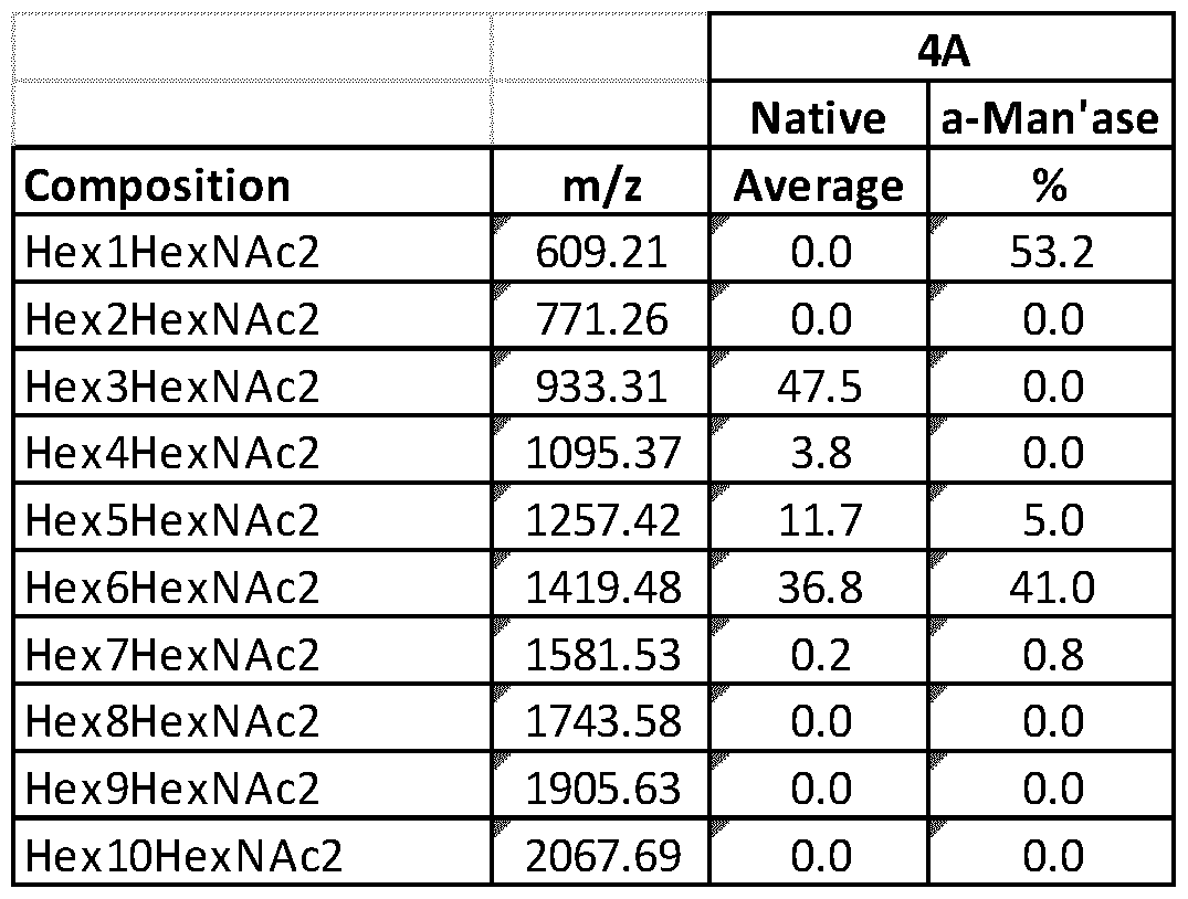


Claims
Priority Applications (13)
| Application Number | Priority Date | Filing Date | Title |
|---|---|---|---|
| DK11790938.2T DK2643456T3 (en) | 2010-11-24 | 2011-11-24 | FUSION enzymes with N-ACETYLGLUKOSAMINYLTRANSFERASE ACTIVITY |
| MX2013005853A MX338560B (en) | 2010-11-24 | 2011-11-24 | Fusion enzymes having n-acetylglucosaminyltransferase activity. |
| JP2013540368A JP6017439B2 (en) | 2010-11-24 | 2011-11-24 | Fusion enzyme having N-acetylglucosaminyltransferase activity |
| CA2819071A CA2819071A1 (en) | 2010-11-24 | 2011-11-24 | Fusion enzymes |
| CN201180064714.3A CN103348003B (en) | 2010-11-24 | 2011-11-24 | There is the fusion enzyme of N-acetylglucosaminyltransferase activity |
| SG2013038088A SG190321A1 (en) | 2010-11-24 | 2011-11-24 | Fusion enzymes having n-acetylglucosaminyltransferase activity |
| EP11790938.2A EP2643456B1 (en) | 2010-11-24 | 2011-11-24 | Fusion enzymes having n-acetylglucosaminyltransferase activity |
| US13/989,084 US9399764B2 (en) | 2010-11-24 | 2011-11-24 | Fusion enzymes |
| KR1020137016445A KR20140018851A (en) | 2010-11-24 | 2011-11-24 | Fusion enzymes having n-acetylglucosaminyltransferase activity |
| AU2011333685A AU2011333685A1 (en) | 2010-11-24 | 2011-11-24 | Fusion enzymes having N-acetylglucosaminyltransferase activity |
| RU2013128619/10A RU2013128619A (en) | 2010-11-24 | 2011-11-24 | CONNECTED ENZYMES |
| IL226481A IL226481A0 (en) | 2010-11-24 | 2013-05-21 | Fusion enzymes |
| US15/205,577 US20170002337A1 (en) | 2010-11-24 | 2016-07-08 | Fusion Enzymes |
Applications Claiming Priority (2)
| Application Number | Priority Date | Filing Date | Title |
|---|---|---|---|
| US41714410P | 2010-11-24 | 2010-11-24 | |
| US61/417,144 | 2010-11-24 |
Related Child Applications (2)
| Application Number | Title | Priority Date | Filing Date |
|---|---|---|---|
| US13/989,084 A-371-Of-International US9399764B2 (en) | 2010-11-24 | 2011-11-24 | Fusion enzymes |
| US15/205,577 Continuation US20170002337A1 (en) | 2010-11-24 | 2016-07-08 | Fusion Enzymes |
Publications (2)
| Publication Number | Publication Date |
|---|---|
| WO2012069593A2 true WO2012069593A2 (en) | 2012-05-31 |
| WO2012069593A3 WO2012069593A3 (en) | 2013-01-03 |
Family
ID=45093734
Family Applications (1)
| Application Number | Title | Priority Date | Filing Date |
|---|---|---|---|
| PCT/EP2011/070956 WO2012069593A2 (en) | 2010-11-24 | 2011-11-24 | Fusion enzymes |
Country Status (13)
| Country | Link |
|---|---|
| US (2) | US9399764B2 (en) |
| EP (1) | EP2643456B1 (en) |
| JP (1) | JP6017439B2 (en) |
| KR (1) | KR20140018851A (en) |
| CN (1) | CN103348003B (en) |
| AU (1) | AU2011333685A1 (en) |
| CA (1) | CA2819071A1 (en) |
| DK (1) | DK2643456T3 (en) |
| IL (1) | IL226481A0 (en) |
| MX (1) | MX338560B (en) |
| RU (1) | RU2013128619A (en) |
| SG (1) | SG190321A1 (en) |
| WO (1) | WO2012069593A2 (en) |
Cited By (6)
| Publication number | Priority date | Publication date | Assignee | Title |
|---|---|---|---|---|
| WO2013174927A1 (en) | 2012-05-23 | 2013-11-28 | Novartis International Pharmaceutical Limited | Production of fucosylated glycoproteins |
| WO2015001049A1 (en) | 2013-07-04 | 2015-01-08 | Novartis Ag | O-mannosyltransferase deficient filamentous fungal cells and methods of use thereof |
| WO2015004239A1 (en) | 2013-07-10 | 2015-01-15 | Novartis Ag | Production of glycoproteins having increased n-glycosylation site occupancy |
| US9399764B2 (en) | 2010-11-24 | 2016-07-26 | Novartis International Pharmaceutical, Ltd. | Fusion enzymes |
| JP2016523552A (en) * | 2013-07-10 | 2016-08-12 | ノバルティス アーゲー | Multiple protease-deficient filamentous fungal cells and methods of use thereof |
| US10081802B2 (en) | 2013-07-29 | 2018-09-25 | Danisco Us Inc. | Variant Enzymes |
Families Citing this family (6)
| Publication number | Priority date | Publication date | Assignee | Title |
|---|---|---|---|---|
| US20160177355A1 (en) * | 2013-03-14 | 2016-06-23 | Adam C. Fisher | Oligosaccharide compositions, glycoproteins and methods to produce the same in prokaryotes |
| CN108064266A (en) | 2014-07-21 | 2018-05-22 | 格利科斯芬兰公司 | The preparation of the glycoprotein with mammal sample N- glycan in filamentous fungi |
| CN105695439B (en) * | 2014-11-27 | 2023-08-01 | 丰益(上海)生物技术研发中心有限公司 | Recombinant expression method of beta-glucosidase gene |
| JP2021534763A (en) * | 2018-08-21 | 2021-12-16 | クララ フーズ カンパニー | Modification of Glycosylation of Proteins in Microorganisms |
| CN111699252A (en) * | 2019-01-16 | 2020-09-22 | 福尼亚生物处理股份有限公司 | Endoglucanase compositions and methods |
| CN110448719B (en) * | 2019-06-28 | 2020-05-22 | 浙江大学 | Silk fibroin-polypeptide electrospun membrane for promoting blood coagulation and preparation method thereof |
Citations (21)
| Publication number | Priority date | Publication date | Assignee | Title |
|---|---|---|---|---|
| US4439196A (en) | 1982-03-18 | 1984-03-27 | Merck & Co., Inc. | Osmotic drug delivery system |
| US4447224A (en) | 1982-09-20 | 1984-05-08 | Infusaid Corporation | Variable flow implantable infusion apparatus |
| US4447233A (en) | 1981-04-10 | 1984-05-08 | Parker-Hannifin Corporation | Medication infusion pump |
| US4475196A (en) | 1981-03-06 | 1984-10-02 | Zor Clair G | Instrument for locating faults in aircraft passenger reading light and attendant call control system |
| US4486194A (en) | 1983-06-08 | 1984-12-04 | James Ferrara | Therapeutic device for administering medicaments through the skin |
| US4487603A (en) | 1982-11-26 | 1984-12-11 | Cordis Corporation | Implantable microinfusion pump system |
| US4500707A (en) | 1980-02-29 | 1985-02-19 | University Patents, Inc. | Nucleosides useful in the preparation of polynucleotides |
| US4596556A (en) | 1985-03-25 | 1986-06-24 | Bioject, Inc. | Hypodermic injection apparatus |
| US4683195A (en) | 1986-01-30 | 1987-07-28 | Cetus Corporation | Process for amplifying, detecting, and/or-cloning nucleic acid sequences |
| US4743546A (en) | 1985-02-13 | 1988-05-10 | Biotechnica International, Inc. | Controlled gene excision |
| US4790824A (en) | 1987-06-19 | 1988-12-13 | Bioject, Inc. | Non-invasive hypodermic injection device |
| US4941880A (en) | 1987-06-19 | 1990-07-17 | Bioject, Inc. | Pre-filled ampule and non-invasive hypodermic injection device assembly |
| US5064413A (en) | 1989-11-09 | 1991-11-12 | Bioject, Inc. | Needleless hypodermic injection device |
| US5312335A (en) | 1989-11-09 | 1994-05-17 | Bioject Inc. | Needleless hypodermic injection device |
| US5383851A (en) | 1992-07-24 | 1995-01-24 | Bioject Inc. | Needleless hypodermic injection device |
| US5436327A (en) | 1988-09-21 | 1995-07-25 | Isis Innovation Limited | Support-bound oligonucleotides |
| US5700637A (en) | 1988-05-03 | 1997-12-23 | Isis Innovation Limited | Apparatus and method for analyzing polynucleotide sequences and method of generating oligonucleotide arrays |
| WO2000024883A1 (en) | 1998-10-26 | 2000-05-04 | Novozymes A/S | Constructing and screening a dna library of interest in filamentous fungal cells |
| US6653459B1 (en) | 1997-12-09 | 2003-11-25 | Antje Von Schaewen | Plant GntI sequences and the use thereof for the production of plants having reduced or lacking N-acetyl glucosaminyl transferase I(GnTI) activity |
| WO2008040387A1 (en) | 2006-10-03 | 2008-04-10 | Metabolic Explorer | Process for chromosomal integration and dna sequence replacement in clostridia |
| US7501275B2 (en) | 2004-10-27 | 2009-03-10 | The United States Of America As Represented By The Secretary Of Agriculture | Yeast transformation system |
Family Cites Families (10)
| Publication number | Priority date | Publication date | Assignee | Title |
|---|---|---|---|---|
| US7244601B2 (en) * | 1997-12-15 | 2007-07-17 | National Research Council Of Canada | Fusion proteins for use in enzymatic synthesis of oligosaccharides |
| AU784960B2 (en) * | 1999-08-19 | 2006-08-10 | Kirin Pharma Kabushiki Kaisha | Novel yeast variants and process for producing glycoprotein containing mammalian type sugar chain |
| US7795002B2 (en) * | 2000-06-28 | 2010-09-14 | Glycofi, Inc. | Production of galactosylated glycoproteins in lower eukaryotes |
| US7598055B2 (en) * | 2000-06-28 | 2009-10-06 | Glycofi, Inc. | N-acetylglucosaminyltransferase III expression in lower eukaryotes |
| WO2003056914A1 (en) * | 2001-12-27 | 2003-07-17 | Glycofi, Inc. | Methods to engineer mammalian-type carbohydrate structures |
| AU2003239365A1 (en) * | 2002-05-03 | 2003-11-17 | Neose Technologies, Inc. | Recombinant glycosyltransferase fusion proteins |
| WO2006026992A1 (en) | 2004-09-07 | 2006-03-16 | Novozymes A/S | Altered structure of n-glycans in a fungus |
| US20080026376A1 (en) | 2006-07-11 | 2008-01-31 | Huaming Wang | KEX2 cleavage regions of recombinant fusion proteins |
| TW201028431A (en) | 2008-10-31 | 2010-08-01 | Lonza Ag | Novel tools for the production of glycosylated proteins in host cells |
| SG190321A1 (en) | 2010-11-24 | 2013-06-28 | Novartis Int Pharm Ltd | Fusion enzymes having n-acetylglucosaminyltransferase activity |
-
2011
- 2011-11-24 SG SG2013038088A patent/SG190321A1/en unknown
- 2011-11-24 JP JP2013540368A patent/JP6017439B2/en not_active Expired - Fee Related
- 2011-11-24 MX MX2013005853A patent/MX338560B/en active IP Right Grant
- 2011-11-24 CA CA2819071A patent/CA2819071A1/en not_active Abandoned
- 2011-11-24 EP EP11790938.2A patent/EP2643456B1/en not_active Not-in-force
- 2011-11-24 WO PCT/EP2011/070956 patent/WO2012069593A2/en active Application Filing
- 2011-11-24 US US13/989,084 patent/US9399764B2/en active Active
- 2011-11-24 KR KR1020137016445A patent/KR20140018851A/en not_active Application Discontinuation
- 2011-11-24 DK DK11790938.2T patent/DK2643456T3/en active
- 2011-11-24 AU AU2011333685A patent/AU2011333685A1/en not_active Abandoned
- 2011-11-24 CN CN201180064714.3A patent/CN103348003B/en not_active Expired - Fee Related
- 2011-11-24 RU RU2013128619/10A patent/RU2013128619A/en not_active Application Discontinuation
-
2013
- 2013-05-21 IL IL226481A patent/IL226481A0/en unknown
-
2016
- 2016-07-08 US US15/205,577 patent/US20170002337A1/en not_active Abandoned
Patent Citations (23)
| Publication number | Priority date | Publication date | Assignee | Title |
|---|---|---|---|---|
| US4500707A (en) | 1980-02-29 | 1985-02-19 | University Patents, Inc. | Nucleosides useful in the preparation of polynucleotides |
| US4475196A (en) | 1981-03-06 | 1984-10-02 | Zor Clair G | Instrument for locating faults in aircraft passenger reading light and attendant call control system |
| US4447233A (en) | 1981-04-10 | 1984-05-08 | Parker-Hannifin Corporation | Medication infusion pump |
| US4439196A (en) | 1982-03-18 | 1984-03-27 | Merck & Co., Inc. | Osmotic drug delivery system |
| US4447224A (en) | 1982-09-20 | 1984-05-08 | Infusaid Corporation | Variable flow implantable infusion apparatus |
| US4487603A (en) | 1982-11-26 | 1984-12-11 | Cordis Corporation | Implantable microinfusion pump system |
| US4486194A (en) | 1983-06-08 | 1984-12-04 | James Ferrara | Therapeutic device for administering medicaments through the skin |
| US4743546A (en) | 1985-02-13 | 1988-05-10 | Biotechnica International, Inc. | Controlled gene excision |
| US4596556A (en) | 1985-03-25 | 1986-06-24 | Bioject, Inc. | Hypodermic injection apparatus |
| US4683195B1 (en) | 1986-01-30 | 1990-11-27 | Cetus Corp | |
| US4683195A (en) | 1986-01-30 | 1987-07-28 | Cetus Corporation | Process for amplifying, detecting, and/or-cloning nucleic acid sequences |
| US4790824A (en) | 1987-06-19 | 1988-12-13 | Bioject, Inc. | Non-invasive hypodermic injection device |
| US4941880A (en) | 1987-06-19 | 1990-07-17 | Bioject, Inc. | Pre-filled ampule and non-invasive hypodermic injection device assembly |
| US5700637A (en) | 1988-05-03 | 1997-12-23 | Isis Innovation Limited | Apparatus and method for analyzing polynucleotide sequences and method of generating oligonucleotide arrays |
| US5436327A (en) | 1988-09-21 | 1995-07-25 | Isis Innovation Limited | Support-bound oligonucleotides |
| US5064413A (en) | 1989-11-09 | 1991-11-12 | Bioject, Inc. | Needleless hypodermic injection device |
| US5312335A (en) | 1989-11-09 | 1994-05-17 | Bioject Inc. | Needleless hypodermic injection device |
| US5383851A (en) | 1992-07-24 | 1995-01-24 | Bioject Inc. | Needleless hypodermic injection device |
| US5399163A (en) | 1992-07-24 | 1995-03-21 | Bioject Inc. | Needleless hypodermic injection methods and device |
| US6653459B1 (en) | 1997-12-09 | 2003-11-25 | Antje Von Schaewen | Plant GntI sequences and the use thereof for the production of plants having reduced or lacking N-acetyl glucosaminyl transferase I(GnTI) activity |
| WO2000024883A1 (en) | 1998-10-26 | 2000-05-04 | Novozymes A/S | Constructing and screening a dna library of interest in filamentous fungal cells |
| US7501275B2 (en) | 2004-10-27 | 2009-03-10 | The United States Of America As Represented By The Secretary Of Agriculture | Yeast transformation system |
| WO2008040387A1 (en) | 2006-10-03 | 2008-04-10 | Metabolic Explorer | Process for chromosomal integration and dna sequence replacement in clostridia |
Non-Patent Citations (55)
| Title |
|---|
| "Biological Regulation and Development", vol. 1, PLENUM, pages: 349 - 399 |
| "Biology and Activities of Yeast", 1980 |
| "Protein Purification Methods: A Practical Approach", 1989, IRL PRESS |
| "Sustained and Controlled Release Drug Delivery Systems", 1978, MARCEL DEKKER, INC. |
| BERGE, S.M. ET AL., J. PHARM. SCI., vol. 66, 1977, pages 1 - 19 |
| BIOCHEM., vol. 9, 1970, pages 4022 |
| CALLEWAERT, FEBS LETT, 2001, pages 173 - 178 |
| CARBOHYDRATE RES., vol. 297, 1997, pages 1 |
| CARBOHYDRATE RES., vol. 312, 1998, pages 167 |
| CHEN S. ET AL., J. BIOL.CHEM, vol. 274, no. 1, 1999, pages 288 - 97 |
| CHIBA ET AL., JBIOL CHEM, vol. 273, 1998, pages 26298 - 304 |
| CHIBA, JBC, 1998, pages 26298 - 304 |
| DAVIDSON ET AL., GLYCOBIOLOGY, 2004, pages 399 - 407 |
| DE BOER ET AL., PROC NATL ACAD SCI USA, vol. 80, no. 1, 1983, pages 21 - 25 |
| DE POURCQ ET AL., APPL MICROBIOL BIOTECHNOL, vol. 87, no. 5, pages 1617 - 31 |
| EUR. J. BIOCHEM., vol. 257, 1998, pages 29 |
| GUO S. ET AL., GLYCOBIOLOGY, vol. 11, 2001, pages 813 - 20 |
| HAMILTON ET AL., SCIENCE, vol. 301, 2003, pages 1244 - 6 |
| HAWKSWORTH ET AL.: "Ainsworth and Bisby's Dictionary of The Fungi", 1995, UNIVERSITY PRESS |
| J.MOL.BIOL., vol. 357, 2006, pages 1619 - 1633 |
| JEREMIAH D. READ ET AL., APPLIED AND ENVIRONMENTAL MICROBIOLOGY, August 2007 (2007-08-01), pages 5088 - 5096 |
| KAINZ ET AL., APPL ENVIRON MICROB., 2008, pages 1076 - 86 |
| KAINZ ET AL., APPL ENVIRON MICROBIOL, vol. 74, 2008, pages 1076 - 86 |
| KAINZ ET AL., APPL. ENVIRON MICROB, 2008, pages 1076 - 86 |
| KALSNER ET AL., GLYCOCON. J, 1995, pages 360 - 370 |
| KASAJIMA ET AL., BIOSCI BIOTECH BIOCHEM, 2006, pages 2662 - 8 |
| KUMAR R. ET AL., PROC NATL ACAD SCI U S A., vol. 87, no. 24, December 1990 (1990-12-01), pages 9948 - 52 |
| MARAS ET AL., EURO JBIOCHEM, vol. 249, 1997, pages 701 - 07 |
| MARAS ET AL., FEBS LETT, 1999, pages 365 - 70 |
| MARAS ET AL., FEBS LETTERS, vol. 452, 1999, pages 365 - 70 |
| MARAS M ET AL., J BIOTECH., vol. 77, 2000, pages 255 |
| MARTINET ET AL., BIOTECH LETT, 1998, pages 1171 |
| MATTEUCCI ET AL., TETRAHEDRON LETT, vol. 21, 1980, pages 719 - 722 |
| OPAT AS ET AL., BIOCHEM J., vol. 336, 15 December 1998 (1998-12-15), pages 593 - 8 |
| OSVALDO DELGADO ET AL., FEMS MICROBIOLOGY LETTERS, vol. 132, 1995, pages 23 - 26 |
| PUTHALAKATH H. ET AL., J. BIOL.CHEM, vol. 271, no. 44, 1996, pages 27818 - 22 |
| SALAMIN ET AL., APPL. ENVIRON. MICROBIOL., vol. 76, 2010, pages 4269 - 4276 |
| SARKAR ET AL., GLYCOCONJUGATE J, vol. 15, 1998, pages 193 - 197 |
| SARKAR M ET AL., PROC NATL ACAD SCI U S A., vol. 88, no. 1, 1 January 1991 (1991-01-01), pages 234 - 8 |
| SARKAR; SCHACHTER, BIOL CHEM., vol. 382, no. 2, February 2001 (2001-02-01), pages 209 - 17 |
| SCHWIENTEK, J.BIOL. CHEM, 1996, pages 3398 |
| SELINHEIMO ET AL., FEBSJ., vol. 273, no. 18, 2006, pages 4322 - 35 |
| SHAPER N.L. ET AL., PROC. NATL. ACAD. SCI. U.S.A., vol. 83, no. 6, 1986, pages 1573 - 1577 |
| SHAPER, N.L. ET AL., J. BIOL. CHEM., vol. 263, no. 21, 1998, pages 10420 - 10428 |
| SHINE; DALGARNO, NATURE, vol. 254, no. 5495, 1975, pages 34 - 38 |
| STALS ET AL., GLYCOBIOLOGY, vol. 14, 2004, pages 713 - 724 |
| STALS ET AL., GLYCOBIOLOGY, vol. 14, 2004, pages 725 |
| STALS ET AL., GLYCOBIOLOGY, vol. 14, 2004, pages 725 - 737 |
| UNLIGIL UM ET AL., EMBO J., vol. 19, no. 20, 16 October 2000 (2000-10-16), pages 5269 - 80 |
| VAD ET AL., J BIOTECHNOL., vol. 116, 2005, pages 251 - 260 |
| VAN DEN ELSEN J.M. ET AL., EMBO J., vol. 20, 2001, pages 3008 - 3017 |
| VERVECKEN, APPL. ENVIRON MICROB, 2004, pages 2639 - 46 |
| WILDT; GERNGROSS, NATURE REV BIOTECH, 2005, pages 119 |
| WILDT; GERNGROSS, NATURE REVIEWS MICROBIOLOGY, vol. 3, pages 119 - 128 |
| YOSHIDA ET AL., GLYCOBIOLOGY, 1999, pages 53 - 8 |
Cited By (8)
| Publication number | Priority date | Publication date | Assignee | Title |
|---|---|---|---|---|
| US9399764B2 (en) | 2010-11-24 | 2016-07-26 | Novartis International Pharmaceutical, Ltd. | Fusion enzymes |
| WO2013174927A1 (en) | 2012-05-23 | 2013-11-28 | Novartis International Pharmaceutical Limited | Production of fucosylated glycoproteins |
| WO2015001049A1 (en) | 2013-07-04 | 2015-01-08 | Novartis Ag | O-mannosyltransferase deficient filamentous fungal cells and methods of use thereof |
| WO2015004239A1 (en) | 2013-07-10 | 2015-01-15 | Novartis Ag | Production of glycoproteins having increased n-glycosylation site occupancy |
| JP2016523552A (en) * | 2013-07-10 | 2016-08-12 | ノバルティス アーゲー | Multiple protease-deficient filamentous fungal cells and methods of use thereof |
| US10081802B2 (en) | 2013-07-29 | 2018-09-25 | Danisco Us Inc. | Variant Enzymes |
| US10167460B2 (en) | 2013-07-29 | 2019-01-01 | Danisco Us Inc | Variant enzymes |
| US10479983B2 (en) | 2013-07-29 | 2019-11-19 | Danisco Us Inc | Variant enzymes |
Also Published As
| Publication number | Publication date |
|---|---|
| EP2643456B1 (en) | 2016-05-11 |
| IL226481A0 (en) | 2013-07-31 |
| US9399764B2 (en) | 2016-07-26 |
| MX2013005853A (en) | 2013-12-12 |
| US20130330780A1 (en) | 2013-12-12 |
| EP2643456A2 (en) | 2013-10-02 |
| CA2819071A1 (en) | 2012-05-31 |
| SG190321A1 (en) | 2013-06-28 |
| US20170002337A1 (en) | 2017-01-05 |
| CN103348003B (en) | 2016-09-07 |
| MX338560B (en) | 2016-04-20 |
| JP2014500019A (en) | 2014-01-09 |
| DK2643456T3 (en) | 2016-08-22 |
| RU2013128619A (en) | 2014-12-27 |
| CN103348003A (en) | 2013-10-09 |
| WO2012069593A3 (en) | 2013-01-03 |
| JP6017439B2 (en) | 2016-11-02 |
| AU2011333685A1 (en) | 2013-06-27 |
| KR20140018851A (en) | 2014-02-13 |
Similar Documents
| Publication | Publication Date | Title |
|---|---|---|
| US9399764B2 (en) | Fusion enzymes | |
| US9695454B2 (en) | Production of fucosylated glycoproteins | |
| EP3172333B1 (en) | Production of glycoproteins with mammalian-like n-glycans in filamentous fungi | |
| Kainz et al. | N-glycan modification in Aspergillus species | |
| AU2009309925A1 (en) | Novel tools for the production of glycosylated proteins in host cells | |
| AU2011247333A1 (en) | Improved glycosylation of proteins in host cells | |
| US20200362319A1 (en) | O-Mannosyltransferase Deficient Filamentous Fungal Cells and Methods of Use Thereof | |
| US20190276867A1 (en) | Production of Glycoproteins Having Increased N-Glycosylation Site Occupancy | |
| BR112013013045A2 (en) | RECOMBINANT PROTEINS WITH N-ACETYLGLUCOSAMINILTRANSFERASE ACTIVITY, POLYNUCLEOTIDE, EXPRESSION VECTOR, CELLS AND COMPLEX N-GLYCAN PRODUCTION METHODS |
Legal Events
| Date | Code | Title | Description |
|---|---|---|---|
| 121 | Ep: the epo has been informed by wipo that ep was designated in this application |
Ref document number: 11790938 Country of ref document: EP Kind code of ref document: A2 |
|
| ENP | Entry into the national phase |
Ref document number: 2819071 Country of ref document: CA |
|
| WWE | Wipo information: entry into national phase |
Ref document number: 226481 Country of ref document: IL |
|
| WWE | Wipo information: entry into national phase |
Ref document number: MX/A/2013/005853 Country of ref document: MX |
|
| ENP | Entry into the national phase |
Ref document number: 2013540368 Country of ref document: JP Kind code of ref document: A |
|
| NENP | Non-entry into the national phase |
Ref country code: DE |
|
| ENP | Entry into the national phase |
Ref document number: 2013128619 Country of ref document: RU Kind code of ref document: A Ref document number: 20137016445 Country of ref document: KR Kind code of ref document: A |
|
| WWE | Wipo information: entry into national phase |
Ref document number: 2011790938 Country of ref document: EP |
|
| ENP | Entry into the national phase |
Ref document number: 2011333685 Country of ref document: AU Date of ref document: 20111124 Kind code of ref document: A |
|
| 121 | Ep: the epo has been informed by wipo that ep was designated in this application |
Ref document number: 11790938 Country of ref document: EP Kind code of ref document: A2 |
|
| WWE | Wipo information: entry into national phase |
Ref document number: 13989084 Country of ref document: US |
|
| REG | Reference to national code |
Ref country code: BR Ref legal event code: B01A Ref document number: 112013013045 Country of ref document: BR |
|
| REG | Reference to national code |
Ref country code: BR Ref legal event code: B01E Ref document number: 112013013045 Country of ref document: BR Free format text: COM BASE NA RESOLUCAO 81/2013, SOLICITA-SE QUE SEJA APRESENTADO, EM ATE 60 (SESSENTA) DIAS, NOVO CONTEUDO DE LISTAGEM DE SEQUENCIA NOS MOLDES DA PORTARIA 405 DE 21/12/2020, POIS O CONTEUDO APRESENTADOS NA PETICAO NO 020130045631 DE 24/05/2013 NAO PODE SER ASSOCIADO AO PEDIDO EM QUESTAO POR POSSUIR DIVERGENCIAS ENTRE O CONTEUDO DE LISTAGEM APRESENTADO E O PEDIDO EM QUESTAO, COMO DEPOSITANTE DIVERGENTE. |
|
| ENP | Entry into the national phase |
Ref document number: 112013013045 Country of ref document: BR Kind code of ref document: A2 Effective date: 20130524 |