WO2012148477A1 - Digital counting of individual molecules by stochastic attachment of diverse label-tags - Google Patents
Digital counting of individual molecules by stochastic attachment of diverse label-tags Download PDFInfo
- Publication number
- WO2012148477A1 WO2012148477A1 PCT/US2011/065291 US2011065291W WO2012148477A1 WO 2012148477 A1 WO2012148477 A1 WO 2012148477A1 US 2011065291 W US2011065291 W US 2011065291W WO 2012148477 A1 WO2012148477 A1 WO 2012148477A1
- Authority
- WO
- WIPO (PCT)
- Prior art keywords
- label
- target
- tags
- tag
- different
- Prior art date
Links
Classifications
-
- G—PHYSICS
- G16—INFORMATION AND COMMUNICATION TECHNOLOGY [ICT] SPECIALLY ADAPTED FOR SPECIFIC APPLICATION FIELDS
- G16B—BIOINFORMATICS, i.e. INFORMATION AND COMMUNICATION TECHNOLOGY [ICT] SPECIALLY ADAPTED FOR GENETIC OR PROTEIN-RELATED DATA PROCESSING IN COMPUTATIONAL MOLECULAR BIOLOGY
- G16B25/00—ICT specially adapted for hybridisation; ICT specially adapted for gene or protein expression
- G16B25/20—Polymerase chain reaction [PCR]; Primer or probe design; Probe optimisation
-
- C—CHEMISTRY; METALLURGY
- C12—BIOCHEMISTRY; BEER; SPIRITS; WINE; VINEGAR; MICROBIOLOGY; ENZYMOLOGY; MUTATION OR GENETIC ENGINEERING
- C12Q—MEASURING OR TESTING PROCESSES INVOLVING ENZYMES, NUCLEIC ACIDS OR MICROORGANISMS; COMPOSITIONS OR TEST PAPERS THEREFOR; PROCESSES OF PREPARING SUCH COMPOSITIONS; CONDITION-RESPONSIVE CONTROL IN MICROBIOLOGICAL OR ENZYMOLOGICAL PROCESSES
- C12Q1/00—Measuring or testing processes involving enzymes, nucleic acids or microorganisms; Compositions therefor; Processes of preparing such compositions
- C12Q1/68—Measuring or testing processes involving enzymes, nucleic acids or microorganisms; Compositions therefor; Processes of preparing such compositions involving nucleic acids
- C12Q1/6869—Methods for sequencing
- C12Q1/6874—Methods for sequencing involving nucleic acid arrays, e.g. sequencing by hybridisation
-
- C—CHEMISTRY; METALLURGY
- C12—BIOCHEMISTRY; BEER; SPIRITS; WINE; VINEGAR; MICROBIOLOGY; ENZYMOLOGY; MUTATION OR GENETIC ENGINEERING
- C12N—MICROORGANISMS OR ENZYMES; COMPOSITIONS THEREOF; PROPAGATING, PRESERVING, OR MAINTAINING MICROORGANISMS; MUTATION OR GENETIC ENGINEERING; CULTURE MEDIA
- C12N15/00—Mutation or genetic engineering; DNA or RNA concerning genetic engineering, vectors, e.g. plasmids, or their isolation, preparation or purification; Use of hosts therefor
- C12N15/09—Recombinant DNA-technology
- C12N15/10—Processes for the isolation, preparation or purification of DNA or RNA
- C12N15/1034—Isolating an individual clone by screening libraries
- C12N15/1065—Preparation or screening of tagged libraries, e.g. tagged microorganisms by STM-mutagenesis, tagged polynucleotides, gene tags
-
- C—CHEMISTRY; METALLURGY
- C12—BIOCHEMISTRY; BEER; SPIRITS; WINE; VINEGAR; MICROBIOLOGY; ENZYMOLOGY; MUTATION OR GENETIC ENGINEERING
- C12Q—MEASURING OR TESTING PROCESSES INVOLVING ENZYMES, NUCLEIC ACIDS OR MICROORGANISMS; COMPOSITIONS OR TEST PAPERS THEREFOR; PROCESSES OF PREPARING SUCH COMPOSITIONS; CONDITION-RESPONSIVE CONTROL IN MICROBIOLOGICAL OR ENZYMOLOGICAL PROCESSES
- C12Q1/00—Measuring or testing processes involving enzymes, nucleic acids or microorganisms; Compositions therefor; Processes of preparing such compositions
- C12Q1/68—Measuring or testing processes involving enzymes, nucleic acids or microorganisms; Compositions therefor; Processes of preparing such compositions involving nucleic acids
- C12Q1/6809—Methods for determination or identification of nucleic acids involving differential detection
-
- C—CHEMISTRY; METALLURGY
- C12—BIOCHEMISTRY; BEER; SPIRITS; WINE; VINEGAR; MICROBIOLOGY; ENZYMOLOGY; MUTATION OR GENETIC ENGINEERING
- C12Q—MEASURING OR TESTING PROCESSES INVOLVING ENZYMES, NUCLEIC ACIDS OR MICROORGANISMS; COMPOSITIONS OR TEST PAPERS THEREFOR; PROCESSES OF PREPARING SUCH COMPOSITIONS; CONDITION-RESPONSIVE CONTROL IN MICROBIOLOGICAL OR ENZYMOLOGICAL PROCESSES
- C12Q1/00—Measuring or testing processes involving enzymes, nucleic acids or microorganisms; Compositions therefor; Processes of preparing such compositions
- C12Q1/68—Measuring or testing processes involving enzymes, nucleic acids or microorganisms; Compositions therefor; Processes of preparing such compositions involving nucleic acids
- C12Q1/6813—Hybridisation assays
- C12Q1/6834—Enzymatic or biochemical coupling of nucleic acids to a solid phase
- C12Q1/6837—Enzymatic or biochemical coupling of nucleic acids to a solid phase using probe arrays or probe chips
-
- C—CHEMISTRY; METALLURGY
- C12—BIOCHEMISTRY; BEER; SPIRITS; WINE; VINEGAR; MICROBIOLOGY; ENZYMOLOGY; MUTATION OR GENETIC ENGINEERING
- C12Q—MEASURING OR TESTING PROCESSES INVOLVING ENZYMES, NUCLEIC ACIDS OR MICROORGANISMS; COMPOSITIONS OR TEST PAPERS THEREFOR; PROCESSES OF PREPARING SUCH COMPOSITIONS; CONDITION-RESPONSIVE CONTROL IN MICROBIOLOGICAL OR ENZYMOLOGICAL PROCESSES
- C12Q1/00—Measuring or testing processes involving enzymes, nucleic acids or microorganisms; Compositions therefor; Processes of preparing such compositions
- C12Q1/68—Measuring or testing processes involving enzymes, nucleic acids or microorganisms; Compositions therefor; Processes of preparing such compositions involving nucleic acids
- C12Q1/6844—Nucleic acid amplification reactions
- C12Q1/6851—Quantitative amplification
-
- C—CHEMISTRY; METALLURGY
- C12—BIOCHEMISTRY; BEER; SPIRITS; WINE; VINEGAR; MICROBIOLOGY; ENZYMOLOGY; MUTATION OR GENETIC ENGINEERING
- C12Q—MEASURING OR TESTING PROCESSES INVOLVING ENZYMES, NUCLEIC ACIDS OR MICROORGANISMS; COMPOSITIONS OR TEST PAPERS THEREFOR; PROCESSES OF PREPARING SUCH COMPOSITIONS; CONDITION-RESPONSIVE CONTROL IN MICROBIOLOGICAL OR ENZYMOLOGICAL PROCESSES
- C12Q1/00—Measuring or testing processes involving enzymes, nucleic acids or microorganisms; Compositions therefor; Processes of preparing such compositions
- C12Q1/68—Measuring or testing processes involving enzymes, nucleic acids or microorganisms; Compositions therefor; Processes of preparing such compositions involving nucleic acids
- C12Q1/6844—Nucleic acid amplification reactions
- C12Q1/686—Polymerase chain reaction [PCR]
-
- C—CHEMISTRY; METALLURGY
- C12—BIOCHEMISTRY; BEER; SPIRITS; WINE; VINEGAR; MICROBIOLOGY; ENZYMOLOGY; MUTATION OR GENETIC ENGINEERING
- C12Q—MEASURING OR TESTING PROCESSES INVOLVING ENZYMES, NUCLEIC ACIDS OR MICROORGANISMS; COMPOSITIONS OR TEST PAPERS THEREFOR; PROCESSES OF PREPARING SUCH COMPOSITIONS; CONDITION-RESPONSIVE CONTROL IN MICROBIOLOGICAL OR ENZYMOLOGICAL PROCESSES
- C12Q1/00—Measuring or testing processes involving enzymes, nucleic acids or microorganisms; Compositions therefor; Processes of preparing such compositions
- C12Q1/68—Measuring or testing processes involving enzymes, nucleic acids or microorganisms; Compositions therefor; Processes of preparing such compositions involving nucleic acids
- C12Q1/6869—Methods for sequencing
-
- C—CHEMISTRY; METALLURGY
- C12—BIOCHEMISTRY; BEER; SPIRITS; WINE; VINEGAR; MICROBIOLOGY; ENZYMOLOGY; MUTATION OR GENETIC ENGINEERING
- C12Q—MEASURING OR TESTING PROCESSES INVOLVING ENZYMES, NUCLEIC ACIDS OR MICROORGANISMS; COMPOSITIONS OR TEST PAPERS THEREFOR; PROCESSES OF PREPARING SUCH COMPOSITIONS; CONDITION-RESPONSIVE CONTROL IN MICROBIOLOGICAL OR ENZYMOLOGICAL PROCESSES
- C12Q1/00—Measuring or testing processes involving enzymes, nucleic acids or microorganisms; Compositions therefor; Processes of preparing such compositions
- C12Q1/68—Measuring or testing processes involving enzymes, nucleic acids or microorganisms; Compositions therefor; Processes of preparing such compositions involving nucleic acids
- C12Q1/6876—Nucleic acid products used in the analysis of nucleic acids, e.g. primers or probes
-
- G—PHYSICS
- G16—INFORMATION AND COMMUNICATION TECHNOLOGY [ICT] SPECIALLY ADAPTED FOR SPECIFIC APPLICATION FIELDS
- G16B—BIOINFORMATICS, i.e. INFORMATION AND COMMUNICATION TECHNOLOGY [ICT] SPECIALLY ADAPTED FOR GENETIC OR PROTEIN-RELATED DATA PROCESSING IN COMPUTATIONAL MOLECULAR BIOLOGY
- G16B25/00—ICT specially adapted for hybridisation; ICT specially adapted for gene or protein expression
-
- G—PHYSICS
- G16—INFORMATION AND COMMUNICATION TECHNOLOGY [ICT] SPECIALLY ADAPTED FOR SPECIFIC APPLICATION FIELDS
- G16B—BIOINFORMATICS, i.e. INFORMATION AND COMMUNICATION TECHNOLOGY [ICT] SPECIALLY ADAPTED FOR GENETIC OR PROTEIN-RELATED DATA PROCESSING IN COMPUTATIONAL MOLECULAR BIOLOGY
- G16B30/00—ICT specially adapted for sequence analysis involving nucleotides or amino acids
-
- G—PHYSICS
- G16—INFORMATION AND COMMUNICATION TECHNOLOGY [ICT] SPECIALLY ADAPTED FOR SPECIFIC APPLICATION FIELDS
- G16B—BIOINFORMATICS, i.e. INFORMATION AND COMMUNICATION TECHNOLOGY [ICT] SPECIALLY ADAPTED FOR GENETIC OR PROTEIN-RELATED DATA PROCESSING IN COMPUTATIONAL MOLECULAR BIOLOGY
- G16B25/00—ICT specially adapted for hybridisation; ICT specially adapted for gene or protein expression
- G16B25/10—Gene or protein expression profiling; Expression-ratio estimation or normalisation
Definitions
- hybridization-based techniques offer the advantages of scale and the capability of detecting a wide range of gene expression or copy number levels
- measurements may be subject to variability relating to probe hybridization differences and cross-reactivity, eiement-to- element differences within microarrays, and microarray-to-microarray differences, Audic and Claverie, Genomic Res., 7: 986-995 (1997); Wittes and Friedman, J Natl. Cancer Inst. 91 : 400-401 (1999).
- the tags are separated from the rest of the RNA and can be linked together to form long serial molecules that can be cloned and sequenced. Quantitation of the number of times a particular tag is observed provides an estimate of the relative expression level of the corresponding transcript, relative to other tagged transcripts, but not an actual count of the number of times that transcript appears. Other methods based on estimating relative abundance have also been described. See, for example, Wang et al. Nat. Rev. Genet. 10, 57-63 (2009).
- a common feature among all of these approaches is a one-to-one correspondence between probe sequences and oligonucleotide tag sequences. That is, the oligonucleotide tags have been employed as probe surrogates for their favorable hybridizations properties, particularly under multiplex assay conditions.
- PGR and microarray analysis rely on the specificity of hybridization to identify the target of interest for amplification or capture respectively, then yield an analog signal proportional to the original number of molecules.
- a major advantage of these approaches is in the use of hybridization to isolate the specific molecules of interest within the background of many other molecules, generating specificity for the readout or detection step.
- the disadvantage is that the readout signal to noise is proportional to all molecules (specific and non-specific) specified by selective amplification or hybridization. The situation is reversed for sequence counting. No intended sequence specificity is imposed in the sequence capture step, and all molecules are sequenced.
- the major advantage is that the detection step simply yields a digital list of those sequences found, and since there is no specificity in the isolation step, all sequences must be analyzed at a sufficient statistical depth in order to learn about a specific sequence. Although significant technical advances in sequencing speed and throughput have occurred, the statistical requirements imposed to accurately measure small changes in concentration of a specific gene (or target) or a few targets within the background of many other sequences requires measuring many sequences that are not of interest to find the ones that are of interest.
- PCR, array hybridization and sequence counting is a comparative technique in that they primarily measure relative abundance, and do not typically yield an absolute number of molecules in a solution.
- Digital PCR is one method that may be used for absolute counting of nucleic acids (B. Vogelstein, K.
- High-sensitivity single molecule digital counting by the stochastic labeling of a collection of identical molecules is disclosed.
- Each copy of a molecule randomly chooses from a non-depleting reservoir of diverse label-tags.
- the uniqueness of each labeled molecule is determined by the statistics of random choice, and depends on the number of copies of identical molecules in the collection compared to the diversity of label-tags.
- the size of the resulting set of labeled molecules is determined by the stochastic nature of the labeling process, and analysis reveals the original number of molecules.
- the labeled molecules are highly unique, and the digital counting efficiency is high.
- This stochastic transformation relaxes the problem of counting molecules from one of locating and identifying identical molecules to a series of yes/no digital questions detecting whether preprogrammed label- tags are present.
- the conceptual framework for stochastic mapping of a variety of molecule types is developed and the utility of the methods are demonstrated by stochastically labeling 360,000 different fragments of the human genome.
- the labeled fragments for a target molecule of choice are detected with high specificity using a microarray readout system, and with DNA sequencing. The results are consistent with a stochastic process, and yield highly precise relative and absolute counting statistics of selected molecules within a vast background of other molecules.
- the methods disclosed herein for digital counting of individual molecules may be applied to a single target or to a plurality of different targets simultaneously.
- the targets are nucleic acids, but may be a variety of biological or non-biological elements.
- Targets are labeled so that individual occurrences of the same target are marked by attachment of a different label-tag to difference occurrences. The attachment of the label-tag confers a separate,
- the label-tags are different sequences that tag or mark each target occurrence uniquely.
- the resulting modified target comprises the target sequence and the unique label-tag identifier (which also may be referred to herein as label-tag, counter, or identifier).
- the junction of the target and identifier forms a uniquely detectable mechanism for counting the occurrence of that copy of the target.
- the attachment of the identifier to each occurrence of the target is a random sampling event.
- Each occurrence of target could choose any of the identifiers from a pool of identifiers.
- Each identifier is present in multiple copies so selection of one copy does not remove that identifier sequence from the pool of identifiers so it is possible that the same identifier will be selected twice. The probability of that depends on the number of target occurrences relative to the number of different identifier sequences.
- Each stochastic attachment event where a target occurrence is attached to a unique identifier, results in the creation of a novel sequence formed at the junction of the identifier and the target.
- the resulting products will contain the same target portion, but each will contain a different identifier sequence (T1L1 , T1 L2, ...T1LN where N is the number of different occurrences of target 1 .
- Tl and L is the identifier, LI , L2...LN).
- the occurrences are detected by hybridization.
- the methods and systems include a probe array comprising features, wherein each feature has a different combination of target sequence with identifiers, 1 to N wherein N is the number of unique identifiers in the pool of identifiers.
- the array has N features for each target, so if, for example, there are 8 targets to be analyzed there are 8 times N features on the array to interrogate the 8 targets.
- kits for counting the number of occurrences of a two or more different species of target nucleic in a complex mixture of target and non-target species are disclosed.
- the steps may include covalently or non-covalently attaching a label-tag from a pool of label-tags to individual occurrences of the each of the target nucleic acid species of interest to form sets of targets that are distinctly labeled within species so that instead of many undistinguishable molecules of each species there are many distinguishable molecules of each species.
- the species are distinguishable from one another on the basis of differences between species.
- the label-tag are selected from a pool for attaching in an approximately random manner and preferably at least 90% of the labeled targets within a species have a label-tag that is different from all other labeled targets in that species.
- the number of different molecules of a given species is then approximately (within the counting error of the system) equal to the number of different label-tags that are counted.
- the label-tag-targets can be amplified prior to detection.
- Methods of detection include hybridization based methods and sequencing based methods. Other methods such as size separation, mass spec or optical scanning methods may also be used. They nature of the label-tags will guide the detection mechanism.
- Attachment of the label-tags may be by any means available.
- the targets are biological molecules such as nucleic acids or proteins
- the labels are preferably oligonucleotides or peptides.
- Methods for attaching oligonucleotide or nucleic acid sequences to nucleic acids may include ligation, polymerization and primer mediated extension reactions, for example.
- Kits for labeling targets within mixtures and for amplification and detection of the label-tag- targets are also disclosed.
- Kits may include materials or reagents for carrying out a method of the invention and may include systems that allow for the storage, transport, or delivery of reaction reagents (e.g., probes, enzymes, etc. in the appropriate containers) and/or supporting materials (e.g., buffers, written instructions for performing the assay etc.) from one location to another.
- reaction reagents e.g., probes, enzymes, etc. in the appropriate containers
- supporting materials e.g., buffers, written instructions for performing the assay etc.
- kits include one or more enclosures (e.g., boxes) containing the relevant reaction reagents and/or supporting materials for assays of the invention.
- kits of the invention comprises pools of label-tags, pools of label-tag containing primers or pools of label-tag containing adaptors.
- Enzymes that may be included in the disclosed kits include a ligase, a terminal transferase, RNaseH, reverse transcriptase, flap endonuclease, DNA polymerase, and restriction enzymes.
- FIG. 1 shows a schematic of labeling target molecules with a pool of label-tags.
- FIG. 2A shows a schematic of detection of labeled targets on an array having features that are label-tag specific and target specific.
- FIG. 2B shows a schematic of detection of several different targets by hybridization to an array having features containing support bound probes that are specific for junctions between label- tags and each of three different target species to facilitate counting of targets.
- FIG. 2C shows a schematic for detection of individual label-target combinations by label- speci fic and target directed PCR.
- FIG. 3 shows a schematic of a method for circularizing targets and amplifying with gene specific primers followed by detection of the target-label-tag junction.
- FIG. 4 provides an example of a genomic target ligated at either end to a label-tag adaptor.
- FIG. 5 shows a schematic of a method for target preparation wherein the label-tags are ligated to the targets as part of adaptors that have common priming sequences to facilitate amplification.
- FIG. 6 shows a method for stochastic counting by fragmentation where the unique end of the fragment is the label-tag used for counting.
- FIG. 7 shows a schematic of a method for detection of ligation products by hybridization to array probes that are complementary to the sequence resulting from the ligation.
- FIG. 8 shows a schematic of the arrangement and position of the adaptors, PCR primers, and the biotinylated array-ligation probe in one exemplary sample prep method.
- FIG. 9 is a schematic of a method for using ligation based read out on arrays to detect labeled targets and minimize partial target hybridization.
- FIG. 10 shows the arrangement of the adaptors, label-tags and primers used to convert the labeled sample into sequencing template.
- FIG. 1 1 is a plot of the number of label-tags from a non-depleting reservoir of 960 label-tags that are predicted to be captured at least once, exactly once or exactly twice.
- FIG. 12 is a plot of counting results for 4 different DNA copy number titrations using microarray hybridization (on left) or DNA sequencing (on right).
- FIG. 13 shows relative copy ratios of three tested gene targets representing copy number 1, 2 or 3 at different dilutions as analyzed using the disclosed methods.
- FIG. 14 shows a comparison between experimentally observed label-tag usage and predicted label-tag usage from stochastic modeling.
- FIG. 15 shows a plot of the expected label-tag usage (y-axis) when ligating to a given number of target molecules (x-axis).
- FIG. 16 shows a plot of number of target molecules (x-axis) compared to counting efficiency (y-axis).
- the nset is a blow-up of the upper left regi on of the graph.
- FIG. 17 shows a schematic of a method for attaching label-tags to targets using a splint having a variable region.
- FIG. 18 shows a schematic of a method for enriching for molecules that contain label-tags, target or both.
- FIG. 19 is a plot of the intensity observed compared to the number of fragments when fragments are binned according to size.
- FIG. 20 shows label-tags observed by microarray hybridization plotted against intensity (y- axis) for each of 960 label-tags for the chromosome 4 gene target.
- FIG. 21 shows frequency plots (y-axis, log-scale) of intensity distributions of the 960 label- tags in the microarray experiments with the counting threshold applied indicated by the dashed line.
- FIG. 22 shows a simulated PCR run showing the replication outcome for 500 molecules of a target fragment ligated to a library of 960 label-tag counters.
- FIG. 23 shows intensities of 1 , 152 array probes associated with a single gene target on chromosome 4 in the upper panel and a histogram of the intensity data corresponding to 960 label- tags in the lower panel.
- FIG. 24 shows a plot of the number of times each of the 960 label-tags was observed in ligations with low DNA target amounts.
- FIG. 25 shows plots of label-tags observed in the mapped reads from the first sequencing run for chromosome 4 with the horizontal dashed line indicating the counting threshold applied and the vertical dashed line indicating the break separating the 192 negative controls from the expected label-tags (controls to the right of the line).
- FIG. 26 shows a method for adding counting label-tags to polyadenylated RNA.
- FIG. 27 shows another method for adding counting label-tags to polyadenylated RNA.
- FIG. 28A shows a method for adding label-tags to targets using a primer that has a stem loop between two poly (dT) regions.
- FIG. 28B shows amplification of the products of the method followed by fragmentation and adaptor ligation to generate a product to be sequenced.
- an agent includes a plurality of agents, including mixtures thereof.
- An individual is not limited to a human being, but may also be other organisms including, but not limited to, mammals, plants, bacteria, or cells derived from any of the above.
- a range should be considered to have specifically disclosed all the possible subranges as well as individual numerical values within that range.
- description of a range such as from 1 to 6 should be considered to have specifically di sclosed subranges such as from 1 to 3, from 1 to 4, from 1 to 5, from 2 to 4, from 2 to 6, from 3 to 6 etc., as well as individual numbers within that range, for example, 1 , 2, 3, 4, 5, and 6. This applies regardless of the breadth of the range.
- Suitable samples for analysis may be derived from a variety of sources.
- Biological samples may be of any biological tissue or fluid or cells from any organism. Frequently the sample will be a "clinical sample” which is a sample derived from a patient.
- Clinical samples provide a rich source of information regarding the various states of gene expression and copy number.
- Typical clinical samples include, but are not limited to, sputum, blood, tissue or fine needle biopsy samples, urine, peritoneal fluid, and pleural fluid, or cells therefrom.
- Biological samples may also include sections of tissues, such as frozen sections or formalin-fixed sections taken for histological purposes, which may include formalin-fixed, paraffin embedded (FFPE) samples and samples derived therefrom.
- FFPE formalin-fixed, paraffin embedded
- FFPE samples are a particularly important source for study of archived tissue as they can nucleic acids can be recovered from these samples even after long term storage of the samples at room temperature. See, for example, Specht et al. Am J. Path. (2001), 1 58(2):41 9-429. Nucleic acids isolated from fresh-frozen samples may also be analyzed using the disclosed methods.
- the present invention can employ solid substrates, including arrays in some preferred embodiments.
- Methods and techniques applicable to polymer (including protein) array synthesis have been described in U.S. Patent Pub. No. 20050074787, WO 00/58516, U.S. Patent Nos.
- the present invention also contemplates many uses for polymers attached to solid substrates. These uses include gene expression monitoring, transcript profiling, library screening, genotyping, epigenetic analysis, methylation pattern analysis, tumor typing, pharmacogenomics, agrigenetics, pathogen profiling and detection and diagnostics. Gene expression monitoring and profiling methods have been shown in U.S. Patent Nos. 5,800,992, 6,013,449, 6,020, 135, 6,033,860,
- the present invention also contemplates sample preparation methods in certain aspects
- the sample Prior to or concurrent with analysis, the sample may be amplified by a variety of mechanisms.
- nucleic acid amplification methods such as PCR may be combined with the disclosed methods and systems. See, for example, PCR Technology: Principles and Applications for DNA Amplification (Ed. H.A. Erlich, Freeman Press, NY, NY, 1992); PCR Protocols: A Guide to Methods and Applications (Eds. Innis, et al., Academic Press, San Diego, CA, 1990); Mattila et al., Nucleic Acids Res. 19, 4967 (1991); Eckert et al., PCR Methods and Applications 1 , 17 (1991); PCR (Eds.
- Exemplary enzymes include DNA dependent DNA polymerases (such as those shown in Table 1 of Rittie and Perbal), RNA dependent DNA polymerase (see Table 2 of Rittie and Perbal), RNA polymerases (such as T7 and SP6), ligases (see Table 3 of Rittie and Perbal), enzymes for phosphate transfer and removal (see Table 4 of Rittie and Perbal), nucleases (see Table 5 of Rittie and Perbal), and methyl ases.
- DNA dependent DNA polymerases such as those shown in Table 1 of Rittie and Perbal
- RNA dependent DNA polymerase see Table 2 of Rittie and Perbal
- RNA polymerases such as T7 and SP6
- ligases see Table 3 of Rittie and Perbal
- enzymes for phosphate transfer and removal see Table 4 of Rittie and Perbal
- nucleases see Table 5 of Rittie and Perbal
- LCR ligase chain reaction
- nucleic acid based sequence amplification See, U.S. Patent Nos. 5,409,818, 5,554,517, and 6,063,603, each of which is incorporated herein by reference). Other amplification methods that may be used are described in, U.S. Patent Nos.
- DNA may also be amplified by multiplex locus-specific PCR or using adaptor-ligation and single primer PCR (See Kinzler and Vogelstein, NAR (1989) 17:3645- 53.
- Other available methods of amplification such as balanced PCR (Makrigiorgos, et al. (2002), Nat Biotechnol, Vol. 20, pp.936-9), may also be used.
- MIPs Molecular inversion probes
- MIPs may also be used for amplification of selected targets.
- MIPs may be generated so that the ends of the pre-circle probe are complementary to regions that flank the region to be amplified.
- the gap can be closed by extension of the end of the probe so that the complement of the target is incorporated into the MIP prior to ligation of the ends to form a closed circle.
- the closed circle can be amplified and detected by sequencing or hybridization as previously disclosed in Hardenbol et al, Genome Res. 15:269-275 (2005) and in U.S. Patent No. 6,858,412.
- Methods of ligation will be known to those of skill in the art and are described, for example in Sambrook et at. (2001 ) and the New England BioLabs catalog both of which are incorporated herein by reference for all purposes. Methods include using T4 DNA Ligase which catalyzes the formation of a phosphodiester bond between juxtaposed 5' phosphate and 3' hydroxyl termini in duplex DNA or RNA with blunt and sticky ends; Taq DNA Ligase which catalyzes the formation of a phosphodiester bond between juxtaposed 5' phosphate and 3' hydroxyl termini of two adjacent oligonucleotides which are hybridized to a complementary target DNA; E.
- coli DNA ligase which catalyzes the formation of a phosphodiester bond between juxtaposed 5'-phosphate and 3'-hydroxyl termini in duplex DNA containing cohesive ends; and T4 RNA ligase which catalyzes ligation of a 5' phosphoryl-terminated nucleic acid donor to a 3' hydroxyl-terminated nucleic acid acceptor through the formation of a 3'->5' phosphodiester bond
- substrates include single-stranded RNA and DNA as well as dinucleoside pyrophosphates; or any other methods described in the art.
- Fragmented DNA may be treated with one or more enzymes, for example, an endonuclease, prior to ligation of adaptors to one or both ends to facilitate ligation by generating ends that are compatible with ligation.
- enzymes for example, an endonuclease
- Adaptors may be double-stranded, single-stranded or partially single-stranded.
- adaptors are formed from two oligonucleotides that have a region of complementarity, for example, about 10 to 30, or about 15 to 40 bases of perfect complementarity, so that when the two oligonucleotides are hybridized together they form a double stranded region.
- either or both of the oligonucleotides may have a region that is not complementary to the other oligonucleotide and forms a single stranded overhang at one or both ends of the adaptor.
- Single-stranded overhangs may preferably by about 1 to about 8 bases, and most preferably about 2 to about 4.
- the overhang may be complementary to the overhang created by cleavage with a restriction enzyme to facilitate "sticky-end" ligation.
- Adaptors may include other features, such as primer binding sites and restriction sites.
- the restriction site may be for a Type IIS restriction enzyme or another enzyme that cuts outside of its recognition sequence, such as EcoP 1 51 (see, Mucke et al, J Mol Biol 2001 , 312(4):687-698 and US 5,710,000 which is incorporated herein by reference in its entirety).
- mapping arrays see, for example, Applications of microarrays for SNP genotyping have been described in e.g., U.S. Patent Nos 6,300,063, 6,361 ,947, 6,368,799 and US Patent Publication Nos. 20040067493, 20030232353, 20030186279, 20050260628, 20070065816 and 20030186280, and Kennedy et al, Nat. Biotech. 21 : 1233-1237 (2003), Matsuzaki et al, Genome Res. 14:414-425 (2004), Matsuzaki et al, Nat. Meth. 1 : 109-1 1 (2004) and U.S. Patent Pub. Nos.
- Fixed content mapping arrays are available from Affymetrix, for example, the SNP 6.0 array and the AXIOM® array system.
- Selected panels of SNPs and markers can also be interrogated using a panel of locus specific probes in combination with a universal array as described in Hardenbol et al, Genome Res. 15 :269-275 (2005) and in U.S. Patent No. 6,858,412.
- Universal tag arrays and reagent kits for performing such locus specific genotyping using panels of custom molecular inversion probes (MIPs) are available from Affymetrix.
- mapping arrays Methods for analyzing chromosomal copy number using mapping arrays are disclosed, for example, in Bignell et al., Genome Res. 14:287-95 (2004), Lieberfarb, et al, Cancer Res. 63 :4781 - 4785 (2003), Zhao et al, Cancer Res. 64:3060-71 (2004), Huang et al, Hum Genomics 1 :287-299 (2004), Nannya et al, Cancer Res. 65:6071-6079 (2005), Slater et al, Am. J. Hum. Genet. 77:709- 726 (2005) and Ishikawa et al, Biochem. and Biophys. Res. Comm. , 333: 1309-1314 (2005).
- Hybridization assay procedures and conditions will vary depending on the application and are selected in accordance with known general binding methods, including those referred to in: Maniatis et al. Molecular Cloning: A Laboratory Manual (2 nd Ed. Cold Spring Harbor, N.Y, 1989); Berger and Kimmel Methods in Enzymology, Vol. 152, Guide to Molecular Cloning Techniques (Academic Press, Inc., San Diego, CA, 1987); Young and Davis, P.N.A.S, 80: 1 194 (1983).
- the present invention also contemplates signal detection of hybridization between ligands in certain preferred embodiments. See U.S. Patent Nos. 5, 143,854, 5,578,832, 5,63 1 ,734, 5,834,758, 5,936,324, 5,981 ,956, 6,025,601 , 6, 141 ,096, 6, 185,030, 6,201 ,639, 6,218,803, and 6,225,625 in U.S. Patent Pub. No. 20040012676 and in PCT Application PCT/US99/06097 (published as W099/47964), each of which also is hereby incorporated by reference in its entirety for all purposes.
- Computer software products of the invention typically include computer readable medium having computer-executable instructions for performing the logic steps of the method of the invention.
- Suitable computer readable medium include floppy disk, CD- ROM/DVD/DVD-ROM, hard-disk drive, flash memory, ROM/RAM, magnetic tapes, etc.
- the computer-executable instructions may be written in a suitable computer language or combination of several languages.
- the present invention may also make use of various computer program products and software for a variety of purposes, such as probe design, management of data, analysis, and instrument operation. See, U.S. Patent Nos. 5,593,839, 5,795,716, 5,733,729, 5,974,164, 6,066,454, 6,090,555, 6, 185,561 , 6, 188,783, 6,223, 127, 6,229,91 1 and 6,308, 170. Computer methods related to genotyping using high density microarray analysis may also be used in the present methods, see, for example, US Patent Pub. Nos. 20050250151, 20050244883, 20050108197, 20050079536 and 20050042654.
- present disclosure may have preferred embodiments that include methods for providing genetic information over networks such as the Internet as shown in U.S. Patent Pub. Nos. 20030097222, 20020183936, 20030100995, 20030120432, 20040002818, 20040126840, and 20040049354.
- An allele refers to one specific form of a genetic sequence (such as a gene) within a cell, an individual or within a population, the specific form differing from other forms of the same gene in the sequence of at least one, and frequently more than one, variant sites within the sequence of the gene.
- the sequences at these variant sites that differ between different alleles are termed
- an individual At each autosomal specific chromosomal location or "locus" an individual possesses two alleles, one inherited from one parent and one from the other parent, for example one from the mother and one from the father.
- An individual is "heterozygous” at a locus if it has two different alleles at that locus.
- An individual is "homozygous” at a locus if it has two identical alleles at that locus.
- polymorphism refers to the occurrence of two or more genetically determined alternative sequences or alleles in a population.
- a polymorphic marker or site is the locus at which divergence occurs. Preferred markers have at least two alleles, each occurring at frequency of greater than 1%, and more preferably greater than 10% or 20% in a selected population.
- a polymorphism may comprise one or more base changes, an insertion, a repeat, or a deletion of one or more bases. Copy number variants (CNVs), transversions and other
- Polymorphic markers include restriction fragment length polymorphisms, variable number of tandem repeats (VNTR's), hypervariable regions, minisatellites, dinucleotide repeats, trinucleotide repeats, tetranucleotide repeats, simple sequence repeats, and insertion elements such as Alu.
- VNTR's variable number of tandem repeats
- minisatellites dinucleotide repeats, trinucleotide repeats, tetranucleotide repeats, simple sequence repeats, and insertion elements such as Alu.
- the allelic form occurring most frequently in a selected population is sometimes referred to as the wildtype form. Diploid organisms may be homozygous or heterozygous for allelic forms.
- a diallelic polymorphism has two forms.
- a triallelic polymorphism has three forms.
- Single nucleotide polymorphisms are a form of polymorphisms.
- SNPs are a common type of human genetic variation and are useful in the performance of genome wide association studies (GWAS).
- GWAS may be used, for example for the analysis of biological pathways, see Wang and Hakonarson, Nat. Rev. Genet. 2010, 1 1 :843-854.
- genotyping refers to the determination of the genetic information an individual carries at one or more positions in the genome.
- genotyping may comprise the determination of which allele or alleles an individual carries for a single SNP or the determination of which allele or alleles an individual carries for a plurality of SNPs or CNVs.
- a diploid individual may be homozygous for each of the two possible alleles (for example, AA or BB) or heterozygous (for example, AB).
- AA or BB homozygous for each of the two possible alleles
- AB heterozygous
- LOV heterozygosity
- mutant mitotic recombination between normal and mutant genes, followed by formation of daughter cells homozygous for deleted or inactivated (mutant) genes; or loss of the chromosome with the normal gene and duplication of the chromosome with the deleted or inactivated (mutant) gene.
- 1.01 1 may be copy neutral or may result from a deletion or amplification.
- array refers to an intentionally created collection of molecules which can be prepared either synthetically or biosynthetically.
- the molecules in the array can be identical or different from each other.
- the array can assume a variety of formats, for example, libraries of soluble molecules; libraries of compounds tethered to resin beads, silica chips, microparticles, nanoparticles or other solid supports.
- nucleotides or nucleic acids such as, for instance, between the two strands of a double stranded DNA molecule or between an oligonucleotide primer and a primer binding site on a single stranded nucleic acid to be sequenced or amplified. See, M. Kanehisa Nucleic Acids Res. 12:203 (1984), incorporated herein by reference.
- CNV copy number variation
- CNV refers to differences in the copy number of genetic information. In many aspects it refers to differences in the per genome copy number of a genomic region. For example, in a diploid organism the expected copy number for autosomal genomic regions is 2 copies per genome. Such genomic regions should be present at 2 copies per cell. For a recent review see Zhang et al. Annu. Rev. Genomics Hum. Genet. 2009. 10:451 -81.
- CNV is a source of genetic diversity in humans and can be associated with complex disorders and disease, for example, by altering gene dosage, gene disruption, or gene fusion. They can also represent benign polymorphic variants.
- CNVs can be large, for example, larger than 1 Mb, but many are smaller, for example between 100 bp and 1 Mb. More than 38,000 CNVs greater than 100 bp (and less than 3 Mb) have been reported in humans. Along with SNPs these CNVs account for a significant amount of phenotypic variation between individuals. In addition to having deleterious impacts, e.g. causing disease, they may also result in advantageous variation.
- Digital PGR is a technique where a limiting dilut ion of the sample is made across a large number of separate PCR reactions so that most of the reactions have no template molecules and give a negative amplification result. Those reactions that are positive at the reaction endpoint are counted as individual template molecules present in the original sample in a 1 to 1 relationship. See Kalina et al. NAR 25: 1999-2004 (1997) and Vogelstein and Kinzler, PNAS 96:9236-9241 (1999).
- hybridization refers to the process in which two single-stranded polynucleotides bind non-covalently to form a stable double-stranded polynucleotide; triple- stranded hybridization is also theoretically possible.
- the resulting (usually) double-stranded polynucleotide is a "hybrid.”
- the proportion of the population of polynucleotides that forms stable hybrids is referred to herein as the "degree of hybridization.”
- Hybridizations may be performed under stringent conditions, for example, at a salt concentration of no more than 1 M and a temperature of at least 25" C. For example, conditions of 5X SSPE (750 liiM NaCl, 50 mM
- salt concentrations for hybridization are preferably between about 200 mM and about 1M or between about 200 mM and about 500 mM.
- Hybridization temperatures can be as low as 5 " C , but are typically greater than 22 °C, more typically greater than about 30°C, and preferably in excess of about 37 ° C.
- mRNA or sometimes refer by "mRNA transcripts" as used herein, include, but not limited to pre-mRNA transcript(s), transcript processing intermediates, mature mRNA(s) ready for translation and transcripts of the gene or genes, or nucleic acids derived from the mRNA transcript(s). Transcript processing may include splicing, editing and degradation.
- a nucleic acid derived from an mRNA transcript refers to a nucleic acid for whose synthesis the mRNA transcript or a subsequence thereof has ultimately served as a template.
- a cDNA reverse transcribed from an mRNA, an RNA transcribed from that cDNA, a DNA amplified from the cDNA, an RNA transcribed from the amplified DNA, etc. are, all derived from the mRNA transcript and detection of such derived products is indicative of the presence and/or abundance of the original transcript in a sample.
- mRNA derived samples include, but are not limited to, mRNA transcripts of the gene or genes, cDNA reverse transcribed from the mRNA, cRNA transcribed from the cDNA, DNA amplified from the genes, RNA transcribed from amplified DNA, and the like.
- RNAs are also expressed, including, for example, ribosomal RNA, snRNA, miRNA and siRNA. Recent evidence suggests that the human transcriptome contains many functional RNA transcripts which are not translated into proteins. These non-coding RNAs have been recognized as important to a more complete understanding of biology. Mature miRNAs are relatively small (21 -23 nucleotides) RNA duplexes that act as translational repressors of protein expression. The guide strand of a miRNA interacts with proteins to form RNA-Induced Silencing Complexes (RISC) in the cell.
- RISC RNA-Induced Silencing Complexes
- sequence-specific ribonucleoprotein complexes bind target mRNAs typically in the 3'UTR and can subsequently silence gene expression either through directed mRNA degradation or by simply sequestering the target mRNA in an ineffectual form (Lee et al., Cell (1993), 75 : 843-854; Bartel, Cell (2009), 136: 215-233). It has been demonstrated that miRNA based regulation plays a significant role in routine cellular processes including metabolism (Esau et al, Cell Met. 2006, v.3, p 87-98), development (Carthew et al., Cell 2009, v.137, p. 273- 282), and even apoptosis (Cheng et al, Nucl.
- RNAs such as micro RNAs (miRNAs), Piwi- interacting RNAs (piRNAs), snoRNAs, snRNAs, moRNAs, PARs, sdRNAs, tel-sRNAs, crasiRNAs, and small interfering RNAs (siRNAs).
- miRNAs micro RNAs
- piRNAs Piwi- interacting RNAs
- snoRNAs snoRNAs
- snRNAs moRNAs
- PARs sdRNAs
- tel-sRNAs tel-sRNAs
- crasiRNAs small interfering RNAs
- siRNAs small interfering RNAs
- nucleic acid refers to a polymeric form of nucleotides of any length, either ribonucleotides, deoxyribonucleotides or peptide nucleic acids (PNAs), that comprise purine and pyrimidine bases, or other natural, chemically or biochemically modified, non-natural, or derivatized nucleotide bases.
- the backbone of the polynucleotide can comprise sugars and phosphate groups, as may typically be found in RNA or DNA, or modified or substituted sugar or phosphate groups.
- a polynucleotide may comprise modified nucleotides, such as methylated nucleotides and nucleotide analogs.
- the sequence of nucleotides may be interrupted by non- nucleotide components.
- deoxynucleotide generally include analogs such as those described herein. These analogs are those molecules having some structural features in common with a naturally occurring nucleoside or nucleotide such that when incorporated into a nucleic acid or oligonucleoside sequence, they allow hybridization with a naturally occurring nucleic acid sequence in solution. Typically, these analogs are derived from naturally occurring nucleosides and nucleotides by replacing and/or modifying the base, the ribose or the phosphodi ester moiety. The changes can be tailor made to stabilize or destabilize hybrid formation or enhance the specificity of hybridization with a complementary nucleic acid sequence as desired.
- oligonucleotide or sometimes refer by "polynucleotide” as used herein refers to a nucleic acid ranging from at least 2, preferable at least 8, and more preferably at least 20 nucleotides in length or a compound that specifically hybridizes to a polynucleotide.
- Polynucleotides of the present invention include sequences of deoxyribonucleic acid (DNA) or ribonucleic acid (RNA) which may be isolated from natural sources, recombinantly produced or artificially synthesized and mimetics thereof.
- a further example of a polynucleotide of the present invention may include non natural analogs that may increase specificity of hybridization, for example, peptide nucleic acid (PNA) linkages and Locked Nucleic Acid (LNA) linkages.
- PNA peptide nucleic acid
- LNA Locked Nucleic Acid
- probes include: 2'OMe, 2'OAllyl, 2'0-propargyl, 2'O-alkyl, 2' fluoro, 2' arabino, 2' xylo, 2' fluoro arabino, phosphorothioate, phosphorodithioate, phosphoroamidates, 2Amino, 5-alkyl- substituted pyrimidine, 5 -halo-substituted pyrimidine, alkyl-substituted purine, halo-substituted purine, bicyclic nucleotides, 2'MOE, LNA-like molecules and derivatives thereof.
- the invention also encompasses situations in which there is a nontraditional base pairing such as Hoogsteen base pairing which has been identified in certain tRNA molecules and postulated to exist in a triple helix.
- Nontraditional base pairing such as Hoogsteen base pairing which has been identified in certain tRNA molecules and postulated to exist in a triple helix.
- Polynucleotide and oligonucleotide are used interchangeably in this application.
- primer refers to a single-stranded oligonucleotide capable of acting as a point of initiation for template-directed DNA synthesis under suitable conditions for example, buffer and temperature, in the presence of four different nucleoside triphosphates and an agent for polymerization, such as, for example, DNA or RNA polymerase or reverse transcriptase.
- the length of the primer in any given case, depends on, for example, the intended use of the primer, and generally ranges from 15 to 30 nucleotides. Short primer molecules generally require cooler temperatures to form sufficiently stable hybrid complexes with the template.
- a primer need not reflect the exact sequence of the template but must be sufficiently complementary to hybridize with such template.
- the primer site is the area of the template to which a primer hybridizes.
- the primer pair is a set of primers including a 5' upstream primer that hybridizes with the 5' end of the sequence to be amplified and a 3' downstream primer that hybridizes with the complement of the 3' end of the sequence to be amplified.
- probe refers to a surface-immobilized molecule that can be recognized by a particular target. See U.S. Patent No. 6,582,908 for an example of arrays having all possible combinations of probes with 10, 12, and more bases.
- probes that can be investigated by this invention include, but are not restricted to, agonists and antagonists for cell membrane receptors, toxins and venoms, viral epitopes, hormones (for example, opioid peptides, steroids, etc.), hormone receptors, peptides, enzymes, enzyme substrates, cofactors, drugs, lectins, sugars, oligonucleotides, nucleic acids, oligosaccharides, proteins, and monoclonal antibodies.
- solid support refers to a material or group of materials having a rigid or semi-rigid surface or surfaces.
- at least one surface of the solid support will be substantially flat, although in some embodiments it may be desirable to physically separate synthesis regions for different compounds with, for example, wells, raised regions, pins, etched trenches, or the like.
- the solid support(s) will take the form of beads, resins, gels, microspheres, or other geometric configurations. See U.S. Patent No. 5,744,305 and US Patent Pub. Nos.
- label-tag refers to the information that is added to individual occurrences of species of molecules to be counted using the methods disclosed herein.
- Libraries of label-tags having a diversity of unique label-tags for example about 1 ,000, about 5,000, about 10,000, about 100,000 or more than 100,000 may be used to uniquely identify occurrences of target species thereby marking each species with an identifier that can be used to distinguish between two otherwise identical or nearly identical targets.
- each label-tag may be a short string of nucleotides that can be attached to different copies of an mRNA, for example, a first label-tag may be 5 OCATCTTC3 ' and a second may be 5 'CAAGTAAC3'.
- Label-tags should be compounds, structures or elements that are amenable to at least one method of detection that allows for discrimination between different label-tags and should be associable in some means with the elements to be counted.
- a pool of label-tags may be comprises of a collection of different semiconductor nanocrystals, metal compounds, peptides, antibodies, small molecules, isotopes, particles or structures having different shapes, colors, barcodes or diffraction patterns associated therewith or embedded therein, strings of numbers, random fragments of proteins or nucleic acids, or different isotopes (see, Abdelrahman, A.I. et al. Journal of Analytical Atomic Spectrometry 25 (3):260-268, 2010 for use of metal containing polystyrene beads as standards for mass cytometry, incorporated herein by reference). Pools of label-tags may be partitioned into distinct sets that can be attached to separate sample mixtures and then combined for later analysis.
- a set of 1 ,000,000 different label-tags could be physically divided into 10 sets of 100,000 different label-tags and each could be used to label-tag a different mixture.
- the identity of the label-tags in each set can be used as an indication of the original source. Counting of multiple samples in parallel can be facilitated.
- detectable label refers to any chemical moiety attached to a nucleotide, nucleotide polymer, or nucleic acid binding factor, wherein the attachment may be covalent or non-covalent.
- the label is detectable and renders the nucleotide or nucleotide polymer detectable to the practitioner of the invention.
- Detectable labels that may be used in combination with the methods disclosed herein include, for example, a fluorescent label, a chemiluminescent label, a quencher, a radioactive label, biotin and gold, or combinations thereof.
- Detectable labels include luminescent molecules, fluorochromes, fluorescent quenching agents, colored molecules, radioisotopes or scintillants. Detectable labels also include any useful linker molecule (such as biotin, avidin, streptavidin, HRP, protein A, protein G, antibodies or fragments thereof, Grb2, polyhistidine, Ni 2 +, FLAG tags, myc tags), heavy metals, enzymes (examples include alkaline phosphatase, peroxidase and luciferase), electron donors/acceptors, acridinium esters, dyes and calorimetric substrates. It is also envisioned that a change in mass may be considered a detectable label, as is the case of surface plasmon resonance detection. The skilled artisan would readily recognize useful detectable labels that are not mentioned above, which may be employed in the operation of the present invention.
- a stochastic process is the counterpart to a deterministic process. Instead of dealing with only one possible “reality" of how the process might evolve under time, in a stochastic or random process there is some indeterminacy in its future evolution described by probability distributions. This means that even if the initial condition (or starting point) is known, there are many possibilities the process might go to, but some paths are more probable and others less.
- the chemical events follow a stochastic process governed in part by the molecule concentration of each species ( D. T. Gillespie, The Journal of Physical Chemistry 81, 2340 (1977)).
- a stochastic process amounts to a sequence of random variables known as a time series.
- Another basic type of a stochastic process is a random field, whose domain is a region of space, in other words, a random function whose arguments are drawn from a range of continuously changing values.
- One approach to stochastic processes treats them as functions of one or several deterministic arguments ("inputs" or "time”) whose values ("outputs") are random variables: non-deterministic (single) quantities which have certain probability distributions. Random variables corresponding to various times (or points, in the case of random fields) may be completely different. The main requirement is that these different random quantities all have the same "type".
- random values of a stochastic process at different times may be independent random variables, they often exhibit complicated statistical correlations.
- Familiar examples of processes modeled as stochastic time series include stock market and exchange rate fluctuations, signals such as speech, audio and video, medical data such as a patient's EKG, EEG, blood pressure or temperature, and random movement such as Brownian motion or random walks.
- Methods are disclosed herein that may be applied to determining small numbers of biological molecules and their changes in response to, for example, cellular response, differentiation or signal transduction. The methods may also be used in performing a wide variety of clinical measurements. Although many analytical methods have been developed to measure the relative abundance of different molecules through sampling (e.g., microarrays and sequencing), the methods disclosed herein are able to determine the absolute number of molecules in a sample.
- the stochastic labeling methods may also be generalized as follows.
- T ⁇ tj, t2 .... t culinary ⁇ ; where n is the number of copies of T.
- a set of label-tags is defined as /, - ⁇ / / , l 2 .... l m ); where m is the number of different label-tags, / ' reacts stochastically with L, such that each t becomes attached to one /. If the /'s are in non-depleting excess, each t will choose one / randomly, and will take on a new identity /,/,; where /, ⁇ is chosen from L and j is the h copy from the set of n molecules.
- T* I it + t + .... lit; where /, ⁇ is the i th choice from the set of m label-tags. It is important to emphasize that the subscripts of / at this point refer only to the h choice and provide no information about the identity of each /. In fact, / / and l 2 will have some probability of being identical, depending upon the diversity m of the set of label-tags. Overall, T* will contain a set of k unique label-tags resulting from n targets choosing from the non-depleting reservoir of m label-tags.
- T*(m,n) ⁇ // / ⁇ ⁇ ; where k represents the number of unique label-tags that have been captured. In all cases, k will be smaller than m, approaching m only when n becomes very large.
- S(m)T(n) T*(m,n) generating the set ⁇ ht ⁇ .
- the method can simultaneously count copies of multiple target sequences by combining the information of target sequence and label.
- the probability of the number of label-tags generated by the number of trials n, from a diversity of m, can be
- an index random variable, X i may be introduced, which is 1 if a label-tag has been captured at least m
- FIGS. 15 and 1 6 which correspond to Figs. S I and S2 in Fu et al. PNAS,
- FIG. 15 plots the expected label-tag usage (y axis) when ligating to a given number of target molecules (x axis). Each copy of a target molecule randomly ligates to only a single copy of one of 1000 distinct label- tags equally represented in a library pool in non-depleting quantities. The number of occurrences that each label-tag is used is plotted as indicated. The expected label-tag usage was obtained from Eqs. (1) and (3). Ratios can also be expressed as m to n (or m/ri) which may be, for example, greater than 50, 100, 500 or 1 ,000. FIG.
- 16 shows the predicted target molecule counting efficiency (Y-axis) based on libraries of 1000, 2000, 4000, 8000 or 10,000 label-tags (X-axis).
- Counting efficiency is expressed as the ratio of label-tags observed at least once over the number of target molecules present. At lower target numbers (inset plot) a near-linear relationship is expected, but counting efficiency decreases nonlinearly upon increased repetitive usage of the same label-tags. Larger numbers of label-tags have higher counting efficiencies.
- the molecules to be counted are each members of a class that shares some common feature, for example, they may each be a single copy of a particular gene sequence or nucleic acid sequence. Counting may be applied, for example, to mRNA targets, including splicing products and alternatively spliced products and pre-mRNA.
- the methods may be used for counting of short regulatory non-coding RNAs, such as micro RNAs (miRNAs), Piwi-interacting RNAs (piRNAs), snoRNAs, snRNAs, moRNAs, PARs, sdRNAs, tel-sRNAs, crasiRNAs, and small interfering RNAs (siRNAs).
- Methods of the invention can also be used for long non-coding RNAs (long ncRNAs), traditional non-coding tRNAs, and ribosomal RNA (rRNA).
- counting may be applied to DNA, for example, gene copy number, chromosome number, mitochondrial DNA, bacterial genomes, pathogen nucleic acid, viral nucleic acids and the like.
- Counting may be applied in research of disease in humans or other mammals or agricultural organisms, e.g. cattle, chicken, wheat, rice, fish, etc.
- Counting may also be applied to counting aspects of microbes, such as environmental measurements, e.g. water quality testing. The methods may be particularly useful where small numbers of items are to be counted and an accurate count is desirable rather than a relative estimate.
- FIG. 1 illustrates the labeling process schematically.
- the different label-tags or identifiers (represented by different shapes) from the pool of label-tags 201, —l m ⁇ are attached to each of 4 different copies of the same target "t".
- Label-tag 20, l 2 o is attached to ti, label-tag 107 (I 107) to t 2 , label-tag 477 (1 477 ) to t 3 and label-tag 9 (1 9 ) to t 4 .
- the target-label -tag products, t] l 20 , t 2 li 07 , t 3 l47 7 and t 4 l9, are then amplified to generate four unique populations in a pool of amplified targets 205, each population representing a single occurrence of the target in the starting sample.
- Each target molecule randomly captures and joins with a label-tag by choosing from a large, nondepleting reservoir of m label-tags.
- Each resulting labeled target molecule takes on a new identity.
- the subsequent diversity of the labeled molecules is governed by the statistics of random choice, and depends on the number of copies of identical molecules in the collection compared to the number of kinds of label-tags.
- FIG. 2A illustrates a method for comparing two samples, identified as sample 1 and 2.
- the target 203 Gene A is present in 2 copies in sample 1 and 8 copies in sample 2. Both samples have non-target molecules 204.
- the label-tags 201 are combined with the samples and target molecules are attached to individual label-tag molecules in a stochastic manner.
- the targets with attached label-tags are hybridized to an array 211 having many features (illustrated schematically as squares), there is a feature for each possible target-label-tag combination. Some of the features are labeled, for example, 209 and others are not, for example, 207.
- the labeled features indicate the presence of a specific target-label-tag combination and each corresponds to a count. As shown for gene A in sample 1 there are two labeled features so the count is 2. For Gene A in sample 2 there are 8 labeled features so the count is 8.
- FIG. 2C shows a further schematic illustration of one embodiment.
- a library of different label-tag sequences 201 (here shown as identical lines) is combined with a sample that includes an unknown number of each of several targets of interest in a mixture of targets 203. Three different species of target are shown, 203a, 203b and 203c, present at 4, 6 and 3 copies respectively (non- targets may be present but are not shown).
- the individual label-tag oligonucleotides from library 201 are covalently attached to the different targets to form target-label-tag molecules 205.
- Each target has a collection of different label-tag molecules 205a, 205b and 205c (again present at 4, 6 and 3 copies respectively) and within each target-specific collection the members differ in the label- tag oligo that is attached, so that each different target occurrence is uniquely marked with a different label-tag from the pool 201.
- On the array 207 each target is tiled in combination with all possible label-tag combinations represented with each different combination being present at a different known or determinable location on the array.
- each different possible combination of target and label-tag is represented by a single probe for illustration purposes, but on the array each different probe is preferably present in a feature having multiple copies of the same probe sequence.
- the array is divided into subarrays 207a, 207b and 207c for illustrative purposes, each
- the upper portion 209 of the probes (shown as vertical lines) varies at each feature according to the different label-tag.
- the lower portion 213 is the same for all features of each subarray and is complementary to the target.
- a detectable label 211 for example a biotin labeled nucleotide, may be used to detect features where a target-label-tag is hybridized.
- the array illustrated in FIG. 2C is shown as a single planar support, any type of array could be used. In many aspects the methods do not require that the identity of the probe at a given feature be known or determinable and this allows for many different types of arrays to be used. Because the readout can be simply binary (i.e. yes, the feature is labeled or no, the feature is not labeled) it is not necessary to determine which probe sequence is present at a given feature. If a single target is being analyzed such that all features of the array correspond to the same target, the number of labeled features corresponds to the count and it is not necessary to determine which label-tags are being counted.
- the array can be one or more solid supports with different probes arranged in features at known or determinable locations or the probes may be arranged in a random manner where the region of the probe that is complementary to the label-tags is not known. This allows for a variety of synthesis approaches in addition to methods where the sequence to be synthesized or deposited at any given feature is known, for example, synthesis may include a pooled synthesis approach.
- the array may also be beads or microparticles in solution. Different beads may be labeled with different probes and detection may be by arranging the beads on a solid support and imaging the beads on the support, (see for example, Oliphant et. al, Biotechniques 2002, 56-8, 60-1 and Kuhn et al. Genome Res. 2004 14(1 1):2347-56) or the beads may be imaged in solution as they flow through a detection chamber, see for example, Dunbar, SA, Clin Chim Acta. (2006), 363(1- 2):7 1 -82.
- a differentially identifiable bead type can be used for each target and within that bead type different individual beads can be attached to different target-label-tag probes.
- Probes may be attached to a microparticle carrying a distinguishable code that may be characteristic of a target being counted.
- Such microparticles are preferably encoded such that the identity or type of a probe borne by a microparticle can be determined from a distinguishable code.
- the code can be in the form of a tag, which may itself be a probe, such as an oligonucleotide, a detectable label, such as a fluorophore, or embedded in the microparticle, for example, as a bar code.
- a tag which may itself be a probe, such as an oligonucleotide, a detectable label, such as a fluorophore, or embedded in the microparticle, for example, as a bar code.
- Microparticles bearing different probes may have different codes or the code may indicate members of a class of probes, for example, all probes for a given target, the probes varying in the label-tag region.
- Microparticles are typically distributed on a support by a sorting process in which a collection of microparticles are placed on the support and the microparticles distributed on the support.
- the location of the microparticles after distribution on the support can be defined by indentations such as wells or by association to adhesive regions on the support, among other methods.
- the microparticles may be touching or they may be separated so that individual microparticles are not touching.
- populations of microparticles for detection of a single target may be encoded with a single code indicative of that target while other populations may be labeled with a code for another target, even though each microparticle in the population may be specific for a different label-tag.
- FIG. 2C illustrates another method for detection and counting of the number of label-tags and thus the number of individual targets.
- the label-tags, LI , L2, L3 and L4 are attached to the copies of a target 203 to form target-label-tag molecules 205.
- the target-label-tag molecules are then subjected to PGR amplification in individual reactions, each having a single primer pair that includes a first target specific primer "IS” and a label-tag specific primer, "L I", "L2", “L3” or “L4" in the figure.
- the "TS" primer is the same for each reaction because a single target is being interrogated.
- the L I -L4 primers are different for each reaction.
- Each target-label-tag combination that is present after ligation will result in an amplification product in the reaction that includes the corresponding label-tag specific primer (indicated by "YES” in the figure).
- Each label-tag that isn't selected (L3) will result in no amplification (indicated by "NO” in the figure).
- the amplification products can be detected by any method available, including real time PGR or end point PGR where products are separated according to size.
- the label-tags may be attached to the targets through any of a variety of mechanisms.
- Attachment may be, for example, by extension of a primer that has a label-tag portion and a target specific portion.
- the target specific portion may be specific for a single target, for example, a locus specific primer, or it may be specific for a class of targets, for example, an oligo dT region.
- Extension may be polymerase mediated, e.g. DNA or RNA polymerase, and may be template mediated (where the target serves as template for extension of the primer) or template independent.
- template may be RNA or DNA or an RNA:DNA chimera.
- the primer may be RNA or DNA or an RNA : DNA chimera (see.
- the primer may also include modified bases such as locked nucleic acids (LNAs), peptide nucleic acids (PNAs), inosines, isoC, isoG, or the like.
- LNAs locked nucleic acids
- PNAs peptide nucleic acids
- label-tags may be attached by ligation using RNA or DNA ligase that may be heat labile or heat stabile.
- the targets are small RNAs that are not polyadenylated.
- a first pool of identifier primers may be used to generate labeled cDNA product.
- the identifier primers may have (in the 5' to 3' direction) a common priming sequence, an identifier region and a target specific region that is complementary to a first region in the small RNA.
- the priming sequence and the target specific region are common in all identifier primers in the pool and the identifier region varies.
- the primers are hybridized to the targets and extended using a reverse transcriptase. This generates an identifiable cDNA for each small RNA target occurrence.
- the pool of cDNAs can be amplified using a primer specific for the small RNA target and a common primer complementary to the common priming sequence.
- the amplification products can be analyzed to determine how many different identifiers are included in the amplification product.
- the pool of cDNAs can also be analyzed by performing separate PCR amplifications for each identifier in the pool as illustrated in FIG. 2C.
- the reactions may include a primer for one of the identifiers and a primer for the target.
- a separate reaction is used for each identifier so if the pool has 1 ,000 different identifiers the analysis may include 1 ,000 different amplification reactions, although a subset may be analyzed, for example, reactions may be performed for more than 90% of the identifiers, more than 80% or more than 50%. Each reaction may be analyzed for the presence or absence of an amplification product where the number of "present" calls indicates the number of occurrences of the target in the sample.
- Methods for performing many individual amplification reactions by PCR are disclosed, for example, in 1 1 eyries et al. Nature Methods 8, 649-651 (201 1), which is incorporated herein by reference.
- Methods for Digital PCR may be used but the partitioning step may be omitted since each reaction is specific for a single identifier.
- the reactions may be run in micro well plates, capillaries, arrays or miniaturized chambers, or on localized features of an array, e.g. arrays or beads.
- each feature of an array has a different identifier primer attached to the array and an amplification reaction is performed using that primer and a common primer that may be complementary to a specific target to be counted.
- the common primer may be in solution or it may be attached to all of the features of an array for detection of a specific target.
- FIG. 3 shows a schematic of one method for attachment of label-tags to fragments of DNA.
- the nucleic acid target 301 which may be genomic DNA, has fragmentation sites, for example, restriction sites, indicated as vertical lines 303.
- the target 301 is fragmented to generate fragments 305.
- Fragmentation may be site specific, e.g. restriction sites, or non-specific, e.g. by shearing or sonication.
- Adaptors that preferably contain label-tags 306 are ligated to the fragments to generate adaptor-ligated fragments 307.
- the adaptors may ligate to all fragments.
- the adaptor-ligated fragments can be circularized to generate circles 309 that include the two label-tag containing adaptors ligated together 311 and the fragment 305.
- the circularized fragments can be treated with exonuclease to remove demonstrgated fragments.
- Target specific primer pair 313 can be used to amplify fragment 314.
- the circles are linearized prior to amplification (after exonuclease treatment if it is used) by cleavage within fragment 305. preferably at a position between the sites of binding of the primers of the primer pair 313 to facilitate amplification.
- non- target circles will be amplified poorly or not at all because the primers in primer pair 313 are not complementary to the non-targets.
- the PCR product has the label-tag region 311 flanked by target specific regions.
- Counting of the targets is by detection of the junction sequences 315 and/or 317. Detection may be, for example, by sequencing through the junction or by hybridization to a probe complementary to the junction. If an array is used, the array probe is preferably complementary to the junction between the target specific region and the label. The two such junctions 315 and 317 in the PGR product could each be targeted on either strand (since the product is double stranded).
- the products may be fragmented, labeled and hybridized to an array of probes to the junctions.
- the label-target combination can be hybridized to an array for counting.
- the methods disclosed herein and the examples below demonstrate that a population of indistinguishable molecules can be stochastically expanded to a population of uniquely identifiable and countable molecules.
- High-sensitivity threshold detection of single molecules is demonstrated, and the process can be used to count both the absolute and relative number of molecules in a sample.
- the method should be well suited for determining the absolute number of multiple target molecules in a specified container, for example in high-sensitivity clinical assays, or for determining the number of transcripts in single cells.
- the approach should also be compatible with other molecular assay systems. For example, antibodies could be stochastically labeled with DNA fragments and those that bind antigen harvested. After amplification, the number of label-tags detected will reflect the original number of antigens in solutions.
- DNA is used because of the great diversity of sequences available, and because it is easily detectable.
- any molecular label-tag could be used, for example fluorescent groups or mass spectroscopy tags, as long as they are easily detected and they have sufficient diversity for the desired application.
- Microarray and sequencing technologies are commonly used to obtain relative abundance of multiple targets in a sample.
- intensity values reflect the relative amount of hybridization bound target and can be used to compare to the intensity of other targets in the sample.
- sequencing the relative number of times a sequence is found is compared to the number of times other sequences are found.
- digital PCR confers uniqueness by partitioning into unique containers.
- the dynamic range is governed by the number of label-tags used, and the number of label-tags can be easily increased to extend the dynamic range.
- the number of containers in digital PCR plays the same role as the number of label-tags in stochastic labeling and by substituting containers for label- tags we can write identical statistical equations.
- digital PCR stochastically expands identical molecules into physical space, whereas the principle governing stochastic labeling is identity based and expands identical molecules into identity space.
- a diverse set of label-tags is randomly attached to a population of identical molecules and the population of identical molecules is thereby converted into a population of distinct molecules suitable for threshold detection.
- Random attachment refers to a process whereby any label-tag can be attached to a given molecule with the same probability.
- stochastic labeling methods experimentally the absolute and relative number of selected genes were determined after stochastically labeling 360,000 different fragments of the human genome. The approach does not require the physical separation of molecules and may take advantage of highly parallel methods such as microarray and sequencing technologies to simultaneously count absolute numbers of multiple targets.
- stochastic labeling may be used for determining the absolute number of RNA or DNA molecules within single cells.
- the methods disclosed herein may be used to take quantitative measurements of copies of identical molecules in a solution by transformation of the information to a digital process for detecting the presence of different label-tags.
- the stochastic properties of the method have been measured, and the relative and absolute digital counting of nucleic acid molecules is demonstrated.
- the method is extremely sensitive, quantitative, and can be multiplexed to high levels.
- a microarray-based detection method is used, but the method is extendable to many other detection formats.
- Methods for analyzing genomic information often utilize a correlation between a measurement of the amount of material associated with a location.
- the location can be, for example, a feature of an array that contains a specific sequence that is known or can be determined or any type of solid support such as a bead, particle, membrane, etc.
- a common aspect to these methods is often hybridization of a target to be measured to a complementary probe attached to the solid support.
- the probe may be, for example, an oligonucleotide of known or determinable sequence, but may also be BACs, PACs, or PGR amplicons.
- Methods that use signal intensity to estimate relative concentrations of targets typically label the targets with a detectable label, often after an amplification step, and through hybridization of the detectably labeled target to the probe, the probe and thus the feature is also labeled.
- the amount of label-tag is detected and correlated with a measurement of the amount of target in the sample.
- the estimate of amount of a given target in a sample is typically relative to other targets in the sample or to previously obtained measurements and may be based on comparison to targets present in the sample at known or expected levels or to controls within the sample.
- This type of analysis can and has been used successfully, for example, to estimate genomic copy number to detect copy number variation in individuals or in cell populations (see, for example, Pinkel and Albertson, Annu. Rev. Genomics Hum.
- Correlating intensity of hybridization signal or signal intensity with concentration of target molecules has limitations and can typically provide only an estimate of the absolute amount of a target, and may not be an accurate count of the actual amount of target present.
- the estimate may be an under or over estimate, particularly when comparing different targets or different samples. This is the result of many different factors, including but not limited to, differences between probes, feature specific effects, sample specific effects, feature size (as it decreases the ability to correlate accurately decreases) and experimental variation. Much of this variation can be addressed by data analysis methods, but the methods do not provide counting of individual molecules or events and are therefore subject to estimation errors.
- microarrays can be applied to high density applications. For example, at feature sizes of 1 micron square an array can have about 10 s features per cm 2 .
- Array densities that are currently commercially available include feature sizes of between 4 x 4 microns to 10 x 10 microns or feature spacing (distance between the centers of neighboring features of about 4 to about 10 microns).
- the probes may be spaced typically at about 10 nm spacing resulting in about 10 4 molecules in a 1 x 1 micron feature.
- a different label-tag sequence is attached to each molecule of a particular target sequence or more preferably a collection of target sequences of interest.
- the number of different label-tags to be attached to the targets should exceed the number of targets.
- a sample having 100 molecules of target type 1 may be mixed with an excess of about 1000 different-identity label-tag sequences, forming a library of label-tag sequences. Multiple copies of the library of label-tag sequences are used so there are many copies of each different-identity label-tag.
- Different label-tag sequences from the library are appended to each of the 100 target molecules so that each of the 100 molecules of the first target sequence has a unique label-tag sequence appended thereto.
- the target-label-tag molecules may then be amplified to enrich the target-label-tag products relative to other non-targets. Amplification after labeling alters the absolute amount of the target, but because each occurrence in the original sample has been uniquely labeled this will not alter the count.
- the amplified target-label-tag products can then be labeled with a detectable label like biotin, and hybridized to an array of probes or detected by other means.
- the features of the array that have target-label -tag hybridized thereto can be detected, for example, by labeling the hybridization complex with a fluorescent label-tag and detecting the presence of signal at the features.
- the features of the array that have target-label -tag hybridized thereto can be detected, for example, by labeling the hybridization complex with a fluorescent label-tag and detecting the presence of signal at the features.
- there are 1000 different label-tags possible and a single target being analyzed there are 1000 different possible label-target sequences that might be generated so an array having a different feature for each of the 1000 different possibilities can be used. Assuming each target is labeled and no label-tag is used twice, 100 of the 1000 different features should be detectable, indicating the corresponding label-tag has been used.
- a simple detector having m elements for each target sequence can be constructed. An array having 10 8 features or elements could assay 10 5 different targets using 10 3 different label-tags, for example. Other detection methods do not require individual elements for each counter, for example, sequencing.
- Target t ⁇ will choose a label-tag, t 2 has a 0.9 probability of choosing a different label-tag, t 3 has a predictable probability of choosing the same label-tag as ti or t 2 .
- outcomes can be modeled by the binomial distribution as discussed above.
- the probability Pi of being chosen exactly once is (3/10)(l -(l/10)) 2 - 0.243.
- P 2 is 0.027 and the probability P 3 of being chosen 3 times is 0.001.
- the pool of label-tags has approximately the same numbers of copies of each label-tag in the library.
- the label-tag sequences are not target specific, but may be similar to the tags that have been used for other tagging applications, for example, the Affymetrix GENFLEX tag array.
- the label-tags in a pool may be a collection of random sequences, such as a pool of N 6 , N 7 , N 8 , N9 or N10, or mixtures of these lengths, where N can be any of the four bases.
- Preferably all label-tags in a set of label-tags will have similar hybridization characteristics so that the label-tags of the set can be detected under similar conditions.
- the pool of label-tags has similar amounts of each tag, for example, within 2 fold or within 10 fold. In other aspects some label-tags may be present at high amounts relative to other label-tags. For example, some label-tags may be at high concentration in the pool, and others low, for example at a ratio of 100 to 1 or 1000 to 1.
- the label-tag is attached without requirement for it to hybridize to any portion of the target so there is no or minimal bias as to which label-tag sequence is attached.
- the label-tag is attached by ligation of the label-tag to one of the ends of the target.
- the label- tags are attached to all occurrences of a target at the same position. This is facilitated if the termini of each occurrence of a selected target are the same and are known. This may be how the target exists in the unprocessed sample, for example for small RNAs that have defined and known ends such as miPvNAs, siRNAs and other non-coding and structural RNAs or known ends can be generated in a target by cleavage at a specific site of known sequence.
- the label-tag may be attached as a partially double stranded adaptor.
- One strand of the adaptor may serve as a splint to bring the target and the label-tag sequence together for ligation.
- the splint may include a first region that is complementary to one end of the target and a second region that is complementary to at least a portion of the oligonucleotide that carries the label-tag.
- the splint hybridizes to both target and label-tag oligo and brings the ends together for ligation by a ligase enzyme. In some aspects if there is a gap between the two it can be filled by extension of one or the other or by ligation of a gap filling sequence into the gap.
- adaptors may contain a universal or common priming sequence that is 5' of the label-tag sequence. Attachment of universal or common primers to either end of a target can be followed by PCR amplification. The universal primers may be added along with the label-tag or at a subsequent ligation step or primer extension step.
- RNA targets an RNA ligase, such as T4 RNA ligase may be used.
- T4 RNA ligase 1 catalyses the ligation of a 5 ' phosphryl-terminated nucleic acid donor to a 3 ' hydroxyl-terminated nucleic acid acceptor.
- Substrates include single-stranded RNA and DNA. See, for example, Romaniuk, P. and Uhlenbeck, O. (1983) R. Wu, L. Grossman and K. Moldave (Eds.), Methods Enzymol , 100, pp. 52-56. New York: Academic Press and Moore, M.J. and Sharp,
- RNA targets may also be circularized and used as template for rolling circle amplification using an enzyme having reverse transcriptase activity.
- T4 RNA ligase 1 may be used for circularization of RNA by ligating the ends of the molecule together.
- T4 RNA ligase 1 can also be used to ligate RNA to DNA.
- Full-length mRNA can be selected by treating total or poly(A) RNA with calf intestinal phosphatase (CIP) to remove the 5' phosphate from all molecules which contain free 5' phosphates (e.g. ribosomal RNA, fragmented mRNA, tRNA and genomic DNA). Full-length mRNAs are not affected. The RNA can then be treated with tobacco acid pyrophosphatase (TAP) to remove the cap structure from the full-length mRNA leaving a 5'-monophosphate. A synthetic RNA adapter can be ligated to the RNA population. Only molecules containing a 5 '-phosphate, (i.e.
- the adapter has a variable label-tag sequence, and may also have a constant sequence for priming.
- the constant sequence is 5' of the variable sequence.
- the adapter ligated mRNA may then be copied to form a first strand cDNA by, for example, random priming or priming using oligo dT.
- the cDNA may subsequently be amplified by, for example, PCR._T4 RN
- a ligase may also be used for ligation of a DNA oligo to single stranded DNA. See, for example, Troutt et al, (1992) Proc. Natl, Acad. Sci. USA, 89, 9823-9825.
- the ligated target-label-tag molecule may be enriched in the sample relative to other nucleic acids or other molecules. This enrichment may be, for example, by preferentially amplifying the target-label-tag methods, using for example, a DNA or RNA polymerase, or by degrading non target-label -tag molecules preferentially.
- the target-label-tag molecule may be nuclease resistant while the unligated target and unligated label-tag molecules may be nuclease sensitive.
- a nuclease can be added to the sample after ligation so that ligated target-label-tag molecules are not digested but non-ligated molecules are digested.
- the targets may be resistant to a 5' exonuclease (but not a 3' exonuclease) while the label-tags are resistant to a 3' exonuclease but not a 5' exonuclease.
- Ligating target to label-tag generates a molecule that is resistant to 5' and 3' exonuclease activity.
- the sample may be treated with a 5' exonuclease activity, a 3' exonuclease activity or both 5' and 3' exonuclease activities.
- Exo VII for example degrades single stranded DNA from both the 5' and 3 ' ends so the sample could be treated with Exo VII after ligation to degrade molecules that are not ligation products.
- amplification may include a rolling circle amplification (RCA) step.
- RCA rolling circle amplification
- the targets may be ligated so that they have a label-tag and a universal priming (UP) sequence attached to the 5' end of the targets.
- the UP-label-target is then ligated to form a circle.
- a primer complementary to the UP is then hybridized to the circles and extended using a strand displacing polymerase.
- the resulting amplification product contains multiple copies of the complement of the circle, UP-target-L.
- targets may be labeled in a copying step.
- a primer having a 3' target specific region and a 5' variable label-tag region may be hybridized to the targets, either RNA or DNA, and extended to create a single complimentary copy of the target.
- Each extension product will have a different label-tag and the junction between the label-tag and the target specific region is known.
- the target specific region may be common to multiple targets, for example, if the targets are mRNA the target specific region may be oligo dT. Polyadenylated mRNA may optionally be enriched by binding to oligo dT coated beads.
- the oligo dT region hybridizes to the polyA tail at the 3' end of the mRNA and serves as a primer.
- the label-tag is 5' of the oligo dT region and thus becomes part of the cDNA extension product.
- the cDNA extension product is made double stranded in a subsequent step.
- a second strand cDNA can be synthesized, for example, in the presence of RNaseH and a DNA polymerase.
- the amplification primer may be, for example, a promoter sequence for an RNA polymerase such as T7 or SP6 or variants thereof.
- the amplification primer may include a recognition site for a nicking enzyme, such as Nt.BspQ l .
- the dsDNA can be nicked and the nick extended to create another copy and displacing the earlier copy in a strand displacing amplification step.
- the resulting amplified product in all cases can then be analyzed to detect the label-tag and enough of the target sequence to identify the target.
- the target specific region may be a short stretch of random sequence, e.g. N6-label-tag.
- the extension may be performed in the presence of nuclease resistant nucleotides so that the extension product is resistant to nuclease but the unextended primers are not. After extension the reaction is treated with a 3 '-5' exonuclease activity to digest unextended primer. Exonuclease I, for example, removes nucleotides from single stranded DNA in the 3' to 5' direction and Exo III removes nucleotides from the 3' termini of duplex DNA.
- Exonuclease T is a single-stranded RNA or DNA specific nuclease that requires a free 3 ' terminus and removes nucleotides in the 3' to 5' direction.
- the extension products are then detected by hybridization to probes that are complementary to the primers and include the unique label-tag portion and the constant target specific portion. If the target is RNA it can be digested with RNase H after extension.
- the extension product may also be amplified before hybridization.
- FIG. 4 shows a strategy for selecting probes for target fragments.
- Each of these junction regions as shown has a counter region 411 denoted by N's, a fixed sequence 413 that is defined by the restriction enzyme used for fragmentation and a target specific region 415.
- the region 415 is shown as N's but in preferred aspects it is a known or predictable sequence of the target that is adjacent to the selected restriction site and can be used to identify the target.
- the array probes may be complementary to at least a portion of 411, a portion of 415 and all of 413. For each target sequence-counter combination there are 4 different probes that could be included on the array, according to the 4 possible junctions 401, 403, 405 and 407.
- the array could have 192,000 total probes (4 x 10 x 4 x 1200). If sequencing is used for detection it is preferable to obtain sequence information for the region 41 1 and enough of region 415 to determine the identity of the target being sequenced. The length of 415 that need be determined will depend upon the number of different targets being analyzed and the complexity of the sample being analyzed, e.g. the fewer possible targets the less sequence needed to make an identification.
- methods for selecting a collection of label-tags optimized for use in the disclosed methods is contemplated.
- a list of all possible 14 mers may be used as a starting pool (4 14 is -268 million different sequences). Different label-tag lengths can be used resulting in different numbers of starting sequences.
- that list may be processed using one or more of the following steps: eliminate all label- tags that are not at least 50% GC content; eliminate all label-tags that do not use each of the 4 possible nucleotides at least twice; eliminate all label-tags that have more than two Gs or Cs in tandem, e.g.
- a probe with GGG or CCC would be eliminated, or with more than three As or Ts in tandem, e.g. AAAA or TTTT would be removed; remove label-tags that contain a selected restriction site; remove label-tags having a Tm that is outside of a range (for example 38.5 to 39.5 °C, 38 to 40 °C, 38-39 °C, or 39-40 °C); and remove probes that have self complementarity beyond a selected threshold.
- Further processing steps may include: perform a hierarchical clustering to maximize sequence differences between label-tags to minimize cross hybridization, same label-tag to same probe and minimize self-complementarity within the collection to reduce tendency of two label-tags binding to each other.
- FIG. 5 shows a counter adaptor 501 that includes a counter region 505, a constant region for priming 503 and a sticky end 507 for ligation to an overhang created, in the fragments, by restriction digestion of the sample, for example with Baml 11.
- adaptors 501 are ligated to target fragments 509. After ligation of the adaptors 501 to the target fragment 509 there are typically two adaptors ligated to the target fragment, one at either end. It is probable that the counters on the two ends will be different, as shown, although there is a predictable probability of having the same counter ligated to both ends of the same fragment.
- the adaptor-ligated fragments 511 can be amplified in step 5002 by PCR using a common primer to the 503 region of the adaptor.
- the adaptor may first be filled in to make it double stranded using a DNA polymerase.
- the PCR amplification may be used to preferentially amplify fragments of a selected size range, for example, 300 to 2 kb. Smaller fragments are not amplified as efficiently because of self complementarity between the ends of the individual strands (capable of forming a panhandle structure that inhibits amplification) and longer fragments (longer than about 3 kb) also don't amplify well.
- Matsuzaki et al. Genome Res. 2004, 14(3):414-425 which is incorporated herein by reference in its entirety.
- the PCR product 511 may be circularized by ligating the adaptor ends together at circularization point 515. After circularization, the uncircularized fragments can be digested using an exonuclease, for example. The circularized fragments can be amplified using target specific primers to generate amplification product 513.
- Step 5002 includes formation of the circularized product (not shown) followed by PCR amplification to generate product 513.
- the target specific primers are identified as "TS primer F" and "TS primer R". They are primers that are specific for the target sequences being counted.
- the primers used to amplify 511 are common to all adaptor ligated fragments and will amplify all fragments that are in the size range to be amplified using PCR
- the TS primers are specific for selected targets to be analyzed and this amplification step will reduce the complexity of the sample by preferentially amplifying the targets to be counted.
- Non-targets are amplified in step 5001 but not efficiently in step 5002.
- the amplification product 513 has in the 5' to 3' direction, target specific sequence, overhang sequence, a first counter "Li", first adaptor sequence, circularization junction 515, second adaptor sequence, second counter "L 2 ", second overhang sequence and a second target specific sequence.
- the first and second counter are generally different (although they may be the same at a low probability depending on the number of species of counters) and the first and second target sequence are different.
- the product 513 or preferably fragments thereof can be detected by a variety of methods, for example, an array of probes as exemplified by probe 517 can be used or the product can be sequenced using any available method. As shown in FIG. 5 step 5003, the array probe 517 is complementary to a region of the target, the overhang region and the counter. When hybridized the target will have an overhanging single stranded region that corresponds to the adaptor sequence.
- a labeled probe 519 that is complementary to one strand of the adaptor can be hybridized and the ligated to the array probe as shown in step 8004, and as described below.
- the labeling of the array probe with biotin indicates the presence of that target-counter L I combination and is indicative of the presence of one occurrence of the target.
- the array has probes (probe features comprising multiple copies of the sample probe sequence) for each possible target-counter combination.
- the label-tags are generated within the target to be counted.
- the label-tag may be a unique cleavage site in a target fragment as shown in FIG. 6.
- Each of the copies of the target to be counted 601 have a common sequence at one end identified in the figure as 603. This may be a common sequence that has been added to the targets through ligation or primer extension or it may be a naturally occurring sequence in the target.
- the targets are fragmented randomly, for example by shearing or sonication resulting in cleavage at the points indicated by the arrows to generate cleavage products 604.
- Cleavage is at a different and unique site in each of the fragments and results in a unique sequence in the target immediately to the left of the point of cleavage in the illustration (indicated by circles in fragments 607).
- This unique sequence can function as a label-tag for the disclosed methods.
- a second common sequence 605 may be attached to each of the targets immediately downstream of the cleavage point, through for example ligation of an adaptor sequence.
- the resulting targets 607 can be analyzed directly to determine how many unique sequences are present and using this number as an indication of the number of targets in the starting sample. This is illustrated for nucleic acids, but could similarly be applied to proteins or other contiguous strings of monomers or units that are assembled in a non repeating pattern.
- FIG. 7 An exemplary method for detection is diagramed in FIG. 7.
- the 3840 distinct label-tag oligos (counters) were single stranded oligos pooled from the Ddel TACL primer panel (40 primer plates by 96 wells per plate for 3840 different oligos).
- An example label-tag oligo 701 is shown:
- the "label” or "counter” oligo has an 18 nt common sequence at the 5' end and a 15-28 nt “label” (or “counter”) sequence at the 3' end.
- An example “label” 705 is shown.
- the universal primer 703 common to all or a group of the label-tag oligos has sequence 5 ' TCGATGGTTTGGCGCGCC-3 ' (SEQ ID NO: 1966) at the 5' end and each target oligonucleotide has common sequence 711 that is 5' AATCGATCCTGCATGCCA-3 ' (SEQ ID NO: 1967) at the 3 ' end as universal priming sequence.
- the target oligos vary in sequence at the 5' ends 709.
- genomic DMA 301 is fragmented with a restriction enzyme, for example, BamHI, which generates a single stranded overhang for sticky ended ligation.
- the fragments 305 are ligated to adaptors 501 that include a label-tag 505 and a universal priming site 503. Different adaptors vary in the label-tag portion 505 but have a common priming site 503.
- the label-tag is 3' of the universal priming site so that it is between the fragment and the universal priming site.
- the adaptor ligated fragments are amplified by PGR using primer 313.
- the PCR product can be fragmented, labeled with a detectable label and hybridized to an array.
- the resulting strands are detected by hybridization to an array having target- label-tag probes to the junction between target and label-tag as illustrated in FIG. 7.
- the array may have a different probe feature for each target-label-tag combination.
- the figure shows a collection of features for each of Target 1 and Target 2 where each different probe feature is represented by a single vertical line.
- the PCR amplicons may be fragmented prior to array hybridization. Preferably the fragments are labeled, for example, by TdT incorporation of a nucleotide that can be detected, such as a biotin containing nucleotide.
- the probes of the array are complementary to the junction between the label-tag and the restriction fragment.
- the sequences at the ends of the individual strands of the restriction fragments are predicted based on in silico digestion of the human genome. Also, fragments are targeted that are within the size range that is known to amplify efficiently by adaptor ligation PCR, for example, 200 bases to 2 kb.
- the adaptor 501 has two segments, a constant priming region 503 and a variable label-tag region 505. Together 503 and 505 form the label-tag adaptor 501.
- the primer 313 has the same sequence 5' to 3' as 503.
- the genomic DNA is double stranded and the restriction fragments have two strands, which may be referred to as a top strand and a bottom strand.
- the convention is that the top strand is drawn 5' to 3 ' left to right and the bottom strand is the complement of the top strand and is drawn 3' to 5' left to right.
- Adaptors are preferably double stranded for at least a portion of the adaptor. They may have single stranded overhangs, for example to have "sticky ends" that facilitate hybridization and ligation to the overhang resulting from restriction digestion.
- the same adaptor can be ligated to the two ends of a strand of a restriction fragment and may be ligated to one or both strands.
- the adaptor may be ligated to the ends of the top strand in opposite orientations as shown in FIG. 8, so that the label-tag 505 is internal to the priming site at both ends.
- the adaptor may have a top and a bottom strand and the top strand may be ligated to the top strand of the fragment and the bottom strand ligated to the bottom strand of the fragment.
- the top and bottom strands of the adaptor may be complementary over the entire length, but often have single stranded regions at one end to facilitate sticky ended ligation to an overhang created by a restriction enzyme.
- FIG. 9 shows a method for reading out the counter-labeled targets on arrays by ligation of biotin-labeled oligos 901 to the array probes if the cognate counter-labeled targets are present.
- the left panel shows the results when target region Gi ligated to label-tag L ⁇ to form GiLj hybridizes to the complementary GjLi probe on the array.
- the constant region in white can hybridize to its labeled complement so that the 3' end of the biotin-labeled oligo is juxtaposed with the 5' end of the Li region of the probe on the array and the ends can be ligated to label-tag the array probe.
- the target hybridizing to the G]Li probe is non-cognate; the label-tag region is L 2 and not Li so it does not hybridize to the Li region of the probe.
- the biotin-labeled oligo can hybridize to the partially hybridized target but it is not juxtaposed with the 5' end of the L ⁇ region of the probe so it should not ligate to the probe.
- the target shown hybridized has the Li region and is complementary to the array probe at that region, but the array probe has a G region that is not G ⁇ so the G ⁇ L ⁇ target does not hybridize.
- the labeled oligo can hybridize to the target but because the Li :Lj region is short the duplex is not stable and the labeled oligo does not ligate to the end of the array probe efficiently. This allows for labeling of properly hybridized target-label- tag pairs with both hybridization and ligation discrimination.
- each label-tag species has a different sequence but multiple copies of each species are preferably present. All species may be present at the same amount or there may be variable amounts of some species, for example, some species may be present at 10 M and others at 1M. Other ratios are also possible, e.g. 1 : 100 or 1 : 1000 for species in the collection. It may be desirable to vary the relative ratios of species in the label-tag pool in accordance with the expected ratios of targets to be detected. In many aspects it is preferable that the number of different species of label-tags in the pool is at least 10 times the number of copies of the most abundant target to be counted.
- the label-tags are a collection of known sequences such as a collection of all possible 6mers (N 6 ), (or 7- 14mers) or a subset of all possible N-mers of a given length. Subsets may be selected to optimize or normalize hybridization conditions or to minimize cross hybridization. Sets of label-tags may be further subdivided into subsets of [0149]
- the label-tag sequences are covalently associated with the targets. Attachment, by for example, ligation, is random so that any given label-tag has about the same probability of ligating to any one target occurrence.
- the probability that any two target occurrences will have the same label-tag ligated is preferably low and depends on the number of label-tags available. Because the attachment is a random stochastic process there is a known probability that if there are C copies of a given target and N different label-tags that any two copies of a target T will have the same label.
- the relationship between the number of targets and the number of label-tags determines the probability that two C's will have the same L.
- Y is greater than X for each target to be counted. This reduces the probability of undercounting due to double labeling. If CI and C2 of Tl are both labeled with L3 both copies will be counted as a single occurrence, resulting in under counting. Undercounting can also be adjusted for by estimating the number of copies that are likely to be multiply labeled and adjusting the final count upwards to take those into account. For example, if there is a likelihood that 5 of 1000 copies will be labeled with the same label-tag then the final number should be adjusted up by 0.5%.
- the detection is by hybridization to an array of probes.
- the array has a collection of features for each target that includes a different feature for each label-tag. For example, if there are X label-tags there are X features for each target, T1L1 , T1L2,...T1LX and the same for target 2, T2L 1 , T2L2, .. .T2LX, out to TNL1 , TNL2, .. .TNLX.
- the number of features of the array is on the order of X times N.
- Each probe has a target complementary sequence and a label-tag complementary sequence. Within a set of probes for a given target the target segment of the probe would remain constant and the label-tag portion varies from feature to feature so that each label-tag sequence is represented by at least one feature for each target.
- the methods may be used to count the number of copies of each of a plurality of targets in a sample.
- the amount of target containing sample mixed with the label-tags may be diluted so that the number of copies of each target to be counted is less than the number of label- tags. For example, if the targets to be counted are present at about 1 ,000 copies per cell and there are 10,000 label-tags you want to have the amount of sample in the mixture to be about the equivalent of one cell's worth of RNA. You can mix that with multiple copies of each label-tag, but you want to keep the absolute number of copies of target below the number of types of label-tag sequences. Dilution of the sample and use of an appropriately small amount of starting material may be used.
- a target sequence is present at low copy number per cell it is possible to use the nucleic acid from a larger number of cells. For example, to measure the DNA copy number of a chromosomal region relative to other chromosomal regions the expected copy number is low (e.g. 2 for normal) so if there are 10,000 different label-tags, the number of genomes that can be added to the sample for attachment of label-tags can be high, e.g. 500 to 1000.
- the methods are used to identify regions of genomic amplification and chromosomal abnormalities.
- the methods may be used to detect trisomy or other forms of aneuploidy.
- Trisomy is a type of aneuploidy where most of the chromosomal regions will be present in 2 copies per cell and the region of trisomy will be present in 3 copies per cell.
- Trisomy may also be partial, i.e. there is an extra copy of part of a chromosome. Trisomy 21 and 18 are the most common in humans. Trisomy may be indicated if 3 :2 ratio is observed in a counting analysis performed using the disclosed methods. For example, if the sample contains about 500 genomes the observed count may be 1000 copies of most regions and 1500 copies of the trisomy regions. Small errors in the counting, resulting from undercounting, would have little or no effect on the conclusion.
- controls of known copy number may be spiked in to a sample as an internal control or to determine accuracy.
- Counting alleles can be used to measure allelic ratios or mosaicism. For example, two populations of cells within the sample individual may have different genotypes resulting in different numbers of each allele of one or more SNPs.
- the methods allow for precise measurement of the numbers of copies of each allele of a given SNP in a sample.
- the methods impart a clonal labeling, that allows a single copy to be separately detected and separately isolated from the solution.
- Variants that occur as the result of amplification after the label-tag has been attached, e.g. resulting from polymerase errors, can also be detected as they will have the same label-tag but different sequences.
- Some nucleic acid sequencing reactions use methods that stochastically attach targets to a solid support followed by amplification of the attached target and analysis.
- the target attaches in an unknown location and the location can be determined by sequencing the amplified target at specific locations.
- the disclosed methods provide for clonal amplification of known targets in a known location.
- the stochastic nature of the formation of the target-label-tag molecule provides a mechanism for isolating single occurrences of selected targets that can be subsequently amplified and analyzed.
- the label-tag can be used as a handle for isolating clonal populations of targets.
- the labeling step generates an indexed library that has a variety of applications. For example, the indexed library could be used for sequencing applications.
- the method adds distinguishability to any set of molecules, even molecules that are not distinguishable by other mechanisms because they may share common regions or even been identical.
- the indexed library can be stored and used multiple times to generate samples for analysis. Some applications include, for example, genotyping polymorphisms, studying RNA processing, and selecting clonal representatives to do sequencing.
- the methods are used to stochastically label-tag a polyclonal antibody population. This may be used to identify different polyclonal populations.
- the methods may be used to convert an analog readout of hybridization signal intensities on arrays into a measurable process that can be scored digitally on the arrays.
- the method leverages a random process where the tagging of assayed molecules is governed by stochastic behavior. In a random process, the more copies of a given target, the greater the probability of being tagged with multiple label-tags. A count of the number of incorporated label-tags for each target can approximate the abundance level of a given target of interest. The ability to count label-tags on microarrays would be a clear cost-advantage over the other existing techniques.
- a stochastic counting assay system as described herein can also be a sub-system within a much larger bio-analysis system.
- the bio-analysis system could include all the aspects of sample preparation prior to, for example, optical detection, the post processing of data collected in the optical detection phase and finally decision making based on these results.
- Sample preparation may include steps such as: extraction of the sample from the tested subject (human, animal, plant environment etc.); separation of different parts of the sample to achieve higher concentration and purity of the molecules under investigation; sample amplification (e.g. through PCR); attachment of detectable labels to different parts of the sample; and transfer of the sample or a portion of the sample into a reaction vessel or site on a substrate.
- the post processing of the collected data may include: normalization; background and noise reduction; and statistical analysis such as averaging over repeated tests or correlation between different tests.
- the decision making may include: testing against a predefined set of rules and comparison to information stored in external data-bases.
- a method of diagnosing a disease comprises reviewing or analyzing data relating to the presence and/or the concentration level of a target in a sample.
- a conclusion based review or analysis of the data can be provided to a patient, a health care provider or a health care manager.
- the conclusion is based on the review or analysis of data regarding a disease diagnosis. It is envisioned that in another embodiment that providing a conclusion to a patient, a health care provider or a health care manager includes transmission of the data over a network.
- One aspect of the invention is a business method comprising screening patient test samples for the amount of a biologically active analyte present in the sample to produce data regarding the analyte, collecting the analyte data, providing the analyte data to a patient, a health care provider or a health care manager for making a conclusion based on review or analysis of the data regarding a disease diagnosis or prognosis or to determine a treatment regimen.
- the conclusion is provided to a patient, a health care provider or a health care manager includes transmission of the data over a network.
- Applications for the disclosed methods include diagnosing a cancerous condition or diagnosing viral, bacterial, and other pathological or nonpathological infections, as described in U.S. Pat. No. 5,800,992. Additional applications of the disclosed methods and systems include, pathogens detection and classification; chemical/biological warfare real-time detection; chemical concentration control; dangerous substance (e.g., gas, liquid) detection and alarm; sugar and insulin levels detection in diabetic patients; pregnancy testing; detection of viral and bacterial infectious diseases (e.g. AIDS, Bird Flu, SARS, West Nile virus); environmental pollution monitoring (e.g., water, air); and quality control in food processing.
- pathogens detection and classification include, pathogens detection and classification; chemical/biological warfare real-time detection; chemical concentration control; dangerous substance (e.g., gas, liquid) detection and alarm; sugar and insulin levels detection in diabetic patients; pregnancy testing; detection of viral and bacterial infectious diseases (e.g. AIDS, Bird Flu, SARS, West Nile virus); environmental pollution monitoring (e.g., water, air);
- any available mechanism for detection of the label-tags may be used. While many of the embodiments discussed above use an array readout form, it will be obvious to one of skill in the art that other methods for readout may be used. For example, sequencing may be preferred in some embodiments, but detection may be by hybridization, PCR, real time PCR or reverse transcription, optionally followed by PCR. Amplification may be by PCR but may also be by any method of amplification available, such as random priming by a strand displacing polymerase.
- the readout is on an array.
- the array may be a solid support having immobilized nucleic acid probes attached to the surface in an ordered arrangement.
- the probes may be, for example, synthesized in situ on the support in known locations using photolithography or the probes may be spotted onto the support in an array format.
- the array includes a probe feature for each possible label-target combination.
- a feature preferably includes many copies of a single probe sequence.
- the feature may also have some probes that are not full length, resulting from truncation of synthesis. The photo activation process may not be 100% efficient so some probes are terminated at each step without having subsequent bases added. These truncated probes have the sequence of a portion of the full length probe.
- the detection of the unique label-tag-target molecules can be done by using a sequencing readout. It is necessary only to determine the sequence of enough of the label-tag to unambiguously distinguish between other label-tags in the pool used and enough of the target to unambiguously distinguish between other targets being analyzed.
- the targets may be amplified according to any of the methods disclosed herein prior to sequencing analysis of the product. Amplification is not required for some sequencing methods that are capable of sequencing single molecules, for example, methods that sequence a molecule as it travels through a narrow opening or nanopore, the methods developed by Helicos and sequencing by synthesis in zero-mode wave-guides (Pacific Biosciences).
- the methods of the invention can be used in conjunction with essentially any sequencing methodology. Suitable techniques include, for example, Pyrosequencing, FISSEQ (fluorescent in situ sequencing), MPSS (massively parallel signature sequencing) sequencing by ligation-based methods, sequencing by synthesis, sequencing by hybridization. Methods that uses arrays of chemical-sensitive field effect transistors, or FETs, to detect how nucleotides incorporate into growing strands of DNA by measuring changes in current or pH for sequencing may also be applied to the methods disclosed herein.
- a number of alternative sequencing techniques have been developed and many are available commercially. For a review see, for example, Ansorge New Biotechnology 25(4): 195-203 (2009), which is incorporated herein by reference. These include the use of microarrays of genetic material that can be manipulated so as to permit parallel detection of the ordering of nucleotides in a multitude of fragments of genetic material.
- the arrays typically include many sites formed or disposed on a substrate. Additional materials, typically single nucleotides or strands of nucleotides (oligonucleotides) are introduced and permitted or encouraged to bind to the template of genetic material to be sequenced, thereby selectively marking the template in a sequence dependent manner. Sequence information may then be gathered by imaging the sites.
- each nucleotide type is tagged with a fluorescent tag or dye that permits analysis of the nucleotide attached at a particular site to be determined by analysis of image data.
- a fluorescent tag or dye that permits analysis of the nucleotide attached at a particular site to be determined by analysis of image data.
- any method of sequencing that is available could be applied to detection of the target-label- tags to count the number of unique target-label tag combinations to count the number of a given target in a sample.
- mass spec analysis may be used to detect the label-tags and count the targets.
- the label-tags can be distinguishable based on size or other property that can be detected. Many of the examples provided herein identify the label-tag based on unique nucleic acid sequence but any distinguishable label-tag may be used, for example, the pool of label-tags may be label-tags that are differentially detectable based on fluorescence emission at a unique wavelength.
- FIG. 9 shows a method for reading out the labeled targets on arrays.
- the target with Gi ligated to Li "GiLi”
- the target G i ligated to label-tag L 2 is shown partially hybridized to the G1L1 probe on the array.
- the biotin labeled constant segment can hybridize to the GiL) target and ligate to the 5' end of the GiLj array probe.
- the constant segment can hybridize to the L 2 segment but will not ligate to Lj . This allows for labeling of properly hybridized target- label -tag pairs with both hybridization and ligation discrimination.
- the lower panel shows an example where the target or G portion is not matching with the probe on the array. This will not ligate efficiently because it hybridizes less stably.
- the left panel shows the results when target Gj ligated to label-tag Lj to form GjLi hybridizes to the complementary GjLi probe on the array.
- the constant region in white
- the target hybridizing to the GjLi probe is non-cognate
- the label-tag region is L 2 and not Lj so it does not hybridize to the Li region of the probe.
- the labeled oligo can hybridize to the partially hybridized target but it is not juxtaposed with the 5' end of the Li region of the probe so it should not ligate to the probe.
- the target shown hybridized has the L] region and is complementary to the array probe at that region, but the array probe has a G region that is not Gi so the GjLi target does not hybridize.
- the labeled oligo can hybridize to the target but because the L I :L1 region is short the duplex is not stable and the labeled oligo does not ligate to the end of the array probe.
- the methods are broadly applicable to counting a population of molecules by performing a stochastic operation on the population to generate a stochastic population of identifiable molecules.
- the targets need not be identical.
- the methods may be used to count the absolute number of members of a group.
- a sample having an unknown number of copies of a selected nucleic acid target is fragmented randomly so that on average each copy of the target has a different end resulting from a distinct fragmentation event.
- a common adaptor sequence can be ligated to the end of each fragment and used for amplification of the fragments. Each ligation event generates a new molecule having a junction formed by the end of the random fragment and the adaptor sequence.
- FIG. 26 shows another method for attaching label-tags.
- Polyadenylated RNA 4001 is reverse transcribed using a first primer f UR 1 label ] 4002.
- the primer may include several regions including an oligo dT region 4003 3 ' of a label -tag region 4005.
- a sequencing primer region 4007 may be 5' of the label-tag and another priming region 4008 may be 5 ' of that.
- the primer region 4007 may be used as a first round sequencing primer.
- the reaction time can be controlled to shorten transcript length in the first round synthesis reaction.
- the primer preferably terminates at the 3' end with at least one non-T base so that it can hybridize at the beginning of the polyA tail rather than at a random position within the tail. For example, if the primer has VN at the 3' end (as shown in the figure) where V is A, C or G but not T, and N is A, C, G or T, then the V should hybridize in the mRNA and not within the polyA tail. This serves to anchor the primer at the junction between the end of the mRNA sequence and the beginning of the polyA tail (as shown).
- the label-tag 4005 may be a series of N's 5 ' of the oligo dT region.
- the primer may optionally include a restriction site 5' of the label-tag and optionally one or more U bases (shown between the label-tag and the oligo(dT)) that can be subsequently made abasic by UDG. Abasic sites do not serve as efficient template for extension and can thus be used to block or mitigate amplification of the input primer.
- the primer is extended to make a copy of the transcript.
- the extended region is shown by the dashed arrow.
- Exo I may be used to remove residual oligos that haven't been extended.
- the products are treated with RNase H and then subjected to poly dA tailing with TdT (step 3001).
- a second strand is synthesized using a second set of label-tag primers 4009.
- the second set of primers has from the 5 ' end a constant region 4010, a 1 st round sequencing primer 4011, a 5' label- tag 4013, a poly(dT) 4003 stretch and a VN anchor.
- the sample is treated with Exo I and UDG to remove or disable the residual oligos and the reaction is amplified using PCR using the constant priming regions 4008 and 4009.
- the label-tags 4013 and 4005 are flanked by sequencing primers 4007 and 4011.
- the fragments may be separated by size, for example, by gel sizing, and subjected to sequencing using, for example, Illumina paired end sequencing. Efficiency of amplification may be affected by fragment size. Higher primer concentrations, e.g. greater than 3 uM, may be used.
- a first round sequencing primer (5' ACACTCTTTCCCTACACGACGCTCTTCCGATCT-3' SEQ ID NO.1978 ) and a second round sequencing primer (5'
- CGGTCTCGGCATTCCTGCTGAACCGCTCTTCCGATCT-3 ' SEQ ID NO. 1979 may be used in subsequent steps to perform primer extension based sequencing using standard methods, for example, next generation sequencing methods commercialized by Illumina, Inc. , Life Technologies and Roche 454.
- the sample prep methods for these manufacturers use adaptor ligation and can be modified to include counters within the adaptors that are added during sample prep.
- Methods for sequencing have been reviewed, for example, in Metzker, Nature Reviews Genetics Vol. 1 1 , pp31- 46 (2010) which is incorporated herein by reference in its entirety. Methods include, for example, use of reversible terminator chemistry, virtual terminators and ligation based methods.
- FIG. 27 shows another method for attaching label-tags.
- polyadenyiated RNA 4001 is reverse transcribed using a first primer [URl_label] that has from the 5' end, a priming region 4008, a 2 nd round sequencing primer 4007, a 3' label-tag 4005, a region of uracil bases, a poly(dT) region 4003 and a VN at the 3 ' end as described above where V is A, C or G but not T, and N is A, C, G or T.
- the [UR1 label] primer is extended to make a copy of the transcript. Exo I may be used to remove residual oligos that haven't been extended.
- a second strand is synthesized using a second set of label-tag primers [UF Mabel].
- the second set of primers has from the 5' end a constant region 4009, a 1 st round sequencing primer 4011, a 5' label-tag 4013, a poly(dC) 4015 stretch and a DN anchor where D is any base but C.
- the sample is treated with Exo I and UDG to remove or disable the residual oligos and the reaction is amplified using PCR using the constant priming regions 4008 and 4009.
- the label-tags 4013 and 4005 are flanked by sequencing primers 4007 and 4011.
- the fragments may be separated by size, for example, by gel sizing, and subjected to sequencing using, for example, Illumina paired end sequencing.
- polyG tailing after 1 st strand synthesis reduces the self complementarity between the ends of the strands.
- poly A and polyT which are complementary the strands have polyG and polyT or polyC and poly A.
- the primer may have the following sequence in the following order (5' to 3'): oligo dT, a first sequence, a restriction site, a label-tag sequence, a second sequence that is complementary to the first sequence, another dT region, 1-4 U's, V and N.
- oligo dT a first sequence
- restriction site a restriction site
- label-tag sequence a second sequence that is complementary to the first sequence
- another dT region 1-4 U's, V and N.
- the first and second complementary sequences for the step of a hair pin and the label-tag and the restriction site form the loop.
- the formation of the stem-loop structure allows for a shorter length of oligo dT between the label-tag and the target mRNA.
- the primer is hybridized to the mRNA and extended using the hybridized mRNA as template.
- the mRNA can then be digested and the cDNA resulting from the extension of the primer is tailed at the 3' end, for example, by the addition of a run of poly (dA) using polyA polymerase (PAP) or tailing using TdT.
- Primers that have not been extended can be digested with a 3' to 5 ' single stranded exonuclease like Exo I.
- a second strand cDNA can be synthesized by extending a primer complementary to the added 3' tail, for example polyT.
- the now double stranded products can be randomly fragmented with DNase, and digested with the restriction enzyme (e.g. Not I). This is shown schematically in FIG. 28B.
- Adaptors can be ligated to the fragments and the fragments can be amplified.
- the adaptors may contain priming sites for sequencing.
- the ligation of an adaptor at the Not I cleavage site positions a sequencing primer near the label-tag and facilitates sequencing of the label.
- the sample Prior to the PGR amplification step the sample can be treated with UDG to remove excess unincorporated primer with label-tags.
- the UDG treatment removes the uracils from the label-tag primer extension product so that the strand is not copied 5 ' of that region.
- the oligo dT regions 3203 flank the stem regions 3205 which flank the label-tag 3007 and the restriction site 3009.
- the target region 3211 is between the region added by tailing 3213 and the second oligo dT region 3203b.
- the full length product is amplified by PCR, fragmented with DNAse to generate fragments with blunt ends. Digested with the restriction enzyme to generate a site for adaptor ligation adjacent to the label.
- Adaptors 3215 are ligated, resulting in a sequencing primer being positioned near the label. The sequencing is performed through the label-tag, the second step region 3205b, the second oligo dT region 3203b and into the target 3211.
- the oligon dT region a region containing 1 or more uracil bases, a 3' label-tag region and a constant region for subsequent analysis.
- An exemplary primer may be, (SEQ ID NO. 1976) NVTTTTTTTTTTTTTTUUUCANNNN NNNNNgatctagccttctcgccaagtcgtccttacggctctggctaga gcatacggcagaag
- Exo I is used to remove the residual oligos and RNase 11 plus poly dA tailing with TdT is used to add a 3' poly A tail to the first strand cDNA.
- a second strand cDNA is synthesized using a primer having a second label-tag, an oligo dT region and a second constant region.
- Exo 1 and UdG are used to remove residual oligos.
- N is A, C, G or T and V is A, C or G but no I . This facilitates the primer docking at the start of the A homopolymer rather than further down the run of A's.
- a 72 base single read or paired end read sequencing run can be performed to provide 39 bases of the 3 ' mRNA end ahead of the polyA site.
- the probability that any two targets are labeled with the same label-tag may be decreased by using two or more labeling steps. For example, a first labeling step where each target has a label-tag selected from a set of label-tags followed by a second labeling set using the same set of label-tags.
- the first labeling event will be independent of the second so the probability that the first and second labeling events will both be the same in two independent targets is the product of the probability of two targets having the same label-tag in either step.
- the probability that a second target will be labeled also with Nl and then N4 is 1/N 2 . So if there are 100 different label-tags, the probability that two targets will be labeled with the same label-tag in the first round and the same label-tag in the second round is 1/10,000.
- a first round of labeling may be done with 16 probes (for example, all possible 2 base combinations) and then a second round of labeling is done using the same 16 probes.
- the chance of any one probe attaching to a given target occurrence in the first round is 1 out of 16, the chance that the same probe will attach to the second target is 1/16 and the chance that the same two probes will attach is 1/16 X 1/16 or 1/256.
- reversible terminators are used to add a sequence to the end of each target being counted. For example, a 6 base sequence may be added and the chance of two being the same is 1 in 4 6 or 1 in 4096. See, for example, WO 93/06121 and US 6, 140,493 which disclose stochastic methods for synthesizing random oligomers.
- each target to be detected is present in the sample at a certain integer occurrence (Tl 1-t ', T2i. t 2 , ...TNikie t n ).
- the method is used to count the number of each of the different targets, (e.g. how many occurrences of Tl , how many of T2,...how many of TN) in the sample.
- the targets are independently labeled with the label-tag molecules. Labeling is stochastic, so that any given target occurrence can be labeled with any one of the label-tags. For example, T1-1/L689, T1 -2/L3, T 1 -3/1.4,567 and so on.
- any given occurrence can also be labeled with any of the label-tag molecules.
- This might generate, for example, (T2-1 , L5), (T2-2, LI 98), (T2-3, L34) and so on.
- T2-1 might be labeled with L5
- Tl-500 may also be labeled with L5.
- the methods disclosed herein may be used to measure random cell-to-cell variations in gene expression within an isogenic population of cells. Such variation can lead to transitions between alternative states for individual cells. For example, cell-to-cell variation in the expression of comK in B. subtilis has been shown to select cells for transition to the competent state in which genes encoding for DNA uptake proteins are expressed. See, Maamar et al. Science 317:526-529 (2007) which is incorporated herein by reference.
- the methods disclosed herein may be used to count the number of transcripts or DNA targets of a single type in a single cell (or small group of cells) and to separately count many different transcript types from a single cell (or small group of cells).
- the transcriptome or genome from a single cell or a few cells can thus be compared to the transcriptome or genome from another cell or group of cells. This comparison may be between different cell types, the same cell type at different developmental stages or under different environmental circumstances or between different portions of a tissue that is a mosaic.
- Stochastic fluctuations in the levels of cellular components may play an essential role in cellular activities and may facilitate probabilistic differentiation strategies.
- Pooling of the nucleic acids from a larger number of cells may mask differences between individual cells or clusters of cells. Single cell analysis methods have been reported previously. See, for example, Aumann et al., (2008) Exp. Neurol. 213:419-430, Cauli et al., (1997) J. Neurosci. 17:3894-3906, Neuhoff et al. (2002) J. Neurosci.
- RNA concentrations for improving the yield of cDNA from reverse transcription of small amounts of RNA, such as those amounts that might be obtained from a single cell have been developed and may be used in conjunction with the reverse transcription methods for associating unique identifiers with individual molecules that are disclosed herein.
- methods for improved mixing of the reverse transcription reaction may be used.
- acoustic microstreaming is described in Boon et al, BioTechniques 50(2), 1 16-1 19.
- Acoustic microstreaming is a phenomenon where sound waves propagating around a small obstacle create a mean flow near the obstacle.
- Boon et al. describe using acoustic microstreaming at audio frequencies by ensuring the system has a liquid-air interface with a small radius of curvature that causes the entire drop to oscillate. This method was most effective at RNA concentrations of 0.1 to 1 pg/ul).
- polyadenylated RNA from a single cell is analyzed by the methods disclosed herein.
- the poly A RNA may be enriched by capture on a solid support, such as a bead, having oligo dT attached or the amplification can be performed on the lysate.
- a labeled- cDNA copy of the RNA is made by hybridizing a primer that has an oligo dT region and a label-tag region. The label-tag region being 5' of the oligo dT region.
- amplification sequence that is 5' of the label-tag region so that the label-tag region, which is variable between primers, is between a 5' common amplification primer sequence and a 3' oligo dT region.
- Second strand cDNA is then synthesized using standard methods, for example use of RNaseH and DNA polymerase.
- the resulting dsDNA can then be linearly amplified depending on the amplification primer sequence.
- the amplification primer sequence is a T7 RNA polymerase promoter sequence
- antisense RNA can be generated by IVT using T7 RNA pol.
- amplification prime sequence includes a site for s nicking enzyme (e.g. Nt. BspQl), nicking enzyme strand displacement can be used to generate DNA copies of the RNA targets.
- the copies can then be modified to include sequencing primers at one or both ends and the products can be sequenced. Sequence information is collected for the tag and enough of the adjacent sequence to provide an identification of the target.
- the examples provided herein demonstrate the concept of using a stochastic labeling strategy in the high sensitivity detection and counting of individual DNA molecules.
- the difficult task of quantifying single nucleic acid molecules is converted into a simple qualitative assay that leverages the statistics of random probability; and at the same time, the requirement of single molecule detection sensitivity is achieved with PCR for the robust amplification of single DNA molecules.
- improved methods for amplification will be used.
- linear amplification methods may be used to mitigate the representation distortions created by exponential cycling in PCR.
- Genomic DNA from a male individual with Trisomy 21 was used to determine the absolute and relative number of DNA copies of chromosomes X, 4 and 21 , representing 1 , 2 and 3 target copies of each chromosome, respectively.
- Genomic DNA isolated from cultured B-Lymphocytes of a male Caucasian with Trisomy 21 was purchased from The Coriell Institute for Medical Research (Catalog # GM01921). The DNA quantity was determined by PICOGREEN dye (Invitrogen) measurements using the lambda phage DNA provided in the kit as reference standard. DNA quality was assessed by agarose gel electrophoresis.
- the DNA concentration in the stock solution was measured by quantitative staining with picogreen fluorescent dye, and dilutions containing 3.62 ng, 1.45 ng, 0.36 ng and 0.036 ng were prepared. In each dilution, the number of copies of target molecules in the sample was calculated from a total DNA mass of 3.5 pg per haploid nucleus (see, T. R. Gregory et al , Nucleic Acids Res 35, D332 (2007) , and represent approximately 500, 200, 50 and 5 haploid genomes.
- the absolute quantity of DNA in the sample was determined by optical density measurements and quantitative staining with PICOGREEN fluorescent dye (Invitrogen) prior to making dilutions.
- genomic DNA sample 301 was first digested to completion with the Bam HI restriction endonuclease to produce 360,679 DNA fragments 305.
- a diverse set of label- tags consisting of 960 14-nucleotide sequences was synthesized as adaptors harboring BamHI overhangs (SEQ ID Nos. 44- 1963).
- Genomic DNA was digested to completion with BamHI (Now England BioLabs, NEB) and ligated to a pool of adaptors consisting of an equal concentration of 960 distinct label-tags (as shown in FIG. 3).
- Each adaptor consists of a universal PCR priming site, a 14 nucleotide long counter sequence and a BamHI overhang (similar to the form of the adaptor shown in FIG. 4.
- the sequence of the label-tag adaptors SEQ ID Nos. 44-1963 were selected from an all-possible 4 14 nucleotide combination to be of similar melting temperature, minimal self- complementation, and maximal differences between one-another. Homopolymer runs and the sequence of the BamHI restriction site were avoided.
- Each pair for example, SEQ ID Nos. 44 and 45, form an adaptor pair that has a region of complementarity starting at base 12 in SEQ ID No. 44 and base 5 in SEQ ID No. 45:
- the adaptors have a 5' overhang of 1 1 bases in the even numbered SEQ IDs and 4 bases (GATC) in the odd numbered SEQ IDs.
- Oligonucleotides were synthesized (Integrated DNA Technologies) and annealed to form double-stranded adaptors prior to pooling.
- the digested DNA was diluted to the desired quantity and added to l OOpmoles (equivalent to 6 x 10 13 molecules) of pooled label-adaptors, and 2xl0 6 units (equivalent to 1 x 10 13 molecules) of T4 DNA ligase (NEB) in a 30 ⁇ 1 reaction.
- the reaction was incubated at 20°C for 3 hours until inactivation at 65°C for 20 minutes.
- each DNA fragment-end randomly attaches to a single label-tag by means of enzymatic ligation of compatible cohesive DNA ends to generate labeled fragments.
- High coupling efficiency is achieved through incubation with a large molar excess of label-tags and DNA ligase enzyme ( ⁇ 10 13 molecules each).
- a universal primer may be added either by including as part of the adaptor or by extension of a primer containing the universal priming region in a subsequent step, and the entire population of labeled DNA fragments may be PCR amplified.
- the PGR reaction preferentially amplifies approximately fragments in the 150 bp-2 kb size range. Larger fragments, greater than about 9000 bases, for example, do not amplify efficiently under the selected PCR conditions. There are approximately 80,000 such fragments when the human genome is fragmented with BamHI. (FIG. 19).
- Adaptor- ligated fragments were amplified in a 50 ⁇ 1 reaction containing IX TITANIUM Taq PCR buffer (Clontech), 1M betaine (Sigma-Aldrich), 0.3mM dNTPs, 4 ⁇ PCR004StuA primer (SEQ ID No. 1974), 2.5U Taq DNA Polymerase (USB), and IX TITANIUM Taq DNA polymerase (Ciontech).
- PCR extension was performed with 5 minutes at 72°C; 3 minutes at 94°C; followed by 5 cycles of 94°C for 30 seconds, 45°C for 45 seconds and 68°C for 15 seconds. This was followed by 25 cycles of 94°C for 30 seconds, 60°C for 45 seconds and 68°C for 15 seconds and a final extension step of 7 minutes at 68°C.
- PCR products were assessed with agarose gel electrophoresis and purified using the Q1AQUICK PCR purification kit (Qiagen).
- the purified PCR product was denatured at 95°C for 3 minutes prior to phosphorylation with T4 polynucleotide kinase (NEB).
- the phosphorylated DNA was ethanol precipitated and circularized using the CIRCLIGASE II ssDNA Ligase Kit (Epicentre). Circularization was performed at 60°C for 2 hours followed by 80°C inactivation for 10 minutes in a 40 ⁇ 1 reaction consisting of IX CIRCLIGASE II reaction buffer, 2.5 mM MnCl 2 , 1M betaine, and 200U
- Exonuclease I (Epicentre) at 37°C for 30 minutes. Remaining DNA was purified with ethanol precipitation and quantified with OD 2 6o measurement.
- chromosomes 4, 21 and X The genomic location (fragment starting and ending positions are provided), of the selected fragments are as follows: Chr4 106415806 106416680 (SEQ ID No. 1), Chr21 38298439J8299372 (SEQ ID No. 2), and ChrX 133694723_133695365 (SEQ ID No. 3). The lengths are 875, 934 and 643 bases respectively.
- the circularized DNA was amplified with gene specific primers (SEQ ID Nos. 4-9) in a multiplex inverse PCR reaction. PCR primers were picked using Primer3 (available from the FRODO web site hosted by MIT) to yield amplicons ranging between 121 and 168 bp.
- PCR was carried out with IX TITANIUM Taq PCR buffer (Clontech), 0.3mM dNTPs, 0.4 ⁇ each primer, IX TITANIUM Taq DNA Polymerase (Clontech), and ⁇ 200ng of the circularized DNA. After denaturation at 94°C for 2 minutes, reactions were cycled 30 times as follows: 94°C for 20 seconds, 60°C for 20 seconds, and 68 °C for 20 seconds, with a 68°C final hold for 4 minutes. PCR products were assessed on a 4-20% gradient
- the amplified DNA was fragmented with DNase I, end-labeled with Biotin, and hybridized to a whole-genome tiling array which spans the entire non-repetitive portion of the genome with uniform coverage at an average probe spacing of -200 nt (see Matsuzaki et at, Genome Biol 10, R125 (2009) and Wagner et at Systematic Biology 43, 250(1994)).
- probe intensity ("Array Intensity" from a whole-genome tiling array (y-axis) is grouped into 200 nt bins by the length of the BamHI fragment on which it resides.
- the sizes and sequences of restriction fragments from a selected genome can be predicted and binned according to size.
- High probe intensity demonstrates the preferential amplification of fragments in the 600bp ⁇ 1.2kb size range (x-axis, log-scale).
- the computed size distribution of BamHI restricted fragments in the reference genome sequence (NCB1 Build 36) is shown by the curve labeled "Number of Fragments”.
- the three labeled targets were counted using two sampling techniques: DNA microarrays and next-generation sequencing.
- array counting a custom DNA array detector capable of distinguishing the set of label-tags bound to the targets was constructed by dedicating one array element for each of the 960 target-label-tag combinations.
- Each array element consists of a complementary target sequence adjacent to one of the complements of the 960 label-tag sequences (as shown in FIG. 2C).
- Array Design For each gene target assayed, the array probes tiled consist of all possible combinations of the 960 counter sequences connected to the two BamHI genomic fragment ends (FIG. 5). An additional 192 counter sequences that were not included in the adaptor pool were also tiled to serve as non-specific controls. This tiling strategy enables counter detection separately at each paired end, since each target fragment is ligated to two independent counters (one on either end).
- Arrays were synthesized following standard Affymetrix GENECHIP manufacturing methods utilizing lithography and phosphoramidite nucleoside monomers bearing photolabile 5'- protecting groups. Array probes were synthesized with 5' phosphate ends to allow for ligation. Fused-silica wafer substrates were prepared by standard methods with trialkoxy aminosilane as previously described in Fodor et al, Science 25 1 :767 (1991 ). After the final lithographic exposure step, the wafer was de-protected in an ethanolic amine solution for a total of 8 hrs prior to dicing and packaging.
- Hybridization to Arrays PCR products were digested with Stu I (NEB), and treated with Lambda exonuclease (USB). 5 ⁇ g of the digested DNA was hybridized to a GeneChip array in 1 12.5 ⁇ 1 of hybridization solution containing 80 ⁇ g denatured Herring sperm DNA (Promega), 25% formamide, 2.5pM biotin-labeled gridding oligo, and 70 ⁇ 1 hybridization buffer (4.8M TMAC1, 15mM Tris pH 8, and 0.015% Triton X-100). Hybridizations were carried out in ovens for 16 hours at 50°C with rotation at 30 rpm.
- arrays were washed in 0.2X SSPE containing 0.005% Trition X-100 for 30 minutes at 37°C, and with TE ( 10 mM Tris, 1 mM EDTA, pH 8) for 15 minutes at 37°C.
- TE 10 mM Tris, 1 mM EDTA, pH 8
- a short biotin-labeled oligonucleotide was annealed to the hybridized DNAs, and ligated to the array probes with E.coli DNA ligase (USB). Excess unligated oligonucleotides were removed with TE wash for 10 minutes at 50°C.
- the arrays were stained with Streptavidin, R-phycoerythrin conjugate (Invitrogen) and scanned on the
- the function normalmixEM in the R package mixtools was used to classify label-tags.
- This function uses the Expectation Maximization (EM) algorithm to fit the data by mixtures of two normal distributions iteratively. The two normal distributions correspond to the two clusters to be identified. The cluster of label-tags with a high value is counted as "used", and the other as "not used”. The average of the minimum and maximum of the two clusters, (7 rain + 7 max ) / 2 , was applied as the threshold for separating the two clusters.
- the number of molecules for each chromosome target in the stock DNA solution was determined and numerical simulations were performed to follow the dilution steps in preparing the test samples (3.62ng, 1.45ng, 0.36ng and 0.036ng).
- the dilution step is sampling ⁇ ⁇ of a 25 ⁇ 1 solution containing 250 molecules
- 25 bins are created and each of the 250 molecules is randomly assigned into one of the bins.
- a bin was chosen at random and the number of molecules assigned to that bin was counted to simulate the process of sampling l/25 th of the entire solution. If a serial dilution was performed, the simulation process could be repeated accordingly.
- the observed copy number ratios of Chr 4:X or 21 :X for 10,000 independent trials are summarized as observed medians, along with the 10 th and 90 th percentiles and shown in FIGS. 12 and 13.
- the labeled samples can be sequenced using second- generation sequencing methods. This was demonstrated by submitting samples to two independent DNA sequencing runs using SOLID sequencing (ABI). The arrangement and position of the adaptors and PCR primers used to convert the DNA sample hybridized to microarrays into sequencing templates are shown schematically in FIG. 10. For additional detail and exemplary sequences see also, Fig. S5 of Fu et al. PNAS, vol. 108(22):9026-9031 (201 1), which is
- PCR primers (3116-3118) that have restriction sites are used to amplify two fragments.
- Exemplary PCR primers are as follows “Fsac” 3116, “PCR004StuA_Bgl, 3117 and “Rsac” 3118.
- the PCR amplification introduces restriction sites at the ends of the fragments.
- the fragments are digested with the restriction enzymes to generate ends compatible with ligation to sequencing adaptors "P2 Sac 1 adaptor” and "PI Bgl II adaptor” which also may include an encoder sequence to identify different samples.
- the digestion is with Bgl II and Sac I because those are the sites that were included in the primers 3116-3118, but a variety of restriction enzymes could be used.
- the sequencing adaptors including the priming sites for the sequencing reaction, are ligated to the ends to generate fragments that have a label-tag sequence and a portion of the target that is about 48 to 94 base pairs in length flanked by sequences for use with SOLID sequencing.
- the sequence of the entire label-tag is determined, for example 14 bases, and enough of the target to determine identity of the target.
- Read lengths may be, for example, 35 to 50 bases. The number of bases of sequence needed to unambiguously assign an identity to each read of a fragment will vary depending on the number of possible targets and the difference between targets.
- Each adaptor includes two strands, SEQ ID Nos. 34 and 35, 36 and 37, 38 and 39, 40 and 41 or 42 and 43, that hybridize to form a double stranded region of 29 base pairs and a single stranded 4 base overhang (GATC).
- GTC single stranded 4 base overhang
- Individual libraries were prepared for each sample, and quantified with picogreen before equal amounts of each sample was combined into a single pooled library. DNA sequencing was performed on SOLID v3 by Cofactor Genomics. A total of -46 million 50 base reads were generated. Each read is composed of three segments, corresponding to the sample encoder, label-tag sequence and gene fragment (FIG. 10).
- Reads were removed if: uncalled color bases were present, the average Quality Value (aQV) of the whole read ⁇ 10, the aQV of the sample encoder ⁇ 20, or the aQV of the label-tag sequence ⁇ 18. 40% of the raw reads were removed. Filtered reads were mapped to reference sequences using the program Short Oligonucleotide Color Space (SOCS), available from ABI with a maximum tolerance of 4 color mismatches between the first 45 color bases in each read and reference sequences (the last 5 color bases on 3'end of each read were trimmed in alignment).
- SOCS Short Oligonucleotide Color Space
- Thresholds were set for the number of sequencing reads observed for each label-tag. and score a label-tag as "present” and counted if the number of sequencing reads exceeded the threshold.
- Label-tag usage summaries from experimental observations or from the stochastic modeling are shown in Table 2.
- the number of attached label-tags, k, detected for each target in each dilution either by microarray counting or sequence counting is presented in Table 2, and plotted in FIG. 12.
- FIG. 13 Relative copy ratios of the three gene targets (FIG. 13): ChrX (right bar), Chr4 (left bar) and Chr21 (center bar) representing one, two and three copies per cell, respectively.
- the calculated number of target molecules was determined from the number of label-tags detected on microarrays (Table 4, column 9) or from the SOLiD sequencing data.
- the copy number ratio of each gene target relative to ChrX is shown for the microarray (FIG. 13 left) and for SOLiD sequencing (FIG. 13 right).
- FIG. 13 also shows relative copy ratios obtained from in silico sampling simulations, where circles indicate the median values from 10,000 independent trials and error bars indicate the 10 th and 90 th percentiles.
- the 90 th percentile values of the relative copy ratios at the lowest concentration (0.036 ng) are provided (4.5 and 6.67).
- the counting results span a range of approximately 1 ,500 to 5 molecules, and it is useful to consider the results in two counting regimes, below and above 200 molecules. There is a striking agreement between the experimentally observed number of molecules and that expected from dilution in the first regime where the ratio of molecules to label-tags (n/m) ⁇ 0.2 (Table 2). Below 200 molecules the data are in tight agreement, including the data from the lowest number of molecules, 5, 10 and 15 where the counting results are all within the expected sampling error for the experiment (The sampling error for 10 molecules is estimated to be 10 ⁇ 6.4, where 10 and 6.4 are the mean and two standard deviations from 10,000 independent simulation trials).
- This effect can be reduced by carrying out the reaction at n/m ratios near or less than 0.2, increasing the number of label-tags m, further optimization of the amplification reaction, or by employing a linear amplification method. Other factors, such as errors from inaccurate pipetting, could also contribute.
- FIG. 13 presents the relative number of molecules from all three chromosomes normalized to copy number 1 for the X chromosome. As shown, the measurements above 50 molecules all yield highly precise relative copy number values. At low numbers of molecules (0.036 ng), uncertainty results because the stochastic variation of molecules captured by sampling an aliquot for dilution are significant.
- PCR was used as an independent method to investigate isolated cases of disagreement, and demonstrated that the label-tags were present in the samples used for the sequencing runs.
- PCR was used to detect the presence of 16 label-tag sequences (Table 3) which were either observed as high or low hybridization intensity on microarrays, and observed with either high or low numbers of mapped reads in SOLID sequencing.
- the Chr4 gene target was PCR amplified with 3 dilutions (0. lpg, lpg, and l Opg) of the 3.62ng NA01921 sample, using the DNA target that was hybridized to microarrays, or the prepared SOLID library template. PCR products were resolved on 4% agarose gels and fluorescent DNA bands were detected after ethidium bromide staining.
- Table 3 PCR detection for the presence of label-tag sequences in the processed DNA sample that was hybridized to microarray, or in the DNA sequencing library. Each PCR contained O. lpg of template, which represents approximately 1 x 10 6 DNA molecules. The number of mapped sequencing reads and the microarray intensity of each of the 1 6 label-tags for this selected gene target (Chr4, 3.62ng) are listed. The last 2 columns show the gel lane number containing the indicated sample. Those numbers indicated by an * correspond to reactions where PCR failed to detect the label-tag sequence in the sample.
- FIG. 14 shows that the number of times each label-tag is used closely follows modeling for 1 ,064 data points obtained from microarray counting.
- the graph is a comparison between experimentally observed label-tag usage rates (microarray results) with those predicted from stochastic model (stochastic model). At low target molecule numbers, the chance of multiple target ligations to the same label-tag sequence is low.
- column 1 name of tested gene targets
- column 2 estimated number of target molecules at either left or right end, this number is determined by PICOGREEN dye measurement (Molecular Probes, Inc.), the DNA concentration is also listed in this column
- column 3 number of label-tags expected to be observed/used at either end (predicted by theoretical models), given the estimated number of target molecules in 2nd column
- column 4 number of label-tags expected to be observed in common across the paired-ends (predicted by theoretical models), given the estimated number of target molecules in 2nd column
- column 5 empirically observed number of label-tags used at the left end of gene target
- column 6 empirically observed number of label-tags used at the right end of gene target
- column 7
- column 8 number of target molecules predicted by theoretical models, based on the empirically observed number of label-tags used (i.e., number in 7th column);
- column 9 number of label-tags expected to be observed in common across the paired-ends, given the number of target molecules in 8th column;
- column 10 empirically observed number of label-tags that were used in common across the paired-ends of the gene target.
- An array was designed having 48 target sequences. Each target was paired with one of 3840 label-tags or "counters" for a total of 48 x 3840 or 184,320 probes.
- the probes were 30 mers (30 nucleotides in length) and the assay was designed to test whether or not the 30 mer imparts sufficient discrimination. Of the 30 bases, 15 bases are from the label-tags and the other 15 bases are derived from the targets. The probes were assayed to determine if each label-target combination hybridizes specifically. A phage RNA ligase was used to join label-tags with targets.
- Universal priming sites of 18 bases were included on the 5' end of the label-tags and the 3 ' end of the targets, facilitating PCR amplification of the joined label-targets.
- a 1 : 1 dilution of each of the 3840 counters was mixed with various dilutions of each of the 48 target oligos to simulate different expression levels under ligation conditions so that the 5' end of the target oligos can be ligated to the 3' end of the label-tag oligos.
- T4 RNA ligase may be used to join the ends of the single stranded oligos.
- the 5' and 3 ' ends of the target oligos are phosphorylated and the 5' and 3 ' ends of the label-tag oligos are hydroxylated.
- the products are amplified by PCR using primers to the universal priming sequences. Only those fragments that have both universal priming sequences 703 and 711 will amplify efficiently.
- the product of the ligation and amplification reactions is hybridized to the array.
- the number of features that light up is determined and can be compared to the known copy number of each target in the input sample.
- each has a common 5' universal priming sequence, preferably 15-20 bases in length to facilitate amplification, and a 3' label-tag sequence, preferably 17-21 bases in length.
- Each type of primer in the collection has the same universal priming sequence but each type has a label-tag sequence that is different from all of the other types in the collection. In one aspect there are about 4,000 to 5,000 different types of label-tag sequences in the collection to be used. For testing the method, a set of 50 target sequences was synthesized.
- the target sequences each have a universal priming sequence at the 3 ' end (5 ' GCTAGGGCTAATATC-3 ' SEQ ID NO: 1968, was used in this experiment).
- Each of the 50 oligo target sequences that were generated has a different 21 base sequence from the GENFLEX array collection of sequences, for example, 5' GCCATTTACAAACTAGGTATT'3 ' SEQ ID NO: 1970.
- the collection of label-tag oligos and the collection of target oligos was mixed.
- Various dilutions of the different targets were used in the mixture of targets to simulate a mixed population present at different levels, for example, different expression or copy number levels.
- T4 RNA ligase was added to ligate the label-tag oligos to the target oligos.
- the ligated target/label-tag oligos are then amplified using primers to the universal priming sites. Label-tags can be incorporated during amplification.
- the labeled amplification product is then hybridized to an array. For each different possible combination of target (50) and label-tag (5000) there is a different probe on the array that targets that junction of the target ligated to the label. There will therefore be 50 X 5000 different probes on the array or 250,000 different probes.
- PCR products were labeled and hybridized to the array and the scan images after array hybridization were analyzed. As expected no signal was observed for ligation 3, all of the targets were observed for ligation 2 and the single expected target was observed for ligation l . The single target lights up in the correct region of the chip, but background signal was also observed in unexpected locations. Increased stringency of hybridization conditions can be used to minimize hybridization to unexpected probes of the array.
- probes may be selected to have similar melting temps for the counters or the target-counter combination may be optimized to obtain similar hybridization stabilities. For example, if two targets are to be analyzed the portions of each target that are to be part of the probe may be selected to have similar TMs.
- DNA ligase from E. coli was tested. This required a slight modification of the sample prep (as depicted in FIG. 17A) by creating an overhang site for duplex ligation.
- the target in this example has a double stranded target specific portion and a single stranded overhang.
- the overhang may be, for example, a run of inosine bases, for example 6 to 9, or a string of random bases, for example, N6-16.
- DNA ligase is used to close the gap and generate a ligated product that includes the target strand and a label/counter from the pool of 3,840 counters.
- the PCR is carried out in the same manner as above using common primers.
- the targets have a 3' random region, that can be, for example, a degenerate region or an inosine region.
- the label-tags hybridize to the random region and the random region is used as a primer for extension through the label-tag during the PCR step to append a copy of the label-tag and the universal priming site at the 5' end of the label-tag oligo to the 3' end of the target.
- the extended target has a copy of the label-tag sequence and the universal priming sequence and can be amplified by PC .
- a purification method for removing excess un-ligated counters was tested.
- the method is shown schematically in FIG. 18.
- the counters 101 and the targets 2103 are ligated to form counter-target molecules as shown previously.
- a support bound probe that is complementary to the universal primer at the end of the target oligonucleotides 2105, is used to separate counter-targets and targets from un-ligated counters.
- the support 2109 may be, for example, a magnetic bead.
- a second separation can be used to separate counter-targets from un- ligated targets.
- the second separation uses a support bound probe complementary to the universal priming sequence at the end of the counters 2107.
- the single capture reduces background amplification.
- a double round of capture may also be used.
- test adaptor has PCR002 (SEQ ID No. 1969) as top or sense strand and BamAdaAS (SEQ ID No. 1970) as bottom or antisense strand.
- the single stranded region on the right is the BamHI single stranded overhang.
- the adaptor also has a half Bgl II site.
- the "full length-label" adaptor has SEQ ID No. 1972 as top or sense strand and SEQ ID No. 1 973 as bottom or antisense strand.
- Ant iSense 3 AATACTCGTGCTGTCTGCGGACTAGA NNNNNNNNNNNACTAG
- a 5' phosphate may be added to one or both strands of the adaptor using, for example, T4 polynucleotide kinase.
- a truncated adaptor such as the one shown in FIG. 4, may be used.
- the top or sense strand is SEQ ID No. 1974 and the bottom or antisense strand is SEQ ID No. 1975.
- SEQ ID NOs. 1974 and 1975 are subsequences of SEQ ID NOs. 1972 and 1973 respectively.
- the N's in SEQ ID Nos. 1972-1975 indicate a variable sequence in the adaptor that is different for each different label. For example, if there are 1,920 different label-tags to be used then the Ni4 represents the 1 ,920 different label-tags.
- the full length adaptor is 87 bases while the truncated is 57 bases. It is more economical to use shorter sequences particularly when considering that for each different label-tag 2 oligos must be synthesized (e.g. 1 ,920 label-tags requires 3,840 different oligos).
- the separate oligos are preferably annealed together prior to being combined into a pool for ligation to fragments.
- the primer used for amplification may be, for example, SEQ ID NO. 1969 or the 5' 17 bases of SEQ ID No. 1974.
- a truncated label-tag adaptor was used (SEQ ID Nos. 1974 and 1975).
- the adaptor ligated fragments were extended to fill in the ends with polymerase prior to PCR.
- Hybridization was done in duplicate to either the CNV-type array or HG49 design C. Fragmented DNA and non-fragmented DNA were plotted. The intensity of the DNA that was not fragmented prior to hybridization is less than the intensity of the fragmented DNA. The peak of the intensity for both plots is at a fragment size of about 900 base pairs.
- FIG. 12 shows counting results for DNA copy number titrations using microarray hybridization on the left or DNA sequencing on the right.
- Dilutions (3.62ng, 1.45ng, 0.36ng and 0.036ng) of a DNA sample isolated from cultured lymphoblasts of a Trisomy 21 male individual were processed for microarray hybridization (left) and DNA sequencing (right).
- Three chromosome targets were tested and observed numbers of counters (Y-axis) are shown (curve 1201).
- the number of target molecules for each sample (X-axis) was determined from the amount of DNA used, assuming a single cell corresponds to lOpg.
- the theoretical counter usage rates from the stochastic model equation are also plotted 1202. Numerical values are provided in Table 4.
- BamHI cuts the human genome into an estimated 360,679 fragments with a size distribution of 6 bp to 30,020,000 bp.
- the median size is 5142 bp and the mean is 8320 bp.
- the upper panel of FIG. 23 shows the intensities of 1 , 152 array probes associated with one gene target on chromosome 4, chr4_01 s.
- the data are from the array with 5ng DNA, i.e., 1000 copies of the tested gene target.
- the 1, 152 probes shown share the same genomic sequence portion, but have different label-tag sequences. Each black dot represents one label-tag sequence.
- the left 960 dots (on the left side of the dashed vertical line) correspond to specific label-tags (i.e., label- tags used in ligation reaction), and the 192 dots to the ri8ght of the dashed vertical line correspond to non-specific label-tags (i.e., label-tags not used in ligation reaction).
- the probe intensities were plotted in natural log scale on the y-axis.
- the horizontal dashed line is the threshold determined by analysis algorithm, which has a value of 3,800.
- the array design for the experiment represented in FIG. 23 is as follows. For each gene target assayed, the array probe consists of all possible combinations of the 960 label-tag sequence and either of the two BamHI genomic fragment ends. An additional 192 label-tag sequences that were not included in the adaptor pool were also tiled to serve as non-specific controls. This tiling strategy enables consistency check on the number of label-tags used at the paired ends, since each target fragment is ligated to two independent label-tags (one on either end), and for the same target fragment, the counts on the left and right side should be very similar.
- the lower panel of FIG. 23 shows the histogram of the intensity data corresponding to 960 specific label-tags. Also shown in the figure are the 2 fitted normal distributions. The fitted distributions have the mean and standard deviation of 1447 ⁇ 680 and 12186 ⁇ 3580, respectively.
- the vertical dashed line in the lower panel is the threshold, which has the same value as the horizontal dashed line shown in the upper panel. Based on such threshold, 501 probes (i.e., label- tags) were counted as "used".
- FIG. 24 shows the number of times observed for each of the 960 specific label-tags.
- N° is the initial number of/7, .
- the PCR process at cycle c+1 can be modeled as a series of N- independent trials that determine the replicability of each of the N' molecules with replication probability p.
- the relative ratio of its abundance to that of the counter with the lowest abundance is plotted (y-axis) for the onset of PCR (panel A), or after 5, 10 or 15 cycles of amplification (panels B-D respectively).
- the x-axis represents the different labeled molecules.
- the ratio of the most abundant to the least abundant is 30. This demonstrates that the degree of dispersion increases with the incidence of replicate use of identical counters, which may be in-part responsible for the deviation observed when assaying high target copy numbers.
- Table 6 lists the ratio and CV for distributions corresponding to different concentrations and replication probabilities (one sided ligation considered).
- Step 1 clustering based on the last 7 bases: all sequences with the same last 7 bases are grouped together; within each cluster, randomly pick one sequence, this gives us 1 1 ,025 sequences, denoted by set A.
- Step 2 clustering based on the first 7 bases: all sequences with the same first 7 bases are grouped together; within each cluster, randomly pick one sequence, this gives us 13,377 sequences, denoted by set B.
- Step 3 get the union set of set A and B, the combined set has 24,073 sequences.
- Step 4 calculate the all-against-all alignment score of set C, which gives us a 3,084 X 3,084 self- similarity score matrix, denoted by S.
- Step 5 filter based on the score matrix. If an element of the score matrix S(i,j) has a high value, that means, the corresponding sequences i and j are very similar to each other. Starting from the elements with top self-similarity score, randomly pick one and discard the other; repeat this process until the number of retained sequences ⁇ 2000. Until this step, 1,927 sequences were retained.
- a collection of label-tags is selected using the following steps. Starting with all possible 14 mers (4 14 or -268 million possible label-tags) eliminate all that do not have 50% GC content. Eliminate those were each nucleotide does not occur at least twice.
- Regions selected to assay as targets included Chr X, Chr Y. Chr 4 as a reference and Chr 21 for Trisomy. Locations on the chromosomes for assaying were selected to avoid centromeres and telomeres. Fragments were selected based on Bam HI fragments of between about 400 and 600 base pairs. Fragment intensity was checked using HG49 array hybridization. The first and the last 26 nucleotides of the fragments (from and including the Bam HI site) were tiled. Repeats were avoided and GC% was optimized.
- the array format was 100/25. Feature size is Sum. There are 436 x 436 features or 190,096 features. Synthesis was NNPOC, 43 mer probes, 5' up, no phosphate. The chip name is STCL- test2. The gridding probe used was the same as the HG49. No quality control probes were included.
- restriction digestion also serves to reduce the overall sequence complexity of the sample, as only an estimated 79,517 fragments reside in the 150bp-2kb size range that is effectively amplified by PCR.
- the labeled genomic target fragments may be circularized and PCR amplified to prepare for analysis, for example, using microarray hybridization or DNA sequencing. A representative BamHI target fragment was sampled for each of the three test chromosomes. Simultaneous measurements of all three chromosomes serve as an internal control independent of dilution or other systematic errors.
- a suitable DNA array detector capable of distinguishing the set of counters bound to copies of the target molecules was constructed using photolithography ( S. P. Fodor et ah, Science 251, 767 (Feb 15, 1991).
- Each array element for a target to be evaluated consists of a target complementary sequence adjacent to one of the complements to the 960 counter sequences.
- a ligation-readout was performed on the microarray after hybridization to avoid false positive detection of the cross-hybridization of identical targets with different counters.
- samples hybridized to microarrays were subsequently sampled by DNA sequencing (FIGS. 10 and 25).
- the number of counters used at least once is an important measure because it serves as the basis for drawing yes/no conclusions in our digital readout on microarrays. Under stochastic labeling conditions, we expect that the absolute quantity of single DNA molecules can be accurately determined by proxy counts of labeling events. Indeed, microarray experiments demonstrate a high degree of correlation between the number of copies of target molecules added to the reaction and the number of counters used, as detected on microarrays (FIG. 12). In particular, counter usage precisely profiles the number of target molecules under conditions of high counting efficiency. Subtle deviations from the model may represent minor dilution errors in the preparation of the sample. However, within that sample dilution, the relative counter ratios of the three internally built-in controls are highly accurate (FIG. 13). FIG.
Abstract
Compositions, methods and kits are disclosed for high-sensitivity counting of individual molecules by stochastic labeling of a identical molecules in mixtures of molecules by attachment of a unique label-tags from a diverse pool of label tags to confer uniqueness to otherwise identical or indistinguishable events. Individual occurrences of target molecules randomly choose from a non- depleting reservoir of diverse label-tags. Labeled molecules may be detected by hybridization or sequencing based methods. Molecules that would otherwise be identical in information content are labeled to create a separately detectable product that can be distinctly detected. The disclosed stochastic transformation methods reduce the problem of counting molecules from one of locating and identifying identical molecules to a series of binary digital questions detecting whether preprogrammed label-tags are present. The methods may be used, for example, to count a given species of molecule within a sample.
Description
DIGITAL COUNTING OF INDIVIDUAL MOLECULES BY STOCHASTIC ATTACHMENT
OF DIVERSE LABEL-TAGS
RELATED APPLICATIONS
[0001] This application claims priority to U.S. Provisional Application No. 61/286,768 filed
December 15, 2009, and is a continuation-in-part of U.S. Application No. 12/969,581 , filed
December 15, 2010, the contents of which are incorporated herein in their entireties.
FIELD OF THE INVENTION
[0002] Methods, compositions and products for counting individual molecules by stochastic attachment of diverse label-tags from a set of label-tags, followed by amplification and detection are disclosed.
INCORPORATION OF SEQUENCE LISTING
[0003] The content of the Sequence Listing filed electronically concurrently with the filing of this application is incorporated herein by reference in its entirety.
BACKGROUND OF THE INVENTION
[0004] Many processes are characterized or regulated by the absolute or relative amounts of a plurality of items. For example, in biology, the level of expression of particular genes or groups of genes or the number of copies of chromosomal regions can be used to characterize the status of a cell or tissue. Analog methods such as microarray hybridization methods and real-time PCR are alternatives, but digital readout methods such as those disclosed herein have advantages over analog methods. Methods for estimating the abundance or relative abundance of genetic material having increased accuracy of counting would be beneficial.
[0005] The availability of convenient and efficient methods for the accurate identi fication o genetic variation and expression patterns among large sets of genes may be applied to
understanding the relationship between an organism's genetic make-up and the state of its health or disease, Collins et al, Science, 282: 682-689 (1998). In this regard, techniques have been developed for the analysis of large populations of polynucleotides based either on specific hybridization of probes to microarrays, e.g. Lockhart et al. Hacia et al, Nature Genetics, 21 : 4247 (1999), or on the
counting of tags or signatures of DTSIA fragments, e.g. Velculescu et al, Science, 270: 484-487 (1995); Brenner et al, Nature Biotechnology, 1 8: 630-634 (2000). These techniques have been used in discovery research to identify subsets of genes that have coordinated patterns of expression under a variety of circumstances or that are correlated with, and predictive of events, of interest, such as toxicity, drug responsiveness, risk of relapse, and the like, e.g. Golub et al, Science, 286: 531-537 (1999); Alizadeh et al, Nature, 403 : 503-5 1 1 (2000); Perou et al, Nature, 406: 747-752 (2000); Shipp et al, Nature Medicine, 8: 68-74 (2002); Hakak et al, Proc. Natl. Acad. ScL, 98: 47454751 (2001); Thomas et al, Mol. Pharmacol, 60: 1 189-1 194 (2001); De Primo et al, BMC Cancer 2003, 3 :3. Not infrequently the subset of genes found to be relevant has a size in the range of from ten or under to a few hundred.
[0006] In addition to gene expression, techniques have also been developed to measure genome- wide variation in gene copy number. For example, in the field of oncology, there is interest in measuring genome-wide copy number variation of local regions that characterize many cancers and that may have diagnostic or prognostic implications. For a review see Zhang et al. Annu. Rev. Genomics Hum. Genet. 2009. 10:451 -81 .
[0007] While such hybridization-based techniques offer the advantages of scale and the capability of detecting a wide range of gene expression or copy number levels, such measurements may be subject to variability relating to probe hybridization differences and cross-reactivity, eiement-to- element differences within microarrays, and microarray-to-microarray differences, Audic and Claverie, Genomic Res., 7: 986-995 (1997); Wittes and Friedman, J Natl. Cancer Inst. 91 : 400-401 (1999).
[0008] On the other hand, techniques that provide digital representations of abundance, such as SAGE (Velculescu et al. Science 270, 484-487 (1995) and Velculescu et al. Cell 88 (1997) or MPSS (Brenner et al, cited above), are statistically more robust; they do not require repetition or standardization of counting experiments as counting statistics are well-modeled by the Poisson distribution, and the precision and accuracy of relative abundance measurements may be increased by increasing the size of the sample of tags or signatures counted, e.g. Audic and Claverie (cited above). SAGE relies on short sequence tags (10-14bp) within transcripts as an indicator of the presence of a given transcript. The tags are separated from the rest of the RNA and can be linked together to form long serial molecules that can be cloned and sequenced. Quantitation of the number of times a particular tag is observed provides an estimate of the relative expression level of
the corresponding transcript, relative to other tagged transcripts, but not an actual count of the number of times that transcript appears. Other methods based on estimating relative abundance have also been described. See, for example, Wang et al. Nat. Rev. Genet. 10, 57-63 (2009).
[0009] Both digital and non-digital hybridization-based assays have been implemented using oligonucleotide tags that are hybridized to their complements, typically as part of a detection or signal generation schemes that may include solid phase supports, such as microarrays, microbeads, or the like, e.g. Brenner et al, Proc. Natl. Acad. Sci., 97: 1665-1670 (2000); Church et al, Science, 240: 185-188 (1988); Chee, Nucleic Acids Research, 19: 3301 -3305 (1991); Shoemaker et al, Nature Genetics, 14: 450456 (1996); Wallace, U.S. Pat. No. 5,981 , 179; Gerry et al, J. Mol. Biol, 292: 251-262 (1999); Fan et al, Genome Research, 10: 853-860 (2000); Ye et al, Human Mutation, 17: 305-316 (2001); and the like. Bacterial transcript imaging by hybridization of total RNA to nucleic acid arrays may be conducted as described in Saizieu et al, Nature Biotechnology, 16:45-48 (1998). Accessing genetic information using high density DNA arrays is further described in Chee et al., Science 274:610-614 (1996). Additional methods for digital profiling are disclosed, for example, in U.S. Patent Pub. 20050250147 and US 7,537,897. Tagging approaches have also been used in combination with next-generation sequencing methods, see for example, Smith et al. NAR (May 1 1 , 2010), 1 -7.
[0010] A common feature among all of these approaches is a one-to-one correspondence between probe sequences and oligonucleotide tag sequences. That is, the oligonucleotide tags have been employed as probe surrogates for their favorable hybridizations properties, particularly under multiplex assay conditions.
[0011] Determining small numbers of biological molecules and their changes is essential when unraveling mechanisms of cellular response, differentiation or signal transduction, and in performing a wide variety of clinical measurements. Although many analytical methods have been developed to measure the relative abundance of different molecules through sampling (e.g., microarrays and sequencing), few techniques are available to determine the absolute number of molecules in a sample. This can be an important goal, for example in single cell measurements of copy number or stochastic gene expression, and is especially challenging when the number of molecules of interest is low in a background of many other species. As an example, measuring the relative copy number or expression level of a gene across a wide number of genes can currently be performed using PGR, hybridization to a microarray or by direct sequence counting. PGR and
microarray analysis rely on the specificity of hybridization to identify the target of interest for amplification or capture respectively, then yield an analog signal proportional to the original number of molecules. A major advantage of these approaches is in the use of hybridization to isolate the specific molecules of interest within the background of many other molecules, generating specificity for the readout or detection step. The disadvantage is that the readout signal to noise is proportional to all molecules (specific and non-specific) specified by selective amplification or hybridization. The situation is reversed for sequence counting. No intended sequence specificity is imposed in the sequence capture step, and all molecules are sequenced. The major advantage is that the detection step simply yields a digital list of those sequences found, and since there is no specificity in the isolation step, all sequences must be analyzed at a sufficient statistical depth in order to learn about a specific sequence. Although significant technical advances in sequencing speed and throughput have occurred, the statistical requirements imposed to accurately measure small changes in concentration of a specific gene (or target) or a few targets within the background of many other sequences requires measuring many sequences that are not of interest to find the ones that are of interest. Each of these techniques, PCR, array hybridization and sequence counting is a comparative technique in that they primarily measure relative abundance, and do not typically yield an absolute number of molecules in a solution. Digital PCR is one method that may be used for absolute counting of nucleic acids (B. Vogelstein, K. W. Kinzler, Proc Natl Acad Sci USA 96, 9236 (Aug 3, 1999). In this application of PCR solutions are progressively diluted into individual compartments until there is an average probability of one molecule per two wells, then detected by PCR. Although digital PCR can be used as a measure of absolute abundance, the dilutions must be customized for each type of molecule, and thus in practice the method is generally limited to the analysis of a small number of different molecules.
SUMMARY OF THE INVENTION
[0012] High-sensitivity single molecule digital counting by the stochastic labeling of a collection of identical molecules is disclosed. Each copy of a molecule randomly chooses from a non-depleting reservoir of diverse label-tags. The uniqueness of each labeled molecule is determined by the statistics of random choice, and depends on the number of copies of identical molecules in the collection compared to the diversity of label-tags. The size of the resulting set of labeled molecules is determined by the stochastic nature of the labeling process, and analysis reveals the original
number of molecules. When the number of copies of a molecule to the diversity of label-tags is low, the labeled molecules are highly unique, and the digital counting efficiency is high. This stochastic transformation relaxes the problem of counting molecules from one of locating and identifying identical molecules to a series of yes/no digital questions detecting whether preprogrammed label- tags are present. The conceptual framework for stochastic mapping of a variety of molecule types is developed and the utility of the methods are demonstrated by stochastically labeling 360,000 different fragments of the human genome. The labeled fragments for a target molecule of choice are detected with high specificity using a microarray readout system, and with DNA sequencing. The results are consistent with a stochastic process, and yield highly precise relative and absolute counting statistics of selected molecules within a vast background of other molecules.
[0013] The methods disclosed herein for digital counting of individual molecules may be applied to a single target or to a plurality of different targets simultaneously. In preferred embodiments the targets are nucleic acids, but may be a variety of biological or non-biological elements. Targets are labeled so that individual occurrences of the same target are marked by attachment of a different label-tag to difference occurrences. The attachment of the label-tag confers a separate,
determinable identity to each occurrence of targets that may otherwise be indistinguishable.
Preferably the label-tags are different sequences that tag or mark each target occurrence uniquely. The resulting modified target comprises the target sequence and the unique label-tag identifier (which also may be referred to herein as label-tag, counter, or identifier). The junction of the target and identifier forms a uniquely detectable mechanism for counting the occurrence of that copy of the target. The attachment of the identifier to each occurrence of the target is a random sampling event. Each occurrence of target could choose any of the identifiers from a pool of identifiers. Each identifier is present in multiple copies so selection of one copy does not remove that identifier sequence from the pool of identifiers so it is possible that the same identifier will be selected twice. The probability of that depends on the number of target occurrences relative to the number of different identifier sequences.
[0014] Each stochastic attachment event, where a target occurrence is attached to a unique identifier, results in the creation of a novel sequence formed at the junction of the identifier and the target. For a given target, the resulting products will contain the same target portion, but each will contain a different identifier sequence (T1L1 , T1 L2, ...T1LN where N is the number of different occurrences of target 1 . "Tl" and L is the identifier, LI , L2...LN). In preferred aspects the
occurrences are detected by hybridization. In some aspects the methods and systems include a probe array comprising features, wherein each feature has a different combination of target sequence with identifiers, 1 to N wherein N is the number of unique identifiers in the pool of identifiers. The array has N features for each target, so if, for example, there are 8 targets to be analyzed there are 8 times N features on the array to interrogate the 8 targets.
[0015] In some aspects methods and kits for counting the number of occurrences of a two or more different species of target nucleic in a complex mixture of target and non-target species are disclosed. The steps may include covalently or non-covalently attaching a label-tag from a pool of label-tags to individual occurrences of the each of the target nucleic acid species of interest to form sets of targets that are distinctly labeled within species so that instead of many undistinguishable molecules of each species there are many distinguishable molecules of each species. The species are distinguishable from one another on the basis of differences between species. The label-tag are selected from a pool for attaching in an approximately random manner and preferably at least 90% of the labeled targets within a species have a label-tag that is different from all other labeled targets in that species. The number of different molecules of a given species is then approximately (within the counting error of the system) equal to the number of different label-tags that are counted. In many aspects the label-tag-targets can be amplified prior to detection.
[0016] Methods of detection include hybridization based methods and sequencing based methods. Other methods such as size separation, mass spec or optical scanning methods may also be used. They nature of the label-tags will guide the detection mechanism.
[0017] Attachment of the label-tags may be by any means available. In preferred aspects where the targets are biological molecules such as nucleic acids or proteins the labels are preferably oligonucleotides or peptides. Methods for attaching oligonucleotide or nucleic acid sequences to nucleic acids may include ligation, polymerization and primer mediated extension reactions, for example.
[0018] Kits for labeling targets within mixtures and for amplification and detection of the label-tag- targets are also disclosed. Kits may include materials or reagents for carrying out a method of the invention and may include systems that allow for the storage, transport, or delivery of reaction reagents (e.g., probes, enzymes, etc. in the appropriate containers) and/or supporting materials (e.g., buffers, written instructions for performing the assay etc.) from one location to another. For example, kits include one or more enclosures (e.g., boxes) containing the relevant reaction reagents
and/or supporting materials for assays of the invention. In one aspect, kits of the invention comprises pools of label-tags, pools of label-tag containing primers or pools of label-tag containing adaptors. Enzymes that may be included in the disclosed kits include a ligase, a terminal transferase, RNaseH, reverse transcriptase, flap endonuclease, DNA polymerase, and restriction enzymes.
[0019] It is to be understood that both the foregoing general description and the following detailed description are exemplary and explanatory only and are not restrictive of the invention, as claimed. The accompanying drawings, which are incorporated in and constitute a part of this specification, illustrate several exemplary embodiments of the disclosure and together with the description, serve to explain certain teachings.
BRIEF DESCRIPTION OF THE DRAWINGS
[0020] The skilled artisan will understand that the drawings described below are for illustration purposes only. The drawings are not intended to limit the scope of the present teachings in any way.
[0021] FIG. 1 shows a schematic of labeling target molecules with a pool of label-tags.
[0022] FIG. 2A shows a schematic of detection of labeled targets on an array having features that are label-tag specific and target specific.
[0023] FIG. 2B shows a schematic of detection of several different targets by hybridization to an array having features containing support bound probes that are specific for junctions between label- tags and each of three different target species to facilitate counting of targets.
[0024] FIG. 2C shows a schematic for detection of individual label-target combinations by label- speci fic and target directed PCR.
[0025] FIG. 3 shows a schematic of a method for circularizing targets and amplifying with gene specific primers followed by detection of the target-label-tag junction.
[0026] FIG. 4 provides an example of a genomic target ligated at either end to a label-tag adaptor.
[0027] FIG. 5 shows a schematic of a method for target preparation wherein the label-tags are ligated to the targets as part of adaptors that have common priming sequences to facilitate amplification.
[0028] FIG. 6 shows a method for stochastic counting by fragmentation where the unique end of the fragment is the label-tag used for counting.
[0029] FIG. 7 shows a schematic of a method for detection of ligation products by hybridization to array probes that are complementary to the sequence resulting from the ligation.
[0030] FIG. 8 shows a schematic of the arrangement and position of the adaptors, PCR primers, and the biotinylated array-ligation probe in one exemplary sample prep method.
[0031] FIG. 9 is a schematic of a method for using ligation based read out on arrays to detect labeled targets and minimize partial target hybridization.
[0032] FIG. 10 shows the arrangement of the adaptors, label-tags and primers used to convert the labeled sample into sequencing template.
[0033] FIG. 1 1 is a plot of the number of label-tags from a non-depleting reservoir of 960 label-tags that are predicted to be captured at least once, exactly once or exactly twice.
[0034] FIG. 12 is a plot of counting results for 4 different DNA copy number titrations using microarray hybridization (on left) or DNA sequencing (on right).
[0035] FIG. 13 shows relative copy ratios of three tested gene targets representing copy number 1, 2 or 3 at different dilutions as analyzed using the disclosed methods.
[0036] FIG. 14 shows a comparison between experimentally observed label-tag usage and predicted label-tag usage from stochastic modeling.
[0037] FIG. 15 shows a plot of the expected label-tag usage (y-axis) when ligating to a given number of target molecules (x-axis).
[0038] FIG. 16 shows a plot of number of target molecules (x-axis) compared to counting efficiency (y-axis). The nset is a blow-up of the upper left regi on of the graph.
[0039] FIG. 17 shows a schematic of a method for attaching label-tags to targets using a splint having a variable region.
[0040] FIG. 18 shows a schematic of a method for enriching for molecules that contain label-tags, target or both.
[0041] FIG. 19 is a plot of the intensity observed compared to the number of fragments when fragments are binned according to size.
[0042] FIG. 20 shows label-tags observed by microarray hybridization plotted against intensity (y- axis) for each of 960 label-tags for the chromosome 4 gene target.
[0043] FIG. 21 shows frequency plots (y-axis, log-scale) of intensity distributions of the 960 label- tags in the microarray experiments with the counting threshold applied indicated by the dashed line.
[0044] FIG. 22 shows a simulated PCR run showing the replication outcome for 500 molecules of a target fragment ligated to a library of 960 label-tag counters.
[0045] FIG. 23 shows intensities of 1 , 152 array probes associated with a single gene target on chromosome 4 in the upper panel and a histogram of the intensity data corresponding to 960 label- tags in the lower panel.
[0046] FIG. 24 shows a plot of the number of times each of the 960 label-tags was observed in ligations with low DNA target amounts.
[0047] FIG. 25 shows plots of label-tags observed in the mapped reads from the first sequencing run for chromosome 4 with the horizontal dashed line indicating the counting threshold applied and the vertical dashed line indicating the break separating the 192 negative controls from the expected label-tags (controls to the right of the line).
[0048] FIG. 26 shows a method for adding counting label-tags to polyadenylated RNA.
[0049] FIG. 27 shows another method for adding counting label-tags to polyadenylated RNA.
[0050] FIG. 28A shows a method for adding label-tags to targets using a primer that has a stem loop between two poly (dT) regions.
[0051] FIG. 28B shows amplification of the products of the method followed by fragmentation and adaptor ligation to generate a product to be sequenced.
DETAILED DESCRIPTION OF THE INVENTION
[0052] Reference will now be made in detail to exemplary embodiments of the invention. While the invention will be described in conjunction with the exemplary embodiments, it will be understood that they are not intended to limit the invention to these embodiments. On the contrary, the invention is intended to cover alternatives, modifications and equivalents, which may be included within the spirit and scope of the invention.
[0053] The invention has many preferred embodiments and relies on many patents, applications and other references for details known to those of the art. Therefore, when a patent, application, or other reference, such as a printed publication, is cited or repeated below, it should be understood that it is incorporated by reference in its entirety for all purposes and particularly for the proposition that is recited.
[0054] As used in this application, the singular form "a," "an," and "the" include plural references unless the context clearly dictates otherwise. For example, the term "an agent" includes a plurality of agents, including mixtures thereof.
[0055] An individual is not limited to a human being, but may also be other organisms including, but not limited to, mammals, plants, bacteria, or cells derived from any of the above.
[0056] Throughout this disclosure, various aspects of this invention can be presented in a range format. It should be understood that the description in range format is merely for convenience and brevity and should not be construed as an inflexible limitation on the scope of the invention.
Accordingly, the description of a range should be considered to have specifically disclosed all the possible subranges as well as individual numerical values within that range. For example, description of a range such as from 1 to 6 should be considered to have specifically di sclosed subranges such as from 1 to 3, from 1 to 4, from 1 to 5, from 2 to 4, from 2 to 6, from 3 to 6 etc., as well as individual numbers within that range, for example, 1 , 2, 3, 4, 5, and 6. This applies regardless of the breadth of the range.
[0057] Suitable samples for analysis may be derived from a variety of sources. Biological samples may be of any biological tissue or fluid or cells from any organism. Frequently the sample will be a "clinical sample" which is a sample derived from a patient. Clinical samples provide a rich source of information regarding the various states of gene expression and copy number. Typical clinical samples include, but are not limited to, sputum, blood, tissue or fine needle biopsy samples, urine, peritoneal fluid, and pleural fluid, or cells therefrom. Biological samples may also include sections of tissues, such as frozen sections or formalin-fixed sections taken for histological purposes, which may include formalin-fixed, paraffin embedded (FFPE) samples and samples derived therefrom. FFPE samples are a particularly important source for study of archived tissue as they can nucleic acids can be recovered from these samples even after long term storage of the samples at room temperature. See, for example, Specht et al. Am J. Path. (2001), 1 58(2):41 9-429. Nucleic acids isolated from fresh-frozen samples may also be analyzed using the disclosed methods.
[0058] The practice of the present invention may employ, unless otherwise indicated, conventional techniques and descriptions of organic chemistry, polymer technology, molecular biology
(including recombinant techniques), cell biology, biochemistry, and immunology, which are within the skill of the art. Such conventional techniques include polymer array synthesis, hybridization, ligation, and detection of hybridization using a label. Specific illustrations of suitable techniques can be had by reference to the example herein below. However, other equivalent conventional procedures can, of course, also be used. Such conventional techniques and descriptions can be found in standard laboratory manuals such as Genome Analysis: A Laboratory Manual Series (Vols.
I-IV), Using Antibodies: A Laboratory Manual, Cells: A Laboratory Manual, PCR Primer: A Laboratory Manual, and Molecular Cloning: A Laboratory Manual (all from Cold Spring Harbor Laboratory Press), Gait, "Oligonucleotide Synthesis: A Practical Approach " 1984, IRL Press, London, Nelson and Cox (2000), Lehninger et ah, (2008) Principles of Biochemistry 5th Ed., W.H. Freeman Pub., New York, NY and Berg et ah (2006) Biochemistry, 6th Ed., W.H. Freeman Pub., New York, NY, all of which are herein incorporated in their entirety by reference for all purposes.
[0059] The present invention can employ solid substrates, including arrays in some preferred embodiments. Methods and techniques applicable to polymer (including protein) array synthesis have been described in U.S. Patent Pub. No. 20050074787, WO 00/58516, U.S. Patent Nos.
5, 143,854, 5,242,974, 5,252,743, 5,324,633, 5,384,261 , 5,405,783, 5,424, 186, 5,451 ,683,
5,482,867, 5,491 ,074, 5,527,681 , 5,550,215, 5,571 ,639, 5,578,832, 5,593,839, 5,599,695,
5,624,71 1 , 5,631 ,734, 5,795,716, 5,831 ,070, 5,837,832, 5,856, 101 , 5,858,659, 5,936,324,
5,968,740, 5,974, 164, 5,981 , 185, 5,981 ,956, 6,025,601 , 6,033,860, 6,040, 193, 6,090,555,
6, 136,269, 6,269,846 and 6,428,752, in PCT Publication No. WO 99/36760 and WO 01/58593, which are all incorporated herein by reference in their entirety for all purposes. Patents that describe synthesis techniques in specific embodiments include U.S. Patent Nos. 5,412,087, 6, 147,205, 6,262,216, 6,310, 189, 5,889, 165, and 5,959,098. Nucleic acid arrays are described in many of the above patents, but many of the same techniques may be applied to polypeptide arrays.
[0060] The present invention also contemplates many uses for polymers attached to solid substrates. These uses include gene expression monitoring, transcript profiling, library screening, genotyping, epigenetic analysis, methylation pattern analysis, tumor typing, pharmacogenomics, agrigenetics, pathogen profiling and detection and diagnostics. Gene expression monitoring and profiling methods have been shown in U.S. Patent Nos. 5,800,992, 6,013,449, 6,020, 135, 6,033,860,
6,040, 138, 6, 177,248 and 6,309,822. Genotyping and uses therefore are shown in U.S. Patent Publication Nos. 20030036069 and 20070065816 and U.S. Patent Nos. 5,856,092, 6,300,063, 5,858,659, 6,284,460, 6,361 ,947, 6,368,799 and 6,333, 179. Other uses are embodied in U.S. Patent Nos. 5,871 ,928, 5,902,723, 6,045,996, 5,541 ,061 , and 6, 197,506.
[0061] The present invention also contemplates sample preparation methods in certain
embodiments. Prior to or concurrent with analysis, the sample may be amplified by a variety of mechanisms. In some aspects nucleic acid amplification methods such as PCR may be combined with the disclosed methods and systems. See, for example, PCR Technology: Principles and
Applications for DNA Amplification (Ed. H.A. Erlich, Freeman Press, NY, NY, 1992); PCR Protocols: A Guide to Methods and Applications (Eds. Innis, et al., Academic Press, San Diego, CA, 1990); Mattila et al., Nucleic Acids Res. 19, 4967 (1991); Eckert et al., PCR Methods and Applications 1 , 17 (1991); PCR (Eds. McPherson et al., IRL Press, Oxford); and U.S. Patent Nos. 4,683,202, 4,683, 195, 4,800, 159, 4,965, 188, and 5,333,675, each of which is incorporated herein by reference in their entireties for all purposes. Additional methods of sample preparation and techniques for reducing the complexity of a nucleic sample are described in Dong et al., Genome Research 1 1 , 1418 (2001 ), in U.S. Patent Nos. 6,300,070 (amplification on an array), 6,361,947, 6,391 ,592, 6,872,529 and 6,458,530 and U.S. Patent Pub. Nos. 20030096235, 20030082543, 20030039069, 20050079536, 20040072217, 20050142577, 20050233354, 20050227244,
20050208555, 20050074799, 20050042654, and 20040067493, which are each incorporated herein by reference in their entireties..
[0062] Many of the methods and systems disclosed herein utilize enzyme activities. Enzymes and related methods of use in molecular biology that may be used in combination with the disclosed methods and systems are reviewed, for example, in Rittie and Perbal, J. Cell Commun. Signal. (2008) 2:25-45, incorporated herein by reference in its entirety. A variety of enzymes are well known, have been characterized and many are commercially available from one or more supplier. Exemplary enzymes include DNA dependent DNA polymerases (such as those shown in Table 1 of Rittie and Perbal), RNA dependent DNA polymerase (see Table 2 of Rittie and Perbal), RNA polymerases (such as T7 and SP6), ligases (see Table 3 of Rittie and Perbal), enzymes for phosphate transfer and removal (see Table 4 of Rittie and Perbal), nucleases (see Table 5 of Rittie and Perbal), and methyl ases.
[0063] Other methods of genome analysis and complexity reduction include, for example, AFLP, see US Patent 6,045,994, which is incorporated herein by reference, and arbitrarily primed-PCR (AP-PCR) see McClelland and Welsh, in PCR Primer: A laboratory Manual, (1995) eds. C.
Dieffenbach and G. Dveksler, Cold Spring Harbor Lab Press, for example, at p 203, which is incorporated herein by reference in its entirety.
[0064] Other suitable amplification methods include the ligase chain reaction (LCR) (for example, Wu and Wallace, Genomics 4, 560 (1989), Landegren et al., Science 241 , 1077 (1988) and
Barringer et al. Gene 89: 1 17 (1990)), transcription amplification (Kwoh et al, Proc. Natl. Acad. Sci. USA 86, 1 173 (1989) and WO88/10315), self-sustained sequence replication (Guatelli et al.,
Proc. Nat. Acad. Set USA, 87, 1874 (1990) and WO90/06995), selective amplification of target polynucleotide sequences (U.S. Patent No. 6,410,276), consensus sequence primed polymerase chain reaction (CP-PCR) (U.S. Patent No. 4,437,975), arbitrarily primed polymerase chain reaction (AP-PCPv) (U.S. Patent Nos. 5, 413,909, 5,861 ,245), rolling circle amplification (RCA) (for example, Fire and Xu, PNAS 92:4641 ( 1995) and Liu et al., J. Am. Chem. Soc. 1 18: 1587 (1996)) and U.S. Pat. No. 5,648,245, strand displacement amplification (see Lasken and Egholm, Trends Biotechnol. 2003 21 (12):531 -5; Barker et al. Genome Res. 2004 May; 14(5):901 -7; Dean et al. Proc Natl Acad Sci U S A. 2002; 99(8 ):526 1 -6; Walker et al. 1992, Nucleic Acids Res. 20(7): 1 691 -6. 1992 and Paez, J.G., et al. Nucleic Acids Res. 2004; 32(9):e71), Qbeta Replicase, described in PCX Patent Application No. PCT/US87/00880 and nucleic acid based sequence amplification (NABSA). (See, U.S. Patent Nos. 5,409,818, 5,554,517, and 6,063,603, each of which is incorporated herein by reference). Other amplification methods that may be used are described in, U.S. Patent Nos.
6,582,938, 5,242,794, 5,494,810, 4,988,617, and US Pub. No. 20030143599 each of which is incorporated herein by reference. DNA may also be amplified by multiplex locus-specific PCR or using adaptor-ligation and single primer PCR (See Kinzler and Vogelstein, NAR (1989) 17:3645- 53. Other available methods of amplification, such as balanced PCR (Makrigiorgos, et al. (2002), Nat Biotechnol, Vol. 20, pp.936-9), may also be used.
[0065] Molecular inversion probes("MIPs") may also be used for amplification of selected targets. MIPs may be generated so that the ends of the pre-circle probe are complementary to regions that flank the region to be amplified. The gap can be closed by extension of the end of the probe so that the complement of the target is incorporated into the MIP prior to ligation of the ends to form a closed circle. The closed circle can be amplified and detected by sequencing or hybridization as previously disclosed in Hardenbol et al, Genome Res. 15:269-275 (2005) and in U.S. Patent No. 6,858,412.
[0066] Methods of ligation will be known to those of skill in the art and are described, for example in Sambrook et at. (2001 ) and the New England BioLabs catalog both of which are incorporated herein by reference for all purposes. Methods include using T4 DNA Ligase which catalyzes the formation of a phosphodiester bond between juxtaposed 5' phosphate and 3' hydroxyl termini in duplex DNA or RNA with blunt and sticky ends; Taq DNA Ligase which catalyzes the formation of a phosphodiester bond between juxtaposed 5' phosphate and 3' hydroxyl termini of two adjacent oligonucleotides which are hybridized to a complementary target DNA; E. coli DNA ligase which
catalyzes the formation of a phosphodiester bond between juxtaposed 5'-phosphate and 3'-hydroxyl termini in duplex DNA containing cohesive ends; and T4 RNA ligase which catalyzes ligation of a 5' phosphoryl-terminated nucleic acid donor to a 3' hydroxyl-terminated nucleic acid acceptor through the formation of a 3'->5' phosphodiester bond, substrates include single-stranded RNA and DNA as well as dinucleoside pyrophosphates; or any other methods described in the art.
Fragmented DNA may be treated with one or more enzymes, for example, an endonuclease, prior to ligation of adaptors to one or both ends to facilitate ligation by generating ends that are compatible with ligation.
[0067] Methods for ligating adaptors to fragments of nucleic acid are well known. Adaptors may be double-stranded, single-stranded or partially single-stranded. In preferred aspects adaptors are formed from two oligonucleotides that have a region of complementarity, for example, about 10 to 30, or about 15 to 40 bases of perfect complementarity, so that when the two oligonucleotides are hybridized together they form a double stranded region. Optionally, either or both of the oligonucleotides may have a region that is not complementary to the other oligonucleotide and forms a single stranded overhang at one or both ends of the adaptor. Single-stranded overhangs may preferably by about 1 to about 8 bases, and most preferably about 2 to about 4. The overhang may be complementary to the overhang created by cleavage with a restriction enzyme to facilitate "sticky-end" ligation. Adaptors may include other features, such as primer binding sites and restriction sites. In some aspects the restriction site may be for a Type IIS restriction enzyme or another enzyme that cuts outside of its recognition sequence, such as EcoP 1 51 (see, Mucke et al, J Mol Biol 2001 , 312(4):687-698 and US 5,710,000 which is incorporated herein by reference in its entirety).
[0068] Methods for using mapping arrays see, for example, Applications of microarrays for SNP genotyping have been described in e.g., U.S. Patent Nos 6,300,063, 6,361 ,947, 6,368,799 and US Patent Publication Nos. 20040067493, 20030232353, 20030186279, 20050260628, 20070065816 and 20030186280, and Kennedy et al, Nat. Biotech. 21 : 1233-1237 (2003), Matsuzaki et al, Genome Res. 14:414-425 (2004), Matsuzaki et al, Nat. Meth. 1 : 109-1 1 1 (2004) and U.S. Patent Pub. Nos. 20040146890 and 20050042654, each incorporated herein by reference. Fixed content mapping arrays are available from Affymetrix, for example, the SNP 6.0 array and the AXIOM® array system. Selected panels of SNPs and markers (e.g. copy number markers) can also be interrogated using a panel of locus specific probes in combination with a universal array as
described in Hardenbol et al, Genome Res. 15 :269-275 (2005) and in U.S. Patent No. 6,858,412. Universal tag arrays and reagent kits for performing such locus specific genotyping using panels of custom molecular inversion probes (MIPs) are available from Affymetrix.
[0069] Methods for analyzing chromosomal copy number using mapping arrays are disclosed, for example, in Bignell et al., Genome Res. 14:287-95 (2004), Lieberfarb, et al, Cancer Res. 63 :4781 - 4785 (2003), Zhao et al, Cancer Res. 64:3060-71 (2004), Huang et al, Hum Genomics 1 :287-299 (2004), Nannya et al, Cancer Res. 65:6071-6079 (2005), Slater et al, Am. J. Hum. Genet. 77:709- 726 (2005) and Ishikawa et al, Biochem. and Biophys. Res. Comm. , 333: 1309-1314 (2005).
Computer implemented methods for estimation of copy number based on hybridization intensity are disclosed in U.S. Patent Pub. Nos. 20040157243, 20050064476, 20050130217, 20060035258, 20060134674 and 20060194243.
[0070] Methods for conducting polynucleotide hybridization assays have been well developed in the art. Hybridization assay procedures and conditions will vary depending on the application and are selected in accordance with known general binding methods, including those referred to in: Maniatis et al. Molecular Cloning: A Laboratory Manual (2nd Ed. Cold Spring Harbor, N.Y, 1989); Berger and Kimmel Methods in Enzymology, Vol. 152, Guide to Molecular Cloning Techniques (Academic Press, Inc., San Diego, CA, 1987); Young and Davis, P.N.A.S, 80: 1 194 (1983).
Methods and apparatus for carrying out repeated and controlled hybridization reactions have been described in U.S. Patent Nos. 5,871 ,928, 5,874,219, 6,045,996 and 6,386,749, 6,391 ,623 each of which are incorporated herein by reference.
[0071] The present invention also contemplates signal detection of hybridization between ligands in certain preferred embodiments. See U.S. Patent Nos. 5, 143,854, 5,578,832, 5,63 1 ,734, 5,834,758, 5,936,324, 5,981 ,956, 6,025,601 , 6, 141 ,096, 6, 185,030, 6,201 ,639, 6,218,803, and 6,225,625 in U.S. Patent Pub. No. 20040012676 and in PCT Application PCT/US99/06097 (published as W099/47964), each of which also is hereby incorporated by reference in its entirety for all purposes.
[0072] Methods and apparatus for signal detection and processing of intensity data are disclosed in, for example, U.S. Patents Nos. 5, 143,854, 5,547,839, 5,578,832, 5,631 ,734, 5,800,992, 5,834,758, 5,856,092, 5,902,723, 5,936,324, 5,981 ,956, 6,025,601 , 6,090,555, 6, 141 ,096, 6, 185,030,
6,201 ,639; 6,218,803; and 6,225,625, in U.S. Patent Pub. Nos. 20040012676 and 20050059062 and
in PCT Application PCT/US99/06097 (published as W099/47964), each of which also is hereby incorporated by reference in its entirety for all purposes.
[0073] The practice of the present invention may also employ conventional biology methods, software and systems. Computer software products of the invention typically include computer readable medium having computer-executable instructions for performing the logic steps of the method of the invention. Suitable computer readable medium include floppy disk, CD- ROM/DVD/DVD-ROM, hard-disk drive, flash memory, ROM/RAM, magnetic tapes, etc. The computer-executable instructions may be written in a suitable computer language or combination of several languages. Basic computational biology methods are described in, for example, Setubal and Meidanis et al., Introduction to Computational Biology Methods (PWS Publishing Company, Boston, 1997); Salzberg, Searles, Kasif, (Ed.), Computational Methods in Molecular Biology, (Elsevier, Amsterdam, 1998); Rashidi and Buehler, Bioinformatics Basics: Application in
Biological Science and Medicine (CRC Press, London, 2000) and Ouelette and Bzevanis
Bioinformatics: A Practical Guide for Analysis of Gene and Proteins (Wiley & Sons, Inc., 2nd ed., 2001). See also US 6,420, 108.
[0074] The present invention may also make use of various computer program products and software for a variety of purposes, such as probe design, management of data, analysis, and instrument operation. See, U.S. Patent Nos. 5,593,839, 5,795,716, 5,733,729, 5,974,164, 6,066,454, 6,090,555, 6, 185,561 , 6, 188,783, 6,223, 127, 6,229,91 1 and 6,308, 170. Computer methods related to genotyping using high density microarray analysis may also be used in the present methods, see, for example, US Patent Pub. Nos. 20050250151, 20050244883, 20050108197, 20050079536 and 20050042654. Additionally, the present disclosure may have preferred embodiments that include methods for providing genetic information over networks such as the Internet as shown in U.S. Patent Pub. Nos. 20030097222, 20020183936, 20030100995, 20030120432, 20040002818, 20040126840, and 20040049354.
[0075] An allele refers to one specific form of a genetic sequence (such as a gene) within a cell, an individual or within a population, the specific form differing from other forms of the same gene in the sequence of at least one, and frequently more than one, variant sites within the sequence of the gene. The sequences at these variant sites that differ between different alleles are termed
"variances", "polymorphisms", or "mutations". At each autosomal specific chromosomal location or "locus" an individual possesses two alleles, one inherited from one parent and one from the other
parent, for example one from the mother and one from the father. An individual is "heterozygous" at a locus if it has two different alleles at that locus. An individual is "homozygous" at a locus if it has two identical alleles at that locus.
[0076] The term "polymorphism" as used herein refers to the occurrence of two or more genetically determined alternative sequences or alleles in a population. A polymorphic marker or site is the locus at which divergence occurs. Preferred markers have at least two alleles, each occurring at frequency of greater than 1%, and more preferably greater than 10% or 20% in a selected population. A polymorphism may comprise one or more base changes, an insertion, a repeat, or a deletion of one or more bases. Copy number variants (CNVs), transversions and other
rearrangements are also forms of genetic variation. Polymorphic markers include restriction fragment length polymorphisms, variable number of tandem repeats (VNTR's), hypervariable regions, minisatellites, dinucleotide repeats, trinucleotide repeats, tetranucleotide repeats, simple sequence repeats, and insertion elements such as Alu. The allelic form occurring most frequently in a selected population is sometimes referred to as the wildtype form. Diploid organisms may be homozygous or heterozygous for allelic forms. A diallelic polymorphism has two forms. A triallelic polymorphism has three forms. Single nucleotide polymorphisms (SNPs) are a form of polymorphisms. SNPs are a common type of human genetic variation and are useful in the performance of genome wide association studies (GWAS). GWAS may be used, for example for the analysis of biological pathways, see Wang and Hakonarson, Nat. Rev. Genet. 2010, 1 1 :843-854.
[0077] The term genotyping refers to the determination of the genetic information an individual carries at one or more positions in the genome. For example, genotyping may comprise the determination of which allele or alleles an individual carries for a single SNP or the determination of which allele or alleles an individual carries for a plurality of SNPs or CNVs. A diploid individual may be homozygous for each of the two possible alleles (for example, AA or BB) or heterozygous (for example, AB). For additional information regarding genotyping and genome structure see, Color Atlas of Genetics, Ed. Passarge, Thieme, New York, NY (2001), which is incorporated by reference.
[0078] Normal cells that are heterozygous at one or more loci may give rise to tumor cells that are homozygous at those loci. This loss of heterozygosity (LOH) may result from structural deletion of normal genes or loss of the chromosome carrying the normal gene, mitotic recombination between normal and mutant genes, followed by formation of daughter cells homozygous for deleted or
inactivated (mutant) genes; or loss of the chromosome with the normal gene and duplication of the chromosome with the deleted or inactivated (mutant) gene. 1.01 1 may be copy neutral or may result from a deletion or amplification.
[0079] The term "array" as used herein refers to an intentionally created collection of molecules which can be prepared either synthetically or biosynthetically. The molecules in the array can be identical or different from each other. The array can assume a variety of formats, for example, libraries of soluble molecules; libraries of compounds tethered to resin beads, silica chips, microparticles, nanoparticles or other solid supports.
[0080] The term "complementary" as used herein refers to the hybridization or base pairing between nucleotides or nucleic acids, such as, for instance, between the two strands of a double stranded DNA molecule or between an oligonucleotide primer and a primer binding site on a single stranded nucleic acid to be sequenced or amplified. See, M. Kanehisa Nucleic Acids Res. 12:203 (1984), incorporated herein by reference.
[0081] The term "copy number variation" or "CNV" refers to differences in the copy number of genetic information. In many aspects it refers to differences in the per genome copy number of a genomic region. For example, in a diploid organism the expected copy number for autosomal genomic regions is 2 copies per genome. Such genomic regions should be present at 2 copies per cell. For a recent review see Zhang et al. Annu. Rev. Genomics Hum. Genet. 2009. 10:451 -81. CNV is a source of genetic diversity in humans and can be associated with complex disorders and disease, for example, by altering gene dosage, gene disruption, or gene fusion. They can also represent benign polymorphic variants. CNVs can be large, for example, larger than 1 Mb, but many are smaller, for example between 100 bp and 1 Mb. More than 38,000 CNVs greater than 100 bp (and less than 3 Mb) have been reported in humans. Along with SNPs these CNVs account for a significant amount of phenotypic variation between individuals. In addition to having deleterious impacts, e.g. causing disease, they may also result in advantageous variation.
[0082] Digital PGR is a technique where a limiting dilut ion of the sample is made across a large number of separate PCR reactions so that most of the reactions have no template molecules and give a negative amplification result. Those reactions that are positive at the reaction endpoint are counted as individual template molecules present in the original sample in a 1 to 1 relationship. See Kalina et al. NAR 25: 1999-2004 (1997) and Vogelstein and Kinzler, PNAS 96:9236-9241 (1999). This method is an absolute counting method where solutions are partitioned into containers until
there is an average probability of one molecule per two containers or when, P0 = ( l -e~" c) = ½; where n is the number of molecules and c is the number of containers, or n/c is 0.693. Quantitative partitioning is assumed, and the dynamic range is governed by the number of containers available for stochastic separation. The molecules are then detected by PCR and the number of positive containers is counted. Each successful amplification is counted as one molecule, independent of the actual amount of product. PCR-based techniques have the additional advantage of only counting molecules that can be amplified, e.g. that are relevant to the massively parallel PCR step in the sequencing workflow. Because digital PCR has single molecule sensitivity, only a few hundred library molecules are required for accurate quantification. Elimination of the quantification bottleneck reduces the sample input requirement from micrograms to nanograms or less, opening the way for minute and/or precious samples onto the next-generation sequencing platforms without the distorting effects of pre-amplification. Digital PCR has been used to quantify sequencing libraries to eliminate uncertainty associated with the construction and application of standard curves to PCR-based quantification and enable direct sequencing without titration runs. See White et al. BMC Genomics 10: 1 16 (2009). To vary dynamic range, micro-fabrication can be used to substantially increase the number of containers. See, Fan et al. Am J Obstet Gynecol 200. 543 el (May, 2009).
[0083] Similarly, in stochastic labeling as disclosed herein, the same statistical conditions are met when Po = (l-e"!/m) = ½; where m is the number of label-tags, and one half of the label-tags will be used at least once when n/m = 0.693. The dynamic range is governed by the number of label-tags used, and the number of label-tags can be easily increased to extend the dynamic range. The number of containers in digital PCR plays the same role as the number of label-tags in stochastic labeling and by substituting containers for label-tags identical statistical equations may be applied. Using the principles of physical separation, digital PCR stochastically expands identical molecules into physical space, whereas the principle governing stochastic labeling is identity based and expands identical molecules into identity space.
[0084] The term "hybridization" as used herein refers to the process in which two single-stranded polynucleotides bind non-covalently to form a stable double-stranded polynucleotide; triple- stranded hybridization is also theoretically possible. The resulting (usually) double-stranded polynucleotide is a "hybrid." The proportion of the population of polynucleotides that forms stable hybrids is referred to herein as the "degree of hybridization." Hybridizations may be performed
under stringent conditions, for example, at a salt concentration of no more than 1 M and a temperature of at least 25" C. For example, conditions of 5X SSPE (750 liiM NaCl, 50 mM
NaPhosphate, 5 mM EDTA, pH 7.4) and a temperature of 25-30°C are suitable for allele-specific probe hybridizations. For stringent conditions, see, for example, Sambrook, Fritsche and Maniatis. "Molecular Cloning A laboratory Manual" 2nd Ed. Cold Spring Harbor Press (1989) which is hereby incorporated by reference in its entirety for all purposes above. In some aspects salt concentrations for hybridization are preferably between about 200 mM and about 1M or between about 200 mM and about 500 mM. Hybridization temperatures can be as low as 5 "C , but are typically greater than 22 °C, more typically greater than about 30°C, and preferably in excess of about 37°C. Longer fragments may require higher hybridization temperatures for specific hybridization. As other factors may affect the stringency of hybridization, including base composition and length of the complementary strands, presence of organic solvents and extent of base mismatching, the combination of parameters is more important than the absolute measure of any one alone.
[0085] The term "mRNA" or sometimes refer by "mRNA transcripts" as used herein, include, but not limited to pre-mRNA transcript(s), transcript processing intermediates, mature mRNA(s) ready for translation and transcripts of the gene or genes, or nucleic acids derived from the mRNA transcript(s). Transcript processing may include splicing, editing and degradation. As used herein, a nucleic acid derived from an mRNA transcript refers to a nucleic acid for whose synthesis the mRNA transcript or a subsequence thereof has ultimately served as a template. Thus, a cDNA reverse transcribed from an mRNA, an RNA transcribed from that cDNA, a DNA amplified from the cDNA, an RNA transcribed from the amplified DNA, etc., are, all derived from the mRNA transcript and detection of such derived products is indicative of the presence and/or abundance of the original transcript in a sample. Thus, mRNA derived samples include, but are not limited to, mRNA transcripts of the gene or genes, cDNA reverse transcribed from the mRNA, cRNA transcribed from the cDNA, DNA amplified from the genes, RNA transcribed from amplified DNA, and the like.
[0086] Other classes or RNAs are also expressed, including, for example, ribosomal RNA, snRNA, miRNA and siRNA. Recent evidence suggests that the human transcriptome contains many functional RNA transcripts which are not translated into proteins. These non-coding RNAs have been recognized as important to a more complete understanding of biology. Mature miRNAs are relatively small (21 -23 nucleotides) RNA duplexes that act as translational repressors of protein
expression. The guide strand of a miRNA interacts with proteins to form RNA-Induced Silencing Complexes (RISC) in the cell. These sequence-specific ribonucleoprotein complexes bind target mRNAs typically in the 3'UTR and can subsequently silence gene expression either through directed mRNA degradation or by simply sequestering the target mRNA in an ineffectual form (Lee et al., Cell (1993), 75 : 843-854; Bartel, Cell (2009), 136: 215-233). It has been demonstrated that miRNA based regulation plays a significant role in routine cellular processes including metabolism (Esau et al, Cell Met. 2006, v.3, p 87-98), development (Carthew et al., Cell 2009, v.137, p. 273- 282), and even apoptosis (Cheng et al, Nucl. Acids Res. 2005, v.33, pi 290- 1297). Further research has revealed that miRNAs play critical roles in diverse disease processes such as hepatitis C (Jopling et al, Science 2005, v.309, p. 1577-1581), diabetes (Poy et al., Nature 2004, v.432, p. 226- 230), and most notably multiple cancer types (Hammond, Can. Chemo. Pharma. 2006 v.58, s63- s68; Calin et al, Cancer Res. 2006, v.66, p. 7390-7394) including leukemia (Calin et al., PNAS 2002, v. l O l , p. 2999-3004) and glioma (Corsten et al., Cancer Res. 2007, v.67, p. 8994-9000). Over one thousand miRNAs have now been identified in animals, but only a few individual miRNAs have been linked to specific functions. Methods of the invention disclosed herein can be used for inactivation of relatively short regulatory non-coding RNAs, such as micro RNAs (miRNAs), Piwi- interacting RNAs (piRNAs), snoRNAs, snRNAs, moRNAs, PARs, sdRNAs, tel-sRNAs, crasiRNAs, and small interfering RNAs (siRNAs). Methods of the invention can also be used for long non-coding RNAs (long ncRNAs), traditional non-coding tRNAs and ribosomal RNA (rRNA).
[0087] The term "nucleic acid" as used herein refers to a polymeric form of nucleotides of any length, either ribonucleotides, deoxyribonucleotides or peptide nucleic acids (PNAs), that comprise purine and pyrimidine bases, or other natural, chemically or biochemically modified, non-natural, or derivatized nucleotide bases. The backbone of the polynucleotide can comprise sugars and phosphate groups, as may typically be found in RNA or DNA, or modified or substituted sugar or phosphate groups. A polynucleotide may comprise modified nucleotides, such as methylated nucleotides and nucleotide analogs. The sequence of nucleotides may be interrupted by non- nucleotide components. Thus the terms nucleoside, nucleotide, deoxynucleoside and
deoxynucleotide generally include analogs such as those described herein. These analogs are those molecules having some structural features in common with a naturally occurring nucleoside or nucleotide such that when incorporated into a nucleic acid or oligonucleoside sequence, they allow hybridization with a naturally occurring nucleic acid sequence in solution. Typically, these analogs
are derived from naturally occurring nucleosides and nucleotides by replacing and/or modifying the base, the ribose or the phosphodi ester moiety. The changes can be tailor made to stabilize or destabilize hybrid formation or enhance the specificity of hybridization with a complementary nucleic acid sequence as desired.
[0088] The term "oligonucleotide" or sometimes refer by "polynucleotide" as used herein refers to a nucleic acid ranging from at least 2, preferable at least 8, and more preferably at least 20 nucleotides in length or a compound that specifically hybridizes to a polynucleotide.
Polynucleotides of the present invention include sequences of deoxyribonucleic acid (DNA) or ribonucleic acid (RNA) which may be isolated from natural sources, recombinantly produced or artificially synthesized and mimetics thereof. A further example of a polynucleotide of the present invention may include non natural analogs that may increase specificity of hybridization, for example, peptide nucleic acid (PNA) linkages and Locked Nucleic Acid (LNA) linkages. The LNA linkages are conformationally restricted nucleotide analogs that bind to complementary target with a higher melting temperature and greater mismatch discrimination. Other modifications that may be included in probes include: 2'OMe, 2'OAllyl, 2'0-propargyl, 2'O-alkyl, 2' fluoro, 2' arabino, 2' xylo, 2' fluoro arabino, phosphorothioate, phosphorodithioate, phosphoroamidates, 2Amino, 5-alkyl- substituted pyrimidine, 5 -halo-substituted pyrimidine, alkyl-substituted purine, halo-substituted purine, bicyclic nucleotides, 2'MOE, LNA-like molecules and derivatives thereof. The invention also encompasses situations in which there is a nontraditional base pairing such as Hoogsteen base pairing which has been identified in certain tRNA molecules and postulated to exist in a triple helix. "Polynucleotide" and "oligonucleotide" are used interchangeably in this application.
[0089] The term "primer" as used herein refers to a single-stranded oligonucleotide capable of acting as a point of initiation for template-directed DNA synthesis under suitable conditions for example, buffer and temperature, in the presence of four different nucleoside triphosphates and an agent for polymerization, such as, for example, DNA or RNA polymerase or reverse transcriptase. The length of the primer, in any given case, depends on, for example, the intended use of the primer, and generally ranges from 15 to 30 nucleotides. Short primer molecules generally require cooler temperatures to form sufficiently stable hybrid complexes with the template. A primer need not reflect the exact sequence of the template but must be sufficiently complementary to hybridize with such template. The primer site is the area of the template to which a primer hybridizes. The primer pair is a set of primers including a 5' upstream primer that hybridizes with the 5' end of the
sequence to be amplified and a 3' downstream primer that hybridizes with the complement of the 3' end of the sequence to be amplified.
[0090] The term "probe" as used herein refers to a surface-immobilized molecule that can be recognized by a particular target. See U.S. Patent No. 6,582,908 for an example of arrays having all possible combinations of probes with 10, 12, and more bases. Examples of probes that can be investigated by this invention include, but are not restricted to, agonists and antagonists for cell membrane receptors, toxins and venoms, viral epitopes, hormones (for example, opioid peptides, steroids, etc.), hormone receptors, peptides, enzymes, enzyme substrates, cofactors, drugs, lectins, sugars, oligonucleotides, nucleic acids, oligosaccharides, proteins, and monoclonal antibodies.
[0091] The term "solid support", "support", and "substrate" as used herein are used interchangeably and refer to a material or group of materials having a rigid or semi-rigid surface or surfaces. In many embodiments, at least one surface of the solid support will be substantially flat, although in some embodiments it may be desirable to physically separate synthesis regions for different compounds with, for example, wells, raised regions, pins, etched trenches, or the like. According to other embodiments, the solid support(s) will take the form of beads, resins, gels, microspheres, or other geometric configurations. See U.S. Patent No. 5,744,305 and US Patent Pub. Nos.
20090149340 and 20080038559 for exemplary substrates.
[0092] The terms "label-tag", "identifier" and "counter" as used herein refer to the information that is added to individual occurrences of species of molecules to be counted using the methods disclosed herein. Libraries of label-tags having a diversity of unique label-tags, for example about 1 ,000, about 5,000, about 10,000, about 100,000 or more than 100,000 may be used to uniquely identify occurrences of target species thereby marking each species with an identifier that can be used to distinguish between two otherwise identical or nearly identical targets. For example, each label-tag may be a short string of nucleotides that can be attached to different copies of an mRNA, for example, a first label-tag may be 5 OCATCTTC3 ' and a second may be 5 'CAAGTAAC3'. Each has a unique identity that can be determined by determining the identity and order of the bases in the label-tag. Although nucleic acids are used throughout as a preferred embodiment of label-tag, one of skill in the art will appreciate that a number of types of molecules or items that can be generated with the diversity needed may be used as label-tags. Label-tags should be compounds, structures or elements that are amenable to at least one method of detection that allows for discrimination between different label-tags and should be associable in some means with the
elements to be counted. For example, a pool of label-tags may be comprises of a collection of different semiconductor nanocrystals, metal compounds, peptides, antibodies, small molecules, isotopes, particles or structures having different shapes, colors, barcodes or diffraction patterns associated therewith or embedded therein, strings of numbers, random fragments of proteins or nucleic acids, or different isotopes (see, Abdelrahman, A.I. et al. Journal of Analytical Atomic Spectrometry 25 (3):260-268, 2010 for use of metal containing polystyrene beads as standards for mass cytometry, incorporated herein by reference). Pools of label-tags may be partitioned into distinct sets that can be attached to separate sample mixtures and then combined for later analysis. For example, a set of 1 ,000,000 different label-tags could be physically divided into 10 sets of 100,000 different label-tags and each could be used to label-tag a different mixture. The identity of the label-tags in each set can be used as an indication of the original source. Counting of multiple samples in parallel can be facilitated.
[0093] The term "detectable label" as used herein refers to any chemical moiety attached to a nucleotide, nucleotide polymer, or nucleic acid binding factor, wherein the attachment may be covalent or non-covalent. Preferably, the label is detectable and renders the nucleotide or nucleotide polymer detectable to the practitioner of the invention. Detectable labels that may be used in combination with the methods disclosed herein include, for example, a fluorescent label, a chemiluminescent label, a quencher, a radioactive label, biotin and gold, or combinations thereof. Detectable labels include luminescent molecules, fluorochromes, fluorescent quenching agents, colored molecules, radioisotopes or scintillants. Detectable labels also include any useful linker molecule (such as biotin, avidin, streptavidin, HRP, protein A, protein G, antibodies or fragments thereof, Grb2, polyhistidine, Ni2+, FLAG tags, myc tags), heavy metals, enzymes (examples include alkaline phosphatase, peroxidase and luciferase), electron donors/acceptors, acridinium esters, dyes and calorimetric substrates. It is also envisioned that a change in mass may be considered a detectable label, as is the case of surface plasmon resonance detection. The skilled artisan would readily recognize useful detectable labels that are not mentioned above, which may be employed in the operation of the present invention.
[0094] A stochastic process is the counterpart to a deterministic process. Instead of dealing with only one possible "reality" of how the process might evolve under time, in a stochastic or random process there is some indeterminacy in its future evolution described by probability distributions. This means that even if the initial condition (or starting point) is known, there are many possibilities
the process might go to, but some paths are more probable and others less. When all of the molecules in a uniform mixture of fixed volume collide and react randomly, the chemical events follow a stochastic process governed in part by the molecule concentration of each species ( D. T. Gillespie, The Journal of Physical Chemistry 81, 2340 (1977)).
[0095] In the simplest possible case, a stochastic process amounts to a sequence of random variables known as a time series. Another basic type of a stochastic process is a random field, whose domain is a region of space, in other words, a random function whose arguments are drawn from a range of continuously changing values. One approach to stochastic processes treats them as functions of one or several deterministic arguments ("inputs" or "time") whose values ("outputs") are random variables: non-deterministic (single) quantities which have certain probability distributions. Random variables corresponding to various times (or points, in the case of random fields) may be completely different. The main requirement is that these different random quantities all have the same "type". Although the random values of a stochastic process at different times may be independent random variables, they often exhibit complicated statistical correlations. Familiar examples of processes modeled as stochastic time series include stock market and exchange rate fluctuations, signals such as speech, audio and video, medical data such as a patient's EKG, EEG, blood pressure or temperature, and random movement such as Brownian motion or random walks.
STOCHASTIC LABELING OF INDIVIDUAL MOLECULES
[0096] Methods are disclosed herein that may be applied to determining small numbers of biological molecules and their changes in response to, for example, cellular response, differentiation or signal transduction. The methods may also be used in performing a wide variety of clinical measurements. Although many analytical methods have been developed to measure the relative abundance of different molecules through sampling (e.g., microarrays and sequencing), the methods disclosed herein are able to determine the absolute number of molecules in a sample.
[0097] The stochastic labeling methods may also be generalized as follows. Consider a given target sequence defined as T = {tj, t2 .... t„}; where n is the number of copies of T. A set of label-tags is defined as /, - {// , l2 .... lm); where m is the number of different label-tags, /' reacts stochastically with L, such that each t becomes attached to one /. If the /'s are in non-depleting excess, each t will choose one / randomly, and will take on a new identity /,/,; where /,· is chosen from L and j is the h copy from the set of n molecules. We identify each new molecule /,· ,· by its label-tag subscript and
drop the subscript for the copies of T, because they are identical. The new collection of molecules becomes T* = I it + t + .... lit; where /,· is the ith choice from the set of m label-tags. It is important to emphasize that the subscripts of / at this point refer only to the h choice and provide no information about the identity of each /. In fact, // and l2 will have some probability of being identical, depending upon the diversity m of the set of label-tags. Overall, T* will contain a set of k unique label-tags resulting from n targets choosing from the non-depleting reservoir of m label-tags. Or, T*(m,n) = {///<}; where k represents the number of unique label-tags that have been captured. In all cases, k will be smaller than m, approaching m only when n becomes very large. We can define the stochastic attachment of the set of label-tags on a target using a stochastic operator S with m members, acting upon a target population of n, such that S(m)T(n) = T*(m,n) generating the set {ht} .
[0098] Furthermore, because 5* operates on all molecules independently, it can act on many different target sequences. Hence, the method can simultaneously count copies of multiple target sequences by combining the information of target sequence and label. The probability of the number of label-tags generated by the number of trials n, from a diversity of m, can be
approximated by the Poisson equation, PX = [(n / m)x / xl]e~("'m) . Then P0 is the probability that a label-tag will not be chosen in n trials, and therefore, 1- PQ is the probability that a label-tag will occur at least once. It follows that the number of unique label-tags captured is given by: k = m(l - Po) = m[l- e ~(n/m) ]. Given k, we can calculate n. In addition to using the Poisson approximation, the relationship for k, n and m can be described analytically using the binomial distribution, or simulated using a random number generator, each yielding similar results.
[0099] The stochastic labeling process employed herein can be generalized as follows for illustrative purposes. Consider n copies of a given target molecule T, where T = {t p j = 1,2, · · · , «}, and a non-depleting reservoir of m diverse label-tags L, where L = {/. , ?' = 1,2, · · · , m} . T reacts with L stochastically, such that each t; will choose exactly one I < i{j) < m to take on a new
identity i.{ j)t . , and may be identified by its label-tag subscript (the subscript j for the copy of target molecult // may be dropped). Therefore, the new collection of molecules Γ* may be denoted as T* = {lIU)t, 7 = 1,2,- , /!, \ < i(j)≤m } .
[00100] When different copies of the target molecules react with the same label-tag, for those molecules will assume the same value, therefore, the number of uniquely labeled target
molecules k cannot be greater than m. The stochastic reaction of the set of label-tags on a target may be described by a stochastic operator S with m members, acting upon a target population of n, such that S (m)T(n) = ST = T*(m,n) generating the set τ* = {li{i)t, j = \,2,---,n, \ < i(j)<m}. For simplicity, we may writer* = {lkt}■ Furthermore, since S operates on all molecules independently, it will act on many different target sequences and the method can be expanded to count copies of multiple target sequences, w,
simultaneously: ST w = ST, + ST2+-- + STW = *+T2 * + ··· + TW* = {lkt}l +{lkt}2 + ··· +{lkt}w, where each T*, i = 1,2,■■■ ,w consists of a set{/ti}(.. The net result of S operating on a specific target population is to map the number of molecules, n, of that target, to the number of distinct label-tags captured, k, which is a random variable.
[0100] Because target molecules randomly react with a label-tag with probability - , the
m
probability of a label-tag being captured by exactly x out of n copies of a target molecule can be modeled as a Binomial distribution, P(x) — (—)* (1 -—)"~x , where xl denotes the factorial x\(n-x) m m
of x. The probability that a label-tag will not be captured by any copy of the target molecule is (0) = (1 - 1 / m)" , and the probability that a label-tag will be captured at least once is 1 - P(0) . When n→∞ and Mm -» 0 in the way thatn/m - λ , P{x) converges to the Poisson distribution with mean A, i.e., P(x) =— β'λ .
x\
[0101] To compute the number of unique label-tags captured by n copies of a target molecule, an index random variable, Xi may be introduced, which is 1 if a label-tag has been captured at least m
once, and 0 otherwise. The number of unique label-tags captured is thus k = Xt .. The mean and variance of k can be derived,
E[k] = rn[l -(}--)"] (1)
m
Var[k] = m[\ - (1 - 1)" ](i - 1)» + m(m - 1)[(1 - - (1 - ^)2" ] (2)
m m m m
[0102] Similarly, to compute the number of label-tags captured by exactly x copies of a target molecule, we introduce another index random variable, Yj , which is 1 if a label-tag has been
captured exactly x times, and 0 otherwise. The number of label-tags captured x times is m
thus t = Yt . . The mean and variance of / are,
4)
w
 here
here
The equations were experimentally validated by performing numerical simulations with 5000 independent runs for each simulated case. Complete agreement with the analytical solutions was observed.
[0103] The theoretical outcome of stochastic labeling is illustrated by examining the graph of k verses n (curve 1101 in FIG. 1 1) calculated using a label-tag diversity (m) of 960 and assuming a non-depleting reservoir. As expected, the number of unique label-tags captured depends on the ratio of molecules to label-tags, n/m. When n is much smaller than m, each molecule almost always captures a unique label-tag, and counting k is equivalent to counting n. As n increases, k increases more slowly as given by equation 1 , and yet remains a very precise estimate of n. For example, when n/m is -0.01 , the ratio of unique label-tags to molecules k/n -0.99, and we expect an increase of 1 0 molecules will generate 10 +/- X new label-tags. As n/m approaches 0.5 (i.e., -480 molecules reacted with 960 label-tags), k/n -0.79 and -6 +/- X new label-tags are expected with an increase of 10 molecules. At high n/m, k increases more slowly as label-tags in the library are more likely to be captured more than once. Curve 1 101 shows the number of label-tags captured at least once, curve 1102 is the number of label-tags chosen exactly once, and curve 1103 shows the number of label- tags chosen exactly twice as n increases. Curve 1101 shows the number of label-tags captured at least once. Equation (1 ) above was used to calculate the at least once curve, equation (2) the exactly once curve and equation (3) the exactly twice curve. The error bars indicate one standard deviation from the corresponding mean value.
[0104] A more complete description of the number of times a label-tag is chosen as a function of n is shown in FIGS. 15 and 1 6 (which correspond to Figs. S I and S2 in Fu et al. PNAS,
108(22):9026-9031 (201 1 ), which is incorporated herein in its entirety by reference). FIG. 15, plots
the expected label-tag usage (y axis) when ligating to a given number of target molecules (x axis). Each copy of a target molecule randomly ligates to only a single copy of one of 1000 distinct label- tags equally represented in a library pool in non-depleting quantities. The number of occurrences that each label-tag is used is plotted as indicated. The expected label-tag usage was obtained from Eqs. (1) and (3). Ratios can also be expressed as m to n (or m/ri) which may be, for example, greater than 50, 100, 500 or 1 ,000. FIG. 16 shows the predicted target molecule counting efficiency (Y-axis) based on libraries of 1000, 2000, 4000, 8000 or 10,000 label-tags (X-axis). Counting efficiency is expressed as the ratio of label-tags observed at least once over the number of target molecules present. At lower target numbers (inset plot) a near-linear relationship is expected, but counting efficiency decreases nonlinearly upon increased repetitive usage of the same label-tags. Larger numbers of label-tags have higher counting efficiencies.
[0105] Methods for performing single molecule digital counting by the stochastic labeling of a collection of identical molecules are disclosed. As illustrated in FIGS. 1 , 2A and 2B, each copy of a molecule (from a collection of identical target molecules (target types 203a, 203b and 203c, in a mixture 203) randomly captures a label-tag by choosing from a large, non-depleting reservoir of diverse label-tags 201. The uniqueness of each labeled molecule is governed by the statistics of random choice, and depends on the number of copies of identical molecules in the collection compared to the diversity of label-tags. Once the molecules are labeled each has been given a unique identity and can now be separately detected. In some aspects, it is preferable to first amplify the labeled targets prior to detection so that simple present/absent threshold detection methods can be used. Counting the number of label-tags is used to determine the original number of molecules in solution. In further aspects, the molecules to be counted are each members of a class that shares some common feature, for example, they may each be a single copy of a particular gene sequence or nucleic acid sequence. Counting may be applied, for example, to mRNA targets, including splicing products and alternatively spliced products and pre-mRNA. The methods may be used for counting of short regulatory non-coding RNAs, such as micro RNAs (miRNAs), Piwi-interacting RNAs (piRNAs), snoRNAs, snRNAs, moRNAs, PARs, sdRNAs, tel-sRNAs, crasiRNAs, and small interfering RNAs (siRNAs). Methods of the invention can also be used for long non-coding RNAs (long ncRNAs), traditional non-coding tRNAs, and ribosomal RNA (rRNA).
[0106] Similarly, counting may be applied to DNA, for example, gene copy number, chromosome number, mitochondrial DNA, bacterial genomes, pathogen nucleic acid, viral nucleic acids and the
like. Counting may be applied in research of disease in humans or other mammals or agricultural organisms, e.g. cattle, chicken, wheat, rice, fish, etc. Counting may also be applied to counting aspects of microbes, such as environmental measurements, e.g. water quality testing. The methods may be particularly useful where small numbers of items are to be counted and an accurate count is desirable rather than a relative estimate.
[0107] FIG. 1 illustrates the labeling process schematically. The different label-tags or identifiers (represented by different shapes) from the pool of label-tags 201, —lm} are attached to each of 4 different copies of the same target "t". Label-tag 20, l2o is attached to ti, label-tag 107 (I 107) to t2, label-tag 477 (1477) to t3 and label-tag 9 (19) to t4. The target-label -tag products, t] l20, t2li07, t3l477 and t4l9, are then amplified to generate four unique populations in a pool of amplified targets 205, each population representing a single occurrence of the target in the starting sample. Each target molecule randomly captures and joins with a label-tag by choosing from a large, nondepleting reservoir of m label-tags. Each resulting labeled target molecule takes on a new identity. The subsequent diversity of the labeled molecules is governed by the statistics of random choice, and depends on the number of copies of identical molecules in the collection compared to the number of kinds of label-tags. Once the molecules are labeled, they can be amplified so that simple present/absent threshold detection methods can be used for each. Counting the number of distinctly labeled targets reveals the original number of molecules of each species.
[0108] FIG. 2A illustrates a method for comparing two samples, identified as sample 1 and 2. The target 203 Gene A is present in 2 copies in sample 1 and 8 copies in sample 2. Both samples have non-target molecules 204. The label-tags 201 are combined with the samples and target molecules are attached to individual label-tag molecules in a stochastic manner. The targets with attached label-tags are hybridized to an array 211 having many features (illustrated schematically as squares), there is a feature for each possible target-label-tag combination. Some of the features are labeled, for example, 209 and others are not, for example, 207. The labeled features indicate the presence of a specific target-label-tag combination and each corresponds to a count. As shown for gene A in sample 1 there are two labeled features so the count is 2. For Gene A in sample 2 there are 8 labeled features so the count is 8.
[0109] FIG. 2C shows a further schematic illustration of one embodiment. A library of different label-tag sequences 201 (here shown as identical lines) is combined with a sample that includes an unknown number of each of several targets of interest in a mixture of targets 203. Three different
species of target are shown, 203a, 203b and 203c, present at 4, 6 and 3 copies respectively (non- targets may be present but are not shown). The individual label-tag oligonucleotides from library 201 are covalently attached to the different targets to form target-label-tag molecules 205. Each target has a collection of different label-tag molecules 205a, 205b and 205c (again present at 4, 6 and 3 copies respectively) and within each target-specific collection the members differ in the label- tag oligo that is attached, so that each different target occurrence is uniquely marked with a different label-tag from the pool 201. On the array 207, each target is tiled in combination with all possible label-tag combinations represented with each different combination being present at a different known or determinable location on the array. In the figure each different possible combination of target and label-tag is represented by a single probe for illustration purposes, but on the array each different probe is preferably present in a feature having multiple copies of the same probe sequence. The array is divided into subarrays 207a, 207b and 207c for illustrative purposes, each
corresponding to a different target species (203a, 203b or 203c respectively). The upper portion 209 of the probes (shown as vertical lines) varies at each feature according to the different label-tag. The lower portion 213 is the same for all features of each subarray and is complementary to the target. Preferably there is a different probe for each possible combination of label-tag and target. For example, if there are three targets being interrogated and 1000 different label-tags there would be 3,000 different probe features (3 x 1000). After hybridization individual features of the array are labeled through hybridization of the complementary target-label-tag molecule to the feature. The figure shows a detectable label 211, for example a biotin labeled nucleotide, may be used to detect features where a target-label-tag is hybridized.
[0110] Although the array illustrated in FIG. 2C is shown as a single planar support, any type of array could be used. In many aspects the methods do not require that the identity of the probe at a given feature be known or determinable and this allows for many different types of arrays to be used. Because the readout can be simply binary (i.e. yes, the feature is labeled or no, the feature is not labeled) it is not necessary to determine which probe sequence is present at a given feature. If a single target is being analyzed such that all features of the array correspond to the same target, the number of labeled features corresponds to the count and it is not necessary to determine which label-tags are being counted. The array can be one or more solid supports with different probes arranged in features at known or determinable locations or the probes may be arranged in a random manner where the region of the probe that is complementary to the label-tags is not known. This
allows for a variety of synthesis approaches in addition to methods where the sequence to be synthesized or deposited at any given feature is known, for example, synthesis may include a pooled synthesis approach.
[0111] The array may also be beads or microparticles in solution. Different beads may be labeled with different probes and detection may be by arranging the beads on a solid support and imaging the beads on the support, (see for example, Oliphant et. al, Biotechniques 2002, 56-8, 60-1 and Kuhn et al. Genome Res. 2004 14(1 1):2347-56) or the beads may be imaged in solution as they flow through a detection chamber, see for example, Dunbar, SA, Clin Chim Acta. (2006), 363(1- 2):7 1 -82. A differentially identifiable bead type can be used for each target and within that bead type different individual beads can be attached to different target-label-tag probes. Each bead that is labeled can be counted as an occurrence of that target. Methods for making and using microparticles are also disclosed in US 7,745,091 and 7,745,092 and PCT Publication No. WO 2007/081410, each of which is incorporated herein by reference in their entireties. Probes may be attached to a microparticle carrying a distinguishable code that may be characteristic of a target being counted. Such microparticles are preferably encoded such that the identity or type of a probe borne by a microparticle can be determined from a distinguishable code. The code can be in the form of a tag, which may itself be a probe, such as an oligonucleotide, a detectable label, such as a fluorophore, or embedded in the microparticle, for example, as a bar code. Microparticles bearing different probes may have different codes or the code may indicate members of a class of probes, for example, all probes for a given target, the probes varying in the label-tag region. Microparticles are typically distributed on a support by a sorting process in which a collection of microparticles are placed on the support and the microparticles distributed on the support. The location of the microparticles after distribution on the support can be defined by indentations such as wells or by association to adhesive regions on the support, among other methods. The microparticles may be touching or they may be separated so that individual microparticles are not touching. In many aspects, populations of microparticles for detection of a single target may be encoded with a single code indicative of that target while other populations may be labeled with a code for another target, even though each microparticle in the population may be specific for a different label-tag.
[0112] FIG. 2C illustrates another method for detection and counting of the number of label-tags and thus the number of individual targets. As above the label-tags, LI , L2, L3 and L4 are attached to the copies of a target 203 to form target-label-tag molecules 205. The target-label-tag molecules
are then subjected to PGR amplification in individual reactions, each having a single primer pair that includes a first target specific primer "IS" and a label-tag specific primer, "L I", "L2", "L3" or "L4" in the figure. The "TS" primer is the same for each reaction because a single target is being interrogated. The L I -L4 primers are different for each reaction. Each target-label-tag combination that is present after ligation will result in an amplification product in the reaction that includes the corresponding label-tag specific primer (indicated by "YES" in the figure). Each label-tag that isn't selected (L3) will result in no amplification (indicated by "NO" in the figure). The amplification products can be detected by any method available, including real time PGR or end point PGR where products are separated according to size.
[0113] The label-tags may be attached to the targets through any of a variety of mechanisms.
Examples for methods for attachment of label-tags to targets are provided herein. Attachment may be, for example, by extension of a primer that has a label-tag portion and a target specific portion. The target specific portion may be specific for a single target, for example, a locus specific primer, or it may be specific for a class of targets, for example, an oligo dT region. Extension may be polymerase mediated, e.g. DNA or RNA polymerase, and may be template mediated (where the target serves as template for extension of the primer) or template independent. For template mediated extension the template may be RNA or DNA or an RNA:DNA chimera. The primer may be RNA or DNA or an RNA : DNA chimera (see. for example, US 6,251 ,639 which is incorporated herein by reference). The primer may also include modified bases such as locked nucleic acids (LNAs), peptide nucleic acids (PNAs), inosines, isoC, isoG, or the like. In some aspects label-tags may be attached by ligation using RNA or DNA ligase that may be heat labile or heat stabile.
[0114] In one aspect, the targets are small RNAs that are not polyadenylated. To attach identifiers to these small RNAs a first pool of identifier primers may be used to generate labeled cDNA product. The identifier primers may have (in the 5' to 3' direction) a common priming sequence, an identifier region and a target specific region that is complementary to a first region in the small RNA. The priming sequence and the target specific region are common in all identifier primers in the pool and the identifier region varies. The primers are hybridized to the targets and extended using a reverse transcriptase. This generates an identifiable cDNA for each small RNA target occurrence. The pool of cDNAs can be amplified using a primer specific for the small RNA target and a common primer complementary to the common priming sequence. The amplification products can be analyzed to determine how many different identifiers are included in the
amplification product. The pool of cDNAs can also be analyzed by performing separate PCR amplifications for each identifier in the pool as illustrated in FIG. 2C. The reactions may include a primer for one of the identifiers and a primer for the target. A separate reaction is used for each identifier so if the pool has 1 ,000 different identifiers the analysis may include 1 ,000 different amplification reactions, although a subset may be analyzed, for example, reactions may be performed for more than 90% of the identifiers, more than 80% or more than 50%. Each reaction may be analyzed for the presence or absence of an amplification product where the number of "present" calls indicates the number of occurrences of the target in the sample. Methods for performing many individual amplification reactions by PCR are disclosed, for example, in 1 1 eyries et al. Nature Methods 8, 649-651 (201 1), which is incorporated herein by reference. Methods for Digital PCR may be used but the partitioning step may be omitted since each reaction is specific for a single identifier. The reactions may be run in micro well plates, capillaries, arrays or miniaturized chambers, or on localized features of an array, e.g. arrays or beads. In some aspects each feature of an array has a different identifier primer attached to the array and an amplification reaction is performed using that primer and a common primer that may be complementary to a specific target to be counted. The common primer may be in solution or it may be attached to all of the features of an array for detection of a specific target.
[0115] FIG. 3 shows a schematic of one method for attachment of label-tags to fragments of DNA. The nucleic acid target 301, which may be genomic DNA, has fragmentation sites, for example, restriction sites, indicated as vertical lines 303. The target 301 is fragmented to generate fragments 305. Fragmentation may be site specific, e.g. restriction sites, or non-specific, e.g. by shearing or sonication. Adaptors that preferably contain label-tags 306 are ligated to the fragments to generate adaptor-ligated fragments 307. The adaptors may ligate to all fragments. The adaptor-ligated fragments can be circularized to generate circles 309 that include the two label-tag containing adaptors ligated together 311 and the fragment 305. The circularized fragments can be treated with exonuclease to remove uniigated fragments. Target specific primer pair 313 can be used to amplify fragment 314. In some aspects the circles are linearized prior to amplification (after exonuclease treatment if it is used) by cleavage within fragment 305. preferably at a position between the sites of binding of the primers of the primer pair 313 to facilitate amplification. During amplification non- target circles will be amplified poorly or not at all because the primers in primer pair 313 are not complementary to the non-targets. The PCR product has the label-tag region 311 flanked by target
specific regions. Counting of the targets is by detection of the junction sequences 315 and/or 317. Detection may be, for example, by sequencing through the junction or by hybridization to a probe complementary to the junction. If an array is used, the array probe is preferably complementary to the junction between the target specific region and the label. The two such junctions 315 and 317 in the PGR product could each be targeted on either strand (since the product is double stranded). The products may be fragmented, labeled and hybridized to an array of probes to the junctions. The label-target combination can be hybridized to an array for counting.
[0116] The methods disclosed herein and the examples below demonstrate that a population of indistinguishable molecules can be stochastically expanded to a population of uniquely identifiable and countable molecules. High-sensitivity threshold detection of single molecules is demonstrated, and the process can be used to count both the absolute and relative number of molecules in a sample. The method should be well suited for determining the absolute number of multiple target molecules in a specified container, for example in high-sensitivity clinical assays, or for determining the number of transcripts in single cells. The approach should also be compatible with other molecular assay systems. For example, antibodies could be stochastically labeled with DNA fragments and those that bind antigen harvested. After amplification, the number of label-tags detected will reflect the original number of antigens in solutions. In the examples shown here, DNA is used because of the great diversity of sequences available, and because it is easily detectable. In principle, any molecular label-tag could be used, for example fluorescent groups or mass spectroscopy tags, as long as they are easily detected and they have sufficient diversity for the desired application. Although many of the examples refer to populations
[0117] It is instructive to contrast the attributes of stochastic labeling with other quantitative methods. Microarray and sequencing technologies are commonly used to obtain relative abundance of multiple targets in a sample. In the case of microarray analysis, intensity values reflect the relative amount of hybridization bound target and can be used to compare to the intensity of other targets in the sample. In the case of sequencing, the relative number of times a sequence is found is compared to the number of times other sequences are found. Although the techniques differ by using intensity in one case and a digital count in the other, they both provide relative comparisons of the number of molecules in solution. In order to obtain absolute numbers, quantitative capture of all sequences would need to be assured; however in practice the efficiency of capture with microarray and sequencing technologies is unknown.
[0118] Unlike the stochastic labeling methods disclosed herein, digital PCR confers uniqueness by partitioning into unique containers. Digital PCR is an absolute counting method where solutions are stochastically partitioned into multi-well containers until there is an average probability of one molecule per two containers, then detected by PCR. This condition is satisfied when, P0 = (l-e""/c) = ½: where n is the number of molecules and c is the number of containers, or n/c is 0.693.
Quantitative partitioning is assumed, and the dynamic range is governed by the number of containers available for stochastic separation. Once the molecules are partitioned, high efficiency PCR detection gives the yes/no answer and absolute counting enabled. To vary dynamic range, micro-fabrication can be used to substantially increase the number of containers. Similarly, in stochastic labeling, the same statistical conditions are met when Po = (l-e "/m) = ½; where m is the number of label-tags, and one half of the label-tags will be used at least once when n/m = 0.693. The dynamic range is governed by the number of label-tags used, and the number of label-tags can be easily increased to extend the dynamic range. The number of containers in digital PCR plays the same role as the number of label-tags in stochastic labeling and by substituting containers for label- tags we can write identical statistical equations. Using the principles of physical separation, digital PCR stochastically expands identical molecules into physical space, whereas the principle governing stochastic labeling is identity based and expands identical molecules into identity space.
[0119] In preferred aspects, of the presently disclosed methods, a diverse set of label-tags is randomly attached to a population of identical molecules and the population of identical molecules is thereby converted into a population of distinct molecules suitable for threshold detection.
Random attachment as used herein refers to a process whereby any label-tag can be attached to a given molecule with the same probability. To demonstrate stochastic labeling methods experimentally the absolute and relative number of selected genes were determined after stochastically labeling 360,000 different fragments of the human genome. The approach does not require the physical separation of molecules and may take advantage of highly parallel methods such as microarray and sequencing technologies to simultaneously count absolute numbers of multiple targets. In some embodiments, stochastic labeling may be used for determining the absolute number of RNA or DNA molecules within single cells.
[0120] The methods disclosed herein may be used to take quantitative measurements of copies of identical molecules in a solution by transformation of the information to a digital process for detecting the presence of different label-tags. The stochastic properties of the method have been
measured, and the relative and absolute digital counting of nucleic acid molecules is demonstrated. The method is extremely sensitive, quantitative, and can be multiplexed to high levels. In some aspects a microarray-based detection method is used, but the method is extendable to many other detection formats.
[0121] Methods for analyzing genomic information often utilize a correlation between a measurement of the amount of material associated with a location. The location can be, for example, a feature of an array that contains a specific sequence that is known or can be determined or any type of solid support such as a bead, particle, membrane, etc. A common aspect to these methods is often hybridization of a target to be measured to a complementary probe attached to the solid support. The probe may be, for example, an oligonucleotide of known or determinable sequence, but may also be BACs, PACs, or PGR amplicons.
[0122] Methods that use signal intensity to estimate relative concentrations of targets typically label the targets with a detectable label, often after an amplification step, and through hybridization of the detectably labeled target to the probe, the probe and thus the feature is also labeled. The amount of label-tag is detected and correlated with a measurement of the amount of target in the sample. The estimate of amount of a given target in a sample is typically relative to other targets in the sample or to previously obtained measurements and may be based on comparison to targets present in the sample at known or expected levels or to controls within the sample. This type of analysis can and has been used successfully, for example, to estimate genomic copy number to detect copy number variation in individuals or in cell populations (see, for example, Pinkel and Albertson, Annu. Rev. Genomics Hum. Genet. 6, 331-354 (2005), Lucito et al. Genome Res. 13, 229102305 (2004), Sebat et al. Science 305, 525-528 (2004), Zhou et al, Nat. Biotechnol. 19, 78-81 (2001) and Zhao et al. Cancer Res. 65, 5561 -5570 (2005) and US Patent Pub. Nos. 200401 57243 and 20060035258) or to estimate gene expression levels (see, for example, Lockhart et al, Nat. Biotechnol. 14: 1675-1680 ( 1996), and Wodicka et al, Nat. Biotechnol. 15 : 1359- 1367 (1997)).
[0123] Correlating intensity of hybridization signal or signal intensity with concentration of target molecules has limitations and can typically provide only an estimate of the absolute amount of a target, and may not be an accurate count of the actual amount of target present. The estimate may be an under or over estimate, particularly when comparing different targets or different samples. This is the result of many different factors, including but not limited to, differences between probes, feature specific effects, sample specific effects, feature size (as it decreases the ability to correlate
accurately decreases) and experimental variation. Much of this variation can be addressed by data analysis methods, but the methods do not provide counting of individual molecules or events and are therefore subject to estimation errors.
[0124] Because of the density of different features that can be obtained using synthesis methods such as photolithography, microarrays can be applied to high density applications. For example, at feature sizes of 1 micron square an array can have about 10s features per cm2. Array densities that are currently commercially available include feature sizes of between 4 x 4 microns to 10 x 10 microns or feature spacing (distance between the centers of neighboring features of about 4 to about 10 microns). Within a feature, depending on the chemistry used for synthesis, the probes may be spaced typically at about 10 nm spacing resulting in about 104 molecules in a 1 x 1 micron feature. At approximately full saturation about 10% of those probes are hybridized with target (with some photolithographic synthesis methods some portion of the probes are not full length and may not participate in specific hybridization to the target). Assuming that only about 80% of the size of the feature is being analyzed (to mitigate for overlap between features at the edge) there are only about -800 nm2 functional area for the probe so about 640 functional molecules in an array having 1 micron2 spacing between features. This relatively small number of functional molecules in a feature limits the dynamic range for estimating relative concentration from hybridization signal intensity. The methods disclosed herein do not require estimation of relative concentration; rather it is sufficient to have a binary readout that is not quantitative with respect to feature intensity. The dynamic range limitations observed with small feature sizes and small numbers of molecules on the array surface, are mitigated by the disclosed methods by using a counting or digital readout as a substitute for the typical analog signal resulting from array hybridization.
[0125] In further aspects a different label-tag sequence is attached to each molecule of a particular target sequence or more preferably a collection of target sequences of interest. To achieve unique or nearly unique labeling of targets the number of different label-tags to be attached to the targets should exceed the number of targets. For example, a sample having 100 molecules of target type 1 may be mixed with an excess of about 1000 different-identity label-tag sequences, forming a library of label-tag sequences. Multiple copies of the library of label-tag sequences are used so there are many copies of each different-identity label-tag. Different label-tag sequences from the library are appended to each of the 100 target molecules so that each of the 100 molecules of the first target sequence has a unique label-tag sequence appended thereto. This results in 100 different target-
label-tag combinations that are each unique. The target-label-tag molecules may then be amplified to enrich the target-label-tag products relative to other non-targets. Amplification after labeling alters the absolute amount of the target, but because each occurrence in the original sample has been uniquely labeled this will not alter the count. The amplified target-label-tag products, whether amplified or not, can then be labeled with a detectable label like biotin, and hybridized to an array of probes or detected by other means.
[0126] If an array is used for detection, the features of the array that have target-label -tag hybridized thereto can be detected, for example, by labeling the hybridization complex with a fluorescent label-tag and detecting the presence of signal at the features. In this example, because there are 1000 different label-tags possible and a single target being analyzed, there are 1000 different possible label-target sequences that might be generated so an array having a different feature for each of the 1000 different possibilities can be used. Assuming each target is labeled and no label-tag is used twice, 100 of the 1000 different features should be detectable, indicating the corresponding label-tag has been used. A simple detector having m elements for each target sequence can be constructed. An array having 108 features or elements could assay 105 different targets using 103 different label-tags, for example. Other detection methods do not require individual elements for each counter, for example, sequencing.
[0127] The number of different-identity label-tags that are needed depends upon the maximum number of expected targets and the requirement for accuracy in the count. Consider 1 copy of a target molecule in solution identified as ti reacted against a set of 10 label-tags, Lm
 12, ... 1 10} · Each label-tag has a 0.1 probability of being chosen. Next consider multiple copies of the target, tn, reacted against the set of Lm (assume a non-depleting reservoir of label-tags). For simplicity, consider 3 copies of t: ti, t2 and t3. Target t\ will choose a label-tag, t2 has a 0.9 probability of choosing a different label-tag, t3 has a predictable probability of choosing the same label-tag as ti or t2. For n copies choosing from m label-tags, outcomes can be modeled by the binomial distribution as discussed above. For 3 targets and 10 label-tags, the probability of a label-tag not being chosen, Po is (1-(1/10))3 = 0.729. The probability Pi of being chosen exactly once is (3/10)(l -(l/10))2 - 0.243. The probability of being chosen twice, P2 is 0.027 and the probability P3 of being chosen 3 times is 0.001. Since P0 is the probability of not being chosen, 1 -P0 is the probability of being chosen at least once. We define k = m{\ -Po) as the number of label-tags we expect to see in an experiment. Conversely, if we know m, and observe k we can solve for the number of molecules.
In the previous example where n=3 and m=l 0 we expect to see 10(1 -P0) or 2.71 label-tags as our most probable outcome. Increasing m dramatically increases the counting efficiency, accuracy and dynamic range, e.g. for m = 1 ,000, ^(number of label-tags expected for «=10, k = 9.96, for n = 20, k = 19.8. However, it may be desirable to use a lower number of label-tags to reduce cost and complexity of the analysis with the understanding that there is an increased probability of undercounting because label-tags are used multiple times for the same target.
12, ... 1 10} · Each label-tag has a 0.1 probability of being chosen. Next consider multiple copies of the target, tn, reacted against the set of Lm (assume a non-depleting reservoir of label-tags). For simplicity, consider 3 copies of t: ti, t2 and t3. Target t\ will choose a label-tag, t2 has a 0.9 probability of choosing a different label-tag, t3 has a predictable probability of choosing the same label-tag as ti or t2. For n copies choosing from m label-tags, outcomes can be modeled by the binomial distribution as discussed above. For 3 targets and 10 label-tags, the probability of a label-tag not being chosen, Po is (1-(1/10))3 = 0.729. The probability Pi of being chosen exactly once is (3/10)(l -(l/10))2 - 0.243. The probability of being chosen twice, P2 is 0.027 and the probability P3 of being chosen 3 times is 0.001. Since P0 is the probability of not being chosen, 1 -P0 is the probability of being chosen at least once. We define k = m{\ -Po) as the number of label-tags we expect to see in an experiment. Conversely, if we know m, and observe k we can solve for the number of molecules.
In the previous example where n=3 and m=l 0 we expect to see 10(1 -P0) or 2.71 label-tags as our most probable outcome. Increasing m dramatically increases the counting efficiency, accuracy and dynamic range, e.g. for m = 1 ,000, ^(number of label-tags expected for «=10, k = 9.96, for n = 20, k = 19.8. However, it may be desirable to use a lower number of label-tags to reduce cost and complexity of the analysis with the understanding that there is an increased probability of undercounting because label-tags are used multiple times for the same target.
[0128] In some aspects the pool of label-tags has approximately the same numbers of copies of each label-tag in the library. The label-tag sequences are not target specific, but may be similar to the tags that have been used for other tagging applications, for example, the Affymetrix GENFLEX tag array. The label-tags in a pool may be a collection of random sequences, such as a pool of N6, N7, N8, N9 or N10, or mixtures of these lengths, where N can be any of the four bases. Preferably all label-tags in a set of label-tags will have similar hybridization characteristics so that the label-tags of the set can be detected under similar conditions. In some aspects the pool of label-tags has similar amounts of each tag, for example, within 2 fold or within 10 fold. In other aspects some label-tags may be present at high amounts relative to other label-tags. For example, some label-tags may be at high concentration in the pool, and others low, for example at a ratio of 100 to 1 or 1000 to 1.
[0129] In some aspects, the label-tag is attached without requirement for it to hybridize to any portion of the target so there is no or minimal bias as to which label-tag sequence is attached. In one embodiment, the label-tag is attached by ligation of the label-tag to one of the ends of the target. When array hybridization is used for detection the probes of the array may be
complementary to a predicted junction between target and label-tag so it is preferable that the label- tags are attached to all occurrences of a target at the same position. This is facilitated if the termini of each occurrence of a selected target are the same and are known. This may be how the target exists in the unprocessed sample, for example for small RNAs that have defined and known ends such as miPvNAs, siRNAs and other non-coding and structural RNAs or known ends can be generated in a target by cleavage at a specific site of known sequence. Such cleavage can be enzymatic, for example site specific restriction digestion or at a site that is targeted by hybridization of an oligo to facilitate cleavage by RNaseH or by an enzyme that has flap endonucleoase activity such as FEN 1 or Taq DNA polymerase.
[0130] In another aspect the label-tag may be attached as a partially double stranded adaptor. One strand of the adaptor may serve as a splint to bring the target and the label-tag sequence together for ligation. The splint may include a first region that is complementary to one end of the target and a second region that is complementary to at least a portion of the oligonucleotide that carries the label-tag. The splint hybridizes to both target and label-tag oligo and brings the ends together for ligation by a ligase enzyme. In some aspects if there is a gap between the two it can be filled by extension of one or the other or by ligation of a gap filling sequence into the gap.
[0131] To facilitate amplification, adaptors may contain a universal or common priming sequence that is 5' of the label-tag sequence. Attachment of universal or common primers to either end of a target can be followed by PCR amplification. The universal primers may be added along with the label-tag or at a subsequent ligation step or primer extension step.
[0132] For RNA targets an RNA ligase, such as T4 RNA ligase may be used. T4 RNA ligase 1 catalyses the ligation of a 5 ' phosphryl-terminated nucleic acid donor to a 3 ' hydroxyl-terminated nucleic acid acceptor. Substrates include single-stranded RNA and DNA. See, for example, Romaniuk, P. and Uhlenbeck, O. (1983) R. Wu, L. Grossman and K. Moldave (Eds.), Methods Enzymol , 100, pp. 52-56. New York: Academic Press and Moore, M.J. and Sharp,
P.A. (1992) Science, 256, 992-997. RNA targets may also be circularized and used as template for rolling circle amplification using an enzyme having reverse transcriptase activity. T4 RNA ligase 1 may be used for circularization of RNA by ligating the ends of the molecule together. T4 RNA ligase 1 can also be used to ligate RNA to DNA.
[0133] Full-length mRNA can be selected by treating total or poly(A) RNA with calf intestinal phosphatase (CIP) to remove the 5' phosphate from all molecules which contain free 5' phosphates (e.g. ribosomal RNA, fragmented mRNA, tRNA and genomic DNA). Full-length mRNAs are not affected. The RNA can then be treated with tobacco acid pyrophosphatase (TAP) to remove the cap structure from the full-length mRNA leaving a 5'-monophosphate. A synthetic RNA adapter can be ligated to the RNA population. Only molecules containing a 5 '-phosphate, (i.e. the uncapped, full- length mRNAs) will ligate to the adapters. Preferably the adapter has a variable label-tag sequence, and may also have a constant sequence for priming. Preferably, the constant sequence is 5' of the variable sequence. In some aspects, the adapter ligated mRNA may then be copied to form a first strand cDNA by, for example, random priming or priming using oligo dT. The cDNA may subsequently be amplified by, for example, PCR._T4 RN A ligase may also be used for ligation of a
DNA oligo to single stranded DNA. See, for example, Troutt et al, (1992) Proc. Natl, Acad. Sci. USA, 89, 9823-9825.
[0134] In other aspects, the ligated target-label-tag molecule may be enriched in the sample relative to other nucleic acids or other molecules. This enrichment may be, for example, by preferentially amplifying the target-label-tag methods, using for example, a DNA or RNA polymerase, or by degrading non target-label -tag molecules preferentially.
[0135] In one aspect, the target-label-tag molecule may be nuclease resistant while the unligated target and unligated label-tag molecules may be nuclease sensitive. A nuclease can be added to the sample after ligation so that ligated target-label-tag molecules are not digested but non-ligated molecules are digested. For example, the targets may be resistant to a 5' exonuclease (but not a 3' exonuclease) while the label-tags are resistant to a 3' exonuclease but not a 5' exonuclease.
Ligating target to label-tag generates a molecule that is resistant to 5' and 3' exonuclease activity. After ligation the sample may be treated with a 5' exonuclease activity, a 3' exonuclease activity or both 5' and 3' exonuclease activities. For examples of nucleases see Rittie and Perbal, J Cell Commun. Signal. (2008) 2:25-45, which is incorporated by reference (in particular see Table 5). Exo VII, for example degrades single stranded DNA from both the 5' and 3 ' ends so the sample could be treated with Exo VII after ligation to degrade molecules that are not ligation products.
[0136] In another aspect amplification may include a rolling circle amplification (RCA) step. See for example, Baner et al. (1998) NAR 26:5073, Lizardi et al. (1998) Nat. Genet. 19:225, Fire and Xu, (1995) PNAS 92:4641-5, Zhao et al. Angew Chem Int Ed Engl. 2008; 47:6330-6337 and Nilsson et al. (2008), Trends in Biotechnology, 24:83-88. The targets may be ligated so that they have a label-tag and a universal priming (UP) sequence attached to the 5' end of the targets. The UP-label-target is then ligated to form a circle. A primer complementary to the UP is then hybridized to the circles and extended using a strand displacing polymerase. The resulting amplification product contains multiple copies of the complement of the circle, UP-target-L.
[0137] In another aspect, targets may be labeled in a copying step. For example, a primer having a 3' target specific region and a 5' variable label-tag region may be hybridized to the targets, either RNA or DNA, and extended to create a single complimentary copy of the target. Each extension product will have a different label-tag and the junction between the label-tag and the target specific region is known. The target specific region may be common to multiple targets, for example, if the targets are mRNA the target specific region may be oligo dT. Polyadenylated mRNA may
optionally be enriched by binding to oligo dT coated beads. The oligo dT region hybridizes to the polyA tail at the 3' end of the mRNA and serves as a primer. The label-tag is 5' of the oligo dT region and thus becomes part of the cDNA extension product. There is preferably an amplification primer sequence 5' of the label-tag region that also becomes part of the extension product and can be used in a subsequent amplification step. In preferred aspects the cDNA extension product is made double stranded in a subsequent step. A second strand cDNA can be synthesized, for example, in the presence of RNaseH and a DNA polymerase. The amplification primer may be, for example, a promoter sequence for an RNA polymerase such as T7 or SP6 or variants thereof. Multiple labeled aRNA copies can be generated by IVT. In other aspects the amplification primer may include a recognition site for a nicking enzyme, such as Nt.BspQ l . The dsDNA can be nicked and the nick extended to create another copy and displacing the earlier copy in a strand displacing amplification step. The resulting amplified product in all cases can then be analyzed to detect the label-tag and enough of the target sequence to identify the target.
[0138] In another aspect the target specific region may be a short stretch of random sequence, e.g. N6-label-tag. The extension may be performed in the presence of nuclease resistant nucleotides so that the extension product is resistant to nuclease but the unextended primers are not. After extension the reaction is treated with a 3 '-5' exonuclease activity to digest unextended primer. Exonuclease I, for example, removes nucleotides from single stranded DNA in the 3' to 5' direction and Exo III removes nucleotides from the 3' termini of duplex DNA. Exonuclease T (or RNase T) is a single-stranded RNA or DNA specific nuclease that requires a free 3 ' terminus and removes nucleotides in the 3' to 5' direction. The extension products are then detected by hybridization to probes that are complementary to the primers and include the unique label-tag portion and the constant target specific portion. If the target is RNA it can be digested with RNase H after extension. The extension product may also be amplified before hybridization.
[0139] FIG. 4 shows a strategy for selecting probes for target fragments. Each of these junction regions as shown has a counter region 411 denoted by N's, a fixed sequence 413 that is defined by the restriction enzyme used for fragmentation and a target specific region 415. The region 415 is shown as N's but in preferred aspects it is a known or predictable sequence of the target that is adjacent to the selected restriction site and can be used to identify the target. In an array is used for detection the array probes may be complementary to at least a portion of 411, a portion of 415 and all of 413. For each target sequence-counter combination there are 4 different probes that could be
included on the array, according to the 4 possible junctions 401, 403, 405 and 407. For example, if the targets are 10 loci from each of 4 chromosomes and 4 probes per fragment are included for 1200 different label-tags (1000 specific plus 200 non-specific) the array could have 192,000 total probes (4 x 10 x 4 x 1200). If sequencing is used for detection it is preferable to obtain sequence information for the region 41 1 and enough of region 415 to determine the identity of the target being sequenced. The length of 415 that need be determined will depend upon the number of different targets being analyzed and the complexity of the sample being analyzed, e.g. the fewer possible targets the less sequence needed to make an identification.
[0140] in some aspects methods for selecting a collection of label-tags optimized for use in the disclosed methods is contemplated. For example, a list of all possible 14 mers may be used as a starting pool (414 is -268 million different sequences). Different label-tag lengths can be used resulting in different numbers of starting sequences. Once a list of starting sequences has been generated, that list may be processed using one or more of the following steps: eliminate all label- tags that are not at least 50% GC content; eliminate all label-tags that do not use each of the 4 possible nucleotides at least twice; eliminate all label-tags that have more than two Gs or Cs in tandem, e.g. a probe with GGG or CCC would be eliminated, or with more than three As or Ts in tandem, e.g. AAAA or TTTT would be removed; remove label-tags that contain a selected restriction site; remove label-tags having a Tm that is outside of a range (for example 38.5 to 39.5 °C, 38 to 40 °C, 38-39 °C, or 39-40 °C); and remove probes that have self complementarity beyond a selected threshold. Further processing steps may include: perform a hierarchical clustering to maximize sequence differences between label-tags to minimize cross hybridization, same label-tag to same probe and minimize self-complementarity within the collection to reduce tendency of two label-tags binding to each other.
[0141] FIG. 5 shows a counter adaptor 501 that includes a counter region 505, a constant region for priming 503 and a sticky end 507 for ligation to an overhang created, in the fragments, by restriction digestion of the sample, for example with Baml 11. In step 5001 adaptors 501 are ligated to target fragments 509. After ligation of the adaptors 501 to the target fragment 509 there are typically two adaptors ligated to the target fragment, one at either end. It is probable that the counters on the two ends will be different, as shown, although there is a predictable probability of having the same counter ligated to both ends of the same fragment. After adaptor ligation the adaptor-ligated fragments 511 can be amplified in step 5002 by PCR using a common primer to the
503 region of the adaptor. The adaptor may first be filled in to make it double stranded using a DNA polymerase. The PCR amplification may be used to preferentially amplify fragments of a selected size range, for example, 300 to 2 kb. Smaller fragments are not amplified as efficiently because of self complementarity between the ends of the individual strands (capable of forming a panhandle structure that inhibits amplification) and longer fragments (longer than about 3 kb) also don't amplify well. For a discussion of one-primer multiplex PCR see, for example, Matsuzaki et al. Genome Res. 2004, 14(3):414-425, which is incorporated herein by reference in its entirety.
[0142] The PCR product 511 may be circularized by ligating the adaptor ends together at circularization point 515. After circularization, the uncircularized fragments can be digested using an exonuclease, for example. The circularized fragments can be amplified using target specific primers to generate amplification product 513. Step 5002 includes formation of the circularized product (not shown) followed by PCR amplification to generate product 513. In FIG. 5 the target specific primers are identified as "TS primer F" and "TS primer R". They are primers that are specific for the target sequences being counted. Whereas the primers used to amplify 511 are common to all adaptor ligated fragments and will amplify all fragments that are in the size range to be amplified using PCR, the TS primers are specific for selected targets to be analyzed and this amplification step will reduce the complexity of the sample by preferentially amplifying the targets to be counted. Non-targets are amplified in step 5001 but not efficiently in step 5002. The amplification product 513 has in the 5' to 3' direction, target specific sequence, overhang sequence, a first counter "Li", first adaptor sequence, circularization junction 515, second adaptor sequence, second counter "L2", second overhang sequence and a second target specific sequence. The first and second counter are generally different (although they may be the same at a low probability depending on the number of species of counters) and the first and second target sequence are different. The product 513 or preferably fragments thereof can be detected by a variety of methods, for example, an array of probes as exemplified by probe 517 can be used or the product can be sequenced using any available method. As shown in FIG. 5 step 5003, the array probe 517 is complementary to a region of the target, the overhang region and the counter. When hybridized the target will have an overhanging single stranded region that corresponds to the adaptor sequence. A labeled probe 519 that is complementary to one strand of the adaptor can be hybridized and the ligated to the array probe as shown in step 8004, and as described below. The labeling of the array probe with biotin indicates the presence of that target-counter L I combination and is indicative of
the presence of one occurrence of the target. The array has probes (probe features comprising multiple copies of the sample probe sequence) for each possible target-counter combination.
[0143] In some aspects the label-tags are generated within the target to be counted. For example, the label-tag may be a unique cleavage site in a target fragment as shown in FIG. 6. Each of the copies of the target to be counted 601 have a common sequence at one end identified in the figure as 603. This may be a common sequence that has been added to the targets through ligation or primer extension or it may be a naturally occurring sequence in the target. The targets are fragmented randomly, for example by shearing or sonication resulting in cleavage at the points indicated by the arrows to generate cleavage products 604. Cleavage is at a different and unique site in each of the fragments and results in a unique sequence in the target immediately to the left of the point of cleavage in the illustration (indicated by circles in fragments 607). This unique sequence can function as a label-tag for the disclosed methods. A second common sequence 605 may be attached to each of the targets immediately downstream of the cleavage point, through for example ligation of an adaptor sequence. The resulting targets 607 can be analyzed directly to determine how many unique sequences are present and using this number as an indication of the number of targets in the starting sample. This is illustrated for nucleic acids, but could similarly be applied to proteins or other contiguous strings of monomers or units that are assembled in a non repeating pattern.
[0144] An exemplary method for detection is diagramed in FIG. 7. The 3840 distinct label-tag oligos (counters) were single stranded oligos pooled from the Ddel TACL primer panel (40 primer plates by 96 wells per plate for 3840 different oligos). An example label-tag oligo 701 is shown:
(SEQ ID NO: 1964) 5 ' TCGATGGTTTGGCGCGCCGGTAGTTTGAACCATCCAT-3 ' . The 48 different primers used as "targets" were synthesized using as target 48 different 21 nucleotide sequences from the Affymetrix TrueTag 5K_A array. An example target oligo 707 is shown (SEQ ID NO: 1965) 5 ' GCCATTTACAAACTAGGTATTAATCGATCCTGCATGCC-3 ' .
The "label" or "counter" oligo has an 18 nt common sequence at the 5' end and a 15-28 nt "label" (or "counter") sequence at the 3' end. An example "label" 705 is shown. The universal primer 703 common to all or a group of the label-tag oligos has sequence 5 ' TCGATGGTTTGGCGCGCC-3 ' (SEQ ID NO: 1966) at the 5' end and each target oligonucleotide has common sequence 711 that is 5' AATCGATCCTGCATGCCA-3 ' (SEQ ID NO: 1967) at the 3 ' end as universal priming sequence. The target oligos vary in sequence at the 5' ends 709.
[0145] In another embodiment, illustrated schematically in FIG. 8, genomic DMA 301 is fragmented with a restriction enzyme, for example, BamHI, which generates a single stranded overhang for sticky ended ligation. The fragments 305 are ligated to adaptors 501 that include a label-tag 505 and a universal priming site 503. Different adaptors vary in the label-tag portion 505 but have a common priming site 503. The label-tag is 3' of the universal priming site so that it is between the fragment and the universal priming site. The adaptor ligated fragments are amplified by PGR using primer 313. The PCR product can be fragmented, labeled with a detectable label and hybridized to an array. The resulting strands are detected by hybridization to an array having target- label-tag probes to the junction between target and label-tag as illustrated in FIG. 7. The array may have a different probe feature for each target-label-tag combination. The figure shows a collection of features for each of Target 1 and Target 2 where each different probe feature is represented by a single vertical line. The PCR amplicons may be fragmented prior to array hybridization. Preferably the fragments are labeled, for example, by TdT incorporation of a nucleotide that can be detected, such as a biotin containing nucleotide.
[0146] The probes of the array are complementary to the junction between the label-tag and the restriction fragment. The sequences at the ends of the individual strands of the restriction fragments are predicted based on in silico digestion of the human genome. Also, fragments are targeted that are within the size range that is known to amplify efficiently by adaptor ligation PCR, for example, 200 bases to 2 kb. The adaptor 501 has two segments, a constant priming region 503 and a variable label-tag region 505. Together 503 and 505 form the label-tag adaptor 501. The primer 313 has the same sequence 5' to 3' as 503. The schematic is drawn showing only one strand, but one of skill in the art would understand that in a preferred embodiment the genomic DNA is double stranded and the restriction fragments have two strands, which may be referred to as a top strand and a bottom strand. The convention is that the top strand is drawn 5' to 3 ' left to right and the bottom strand is the complement of the top strand and is drawn 3' to 5' left to right. Adaptors are preferably double stranded for at least a portion of the adaptor. They may have single stranded overhangs, for example to have "sticky ends" that facilitate hybridization and ligation to the overhang resulting from restriction digestion. In a preferred aspect, the same adaptor can be ligated to the two ends of a strand of a restriction fragment and may be ligated to one or both strands. The adaptor may be ligated to the ends of the top strand in opposite orientations as shown in FIG. 8, so that the label-tag 505 is internal to the priming site at both ends. The adaptor may have a top and a bottom strand and
the top strand may be ligated to the top strand of the fragment and the bottom strand ligated to the bottom strand of the fragment. The top and bottom strands of the adaptor may be complementary over the entire length, but often have single stranded regions at one end to facilitate sticky ended ligation to an overhang created by a restriction enzyme.
[0147] FIG. 9 shows a method for reading out the counter-labeled targets on arrays by ligation of biotin-labeled oligos 901 to the array probes if the cognate counter-labeled targets are present. The left panel shows the results when target region Gi ligated to label-tag L\ to form GiLj hybridizes to the complementary GjLi probe on the array. The constant region (in white) can hybridize to its labeled complement so that the 3' end of the biotin-labeled oligo is juxtaposed with the 5' end of the Li region of the probe on the array and the ends can be ligated to label-tag the array probe. In the center panel the target hybridizing to the G]Li probe is non-cognate; the label-tag region is L2 and not Li so it does not hybridize to the Li region of the probe. The biotin-labeled oligo can hybridize to the partially hybridized target but it is not juxtaposed with the 5' end of the L\ region of the probe so it should not ligate to the probe. In the right panel the target shown hybridized has the Li region and is complementary to the array probe at that region, but the array probe has a G region that is not G\ so the G\L\ target does not hybridize. The labeled oligo can hybridize to the target but because the Li :Lj region is short the duplex is not stable and the labeled oligo does not ligate to the end of the array probe efficiently. This allows for labeling of properly hybridized target-label- tag pairs with both hybridization and ligation discrimination.
[0148] In the collection of label-tag sequences, each label-tag species has a different sequence but multiple copies of each species are preferably present. All species may be present at the same amount or there may be variable amounts of some species, for example, some species may be present at 10 M and others at 1M. Other ratios are also possible, e.g. 1 : 100 or 1 : 1000 for species in the collection. It may be desirable to vary the relative ratios of species in the label-tag pool in accordance with the expected ratios of targets to be detected. In many aspects it is preferable that the number of different species of label-tags in the pool is at least 10 times the number of copies of the most abundant target to be counted. In a preferred aspect, the label-tags are a collection of known sequences such as a collection of all possible 6mers (N6), (or 7- 14mers) or a subset of all possible N-mers of a given length. Subsets may be selected to optimize or normalize hybridization conditions or to minimize cross hybridization. Sets of label-tags may be further subdivided into subsets of
[0149] In preferred aspects the label-tag sequences are covalently associated with the targets. Attachment, by for example, ligation, is random so that any given label-tag has about the same probability of ligating to any one target occurrence. So if there are 1000 different occurrences of a target each could be ligated to a different label-tag sequence and the probability that any two target occurrences will have the same label-tag ligated is preferably low and depends on the number of label-tags available. Because the attachment is a random stochastic process there is a known probability that if there are C copies of a given target and N different label-tags that any two copies of a target T will have the same label.
[0150] The relationship between the number of targets and the number of label-tags determines the probability that two C's will have the same L. In preferred aspects Y is greater than X for each target to be counted. This reduces the probability of undercounting due to double labeling. If CI and C2 of Tl are both labeled with L3 both copies will be counted as a single occurrence, resulting in under counting. Undercounting can also be adjusted for by estimating the number of copies that are likely to be multiply labeled and adjusting the final count upwards to take those into account. For example, if there is a likelihood that 5 of 1000 copies will be labeled with the same label-tag then the final number should be adjusted up by 0.5%.
[0151] In preferred aspects, the detection is by hybridization to an array of probes. The array has a collection of features for each target that includes a different feature for each label-tag. For example, if there are X label-tags there are X features for each target, T1L1 , T1L2,...T1LX and the same for target 2, T2L 1 , T2L2, .. .T2LX, out to TNL1 , TNL2, .. .TNLX. The number of features of the array is on the order of X times N. Each probe has a target complementary sequence and a label-tag complementary sequence. Within a set of probes for a given target the target segment of the probe would remain constant and the label-tag portion varies from feature to feature so that each label-tag sequence is represented by at least one feature for each target.
[0152] In one aspect, the methods may be used to count the number of copies of each of a plurality of targets in a sample. The amount of target containing sample mixed with the label-tags may be diluted so that the number of copies of each target to be counted is less than the number of label- tags. For example, if the targets to be counted are present at about 1 ,000 copies per cell and there are 10,000 label-tags you want to have the amount of sample in the mixture to be about the equivalent of one cell's worth of RNA. You can mix that with multiple copies of each label-tag, but you want to keep the absolute number of copies of target below the number of types of label-tag
sequences. Dilution of the sample and use of an appropriately small amount of starting material may be used. If a target sequence is present at low copy number per cell it is possible to use the nucleic acid from a larger number of cells. For example, to measure the DNA copy number of a chromosomal region relative to other chromosomal regions the expected copy number is low (e.g. 2 for normal) so if there are 10,000 different label-tags, the number of genomes that can be added to the sample for attachment of label-tags can be high, e.g. 500 to 1000.
[0153] In one aspect, the methods are used to identify regions of genomic amplification and chromosomal abnormalities. For example, the methods may be used to detect trisomy or other forms of aneuploidy. Trisomy is a type of aneuploidy where most of the chromosomal regions will be present in 2 copies per cell and the region of trisomy will be present in 3 copies per cell.
Trisomy may also be partial, i.e. there is an extra copy of part of a chromosome. Trisomy 21 and 18 are the most common in humans. Trisomy may be indicated if 3 :2 ratio is observed in a counting analysis performed using the disclosed methods. For example, if the sample contains about 500 genomes the observed count may be 1000 copies of most regions and 1500 copies of the trisomy regions. Small errors in the counting, resulting from undercounting, would have little or no effect on the conclusion.
[0154] In some aspects, controls of known copy number may be spiked in to a sample as an internal control or to determine accuracy.
[0155] Stochastic labeling of tl ,N (collection of essential identical molecules of copy 1 , 2....N of target 1 ) by L^m (effectively an infinite reservoir of diversity m when m is much greater than N). This allows for complete or near complete resolution of members of tijN, by imparting separate identities to the members of the collection of tijN (provided that M is sufficiently smaller than N in the labeling). This provides for a stochastic or random projection of tijN onto Li m. In some aspects Li>m is a library and the members of the library that are associated with ti;N can be counted to determine the number of copies of the target. In some aspects the methods can be described as indexing the members of the target. This provides a method to follow individual molecules that are members of a type of molecule that would not otherwise be distinguishable one from another.
[0156] Because stochastic labeling can impart identi liability to otherwise non-identifiable molecules it can impart identifiability to any two targets that may be very similar, but different. Examples of targets that may be highly similar but could be separately counted using the disclosed methods, include, for example, alternative splice forms of a gene, and sequences that have one or
more variations, including a variation in a single base (e.g. SNP or indels (insertion or deletions of short regions, e.g. 1-5 bases). Counting alleles can be used to measure allelic ratios or mosaicism. For example, two populations of cells within the sample individual may have different genotypes resulting in different numbers of each allele of one or more SNPs. The methods allow for precise measurement of the numbers of copies of each allele of a given SNP in a sample. In some aspects the methods impart a clonal labeling, that allows a single copy to be separately detected and separately isolated from the solution. Variants that occur as the result of amplification after the label-tag has been attached, e.g. resulting from polymerase errors, can also be detected as they will have the same label-tag but different sequences.
[0157] Some nucleic acid sequencing reactions use methods that stochastically attach targets to a solid support followed by amplification of the attached target and analysis. The target attaches in an unknown location and the location can be determined by sequencing the amplified target at specific locations. In contrast, the disclosed methods provide for clonal amplification of known targets in a known location. The stochastic nature of the formation of the target-label-tag molecule provides a mechanism for isolating single occurrences of selected targets that can be subsequently amplified and analyzed. In some aspects the label-tag can be used as a handle for isolating clonal populations of targets. The labeling step generates an indexed library that has a variety of applications. For example, the indexed library could be used for sequencing applications. The method adds distinguishability to any set of molecules, even molecules that are not distinguishable by other mechanisms because they may share common regions or even been identical. The indexed library can be stored and used multiple times to generate samples for analysis. Some applications include, for example, genotyping polymorphisms, studying RNA processing, and selecting clonal representatives to do sequencing.
[0158] In some aspects the methods are used to stochastically label-tag a polyclonal antibody population. This may be used to identify different polyclonal populations.
[0159] The methods may be used to convert an analog readout of hybridization signal intensities on arrays into a measurable process that can be scored digitally on the arrays. The method leverages a random process where the tagging of assayed molecules is governed by stochastic behavior. In a random process, the more copies of a given target, the greater the probability of being tagged with multiple label-tags. A count of the number of incorporated label-tags for each target can
approximate the abundance level of a given target of interest. The ability to count label-tags on microarrays would be a clear cost-advantage over the other existing techniques.
[0160] A stochastic counting assay system as described herein can also be a sub-system within a much larger bio-analysis system. The bio-analysis system could include all the aspects of sample preparation prior to, for example, optical detection, the post processing of data collected in the optical detection phase and finally decision making based on these results. Sample preparation may include steps such as: extraction of the sample from the tested subject (human, animal, plant environment etc.); separation of different parts of the sample to achieve higher concentration and purity of the molecules under investigation; sample amplification (e.g. through PCR); attachment of detectable labels to different parts of the sample; and transfer of the sample or a portion of the sample into a reaction vessel or site on a substrate. The post processing of the collected data may include: normalization; background and noise reduction; and statistical analysis such as averaging over repeated tests or correlation between different tests. The decision making may include: testing against a predefined set of rules and comparison to information stored in external data-bases.
[0161] The applications and uses of the stochastic labeling and counting methods and systems described herein can produce one or more result useful to diagnose a disease state of an individual, for example, a patient. In one embodiment, a method of diagnosing a disease comprises reviewing or analyzing data relating to the presence and/or the concentration level of a target in a sample. A conclusion based review or analysis of the data can be provided to a patient, a health care provider or a health care manager. In one embodiment the conclusion is based on the review or analysis of data regarding a disease diagnosis. It is envisioned that in another embodiment that providing a conclusion to a patient, a health care provider or a health care manager includes transmission of the data over a network.
[0162] Accordingly, business methods relating to the stochastic labeling and counting methods and methods related to use thereof as described herein are provided. One aspect of the invention is a business method comprising screening patient test samples for the amount of a biologically active analyte present in the sample to produce data regarding the analyte, collecting the analyte data, providing the analyte data to a patient, a health care provider or a health care manager for making a conclusion based on review or analysis of the data regarding a disease diagnosis or prognosis or to determine a treatment regimen. In one embodiment the conclusion is provided to a patient, a health care provider or a health care manager includes transmission of the data over a network.
[0163] Applications for the disclosed methods include diagnosing a cancerous condition or diagnosing viral, bacterial, and other pathological or nonpathological infections, as described in U.S. Pat. No. 5,800,992. Additional applications of the disclosed methods and systems include, pathogens detection and classification; chemical/biological warfare real-time detection; chemical concentration control; dangerous substance (e.g., gas, liquid) detection and alarm; sugar and insulin levels detection in diabetic patients; pregnancy testing; detection of viral and bacterial infectious diseases (e.g. AIDS, Bird Flu, SARS, West Nile virus); environmental pollution monitoring (e.g., water, air); and quality control in food processing.
[0164] Any available mechanism for detection of the label-tags may be used. While many of the embodiments discussed above use an array readout form, it will be obvious to one of skill in the art that other methods for readout may be used. For example, sequencing may be preferred in some embodiments, but detection may be by hybridization, PCR, real time PCR or reverse transcription, optionally followed by PCR. Amplification may be by PCR but may also be by any method of amplification available, such as random priming by a strand displacing polymerase.
[0165] In some aspects the readout is on an array. The array may be a solid support having immobilized nucleic acid probes attached to the surface in an ordered arrangement. The probes may be, for example, synthesized in situ on the support in known locations using photolithography or the probes may be spotted onto the support in an array format. As discussed above, in some embodiments the array includes a probe feature for each possible label-target combination. A feature preferably includes many copies of a single probe sequence. The feature may also have some probes that are not full length, resulting from truncation of synthesis. The photo activation process may not be 100% efficient so some probes are terminated at each step without having subsequent bases added. These truncated probes have the sequence of a portion of the full length probe.
[0166] In many aspects the detection of the unique label-tag-target molecules can be done by using a sequencing readout. It is necessary only to determine the sequence of enough of the label-tag to unambiguously distinguish between other label-tags in the pool used and enough of the target to unambiguously distinguish between other targets being analyzed. After attachment of the label-tags to the targets in a stochastic manner, the targets may be amplified according to any of the methods disclosed herein prior to sequencing analysis of the product. Amplification is not required for some sequencing methods that are capable of sequencing single molecules, for example, methods that
sequence a molecule as it travels through a narrow opening or nanopore, the methods developed by Helicos and sequencing by synthesis in zero-mode wave-guides (Pacific Biosciences).
[0167] The methods of the invention can be used in conjunction with essentially any sequencing methodology. Suitable techniques include, for example, Pyrosequencing, FISSEQ (fluorescent in situ sequencing), MPSS (massively parallel signature sequencing) sequencing by ligation-based methods, sequencing by synthesis, sequencing by hybridization. Methods that uses arrays of chemical-sensitive field effect transistors, or FETs, to detect how nucleotides incorporate into growing strands of DNA by measuring changes in current or pH for sequencing may also be applied to the methods disclosed herein.
[0168] A number of alternative sequencing techniques have been developed and many are available commercially. For a review see, for example, Ansorge New Biotechnology 25(4): 195-203 (2009), which is incorporated herein by reference. These include the use of microarrays of genetic material that can be manipulated so as to permit parallel detection of the ordering of nucleotides in a multitude of fragments of genetic material. The arrays typically include many sites formed or disposed on a substrate. Additional materials, typically single nucleotides or strands of nucleotides (oligonucleotides) are introduced and permitted or encouraged to bind to the template of genetic material to be sequenced, thereby selectively marking the template in a sequence dependent manner. Sequence information may then be gathered by imaging the sites. In certain current techniques, for example, each nucleotide type is tagged with a fluorescent tag or dye that permits analysis of the nucleotide attached at a particular site to be determined by analysis of image data. As indicated above, any method of sequencing that is available could be applied to detection of the target-label- tags to count the number of unique target-label tag combinations to count the number of a given target in a sample.
[0169] In another aspect, mass spec analysis may be used to detect the label-tags and count the targets. The label-tags can be distinguishable based on size or other property that can be detected. Many of the examples provided herein identify the label-tag based on unique nucleic acid sequence but any distinguishable label-tag may be used, for example, the pool of label-tags may be label-tags that are differentially detectable based on fluorescence emission at a unique wavelength.
[0170] FIG. 9 shows a method for reading out the labeled targets on arrays. On the left, the target with Gi ligated to Li , "GiLi", is shown hybridizing to the complementary array probe over the entire length of the probe. On the right target G i ligated to label-tag L2 is shown partially
hybridized to the G1L1 probe on the array. On the left the biotin labeled constant segment can hybridize to the GiL) target and ligate to the 5' end of the GiLj array probe. The constant segment can hybridize to the L2 segment but will not ligate to Lj . This allows for labeling of properly hybridized target- label -tag pairs with both hybridization and ligation discrimination. The lower panel shows an example where the target or G portion is not matching with the probe on the array. This will not ligate efficiently because it hybridizes less stably.
[0171] The left panel shows the results when target Gj ligated to label-tag Lj to form GjLi hybridizes to the complementary GjLi probe on the array. The constant region (in white) can hybridize to its labeled complement so that the 3' end of the labeled complement is juxtaposed with the 5' end of the Li region of the probe on the array and the ends can be ligated. In the center panel the target hybridizing to the GjLi probe is non-cognate, the label-tag region is L2 and not Lj so it does not hybridize to the Li region of the probe. The labeled oligo can hybridize to the partially hybridized target but it is not juxtaposed with the 5' end of the Li region of the probe so it should not ligate to the probe. In the right panel the target shown hybridized has the L] region and is complementary to the array probe at that region, but the array probe has a G region that is not Gi so the GjLi target does not hybridize. The labeled oligo can hybridize to the target but because the L I :L1 region is short the duplex is not stable and the labeled oligo does not ligate to the end of the array probe.
[0172] The methods are broadly applicable to counting a population of molecules by performing a stochastic operation on the population to generate a stochastic population of identifiable molecules. The targets need not be identical. For example, the methods may be used to count the absolute number of members of a group. In one aspect, a sample having an unknown number of copies of a selected nucleic acid target is fragmented randomly so that on average each copy of the target has a different end resulting from a distinct fragmentation event. A common adaptor sequence can be ligated to the end of each fragment and used for amplification of the fragments. Each ligation event generates a new molecule having a junction formed by the end of the random fragment and the adaptor sequence. The new junction can be detected by, for example, sequencing using a primer complementary to the adaptor or a region of the adaptor. Because the fragmentation was a stochastic process the number of different ends detected is a count of the number of different starting target molecules, assuming one fragment per starting target molecule.
[0173] FIG. 26 shows another method for attaching label-tags. Polyadenylated RNA 4001 is reverse transcribed using a first primer f UR 1 label ] 4002. The primer may include several regions including an oligo dT region 4003 3 ' of a label -tag region 4005. A sequencing primer region 4007 may be 5' of the label-tag and another priming region 4008 may be 5 ' of that. The primer region 4007 may be used as a first round sequencing primer. The reaction time can be controlled to shorten transcript length in the first round synthesis reaction. The primer preferably terminates at the 3' end with at least one non-T base so that it can hybridize at the beginning of the polyA tail rather than at a random position within the tail. For example, if the primer has VN at the 3' end (as shown in the figure) where V is A, C or G but not T, and N is A, C, G or T, then the V should hybridize in the mRNA and not within the polyA tail. This serves to anchor the primer at the junction between the end of the mRNA sequence and the beginning of the polyA tail (as shown). The label-tag 4005 may be a series of N's 5 ' of the oligo dT region. The primer may optionally include a restriction site 5' of the label-tag and optionally one or more U bases (shown between the label-tag and the oligo(dT)) that can be subsequently made abasic by UDG. Abasic sites do not serve as efficient template for extension and can thus be used to block or mitigate amplification of the input primer.
[0174] The primer is extended to make a copy of the transcript. The extended region is shown by the dashed arrow. Exo I may be used to remove residual oligos that haven't been extended. The products are treated with RNase H and then subjected to poly dA tailing with TdT (step 3001). A second strand is synthesized using a second set of label-tag primers 4009. The second set of primers has from the 5 ' end a constant region 4010, a 1st round sequencing primer 4011, a 5' label- tag 4013, a poly(dT) 4003 stretch and a VN anchor. After the second set of label-tag primers is used to primer second strand synthesis, the sample is treated with Exo I and UDG to remove or disable the residual oligos and the reaction is amplified using PCR using the constant priming regions 4008 and 4009. The label-tags 4013 and 4005 are flanked by sequencing primers 4007 and 4011. The fragments may be separated by size, for example, by gel sizing, and subjected to sequencing using, for example, Illumina paired end sequencing. Efficiency of amplification may be affected by fragment size. Higher primer concentrations, e.g. greater than 3 uM, may be used. A first round sequencing primer (5' ACACTCTTTCCCTACACGACGCTCTTCCGATCT-3' SEQ ID NO.1978 ) and a second round sequencing primer (5'
CGGTCTCGGCATTCCTGCTGAACCGCTCTTCCGATCT-3 ' SEQ ID NO. 1979 ) may be used
in subsequent steps to perform primer extension based sequencing using standard methods, for example, next generation sequencing methods commercialized by Illumina, Inc. , Life Technologies and Roche 454. The sample prep methods for these manufacturers use adaptor ligation and can be modified to include counters within the adaptors that are added during sample prep. Methods for sequencing have been reviewed, for example, in Metzker, Nature Reviews Genetics Vol. 1 1 , pp31- 46 (2010) which is incorporated herein by reference in its entirety. Methods include, for example, use of reversible terminator chemistry, virtual terminators and ligation based methods.
[0175] FIG. 27 shows another method for attaching label-tags. As in FIG. 26, polyadenyiated RNA 4001 is reverse transcribed using a first primer [URl_label] that has from the 5' end, a priming region 4008, a 2nd round sequencing primer 4007, a 3' label-tag 4005, a region of uracil bases, a poly(dT) region 4003 and a VN at the 3 ' end as described above where V is A, C or G but not T, and N is A, C, G or T. The [UR1 label] primer is extended to make a copy of the transcript. Exo I may be used to remove residual oligos that haven't been extended. The products are treated with RNase H and then subjected to poly dG tailing with TdT. A second strand is synthesized using a second set of label-tag primers [UF Mabel]. The second set of primers has from the 5' end a constant region 4009, a 1st round sequencing primer 4011, a 5' label-tag 4013, a poly(dC) 4015 stretch and a DN anchor where D is any base but C. After the second set of label-tag primers is used to primer second strand synthesis, the sample is treated with Exo I and UDG to remove or disable the residual oligos and the reaction is amplified using PCR using the constant priming regions 4008 and 4009. The label-tags 4013 and 4005 are flanked by sequencing primers 4007 and 4011. The fragments may be separated by size, for example, by gel sizing, and subjected to sequencing using, for example, Illumina paired end sequencing. Unlike the method shown in FIG. 26 the use of polyG tailing after 1st strand synthesis reduces the self complementarity between the ends of the strands. Instead of poly A and polyT which are complementary the strands have polyG and polyT or polyC and poly A.
[0176] In another aspect shown in FIG. 28A the primer may have the following sequence in the following order (5' to 3'): oligo dT, a first sequence, a restriction site, a label-tag sequence, a second sequence that is complementary to the first sequence, another dT region, 1-4 U's, V and N. Such a sequence is shown in FIG. 28A (SEQ ID No. 1977). The first and second complementary sequences for the step of a hair pin and the label-tag and the restriction site (Not I in the example
shown), form the loop. The formation of the stem-loop structure allows for a shorter length of oligo dT between the label-tag and the target mRNA.
[0177] The primer is hybridized to the mRNA and extended using the hybridized mRNA as template. The mRNA can then be digested and the cDNA resulting from the extension of the primer is tailed at the 3' end, for example, by the addition of a run of poly (dA) using polyA polymerase (PAP) or tailing using TdT. Primers that have not been extended can be digested with a 3' to 5 ' single stranded exonuclease like Exo I. A second strand cDNA can be synthesized by extending a primer complementary to the added 3' tail, for example polyT. The now double stranded products can be randomly fragmented with DNase, and digested with the restriction enzyme (e.g. Not I). This is shown schematically in FIG. 28B. Adaptors can be ligated to the fragments and the fragments can be amplified. The adaptors may contain priming sites for sequencing. The ligation of an adaptor at the Not I cleavage site positions a sequencing primer near the label-tag and facilitates sequencing of the label. Prior to the PGR amplification step the sample can be treated with UDG to remove excess unincorporated primer with label-tags. The UDG treatment removes the uracils from the label-tag primer extension product so that the strand is not copied 5 ' of that region. The oligo dT regions 3203 flank the stem regions 3205 which flank the label-tag 3007 and the restriction site 3009. The target region 3211 is between the region added by tailing 3213 and the second oligo dT region 3203b. The full length product is amplified by PCR, fragmented with DNAse to generate fragments with blunt ends. Digested with the restriction enzyme to generate a site for adaptor ligation adjacent to the label. Adaptors 3215 are ligated, resulting in a sequencing primer being positioned near the label. The sequencing is performed through the label-tag, the second step region 3205b, the second oligo dT region 3203b and into the target 3211.
[0178] The oligon dT region, a region containing 1 or more uracil bases, a 3' label-tag region and a constant region for subsequent analysis. An exemplary primer may be, (SEQ ID NO. 1976) NVTTTTTTTTTTTTTTUUUCANNNN NNNNNNNgatctagccttctcgccaagtcgtccttacggctctggctaga gcatacggcagaag
[0179] Exo I is used to remove the residual oligos and RNase 11 plus poly dA tailing with TdT is used to add a 3' poly A tail to the first strand cDNA. A second strand cDNA is synthesized using a primer having a second label-tag, an oligo dT region and a second constant region. Exo 1 and UdG are used to remove residual oligos. In the oligos N is A, C, G or T and V is A, C or G but no I .
This facilitates the primer docking at the start of the A homopolymer rather than further down the run of A's. A 72 base single read or paired end read sequencing run can be performed to provide 39 bases of the 3 ' mRNA end ahead of the polyA site.
[0180] In some aspects the probability that any two targets are labeled with the same label-tag may be decreased by using two or more labeling steps. For example, a first labeling step where each target has a label-tag selected from a set of label-tags followed by a second labeling set using the same set of label-tags. The first labeling event will be independent of the second so the probability that the first and second labeling events will both be the same in two independent targets is the product of the probability of two targets having the same label-tag in either step. If there are N possible label-tags, and the first target is labeled first with, label-tag Nl and then with label-tag N4, the probability that a second target will be labeled also with Nl and then N4 is 1/N2. So if there are 100 different label-tags, the probability that two targets will be labeled with the same label-tag in the first round and the same label-tag in the second round is 1/10,000.
[0181] In another aspect a first round of labeling may be done with 16 probes (for example, all possible 2 base combinations) and then a second round of labeling is done using the same 16 probes. The chance of any one probe attaching to a given target occurrence in the first round is 1 out of 16, the chance that the same probe will attach to the second target is 1/16 and the chance that the same two probes will attach is 1/16 X 1/16 or 1/256.
[0182] In another aspect reversible terminators are used to add a sequence to the end of each target being counted. For example, a 6 base sequence may be added and the chance of two being the same is 1 in 46 or 1 in 4096. See, for example, WO 93/06121 and US 6, 140,493 which disclose stochastic methods for synthesizing random oligomers.
[0183] There is a finite set of label-tags, Li-X and each target to be detected is present in the sample at a certain integer occurrence (Tl 1-t', T2i.t 2, ...TNi„t n). In a preferred aspect, the method is used to count the number of each of the different targets, (e.g. how many occurrences of Tl , how many of T2,...how many of TN) in the sample. The targets are independently labeled with the label-tag molecules. Labeling is stochastic, so that any given target occurrence can be labeled with any one of the label-tags. For example, T1-1/L689, T1 -2/L3, T 1 -3/1.4,567 and so on. For Target 2, any given occurrence can also be labeled with any of the label-tag molecules. This might generate, for example, (T2-1 , L5), (T2-2, LI 98), (T2-3, L34) and so on. There are multiple copies of each label- tag so T2-1 might be labeled with L5 and Tl-500 may also be labeled with L5.
[0184] The methods disclosed herein may be used to measure random cell-to-cell variations in gene expression within an isogenic population of cells. Such variation can lead to transitions between alternative states for individual cells. For example, cell-to-cell variation in the expression of comK in B. subtilis has been shown to select cells for transition to the competent state in which genes encoding for DNA uptake proteins are expressed. See, Maamar et al. Science 317:526-529 (2007) which is incorporated herein by reference.
[0185] The methods disclosed herein may be used to count the number of transcripts or DNA targets of a single type in a single cell (or small group of cells) and to separately count many different transcript types from a single cell (or small group of cells). The transcriptome or genome from a single cell or a few cells can thus be compared to the transcriptome or genome from another cell or group of cells. This comparison may be between different cell types, the same cell type at different developmental stages or under different environmental circumstances or between different portions of a tissue that is a mosaic.
[0186] Stochastic fluctuations in the levels of cellular components may play an essential role in cellular activities and may facilitate probabilistic differentiation strategies. For a review see Eldar and Elowitz, Nature 467, 167-173 (2010), which is incorporated herein by reference. Pooling of the nucleic acids from a larger number of cells may mask differences between individual cells or clusters of cells. Single cell analysis methods have been reported previously. See, for example, Aumann et al., (2008) Exp. Neurol. 213:419-430, Cauli et al., (1997) J. Neurosci. 17:3894-3906, Neuhoff et al. (2002) J. Neurosci. 22: 1290-1302, Stahlberg and Bengtsson (2010) PGR Methods 50:282-288 and Esumi et al. Neurosci Res. 2008, 6(4):439-51 , Other methods for analysis or the transcriptome of single cells that do not provide unique label-tags are impacted by amplification bias, for example, Tang et al. (Nature Protocols 5, 516-535 (2010) uses deep sequencing after 29 cycles of PCR to analyze the transcriptome of single mouse oocytes. Targets that did not amplify efficiently in the initial rounds of amplification may go undetected by sequencing.
[0187] Methods for analyzing gene expression at the single cell level is difficult because of the relatively small amount of niR A available, sometimes being less than about 1 pg. Standard methods of mixing microliter volumes such as shaking, triturating or vortexing can fail to product sufficient turbulence at small scales so other methods of mixing may be desirable. Acoustic microstreaming uses sound waves in the audible spectrum to rapidly and effectively mix solutions in volumes of about 10 to about 100 ul (see Petkovic-Duran et al. (2009) Biofechniques 47:827-
834 and US Patent 20090034360). Methods for improving the yield of cDNA from reverse transcription of small amounts of RNA, such as those amounts that might be obtained from a single cell have been developed and may be used in conjunction with the reverse transcription methods for associating unique identifiers with individual molecules that are disclosed herein. In one aspect methods for improved mixing of the reverse transcription reaction may be used. One such method, acoustic microstreaming, is described in Boon et al, BioTechniques 50(2), 1 16-1 19. Acoustic microstreaming is a phenomenon where sound waves propagating around a small obstacle create a mean flow near the obstacle. Boon et al. describe using acoustic microstreaming at audio frequencies by ensuring the system has a liquid-air interface with a small radius of curvature that causes the entire drop to oscillate. This method was most effective at RNA concentrations of 0.1 to 1 pg/ul).
[0188] In one aspect, polyadenylated RNA from a single cell is analyzed by the methods disclosed herein. After cell lysis the poly A RNA may be enriched by capture on a solid support, such as a bead, having oligo dT attached or the amplification can be performed on the lysate. A labeled- cDNA copy of the RNA is made by hybridizing a primer that has an oligo dT region and a label-tag region. The label-tag region being 5' of the oligo dT region. Preferably there is an amplification sequence that is 5' of the label-tag region so that the label-tag region, which is variable between primers, is between a 5' common amplification primer sequence and a 3' oligo dT region. Second strand cDNA is then synthesized using standard methods, for example use of RNaseH and DNA polymerase. The resulting dsDNA can then be linearly amplified depending on the amplification primer sequence. For example, if the amplification primer sequence is a T7 RNA polymerase promoter sequence, antisense RNA can be generated by IVT using T7 RNA pol. If the
amplification prime sequence includes a site for s nicking enzyme (e.g. Nt. BspQl), nicking enzyme strand displacement can be used to generate DNA copies of the RNA targets. The copies can then be modified to include sequencing primers at one or both ends and the products can be sequenced. Sequence information is collected for the tag and enough of the adjacent sequence to provide an identification of the target.
[0189] The examples provided herein demonstrate the concept of using a stochastic labeling strategy in the high sensitivity detection and counting of individual DNA molecules. The difficult task of quantifying single nucleic acid molecules is converted into a simple qualitative assay that leverages the statistics of random probability; and at the same time, the requirement of single
molecule detection sensitivity is achieved with PCR for the robust amplification of single DNA molecules. In some aspects improved methods for amplification will be used. For example, linear amplification methods may be used to mitigate the representation distortions created by exponential cycling in PCR. Given the lack of available techniques for single molecule counting, and the increasing need for its use, the new concept of stochastic labeling is likely to find numerous applications in the near future.
[0190] The examples of the present invention presented below are provided only for illustrative purposes and not to limit the scope of the invention.
EXAMPLES
[0191] To demonstrate stochastic labeling, an experiment was performed to count small numbers of nucleic acid molecules in solution. Genomic DNA from a male individual with Trisomy 21 was used to determine the absolute and relative number of DNA copies of chromosomes X, 4 and 21 , representing 1 , 2 and 3 target copies of each chromosome, respectively. Genomic DNA isolated from cultured B-Lymphocytes of a male Caucasian with Trisomy 21 was purchased from The Coriell Institute for Medical Research (Catalog # GM01921). The DNA quantity was determined by PICOGREEN dye (Invitrogen) measurements using the lambda phage DNA provided in the kit as reference standard. DNA quality was assessed by agarose gel electrophoresis.
[0192] The DNA concentration in the stock solution was measured by quantitative staining with picogreen fluorescent dye, and dilutions containing 3.62 ng, 1.45 ng, 0.36 ng and 0.036 ng were prepared. In each dilution, the number of copies of target molecules in the sample was calculated from a total DNA mass of 3.5 pg per haploid nucleus (see, T. R. Gregory et al , Nucleic Acids Res 35, D332 (2007) , and represent approximately 500, 200, 50 and 5 haploid genomes. The absolute quantity of DNA in the sample was determined by optical density measurements and quantitative staining with PICOGREEN fluorescent dye (Invitrogen) prior to making dilutions.
[0193] As outlined in FIG. 3, the genomic DNA sample 301 was first digested to completion with the Bam HI restriction endonuclease to produce 360,679 DNA fragments 305. A diverse set of label- tags consisting of 960 14-nucleotide sequences was synthesized as adaptors harboring BamHI overhangs (SEQ ID Nos. 44- 1963). Genomic DNA was digested to completion with BamHI (Now England BioLabs, NEB) and ligated to a pool of adaptors consisting of an equal concentration of 960 distinct label-tags (as shown in FIG. 3). Each adaptor consists of a universal PCR priming site,
a 14 nucleotide long counter sequence and a BamHI overhang (similar to the form of the adaptor shown in FIG. 4. The sequence of the label-tag adaptors SEQ ID Nos. 44-1963 were selected from an all-possible 414 nucleotide combination to be of similar melting temperature, minimal self- complementation, and maximal differences between one-another. Homopolymer runs and the sequence of the BamHI restriction site were avoided. Each pair, for example, SEQ ID Nos. 44 and 45, form an adaptor pair that has a region of complementarity starting at base 12 in SEQ ID No. 44 and base 5 in SEQ ID No. 45:
SEQ ID 44 5 ' CGACAGACGCCTGATCTTTTGTTAGCCGGAGT 3 '
3 ' ACTAGAAAACAATCGGCCTCACTAG5 ' SEQ ID 45
[0194] The adaptors have a 5' overhang of 1 1 bases in the even numbered SEQ IDs and 4 bases (GATC) in the odd numbered SEQ IDs. Oligonucleotides were synthesized (Integrated DNA Technologies) and annealed to form double-stranded adaptors prior to pooling. For ligation, the digested DNA was diluted to the desired quantity and added to l OOpmoles (equivalent to 6 x 10¹³ molecules) of pooled label-adaptors, and 2xl06 units (equivalent to 1 x 10¹³ molecules) of T4 DNA ligase (NEB) in a 30μ1 reaction. The reaction was incubated at 20°C for 3 hours until inactivation at 65°C for 20 minutes.
[0195] In some aspects of the stochastic labeling reaction, each DNA fragment-end randomly attaches to a single label-tag by means of enzymatic ligation of compatible cohesive DNA ends to generate labeled fragments. High coupling efficiency is achieved through incubation with a large molar excess of label-tags and DNA ligase enzyme (~1013 molecules each). At this stage, the labeling process is complete, and the samples can be amplified as desired for detection. A universal primer may be added either by including as part of the adaptor or by extension of a primer containing the universal priming region in a subsequent step, and the entire population of labeled DNA fragments may be PCR amplified. The PGR reaction preferentially amplifies approximately fragments in the 150 bp-2 kb size range. Larger fragments, greater than about 9000 bases, for example, do not amplify efficiently under the selected PCR conditions. There are approximately 80,000 such fragments when the human genome is fragmented with BamHI. (FIG. 19). Adaptor- ligated fragments were amplified in a 50μ1 reaction containing IX TITANIUM Taq PCR buffer (Clontech), 1M betaine (Sigma-Aldrich), 0.3mM dNTPs, 4μΜ PCR004StuA primer (SEQ ID No. 1974), 2.5U Taq DNA Polymerase (USB), and IX TITANIUM Taq DNA polymerase (Ciontech).
An initial PCR extension was performed with 5 minutes at 72°C; 3 minutes at 94°C; followed by 5 cycles of 94°C for 30 seconds, 45°C for 45 seconds and 68°C for 15 seconds. This was followed by 25 cycles of 94°C for 30 seconds, 60°C for 45 seconds and 68°C for 15 seconds and a final extension step of 7 minutes at 68°C. PCR products were assessed with agarose gel electrophoresis and purified using the Q1AQUICK PCR purification kit (Qiagen).
[0196] The purified PCR product was denatured at 95°C for 3 minutes prior to phosphorylation with T4 polynucleotide kinase (NEB). The phosphorylated DNA was ethanol precipitated and circularized using the CIRCLIGASE II ssDNA Ligase Kit (Epicentre). Circularization was performed at 60°C for 2 hours followed by 80°C inactivation for 10 minutes in a 40μ1 reaction consisting of IX CIRCLIGASE II reaction buffer, 2.5 mM MnCl2, 1M betaine, and 200U
CIRCLIGASE II ssDNA ligase. Uncirculated DNAs were removed by treatment with 20U
Exonuclease I (Epicentre) at 37°C for 30 minutes. Remaining DNA was purified with ethanol precipitation and quantified with OD26o measurement.
[0197] Amplification of gene targets. Three assay regions were tested: One on each of
chromosomes 4, 21 and X. The genomic location (fragment starting and ending positions are provided), of the selected fragments are as follows: Chr4 106415806 106416680 (SEQ ID No. 1), Chr21 38298439J8299372 (SEQ ID No. 2), and ChrX 133694723_133695365 (SEQ ID No. 3). The lengths are 875, 934 and 643 bases respectively. The circularized DNA was amplified with gene specific primers (SEQ ID Nos. 4-9) in a multiplex inverse PCR reaction. PCR primers were picked using Primer3 (available from the FRODO web site hosted by MIT) to yield amplicons ranging between 121 and 168 bp. PCR was carried out with IX TITANIUM Taq PCR buffer (Clontech), 0.3mM dNTPs, 0.4μΜ each primer, IX TITANIUM Taq DNA Polymerase (Clontech), and ~200ng of the circularized DNA. After denaturation at 94°C for 2 minutes, reactions were cycled 30 times as follows: 94°C for 20 seconds, 60°C for 20 seconds, and 68 °C for 20 seconds, with a 68°C final hold for 4 minutes. PCR products were assessed on a 4-20% gradient
polyacrylamide gel (Invitrogen) and precipitated with ethanol.
[0198] The amplified DNA was fragmented with DNase I, end-labeled with Biotin, and hybridized to a whole-genome tiling array which spans the entire non-repetitive portion of the genome with uniform coverage at an average probe spacing of -200 nt (see Matsuzaki et at, Genome Biol 10, R125 (2009) and Wagner et at Systematic Biology 43, 250(1994)). In FIG. 19, probe intensity ("Array Intensity") from a whole-genome tiling array (y-axis) is grouped into 200 nt bins by the
length of the BamHI fragment on which it resides. The sizes and sequences of restriction fragments from a selected genome can be predicted and binned according to size. High probe intensity demonstrates the preferential amplification of fragments in the 600bp ~ 1.2kb size range (x-axis, log-scale). The computed size distribution of BamHI restricted fragments in the reference genome sequence (NCB1 Build 36) is shown by the curve labeled "Number of Fragments". After circularization of the amplified products to obtain circles as shown in FIG. 3, three test target fragments were isolated using gene-specific PCR; one on each of chromosomes X, 4, and 21 , and prepared for detection.
[0199] The three labeled targets were counted using two sampling techniques: DNA microarrays and next-generation sequencing. For the array counting, a custom DNA array detector capable of distinguishing the set of label-tags bound to the targets was constructed by dedicating one array element for each of the 960 target-label-tag combinations. Each array element consists of a complementary target sequence adjacent to one of the complements of the 960 label-tag sequences (as shown in FIG. 2C).
[0200] Array Design: For each gene target assayed, the array probes tiled consist of all possible combinations of the 960 counter sequences connected to the two BamHI genomic fragment ends (FIG. 5). An additional 192 counter sequences that were not included in the adaptor pool were also tiled to serve as non-specific controls. This tiling strategy enables counter detection separately at each paired end, since each target fragment is ligated to two independent counters (one on either end).
[0201] Arrays were synthesized following standard Affymetrix GENECHIP manufacturing methods utilizing lithography and phosphoramidite nucleoside monomers bearing photolabile 5'- protecting groups. Array probes were synthesized with 5' phosphate ends to allow for ligation. Fused-silica wafer substrates were prepared by standard methods with trialkoxy aminosilane as previously described in Fodor et al, Science 25 1 :767 (1991 ). After the final lithographic exposure step, the wafer was de-protected in an ethanolic amine solution for a total of 8 hrs prior to dicing and packaging.
[0202] Hybridization to Arrays: PCR products were digested with Stu I (NEB), and treated with Lambda exonuclease (USB). 5μg of the digested DNA was hybridized to a GeneChip array in 1 12.5μ1 of hybridization solution containing 80μg denatured Herring sperm DNA (Promega), 25% formamide, 2.5pM biotin-labeled gridding oligo, and 70μ1 hybridization buffer (4.8M TMAC1,
15mM Tris pH 8, and 0.015% Triton X-100). Hybridizations were carried out in ovens for 16 hours at 50°C with rotation at 30 rpm. Following hybridization, arrays were washed in 0.2X SSPE containing 0.005% Trition X-100 for 30 minutes at 37°C, and with TE ( 10 mM Tris, 1 mM EDTA, pH 8) for 15 minutes at 37°C. A short biotin-labeled oligonucleotide (see 519 in FIG. 5) was annealed to the hybridized DNAs, and ligated to the array probes with E.coli DNA ligase (USB). Excess unligated oligonucleotides were removed with TE wash for 10 minutes at 50°C. The arrays were stained with Streptavidin, R-phycoerythrin conjugate (Invitrogen) and scanned on the
GCS3000 instrument (Affymetrix).
[0203] In order to maximize the specificity of target-label-tag hybridization and scoring, a ligation labeling procedure was employed on the captured sequences (FIG. 5). Thresholds were set to best separate the intensity data from the array into two clusters, one of low intensity and one of high intensity to classify label-tags as either being used or not (FIGS. 20, 21 and 25). A label-tag was scored as "present" and counted if its signal intensity exceeded the threshold. To count label-tags thresholds for the array intensity, or the number of sequencing reads were set. Appropriate thresholds were straightforward to determine when used and un-used label-tags fall into two distinct clusters separated by a significant gap. In situations where a gap was not obvious, the function normalmixEM in the R package mixtools was used to classify label-tags. This function uses the Expectation Maximization (EM) algorithm to fit the data by mixtures of two normal distributions iteratively. The two normal distributions correspond to the two clusters to be identified. The cluster of label-tags with a high value is counted as "used", and the other as "not used". The average of the minimum and maximum of the two clusters, (7rain + 7max ) / 2 , was applied as the threshold for separating the two clusters.
[0204] Sampling error calculation. A sampling error can be introduced when preparing dilutions of the stock DNA solution. This error is a direct consequence of random fluctuations in the number of molecules in the volume of solution sampled. For example, when exactly Ι ΟμΙ of a ΙΟΟμΙ solution containing 100 molecules is measured, the actual number of molecules in the sampled aliquot may not be exactly 10. The lower the concentration of the molecules in the entire solution, the higher the sampling error, and the more likely the actual abundance in the sampled aliquot will deviate from the expected abundance (n = 10). To calculate sampling errors, the number of molecules for each chromosome target in the stock DNA solution was determined and numerical simulations were performed to follow the dilution steps in preparing the test samples (3.62ng, 1.45ng, 0.36ng and
0.036ng). To illustrate, if the dilution step is sampling Ι μΐ of a 25μ1 solution containing 250 molecules, 25 bins are created and each of the 250 molecules is randomly assigned into one of the bins. A bin was chosen at random and the number of molecules assigned to that bin was counted to simulate the process of sampling l/25th of the entire solution. If a serial dilution was performed, the simulation process could be repeated accordingly. For each dilution, the observed copy number ratios of Chr 4:X or 21 :X for 10,000 independent trials are summarized as observed medians, along with the 10th and 90th percentiles and shown in FIGS. 12 and 13.
[0205] As an alternate form of detection, the labeled samples can be sequenced using second- generation sequencing methods. This was demonstrated by submitting samples to two independent DNA sequencing runs using SOLID sequencing (ABI). The arrangement and position of the adaptors and PCR primers used to convert the DNA sample hybridized to microarrays into sequencing templates are shown schematically in FIG. 10. For additional detail and exemplary sequences see also, Fig. S5 of Fu et al. PNAS, vol. 108(22):9026-9031 (201 1), which is
incorporated herein in its entirety by reference. The circularized junction 515 is located between the two counter label-tags. PCR primers (3116-3118) that have restriction sites are used to amplify two fragments. Exemplary PCR primers are as follows "Fsac" 3116, "PCR004StuA_Bgl, 3117 and "Rsac" 3118. The PCR amplification introduces restriction sites at the ends of the fragments. The fragments are digested with the restriction enzymes to generate ends compatible with ligation to sequencing adaptors "P2 Sac 1 adaptor" and "PI Bgl II adaptor" which also may include an encoder sequence to identify different samples. In the example the digestion is with Bgl II and Sac I because those are the sites that were included in the primers 3116-3118, but a variety of restriction enzymes could be used. The sequencing adaptors, including the priming sites for the sequencing reaction, are ligated to the ends to generate fragments that have a label-tag sequence and a portion of the target that is about 48 to 94 base pairs in length flanked by sequences for use with SOLID sequencing. In one embodiment the sequence of the entire label-tag is determined, for example 14 bases, and enough of the target to determine identity of the target. Read lengths may be, for example, 35 to 50 bases. The number of bases of sequence needed to unambiguously assign an identity to each read of a fragment will vary depending on the number of possible targets and the difference between targets. Reads of 6 to 21 bases of the target were used in the present examples. If fragments are very similar, for example, different alleles of a variant, such as a SNP or indel, it may be necessary to sequence the variable region or a portion of the variable region.
[0206] Validation by DNA sequencing (First SOLID run). DNA targets that were used for hybridization to arrays were converted to libraries for sequencing on the SOLID instrument (ABI). P I and P2 SOLID amplification primers were added to the DNA ends through adaptor ligation and strand extension from gene-specific primers flanked by P I or P2 sequences (FIG. 10). Each sample received a unique ligation adaptor harboring a 4-base encoder (SEQ ID Nos. 34-43) that unambiguously identifies the originating sample of any resulting read. Each adaptor includes two strands, SEQ ID Nos. 34 and 35, 36 and 37, 38 and 39, 40 and 41 or 42 and 43, that hybridize to form a double stranded region of 29 base pairs and a single stranded 4 base overhang (GATC). Individual libraries were prepared for each sample, and quantified with picogreen before equal amounts of each sample was combined into a single pooled library. DNA sequencing was performed on SOLID v3 by Cofactor Genomics. A total of -46 million 50 base reads were generated. Each read is composed of three segments, corresponding to the sample encoder, label-tag sequence and gene fragment (FIG. 10). Reads were removed if: uncalled color bases were present, the average Quality Value (aQV) of the whole read < 10, the aQV of the sample encoder < 20, or the aQV of the label-tag sequence < 18. 40% of the raw reads were removed. Filtered reads were mapped to reference sequences using the program Short Oligonucleotide Color Space (SOCS), available from ABI with a maximum tolerance of 4 color mismatches between the first 45 color bases in each read and reference sequences (the last 5 color bases on 3'end of each read were trimmed in alignment). About 64.3% reads were uniquely mapped to reference sequences, of which 89.5% (16 million) have high mapping quality, i.e., with no mismatch in the sample encoder and at most 1 mismatch in the label-tag sequence. These high-quality reads, accounting for -35% of the total reads generated, were used in subsequent counting analysis.
[0207] Sequencing replication (Second SOLID run). An aliquot of the exact same DNA library originally sequenced by Cofactor Genomics was subsequently re-sequenced by Bee km an Coulter Genomics. Approximately 50 million 35 base reads were generated, and processed following the same rules. Approximately 61 % of the raw reads passed quality filters, of which 81% uniquely mapped to a reference sequence with a maximum tolerance of 3 color mismatches (An adjusted mismatch tolerance was applied in the alignment step to account for the shorter length of these reads). Of the mapped reads, 91% (22.5 million) are of high mapping quality, i.e., with perfect match in the sample encoder and at most 1 mismatch in the label-tag sequence. These high-quality reads (45% of the total raw reads generated) were used for counting analysis.
[0208] Between several hundred thousand to several million reads were used to score the captured label-tags. Table 1 shows the number of mapped reads from SOLID DNA sequencing.
Table 1

[0209] Thresholds were set for the number of sequencing reads observed for each label-tag. and score a label-tag as "present" and counted if the number of sequencing reads exceeded the threshold. Label-tag usage summaries from experimental observations or from the stochastic modeling are shown in Table 2. The number of attached label-tags, k, detected for each target in each dilution either by microarray counting or sequence counting is presented in Table 2, and plotted in FIG. 12.
[0210] Several dilutions (3.62 ng, 1 .45 ng, 0.36 ng and 0.036 ng) of DNA isolated from cultured lymphoblasts of a Trisomy 21 male individual were processed for microarray hybridization (FIG. 12 left) and DNA sequencing (FIG. 12 right). Three gene targets were tested from chromosome X, 4 and 21 , and observed numbers of detected label-tags are shown (lines 1201). The number of target molecules for each sample was determined from the amount of DNA used, assuming a single haploid nucleus corresponds to 3.5 pg. For comparison, the calculated number of label-tags expected to appear using a stochastic model are also plotted, lines 1202. Numerical values are provided in Table 4. Relative copy ratios of the three gene targets (FIG. 13): ChrX (right bar), Chr4 (left bar) and Chr21 (center bar) representing one, two and three copies per cell, respectively. The calculated number of target molecules was determined from the number of label-tags detected on
microarrays (Table 4, column 9) or from the SOLiD sequencing data. For each sample dilution, the copy number ratio of each gene target relative to ChrX is shown for the microarray (FIG. 13 left) and for SOLiD sequencing (FIG. 13 right). For comparison, FIG. 13 also shows relative copy ratios obtained from in silico sampling simulations, where circles indicate the median values from 10,000 independent trials and error bars indicate the 10th and 90th percentiles. The 90th percentile values of the relative copy ratios at the lowest concentration (0.036 ng) are provided (4.5 and 6.67).
Table 2

[0211] The counting results span a range of approximately 1 ,500 to 5 molecules, and it is useful to consider the results in two counting regimes, below and above 200 molecules. There is a striking agreement between the experimentally observed number of molecules and that expected from
dilution in the first regime where the ratio of molecules to label-tags (n/m) < 0.2 (Table 2). Below 200 molecules the data are in tight agreement, including the data from the lowest number of molecules, 5, 10 and 15 where the counting results are all within the expected sampling error for the experiment (The sampling error for 10 molecules is estimated to be 10 ± 6.4, where 10 and 6.4 are the mean and two standard deviations from 10,000 independent simulation trials).
[0212] In the second regime above 200 molecules, there is an approximate 10-25% undercounting of molecules, increasing as the number of molecules increases. This deviation may be due to a distortion in the amplification reaction. PCR-introduced distortion occurs from small amounts of any complex template due to the differences in amplification efficiency between individual templates (2, 3), In the present case, stochastic labeling will produce only one (at low n/m ratios), and increasingly several copies (at higher n/m ratios) of each template. Modeling suggests that simple random dropout of sequences (PCR efficiencies under 100%) generate significant distortion in the final numbers of each molecule after amplification. At any labeling ratio, random dropout of sequences due to PCR efficiency will result in an undercount of the original number of molecules. At high n/m ratios, the number of label-tags residing on multiple targets will increase and have a statistical survival advantage through the PCR reaction causing greater distortion. In support of this argument, a wide range of intensities on the microarray and a wide range in the number of occurrences of specific sequences in the sequencing experiments were observed (FIGS. 20 and 25). For additional plots of label-tags observed in the microarray experiments and by sequencing see Figures S4A and S4B of Fu et al. PNAS, vol. 108(22):9026-9031 (201 1). This effect can be reduced by carrying out the reaction at n/m ratios near or less than 0.2, increasing the number of label-tags m, further optimization of the amplification reaction, or by employing a linear amplification method. Other factors, such as errors from inaccurate pipetting, could also contribute.
[0213] The lymphoblast cell line used in this study provides an internal control for the relative measurement of copy number for genes residing on chromosomes X, 4 and 21. FIG. 13 presents the relative number of molecules from all three chromosomes normalized to copy number 1 for the X chromosome. As shown, the measurements above 50 molecules all yield highly precise relative copy number values. At low numbers of molecules (0.036 ng), uncertainty results because the stochastic variation of molecules captured by sampling an aliquot for dilution are significant.
Numerical simulations were performed to estimate the sampling variation, and summarized medians, along with the 10th and 90th percentiles of the copy number ratios and are shown in FIG.
13 as circles and range bars, respectively. At the most extreme dilutions, where ~5, 10, and 15 molecules are expected for the chromosome X, 4 and 21 genes, the copy number ratios fall within the expected sampling error.
[0214] Overall, the identity of label-tags detected on the microarrays and in sequencing are in good agreement, with only a small subset of label-tags unique to each process (Table 7). Despite a high sequencing sampling depth (Table 1 ), a small number of label-tags with high microarray intensity appear to be missing or under-represented in the sequencing results. In contrast, label-tags that appear in high numbers in the sequencing reaction always correlate with high microarray intensities. No trivial explanation could be found for the label-tags that are missing from any given sequencing experiment. While under-represented in some experiments, the same label-tags appear as present with high sequence counts in other experiments, suggesting that the sequences are compatible with the sequencing reactions.
[0215] PGR was used as an independent method to investigate isolated cases of disagreement, and demonstrated that the label-tags were present in the samples used for the sequencing runs. PCR was used to detect the presence of 16 label-tag sequences (Table 3) which were either observed as high or low hybridization intensity on microarrays, and observed with either high or low numbers of mapped reads in SOLID sequencing. The Chr4 gene target was PCR amplified with 3 dilutions (0. lpg, lpg, and l Opg) of the 3.62ng NA01921 sample, using the DNA target that was hybridized to microarrays, or the prepared SOLID library template. PCR products were resolved on 4% agarose gels and fluorescent DNA bands were detected after ethidium bromide staining.
Table 3

[0216] Table 3. PCR detection for the presence of label-tag sequences in the processed DNA sample that was hybridized to microarray, or in the DNA sequencing library. Each PCR contained O. lpg of template, which represents approximately 1 x 10⁶ DNA molecules. The number of mapped sequencing reads and the microarray intensity of each of the 1 6 label-tags for this selected gene target (Chr4, 3.62ng) are listed. The last 2 columns show the gel lane number containing the indicated sample. Those numbers indicated by an * correspond to reactions where PCR failed to detect the label-tag sequence in the sample.
[0217] Although their presence in the sequencing libraries is confirmed, it is unclear why these label-tags are missing or under-represented in the final sequencing data.
[0218] To test the stochastic behavior of label-tag selection, the results of multiple reactions at low target concentrations (0.36 and 0.036 ng), where the probability that a label-tag will be chosen more than once is small, were pooled. FIG. 14 shows that the number of times each label-tag is used closely follows modeling for 1 ,064 data points obtained from microarray counting. The graph is a
comparison between experimentally observed label-tag usage rates (microarray results) with those predicted from stochastic model (stochastic model). At low target molecule numbers, the chance of multiple target ligations to the same label-tag sequence is low. It is therefore reasonable to consider data from experiments with low target numbers (0.036ng and 0.36ng of DNA), from those experiments, a total of 1 ,064 label-tags were observed, with the total frequency of label-tag usage ranging from 0 to 6. The theoretically expected label-tag usage frequency for 1 ,064 target molecules was obtained by performing 5000 simulation runs, with multiple independent reactions simulated in each run. The error bars indicate one standard deviation from the corresponding means.
[0219] Furthermore, since each end of a target sequence chooses a label-tag independently, the likely hood of the same label-tag occurring on both ends of a target at high copy numbers can be compared. Table 4 columns 10-1 1 present the experimentally observed frequency of label-tags occurring in common across both ends of a target and their expected frequency from numerical simulations. No evidence of non-stochastic behavior was observed in this data.
Table 4

Label-tags detected on microarray experiments are quantified in Table 4. Indicated quantities (col.
2) of genomic DNA derived from a Trisomy 21 male sample were tested on 3 chromosome targets (col. 1). The estimated number of copies of target molecules (or haploid genome equivalents, col.
3) , the number of label-tags expected by the stochastic model (col. 4), and the actual number of label-tags detected on microarrays (col. 6-8) are summarized. Because each gene target fragment paired-end consists of random, independent label-tag ligation events at the left (L) and the right (R)
termini, the number of identical label-tags expected (col. 5) can be predicted from computer simulations, and compared to the number actually detected (col. 1 1 ). Given the number of label-tags detected (col. 8), the corresponding number of copies of target molecules (col. 9) in the stochastic model, and the predicted occurrences of identical label-tags across paired-ends (col. 10) were obtained. The numbers in col. 5 and 10 are the means from 5,000 independent simulation runs along with one standard deviation of the corresponding means, given the number of label-tags at either end (col. 4 and col. 9).
[0220] The detailed column information for Table 4 is as follows: column 1 : name of tested gene targets; column 2: estimated number of target molecules at either left or right end, this number is determined by PICOGREEN dye measurement (Molecular Probes, Inc.), the DNA concentration is also listed in this column; column 3 : number of label-tags expected to be observed/used at either end (predicted by theoretical models), given the estimated number of target molecules in 2nd column; column 4: number of label-tags expected to be observed in common across the paired-ends (predicted by theoretical models), given the estimated number of target molecules in 2nd column; column 5 : empirically observed number of label-tags used at the left end of gene target; column 6: empirically observed number of label-tags used at the right end of gene target; column 7:
empirically observed number of label-tags used in common across the paired-ends; column 8: number of target molecules predicted by theoretical models, based on the empirically observed number of label-tags used (i.e., number in 7th column); column 9: number of label-tags expected to be observed in common across the paired-ends, given the number of target molecules in 8th column; column 10: empirically observed number of label-tags that were used in common across the paired-ends of the gene target.
[0221] An array was designed having 48 target sequences. Each target was paired with one of 3840 label-tags or "counters" for a total of 48 x 3840 or 184,320 probes. The probes were 30 mers (30 nucleotides in length) and the assay was designed to test whether or not the 30 mer imparts sufficient discrimination. Of the 30 bases, 15 bases are from the label-tags and the other 15 bases are derived from the targets. The probes were assayed to determine if each label-target combination hybridizes specifically. A phage RNA ligase was used to join label-tags with targets. Universal priming sites of 18 bases were included on the 5' end of the label-tags and the 3 ' end of the targets, facilitating PCR amplification of the joined label-targets.
[0222] To test the method illustrated in FIG. 7, a 1 : 1 dilution of each of the 3840 counters was mixed with various dilutions of each of the 48 target oligos to simulate different expression levels under ligation conditions so that the 5' end of the target oligos can be ligated to the 3' end of the label-tag oligos. In preferred aspects T4 RNA ligase may be used to join the ends of the single stranded oligos. The 5' and 3 ' ends of the target oligos are phosphorylated and the 5' and 3 ' ends of the label-tag oligos are hydroxylated. After the ligation the products are amplified by PCR using primers to the universal priming sequences. Only those fragments that have both universal priming sequences 703 and 711 will amplify efficiently.
[0223] Each of the 48 target sequences may be tiled with each of the 3,840 counters, resulting in a total number of features on array = 48 x 3,840 = 184,320. This is the number of different possible combinations of target with label. The product of the ligation and amplification reactions is hybridized to the array. For each target, the number of features that light up is determined and can be compared to the known copy number of each target in the input sample.
[0224] To test the digital counting methods, also referred to as stochastic labeling a collection of label-tag sequences was provided. Each has a common 5' universal priming sequence, preferably 15-20 bases in length to facilitate amplification, and a 3' label-tag sequence, preferably 17-21 bases in length. Each type of primer in the collection has the same universal priming sequence but each type has a label-tag sequence that is different from all of the other types in the collection. In one aspect there are about 4,000 to 5,000 different types of label-tag sequences in the collection to be used. For testing the method, a set of 50 target sequences was synthesized. The target sequences each have a universal priming sequence at the 3 ' end (5 ' GCTAGGGCTAATATC-3 ' SEQ ID NO: 1968, was used in this experiment). Each of the 50 oligo target sequences that were generated has a different 21 base sequence from the GENFLEX array collection of sequences, for example, 5' GCCATTTACAAACTAGGTATT'3 ' SEQ ID NO: 1970. The collection of label-tag oligos and the collection of target oligos was mixed. Various dilutions of the different targets were used in the mixture of targets to simulate a mixed population present at different levels, for example, different expression or copy number levels. T4 RNA ligase was added to ligate the label-tag oligos to the target oligos. There are 5000 different types of label-tag oligos and 50 different types of target oligos so the majority of the target oligos of the same type will be labeled with a type of label-tag oligo that is different from all of the other target oligos of that type. So target oligo type 1 , occurrence 1 will be labeled with a label-tag oligo type A (1 1A) and target oligo type 1 , occurrence
2, will be labeled with a different label-tag oligo, label-tag oligo type B (12B). There is a finite and calculable probability that two or more occurrences of the same target type will be labeled with the same label-tag oligo (1 1A and 12A), but that probability decreases as the number of different types of label-tag oligos increases relative to the number of occurrences of any given type of target.
[0225] The ligated target/label-tag oligos are then amplified using primers to the universal priming sites. Label-tags can be incorporated during amplification. The labeled amplification product is then hybridized to an array. For each different possible combination of target (50) and label-tag (5000) there is a different probe on the array that targets that junction of the target ligated to the label. There will therefore be 50 X 5000 different probes on the array or 250,000 different probes.
[0226] Scanned images of the 48x3840 array were analyzed and compared to expected results. A total of 8 of the 48 targets were ligated to a pool of 3840 label-tags (counters). The assay was as shown in FIG. 3. The conditions were single strand deoxyoligonucleotide ligation using a phage RNA ligase to join the label-tags with targets. Universal priming sites on the targets and label-tags were included to enable PCR amplification of the joined label-targets. The ligation conditions were essentially as described in (Tessier, D.C. et al. (1986) Anal Biochem. 158, 171-178, 50mM Tris- HC1, pH 8, lOmM MgC12; lOug/mL BSA, 25% PEG, ImM HCC, 20uM ATP; 5 : 1 acceptor (label- tags) to donor (the 8 targets) ratio at 25C overnight. The products were amplified using PCR, purified, biotin labeled with TdT, hybridized to the array, washed, stained, and scanned. The expected 8 blocks show hybridization to the array in the expected patterns.
[0227] Different ligation conditions were also tested by ligating either a single target or a pool of 48 targets to the 3,840 counters. The concentrations of the targets used in the experiment were high as in the previous experiment so most counters will be ligated to targets. In ligation 1 a single target was ligated to 3,840 label-tags. In ligation 2, 48 targets at 1 : 1 copy number were ligated to 3,840 label-tags. Ligation 3 is a negative control for PCR so no DNA was added. PCR with the pair of universal primers was performed using the ligation products as template and the products separated on a gel. As expected a band was observed from ligations 1 and 2, but not 3. The PCR products were labeled and hybridized to the array and the scan images after array hybridization were analyzed. As expected no signal was observed for ligation 3, all of the targets were observed for ligation 2 and the single expected target was observed for ligation l .The single target lights up in the correct region of the chip, but background signal was also observed in unexpected locations.
Increased stringency of hybridization conditions can be used to minimize hybridization to unexpected probes of the array.
[0228] In another example, conditions for optimization of hybridization to decrease cross hybridization were tested. The products used were as described above and hybridization was performed with formamide and with or without non-specific competitor (herring sperm DNA). The non-specific signal is significantly decreased in the presence of formamide, with and without non specific competitor. This demonstrates that even though the targets and counters alone have 15 bases of complementarity to probes on the array, the combination of target plus counter and the resulting increase to 30 bases of complementarity to the probes, results in specific hybridization. Within the block of 3,480 probes, there is heterogeneity in the hybridization intensity. Preliminary sequence analysis shows a strong correlation of GC content with high signals. To minimize this array probes may be selected to have similar melting temps for the counters or the target-counter combination may be optimized to obtain similar hybridization stabilities. For example, if two targets are to be analyzed the portions of each target that are to be part of the probe may be selected to have similar TMs.
[0229] To test the efficiency of T4 RNA ligase in the ligation of label-tags to targets, DNA ligase from E. coli was tested. This required a slight modification of the sample prep (as depicted in FIG. 17A) by creating an overhang site for duplex ligation. The target in this example has a double stranded target specific portion and a single stranded overhang. The overhang may be, for example, a run of inosine bases, for example 6 to 9, or a string of random bases, for example, N6-16. DNA ligase is used to close the gap and generate a ligated product that includes the target strand and a label/counter from the pool of 3,840 counters. The PCR is carried out in the same manner as above using common primers.
[0230] The expected targets were observed, but some non-speci fic bands were also detected in the amplified DNA, even in the absence of the target. This suggests that the some of the 3,840 label- tags are combining with each other when this method is used. Selection of an optimized pool of label-tags may be used to mitigate such interference.
[0231] In another example random primed PCR was tested. Instead of a ligation step, the targets have a 3' random region, that can be, for example, a degenerate region or an inosine region. The label-tags hybridize to the random region and the random region is used as a primer for extension through the label-tag during the PCR step to append a copy of the label-tag and the universal
priming site at the 5' end of the label-tag oligo to the 3' end of the target. The extended target has a copy of the label-tag sequence and the universal priming sequence and can be amplified by PC .
[0232] In another example, a purification method for removing excess un-ligated counters was tested. The method is shown schematically in FIG. 18. The counters 101 and the targets 2103 are ligated to form counter-target molecules as shown previously. A support bound probe that is complementary to the universal primer at the end of the target oligonucleotides 2105, is used to separate counter-targets and targets from un-ligated counters. The support 2109 may be, for example, a magnetic bead. A second separation can be used to separate counter-targets from un- ligated targets. The second separation uses a support bound probe complementary to the universal priming sequence at the end of the counters 2107. The single capture reduces background amplification. A double round of capture may also be used.
[0233] To test the method illustrated in FIG. 8 several adaptors were synthesized. The test adaptor has PCR002 (SEQ ID No. 1969) as top or sense strand and BamAdaAS (SEQ ID No. 1970) as bottom or antisense strand.
PCR002 5 ' ATTATGAGCACGACAGACGCCTGATCT ( 1969 )
BamAdaAS 3 ' AATACTCGTGCTGTCTGCGGACTAGACTAG 5 ' P ( 1970 )
[0234] The single stranded region on the right is the BamHI single stranded overhang. The adaptor also has a half Bgl II site. The "full length-label" adaptor has SEQ ID No. 1972 as top or sense strand and SEQ ID No. 1 973 as bottom or antisense strand.
Sense 5 ' ATTATGAGCACGACAGACGCCTGATCTNNNNNNNNNNNNNNT
Ant iSense 3 ' AATACTCGTGCTGTCTGCGGACTAGA NNNNNNNNNNNNNACTAG
[0235] A 5' phosphate may be added to one or both strands of the adaptor using, for example, T4 polynucleotide kinase. In some aspects a truncated adaptor, such as the one shown in FIG. 4, may be used. The top or sense strand is SEQ ID No. 1974 and the bottom or antisense strand is SEQ ID No. 1975. SEQ ID NOs. 1974 and 1975 are subsequences of SEQ ID NOs. 1972 and 1973 respectively. The N's in SEQ ID Nos. 1972-1975 indicate a variable sequence in the adaptor that is different for each different label. For example, if there are 1,920 different label-tags to be used then the Ni4 represents the 1 ,920 different label-tags. In the examples provided, the full length adaptor is 87 bases while the truncated is 57 bases. It is more economical to use shorter sequences particularly when considering that for each different label-tag 2 oligos must be synthesized (e.g. 1 ,920 label-tags
requires 3,840 different oligos). The separate oligos are preferably annealed together prior to being combined into a pool for ligation to fragments. The primer used for amplification may be, for example, SEQ ID NO. 1969 or the 5' 17 bases of SEQ ID No. 1974.
[0236] In another example, a truncated label-tag adaptor was used (SEQ ID Nos. 1974 and 1975). The adaptor ligated fragments were extended to fill in the ends with polymerase prior to PCR. Hybridization was done in duplicate to either the CNV-type array or HG49 design C. Fragmented DNA and non-fragmented DNA were plotted. The intensity of the DNA that was not fragmented prior to hybridization is less than the intensity of the fragmented DNA. The peak of the intensity for both plots is at a fragment size of about 900 base pairs.
[0237] FIG. 12 shows counting results for DNA copy number titrations using microarray hybridization on the left or DNA sequencing on the right. Dilutions (3.62ng, 1.45ng, 0.36ng and 0.036ng) of a DNA sample isolated from cultured lymphoblasts of a Trisomy 21 male individual were processed for microarray hybridization (left) and DNA sequencing (right). Three chromosome targets were tested and observed numbers of counters (Y-axis) are shown (curve 1201). The number of target molecules for each sample (X-axis) was determined from the amount of DNA used, assuming a single cell corresponds to lOpg. For comparison, the theoretical counter usage rates from the stochastic model equation are also plotted 1202. Numerical values are provided in Table 4.
[0238] BamHI cuts the human genome into an estimated 360,679 fragments with a size distribution of 6 bp to 30,020,000 bp. The median size is 5142 bp and the mean is 8320 bp. There are 79,517 fragments in the size range of 150 bp to 2 kb. For testing it may be desirable to choose fragments that meet selected criteria, for example, within a selected size range, select fragments that have more than 1 probe on the I lG4 m array, exclude fragments that are in known CNV regions, or exclude fragments having a SNP in the first or last 20-30 bases.
[0239] The upper panel of FIG. 23 shows the intensities of 1 , 152 array probes associated with one gene target on chromosome 4, chr4_01 s. The data are from the array with 5ng DNA, i.e., 1000 copies of the tested gene target. The 1, 152 probes shown share the same genomic sequence portion, but have different label-tag sequences. Each black dot represents one label-tag sequence. The left 960 dots (on the left side of the dashed vertical line) correspond to specific label-tags (i.e., label- tags used in ligation reaction), and the 192 dots to the ri8ght of the dashed vertical line correspond to non-specific label-tags (i.e., label-tags not used in ligation reaction). The probe intensities were
plotted in natural log scale on the y-axis. The horizontal dashed line is the threshold determined by analysis algorithm, which has a value of 3,800.
[0240] The array design for the experiment represented in FIG. 23 is as follows. For each gene target assayed, the array probe consists of all possible combinations of the 960 label-tag sequence and either of the two BamHI genomic fragment ends. An additional 192 label-tag sequences that were not included in the adaptor pool were also tiled to serve as non-specific controls. This tiling strategy enables consistency check on the number of label-tags used at the paired ends, since each target fragment is ligated to two independent label-tags (one on either end), and for the same target fragment, the counts on the left and right side should be very similar.
[0241] The lower panel of FIG. 23 shows the histogram of the intensity data corresponding to 960 specific label-tags. Also shown in the figure are the 2 fitted normal distributions. The fitted distributions have the mean and standard deviation of 1447 ± 680 and 12186 ± 3580, respectively. The vertical dashed line in the lower panel is the threshold, which has the same value as the horizontal dashed line shown in the upper panel. Based on such threshold, 501 probes (i.e., label- tags) were counted as "used".
[0242] FIG. 24 shows the number of times observed for each of the 960 specific label-tags.
Empirically, 349 label-tags were not observed in any of the 20 cases. By model, we would expect to observe 643.05 ± 9.96 label-tags at least once, which means we expect not to observe 307-327 label-tags. This result shown was obtained by grouping label-tags used in independent ligation reactions together. To more accurately estimate the frequency of usage of label-tags, only data from experiments with low concentrations (0.05ng and 0.5ng of DNA ligation amount) were considered. Under each concentration, 5 different gene targets independently ligated to label-tags at both ends. Therefore, a total of 20 independent reactions (2 concentrations x 5 gene targets x 2 ends) were grouped together. Of these reactions, 1,064 label-tags were observed; some were observed more often than the others, the frequency of usage of label-tags ranges from 0 to 6.
[0243] This suggests that the differential usage of label-tags may be observed before PCR is started. Such difference in the amount of molecules associated with different label-tags will carry on as PCR process goes on.
[0244] For the PCR simulations, we defined n copies of a gene fragment T, each ligated to a single counter randomly selected from an infinite pool of m unique counters to generate a collection of k resulting counter-ligated gene target molecules T* - {tlj , i - 1,2, · · · , £} . We assumed that each
counter-ligated gene target molecule tlt replicates randomly and independently of other target molecules; and that the replication probability p (i.e., amplification efficiency) of different molecules, /7, , remains constant throughout the PCR process. For each /7, , we denote the number of molecules at PCR cycle c as N- . When c = 0, N° is the initial number of/7, . The PCR process at cycle c+1 can be modeled as a series of N- independent trials that determine the replicability of each of the N' molecules with replication probability p. Let AN- represent the number of molecules replicated at cycle c+1, then the number of molecule /// after cycle c+1 is
N,' N. + AN- , where the probability of AN- is p(AN. | N. ) = 0 - p) . We

determined the relative abundance of different counter-ligated gene target molecules //, upon completion of the simulated PCR run for n = 500, 50, or 5, and p = 0.8, 0.7 or 0.6 (Table 5). In each case, we performed 1 ,000 independent runs to simulate 30 cycles of adaptor PCR, followed by 30 cycles of gene-specific PCR. Table 5 shows summary statistics drawn from 100 independent simulation runs modeling PCR, ligation at each end of targets is considered.
Table 5.
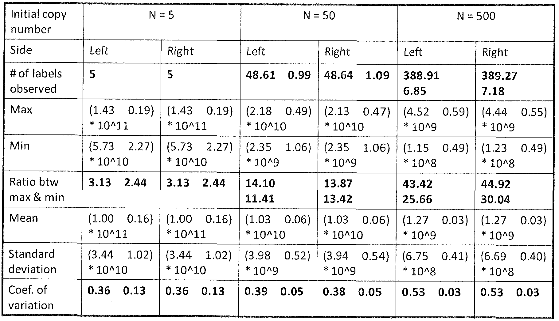
[0245] Focusing on the experiments with concentrations of 0.5 and 0,05ng, (3r and 4 in each group of 5), which provide the most accurate count of label-tags, there are 20 different opportunities
for a given label-tag to be observed (2 concentrations x 5 amplicons x 2 sides (left or right)). We observed 1 ,064 label-tags over the 20 opportunities.
[0246] To observe the distortion of the relative abundance of DNA molecules in the reaction resulting from the PCR process, dispersion in the quantitative distribution of PCR amplified DNA molecules was analyzed. A model of the PCR process was generated to understand the dispersion in the distribution of amplified molecules (FIG. 22, Table 6). A series of 1,000 independent simulation runs were performed to simulate the replication of 500 uniquely labeled target molecules through PCR processes. For each run, the distribution of the final amount of PCR products was determined and the dispersion of distribution was quantified using two measures: ratio of the maximal to the minimal amount, and coefficient of variation of final PCR products. For a library of 960 counters and 500 target molecules the expectation is that about 390 counters will ligate at various redundancies (1 , 2, 3 and 4 are shown). For each of the 390 counters the relative ratio of its abundance to that of the counter with the lowest abundance is plotted (y-axis) for the onset of PCR (panel A), or after 5, 10 or 15 cycles of amplification (panels B-D respectively). The x-axis represents the different labeled molecules. The ratio of the most abundant to the least abundant is 30. This demonstrates that the degree of dispersion increases with the incidence of replicate use of identical counters, which may be in-part responsible for the deviation observed when assaying high target copy numbers. Table 6 lists the ratio and CV for distributions corresponding to different concentrations and replication probabilities (one sided ligation considered).
Table 6

[0248] For the retained 1 ,927 sequences, an all-against-all complement score was calculated for each. This gave a 1 ,927 x 1 ,927 cross complement score matrix. A step similar to step 5 was performed, to avoid label-tags with maximal cross-complement with other label-tags. Starting from the pairs with top cross-complement score, one was randomly pick and the other discarded. This process was repeated until the number of retained sequences was 1920. Next the 1920 label-tags were split into 2 sets, with one set (denoted by set A) consisting of sequences that are maximum different from one-another; and the other set (denoted by set B) consisting of the remaining sequences. The procedure used to split sequences was as follows. Starting from the original 1920 by 1920 similarity score matrix, for each sequence, first, sum up all its similarity scores with the rest of the sequences in the pool, that is, for each sequence, calculate a total similarity score.
Second, sort the total similarity scores of all sequences and select the sequence with the lowest total score, and move it to set A. Third, remove the row and column corresponding to the selected sequence, i.e., both the number of rows and columns in the similarity score matrix are reduced by 1. Repeat steps 1-3, until the number of rows and columns in the similarity score matrix reaches 960 or half of the original. The selected sequences belong to set A and the remaining sequences belong to set B.
[0249] In another embodiment a collection of label-tags is selected using the following steps. Starting with all possible 14 mers (414 or -268 million possible label-tags) eliminate all that do not have 50% GC content. Eliminate those were each nucleotide does not occur at least twice.
Eliminate those that have more than two G/C in tandem or more than three A/T in tandem.
Eliminate those that contain a selected restriction site. That reduces the original set to -33 million or 12.43% of the original set. From that set select those that have a Tm within the range of 38.5°C to 39.5°C. This step results in a set of -7 million or 2.73%) of the original set. Remove those that have regions of self-complementarity. The resulting set in this example was now 521 ,291. A hierarchical clustering was performed to identify a set that has maximum sequence difference between one-another. The resulting set contained 1 ,927 label-tags. Label-tags were removed if the sequence had a tendency to bind to other label-tags in the set. This reduced the set to 1 ,920 label- tags. A final set of 960 label-tags was selected from the 1 ,920 as being maximally different for the "specific" label-tags and 192 additional counters to tile on the array as "non-specific" controls.
[0250] Selection of Targets and design of test array. Regions selected to assay as targets included Chr X, Chr Y. Chr 4 as a reference and Chr 21 for Trisomy. Locations on the chromosomes for assaying were selected to avoid centromeres and telomeres. Fragments were selected based on Bam HI fragments of between about 400 and 600 base pairs. Fragment intensity was checked using HG49 array hybridization. The first and the last 26 nucleotides of the fragments (from and including the Bam HI site) were tiled. Repeats were avoided and GC% was optimized.
[0251] The array format was 100/25. Feature size is Sum. There are 436 x 436 features or 190,096 features. Synthesis was NNPOC, 43 mer probes, 5' up, no phosphate. The chip name is STCL- test2. The gridding probe used was the same as the HG49. No quality control probes were included.
[0252] Aside from reducing whole chromosomes into 360,679 smaller molecular weight DNA pieces more suitable for ligation reactions, restriction digestion also serves to reduce the overall sequence complexity of the sample, as only an estimated 79,517 fragments reside in the 150bp-2kb size range that is effectively amplified by PCR. To detect and quantify counters that have been selected by the target molecules, the labeled genomic target fragments may be circularized and PCR amplified to prepare for analysis, for example, using microarray hybridization or DNA sequencing. A representative BamHI target fragment was sampled for each of the three test chromosomes. Simultaneous measurements of all three chromosomes serve as an internal control independent of
dilution or other systematic errors. A suitable DNA array detector capable of distinguishing the set of counters bound to copies of the target molecules was constructed using photolithography ( S. P. Fodor et ah, Science 251, 767 (Feb 15, 1991). Each array element for a target to be evaluated consists of a target complementary sequence adjacent to one of the complements to the 960 counter sequences. For increased specificity, a ligation-readout was performed on the microarray after hybridization to avoid false positive detection of the cross-hybridization of identical targets with different counters. As a means of validation through a secondary measure, samples hybridized to microarrays were subsequently sampled by DNA sequencing (FIGS. 10 and 25).
[0253] An equation to model the stochastic labeling of target molecules with a small library of 960 counters, and validate our equation model with numerical simulations is disclosed. In the model, the diversity of counters selected by, and ligated to target molecules in the reaction solution (simplified as 'used') is dictated by the number of copies of molecules present for each target fragment in the reaction (FIG. 12). Under conditions where the size of the counter library is much greater than the number of copies of a given target molecule, the counting efficiency is high, and counting the number of counters used is equivalent to counting the number of copies of the original target molecules (FIG. 16). When the number of target copies approaches and exceeds the number of different counters in the reaction, any counter in the library is more likely to be used multiple times (FIG. 15). The number of counters used at least once is an important measure because it serves as the basis for drawing yes/no conclusions in our digital readout on microarrays. Under stochastic labeling conditions, we expect that the absolute quantity of single DNA molecules can be accurately determined by proxy counts of labeling events. Indeed, microarray experiments demonstrate a high degree of correlation between the number of copies of target molecules added to the reaction and the number of counters used, as detected on microarrays (FIG. 12). In particular, counter usage precisely profiles the number of target molecules under conditions of high counting efficiency. Subtle deviations from the model may represent minor dilution errors in the preparation of the sample. However, within that sample dilution, the relative counter ratios of the three internally built-in controls are highly accurate (FIG. 13). FIG. 13 shows comparison of relative copy ratios of the three gene targets tested: ChrX, Chr4 and Chr21 representing genetic material of one, two and three copies per cell. Different dilutions (5ng, 2ng, 0.5ng and 0.05ng) of a DNA sample isolated from cultured lymphoblasts of a Trisomy 21 male individual were processed for microarray hybridization and DN A sequencing. The calculated number of target molecules (see, Table 4,
column 9) was inferred from the number of counters detected on microarrays (A), and was also calculated for the SOLID sequencing data (B). For each sample dilution, the target copy number ratio of each gene target relative to ChrX is shown.
[0254] On the other hand, when target copies exceed -100, detected labeling events appear to indicate fewer than actual molecules in solution (FIG. 12 inset in graph on left). This deviation was reproducible and consistently observed across multiple microarray experiments, and was also observed in the DNA sequencing experiments (FIG. 12 inset in graph on right). Under-counts of expected labeling events may originate from inadequate detection sensitivity of the microarray platform or from other systematic or indeterminate deficiencies in the sample preparation procedure. PGR, for example, is prone to amplification bias ( T. Kanagawa. J Biosci Bioeng 96, 317 (2003) and M. F. Polz, C. M. Cavanaugh, Appl Environ Microbiol 64, 3724 (1998)), which could hinder the comprehensive detection of labeling events that may be genuinely stochastic.
[0255] To confirm the microarray results, a digital sequence counting of individual molecules in the DNA samples hybridized to microarrays was used as a means of validation, and to detect the presence of any false negatives that may have escaped microarray detection. Analysis of mapped sequence reads resulted in counts in agreement to the microarray observations. Furthermore, a second, independent sequencing run was repeated with similar findings (Table 3).
[0256] An additional feature of digital sequence counting is that unlike the microarray intensity data (FIGS. 20 and 21), which may provide lower resolution into the measurement of the concentration dispersion of PGR ampli fied molecules, sequence counting clearly demonstrates significant variation in the representation of amplified targets (FIG. 25), This is consistent with the computed PCR model. Overall, detected counters on the microarray and sequencing experiments correlate well, but a small subset of counters appear to be unique to each process (Table 7). The observed number of label-tags in common between the microarray and the two sets of sequencing
experiments are summarized in the table. The number of label-tags in each category is included. The categories are as follows: A+ l+2 for label-tags detected in each of the 3 experiments, 1+2 for label-tags detected only in sequencing runs 1 and 2, 1+A for label-tags detected in sequencing run 1 and by array, and so on for the amounts of DNA shown in column 3.
Table 7.

[0257] For the reverse scenario, high numbers of mapped sequence reads were always observed to correlate with high microarray intensities in these examples. No systematic or sequence correlations, or explanations were identified for the counters that are absent from any given sequencing experiment for which the microarray readout demonstrates a strong signal. While obviously underrepresented in some experiments, the same counters are sometimes present in high sequence counts in other experiments, suggesting that they are available for sequencing. PGR was used to resolve these isolated cases of disagreement and demonstrate these were false negatives in
the sequencing experiments (Table 3). Despite their presence in the sequencing library, it is unclear why the counters were not observed or were underrepresented in the original sequencing run, and also in the subsequent replicate sequencing run.
[0258] Aside from the comparative analysis of absolute and relative counts of the numbers of target molecules and counter label-tags, additional ways to assess the stochasticity of the labeling process were evaluated. First, if the labeling process is random, the frequency of incorporation of identical counters in independent events across the paired left and right termini of target fragments should closely resemble outcomes from numerical simulation. Observed counts on microarrays do in fact match closely with numbers obtained from computer simulations (Table 4, columns 10-1 1). Second, if the target molecules are labeled randomly with an equal likelihood of incorporation for any member of the 960 counters in the library, we would expect the number of repeated observations of counters to follow a stochastic nature. For this analysis, we accumulated a total of 1 ,064 counter observations over several microarray experiments restricted to low target copy numbers. Exclusion of data from high copy targets was necessary to avoid undercounting labeling events from multiple incidences of identical counters attaching individually to numerous target copies. As a further and final demonstration of stochastic labeling, results show that the frequency of label-tag usage follows a pattern consistent with outcomes from numerical simulation.
[0259] Having now fully described the present invention in some detail by way of illustration and example for purposes of clarity of understanding, it will be obvious to one of ordinary skill in the art that the same can be performed by modifying or changing the invention within a wide and equivalent range of conditions, formulations and other parameters without affecting the scope of the invention or any specific embodiment thereof, and that such modifications or changes are intended to be encompassed within the scope of the appended claims.
[0260] All publications, patents and patent applications mentioned in this specification arc indicative of the level of skill of those skilled in the art to which this invention pertains, and are herein incorporated by reference to the same extent as if each individual publication, patent or patent application was specifically and individually indicated to be incorporated by reference.
Claims
1. A method for counting individual occurrences of at least one species of target molecule in a mixture of nucleic acids having an unknown number of target molecule occurrences, the method comprising:
(a) combining the mixture with a pool of label-tags, wherein the pool of label-tags comprises multiple copies of each of a plurality of different label-tag sequences;
(b) attaching label-tags from the pool of label-tags to individual occurrences of the at least one species of target molecule in a random manner to obtain label-tag-target molecules, wherein different occurrences of a species of target molecule attach to different label-tag sequences from the plurality of different label-tag sequences, thereby obtaining a plurality of unique label-tag-target molecules;
(c) optionally amplifying the unique label-tag-target molecules;
(d) detecting each type of unique label-tag-target molecule; and
(e) counting the number of unique label-tag-target molecules that are detected to obtain a count of the target molecule occurrences of the at least one species of target molecule in the mixture.
2. The method of claim 1 wherein the collection of label-tags comprises a number of different label-tag sequences that is at least twice the number of each target molecule to be estimated.
3. The method of claim 1 wherein the collection of label-tags comprises between 900 and 10,000 different label-tag sequences
4. The method of claim 1 wherein the unique label-tag-target molecules are amplified by PCR in a multiplex amplification reaction so that there are a plurality of copies of each unique label-tag- target molecule that are detected in step (d).
5. The method of claim 1 wherein between 2 and 100 different species of target molecules are counted.
6. The method of claim 1 wherein between 100 and 10,000 different species of target molecules are counted.
7. The method of claim 1 wherein between 10,000 and 50,000 different species of target molecules are counted.
8. The method of claim 1 wherein the mixture of nucleic acids is a genomic DNA sample, the method further comprising:
fragmenting the genomic DNA sample to obtain fragments comprising target molecules prior to step (a),
ligating a first adaptor comprising a common priming sequence to a first end of the fragments;
attaching the label-tags by ligating a second adaptor comprising a label-tag and a second common priming sequence to the second end of the fragments, wherein the second adaptor is randomly selected from a pool of second adaptors that comprises the pool of label-tags, wherein the label-tag is between the target fragment and the second common priming sequence;
thereby obtaining the plurality of unique label-tag-target molecules; and
amplifying the unique label-tag-target molecules by PCR using a primer or a pair of primers to the first and second common priming sequences prior to detecting step (d).
9. The method of claim 1 wherein the number of different label-tags is at least an order of magnitude larger than the expected number of target molecules.
10. The method of claim 1 wherein label-tags are attached to targets by ligation.
1 1. The method of claim 1 wherein the pool of label-tags comprises a pool of label-tag primers that each consist of at least one label-tag and a priming sequence that is complementary to a species of target molecule and wherein the attaching step comprises: (i) hybridizing individual label-tag primers to individual occurrences of the target and (ii) extending the hybridized primers using the target as template to obtain unique primer extension products each comprising a different label-tag and each corresponding to a single occurrence of the target molecule.
12. The method of claim wherein 1 1 wherein the target is RNA and the extending step is by reverse transcriptase.
13. The method of claim wherein 1 1 wherein the target is DNA and the extending step is by a DNA dependent polymerase.
14. The method of claim 1 wherein the ratio of target molecule occurrences to number of different label-tags is less than 1 : 10.
15. The method of claim 1 wherein the ratio of target molecule occurrences to number of different label-tags is less than 1 : 100.
16. The method of claim 1 wherein the ratio of target molecule occurrences to number of different label-tags is less than 1 : 1000.
17. The method of claim 1 wherein the ratio of target molecule occurrences to number of different label-tags is less than 1 : 10,000.
18. The method of claim 1 wherein the step of detecting each type of unique label-tag-target molecule is by hybridizing at least a portion of the label-tag-target molecules to an array of probes comprising a plurality of features wherein each feature contains a different probe sequence and the probe sequence in each feature is specific for a different label-target combination, wherein the array comprises a plurality of features that are complementary to a predicted junction between the label-tag and a region of the target and wherein features that are hybridized to label-tag-target molecules are detectably labeled.
19. The method of claim 18 wherein each feature is a different bead.
20. The method of claim 19 wherein the step of counting comprises counting the number of beads that are detectably labeled.
21. The method of claim 19 wherein the beads are present in the array at random locations and the sequence of the probes on each bead is unknown.
22. The method of claim 1 wherein the step of detecting each type of unique label-tag-target molecule is by sequencing at least a portion of the label-tag-target molecules, wherein enough of the label-tag is sequences to unambiguously identify the label-tag and enough of the target is sequenced to unambiguously identify the target species.
23. The method of claim 22 wherein sequencing is by a method selected from sequencing by synthesis, sequencing by ligation, pyrosequencing, and sequencing by hybridization.
24. The method of claim 22 wherein sequencing is by a method that sequences single molecules as they pass through a nanopore.
25. The method of claim 1 wherein the mixture of nucleic acids is the total mRNA from a single cell.
26. The method of claim 1 wherein the mixture of nucleic acids is the nucleic acid from 10 to 1 ,000 cells, the nucleic acid from 10 to 1,000 cells or the RNA from a 10 to 1 ,000 cells.
27. The method of claim 1 wherein the step of amplifying comprises a method selected from PCR, reverse transcription, in vitro transcription, rolling circle amplification, and isothermal strand displacing amplification.
28. An array comprising a plurality of feature sets, including a feature set for each of a plurality of different target species, wherein each feature set comprises a plurality of different features each feature comprising a plurality of oligonucleotide probes of the same sequence, and wherein the features of a feature set have a common target specific sequence and a variable label-tag sequence, wherein each different feature in the set has the same target specific sequence and a different label- tag specific sequence and wherein each feature set has a feature for each different label-tag in a set of label-tags.
29. The array of claim 28 wherein the array has a first feature set for a first target species, a second feature set for a second target species and a third feature set for a third target species and wherein each feature set comprises at least one feature for each label-tag in a pool of label-tags.
30. The array of claim 28 wherein the array has between 3 and 100 different feature sets, each feature set being specific for a single different target species and each feature set comprising at least one feature for each label-tag in a pool of label-tags.
31 . The array of claim 28 wherein the array has between 100 and 10,000 different feature sets, each feature set being specific for a single different target species and each feature set comprising at least one feature for each label-tag in a pool of label-tags.
32. The array of claim 30 wherein the label-tag set has a diversity of label-tags comprising 1 ,000 to 5,000 different label-tags.
33. The array of claim 30 wherein the label-tag set has a diversity of label-tags comprising 5,000 to 100,000 different label-tags.
34. The array of claim 30 wherein the label-tag set has a diversity of label-tags comprising 100,000 to 1,000,000 different label-tags.
35. A method of determining a number of target polynucleotides, the method comprising the steps of:
providing for each target polynucleotide a plurality of nucleic acid probes specific for the target polynucleotide, each nucleic acid probe having a different oligonucleotide tag;
combining in a reaction mixture the plurality of nucleic acid probes with the target polynucleotides so that substantially every target polynucleotide associates with a nucleic acid probe to form a probe-target complex, the plurality of nucleic acid probes having a number sufficiently greater than the number of target polynucleotides so that substantially every probe- target complex has a unique oligonucleotide tag; treating the sample to enrich the probe-target complexes; and
determining the number of different oligonucleotide tags in a sample of enriched probe- target complexes by hybridization thereby determining the number of target polynucleotide in the mixture.
36. The method of claim 35 wherein the target polynucleotide is a restriction fragment having a first single strand sequence overhang and a second, different single strand overhang, and wherein a first adaptor has a single stranded overhang that is perfectly complementary to the first overhang and a second different adaptor has a single stranded overhang that is perfectly complementary to the second overhang.
37. The method of claim 36 wherein the restriction fragment is formed by digesting genomic DNA with at least one restriction endonuclease.
38. A method for determining a value for n for a plurality of different target species, wherein n is the number of occurrences of a first species of target molecule that are present in a sample, the method comprising:
attaching to each occurrence of the first species of target molecule, a label-tag from a set of diverse label-tags, wherein the set of diverse label-tags comprises m different label-tags and label- tags are chosen at random, thereby generating for each occurrence of the first species of target molecule, a labeled target molecule that comprises a single occurrence of the first species of target molecule and a label-tag from the set of diverse label-tags, wherein at least 90% of the labeled target molecules have a label-tag that is different from the label-tags on all of the other labeled target molecules of the first species of target molecule;
detecting each labeled target molecule by determining at least a partial sequence of the labeled target molecule, wherein at least X bases of the target molecule and at least Y bases of the label-tag are determined; and
counting the number of new molecules, thereby counting the number of molecules of the first target molecule in the sample.
39. The method of claim 38 wherein the ratio of m to n is greater than 50.
40. The method of claim 38 wherein the ratio of m to n is greater than 100.
41. The method of claim 38 wherein the ratio of m to n is greater than 500.
42. The method of claim 38 wherein the ratio of m to n is greater than 1000.
43. The method of claim 38 wherein the target molecules are proteins or peptides.
44. The method of claim 38 wherein target molecules are nucleic acids and the detecting step comprises sequencing.
45. The method of claim 44 wherein sequencing comprises sequencing by extension or sequencing by ligation.
46. The method of claim 44 wherein sequencing comprises sequencing using reversible terminator chemistry.
47. The method of claim 38 wherein the sample consists of a single cell, the nucleic acid from a single cell, the RNA from a single cell or a crude cell extract from a single cell.
48. The method of claim 38 wherein the sample consists of 10 to 1 ,000 cells, the nucleic acid from 10 to 1,000 cells or the RNA from a 10 to 1,000 cells.
49. The method of claim 38 wherein individual diverse label-tags from the set of diverse label- tags are attached to an occurrence of the first species of target molecules by extension of a primer that comprises the diverse label-tag using template dependent primer extension by an enzyme selected from: DNA dependent RNA polymerase, a DNA dependent DNA polymerase and an RNA dependent RNA polymerase.
50. The method of claim 49 wherein the DNA dependent RNA polymerase is selected from T7 RNA polymerase and SP6 RNA polymerase.
51. A method for counting the number of occurrences of each of a plurality of different target molecules in a sample, the method comprising the steps of:
(a) providing a plurality of conjugates each comprising a label-tag selected from a pool of label-tags, and a target molecule, wherein the label-tag is adjacent to a known target sequence in the conjugate so that a label-tag-target junction is formed and wherein more than 90% of the target molecule are attached to a label-tag, the pool of oligonucleotide label-tags comprising a plurality of different label-tag sequences and wherein the probability that any two target molecules is labeled with the same label-tag is less than a selected threshold value;
(b) amplifying the conjugates in a multiplex amplification reaction to obtain amplified conjugates comprising label-tags;
(c) detecting each different amplified conjugate by detecting different label-tags;
(d) counting the number of different lable-tags and conjugates detected in step (c); and, (e) determining the number of target molecules in the sample from the number of different label-tags and conjugates counted in step (d).
52. The method of claim 51 wherein:
(i) the pool of label-tags has m different label-tags;
(ii) the first target species has n occurrences; and
(iii) following the covalent attaching step there are k first targets labeled with a label-tag that is unique in the set of labeled first targets; and
wherein the step of counting comprises empirically determining k and calculating n using k = m(l - e "('?/w) ).
53. The method of claim 52 wherein the number of different label-tags in the pool of label-tags is n and the number of occurrences of the first target is m and wherein n/m is less than 0.10.
54. The method of claim 53 wherein n/m is less than 0.01.
55. The method of claim 53 wherein n/m is less than 0.005.
56. The method of claim 53 wherein n/m is less than 0.001.
57. The method of claim 51 wherein label-tags are selected at random in the step of ligating a label- tag to individual occurrences of first and second target nucleic acid species.
58. The method of claim 51 wherein the label-tags in the pool of label-tags are DNA, the first and second target species are DNA and the ligation comprises ligation with a ligase selected from T4 DNA ligase and E. coli DNA ligase.
59. The method of claim 51 wherein detecting step is by hybridization to an array of probes that are complementary to the label-tag-target junctions.
60. The method of claim 51 wherein the step of detecting and counting comprises:
(a) hybridizing the plurality of amplified labeled first target species and the plurality of amplified labeled second target species to an array of probes comprising a first plurality of probe features each feature complementary to a different first target-label-tag junction and a second plurality of probe features each feature complementary to different second target-label-tag junctions;
(b) detecting hybridized features; and
(c) counting the hybridized features.
61. The method of claim 60 wherein the array of probes comprises a glass support having probes complementary to the junctions.
62. The method of claim 60 wherein the array of probes comprises a plurality of beads having probes complementary to the junctions.
63. The method of claim 60 wherein the array of probes comprises a plurality of microspheres.
64. The method of claim 60 wherein the array of probes comprises a plurality of encoded
microparticles where the microparticles for the first target are encoded with a first code and the microparticles for the second target are encoded with a second code.
65. The method of claim 51 wherein the step of detecting and counting comprises:
sequencing amplified labeled first target species and amplified labeled second target species, wherein at least a portion of the label-tag and at least a portion of the target specific region is sequenced, wherein the number of different sequences obtained for each target is used to determine the count of each target.
66. The method of claim 65 wherein the targets to be counted are the transcriptome of a single cell or a collection of cells of a single type.
67. The method of claim 65 wherein the targets to be counted are the number of chromosomes of a single type in a genome or the number of copies of an allele of a gene in a genome.
68. A kit comprising a pool of label-tags wherein the diversity of the pool of label-tags is selected from between 100 and 1 ,000, between 1 ,000 and 10,000, between 10,000 and 100,000, between 100,000 and 1 ,000,000 and greater than 1 ,000,000 different types of label-tags.
69. The kit of claim 66 wherein the label-tags in the pool of label-tags comprise a variable region that is the same length in all label-tags in the pool and the length is between 6 and 14 bases.
70. The kit of claim 69 wherein the label-tags in the pool of label-tags further comprise a
recognition site for a restriction enzyme, a common priming site or an RNA polymerase promoter sequence.
71 . The kit of claim 69 wherein the label-tags in the pool of label-tags are double stranded adaptors comprising a common priming sequence that is present in all label-tags in a pool of lable-tags.
72. The kit of claim 66 further comprising an oligo d' primer, one or more sequencing primers, and a reverse transcriptase.
Applications Claiming Priority (2)
| Application Number | Priority Date | Filing Date | Title |
|---|---|---|---|
| US12/969,581 | 2010-12-15 | ||
| US12/969,581 US8835358B2 (en) | 2009-12-15 | 2010-12-15 | Digital counting of individual molecules by stochastic attachment of diverse labels |
Publications (1)
| Publication Number | Publication Date |
|---|---|
| WO2012148477A1 true WO2012148477A1 (en) | 2012-11-01 |
Family
ID=44188253
Family Applications (1)
| Application Number | Title | Priority Date | Filing Date |
|---|---|---|---|
| PCT/US2011/065291 WO2012148477A1 (en) | 2010-12-15 | 2011-12-15 | Digital counting of individual molecules by stochastic attachment of diverse label-tags |
Country Status (2)
| Country | Link |
|---|---|
| US (21) | US8835358B2 (en) |
| WO (1) | WO2012148477A1 (en) |
Cited By (70)
| Publication number | Priority date | Publication date | Assignee | Title |
|---|---|---|---|---|
| WO2013123442A1 (en) * | 2012-02-17 | 2013-08-22 | Fred Hutchinson Cancer Research Center | Compositions and methods for accurately identifying mutations |
| US8685678B2 (en) | 2010-09-21 | 2014-04-01 | Population Genetics Technologies Ltd | Increasing confidence of allele calls with molecular counting |
| WO2014108850A2 (en) | 2013-01-09 | 2014-07-17 | Yeda Research And Development Co. Ltd. | High throughput transcriptome analysis |
| US8835358B2 (en) | 2009-12-15 | 2014-09-16 | Cellular Research, Inc. | Digital counting of individual molecules by stochastic attachment of diverse labels |
| US9315857B2 (en) | 2009-12-15 | 2016-04-19 | Cellular Research, Inc. | Digital counting of individual molecules by stochastic attachment of diverse label-tags |
| CN105531375A (en) * | 2012-12-10 | 2016-04-27 | 分析生物科学有限公司 | Methods for targeted genomic analysis |
| US9567645B2 (en) | 2013-08-28 | 2017-02-14 | Cellular Research, Inc. | Massively parallel single cell analysis |
| US9582877B2 (en) | 2013-10-07 | 2017-02-28 | Cellular Research, Inc. | Methods and systems for digitally counting features on arrays |
| US9670529B2 (en) | 2012-02-28 | 2017-06-06 | Population Genetics Technologies Ltd. | Method for attaching a counter sequence to a nucleic acid sample |
| EP2855707B1 (en) | 2012-05-31 | 2017-07-12 | Board Of Regents, The University Of Texas System | Method for accurate sequencing of dna |
| US9727810B2 (en) | 2015-02-27 | 2017-08-08 | Cellular Research, Inc. | Spatially addressable molecular barcoding |
| US9783847B2 (en) | 2009-04-30 | 2017-10-10 | Prognosys Biosciences, Inc. | Nucleic acid constructs and methods of use |
| EP3299472A1 (en) * | 2016-09-27 | 2018-03-28 | Deutsches Krebsforschungszentrum Stiftung des Öffentlichen Rechts | Method for labeling oligonucleotide probes |
| WO2018188856A1 (en) * | 2017-04-13 | 2018-10-18 | Deutsches Krebsforschungszentrum Stiftung des öffentlichen Rechts | Labeling of oligonucleotide probes by multiple-way ligation |
| US10202641B2 (en) | 2016-05-31 | 2019-02-12 | Cellular Research, Inc. | Error correction in amplification of samples |
| EP3460071A1 (en) * | 2017-09-22 | 2019-03-27 | Lexogen GmbH | Estimating pre-pcr fragment numbers from post-pcr frequencies of unique molecular identifiers |
| US10301677B2 (en) | 2016-05-25 | 2019-05-28 | Cellular Research, Inc. | Normalization of nucleic acid libraries |
| US10308982B2 (en) | 2010-04-05 | 2019-06-04 | Prognosys Biosciences, Inc. | Spatially encoded biological assays |
| US10338066B2 (en) | 2016-09-26 | 2019-07-02 | Cellular Research, Inc. | Measurement of protein expression using reagents with barcoded oligonucleotide sequences |
| EP3470533B1 (en) | 2012-09-04 | 2019-11-06 | Guardant Health, Inc. | Systems and methods to detect copy number variation |
| EP3433017A4 (en) * | 2016-03-25 | 2020-01-01 | Bioceryx Inc. | Apparatuses and methods for assessing target sequence numbers |
| EP3378952B1 (en) | 2013-12-28 | 2020-02-05 | Guardant Health, Inc. | Methods and systems for detecting genetic variants |
| WO2020061524A1 (en) * | 2018-09-20 | 2020-03-26 | 13.8, Inc. | METHODS OF MULTIPLEX dPCR ASSAYS AND SHORT-READ SEQUENCING ASSAYS |
| US10619186B2 (en) | 2015-09-11 | 2020-04-14 | Cellular Research, Inc. | Methods and compositions for library normalization |
| US10640763B2 (en) | 2016-05-31 | 2020-05-05 | Cellular Research, Inc. | Molecular indexing of internal sequences |
| US10669570B2 (en) | 2017-06-05 | 2020-06-02 | Becton, Dickinson And Company | Sample indexing for single cells |
| US10697010B2 (en) | 2015-02-19 | 2020-06-30 | Becton, Dickinson And Company | High-throughput single-cell analysis combining proteomic and genomic information |
| US10722880B2 (en) | 2017-01-13 | 2020-07-28 | Cellular Research, Inc. | Hydrophilic coating of fluidic channels |
| US10774374B2 (en) | 2015-04-10 | 2020-09-15 | Spatial Transcriptomics AB and Illumina, Inc. | Spatially distinguished, multiplex nucleic acid analysis of biological specimens |
| US10774372B2 (en) | 2013-06-25 | 2020-09-15 | Prognosy s Biosciences, Inc. | Methods and systems for determining spatial patterns of biological targets in a sample |
| US10787701B2 (en) | 2010-04-05 | 2020-09-29 | Prognosys Biosciences, Inc. | Spatially encoded biological assays |
| WO2020214642A1 (en) * | 2019-04-19 | 2020-10-22 | Becton, Dickinson And Company | Methods of associating phenotypical data and single cell sequencing data |
| US10822643B2 (en) | 2016-05-02 | 2020-11-03 | Cellular Research, Inc. | Accurate molecular barcoding |
| US10941396B2 (en) | 2012-02-27 | 2021-03-09 | Becton, Dickinson And Company | Compositions and kits for molecular counting |
| EP2971168B1 (en) | 2013-03-15 | 2021-05-05 | Guardant Health, Inc. | Method of detecting cancer |
| US11124823B2 (en) | 2015-06-01 | 2021-09-21 | Becton, Dickinson And Company | Methods for RNA quantification |
| US11164659B2 (en) | 2016-11-08 | 2021-11-02 | Becton, Dickinson And Company | Methods for expression profile classification |
| US11168347B2 (en) | 2016-09-23 | 2021-11-09 | California Institute Of Technology | Digital quantification of DNA replication and/or chromosome segregation based determination of antimicrobial susceptibility |
| US11177020B2 (en) | 2012-02-27 | 2021-11-16 | The University Of North Carolina At Chapel Hill | Methods and uses for molecular tags |
| US11319594B2 (en) | 2016-08-25 | 2022-05-03 | Resolution Bioscience, Inc. | Methods for the detection of genomic copy changes in DNA samples |
| US11319583B2 (en) | 2017-02-01 | 2022-05-03 | Becton, Dickinson And Company | Selective amplification using blocking oligonucleotides |
| US11339391B2 (en) | 2015-11-11 | 2022-05-24 | Resolution Bioscience, Inc. | High efficiency construction of DNA libraries |
| US11352659B2 (en) | 2011-04-13 | 2022-06-07 | Spatial Transcriptomics Ab | Methods of detecting analytes |
| US11365409B2 (en) | 2018-05-03 | 2022-06-21 | Becton, Dickinson And Company | Molecular barcoding on opposite transcript ends |
| US11371076B2 (en) | 2019-01-16 | 2022-06-28 | Becton, Dickinson And Company | Polymerase chain reaction normalization through primer titration |
| US11390914B2 (en) | 2015-04-23 | 2022-07-19 | Becton, Dickinson And Company | Methods and compositions for whole transcriptome amplification |
| US11397882B2 (en) | 2016-05-26 | 2022-07-26 | Becton, Dickinson And Company | Molecular label counting adjustment methods |
| US11434523B2 (en) | 2012-09-04 | 2022-09-06 | Guardant Health, Inc. | Systems and methods to detect rare mutations and copy number variation |
| US11492660B2 (en) | 2018-12-13 | 2022-11-08 | Becton, Dickinson And Company | Selective extension in single cell whole transcriptome analysis |
| US11535882B2 (en) | 2015-03-30 | 2022-12-27 | Becton, Dickinson And Company | Methods and compositions for combinatorial barcoding |
| US11584958B2 (en) | 2017-03-31 | 2023-02-21 | Grail, Llc | Library preparation and use thereof for sequencing based error correction and/or variant identification |
| US11608497B2 (en) | 2016-11-08 | 2023-03-21 | Becton, Dickinson And Company | Methods for cell label classification |
| US11639517B2 (en) | 2018-10-01 | 2023-05-02 | Becton, Dickinson And Company | Determining 5′ transcript sequences |
| US11649497B2 (en) | 2020-01-13 | 2023-05-16 | Becton, Dickinson And Company | Methods and compositions for quantitation of proteins and RNA |
| US11649485B2 (en) | 2019-01-06 | 2023-05-16 | 10X Genomics, Inc. | Generating capture probes for spatial analysis |
| US11661631B2 (en) | 2019-01-23 | 2023-05-30 | Becton, Dickinson And Company | Oligonucleotides associated with antibodies |
| US11661625B2 (en) | 2020-05-14 | 2023-05-30 | Becton, Dickinson And Company | Primers for immune repertoire profiling |
| US11733238B2 (en) | 2010-04-05 | 2023-08-22 | Prognosys Biosciences, Inc. | Spatially encoded biological assays |
| US11739443B2 (en) | 2020-11-20 | 2023-08-29 | Becton, Dickinson And Company | Profiling of highly expressed and lowly expressed proteins |
| US11773441B2 (en) | 2018-05-03 | 2023-10-03 | Becton, Dickinson And Company | High throughput multiomics sample analysis |
| US11773436B2 (en) | 2019-11-08 | 2023-10-03 | Becton, Dickinson And Company | Using random priming to obtain full-length V(D)J information for immune repertoire sequencing |
| US11827944B2 (en) | 2017-10-11 | 2023-11-28 | California Institute Of Technology | Antibiotic susceptibility of microorganisms and related compositions, methods and systems |
| US11913065B2 (en) | 2012-09-04 | 2024-02-27 | Guardent Health, Inc. | Systems and methods to detect rare mutations and copy number variation |
| US11926867B2 (en) | 2019-01-06 | 2024-03-12 | 10X Genomics, Inc. | Generating capture probes for spatial analysis |
| US11933957B1 (en) | 2018-12-10 | 2024-03-19 | 10X Genomics, Inc. | Imaging system hardware |
| US11932901B2 (en) | 2020-07-13 | 2024-03-19 | Becton, Dickinson And Company | Target enrichment using nucleic acid probes for scRNAseq |
| US11932849B2 (en) | 2018-11-08 | 2024-03-19 | Becton, Dickinson And Company | Whole transcriptome analysis of single cells using random priming |
| US11939622B2 (en) | 2019-07-22 | 2024-03-26 | Becton, Dickinson And Company | Single cell chromatin immunoprecipitation sequencing assay |
| US11946095B2 (en) | 2017-12-19 | 2024-04-02 | Becton, Dickinson And Company | Particles associated with oligonucleotides |
| US11959139B2 (en) | 2023-05-12 | 2024-04-16 | Guardant Health, Inc. | Methods and systems for detecting genetic variants |
Families Citing this family (362)
| Publication number | Priority date | Publication date | Assignee | Title |
|---|---|---|---|---|
| US7311794B2 (en) | 2004-05-28 | 2007-12-25 | Wafergen, Inc. | Methods of sealing micro wells |
| CA2599683A1 (en) | 2005-03-04 | 2006-09-14 | President And Fellows Of Harvard College | Method and apparatus for forming multiple emulsions |
| US11111543B2 (en) | 2005-07-29 | 2021-09-07 | Natera, Inc. | System and method for cleaning noisy genetic data and determining chromosome copy number |
| US9424392B2 (en) | 2005-11-26 | 2016-08-23 | Natera, Inc. | System and method for cleaning noisy genetic data from target individuals using genetic data from genetically related individuals |
| US10083273B2 (en) | 2005-07-29 | 2018-09-25 | Natera, Inc. | System and method for cleaning noisy genetic data and determining chromosome copy number |
| US11111544B2 (en) | 2005-07-29 | 2021-09-07 | Natera, Inc. | System and method for cleaning noisy genetic data and determining chromosome copy number |
| US10081839B2 (en) | 2005-07-29 | 2018-09-25 | Natera, Inc | System and method for cleaning noisy genetic data and determining chromosome copy number |
| US8053191B2 (en) | 2006-08-31 | 2011-11-08 | Westend Asset Clearinghouse Company, Llc | Iterative nucleic acid assembly using activation of vector-encoded traits |
| WO2008109176A2 (en) | 2007-03-07 | 2008-09-12 | President And Fellows Of Harvard College | Assays and other reactions involving droplets |
| JP5738597B2 (en) | 2007-12-21 | 2015-06-24 | プレジデント アンド フェローズ オブ ハーバード カレッジ | Systems and methods for nucleic acid sequencing |
| WO2010033200A2 (en) | 2008-09-19 | 2010-03-25 | President And Fellows Of Harvard College | Creation of libraries of droplets and related species |
| US8951939B2 (en) | 2011-07-12 | 2015-02-10 | Bio-Rad Laboratories, Inc. | Digital assays with multiplexed detection of two or more targets in the same optical channel |
| US9417190B2 (en) | 2008-09-23 | 2016-08-16 | Bio-Rad Laboratories, Inc. | Calibrations and controls for droplet-based assays |
| US9764322B2 (en) | 2008-09-23 | 2017-09-19 | Bio-Rad Laboratories, Inc. | System for generating droplets with pressure monitoring |
| US20130059754A1 (en) * | 2011-09-01 | 2013-03-07 | Bio-Rad Laboratories, Inc. | Digital assays with reduced measurement uncertainty |
| US11130128B2 (en) | 2008-09-23 | 2021-09-28 | Bio-Rad Laboratories, Inc. | Detection method for a target nucleic acid |
| US8633015B2 (en) | 2008-09-23 | 2014-01-21 | Bio-Rad Laboratories, Inc. | Flow-based thermocycling system with thermoelectric cooler |
| WO2011120024A1 (en) | 2010-03-25 | 2011-09-29 | Quantalife, Inc. | Droplet generation for droplet-based assays |
| US9132394B2 (en) | 2008-09-23 | 2015-09-15 | Bio-Rad Laboratories, Inc. | System for detection of spaced droplets |
| US9492797B2 (en) | 2008-09-23 | 2016-11-15 | Bio-Rad Laboratories, Inc. | System for detection of spaced droplets |
| US9156010B2 (en) | 2008-09-23 | 2015-10-13 | Bio-Rad Laboratories, Inc. | Droplet-based assay system |
| US8709762B2 (en) | 2010-03-02 | 2014-04-29 | Bio-Rad Laboratories, Inc. | System for hot-start amplification via a multiple emulsion |
| US10512910B2 (en) | 2008-09-23 | 2019-12-24 | Bio-Rad Laboratories, Inc. | Droplet-based analysis method |
| US9506119B2 (en) | 2008-11-07 | 2016-11-29 | Adaptive Biotechnologies Corp. | Method of sequence determination using sequence tags |
| US9394567B2 (en) | 2008-11-07 | 2016-07-19 | Adaptive Biotechnologies Corporation | Detection and quantification of sample contamination in immune repertoire analysis |
| US8628927B2 (en) | 2008-11-07 | 2014-01-14 | Sequenta, Inc. | Monitoring health and disease status using clonotype profiles |
| SG195652A1 (en) | 2008-11-07 | 2013-12-30 | Sequenta Inc | Methods of monitoring conditions by sequence analysis |
| US9365901B2 (en) | 2008-11-07 | 2016-06-14 | Adaptive Biotechnologies Corp. | Monitoring immunoglobulin heavy chain evolution in B-cell acute lymphoblastic leukemia |
| US8748103B2 (en) | 2008-11-07 | 2014-06-10 | Sequenta, Inc. | Monitoring health and disease status using clonotype profiles |
| US9528160B2 (en) | 2008-11-07 | 2016-12-27 | Adaptive Biotechnolgies Corp. | Rare clonotypes and uses thereof |
| EP2373812B1 (en) | 2008-12-19 | 2016-11-09 | President and Fellows of Harvard College | Particle-assisted nucleic acid sequencing |
| DK3059337T3 (en) | 2009-01-15 | 2019-07-22 | Adaptive Biotechnologies Corp | Adaptive immunity profiling and methods for producing monoclonal antibodies |
| JP5909095B2 (en) | 2009-03-13 | 2016-04-26 | プレジデント アンド フェローズ オブ ハーバード カレッジ | Scale up microfluidic devices |
| JP2012531202A (en) | 2009-06-25 | 2012-12-10 | フレッド ハチンソン キャンサー リサーチ センター | How to measure adaptive immunity |
| US8298767B2 (en) | 2009-08-20 | 2012-10-30 | Population Genetics Technologies Ltd | Compositions and methods for intramolecular nucleic acid rearrangement |
| JP6155418B2 (en) | 2009-09-02 | 2017-07-05 | バイオ−ラッド・ラボラトリーズ・インコーポレーテッド | System for mixing fluids by combining multiple emulsions |
| BR112012004719A2 (en) | 2009-09-02 | 2016-04-05 | Harvard College | multiple emulsions created by blasting and other techniques |
| ES2640776T3 (en) | 2009-09-30 | 2017-11-06 | Natera, Inc. | Methods for non-invasively calling prenatal ploidy |
| CN102648053B (en) | 2009-10-27 | 2016-04-27 | 哈佛学院院长等 | Drop formation technology |
| US8399198B2 (en) | 2010-03-02 | 2013-03-19 | Bio-Rad Laboratories, Inc. | Assays with droplets transformed into capsules |
| JP2013525087A (en) * | 2010-03-17 | 2013-06-20 | プレジデント アンド フェロウズ オブ ハーバード カレッジ | Melt emulsification |
| CA2767113A1 (en) | 2010-03-25 | 2011-09-29 | Bio-Rad Laboratories, Inc. | Detection system for droplet-based assays |
| EP2556170A4 (en) | 2010-03-25 | 2014-01-01 | Quantalife Inc | Droplet transport system for detection |
| US11339429B2 (en) | 2010-05-18 | 2022-05-24 | Natera, Inc. | Methods for non-invasive prenatal ploidy calling |
| US11322224B2 (en) | 2010-05-18 | 2022-05-03 | Natera, Inc. | Methods for non-invasive prenatal ploidy calling |
| US11939634B2 (en) | 2010-05-18 | 2024-03-26 | Natera, Inc. | Methods for simultaneous amplification of target loci |
| US10316362B2 (en) | 2010-05-18 | 2019-06-11 | Natera, Inc. | Methods for simultaneous amplification of target loci |
| US11326208B2 (en) | 2010-05-18 | 2022-05-10 | Natera, Inc. | Methods for nested PCR amplification of cell-free DNA |
| US11408031B2 (en) | 2010-05-18 | 2022-08-09 | Natera, Inc. | Methods for non-invasive prenatal paternity testing |
| US11332785B2 (en) | 2010-05-18 | 2022-05-17 | Natera, Inc. | Methods for non-invasive prenatal ploidy calling |
| CA2798758C (en) | 2010-05-18 | 2019-05-07 | Natera, Inc. | Methods for non-invasive prenatal ploidy calling |
| US9677118B2 (en) | 2014-04-21 | 2017-06-13 | Natera, Inc. | Methods for simultaneous amplification of target loci |
| US11332793B2 (en) | 2010-05-18 | 2022-05-17 | Natera, Inc. | Methods for simultaneous amplification of target loci |
| US20190010543A1 (en) | 2010-05-18 | 2019-01-10 | Natera, Inc. | Methods for simultaneous amplification of target loci |
| WO2012042374A2 (en) * | 2010-10-01 | 2012-04-05 | Anssi Jussi Nikolai Taipale | Method of determining number or concentration of molecules |
| EP3574990B1 (en) | 2010-11-01 | 2022-04-06 | Bio-Rad Laboratories, Inc. | System for forming emulsions |
| JP6118725B2 (en) | 2010-11-12 | 2017-04-19 | ジェン9・インコーポレイテッドGen9,INC. | Methods and devices for nucleic acid synthesis |
| US10457935B2 (en) | 2010-11-12 | 2019-10-29 | Gen9, Inc. | Protein arrays and methods of using and making the same |
| AU2011348100B2 (en) | 2010-12-22 | 2016-08-25 | Natera, Inc. | Methods for non-invasive prenatal paternity testing |
| ES2687761T3 (en) | 2011-01-31 | 2018-10-29 | F. Hoffmann-La Roche Ag | Methods of identification of multiple epitopes in cells |
| CA2824387C (en) | 2011-02-09 | 2019-09-24 | Natera, Inc. | Methods for non-invasive prenatal ploidy calling |
| AU2012231098B2 (en) | 2011-03-18 | 2016-09-29 | Bio-Rad Laboratories, Inc. | Multiplexed digital assays with combinatorial use of signals |
| US8759036B2 (en) | 2011-03-21 | 2014-06-24 | Affymetrix, Inc. | Methods for synthesizing pools of probes |
| US9260753B2 (en) | 2011-03-24 | 2016-02-16 | President And Fellows Of Harvard College | Single cell nucleic acid detection and analysis |
| CA2834291A1 (en) | 2011-04-25 | 2012-11-01 | Biorad Laboratories, Inc. | Methods and compositions for nucleic acid analysis |
| KR102112205B1 (en) | 2011-04-28 | 2020-06-12 | 더 보드 어브 트러스티스 어브 더 리랜드 스탠포드 주니어 유니버시티 | Identification of polynucleotides associated with a sample |
| JP6122843B2 (en) | 2011-05-23 | 2017-04-26 | プレジデント アンド フェローズ オブ ハーバード カレッジ | Emulsion control including multiple emulsions |
| US9752176B2 (en) * | 2011-06-15 | 2017-09-05 | Ginkgo Bioworks, Inc. | Methods for preparative in vitro cloning |
| CN103764265A (en) | 2011-07-06 | 2014-04-30 | 哈佛学院院长等 | Multiple emulsions and techniques for the formation of multiple emulsions |
| WO2013019751A1 (en) | 2011-07-29 | 2013-02-07 | Bio-Rad Laboratories, Inc., | Library characterization by digital assay |
| US10466160B2 (en) | 2011-08-01 | 2019-11-05 | Celsee Diagnostics, Inc. | System and method for retrieving and analyzing particles |
| US9103754B2 (en) | 2011-08-01 | 2015-08-11 | Denovo Sciences, Inc. | Cell capture system and method of use |
| US9404864B2 (en) | 2013-03-13 | 2016-08-02 | Denovo Sciences, Inc. | System for imaging captured cells |
| JP2014526899A (en) | 2011-08-26 | 2014-10-09 | ジェン9・インコーポレイテッド | Compositions and methods for high fidelity assembly of nucleic acids |
| US10704164B2 (en) | 2011-08-31 | 2020-07-07 | Life Technologies Corporation | Methods, systems, computer readable media, and kits for sample identification |
| US10385475B2 (en) | 2011-09-12 | 2019-08-20 | Adaptive Biotechnologies Corp. | Random array sequencing of low-complexity libraries |
| CA2853088C (en) | 2011-10-21 | 2018-03-13 | Adaptive Biotechnologies Corporation | Quantification of adaptive immune cell genomes in a complex mixture of cells |
| CN102534813B (en) * | 2011-11-15 | 2013-09-04 | 杭州联川生物技术有限公司 | Method for constructing sequencing library of middle-small-segment RNA (Ribonucleic Acid) |
| ES2867924T3 (en) | 2011-12-09 | 2021-10-21 | Adaptive Biotechnologies Corp | Diagnosis of Lymphoid Malignant Tumors and Detection of Minimal Residual Disease |
| WO2013090404A2 (en) * | 2011-12-13 | 2013-06-20 | Single Cell Technology, Inc. | Method of screening a plurality of single secreting cells for functional activity |
| US9499865B2 (en) | 2011-12-13 | 2016-11-22 | Adaptive Biotechnologies Corp. | Detection and measurement of tissue-infiltrating lymphocytes |
| EP2823060B1 (en) | 2012-03-05 | 2018-02-14 | Adaptive Biotechnologies Corporation | Determining paired immune receptor chains from frequency matched subunits |
| EP2825675B1 (en) | 2012-03-13 | 2017-12-27 | Patel, Abhijit Ajit | Measurement of nucleic acid variants using highly-multiplexed error-suppressed deep sequencing |
| PL2828218T3 (en) | 2012-03-20 | 2021-01-11 | University Of Washington Through Its Center For Commercialization | Methods of lowering the error rate of massively parallel dna sequencing using duplex consensus sequencing |
| US9150853B2 (en) | 2012-03-21 | 2015-10-06 | Gen9, Inc. | Methods for screening proteins using DNA encoded chemical libraries as templates for enzyme catalysis |
| WO2013155531A2 (en) | 2012-04-13 | 2013-10-17 | Bio-Rad Laboratories, Inc. | Sample holder with a well having a wicking promoter |
| EP4001427A1 (en) | 2012-04-24 | 2022-05-25 | Gen9, Inc. | Methods for sorting nucleic acids and multiplexed preparative in vitro cloning |
| EP2831276B1 (en) | 2012-05-08 | 2016-04-20 | Adaptive Biotechnologies Corporation | Compositions and method for measuring and calibrating amplification bias in multiplexed pcr reactions |
| AU2013280661A1 (en) | 2012-06-25 | 2015-01-22 | Gen9, Inc. | Methods for nucleic acid assembly and high throughput sequencing |
| EP2882868B1 (en) | 2012-08-08 | 2019-07-31 | H. Hoffnabb-La Roche Ag | Increasing dynamic range for identifying multiple epitopes in cells |
| CA2881783A1 (en) | 2012-08-13 | 2014-02-20 | The Regents Of The University Of California | Methods and systems for detecting biological components |
| US10323279B2 (en) | 2012-08-14 | 2019-06-18 | 10X Genomics, Inc. | Methods and systems for processing polynucleotides |
| US9701998B2 (en) | 2012-12-14 | 2017-07-11 | 10X Genomics, Inc. | Methods and systems for processing polynucleotides |
| US10752949B2 (en) | 2012-08-14 | 2020-08-25 | 10X Genomics, Inc. | Methods and systems for processing polynucleotides |
| US10273541B2 (en) | 2012-08-14 | 2019-04-30 | 10X Genomics, Inc. | Methods and systems for processing polynucleotides |
| US9951386B2 (en) | 2014-06-26 | 2018-04-24 | 10X Genomics, Inc. | Methods and systems for processing polynucleotides |
| CN111748607A (en) | 2012-08-14 | 2020-10-09 | 10X基因组学有限公司 | Microcapsule compositions and methods |
| US10400280B2 (en) | 2012-08-14 | 2019-09-03 | 10X Genomics, Inc. | Methods and systems for processing polynucleotides |
| US11591637B2 (en) | 2012-08-14 | 2023-02-28 | 10X Genomics, Inc. | Compositions and methods for sample processing |
| US10221442B2 (en) | 2012-08-14 | 2019-03-05 | 10X Genomics, Inc. | Compositions and methods for sample processing |
| US20160040229A1 (en) | 2013-08-16 | 2016-02-11 | Guardant Health, Inc. | Systems and methods to detect rare mutations and copy number variation |
| US20160002731A1 (en) | 2012-10-01 | 2016-01-07 | Adaptive Biotechnologies Corporation | Immunocompetence assessment by adaptive immune receptor diversity and clonality characterization |
| US10150996B2 (en) | 2012-10-19 | 2018-12-11 | Adaptive Biotechnologies Corp. | Quantification of adaptive immune cell genomes in a complex mixture of cells |
| JP6454281B2 (en) | 2012-11-05 | 2019-01-16 | タカラ バイオ ユーエスエー, インコーポレイテッド | Nucleic acids to be barcoded |
| US10533221B2 (en) | 2012-12-14 | 2020-01-14 | 10X Genomics, Inc. | Methods and systems for processing polynucleotides |
| EP3567116A1 (en) | 2012-12-14 | 2019-11-13 | 10X Genomics, Inc. | Methods and systems for processing polynucleotides |
| CA2879426A1 (en) | 2013-01-14 | 2014-07-17 | Cellecta, Inc. | Methods and compositions for single cell expression profiling |
| US9606102B2 (en) | 2013-01-26 | 2017-03-28 | Denovo Sciences, Inc. | System and method for capturing and analyzing cells |
| CA2900543C (en) | 2013-02-08 | 2023-01-31 | 10X Genomics, Inc. | Partitioning and processing of analytes and other species |
| US9707562B2 (en) | 2013-03-13 | 2017-07-18 | Denovo Sciences, Inc. | System for capturing and analyzing cells |
| SG10201708498VA (en) | 2013-04-17 | 2017-11-29 | Agency Science Tech & Res | Method for generating extended sequence reads |
| US10391490B2 (en) | 2013-05-31 | 2019-08-27 | Celsee Diagnostics, Inc. | System and method for isolating and analyzing cells |
| US9856535B2 (en) | 2013-05-31 | 2018-01-02 | Denovo Sciences, Inc. | System for isolating cells |
| US10385334B2 (en) | 2013-05-31 | 2019-08-20 | Si Lok | Molecular identity tags and uses thereof in identifying intermolecular ligation products |
| US9708657B2 (en) | 2013-07-01 | 2017-07-18 | Adaptive Biotechnologies Corp. | Method for generating clonotype profiles using sequence tags |
| US9926597B2 (en) | 2013-07-26 | 2018-03-27 | Life Technologies Corporation | Control nucleic acid sequences for use in sequencing-by-synthesis and methods for designing the same |
| US10395758B2 (en) | 2013-08-30 | 2019-08-27 | 10X Genomics, Inc. | Sequencing methods |
| US10577655B2 (en) | 2013-09-27 | 2020-03-03 | Natera, Inc. | Cell free DNA diagnostic testing standards |
| US10262755B2 (en) | 2014-04-21 | 2019-04-16 | Natera, Inc. | Detecting cancer mutations and aneuploidy in chromosomal segments |
| US20160279068A1 (en) | 2013-11-08 | 2016-09-29 | President And Fellows Of Harvard College | Microparticles, methods for their preparation and use |
| ES2693217T3 (en) | 2013-12-02 | 2018-12-10 | Personal Genome Diagnostics Inc. | Method to evaluate minority variants in a sample |
| US11859246B2 (en) | 2013-12-11 | 2024-01-02 | Accuragen Holdings Limited | Methods and compositions for enrichment of amplification products |
| US11286519B2 (en) | 2013-12-11 | 2022-03-29 | Accuragen Holdings Limited | Methods and compositions for enrichment of amplification products |
| SG11201604737VA (en) | 2013-12-11 | 2016-07-28 | Accuragen Inc | Compositions and methods for detecting rare sequence variants |
| US9824068B2 (en) | 2013-12-16 | 2017-11-21 | 10X Genomics, Inc. | Methods and apparatus for sorting data |
| CA2941612A1 (en) | 2014-03-05 | 2015-09-11 | Adaptive Biotechnologies Corporation | Methods using randomer-containing synthetic molecules |
| US10066265B2 (en) | 2014-04-01 | 2018-09-04 | Adaptive Biotechnologies Corp. | Determining antigen-specific t-cells |
| MX2016013156A (en) | 2014-04-10 | 2017-02-14 | 10X Genomics Inc | Fluidic devices, systems, and methods for encapsulating and partitioning reagents, and applications of same. |
| EP3134541B1 (en) | 2014-04-21 | 2020-08-19 | Natera, Inc. | Detecting copy number variations (cnv) of chromosomal segments in cancer |
| US9757707B2 (en) | 2014-06-12 | 2017-09-12 | Takara Bio Usa, Inc. | Single cell capture with capture chips |
| WO2015191684A1 (en) | 2014-06-12 | 2015-12-17 | Wafergen, Inc. | Single cell capture with polymer capture films |
| KR102531677B1 (en) | 2014-06-26 | 2023-05-10 | 10엑스 제노믹스, 인크. | Methods of analyzing nucleic acids from individual cells or cell populations |
| SG11201610691QA (en) | 2014-06-26 | 2017-01-27 | 10X Genomics Inc | Processes and systems for nucleic acid sequence assembly |
| US10697007B2 (en) | 2014-06-27 | 2020-06-30 | The Regents Of The University Of California | PCR-activated sorting (PAS) |
| JP2017532024A (en) | 2014-09-09 | 2017-11-02 | ザ・ブロード・インスティテュート・インコーポレイテッド | Droplet-based methods and instruments for composite single cell nucleic acid analysis |
| CA3001986C (en) | 2014-10-22 | 2023-02-21 | The Regents Of The University Of California | High definition microdroplet printer |
| EP3212807B1 (en) | 2014-10-29 | 2020-09-02 | 10X Genomics, Inc. | Methods and compositions for targeted nucleic acid sequencing |
| US10392663B2 (en) | 2014-10-29 | 2019-08-27 | Adaptive Biotechnologies Corp. | Highly-multiplexed simultaneous detection of nucleic acids encoding paired adaptive immune receptor heterodimers from a large number of samples |
| US9975122B2 (en) | 2014-11-05 | 2018-05-22 | 10X Genomics, Inc. | Instrument systems for integrated sample processing |
| US10246701B2 (en) | 2014-11-14 | 2019-04-02 | Adaptive Biotechnologies Corp. | Multiplexed digital quantitation of rearranged lymphoid receptors in a complex mixture |
| US10233490B2 (en) | 2014-11-21 | 2019-03-19 | Metabiotech Corporation | Methods for assembling and reading nucleic acid sequences from mixed populations |
| US11066705B2 (en) | 2014-11-25 | 2021-07-20 | Adaptive Biotechnologies Corporation | Characterization of adaptive immune response to vaccination or infection using immune repertoire sequencing |
| JP6767978B2 (en) | 2014-12-03 | 2020-10-14 | アイソプレキシス コーポレイション | Analysis and screening of cell secretion profiles |
| US10616219B2 (en) | 2014-12-11 | 2020-04-07 | FlowJo, LLC | Single cell data management and analysis systems and methods |
| MX367432B (en) | 2015-01-12 | 2019-08-08 | 10X Genomics Inc | Processes and systems for preparing nucleic acid sequencing libraries and libraries prepared using same. |
| EP4092681A1 (en) | 2015-01-13 | 2022-11-23 | 10X Genomics, Inc. | Systems and methods for visualizing structural variation and phasing information |
| JP2018508198A (en) | 2015-02-04 | 2018-03-29 | ザ リージェンツ オブ ザ ユニバーシティ オブ カリフォルニア | Nucleic acid sequencing by barcode addition in separate entities |
| CN107208156B (en) | 2015-02-09 | 2021-10-08 | 10X基因组学有限公司 | System and method for determining structural variation and phasing using variation recognition data |
| WO2016134342A1 (en) | 2015-02-20 | 2016-08-25 | Wafergen, Inc. | Method for rapid accurate dispensing, visualization and analysis of single cells |
| WO2016137973A1 (en) | 2015-02-24 | 2016-09-01 | 10X Genomics Inc | Partition processing methods and systems |
| CA2976580A1 (en) | 2015-02-24 | 2016-09-01 | Adaptive Biotechnologies Corp. | Methods for diagnosing infectious disease and determining hla status using immune repertoire sequencing |
| KR20170119710A (en) | 2015-02-24 | 2017-10-27 | 10엑스 제노믹스, 인크. | Targeted nucleic acid sequence coverage method |
| WO2016145409A1 (en) | 2015-03-11 | 2016-09-15 | The Broad Institute, Inc. | Genotype and phenotype coupling |
| WO2016149418A1 (en) * | 2015-03-18 | 2016-09-22 | Cellular Research, Inc. | Methods and compositions for labeling targets and haplotype phasing |
| US11041202B2 (en) | 2015-04-01 | 2021-06-22 | Adaptive Biotechnologies Corporation | Method of identifying human compatible T cell receptors specific for an antigenic target |
| EP3288455B1 (en) | 2015-05-01 | 2023-12-06 | Guardant Health, Inc. | Diagnostic methods |
| US11479812B2 (en) | 2015-05-11 | 2022-10-25 | Natera, Inc. | Methods and compositions for determining ploidy |
| CN114540475A (en) | 2015-05-14 | 2022-05-27 | 生命科技公司 | Bar code sequences and related systems and methods |
| AU2016268089B2 (en) | 2015-05-22 | 2021-09-23 | Sigma-Aldrich Co. Llc | Methods for next generation genome walking and related compositions and kits |
| US10344336B2 (en) | 2015-06-09 | 2019-07-09 | Life Technologies Corporation | Methods, systems, compositions, kits, apparatus and computer-readable media for molecular tagging |
| US10465232B1 (en) | 2015-10-08 | 2019-11-05 | Trace Genomics, Inc. | Methods for quantifying efficiency of nucleic acid extraction and detection |
| KR20180055905A (en) | 2015-10-09 | 2018-05-25 | 아큐라젠 홀딩스 리미티드 | Methods and compositions for the enrichment of amplification products |
| EP3362032A4 (en) | 2015-10-13 | 2019-05-01 | President and Fellows of Harvard College | Systems and methods for making and using gel microspheres |
| US11092607B2 (en) | 2015-10-28 | 2021-08-17 | The Board Institute, Inc. | Multiplex analysis of single cell constituents |
| WO2017075297A1 (en) | 2015-10-28 | 2017-05-04 | The Broad Institute Inc. | High-throughput dynamic reagent delivery system |
| EP3378948B1 (en) | 2015-11-18 | 2020-11-04 | Japan Agency for Marine-Earth Science and Technology | Method for quantifying target nucleic acid and kit therefor |
| US11371094B2 (en) | 2015-11-19 | 2022-06-28 | 10X Genomics, Inc. | Systems and methods for nucleic acid processing using degenerate nucleotides |
| PT3882357T (en) | 2015-12-04 | 2022-09-05 | 10X Genomics Inc | Methods and compositions for nucleic acid analysis |
| EP4043584A1 (en) | 2015-12-08 | 2022-08-17 | Twinstrand Biosciences, Inc. | Improved adapters, methods, and compositions for duplex sequencing |
| WO2017106768A1 (en) | 2015-12-17 | 2017-06-22 | Guardant Health, Inc. | Methods to determine tumor gene copy number by analysis of cell-free dna |
| WO2017138984A1 (en) | 2016-02-11 | 2017-08-17 | 10X Genomics, Inc. | Systems, methods, and media for de novo assembly of whole genome sequence data |
| EP4198146A3 (en) * | 2016-03-25 | 2023-08-23 | Karius, Inc. | Methods using synthetic nucleic acid spike-ins |
| SG11201808775PA (en) | 2016-04-07 | 2018-11-29 | Univ Leland Stanford Junior | Noninvasive diagnostics by sequencing 5-hydroxymethylated cell-free dna |
| US20190085406A1 (en) | 2016-04-14 | 2019-03-21 | Guardant Health, Inc. | Methods for early detection of cancer |
| US11384382B2 (en) | 2016-04-14 | 2022-07-12 | Guardant Health, Inc. | Methods of attaching adapters to sample nucleic acids |
| US10619205B2 (en) | 2016-05-06 | 2020-04-14 | Life Technologies Corporation | Combinatorial barcode sequences, and related systems and methods |
| WO2017197338A1 (en) | 2016-05-13 | 2017-11-16 | 10X Genomics, Inc. | Microfluidic systems and methods of use |
| EP3458586B1 (en) | 2016-05-16 | 2022-12-28 | Accuragen Holdings Limited | Method of improved sequencing by strand identification |
| EP4257701A3 (en) | 2016-06-30 | 2023-12-20 | Grail, LLC | Differential tagging of rna for preparation of a cell-free dna/rna sequencing library |
| WO2018017892A1 (en) | 2016-07-21 | 2018-01-25 | Takara Bio Usa, Inc. | Multi-z imaging and dispensing with multi-well devices |
| CN110088290A (en) | 2016-08-10 | 2019-08-02 | 加利福尼亚大学董事会 | Multiple displacement amplification and PCR are combined in lotion droplet |
| SG11201901296TA (en) | 2016-08-15 | 2019-03-28 | Accuragen Holdings Ltd | Compositions and methods for detecting rare sequence variants |
| CN108300764B (en) * | 2016-08-30 | 2021-11-09 | 武汉康昕瑞基因健康科技有限公司 | Library building method and SNP typing method |
| CN108300773A (en) * | 2016-08-30 | 2018-07-20 | 广州康昕瑞基因健康科技有限公司 | A kind of banking process and SNP classifying methods |
| US10428325B1 (en) | 2016-09-21 | 2019-10-01 | Adaptive Biotechnologies Corporation | Identification of antigen-specific B cell receptors |
| WO2018067517A1 (en) | 2016-10-04 | 2018-04-12 | Natera, Inc. | Methods for characterizing copy number variation using proximity-litigation sequencing |
| ES2870639T3 (en) | 2016-10-24 | 2021-10-27 | Geneinfosec Inc | Hiding information present in nucleic acids |
| DK3538891T3 (en) | 2016-11-11 | 2022-03-28 | Isoplexis Corp | COMPOSITIONS AND PROCEDURES FOR CONTEMPORARY GENOMIC, TRANSCRIPTOMIC AND PROTEOMIC ANALYSIS OF SINGLE CELLS |
| WO2018098372A1 (en) | 2016-11-22 | 2018-05-31 | IsoPlexis Corporation | Systems, devices and methods for cell capture and methods of manufacture thereof |
| US10011870B2 (en) | 2016-12-07 | 2018-07-03 | Natera, Inc. | Compositions and methods for identifying nucleic acid molecules |
| US20180165414A1 (en) | 2016-12-14 | 2018-06-14 | FlowJo, LLC | Applied Computer Technology for Management, Synthesis, Visualization, and Exploration of Parameters in Large Multi-Parameter Data Sets |
| EP3571308A4 (en) | 2016-12-21 | 2020-08-19 | The Regents of The University of California | Single cell genomic sequencing using hydrogel based droplets |
| US10550429B2 (en) | 2016-12-22 | 2020-02-04 | 10X Genomics, Inc. | Methods and systems for processing polynucleotides |
| US10815525B2 (en) | 2016-12-22 | 2020-10-27 | 10X Genomics, Inc. | Methods and systems for processing polynucleotides |
| MX2019007444A (en) | 2016-12-22 | 2019-08-16 | Guardant Health Inc | Methods and systems for analyzing nucleic acid molecules. |
| US10011872B1 (en) | 2016-12-22 | 2018-07-03 | 10X Genomics, Inc. | Methods and systems for processing polynucleotides |
| EP4235676A3 (en) | 2017-01-20 | 2023-10-18 | Sequenom, Inc. | Methods for non-invasive assessment of genetic alterations |
| WO2018140966A1 (en) | 2017-01-30 | 2018-08-02 | 10X Genomics, Inc. | Methods and systems for droplet-based single cell barcoding |
| US10995333B2 (en) | 2017-02-06 | 2021-05-04 | 10X Genomics, Inc. | Systems and methods for nucleic acid preparation |
| US10787699B2 (en) * | 2017-02-08 | 2020-09-29 | Microsoft Technology Licensing, Llc | Generating pluralities of primer and payload designs for retrieval of stored nucleotides |
| EP3585889A1 (en) | 2017-02-21 | 2020-01-01 | Natera, Inc. | Compositions, methods, and kits for isolating nucleic acids |
| WO2018175458A2 (en) * | 2017-03-24 | 2018-09-27 | Cellular Research, Inc. | Synthetic multiplets for multiplets determination |
| EP3885445B1 (en) | 2017-04-14 | 2023-08-23 | Guardant Health, Inc. | Methods of attaching adapters to sample nucleic acids |
| US11072816B2 (en) | 2017-05-03 | 2021-07-27 | The Broad Institute, Inc. | Single-cell proteomic assay using aptamers |
| WO2018213774A1 (en) | 2017-05-19 | 2018-11-22 | 10X Genomics, Inc. | Systems and methods for analyzing datasets |
| WO2018217933A1 (en) | 2017-05-25 | 2018-11-29 | FlowJo, LLC | Visualization, comparative analysis, and automated difference detection for large multi-parameter data sets |
| US10400235B2 (en) | 2017-05-26 | 2019-09-03 | 10X Genomics, Inc. | Single cell analysis of transposase accessible chromatin |
| SG11201901822QA (en) | 2017-05-26 | 2019-03-28 | 10X Genomics Inc | Single cell analysis of transposase accessible chromatin |
| CN111295245B (en) | 2017-08-29 | 2021-03-16 | 伯乐实验室有限公司 | Systems and methods for separating and analyzing cells |
| CA3075932A1 (en) | 2017-09-20 | 2019-03-28 | Guardant Health, Inc. | Methods and systems for differentiating somatic and germline variants |
| EP3688763B1 (en) * | 2017-09-25 | 2023-11-15 | Becton, Dickinson and Company | Immune receptor-barcode error correction |
| US10837047B2 (en) | 2017-10-04 | 2020-11-17 | 10X Genomics, Inc. | Compositions, methods, and systems for bead formation using improved polymers |
| US10501739B2 (en) | 2017-10-18 | 2019-12-10 | Mission Bio, Inc. | Method, systems and apparatus for single cell analysis |
| WO2019084043A1 (en) | 2017-10-26 | 2019-05-02 | 10X Genomics, Inc. | Methods and systems for nuclecic acid preparation and chromatin analysis |
| WO2019084165A1 (en) | 2017-10-27 | 2019-05-02 | 10X Genomics, Inc. | Methods and systems for sample preparation and analysis |
| JP7421474B2 (en) | 2017-11-03 | 2024-01-24 | ガーダント ヘルス, インコーポレイテッド | Normalization of tumor gene mutation burden |
| EP3704265A4 (en) | 2017-11-03 | 2021-09-29 | Guardant Health, Inc. | Correcting for deamination-induced sequence errors |
| AU2018366213A1 (en) | 2017-11-08 | 2020-05-14 | Twinstrand Biosciences, Inc. | Reagents and adapters for nucleic acid sequencing and methods for making such reagents and adapters |
| CN111051523B (en) | 2017-11-15 | 2024-03-19 | 10X基因组学有限公司 | Functionalized gel beads |
| US10829815B2 (en) | 2017-11-17 | 2020-11-10 | 10X Genomics, Inc. | Methods and systems for associating physical and genetic properties of biological particles |
| US11788120B2 (en) | 2017-11-27 | 2023-10-17 | The Trustees Of Columbia University In The City Of New York | RNA printing and sequencing devices, methods, and systems |
| US11254980B1 (en) | 2017-11-29 | 2022-02-22 | Adaptive Biotechnologies Corporation | Methods of profiling targeted polynucleotides while mitigating sequencing depth requirements |
| WO2019108851A1 (en) | 2017-11-30 | 2019-06-06 | 10X Genomics, Inc. | Systems and methods for nucleic acid preparation and analysis |
| AU2018378827B2 (en) | 2017-12-07 | 2023-04-13 | Massachusetts Institute Of Technology | Single cell analyses |
| SG11201903333SA (en) | 2017-12-29 | 2019-08-27 | Clear Labs Inc | Automated priming and library loading services |
| WO2019136058A1 (en) | 2018-01-02 | 2019-07-11 | The Regents Of The University Of Michigan | Multi-droplet capture |
| AU2019212953B2 (en) | 2018-01-29 | 2023-02-02 | St. Jude Children's Research Hospital, Inc. | Method for nucleic acid amplification |
| WO2019157529A1 (en) | 2018-02-12 | 2019-08-15 | 10X Genomics, Inc. | Methods characterizing multiple analytes from individual cells or cell populations |
| US11639928B2 (en) | 2018-02-22 | 2023-05-02 | 10X Genomics, Inc. | Methods and systems for characterizing analytes from individual cells or cell populations |
| KR102099151B1 (en) * | 2018-03-05 | 2020-04-10 | 서강대학교산학협력단 | Method and apparatus of analyzing digital polymerase chain reaction using microwell array |
| US11203782B2 (en) | 2018-03-29 | 2021-12-21 | Accuragen Holdings Limited | Compositions and methods comprising asymmetric barcoding |
| EP3775271A1 (en) | 2018-04-06 | 2021-02-17 | 10X Genomics, Inc. | Systems and methods for quality control in single cell processing |
| CN108595915B (en) * | 2018-04-16 | 2021-06-22 | 北京化工大学 | Third-generation data correction method based on DNA variation detection |
| JP2021526791A (en) | 2018-06-04 | 2021-10-11 | ガーダント ヘルス, インコーポレイテッド | Methods and systems for determining the cellular origin of cell-free nucleic acids |
| US11932899B2 (en) | 2018-06-07 | 2024-03-19 | 10X Genomics, Inc. | Methods and systems for characterizing nucleic acid molecules |
| AU2019288835A1 (en) | 2018-06-22 | 2021-01-28 | Clearnote Health, Inc. | Hydroxymethylation analysis of cell-free nucleic acid samples for assigning tissue of origin, and related methods of use |
| US11703427B2 (en) | 2018-06-25 | 2023-07-18 | 10X Genomics, Inc. | Methods and systems for cell and bead processing |
| US11525159B2 (en) | 2018-07-03 | 2022-12-13 | Natera, Inc. | Methods for detection of donor-derived cell-free DNA |
| CN112673099A (en) | 2018-07-12 | 2021-04-16 | 特温斯特兰德生物科学有限公司 | Methods and reagents for characterizing genome editing, clonal amplification and related applications |
| WO2020023420A2 (en) | 2018-07-23 | 2020-01-30 | Guardant Health, Inc. | Methods and systems for adjusting tumor mutational burden by tumor fraction and coverage |
| US20200032335A1 (en) | 2018-07-27 | 2020-01-30 | 10X Genomics, Inc. | Systems and methods for metabolome analysis |
| US11519033B2 (en) | 2018-08-28 | 2022-12-06 | 10X Genomics, Inc. | Method for transposase-mediated spatial tagging and analyzing genomic DNA in a biological sample |
| AU2019331907A1 (en) | 2018-08-30 | 2021-04-08 | Guardant Health, Inc. | Methods and systems for detecting contamination between samples |
| CA3109539A1 (en) | 2018-08-31 | 2020-03-05 | Guardant Health, Inc. | Microsatellite instability detection in cell-free dna |
| US20200075123A1 (en) | 2018-08-31 | 2020-03-05 | Guardant Health, Inc. | Genetic variant detection based on merged and unmerged reads |
| WO2020096691A2 (en) | 2018-09-04 | 2020-05-14 | Guardant Health, Inc. | Methods and systems for detecting allelic imbalance in cell-free nucleic acid samples |
| WO2020061380A1 (en) | 2018-09-19 | 2020-03-26 | Bluestar Genomics, Inc. | Cell-free dna hydroxymethylation profiles in the evaluation of pancreatic lesions |
| US11104941B2 (en) | 2018-09-28 | 2021-08-31 | Bioo Scientific Corporation | 5′ adapter comprising an internal 5′-5′ linkage |
| US10696994B2 (en) | 2018-09-28 | 2020-06-30 | Bioo Scientific Corporation | Size selection of RNA using poly(A) polymerase |
| JP2022512848A (en) | 2018-10-31 | 2022-02-07 | ガーダント ヘルス, インコーポレイテッド | Methods, compositions and systems for calibrating epigenetic compartment assays |
| CN109576357A (en) * | 2018-11-20 | 2019-04-05 | 上海欧易生物医学科技有限公司 | A kind of method that overall length transcript degree high throughput detects unicellular middle gene mutation |
| US11459607B1 (en) | 2018-12-10 | 2022-10-04 | 10X Genomics, Inc. | Systems and methods for processing-nucleic acid molecules from a single cell using sequential co-partitioning and composite barcodes |
| CN113454218A (en) | 2018-12-20 | 2021-09-28 | 夸登特健康公司 | Methods, compositions, and systems for improved recovery of nucleic acid molecules |
| US20220033811A1 (en) | 2018-12-28 | 2022-02-03 | Biobloxx Ab | Method and kit for preparing complementary dna |
| WO2020142768A1 (en) | 2019-01-04 | 2020-07-09 | Northwestern University | Storing temporal data into dna |
| US11845983B1 (en) | 2019-01-09 | 2023-12-19 | 10X Genomics, Inc. | Methods and systems for multiplexing of droplet based assays |
| CN113661249A (en) | 2019-01-31 | 2021-11-16 | 夸登特健康公司 | Compositions and methods for isolating cell-free DNA |
| US11851683B1 (en) | 2019-02-12 | 2023-12-26 | 10X Genomics, Inc. | Methods and systems for selective analysis of cellular samples |
| US11467153B2 (en) | 2019-02-12 | 2022-10-11 | 10X Genomics, Inc. | Methods for processing nucleic acid molecules |
| EP3924505A1 (en) | 2019-02-12 | 2021-12-22 | 10X Genomics, Inc. | Methods for processing nucleic acid molecules |
| WO2020167830A1 (en) | 2019-02-12 | 2020-08-20 | Becton, Dickinson And Company | Determining expressions of transcript variants and polyadenylation sites |
| EP3924506A1 (en) | 2019-02-14 | 2021-12-22 | Becton Dickinson and Company | Hybrid targeted and whole transcriptome amplification |
| US11655499B1 (en) | 2019-02-25 | 2023-05-23 | 10X Genomics, Inc. | Detection of sequence elements in nucleic acid molecules |
| CN113748467A (en) | 2019-02-27 | 2021-12-03 | 夸登特健康公司 | Loss of function calculation model based on allele frequency |
| WO2020176659A1 (en) | 2019-02-27 | 2020-09-03 | Guardant Health, Inc. | Methods and systems for determining the cellular origin of cell-free dna |
| CN113767178A (en) | 2019-03-11 | 2021-12-07 | 10X基因组学有限公司 | Systems and methods for processing optically labeled beads |
| US10633693B1 (en) | 2019-04-16 | 2020-04-28 | Celsee Diagnostics, Inc. | System and method for leakage control in a particle capture system |
| US11273439B2 (en) | 2019-05-07 | 2022-03-15 | Bio-Rad Laboratories, Inc. | System and method for target material retrieval from microwells |
| US11578322B2 (en) | 2019-05-07 | 2023-02-14 | Bio-Rad Laboratories, Inc. | System and method for automated single cell processing |
| WO2020237222A1 (en) | 2019-05-22 | 2020-11-26 | Mission Bio, Inc. | Method and apparatus for simultaneous targeted sequencing of dna, rna and protein |
| EP3976822A1 (en) | 2019-05-31 | 2022-04-06 | Guardant Health, Inc. | Methods and systems for improving patient monitoring after surgery |
| EP3982716A4 (en) | 2019-06-14 | 2023-09-06 | Bio-Rad Laboratories, Inc. | System and method for automated single cell processing and analyses |
| WO2021003255A1 (en) | 2019-07-01 | 2021-01-07 | Mission Bio | Method and apparatus to normalize quantitative readouts in single-cell experiments |
| EP4025710A4 (en) * | 2019-09-06 | 2023-12-13 | Honeycomb Biotechnologies, Inc. | Methods and systems for rna-seq profiling |
| CA3151538A1 (en) | 2019-09-30 | 2021-04-08 | Stefanie Ann Ward Mortimer | Compositions and methods for analyzing cell-free dna in methylation partitioning assays |
| US11447819B2 (en) | 2019-10-25 | 2022-09-20 | Guardant Health, Inc. | Methods for 3′ overhang repair |
| CA3156589A1 (en) | 2019-11-06 | 2021-05-14 | David M. Kurtz | Methods and systems for analyzing nucleic acid molecules |
| WO2021091611A1 (en) | 2019-11-08 | 2021-05-14 | 10X Genomics, Inc. | Spatially-tagged analyte capture agents for analyte multiplexing |
| EP4025711A2 (en) | 2019-11-08 | 2022-07-13 | 10X Genomics, Inc. | Enhancing specificity of analyte binding |
| EP4065725A1 (en) | 2019-11-26 | 2022-10-05 | Guardant Health, Inc. | Methods, compositions and systems for improving the binding of methylated polynucleotides |
| WO2021113065A1 (en) | 2019-12-04 | 2021-06-10 | Becton, Dickinson And Company | Magnetic capture bead mediated molecular barcoding of nucleic acid targets in single particles and compositions for use in the same |
| ES2946357T3 (en) | 2019-12-23 | 2023-07-17 | 10X Genomics Inc | Methods for spatial analysis using RNA template ligation |
| US20210214770A1 (en) | 2020-01-13 | 2021-07-15 | Becton, Dickinson And Company | Cell capture using du-containing oligonucleotides |
| WO2021146636A1 (en) | 2020-01-17 | 2021-07-22 | Becton, Dickinson And Company | Methods and compositions for single cell secretomics |
| US11702693B2 (en) | 2020-01-21 | 2023-07-18 | 10X Genomics, Inc. | Methods for printing cells and generating arrays of barcoded cells |
| US11732299B2 (en) | 2020-01-21 | 2023-08-22 | 10X Genomics, Inc. | Spatial assays with perturbed cells |
| US11821035B1 (en) | 2020-01-29 | 2023-11-21 | 10X Genomics, Inc. | Compositions and methods of making gene expression libraries |
| CN115335520A (en) | 2020-01-29 | 2022-11-11 | 贝克顿迪金森公司 | Barcoded wells for spatial mapping of single cells by sequencing |
| EP4097724A1 (en) | 2020-01-31 | 2022-12-07 | Guardant Health, Inc. | Significance modeling of clonal-level absence of target variants |
| US11898205B2 (en) | 2020-02-03 | 2024-02-13 | 10X Genomics, Inc. | Increasing capture efficiency of spatial assays |
| US11732300B2 (en) | 2020-02-05 | 2023-08-22 | 10X Genomics, Inc. | Increasing efficiency of spatial analysis in a biological sample |
| US11835462B2 (en) | 2020-02-11 | 2023-12-05 | 10X Genomics, Inc. | Methods and compositions for partitioning a biological sample |
| CN115087746A (en) | 2020-02-12 | 2022-09-20 | 贝克顿迪金森公司 | Intracellular AbSeq |
| US11475981B2 (en) | 2020-02-18 | 2022-10-18 | Tempus Labs, Inc. | Methods and systems for dynamic variant thresholding in a liquid biopsy assay |
| US11211147B2 (en) | 2020-02-18 | 2021-12-28 | Tempus Labs, Inc. | Estimation of circulating tumor fraction using off-target reads of targeted-panel sequencing |
| US11211144B2 (en) | 2020-02-18 | 2021-12-28 | Tempus Labs, Inc. | Methods and systems for refining copy number variation in a liquid biopsy assay |
| US11891654B2 (en) | 2020-02-24 | 2024-02-06 | 10X Genomics, Inc. | Methods of making gene expression libraries |
| US11926863B1 (en) | 2020-02-27 | 2024-03-12 | 10X Genomics, Inc. | Solid state single cell method for analyzing fixed biological cells |
| US11768175B1 (en) | 2020-03-04 | 2023-09-26 | 10X Genomics, Inc. | Electrophoretic methods for spatial analysis |
| US11504719B2 (en) | 2020-03-12 | 2022-11-22 | Bio-Rad Laboratories, Inc. | System and method for receiving and delivering a fluid for sample processing |
| EP4242325A3 (en) | 2020-04-22 | 2023-10-04 | 10X Genomics, Inc. | Methods for spatial analysis using targeted rna depletion |
| WO2021222828A1 (en) | 2020-04-30 | 2021-11-04 | Guardant Health, Inc. | Methods for sequence determination using partitioned nucleic acids |
| US11851700B1 (en) | 2020-05-13 | 2023-12-26 | 10X Genomics, Inc. | Methods, kits, and compositions for processing extracellular molecules |
| US20220025468A1 (en) | 2020-05-14 | 2022-01-27 | Guardant Health, Inc. | Homologous recombination repair deficiency detection |
| WO2021236929A1 (en) | 2020-05-22 | 2021-11-25 | 10X Genomics, Inc. | Simultaneous spatio-temporal measurement of gene expression and cellular activity |
| WO2021237087A1 (en) | 2020-05-22 | 2021-11-25 | 10X Genomics, Inc. | Spatial analysis to detect sequence variants |
| WO2021242834A1 (en) | 2020-05-26 | 2021-12-02 | 10X Genomics, Inc. | Method for resetting an array |
| WO2021247543A2 (en) | 2020-06-02 | 2021-12-09 | 10X Genomics, Inc. | Nucleic acid library methods |
| CN116249785A (en) | 2020-06-02 | 2023-06-09 | 10X基因组学有限公司 | Space transcriptomics for antigen-receptor |
| WO2021252499A1 (en) | 2020-06-08 | 2021-12-16 | 10X Genomics, Inc. | Methods of determining a surgical margin and methods of use thereof |
| EP4165207A1 (en) | 2020-06-10 | 2023-04-19 | 10X Genomics, Inc. | Methods for determining a location of an analyte in a biological sample |
| WO2021263111A1 (en) | 2020-06-25 | 2021-12-30 | 10X Genomics, Inc. | Spatial analysis of dna methylation |
| US11761038B1 (en) | 2020-07-06 | 2023-09-19 | 10X Genomics, Inc. | Methods for identifying a location of an RNA in a biological sample |
| WO2023282916A1 (en) | 2021-07-09 | 2023-01-12 | Guardant Health, Inc. | Methods of detecting genomic rearrangements using cell free nucleic acids |
| US20220010362A1 (en) | 2020-07-13 | 2022-01-13 | Becton, Dickinson And Company | cDNA SPIKE-IN CONTROL FOR SINGLE CELL ANALYSIS |
| EP4189111A1 (en) | 2020-07-30 | 2023-06-07 | Guardant Health, Inc. | Methods for isolating cell-free dna |
| WO2022026909A1 (en) | 2020-07-31 | 2022-02-03 | Becton, Dickinson And Company | Single cell assay for transposase-accessible chromatin |
| EP4205126A1 (en) | 2020-08-25 | 2023-07-05 | Guardant Health, Inc. | Methods and systems for predicting an origin of a variant |
| US20220068433A1 (en) | 2020-08-27 | 2022-03-03 | Guardant Health, Inc. | Computational detection of copy number variation at a locus in the absence of direct measurement of the locus |
| US11926822B1 (en) | 2020-09-23 | 2024-03-12 | 10X Genomics, Inc. | Three-dimensional spatial analysis |
| EP4222278A1 (en) | 2020-09-30 | 2023-08-09 | Guardant Health, Inc. | Compositions and methods for analyzing dna using partitioning and a methylation-dependent nuclease |
| WO2022087309A1 (en) | 2020-10-23 | 2022-04-28 | Guardant Health, Inc. | Compositions and methods for analyzing dna using partitioning and base conversion |
| US20220154288A1 (en) | 2020-11-17 | 2022-05-19 | Becton, Dickinson And Company | Combined analysis of cell-free nucleic acids and single cells for oncology diagnostics |
| US11827935B1 (en) | 2020-11-19 | 2023-11-28 | 10X Genomics, Inc. | Methods for spatial analysis using rolling circle amplification and detection probes |
| US20220162695A1 (en) | 2020-11-20 | 2022-05-26 | Becton, Dickinson And Company | Use of dextramer in single cell analysis |
| WO2022115810A1 (en) | 2020-11-30 | 2022-06-02 | Guardant Health, Inc. | Compositions and methods for enriching methylated polynucleotides |
| CN116569042A (en) | 2020-12-09 | 2023-08-08 | 贝克顿迪金森公司 | Multiplexed single cell immunoassays |
| EP4263859A1 (en) | 2020-12-15 | 2023-10-25 | Becton, Dickinson and Company | Single cell secretome analysis |
| WO2022140028A1 (en) | 2020-12-21 | 2022-06-30 | 10X Genomics, Inc. | Methods, compositions, and systems for capturing probes and/or barcodes |
| EP4267757A1 (en) | 2020-12-23 | 2023-11-01 | Guardant Health, Inc. | Methods and systems for analyzing methylated polynucleotides |
| EP4291679A1 (en) | 2021-02-12 | 2023-12-20 | Guardant Health, Inc. | Methods and compositions for detecting nucleic acid variants |
| WO2022187862A1 (en) | 2021-03-05 | 2022-09-09 | Guardant Health, Inc. | Methods and related aspects for analyzing molecular response |
| EP4305200A1 (en) | 2021-03-09 | 2024-01-17 | Guardant Health, Inc. | Detecting the presence of a tumor based on off-target polynucleotide sequencing data |
| EP4301870A1 (en) | 2021-03-18 | 2024-01-10 | 10X Genomics, Inc. | Multiplex capture of gene and protein expression from a biological sample |
| WO2022204730A1 (en) | 2021-03-25 | 2022-09-29 | Guardant Health, Inc. | Methods and compositions for quantifying immune cell dna |
| US11783912B2 (en) | 2021-05-05 | 2023-10-10 | The Board Of Trustees Of The Leland Stanford Junior University | Methods and systems for analyzing nucleic acid molecules |
| WO2022251655A1 (en) | 2021-05-28 | 2022-12-01 | Guardant Health, Inc. | Compositions and methods for assaying circulating molecules |
| US11859241B2 (en) | 2021-06-17 | 2024-01-02 | Element Biosciences, Inc. | Compositions and methods for pairwise sequencing |
| US11535892B1 (en) | 2021-06-17 | 2022-12-27 | Element Biosciences, Inc. | Compositions and methods for pairwise sequencing |
| WO2022271730A1 (en) | 2021-06-21 | 2022-12-29 | Guardant Health, Inc. | Methods and compositions for copy-number informed tissue-of-origin analysis |
| WO2023023402A2 (en) | 2021-08-20 | 2023-02-23 | Guardant Health, Inc. | Methods for simultaneous molecular and sample barcoding |
| WO2023034790A1 (en) | 2021-08-31 | 2023-03-09 | Becton, Dickinson And Company | Use of decoy polynucleotides in single cell multiomics |
| WO2023034794A1 (en) | 2021-08-31 | 2023-03-09 | Becton, Dickinson And Company | Rna preservation and recovery from fixed cells |
| WO2023034872A1 (en) | 2021-09-01 | 2023-03-09 | Becton, Dickinson And Company | Spatial multiomics using in situ reverse transcription |
| EP4196605A1 (en) | 2021-09-01 | 2023-06-21 | 10X Genomics, Inc. | Methods, compositions, and kits for blocking a capture probe on a spatial array |
| WO2023039433A1 (en) | 2021-09-08 | 2023-03-16 | Becton, Dickinson And Company | Non-sequencing pcr-based method for detection of antibody-conjugated oligonucleotides |
| WO2023056065A1 (en) | 2021-09-30 | 2023-04-06 | Guardant Health, Inc. | Compositions and methods for synthesis and use of probes targeting nucleic acid rearrangements |
| WO2023059599A1 (en) | 2021-10-04 | 2023-04-13 | F. Hoffmann-La Roche Ag | Online base call compression |
| WO2023081722A2 (en) | 2021-11-02 | 2023-05-11 | Guardant Health, Inc. | Quality control method |
| WO2023122623A1 (en) | 2021-12-21 | 2023-06-29 | Guardant Health, Inc. | Methods and systems for combinatorial chromatin-ip sequencing |
| WO2023122740A1 (en) | 2021-12-23 | 2023-06-29 | Guardant Health, Inc. | Compositions and methods for detection of metastasis |
| WO2023150633A2 (en) | 2022-02-02 | 2023-08-10 | Guardant Health, Inc. | Multifunctional primers for paired sequencing reads |
| WO2023150764A1 (en) | 2022-02-07 | 2023-08-10 | Becton, Dickinson And Company | Sorting of mrna and abseq containing barcoded beads by flow |
| WO2023154694A1 (en) | 2022-02-08 | 2023-08-17 | Becton, Dickinson And Company | Reference beads for linking imaging and sequencing readouts with single-cell resolution |
| WO2023172977A1 (en) | 2022-03-09 | 2023-09-14 | Becton, Dickinson And Company | Modified flow proxy assay prior to single-cell cite-seq |
| WO2023194331A1 (en) | 2022-04-04 | 2023-10-12 | Ecole Polytechnique Federale De Lausanne (Epfl) | CONSTRUCTION OF SEQUENCING LIBRARIES FROM A RIBONUCLEIC ACID (RNA) USING TAILING AND LIGATION OF cDNA (TLC) |
| WO2023197004A1 (en) | 2022-04-07 | 2023-10-12 | Guardant Health, Inc. | Detecting the presence of a tumor based on methylation status of cell-free nucleic acid molecules |
| WO2023220602A1 (en) | 2022-05-09 | 2023-11-16 | Guardant Health, Inc. | Detecting degradation based on strand bias |
| WO2024006908A1 (en) | 2022-06-30 | 2024-01-04 | Guardant Health, Inc. | Enrichment of aberrantly methylated dna |
| WO2024020573A1 (en) | 2022-07-21 | 2024-01-25 | Guardant Health, Inc. | Methods for detection and reduction of sample preparation-induced methylation artifacts |
| WO2024059840A1 (en) | 2022-09-16 | 2024-03-21 | Guardant Health, Inc. | Compositions and methods for analyzing soluble proteins |
| WO2024073508A2 (en) | 2022-09-27 | 2024-04-04 | Guardant Health, Inc. | Methods and compositions for quantifying immune cell dna |
Citations (129)
| Publication number | Priority date | Publication date | Assignee | Title |
|---|---|---|---|---|
| US4437975A (en) | 1977-07-20 | 1984-03-20 | Mobil Oil Corporation | Manufacture of lube base stock oil |
| US4683202A (en) | 1985-03-28 | 1987-07-28 | Cetus Corporation | Process for amplifying nucleic acid sequences |
| US4683195A (en) | 1986-01-30 | 1987-07-28 | Cetus Corporation | Process for amplifying, detecting, and/or-cloning nucleic acid sequences |
| WO1988010315A1 (en) | 1987-06-19 | 1988-12-29 | Siska Diagnostics, Inc. | Transcription-based nucleic acid amplification/detection systems |
| US4800159A (en) | 1986-02-07 | 1989-01-24 | Cetus Corporation | Process for amplifying, detecting, and/or cloning nucleic acid sequences |
| WO1990006995A1 (en) | 1988-12-16 | 1990-06-28 | Siska Diagnostics, Inc. | Self-sustained sequence replication system |
| US4965188A (en) | 1986-08-22 | 1990-10-23 | Cetus Corporation | Process for amplifying, detecting, and/or cloning nucleic acid sequences using a thermostable enzyme |
| US4988617A (en) | 1988-03-25 | 1991-01-29 | California Institute Of Technology | Method of detecting a nucleotide change in nucleic acids |
| US5143854A (en) | 1989-06-07 | 1992-09-01 | Affymax Technologies N.V. | Large scale photolithographic solid phase synthesis of polypeptides and receptor binding screening thereof |
| WO1993006121A1 (en) | 1991-09-18 | 1993-04-01 | Affymax Technologies N.V. | Method of synthesizing diverse collections of oligomers |
| US5242794A (en) | 1984-12-13 | 1993-09-07 | Applied Biosystems, Inc. | Detection of specific sequences in nucleic acids |
| US5242974A (en) | 1991-11-22 | 1993-09-07 | Affymax Technologies N.V. | Polymer reversal on solid surfaces |
| US5252743A (en) | 1989-11-13 | 1993-10-12 | Affymax Technologies N.V. | Spatially-addressable immobilization of anti-ligands on surfaces |
| US5324633A (en) | 1991-11-22 | 1994-06-28 | Affymax Technologies N.V. | Method and apparatus for measuring binding affinity |
| US5333675A (en) | 1986-02-25 | 1994-08-02 | Hoffmann-La Roche Inc. | Apparatus and method for performing automated amplification of nucleic acid sequences and assays using heating and cooling steps |
| US5384261A (en) | 1991-11-22 | 1995-01-24 | Affymax Technologies N.V. | Very large scale immobilized polymer synthesis using mechanically directed flow paths |
| US5409818A (en) | 1988-02-24 | 1995-04-25 | Cangene Corporation | Nucleic acid amplification process |
| US5412087A (en) | 1992-04-24 | 1995-05-02 | Affymax Technologies N.V. | Spatially-addressable immobilization of oligonucleotides and other biological polymers on surfaces |
| US5413909A (en) | 1990-08-24 | 1995-05-09 | The University Of Tennessee Research Corp. | Method for profiling nucleic acids of unknown sequence using arbitrary oligonucleotide primers |
| US5424186A (en) | 1989-06-07 | 1995-06-13 | Affymax Technologies N.V. | Very large scale immobilized polymer synthesis |
| US5491074A (en) | 1993-04-01 | 1996-02-13 | Affymax Technologies Nv | Association peptides |
| US5494810A (en) | 1990-05-03 | 1996-02-27 | Cornell Research Foundation, Inc. | Thermostable ligase-mediated DNA amplifications system for the detection of genetic disease |
| US5527681A (en) | 1989-06-07 | 1996-06-18 | Affymax Technologies N.V. | Immobilized molecular synthesis of systematically substituted compounds |
| US5541061A (en) | 1992-04-29 | 1996-07-30 | Affymax Technologies N.V. | Methods for screening factorial chemical libraries |
| US5547839A (en) | 1989-06-07 | 1996-08-20 | Affymax Technologies N.V. | Sequencing of surface immobilized polymers utilizing microflourescence detection |
| US5550215A (en) | 1991-11-22 | 1996-08-27 | Holmes; Christopher P. | Polymer reversal on solid surfaces |
| US5554517A (en) | 1988-02-24 | 1996-09-10 | Akzo Nobel N.V. | Nucleic acid amplification process |
| US5571639A (en) | 1994-05-24 | 1996-11-05 | Affymax Technologies N.V. | Computer-aided engineering system for design of sequence arrays and lithographic masks |
| US5578832A (en) | 1994-09-02 | 1996-11-26 | Affymetrix, Inc. | Method and apparatus for imaging a sample on a device |
| US5599695A (en) | 1995-02-27 | 1997-02-04 | Affymetrix, Inc. | Printing molecular library arrays using deprotection agents solely in the vapor phase |
| US5624711A (en) | 1995-04-27 | 1997-04-29 | Affymax Technologies, N.V. | Derivatization of solid supports and methods for oligomer synthesis |
| US5631734A (en) | 1994-02-10 | 1997-05-20 | Affymetrix, Inc. | Method and apparatus for detection of fluorescently labeled materials |
| US5648245A (en) | 1995-05-09 | 1997-07-15 | Carnegie Institution Of Washington | Method for constructing an oligonucleotide concatamer library by rolling circle replication |
| US5710000A (en) | 1994-09-16 | 1998-01-20 | Affymetrix, Inc. | Capturing sequences adjacent to Type-IIs restriction sites for genomic library mapping |
| US5733729A (en) | 1995-09-14 | 1998-03-31 | Affymetrix, Inc. | Computer-aided probability base calling for arrays of nucleic acid probes on chips |
| US5744305A (en) | 1989-06-07 | 1998-04-28 | Affymetrix, Inc. | Arrays of materials attached to a substrate |
| US5795716A (en) | 1994-10-21 | 1998-08-18 | Chee; Mark S. | Computer-aided visualization and analysis system for sequence evaluation |
| US5800992A (en) | 1989-06-07 | 1998-09-01 | Fodor; Stephen P.A. | Method of detecting nucleic acids |
| US5837832A (en) | 1993-06-25 | 1998-11-17 | Affymetrix, Inc. | Arrays of nucleic acid probes on biological chips |
| US5856092A (en) | 1989-02-13 | 1999-01-05 | Geneco Pty Ltd | Detection of a nucleic acid sequence or a change therein |
| US5858659A (en) | 1995-11-29 | 1999-01-12 | Affymetrix, Inc. | Polymorphism detection |
| US5861245A (en) | 1990-10-15 | 1999-01-19 | Stratagene & California Institute Of Biological Research | Arbitrarily primed polymerase chain reaction method for fingerprinting genomes |
| US5871928A (en) | 1989-06-07 | 1999-02-16 | Fodor; Stephen P. A. | Methods for nucleic acid analysis |
| US5874219A (en) | 1995-06-07 | 1999-02-23 | Affymetrix, Inc. | Methods for concurrently processing multiple biological chip assays |
| WO1999036760A1 (en) | 1998-01-13 | 1999-07-22 | Genetic Microsystems, Inc. | Depositing fluid specimens on substrates, resulting ordered arrays, techniques for analysis of deposited arrays |
| US5936324A (en) | 1998-03-30 | 1999-08-10 | Genetic Microsystems Inc. | Moving magnet scanner |
| WO1999047964A1 (en) | 1998-03-20 | 1999-09-23 | Genetic Microsystems, Inc. | Wide field of view and high speed scanning microscopy |
| US5959098A (en) | 1996-04-17 | 1999-09-28 | Affymetrix, Inc. | Substrate preparation process |
| US5968740A (en) | 1995-07-24 | 1999-10-19 | Affymetrix, Inc. | Method of Identifying a Base in a Nucleic Acid |
| US5981179A (en) | 1991-11-14 | 1999-11-09 | Digene Diagnostics, Inc. | Continuous amplification reaction |
| US5981956A (en) | 1996-05-16 | 1999-11-09 | Affymetrix, Inc. | Systems and methods for detection of labeled materials |
| US5981185A (en) | 1994-05-05 | 1999-11-09 | Beckman Coulter, Inc. | Oligonucleotide repeat arrays |
| US6013449A (en) | 1997-11-26 | 2000-01-11 | The United States Of America As Represented By The Department Of Health And Human Services | Probe-based analysis of heterozygous mutations using two-color labelling |
| US6020135A (en) | 1998-03-27 | 2000-02-01 | Affymetrix, Inc. | P53-regulated genes |
| US6033860A (en) | 1997-10-31 | 2000-03-07 | Affymetrix, Inc. | Expression profiles in adult and fetal organs |
| US6040138A (en) | 1995-09-15 | 2000-03-21 | Affymetrix, Inc. | Expression monitoring by hybridization to high density oligonucleotide arrays |
| US6040193A (en) | 1991-11-22 | 2000-03-21 | Affymetrix, Inc. | Combinatorial strategies for polymer synthesis |
| US6045996A (en) | 1993-10-26 | 2000-04-04 | Affymetrix, Inc. | Hybridization assays on oligonucleotide arrays |
| US6045994A (en) | 1991-09-24 | 2000-04-04 | Keygene N.V. | Selective restriction fragment amplification: fingerprinting |
| US6090555A (en) | 1997-12-11 | 2000-07-18 | Affymetrix, Inc. | Scanned image alignment systems and methods |
| WO2000058516A2 (en) | 1999-03-26 | 2000-10-05 | Whitehead Institute For Biomedical Research | Universal arrays |
| US6147205A (en) | 1995-12-15 | 2000-11-14 | Affymetrix, Inc. | Photocleavable protecting groups and methods for their use |
| US6177248B1 (en) | 1999-02-24 | 2001-01-23 | Affymetrix, Inc. | Downstream genes of tumor suppressor WT1 |
| US6185561B1 (en) | 1998-09-17 | 2001-02-06 | Affymetrix, Inc. | Method and apparatus for providing and expression data mining database |
| US6188783B1 (en) | 1997-07-25 | 2001-02-13 | Affymetrix, Inc. | Method and system for providing a probe array chip design database |
| US6201639B1 (en) | 1998-03-20 | 2001-03-13 | James W. Overbeck | Wide field of view and high speed scanning microscopy |
| US6218803B1 (en) | 1999-06-04 | 2001-04-17 | Genetic Microsystems, Inc. | Position sensing with variable capacitance transducers |
| US6223127B1 (en) | 1997-08-15 | 2001-04-24 | Affymetrix, Inc. | Polymorphism detection utilizing clustering analysis |
| US6251639B1 (en) | 1999-09-13 | 2001-06-26 | Nugen Technologies, Inc. | Methods and compositions for linear isothermal amplification of polynucleotide sequences, using a RNA-DNA composite primer |
| US6262216B1 (en) | 1998-10-13 | 2001-07-17 | Affymetrix, Inc. | Functionalized silicon compounds and methods for their synthesis and use |
| WO2001058593A1 (en) | 2000-02-09 | 2001-08-16 | Affymetrix, Inc. | Cleaning deposit devices that generate microarrays |
| US6284460B1 (en) | 1993-06-25 | 2001-09-04 | Affymetrix Inc. | Hybridization and sequencing of nucleic acids using base pair mismatches |
| US6300070B1 (en) | 1999-06-04 | 2001-10-09 | Mosaic Technologies, Inc. | Solid phase methods for amplifying multiple nucleic acids |
| US6300063B1 (en) | 1995-11-29 | 2001-10-09 | Affymetrix, Inc. | Polymorphism detection |
| US6309822B1 (en) | 1989-06-07 | 2001-10-30 | Affymetrix, Inc. | Method for comparing copy number of nucleic acid sequences |
| US6310189B1 (en) | 1989-06-07 | 2001-10-30 | Affymetrix, Inc. | Nucleotides and analogs having photoremoveable protecting groups |
| US6333179B1 (en) | 1997-06-20 | 2001-12-25 | Affymetrix, Inc. | Methods and compositions for multiplex amplification of nucleic acids |
| US6361947B1 (en) | 1998-10-27 | 2002-03-26 | Affymetrix, Inc. | Complexity management and analysis of genomic DNA |
| US6368799B1 (en) | 1997-06-13 | 2002-04-09 | Affymetrix, Inc. | Method to detect gene polymorphisms and monitor allelic expression employing a probe array |
| US6386749B1 (en) | 2000-06-26 | 2002-05-14 | Affymetrix, Inc. | Systems and methods for heating and mixing fluids |
| US6391592B1 (en) | 2000-12-14 | 2002-05-21 | Affymetrix, Inc. | Blocker-aided target amplification of nucleic acids |
| US6391623B1 (en) | 1996-03-26 | 2002-05-21 | Affymetrix, Inc. | Fluidics station injection needles with distal end and side ports and method of using |
| US6410276B1 (en) | 1987-07-31 | 2002-06-25 | The Board Of Trustees Of The Leland Stanford Junior University | Selective amplification of target polynucleotide sequences |
| US6420108B2 (en) | 1998-02-09 | 2002-07-16 | Affymetrix, Inc. | Computer-aided display for comparative gene expression |
| US6458530B1 (en) | 1996-04-04 | 2002-10-01 | Affymetrix Inc. | Selecting tag nucleic acids |
| US20020183936A1 (en) | 2001-01-24 | 2002-12-05 | Affymetrix, Inc. | Method, system, and computer software for providing a genomic web portal |
| US20030036069A1 (en) | 2001-07-25 | 2003-02-20 | Affymetrix, Inc. | Complexity Managment of genomic DNA |
| US20030039069A1 (en) | 2001-08-23 | 2003-02-27 | International Business Machines Corporation | Method and apparatus for providing tape head assemblies for very high density recording |
| US20030082543A1 (en) | 2001-07-20 | 2003-05-01 | Affymetrix, Inc. | Method of target enrichment and amplification |
| US20030096235A1 (en) | 1999-10-27 | 2003-05-22 | Affymetrix, Inc. | Complexity management of genomic DNA |
| US20030097222A1 (en) | 2000-01-25 | 2003-05-22 | Craford David M. | Method, system, and computer software for providing a genomic web portal |
| US20030100995A1 (en) | 2001-07-16 | 2003-05-29 | Affymetrix, Inc. | Method, system and computer software for variant information via a web portal |
| US6582908B2 (en) | 1990-12-06 | 2003-06-24 | Affymetrix, Inc. | Oligonucleotides |
| US6582938B1 (en) | 2001-05-11 | 2003-06-24 | Affymetrix, Inc. | Amplification of nucleic acids |
| US20030120432A1 (en) | 2001-01-29 | 2003-06-26 | Affymetrix, Inc. | Method, system and computer software for online ordering of custom probe arrays |
| US20030143599A1 (en) | 2001-11-13 | 2003-07-31 | Rubicon Genomics Inc. | DNA amplification and sequencing using DNA molecules generated by random fragmentation |
| US20030186279A1 (en) | 2002-03-28 | 2003-10-02 | Affymetrix, Inc. | Large scale genotyping methods |
| US20030186280A1 (en) | 2002-03-28 | 2003-10-02 | Affymetrix, Inc. | Methods for detecting genomic regions of biological significance |
| US20030232353A1 (en) | 2002-06-17 | 2003-12-18 | Affymetrix, Inc. | Methods of analysis of allelic imbalance |
| US20040002818A1 (en) | 2001-12-21 | 2004-01-01 | Affymetrix, Inc. | Method, system and computer software for providing microarray probe data |
| US20040012676A1 (en) | 2002-03-15 | 2004-01-22 | Affymetrix, Inc., A Corporation Organized Under The Laws Of Delaware | System, method, and product for scanning of biological materials |
| US20040049354A1 (en) | 2002-04-26 | 2004-03-11 | Affymetrix, Inc. | Method, system and computer software providing a genomic web portal for functional analysis of alternative splice variants |
| US20040067493A1 (en) | 2001-07-25 | 2004-04-08 | Affymetrix, Inc. | Complexity management of genomic DNA |
| US20040072217A1 (en) | 2002-06-17 | 2004-04-15 | Affymetrix, Inc. | Methods of analysis of linkage disequilibrium |
| US20040126840A1 (en) | 2002-12-23 | 2004-07-01 | Affymetrix, Inc. | Method, system and computer software for providing genomic ontological data |
| US20040146890A1 (en) | 2002-10-08 | 2004-07-29 | Affymetrix, Inc. | Method for genotyping polymorphisms in humans |
| US20040157243A1 (en) | 2002-11-11 | 2004-08-12 | Affymetrix, Inc. | Methods for identifying DNA copy number changes |
| US6858412B2 (en) | 2000-10-24 | 2005-02-22 | The Board Of Trustees Of The Leland Stanford Junior University | Direct multiplex characterization of genomic DNA |
| US20050042654A1 (en) | 2003-06-27 | 2005-02-24 | Affymetrix, Inc. | Genotyping methods |
| US20050059062A1 (en) | 2003-08-08 | 2005-03-17 | Affymetrix, Inc. | System, method, and product for scanning of biological materials employing dual analog integrators |
| US20050074799A1 (en) | 2003-08-15 | 2005-04-07 | Affymetrix, Inc. | Use of guanine analogs in high-complexity genotyping |
| US20050108197A1 (en) | 2001-03-12 | 2005-05-19 | Affymetrix, Inc. | System, method and computer program product for managing genomic data |
| US20050142577A1 (en) | 2002-10-04 | 2005-06-30 | Affymetrix, Inc. | Methods for genotyping selected polymorphism |
| US20050208555A1 (en) | 2004-03-16 | 2005-09-22 | Affymetrix, Inc. | Methods of genotyping |
| US20050227244A1 (en) | 2002-10-08 | 2005-10-13 | Affymetrix, Inc. | Methods for genotyping polymorphisms in humans |
| US20050233354A1 (en) | 2004-01-22 | 2005-10-20 | Affymetrix, Inc. | Genotyping degraded or mitochandrial DNA samples |
| US20050244883A1 (en) | 2001-12-21 | 2005-11-03 | Affymetrix, Inc. | Method and computer software product for genomic alignment and assessment of the transcriptome |
| US20050250151A1 (en) | 2001-01-11 | 2005-11-10 | Affymetrix, Inc. | System, method, and computer software for genotyping analysis and identification of allelic imbalance |
| US20050250147A1 (en) | 2004-05-10 | 2005-11-10 | Macevicz Stephen C | Digital profiling of polynucleotide populations |
| US20060035258A1 (en) | 2004-08-06 | 2006-02-16 | Affymetrix, Inc. | Methods for identifying DNA copy number changes |
| US20060134674A1 (en) | 2002-11-11 | 2006-06-22 | Affymetrix, Inc. | Methods for identifying DNA copy number changes |
| US20070065816A1 (en) | 2002-05-17 | 2007-03-22 | Affymetrix, Inc. | Methods for genotyping |
| WO2007081410A2 (en) | 2005-09-13 | 2007-07-19 | True Materials Incorporated | Encoded microparticles |
| US20090034360A1 (en) | 2005-04-08 | 2009-02-05 | Commonwealth Scientific And Industrial Research Organisation | Method for microfluidic mixing and mixing device |
| US7537897B2 (en) | 2006-01-23 | 2009-05-26 | Population Genetics Technologies, Ltd. | Molecular counting |
| US20090149340A1 (en) | 2007-06-25 | 2009-06-11 | True Materials, Inc. | Encoded microparticles |
| WO2009152928A2 (en) * | 2008-05-28 | 2009-12-23 | Genxpro Gmbh | Method for the quantitative analysis of nucleic acids, markers therefor and their use |
| WO2012042374A2 (en) * | 2010-10-01 | 2012-04-05 | Anssi Jussi Nikolai Taipale | Method of determining number or concentration of molecules |
| US8700880B2 (en) | 2008-10-10 | 2014-04-15 | Apple Inc. | Dynamic trampoline and structured code generation in a signed code environment |
Family Cites Families (312)
| Publication number | Priority date | Publication date | Assignee | Title |
|---|---|---|---|---|
| US4510244A (en) | 1980-04-17 | 1985-04-09 | The Board Of Trustees Of The Leland Stanford Jr. University | Cell labeling with antigen-coupled microspheres |
| US4725536A (en) | 1985-09-19 | 1988-02-16 | Genetics Institute, Inc. | Reagent polynucleotide complex with multiple target binding regions, and kit and methods |
| US6150517A (en) | 1986-11-24 | 2000-11-21 | Gen-Probe | Methods for making oligonucleotide probes for the detection and/or quantitation of non-viral organisms |
| US5149625A (en) | 1987-08-11 | 1992-09-22 | President And Fellows Of Harvard College | Multiplex analysis of DNA |
| US5124246A (en) | 1987-10-15 | 1992-06-23 | Chiron Corporation | Nucleic acid multimers and amplified nucleic acid hybridization assays using same |
| US5656731A (en) | 1987-10-15 | 1997-08-12 | Chiron Corporation | Nucleic acid-amplified immunoassay probes |
| EP0426756A4 (en) | 1988-07-26 | 1991-11-21 | Genelabs Incorporated | Rna and dna amplification techniques |
| US5925525A (en) | 1989-06-07 | 1999-07-20 | Affymetrix, Inc. | Method of identifying nucleotide differences |
| US6551784B2 (en) | 1989-06-07 | 2003-04-22 | Affymetrix Inc | Method of comparing nucleic acid sequences |
| US5200314A (en) | 1990-03-23 | 1993-04-06 | Chiron Corporation | Polynucleotide capture assay employing in vitro amplification |
| EP0834576B1 (en) | 1990-12-06 | 2002-01-16 | Affymetrix, Inc. (a Delaware Corporation) | Detection of nucleic acid sequences |
| US5424413A (en) | 1992-01-22 | 1995-06-13 | Gen-Probe Incorporated | Branched nucleic acid probes |
| US5573905A (en) | 1992-03-30 | 1996-11-12 | The Scripps Research Institute | Encoded combinatorial chemical libraries |
| US5981176A (en) | 1992-06-17 | 1999-11-09 | City Of Hope | Method of detecting and discriminating between nucleic acid sequences |
| US6087186A (en) | 1993-07-16 | 2000-07-11 | Irori | Methods and apparatus for synthesizing labeled combinatorial chemistry libraries |
| US5500356A (en) | 1993-08-10 | 1996-03-19 | Life Technologies, Inc. | Method of nucleic acid sequence selection |
| US6309823B1 (en) | 1993-10-26 | 2001-10-30 | Affymetrix, Inc. | Arrays of nucleic acid probes for analyzing biotransformation genes and methods of using the same |
| US5681697A (en) | 1993-12-08 | 1997-10-28 | Chiron Corporation | Solution phase nucleic acid sandwich assays having reduced background noise and kits therefor |
| US5714330A (en) | 1994-04-04 | 1998-02-03 | Lynx Therapeutics, Inc. | DNA sequencing by stepwise ligation and cleavage |
| GB2293238A (en) | 1994-09-13 | 1996-03-20 | Inceltec Ltd | Primers for replication and/or amplification reactions |
| US5604097A (en) | 1994-10-13 | 1997-02-18 | Spectragen, Inc. | Methods for sorting polynucleotides using oligonucleotide tags |
| US5695934A (en) | 1994-10-13 | 1997-12-09 | Lynx Therapeutics, Inc. | Massively parallel sequencing of sorted polynucleotides |
| US6406848B1 (en) | 1997-05-23 | 2002-06-18 | Lynx Therapeutics, Inc. | Planar arrays of microparticle-bound polynucleotides |
| US5846719A (en) | 1994-10-13 | 1998-12-08 | Lynx Therapeutics, Inc. | Oligonucleotide tags for sorting and identification |
| US6013445A (en) | 1996-06-06 | 2000-01-11 | Lynx Therapeutics, Inc. | Massively parallel signature sequencing by ligation of encoded adaptors |
| US6600996B2 (en) | 1994-10-21 | 2003-07-29 | Affymetrix, Inc. | Computer-aided techniques for analyzing biological sequences |
| EP0709466B1 (en) | 1994-10-28 | 2006-09-27 | Gen-Probe Incorporated | Compositions and methods for the simultaneous detection and quantification of multiple specific nucleic acid sequences |
| WO1996036436A1 (en) | 1995-04-25 | 1996-11-21 | Irori | Remotely programmable matrices with memories and uses thereof |
| US5763175A (en) | 1995-11-17 | 1998-06-09 | Lynx Therapeutics, Inc. | Simultaneous sequencing of tagged polynucleotides |
| US5854033A (en) | 1995-11-21 | 1998-12-29 | Yale University | Rolling circle replication reporter systems |
| US6613544B1 (en) | 1995-12-22 | 2003-09-02 | Amgen Inc. | Osteoprotegerin |
| US5962271A (en) | 1996-01-03 | 1999-10-05 | Cloutech Laboratories, Inc. | Methods and compositions for generating full-length cDNA having arbitrary nucleotide sequence at the 3'-end |
| EP0912761A4 (en) | 1996-05-29 | 2004-06-09 | Cornell Res Foundation Inc | Detection of nucleic acid sequence differences using coupled ligase detection and polymerase chain reactions |
| JPH1021373A (en) | 1996-07-05 | 1998-01-23 | Fuji Photo Film Co Ltd | Picture analyzing device |
| US5935793A (en) | 1996-09-27 | 1999-08-10 | The Chinese University Of Hong Kong | Parallel polynucleotide sequencing method using tagged primers |
| US6124092A (en) | 1996-10-04 | 2000-09-26 | The Perkin-Elmer Corporation | Multiplex polynucleotide capture methods and compositions |
| US6117631A (en) | 1996-10-29 | 2000-09-12 | Polyprobe, Inc. | Detection of antigens via oligonucleotide antibody conjugates |
| US6046005A (en) | 1997-01-15 | 2000-04-04 | Incyte Pharmaceuticals, Inc. | Nucleic acid sequencing with solid phase capturable terminators comprising a cleavable linking group |
| US6974669B2 (en) | 2000-03-28 | 2005-12-13 | Nanosphere, Inc. | Bio-barcodes based on oligonucleotide-modified nanoparticles |
| US6399334B1 (en) | 1997-09-24 | 2002-06-04 | Invitrogen Corporation | Normalized nucleic acid libraries and methods of production thereof |
| WO1999028505A1 (en) | 1997-12-03 | 1999-06-10 | Curagen Corporation | Methods and devices for measuring differential gene expression |
| US6265163B1 (en) | 1998-01-09 | 2001-07-24 | Lynx Therapeutics, Inc. | Solid phase selection of differentially expressed genes |
| US6787308B2 (en) | 1998-07-30 | 2004-09-07 | Solexa Ltd. | Arrayed biomolecules and their use in sequencing |
| US6653077B1 (en) | 1998-09-04 | 2003-11-25 | Lynx Therapeutics, Inc. | Method of screening for genetic polymorphism |
| US6480791B1 (en) | 1998-10-28 | 2002-11-12 | Michael P. Strathmann | Parallel methods for genomic analysis |
| US6197554B1 (en) | 1998-11-20 | 2001-03-06 | Shi-Lung Lin | Method for generating full-length cDNA library from single cells |
| US6436675B1 (en) | 1999-09-28 | 2002-08-20 | Maxygen, Inc. | Use of codon-varied oligonucleotide synthesis for synthetic shuffling |
| US6629040B1 (en) | 1999-03-19 | 2003-09-30 | University Of Washington | Isotope distribution encoded tags for protein identification |
| US6355431B1 (en) | 1999-04-20 | 2002-03-12 | Illumina, Inc. | Detection of nucleic acid amplification reactions using bead arrays |
| EP1046717B1 (en) | 1999-04-20 | 2010-10-06 | National Institute of Advanced Industrial Science and Technology | Method and probes for determining a concentration of target nucleic acid molecules and method for analyzing data obtained by the method |
| US6372813B1 (en) | 1999-06-25 | 2002-04-16 | Motorola | Methods and compositions for attachment of biomolecules to solid supports, hydrogels, and hydrogel arrays |
| US6326148B1 (en) | 1999-07-12 | 2001-12-04 | The Regents Of The University Of California | Detection of copy number changes in colon cancer |
| US6440706B1 (en) | 1999-08-02 | 2002-08-27 | Johns Hopkins University | Digital amplification |
| SE9903988D0 (en) | 1999-11-03 | 1999-11-03 | Amersham Pharm Biotech Ab | Method of analysis |
| US6489114B2 (en) | 1999-12-17 | 2002-12-03 | Bio Merieux | Process for labeling a ribonucleic acid, and labeled RNA fragments which are obtained thereby |
| US6235483B1 (en) | 2000-01-31 | 2001-05-22 | Agilent Technologies, Inc. | Methods and kits for indirect labeling of nucleic acids |
| US20020006617A1 (en) | 2000-02-07 | 2002-01-17 | Jian-Bing Fan | Nucleic acid detection methods using universal priming |
| DE10011579B4 (en) | 2000-03-09 | 2007-06-06 | BüHLER GMBH | agitating mill |
| US20020072058A1 (en) | 2000-03-24 | 2002-06-13 | Voelker Leroy L. | Method for amplifying quinolone-resistance-determining-regions and identifying polymorphic variants thereof |
| DK2206791T3 (en) | 2000-04-10 | 2016-10-24 | Taxon Biosciences Inc | Methods of study and genetic analysis of populations |
| US20030207300A1 (en) | 2000-04-28 | 2003-11-06 | Matray Tracy J. | Multiplex analytical platform using molecular tags |
| US7630063B2 (en) | 2000-08-02 | 2009-12-08 | Honeywell International Inc. | Miniaturized cytometer for detecting multiple species in a sample |
| CN100462433C (en) | 2000-07-07 | 2009-02-18 | 维西根生物技术公司 | Real-time sequence determination |
| US20020142345A1 (en) | 2000-12-22 | 2002-10-03 | Nelsen Anita J. | Methods for encoding and decoding complex mixtures in arrayed assays |
| US20030049616A1 (en) | 2001-01-08 | 2003-03-13 | Sydney Brenner | Enzymatic synthesis of oligonucleotide tags |
| AU2002249116A1 (en) | 2001-01-11 | 2002-09-19 | Lion Bioscience Ag | Gene library for screening methods |
| DK1364065T3 (en) | 2001-01-25 | 2012-05-14 | Luminex Molecular Diagnostics Inc | Polynucleotides for use as markers and markers, preparation and use thereof |
| JP2005233974A (en) | 2001-03-21 | 2005-09-02 | Olympus Corp | Biochemical inspection method |
| US7027932B2 (en) | 2001-03-21 | 2006-04-11 | Olympus Optical Co., Ltd. | Biochemical examination method |
| AUPR480901A0 (en) | 2001-05-04 | 2001-05-31 | Genomics Research Partners Pty Ltd | Diagnostic method for assessing a condition of a performance animal |
| CA2344599C (en) | 2001-05-07 | 2011-07-12 | Bioneer Corporation | Selective polymerase chain reaction of dna of which base sequence is completely unknown |
| US6905827B2 (en) | 2001-06-08 | 2005-06-14 | Expression Diagnostics, Inc. | Methods and compositions for diagnosing or monitoring auto immune and chronic inflammatory diseases |
| JP4244534B2 (en) | 2001-06-12 | 2009-03-25 | 横河電機株式会社 | Biochip |
| US7473767B2 (en) | 2001-07-03 | 2009-01-06 | The Institute For Systems Biology | Methods for detection and quantification of analytes in complex mixtures |
| US6830931B2 (en) | 2001-07-12 | 2004-12-14 | Automated Cell, Inc. | Method and apparatus for monitoring of proteins and cells |
| US20030175908A1 (en) | 2002-01-29 | 2003-09-18 | Sten Linnarsson | Methods and means for manipulating nucleic acid |
| US20030186251A1 (en) | 2002-04-01 | 2003-10-02 | Brookhaven Science Associates, Llc | Genome sequence tags |
| US20070178478A1 (en) | 2002-05-08 | 2007-08-02 | Dhallan Ravinder S | Methods for detection of genetic disorders |
| AU2003256285A1 (en) | 2002-06-28 | 2004-01-19 | Igen International, Inc. | Improved assay systems and components |
| US20050019776A1 (en) | 2002-06-28 | 2005-01-27 | Callow Matthew James | Universal selective genome amplification and universal genotyping system |
| US7192700B2 (en) | 2002-12-20 | 2007-03-20 | Orchid Cellmark Inc. | Methods and compositions for conducting primer extension and polymorphism detection reactions |
| JP2006515065A (en) | 2002-08-16 | 2006-05-18 | ディシジョン バイオマーカーズ インコーポレイテッド | Fluorescence sequence reading |
| US7361821B2 (en) | 2002-09-20 | 2008-04-22 | Intel Corporation | Controlled alignment of nanobarcodes encoding specific information for scanning probe microscopy (SPM) reading |
| US7919235B2 (en) | 2002-09-30 | 2011-04-05 | F. Hoffman-La Roche Ag | Oligonucleotides for genotyping thymidylate synthase gene |
| WO2004040001A2 (en) | 2002-10-02 | 2004-05-13 | California Institute Of Technology | Microfluidic nucleic acid analysis |
| US7704687B2 (en) | 2002-11-15 | 2010-04-27 | The Johns Hopkins University | Digital karyotyping |
| US7575865B2 (en) | 2003-01-29 | 2009-08-18 | 454 Life Sciences Corporation | Methods of amplifying and sequencing nucleic acids |
| US7269518B2 (en) | 2003-04-30 | 2007-09-11 | Agilent Technologies, Inc. | Chemical array reading |
| US20040259118A1 (en) | 2003-06-23 | 2004-12-23 | Macevicz Stephen C. | Methods and compositions for nucleic acid sequence analysis |
| JP5183063B2 (en) | 2003-07-05 | 2013-04-17 | ザ ジョンズ ホプキンス ユニバーシティ | Methods and compositions for detection and enumeration of genetic variations |
| GB0319332D0 (en) | 2003-08-16 | 2003-09-17 | Astrazeneca Ab | Amplification |
| US20050048498A1 (en) | 2003-08-29 | 2005-03-03 | Applera Corporation | Compositions, methods, and kits for assembling probes |
| US7354706B2 (en) | 2003-09-09 | 2008-04-08 | The Regents Of The University Of Colorado, A Body Corporate | Use of photopolymerization for amplification and detection of a molecular recognition event |
| AU2004286201B2 (en) | 2003-09-10 | 2010-09-09 | Altheadx, Inc. | Expression profiling using microarrays |
| WO2005047521A2 (en) | 2003-11-10 | 2005-05-26 | Investigen, Inc. | Methods of preparing nucleic acid for detection |
| EP1709203A2 (en) | 2004-01-23 | 2006-10-11 | Lingvitae AS | Improving polynucleotide ligation reactions |
| WO2005080604A2 (en) | 2004-02-12 | 2005-09-01 | Compass Genetics, Llc | Genetic analysis by sequence-specific sorting |
| US20100216153A1 (en) | 2004-02-27 | 2010-08-26 | Helicos Biosciences Corporation | Methods for detecting fetal nucleic acids and diagnosing fetal abnormalities |
| US20060002824A1 (en) | 2004-06-07 | 2006-01-05 | Irm, Llc | Dispensing systems, software, and related methods |
| US20060041385A1 (en) | 2004-08-18 | 2006-02-23 | Bauer Kenneth D | Method of quantitating proteins and genes in cells using a combination of immunohistochemistry and in situ hybridization |
| US7713697B2 (en) | 2004-08-27 | 2010-05-11 | Gen-Probe Incorporated | Methods and kits for amplifying DNA |
| US20060073506A1 (en) | 2004-09-17 | 2006-04-06 | Affymetrix, Inc. | Methods for identifying biological samples |
| DK1842226T3 (en) | 2004-11-03 | 2010-10-18 | Iris Molecular Diagnostics Inc | Homogeneous analyte detection |
| US8883487B2 (en) | 2004-12-23 | 2014-11-11 | Abbott Point Of Care Inc. | Molecular diagnostics system and methods |
| DE602005020691D1 (en) | 2004-12-23 | 2010-05-27 | Ge Healthcare Bio Sciences | RNA AMPLIFICATION ON LIGATION BASIS |
| US7393665B2 (en) | 2005-02-10 | 2008-07-01 | Population Genetics Technologies Ltd | Methods and compositions for tagging and identifying polynucleotides |
| US9222129B2 (en) | 2005-02-10 | 2015-12-29 | Riken | Method for amplification of nucleotide sequence |
| EP1856293A2 (en) | 2005-03-16 | 2007-11-21 | Compass Genetics, Llc | Methods and compositions for assay readouts on multiple analytical platforms |
| EP1861512A4 (en) | 2005-03-18 | 2009-12-09 | Fluidigm Corp | Thermal reaction device and method for using the same |
| US20060263789A1 (en) | 2005-05-19 | 2006-11-23 | Robert Kincaid | Unique identifiers for indicating properties associated with entities to which they are attached, and methods for using |
| US7695886B2 (en) | 2005-05-19 | 2010-04-13 | Fuji Xerox Co., Ltd. | Process for producing resin particle liquid dispersion for electrostatic image developing toner, electrostatic image developing toner and production process thereof |
| US8407013B2 (en) | 2005-06-07 | 2013-03-26 | Peter K. Rogan | AB initio generation of single copy genomic probes |
| US7796815B2 (en) | 2005-06-10 | 2010-09-14 | The Cleveland Clinic Foundation | Image analysis of biological objects |
| DE102005032015A1 (en) * | 2005-07-01 | 2007-01-04 | Schering Ag | Case for receiving a drug package and its use |
| US20070020640A1 (en) | 2005-07-21 | 2007-01-25 | Mccloskey Megan L | Molecular encoding of nucleic acid templates for PCR and other forms of sequence analysis |
| EP1943339A2 (en) | 2005-09-16 | 2008-07-16 | 454 Life Sciences Corporation | Cdna library preparation |
| US8313711B2 (en) | 2005-11-01 | 2012-11-20 | Freeslate, Inc. | Liquid dispensing for high-throughput experimentation |
| US7375211B2 (en) | 2005-11-18 | 2008-05-20 | Kou Zhong C | Method for detection and quantification of T-cell receptor Vβ repertoire |
| US20080070303A1 (en) | 2005-11-21 | 2008-03-20 | West Michael D | Methods to accelerate the isolation of novel cell strains from pluripotent stem cells and cells obtained thereby |
| US7636465B2 (en) | 2005-12-09 | 2009-12-22 | Cytyc Corporation | Cross-frame object reconstruction for image-based cytology applications |
| WO2007087310A2 (en) | 2006-01-23 | 2007-08-02 | Population Genetics Technologies Ltd. | Nucleic acid analysis using sequence tokens |
| US20080038727A1 (en) | 2006-03-10 | 2008-02-14 | Applera Corporation | MicroRNA and Messenger RNA Detection on Arrays |
| WO2007114772A1 (en) | 2006-04-04 | 2007-10-11 | Sidec Technologies Ab | Extended electron tomography |
| US20080194414A1 (en) | 2006-04-24 | 2008-08-14 | Albert Thomas J | Enrichment and sequence analysis of genomic regions |
| US9074242B2 (en) | 2010-02-12 | 2015-07-07 | Raindance Technologies, Inc. | Digital analyte analysis |
| WO2007136874A2 (en) | 2006-05-18 | 2007-11-29 | President And Fellows Of Harvard College | Genomic library construction |
| AU2007260676A1 (en) | 2006-06-14 | 2007-12-21 | Artemis Health, Inc. | Rare cell analysis using sample splitting and DNA tags |
| US9012390B2 (en) | 2006-08-07 | 2015-04-21 | Raindance Technologies, Inc. | Fluorocarbon emulsion stabilizing surfactants |
| US8586006B2 (en) | 2006-08-09 | 2013-11-19 | Institute For Systems Biology | Organ-specific proteins and methods of their use |
| EP2069472A4 (en) | 2006-10-05 | 2012-06-20 | Nanopoint Inc | Systems and methods for active microfluidic cell handling |
| US8367051B2 (en) | 2006-10-09 | 2013-02-05 | Carnegie Mellon University | Preparation of functional gel particles with a dual crosslink network |
| EP2518162B1 (en) | 2006-11-15 | 2018-03-07 | Biospherex LLC | Multitag sequencing and ecogenomics analysis |
| US8050476B2 (en) | 2006-11-22 | 2011-11-01 | General Electric Company | Heart rate demodulation of periodic movement in ultrasound data for object identification |
| AU2007334393A1 (en) | 2006-12-14 | 2008-06-26 | Life Technologies Corporation | Methods and apparatus for measuring analytes using large scale FET arrays |
| WO2008087869A1 (en) | 2007-01-16 | 2008-07-24 | Olympus Corporation | Fluorescent signal analyzing apparatus and fluorescent signal analyzing method |
| WO2008109207A2 (en) | 2007-01-30 | 2008-09-12 | The Regents Of The University Of California | Methods and devices for biomolecular arrays |
| US20080269068A1 (en) | 2007-02-06 | 2008-10-30 | President And Fellows Of Harvard College | Multiplex decoding of sequence tags in barcodes |
| WO2008096318A2 (en) | 2007-02-09 | 2008-08-14 | Koninklijke Philips Electronics N.V. | Identification system |
| WO2008109176A2 (en) | 2007-03-07 | 2008-09-12 | President And Fellows Of Harvard College | Assays and other reactions involving droplets |
| KR100882711B1 (en) | 2007-03-12 | 2009-02-06 | 성균관대학교산학협력단 | Uracil-DNA glycosylase of Psychrobacter sp. HJ147 and use thereof |
| JP2008256428A (en) | 2007-04-03 | 2008-10-23 | Intec Systems Institute Inc | Method for detecting block position of microarray image |
| US20080274458A1 (en) | 2007-05-01 | 2008-11-06 | Latham Gary J | Nucleic acid quantitation methods |
| US20090061513A1 (en) | 2007-05-15 | 2009-03-05 | Picovitro Ab | Cell sorting and cell cultivation methods |
| EP2164985A4 (en) | 2007-06-01 | 2014-05-14 | 454 Life Sciences Corp | System and meth0d for identification of individual samples from a multiplex mixture |
| EP2395113A1 (en) | 2007-06-29 | 2011-12-14 | Population Genetics Technologies Ltd. | Methods and compositions for isolating nucleic acid sequence variants |
| PL2036989T3 (en) | 2007-09-12 | 2012-12-31 | Pasteur Institut | Polynucleotide suitable for single cell based reporter assay to monitor gene expression patterns with high spatio-temporal resolution |
| WO2009105670A2 (en) | 2008-02-21 | 2009-08-27 | Gentel Biosciences, Inc. | Substrates for multiplexed assays and uses thereof |
| US8687189B2 (en) | 2008-03-03 | 2014-04-01 | Ajjer, Llc | Analysis of arrays by laser induced breakdown spectroscopy |
| US20090298709A1 (en) | 2008-05-28 | 2009-12-03 | Affymetrix, Inc. | Assays for determining telomere length and repeated sequence copy number |
| US20100120097A1 (en) | 2008-05-30 | 2010-05-13 | Board Of Regents, The University Of Texas System | Methods and compositions for nucleic acid sequencing |
| US20100069250A1 (en) | 2008-08-16 | 2010-03-18 | The Board Of Trustees Of The Leland Stanford Junior University | Digital PCR Calibration for High Throughput Sequencing |
| US8476013B2 (en) | 2008-09-16 | 2013-07-02 | Sequenom, Inc. | Processes and compositions for methylation-based acid enrichment of fetal nucleic acid from a maternal sample useful for non-invasive prenatal diagnoses |
| US9080211B2 (en) | 2008-10-24 | 2015-07-14 | Epicentre Technologies Corporation | Transposon end compositions and methods for modifying nucleic acids |
| CN102317779A (en) | 2008-11-21 | 2012-01-11 | 圣路易大学 | Biosensor for detecting multiple epitopes on a target |
| US20100159533A1 (en) | 2008-11-24 | 2010-06-24 | Helicos Biosciences Corporation | Simplified sample preparation for rna analysis |
| US8492096B2 (en) | 2008-12-24 | 2013-07-23 | Boris Pasche | TGFBR1 expression modifies risk for colorectal cancer |
| AU2010206843B2 (en) | 2009-01-20 | 2015-01-29 | The Board Of Trustees Of The Leland Stanford Junior University | Single cell gene expression for diagnosis, prognosis and identification of drug targets |
| US20100323348A1 (en) | 2009-01-31 | 2010-12-23 | The Regents Of The University Of Colorado, A Body Corporate | Methods and Compositions for Using Error-Detecting and/or Error-Correcting Barcodes in Nucleic Acid Amplification Process |
| BRPI1006113A2 (en) | 2009-03-05 | 2016-02-16 | Daiichi Sankyo Co Ltd | "pyridine derivative". |
| EP2230312A1 (en) | 2009-03-19 | 2010-09-22 | Helmholtz-Zentrum für Infektionsforschung GmbH | Probe compound for detecting and isolating enzymes and means and methods using the same |
| ES2555389T3 (en) | 2009-03-30 | 2015-12-30 | Illumina, Inc. | Analysis of gene expression in individual cells |
| JP5985390B2 (en) | 2009-04-02 | 2016-09-06 | フリューダイム・コーポレイション | Multi-primer amplification method for adding barcode to target nucleic acid |
| WO2010127186A1 (en) | 2009-04-30 | 2010-11-04 | Prognosys Biosciences, Inc. | Nucleic acid constructs and methods of use |
| FR2945545B1 (en) | 2009-05-14 | 2011-08-05 | Univ Aix Marseille Ii | METHOD FOR DETECTION OF PROCARYOTE DNA EXTRACTED FROM A SAMPLE SAMPLE |
| US8673627B2 (en) | 2009-05-29 | 2014-03-18 | Life Technologies Corporation | Apparatus and methods for performing electrochemical reactions |
| US8079418B2 (en) | 2009-06-02 | 2011-12-20 | Baker Hughes Incorporated | Plug in pump for inverted shroud assembly |
| GB0909923D0 (en) | 2009-06-09 | 2009-07-22 | Oxford Gene Tech Ip Ltd | Picowell capture devices for analysing single cells or other particles |
| CN102686728B (en) | 2009-06-29 | 2018-09-18 | 卢米耐克斯公司 | Chimeric primers with hairpin conformation and its application method |
| US8330957B2 (en) | 2009-06-29 | 2012-12-11 | Hager Enviromental and Atmospheric Technologies, LLC | Device and method for quantification of gases in plumes by remote sensing |
| US8298767B2 (en) | 2009-08-20 | 2012-10-30 | Population Genetics Technologies Ltd | Compositions and methods for intramolecular nucleic acid rearrangement |
| BR112012004382A2 (en) | 2009-09-01 | 2016-03-22 | Koninkl Philips Electronics Nv | device for specific target molecule selection, method for specifically targeting molecules and use of a device |
| US8936762B2 (en) | 2009-09-01 | 2015-01-20 | Trustees Of Boston University | High throughput multichannel reader and uses thereof |
| US9625454B2 (en) | 2009-09-04 | 2017-04-18 | The Research Foundation For The State University Of New York | Rapid and continuous analyte processing in droplet microfluidic devices |
| WO2011041308A1 (en) | 2009-09-30 | 2011-04-07 | Quest Diagnostics Investments Incorporated | Bcr-abl truncation mutations |
| US8835358B2 (en) | 2009-12-15 | 2014-09-16 | Cellular Research, Inc. | Digital counting of individual molecules by stochastic attachment of diverse labels |
| US9315857B2 (en) | 2009-12-15 | 2016-04-19 | Cellular Research, Inc. | Digital counting of individual molecules by stochastic attachment of diverse label-tags |
| WO2011091046A1 (en) | 2010-01-19 | 2011-07-28 | Verinata Health, Inc. | Identification of polymorphic sequences in mixtures of genomic dna by whole genome sequencing |
| DK3260555T3 (en) | 2010-01-19 | 2019-01-21 | Verinata Health Inc | Hitherto UNKNOWN PROTOCOL FOR PREPARING SEQUENCE LIBRARIES |
| JP5901046B2 (en) | 2010-02-19 | 2016-04-06 | 国立大学法人 千葉大学 | Novel alternative splicing variant of OATP1B3 mRNA |
| WO2011123246A2 (en) | 2010-04-01 | 2011-10-06 | Illumina, Inc. | Solid-phase clonal amplification and related methods |
| SG185544A1 (en) | 2010-05-14 | 2012-12-28 | Fluidigm Corp | Nucleic acid isolation methods |
| US8828688B2 (en) | 2010-05-27 | 2014-09-09 | Affymetrix, Inc. | Multiplex amplification methods |
| EP2580351B1 (en) | 2010-06-09 | 2018-08-29 | Keygene N.V. | Combinatorial sequence barcodes for high throughput screening |
| EP2407242A1 (en) | 2010-07-13 | 2012-01-18 | Dublin City University | Direct clone analysis and selection technology |
| US20120040843A1 (en) | 2010-08-12 | 2012-02-16 | Dublin City University | Centrifugal capture system |
| ES2690753T3 (en) | 2010-09-21 | 2018-11-22 | Agilent Technologies, Inc. | Increased confidence in allele identifications with molecular count |
| EP2436766A1 (en) | 2010-09-29 | 2012-04-04 | Deutsches Krebsforschungszentrum | Means and methods for improved protein interaction screening |
| US20120088691A1 (en) | 2010-10-08 | 2012-04-12 | Gao Chen | Highly multiplexed real-time pcr using encoded microbeads |
| WO2012048341A1 (en) | 2010-10-08 | 2012-04-12 | President And Fellows Of Harvard College | High-throughput single cell barcoding |
| US20130225623A1 (en) | 2010-10-27 | 2013-08-29 | Mount Sinai School Of Medicine | Methods of Treating Psychiatric or Neurological Disorders with MGLUR Antagonists |
| EP2646795B1 (en) | 2010-11-29 | 2019-02-20 | Dako Denmark A/S | Methods for analyzing images of specimens processed by a programmable quantitative assay |
| US9394511B2 (en) | 2010-12-05 | 2016-07-19 | Wenbin Jiang | Rapid single cell based parallel biological cell sorter |
| CA2858608C (en) | 2010-12-09 | 2020-03-10 | Akonni Biosystems | Sample analysis system |
| EP2652155B1 (en) | 2010-12-16 | 2016-11-16 | Gigagen, Inc. | Methods for massively parallel analysis of nucleic acids in single cells |
| WO2012092515A2 (en) | 2010-12-30 | 2012-07-05 | Life Technologies Corporation | Methods, systems, and computer readable media for nucleic acid sequencing |
| WO2012103154A1 (en) | 2011-01-24 | 2012-08-02 | Nugen Technologies, Inc. | Stem-loop composite rna-dna adaptor-primers: compositions and methods for library generation, amplification and other downstream manipulations |
| WO2012103031A2 (en) | 2011-01-25 | 2012-08-02 | Ariosa Diagnostics, Inc. | Detection of genetic abnormalities |
| WO2012108864A1 (en) | 2011-02-08 | 2012-08-16 | Illumina, Inc. | Selective enrichment of nucleic acids |
| WO2012112804A1 (en) | 2011-02-18 | 2012-08-23 | Raindance Technoligies, Inc. | Compositions and methods for molecular labeling |
| US9260753B2 (en) | 2011-03-24 | 2016-02-16 | President And Fellows Of Harvard College | Single cell nucleic acid detection and analysis |
| KR20120110431A (en) | 2011-03-29 | 2012-10-10 | 에스케이하이닉스 주식회사 | A semiconductor memory apparatus |
| GB201106254D0 (en) | 2011-04-13 | 2011-05-25 | Frisen Jonas | Method and product |
| WO2012142213A2 (en) | 2011-04-15 | 2012-10-18 | The Johns Hopkins University | Safe sequencing system |
| CA2834291A1 (en) | 2011-04-25 | 2012-11-01 | Biorad Laboratories, Inc. | Methods and compositions for nucleic acid analysis |
| GB201108259D0 (en) | 2011-05-17 | 2011-06-29 | Cambridge Entpr Ltd | Gel beads in microfluidic droplets |
| SG10201605049QA (en) | 2011-05-20 | 2016-07-28 | Fluidigm Corp | Nucleic acid encoding reactions |
| RS62497B1 (en) | 2011-05-24 | 2021-11-30 | BioNTech SE | Individualized vaccines for cancer |
| WO2012162621A1 (en) | 2011-05-26 | 2012-11-29 | Brandeis University | Methods for suppression pcr |
| US8841071B2 (en) | 2011-06-02 | 2014-09-23 | Raindance Technologies, Inc. | Sample multiplexing |
| US9752176B2 (en) | 2011-06-15 | 2017-09-05 | Ginkgo Bioworks, Inc. | Methods for preparative in vitro cloning |
| EP2724160A2 (en) | 2011-06-27 | 2014-04-30 | Life Technologies Corporation | Acoustic cytometry methods and protocols |
| KR101854008B1 (en) | 2011-07-12 | 2018-05-02 | 니폰 에이 엔 엘 가부시키가이샤 | Graft copolymer, thermoplastic resin composition, molded article, and method for producing graft copolymer |
| WO2013019075A2 (en) | 2011-08-01 | 2013-02-07 | 연세대학교산학협력단 | Method of preparing nucleic acid molecules |
| US10364464B2 (en) | 2011-08-08 | 2019-07-30 | The Broad Institute, Inc. | Compositions and methods for co-amplifying subsequences of a nucleic acid fragment sequence |
| JP5816486B2 (en) | 2011-08-18 | 2015-11-18 | オリンパス株式会社 | Fluorescence observation apparatus, fluorescence observation system, and fluorescence image processing method of fluorescence observation apparatus |
| EP2776833B1 (en) | 2011-11-11 | 2018-09-05 | Eli N. Glezer | Co-binder assisted assay methods |
| KR101337094B1 (en) | 2011-11-30 | 2013-12-05 | 삼성에스디에스 주식회사 | Apparatus and method for sequence alignment |
| WO2013117595A2 (en) | 2012-02-07 | 2013-08-15 | Illumina Cambridge Limited | Targeted enrichment and amplification of nucleic acids on a support |
| EP3495817A1 (en) | 2012-02-10 | 2019-06-12 | Raindance Technologies, Inc. | Molecular diagnostic screening assay |
| WO2013123125A1 (en) | 2012-02-17 | 2013-08-22 | President And Fellows Of Harvard College | Assembly of nucleic acid sequences in emulsions |
| WO2013126741A1 (en) | 2012-02-24 | 2013-08-29 | Raindance Technologies, Inc. | Labeling and sample preparation for sequencing |
| EP2820174B1 (en) | 2012-02-27 | 2019-12-25 | The University of North Carolina at Chapel Hill | Methods and uses for molecular tags |
| WO2013130674A1 (en) | 2012-02-27 | 2013-09-06 | Cellular Research, Inc. | Compositions and kits for molecular counting |
| WO2013137737A1 (en) | 2012-03-16 | 2013-09-19 | Flexgen B.V. | Methods of creating and screening of dna encoded combinatorial libraries of chemical antibodies |
| US9803239B2 (en) | 2012-03-29 | 2017-10-31 | Complete Genomics, Inc. | Flow cells for high density array chips |
| EP2831276B1 (en) | 2012-05-08 | 2016-04-20 | Adaptive Biotechnologies Corporation | Compositions and method for measuring and calibrating amplification bias in multiplexed pcr reactions |
| US20150132754A1 (en) | 2012-05-14 | 2015-05-14 | Cb Biotechnologies, Inc. | Method for increasing accuracy in quantitative detection of polynucleotides |
| CN104471077B (en) | 2012-05-21 | 2017-05-24 | 富鲁达公司 | Single-particle analysis of particle populations |
| US9617589B2 (en) | 2012-05-25 | 2017-04-11 | The University Of North Carolina At Chapel Hill | Microfluidic devices, solid supports for reagents and related methods |
| EP2861760B1 (en) | 2012-06-15 | 2020-01-01 | The Board of Regents of The University of Texas System | High throughput sequencing of multiple transcripts of a single cell |
| CA2876209A1 (en) | 2012-06-15 | 2013-12-19 | Adaptive Biotechnologies Corporation | Uniquely tagged rearranged adaptive immune receptor genes in a complex gene set |
| SG11201408478QA (en) | 2012-06-18 | 2015-02-27 | Nugen Technologies Inc | Compositions and methods for negative selection of non-desired nucleic acid sequences |
| CN104812947B (en) | 2012-07-17 | 2018-04-27 | 考希尔股份有限公司 | The system and method for detecting hereditary variation |
| EP2875131B1 (en) | 2012-07-18 | 2018-03-14 | Siemens Healthcare Diagnostics Inc. | A method of normalizing biological samples |
| EP2877604A1 (en) | 2012-07-24 | 2015-06-03 | Sequenta, Inc. | Single cell analysis using sequence tags |
| EP2882868B1 (en) | 2012-08-08 | 2019-07-31 | H. Hoffnabb-La Roche Ag | Increasing dynamic range for identifying multiple epitopes in cells |
| US20140378322A1 (en) | 2012-08-14 | 2014-12-25 | 10X Technologies, Inc. | Compositions and methods for sample processing |
| US20140378345A1 (en) | 2012-08-14 | 2014-12-25 | 10X Technologies, Inc. | Compositions and methods for sample processing |
| US10221442B2 (en) | 2012-08-14 | 2019-03-05 | 10X Genomics, Inc. | Compositions and methods for sample processing |
| CN111748607A (en) | 2012-08-14 | 2020-10-09 | 10X基因组学有限公司 | Microcapsule compositions and methods |
| US20150005200A1 (en) | 2012-08-14 | 2015-01-01 | 10X Technologies, Inc. | Compositions and methods for sample processing |
| US9951386B2 (en) | 2014-06-26 | 2018-04-24 | 10X Genomics, Inc. | Methods and systems for processing polynucleotides |
| US20140378349A1 (en) | 2012-08-14 | 2014-12-25 | 10X Technologies, Inc. | Compositions and methods for sample processing |
| US20150005199A1 (en) | 2012-08-14 | 2015-01-01 | 10X Technologies, Inc. | Compositions and methods for sample processing |
| GB201218909D0 (en) | 2012-10-22 | 2012-12-05 | Univ Singapore | Assay for the parallel detection of biological material based on PCR |
| JP6454281B2 (en) | 2012-11-05 | 2019-01-16 | タカラ バイオ ユーエスエー, インコーポレイテッド | Nucleic acids to be barcoded |
| EP3567116A1 (en) | 2012-12-14 | 2019-11-13 | 10X Genomics, Inc. | Methods and systems for processing polynucleotides |
| WO2014108850A2 (en) | 2013-01-09 | 2014-07-17 | Yeda Research And Development Co. Ltd. | High throughput transcriptome analysis |
| US9683230B2 (en) | 2013-01-09 | 2017-06-20 | Illumina Cambridge Limited | Sample preparation on a solid support |
| US9562269B2 (en) | 2013-01-22 | 2017-02-07 | The Board Of Trustees Of The Leland Stanford Junior University | Haplotying of HLA loci with ultra-deep shotgun sequencing |
| WO2014116979A1 (en) | 2013-01-24 | 2014-07-31 | Sabic Innovative Plastics Ip B.V. | Microwell plate made from a polyester-polycarbonate |
| EP2951557B1 (en) | 2013-02-01 | 2022-10-05 | Becton Dickinson and Company | Methods and systems for assessing sample behavior in a flow cytometer |
| CA2900543C (en) | 2013-02-08 | 2023-01-31 | 10X Genomics, Inc. | Partitioning and processing of analytes and other species |
| WO2014126937A1 (en) | 2013-02-14 | 2014-08-21 | The Board Of Trustees Of The Leland Stanford Junior University | Suspension arrays and multiplexed assays based thereon |
| US9621600B2 (en) | 2013-02-26 | 2017-04-11 | PortAura Group | Method and system for providing recommendations using location information |
| US20140274611A1 (en) | 2013-03-12 | 2014-09-18 | Kenneth A. Rex | Recoil Exercise Ball and Methods of Use |
| US20140274811A1 (en) | 2013-03-14 | 2014-09-18 | Lyle J. Arnold | Methods for Amplifying a Complete Genome or Transcriptome |
| GB2525568B (en) | 2013-03-15 | 2020-10-14 | Abvitro Llc | Single cell barcoding for antibody discovery |
| WO2014165762A1 (en) | 2013-04-05 | 2014-10-09 | Raindance Technologies, Inc. | Rare cell analysis after negative selection |
| EP2805769A1 (en) | 2013-05-24 | 2014-11-26 | European Molecular Biology Laboratory | Methods for nano-scale single cell analysis |
| AU2014278537B2 (en) | 2013-06-12 | 2018-04-19 | The General Hospital Corporation | Methods, kits, and systems for multiplexed detection of target molecules and uses thereof |
| WO2014201273A1 (en) | 2013-06-12 | 2014-12-18 | The Broad Institute, Inc. | High-throughput rna-seq |
| US20150011397A1 (en) | 2013-06-17 | 2015-01-08 | Kim Lewis | Methods for quantitative determination of multiple proteins in complex mixtures |
| CN110592182B (en) | 2013-06-27 | 2023-11-28 | 10X基因组学有限公司 | Compositions and methods for sample processing |
| US9708657B2 (en) | 2013-07-01 | 2017-07-18 | Adaptive Biotechnologies Corp. | Method for generating clonotype profiles using sequence tags |
| WO2015031691A1 (en) | 2013-08-28 | 2015-03-05 | Cellular Research, Inc. | Massively parallel single cell analysis |
| US10395758B2 (en) | 2013-08-30 | 2019-08-27 | 10X Genomics, Inc. | Sequencing methods |
| CN105683397A (en) | 2013-09-04 | 2016-06-15 | 富鲁达公司 | Proximity assays for detecting nucleic acids and proteins in a single cell |
| GB201317301D0 (en) | 2013-09-30 | 2013-11-13 | Linnarsson Sten | Method for capturing and encoding nucleic acid from a plurality of single cells |
| WO2015047186A1 (en) | 2013-09-30 | 2015-04-02 | Di Wu | Methods to profile molecular complexes by using proximity bar-coding |
| CN105745528A (en) | 2013-10-07 | 2016-07-06 | 赛卢拉研究公司 | Methods and systems for digitally counting features on arrays |
| KR102433825B1 (en) | 2013-12-30 | 2022-08-31 | 아트레카, 인크. | Analysis of nucleic acids associated with single cells using nucleic acid barcodes |
| AU2015209079B2 (en) | 2014-01-27 | 2021-07-15 | Archerdx, Llc | Methods of preparing nucleic acids for sequencing |
| US10203737B2 (en) | 2014-02-11 | 2019-02-12 | Dell Products L.P. | Power distribution between multiple powering devices and powered device(s) |
| WO2015123608A1 (en) | 2014-02-13 | 2015-08-20 | Cellular Research | Methods and compositions for array-based counting |
| CA2941612A1 (en) | 2014-03-05 | 2015-09-11 | Adaptive Biotechnologies Corporation | Methods using randomer-containing synthetic molecules |
| US20150298091A1 (en) | 2014-04-21 | 2015-10-22 | President And Fellows Of Harvard College | Systems and methods for barcoding nucleic acids |
| AU2015279617A1 (en) | 2014-06-26 | 2017-01-12 | 10X Genomics, Inc. | Analysis of nucleic acid sequences |
| KR102531677B1 (en) | 2014-06-26 | 2023-05-10 | 10엑스 제노믹스, 인크. | Methods of analyzing nucleic acids from individual cells or cell populations |
| US20160017320A1 (en) | 2014-07-15 | 2016-01-21 | Qiagen Sciences, Llc | Semi-random barcodes for nucleic acid analysis |
| EP3536786B1 (en) | 2014-09-15 | 2021-06-30 | AbVitro LLC | High-throughput nucleotide library sequencing |
| KR102472027B1 (en) | 2014-10-17 | 2022-11-30 | 일루미나 케임브리지 리미티드 | Contiguity preserving transposition |
| US10047304B2 (en) | 2014-12-12 | 2018-08-14 | Exxonmobil Research And Engineering Company | Aromatic hydrogenation catalysts and uses thereof |
| US20160289669A1 (en) | 2015-01-22 | 2016-10-06 | Becton, Dickinson And Company | Devices and systems for molecular barcoding of nucleic acid targets in single cells |
| EP3763825B1 (en) | 2015-01-23 | 2023-10-04 | Qiagen Sciences, LLC | High multiplex pcr with molecular barcoding |
| EP3259371B1 (en) | 2015-02-19 | 2020-09-02 | Becton, Dickinson and Company | High-throughput single-cell analysis combining proteomic and genomic information |
| US9727810B2 (en) | 2015-02-27 | 2017-08-08 | Cellular Research, Inc. | Spatially addressable molecular barcoding |
| EP3262189B1 (en) | 2015-02-27 | 2021-12-08 | Becton, Dickinson and Company | Methods for barcoding nucleic acids for sequencing |
| WO2016149418A1 (en) | 2015-03-18 | 2016-09-22 | Cellular Research, Inc. | Methods and compositions for labeling targets and haplotype phasing |
| CN107406888A (en) | 2015-03-30 | 2017-11-28 | 赛卢拉研究公司 | For combining the method and composition of bar coding |
| US10301660B2 (en) | 2015-03-30 | 2019-05-28 | Takara Bio Usa, Inc. | Methods and compositions for repair of DNA ends by multiple enzymatic activities |
| EP3286326A1 (en) | 2015-04-23 | 2018-02-28 | Cellular Research, Inc. | Methods and compositions for whole transcriptome amplification |
| US9618871B2 (en) | 2015-04-28 | 2017-04-11 | Kyocera Document Solutions Inc. | Image forming apparatus |
| AU2016268089B2 (en) | 2015-05-22 | 2021-09-23 | Sigma-Aldrich Co. Llc | Methods for next generation genome walking and related compositions and kits |
| WO2016196229A1 (en) | 2015-06-01 | 2016-12-08 | Cellular Research, Inc. | Methods for rna quantification |
| JP6940484B2 (en) | 2015-09-11 | 2021-09-29 | セルラー リサーチ, インコーポレイテッド | Methods and compositions for library normalization |
| CA3004310A1 (en) | 2015-11-04 | 2017-05-11 | Atreca, Inc. | Combinatorial sets of nucleic acid barcodes for analysis of nucleic acids associated with single cells |
| EP3452614B1 (en) | 2016-05-02 | 2023-06-28 | Becton, Dickinson and Company | Accurate molecular barcoding |
| US10301677B2 (en) | 2016-05-25 | 2019-05-28 | Cellular Research, Inc. | Normalization of nucleic acid libraries |
| CN109074430B (en) | 2016-05-26 | 2022-03-29 | 贝克顿迪金森公司 | Molecular marker counting adjustment method |
| TWI600309B (en) | 2016-05-28 | 2017-09-21 | Hon Hai Prec Ind Co Ltd | Angle adjusting mechanism |
| US10640763B2 (en) | 2016-05-31 | 2020-05-05 | Cellular Research, Inc. | Molecular indexing of internal sequences |
| US10202641B2 (en) | 2016-05-31 | 2019-02-12 | Cellular Research, Inc. | Error correction in amplification of samples |
| US20180037942A1 (en) | 2016-08-03 | 2018-02-08 | Cellular Research, Inc. | Enzyme-independent molecular indexing |
| JP2019528059A (en) | 2016-08-10 | 2019-10-10 | プレジデント アンド フェローズ オブ ハーバード カレッジ | Method for de novo assembly of barcoded genomic DNA fragments |
| AU2017331459B2 (en) | 2016-09-26 | 2023-04-13 | Becton, Dickinson And Company | Measurement of protein expression using reagents with barcoded oligonucleotide sequences |
| US20180208975A1 (en) | 2017-01-20 | 2018-07-26 | Merck Sharp & Dohme Corp. | Assay for simultaneous genomic and proteomic analysis |
| CN110382708A (en) | 2017-02-01 | 2019-10-25 | 赛卢拉研究公司 | Selective amplification is carried out using blocking property oligonucleotides |
| WO2018144813A1 (en) | 2017-02-02 | 2018-08-09 | New York Genome Center | Methods and compositions for identifying or quantifying targets in a biological sample |
| CN110719959B (en) | 2017-06-05 | 2021-08-06 | 贝克顿迪金森公司 | Sample indexing for single cells |
| JP7358388B2 (en) | 2018-05-03 | 2023-10-10 | ベクトン・ディキンソン・アンド・カンパニー | Molecular barcoding at opposite transcript ends |
| AU2019262048A1 (en) | 2018-05-03 | 2020-11-12 | Becton, Dickinson And Company | High throughput multiomics sample analysis |
-
2010
- 2010-12-15 US US12/969,581 patent/US8835358B2/en active Active
-
2011
- 2011-12-15 WO PCT/US2011/065291 patent/WO2012148477A1/en active Application Filing
-
2014
- 2014-05-19 US US14/281,706 patent/US9816137B2/en active Active
- 2014-07-08 US US14/326,443 patent/US20150119257A1/en not_active Abandoned
- 2014-07-08 US US14/326,424 patent/US20150119255A1/en not_active Abandoned
- 2014-07-08 US US14/326,448 patent/US20150118680A1/en not_active Abandoned
- 2014-07-08 US US14/326,452 patent/US20150119258A1/en not_active Abandoned
- 2014-07-08 US US14/326,454 patent/US20150119290A1/en not_active Abandoned
- 2014-07-08 US US14/326,434 patent/US20150119256A1/en not_active Abandoned
- 2014-11-12 US US14/540,007 patent/US9290808B2/en active Active
- 2014-11-12 US US14/540,029 patent/US9708659B2/en active Active
- 2014-11-12 US US14/540,018 patent/US9290809B2/en active Active
-
2015
- 2015-12-18 US US14/975,441 patent/US9845502B2/en active Active
-
2016
- 2016-07-22 US US15/217,896 patent/US10059991B2/en active Active
- 2016-07-22 US US15/217,886 patent/US10619203B2/en active Active
- 2016-07-29 US US15/224,460 patent/US10047394B2/en active Active
-
2018
- 2018-07-18 US US16/038,832 patent/US10202646B2/en active Active
- 2018-07-18 US US16/038,790 patent/US20180327835A1/en not_active Abandoned
- 2018-12-13 US US16/219,553 patent/US10392661B2/en active Active
-
2019
- 2019-08-26 US US16/551,638 patent/US20200224266A1/en active Pending
-
2020
- 2020-04-10 US US16/846,133 patent/US20200354788A1/en active Pending
-
2023
- 2023-03-27 US US18/190,884 patent/US20230366022A1/en active Pending
Patent Citations (165)
| Publication number | Priority date | Publication date | Assignee | Title |
|---|---|---|---|---|
| US4437975A (en) | 1977-07-20 | 1984-03-20 | Mobil Oil Corporation | Manufacture of lube base stock oil |
| US5242794A (en) | 1984-12-13 | 1993-09-07 | Applied Biosystems, Inc. | Detection of specific sequences in nucleic acids |
| US4683202A (en) | 1985-03-28 | 1987-07-28 | Cetus Corporation | Process for amplifying nucleic acid sequences |
| US4683202B1 (en) | 1985-03-28 | 1990-11-27 | Cetus Corp | |
| US4683195B1 (en) | 1986-01-30 | 1990-11-27 | Cetus Corp | |
| US4683195A (en) | 1986-01-30 | 1987-07-28 | Cetus Corporation | Process for amplifying, detecting, and/or-cloning nucleic acid sequences |
| US4800159A (en) | 1986-02-07 | 1989-01-24 | Cetus Corporation | Process for amplifying, detecting, and/or cloning nucleic acid sequences |
| US5333675A (en) | 1986-02-25 | 1994-08-02 | Hoffmann-La Roche Inc. | Apparatus and method for performing automated amplification of nucleic acid sequences and assays using heating and cooling steps |
| US5333675C1 (en) | 1986-02-25 | 2001-05-01 | Perkin Elmer Corp | Apparatus and method for performing automated amplification of nucleic acid sequences and assays using heating and cooling steps |
| US4965188A (en) | 1986-08-22 | 1990-10-23 | Cetus Corporation | Process for amplifying, detecting, and/or cloning nucleic acid sequences using a thermostable enzyme |
| WO1988010315A1 (en) | 1987-06-19 | 1988-12-29 | Siska Diagnostics, Inc. | Transcription-based nucleic acid amplification/detection systems |
| US6410276B1 (en) | 1987-07-31 | 2002-06-25 | The Board Of Trustees Of The Leland Stanford Junior University | Selective amplification of target polynucleotide sequences |
| US5409818A (en) | 1988-02-24 | 1995-04-25 | Cangene Corporation | Nucleic acid amplification process |
| US5554517A (en) | 1988-02-24 | 1996-09-10 | Akzo Nobel N.V. | Nucleic acid amplification process |
| US6063603A (en) | 1988-02-24 | 2000-05-16 | Akzo Nobel N.V. | Nucleic acid amplification process |
| US4988617A (en) | 1988-03-25 | 1991-01-29 | California Institute Of Technology | Method of detecting a nucleotide change in nucleic acids |
| WO1990006995A1 (en) | 1988-12-16 | 1990-06-28 | Siska Diagnostics, Inc. | Self-sustained sequence replication system |
| US5856092A (en) | 1989-02-13 | 1999-01-05 | Geneco Pty Ltd | Detection of a nucleic acid sequence or a change therein |
| US5889165A (en) | 1989-06-07 | 1999-03-30 | Affymetrix, Inc. | Photolabile nucleoside protecting groups |
| US5871928A (en) | 1989-06-07 | 1999-02-16 | Fodor; Stephen P. A. | Methods for nucleic acid analysis |
| US5800992A (en) | 1989-06-07 | 1998-09-01 | Fodor; Stephen P.A. | Method of detecting nucleic acids |
| US5744305A (en) | 1989-06-07 | 1998-04-28 | Affymetrix, Inc. | Arrays of materials attached to a substrate |
| US5424186A (en) | 1989-06-07 | 1995-06-13 | Affymax Technologies N.V. | Very large scale immobilized polymer synthesis |
| US5143854A (en) | 1989-06-07 | 1992-09-01 | Affymax Technologies N.V. | Large scale photolithographic solid phase synthesis of polypeptides and receptor binding screening thereof |
| US6225625B1 (en) | 1989-06-07 | 2001-05-01 | Affymetrix, Inc. | Signal detection methods and apparatus |
| US6310189B1 (en) | 1989-06-07 | 2001-10-30 | Affymetrix, Inc. | Nucleotides and analogs having photoremoveable protecting groups |
| US6309822B1 (en) | 1989-06-07 | 2001-10-30 | Affymetrix, Inc. | Method for comparing copy number of nucleic acid sequences |
| US5527681A (en) | 1989-06-07 | 1996-06-18 | Affymax Technologies N.V. | Immobilized molecular synthesis of systematically substituted compounds |
| US5902723A (en) | 1989-06-07 | 1999-05-11 | Dower; William J. | Analysis of surface immobilized polymers utilizing microfluorescence detection |
| US5547839A (en) | 1989-06-07 | 1996-08-20 | Affymax Technologies N.V. | Sequencing of surface immobilized polymers utilizing microflourescence detection |
| US6197506B1 (en) | 1989-06-07 | 2001-03-06 | Affymetrix, Inc. | Method of detecting nucleic acids |
| US5405783A (en) | 1989-06-07 | 1995-04-11 | Affymax Technologies N.V. | Large scale photolithographic solid phase synthesis of an array of polymers |
| US5252743A (en) | 1989-11-13 | 1993-10-12 | Affymax Technologies N.V. | Spatially-addressable immobilization of anti-ligands on surfaces |
| US5482867A (en) | 1989-11-13 | 1996-01-09 | Affymax Technologies N.V. | Spatially-addressable immobilization of anti-ligands on surfaces |
| US5451683A (en) | 1989-11-13 | 1995-09-19 | Affymax Technologies N.V. | Spatially-addressable immobilization of anti-ligands on surfaces |
| US5494810A (en) | 1990-05-03 | 1996-02-27 | Cornell Research Foundation, Inc. | Thermostable ligase-mediated DNA amplifications system for the detection of genetic disease |
| US5413909A (en) | 1990-08-24 | 1995-05-09 | The University Of Tennessee Research Corp. | Method for profiling nucleic acids of unknown sequence using arbitrary oligonucleotide primers |
| US5861245A (en) | 1990-10-15 | 1999-01-19 | Stratagene & California Institute Of Biological Research | Arbitrarily primed polymerase chain reaction method for fingerprinting genomes |
| US6582908B2 (en) | 1990-12-06 | 2003-06-24 | Affymetrix, Inc. | Oligonucleotides |
| WO1993006121A1 (en) | 1991-09-18 | 1993-04-01 | Affymax Technologies N.V. | Method of synthesizing diverse collections of oligomers |
| US6140493A (en) | 1991-09-18 | 2000-10-31 | Affymax Technologies N.V. | Method of synthesizing diverse collections of oligomers |
| US6045994A (en) | 1991-09-24 | 2000-04-04 | Keygene N.V. | Selective restriction fragment amplification: fingerprinting |
| US5981179A (en) | 1991-11-14 | 1999-11-09 | Digene Diagnostics, Inc. | Continuous amplification reaction |
| US5384261A (en) | 1991-11-22 | 1995-01-24 | Affymax Technologies N.V. | Very large scale immobilized polymer synthesis using mechanically directed flow paths |
| US5550215A (en) | 1991-11-22 | 1996-08-27 | Holmes; Christopher P. | Polymer reversal on solid surfaces |
| US6040193A (en) | 1991-11-22 | 2000-03-21 | Affymetrix, Inc. | Combinatorial strategies for polymer synthesis |
| US5324633A (en) | 1991-11-22 | 1994-06-28 | Affymax Technologies N.V. | Method and apparatus for measuring binding affinity |
| US5242974A (en) | 1991-11-22 | 1993-09-07 | Affymax Technologies N.V. | Polymer reversal on solid surfaces |
| US6136269A (en) | 1991-11-22 | 2000-10-24 | Affymetrix, Inc. | Combinatorial kit for polymer synthesis |
| US5412087A (en) | 1992-04-24 | 1995-05-02 | Affymax Technologies N.V. | Spatially-addressable immobilization of oligonucleotides and other biological polymers on surfaces |
| US5541061A (en) | 1992-04-29 | 1996-07-30 | Affymax Technologies N.V. | Methods for screening factorial chemical libraries |
| US5491074A (en) | 1993-04-01 | 1996-02-13 | Affymax Technologies Nv | Association peptides |
| US5837832A (en) | 1993-06-25 | 1998-11-17 | Affymetrix, Inc. | Arrays of nucleic acid probes on biological chips |
| US6284460B1 (en) | 1993-06-25 | 2001-09-04 | Affymetrix Inc. | Hybridization and sequencing of nucleic acids using base pair mismatches |
| US6045996A (en) | 1993-10-26 | 2000-04-04 | Affymetrix, Inc. | Hybridization assays on oligonucleotide arrays |
| US5631734A (en) | 1994-02-10 | 1997-05-20 | Affymetrix, Inc. | Method and apparatus for detection of fluorescently labeled materials |
| US6141096A (en) | 1994-02-10 | 2000-10-31 | Affymetrix, Inc. | Method and apparatus for detection of fluorescently labeled materials |
| US5981185A (en) | 1994-05-05 | 1999-11-09 | Beckman Coulter, Inc. | Oligonucleotide repeat arrays |
| US5571639A (en) | 1994-05-24 | 1996-11-05 | Affymax Technologies N.V. | Computer-aided engineering system for design of sequence arrays and lithographic masks |
| US5856101A (en) | 1994-05-24 | 1999-01-05 | Affymetrix, Inc. | Computer-aided engineering system for design of sequence arrays and lithographic masks |
| US5593839A (en) | 1994-05-24 | 1997-01-14 | Affymetrix, Inc. | Computer-aided engineering system for design of sequence arrays and lithographic masks |
| US5578832A (en) | 1994-09-02 | 1996-11-26 | Affymetrix, Inc. | Method and apparatus for imaging a sample on a device |
| US5834758A (en) | 1994-09-02 | 1998-11-10 | Affymetrix, Inc. | Method and apparatus for imaging a sample on a device |
| US6025601A (en) | 1994-09-02 | 2000-02-15 | Affymetrix, Inc. | Method and apparatus for imaging a sample on a device |
| US5710000A (en) | 1994-09-16 | 1998-01-20 | Affymetrix, Inc. | Capturing sequences adjacent to Type-IIs restriction sites for genomic library mapping |
| US5974164A (en) | 1994-10-21 | 1999-10-26 | Affymetrix, Inc. | Computer-aided visualization and analysis system for sequence evaluation |
| US5795716A (en) | 1994-10-21 | 1998-08-18 | Chee; Mark S. | Computer-aided visualization and analysis system for sequence evaluation |
| US5599695A (en) | 1995-02-27 | 1997-02-04 | Affymetrix, Inc. | Printing molecular library arrays using deprotection agents solely in the vapor phase |
| US5831070A (en) | 1995-02-27 | 1998-11-03 | Affymetrix, Inc. | Printing oligonucleotide arrays using deprotection agents solely in the vapor phase |
| US5624711A (en) | 1995-04-27 | 1997-04-29 | Affymax Technologies, N.V. | Derivatization of solid supports and methods for oligomer synthesis |
| US5648245A (en) | 1995-05-09 | 1997-07-15 | Carnegie Institution Of Washington | Method for constructing an oligonucleotide concatamer library by rolling circle replication |
| US5874219A (en) | 1995-06-07 | 1999-02-23 | Affymetrix, Inc. | Methods for concurrently processing multiple biological chip assays |
| US5968740A (en) | 1995-07-24 | 1999-10-19 | Affymetrix, Inc. | Method of Identifying a Base in a Nucleic Acid |
| US6066454A (en) | 1995-09-14 | 2000-05-23 | Affymetrix, Inc. | Computer-aided probability base calling for arrays of nucleic acid probes on chips |
| US5733729A (en) | 1995-09-14 | 1998-03-31 | Affymetrix, Inc. | Computer-aided probability base calling for arrays of nucleic acid probes on chips |
| US6040138A (en) | 1995-09-15 | 2000-03-21 | Affymetrix, Inc. | Expression monitoring by hybridization to high density oligonucleotide arrays |
| US6300063B1 (en) | 1995-11-29 | 2001-10-09 | Affymetrix, Inc. | Polymorphism detection |
| US5858659A (en) | 1995-11-29 | 1999-01-12 | Affymetrix, Inc. | Polymorphism detection |
| US6147205A (en) | 1995-12-15 | 2000-11-14 | Affymetrix, Inc. | Photocleavable protecting groups and methods for their use |
| US6391623B1 (en) | 1996-03-26 | 2002-05-21 | Affymetrix, Inc. | Fluidics station injection needles with distal end and side ports and method of using |
| US6458530B1 (en) | 1996-04-04 | 2002-10-01 | Affymetrix Inc. | Selecting tag nucleic acids |
| US5959098A (en) | 1996-04-17 | 1999-09-28 | Affymetrix, Inc. | Substrate preparation process |
| US5981956A (en) | 1996-05-16 | 1999-11-09 | Affymetrix, Inc. | Systems and methods for detection of labeled materials |
| US6368799B1 (en) | 1997-06-13 | 2002-04-09 | Affymetrix, Inc. | Method to detect gene polymorphisms and monitor allelic expression employing a probe array |
| US6333179B1 (en) | 1997-06-20 | 2001-12-25 | Affymetrix, Inc. | Methods and compositions for multiplex amplification of nucleic acids |
| US6188783B1 (en) | 1997-07-25 | 2001-02-13 | Affymetrix, Inc. | Method and system for providing a probe array chip design database |
| US6229911B1 (en) | 1997-07-25 | 2001-05-08 | Affymetrix, Inc. | Method and apparatus for providing a bioinformatics database |
| US6308170B1 (en) | 1997-07-25 | 2001-10-23 | Affymetrix Inc. | Gene expression and evaluation system |
| US6223127B1 (en) | 1997-08-15 | 2001-04-24 | Affymetrix, Inc. | Polymorphism detection utilizing clustering analysis |
| US6033860A (en) | 1997-10-31 | 2000-03-07 | Affymetrix, Inc. | Expression profiles in adult and fetal organs |
| US6013449A (en) | 1997-11-26 | 2000-01-11 | The United States Of America As Represented By The Department Of Health And Human Services | Probe-based analysis of heterozygous mutations using two-color labelling |
| US6090555A (en) | 1997-12-11 | 2000-07-18 | Affymetrix, Inc. | Scanned image alignment systems and methods |
| WO1999036760A1 (en) | 1998-01-13 | 1999-07-22 | Genetic Microsystems, Inc. | Depositing fluid specimens on substrates, resulting ordered arrays, techniques for analysis of deposited arrays |
| US6269846B1 (en) | 1998-01-13 | 2001-08-07 | Genetic Microsystems, Inc. | Depositing fluid specimens on substrates, resulting ordered arrays, techniques for deposition of arrays |
| US6420108B2 (en) | 1998-02-09 | 2002-07-16 | Affymetrix, Inc. | Computer-aided display for comparative gene expression |
| US6185030B1 (en) | 1998-03-20 | 2001-02-06 | James W. Overbeck | Wide field of view and high speed scanning microscopy |
| WO1999047964A1 (en) | 1998-03-20 | 1999-09-23 | Genetic Microsystems, Inc. | Wide field of view and high speed scanning microscopy |
| US6201639B1 (en) | 1998-03-20 | 2001-03-13 | James W. Overbeck | Wide field of view and high speed scanning microscopy |
| US6020135A (en) | 1998-03-27 | 2000-02-01 | Affymetrix, Inc. | P53-regulated genes |
| US5936324A (en) | 1998-03-30 | 1999-08-10 | Genetic Microsystems Inc. | Moving magnet scanner |
| US6428752B1 (en) | 1998-05-14 | 2002-08-06 | Affymetrix, Inc. | Cleaning deposit devices that form microarrays and the like |
| US6185561B1 (en) | 1998-09-17 | 2001-02-06 | Affymetrix, Inc. | Method and apparatus for providing and expression data mining database |
| US6262216B1 (en) | 1998-10-13 | 2001-07-17 | Affymetrix, Inc. | Functionalized silicon compounds and methods for their synthesis and use |
| US6361947B1 (en) | 1998-10-27 | 2002-03-26 | Affymetrix, Inc. | Complexity management and analysis of genomic DNA |
| US6177248B1 (en) | 1999-02-24 | 2001-01-23 | Affymetrix, Inc. | Downstream genes of tumor suppressor WT1 |
| WO2000058516A2 (en) | 1999-03-26 | 2000-10-05 | Whitehead Institute For Biomedical Research | Universal arrays |
| US20050074787A1 (en) | 1999-03-26 | 2005-04-07 | Whitehead Institute For Biomedical Research | Universal arrays |
| US6218803B1 (en) | 1999-06-04 | 2001-04-17 | Genetic Microsystems, Inc. | Position sensing with variable capacitance transducers |
| US6300070B1 (en) | 1999-06-04 | 2001-10-09 | Mosaic Technologies, Inc. | Solid phase methods for amplifying multiple nucleic acids |
| US6251639B1 (en) | 1999-09-13 | 2001-06-26 | Nugen Technologies, Inc. | Methods and compositions for linear isothermal amplification of polynucleotide sequences, using a RNA-DNA composite primer |
| US20030096235A1 (en) | 1999-10-27 | 2003-05-22 | Affymetrix, Inc. | Complexity management of genomic DNA |
| US20030097222A1 (en) | 2000-01-25 | 2003-05-22 | Craford David M. | Method, system, and computer software for providing a genomic web portal |
| WO2001058593A1 (en) | 2000-02-09 | 2001-08-16 | Affymetrix, Inc. | Cleaning deposit devices that generate microarrays |
| US6386749B1 (en) | 2000-06-26 | 2002-05-14 | Affymetrix, Inc. | Systems and methods for heating and mixing fluids |
| US6858412B2 (en) | 2000-10-24 | 2005-02-22 | The Board Of Trustees Of The Leland Stanford Junior University | Direct multiplex characterization of genomic DNA |
| US6391592B1 (en) | 2000-12-14 | 2002-05-21 | Affymetrix, Inc. | Blocker-aided target amplification of nucleic acids |
| US20050250151A1 (en) | 2001-01-11 | 2005-11-10 | Affymetrix, Inc. | System, method, and computer software for genotyping analysis and identification of allelic imbalance |
| US20020183936A1 (en) | 2001-01-24 | 2002-12-05 | Affymetrix, Inc. | Method, system, and computer software for providing a genomic web portal |
| US20030120432A1 (en) | 2001-01-29 | 2003-06-26 | Affymetrix, Inc. | Method, system and computer software for online ordering of custom probe arrays |
| US20050108197A1 (en) | 2001-03-12 | 2005-05-19 | Affymetrix, Inc. | System, method and computer program product for managing genomic data |
| US6582938B1 (en) | 2001-05-11 | 2003-06-24 | Affymetrix, Inc. | Amplification of nucleic acids |
| US20030100995A1 (en) | 2001-07-16 | 2003-05-29 | Affymetrix, Inc. | Method, system and computer software for variant information via a web portal |
| US20030082543A1 (en) | 2001-07-20 | 2003-05-01 | Affymetrix, Inc. | Method of target enrichment and amplification |
| US20050260628A1 (en) | 2001-07-25 | 2005-11-24 | Affymetrix, Inc. | Complexity management of genomic DNA |
| US20050079536A1 (en) | 2001-07-25 | 2005-04-14 | Affymetrix, Inc. | Complexity management of genomic DNA |
| US20030036069A1 (en) | 2001-07-25 | 2003-02-20 | Affymetrix, Inc. | Complexity Managment of genomic DNA |
| US20040067493A1 (en) | 2001-07-25 | 2004-04-08 | Affymetrix, Inc. | Complexity management of genomic DNA |
| US6872529B2 (en) | 2001-07-25 | 2005-03-29 | Affymetrix, Inc. | Complexity management of genomic DNA |
| US20030039069A1 (en) | 2001-08-23 | 2003-02-27 | International Business Machines Corporation | Method and apparatus for providing tape head assemblies for very high density recording |
| US20030143599A1 (en) | 2001-11-13 | 2003-07-31 | Rubicon Genomics Inc. | DNA amplification and sequencing using DNA molecules generated by random fragmentation |
| US20050244883A1 (en) | 2001-12-21 | 2005-11-03 | Affymetrix, Inc. | Method and computer software product for genomic alignment and assessment of the transcriptome |
| US20040002818A1 (en) | 2001-12-21 | 2004-01-01 | Affymetrix, Inc. | Method, system and computer software for providing microarray probe data |
| US20040012676A1 (en) | 2002-03-15 | 2004-01-22 | Affymetrix, Inc., A Corporation Organized Under The Laws Of Delaware | System, method, and product for scanning of biological materials |
| US20030186279A1 (en) | 2002-03-28 | 2003-10-02 | Affymetrix, Inc. | Large scale genotyping methods |
| US20030186280A1 (en) | 2002-03-28 | 2003-10-02 | Affymetrix, Inc. | Methods for detecting genomic regions of biological significance |
| US20040049354A1 (en) | 2002-04-26 | 2004-03-11 | Affymetrix, Inc. | Method, system and computer software providing a genomic web portal for functional analysis of alternative splice variants |
| US20070065816A1 (en) | 2002-05-17 | 2007-03-22 | Affymetrix, Inc. | Methods for genotyping |
| US20030232353A1 (en) | 2002-06-17 | 2003-12-18 | Affymetrix, Inc. | Methods of analysis of allelic imbalance |
| US20040072217A1 (en) | 2002-06-17 | 2004-04-15 | Affymetrix, Inc. | Methods of analysis of linkage disequilibrium |
| US20050142577A1 (en) | 2002-10-04 | 2005-06-30 | Affymetrix, Inc. | Methods for genotyping selected polymorphism |
| US20050227244A1 (en) | 2002-10-08 | 2005-10-13 | Affymetrix, Inc. | Methods for genotyping polymorphisms in humans |
| US20040146890A1 (en) | 2002-10-08 | 2004-07-29 | Affymetrix, Inc. | Method for genotyping polymorphisms in humans |
| US20050064476A1 (en) | 2002-11-11 | 2005-03-24 | Affymetrix, Inc. | Methods for identifying DNA copy number changes |
| US20060134674A1 (en) | 2002-11-11 | 2006-06-22 | Affymetrix, Inc. | Methods for identifying DNA copy number changes |
| US20050130217A1 (en) | 2002-11-11 | 2005-06-16 | Affymetrix, Inc. | Methods for identifying DNA copy number changes |
| US20040157243A1 (en) | 2002-11-11 | 2004-08-12 | Affymetrix, Inc. | Methods for identifying DNA copy number changes |
| US20040126840A1 (en) | 2002-12-23 | 2004-07-01 | Affymetrix, Inc. | Method, system and computer software for providing genomic ontological data |
| US20050042654A1 (en) | 2003-06-27 | 2005-02-24 | Affymetrix, Inc. | Genotyping methods |
| US20050059062A1 (en) | 2003-08-08 | 2005-03-17 | Affymetrix, Inc. | System, method, and product for scanning of biological materials employing dual analog integrators |
| US20050074799A1 (en) | 2003-08-15 | 2005-04-07 | Affymetrix, Inc. | Use of guanine analogs in high-complexity genotyping |
| US20050233354A1 (en) | 2004-01-22 | 2005-10-20 | Affymetrix, Inc. | Genotyping degraded or mitochandrial DNA samples |
| US20050208555A1 (en) | 2004-03-16 | 2005-09-22 | Affymetrix, Inc. | Methods of genotyping |
| US20050250147A1 (en) | 2004-05-10 | 2005-11-10 | Macevicz Stephen C | Digital profiling of polynucleotide populations |
| US20060194243A1 (en) | 2004-08-06 | 2006-08-31 | Affymetrix, Inc. | Methods for Identifying DNA Copy Number Changes |
| US20060035258A1 (en) | 2004-08-06 | 2006-02-16 | Affymetrix, Inc. | Methods for identifying DNA copy number changes |
| US20090034360A1 (en) | 2005-04-08 | 2009-02-05 | Commonwealth Scientific And Industrial Research Organisation | Method for microfluidic mixing and mixing device |
| US7745092B2 (en) | 2005-09-13 | 2010-06-29 | Affymetrix, Inc. | Multiple step printing methods for microbarcodes |
| US20080038559A1 (en) | 2005-09-13 | 2008-02-14 | Randall True | Miniaturized microparticles |
| WO2007081410A2 (en) | 2005-09-13 | 2007-07-19 | True Materials Incorporated | Encoded microparticles |
| US7745091B2 (en) | 2005-09-13 | 2010-06-29 | Affymetrix, Inc. | Miniaturized microparticles |
| US7537897B2 (en) | 2006-01-23 | 2009-05-26 | Population Genetics Technologies, Ltd. | Molecular counting |
| US20090149340A1 (en) | 2007-06-25 | 2009-06-11 | True Materials, Inc. | Encoded microparticles |
| WO2009152928A2 (en) * | 2008-05-28 | 2009-12-23 | Genxpro Gmbh | Method for the quantitative analysis of nucleic acids, markers therefor and their use |
| US8700880B2 (en) | 2008-10-10 | 2014-04-15 | Apple Inc. | Dynamic trampoline and structured code generation in a signed code environment |
| WO2012042374A2 (en) * | 2010-10-01 | 2012-04-05 | Anssi Jussi Nikolai Taipale | Method of determining number or concentration of molecules |
Non-Patent Citations (141)
| Title |
|---|
| "Color Atlas of Genetics", 2001, THIEME |
| "Computational Methods in Molecular Biology", 1998, ELSEVIER |
| "DNA Amplification", 1992, FREEMAN PRESS |
| "Molecular Cloning: A Laboratory Manual", COLD SPRING HARBOR LABORATORY PRESS |
| "PCR Protocols: A Guide to Methods and Applications", 1990, ACADEMIC PRESS |
| "PCR", IRL PRESS |
| "Using Antibodies: A Laboratory Manual, Cells: A Laboratory Manual, PCR Primer: A Laboratory Manual", vol. I-IV |
| ABDELRAHMAN, A.I. ET AL., JOURNAL OFANALYTICAL ATOMIC SPECTROMETRY, vol. 25, no. 3, 2010, pages 260 - 268 |
| ALIZADEH ET AL., NATURE, vol. 403, 2000, pages 503 - 511 |
| ANSORGE, NEW BIOTECHNOLOGY, vol. 25, no. 4, 2009, pages 195 - 203 |
| AUDIC; CLAVERIE, GENOMIC RES., vol. 7, 1997, pages 986 - 995 |
| AUMANN ET AL., EXP. NEUROL., vol. 213, 2008, pages 419 - 430 |
| B. VOGELSTEIN; K. W. KINZLER, PROC NATL ACAD SCI USA, vol. 96, 3 August 1999 (1999-08-03), pages 9236 |
| BANER ET AL., NAR, vol. 26, 1998, pages 5073 |
| BARKER ET AL., GENOME RES., vol. 14, no. 5, May 2004 (2004-05-01), pages 901 - 907 |
| BARRINGER ET AL., GENE, vol. 89, 1990, pages 117 |
| BARTEL, CELL, vol. 136, 2009, pages 215 - 233 |
| BERG ET AL.: "Biochemistry", 2006, W.H. FREEMAN PUB. |
| BERGER; KIMMEL: "Guide to Molecular Cloning Techniques", vol. 152, 1987, ACADEMIC PRESS, INC., article "Methods in Enzymology" |
| BIGNELL ET AL., GENOME RES., vol. 14, 2004, pages 287 - 295 |
| BOON ET AL., BIOTECHNIQUES, vol. 50, no. 2, pages 116 - 119 |
| BRENNER ET AL., NATURE BIOTECHNOLOGY, vol. 18, 2000, pages 630 - 634 |
| BRENNER ET AL., PROC. NATL. ACAD. SCI., vol. 97, 2000, pages 1665 - 1670 |
| CALIN ET AL., CANCER RES., vol. 66, 2006, pages 7390 - 7394 |
| CALIN ET AL., PNAS, vol. 101, 2002, pages 2999 - 3004 |
| CARTHEW ET AL., CELL, vol. 137, 2009, pages 273 - 282 |
| CAULI ET AL., J. NEUROSCI., vol. 17, 1997, pages 3894 - 3906 |
| CHEE ET AL., SCIENCE, vol. 274, 1996, pages 610 - 614 |
| CHEE, NUCLEIC ACIDS RESEARCH, vol. 19, 1991, pages 3301 - 3305 |
| CHENG ET AL., NUCL. ACIDS RES., vol. 33, 2005, pages L290 - 1297 |
| CHURCH, SCIENCE, vol. 240, 1988, pages 185 - 188 |
| COLLINS ET AL., SCIENCE, vol. 282, 1998, pages 682 - 689 |
| CORSTEN ET AL., CANCER RES., vol. 67, 2007, pages 8994 - 9000 |
| D. T. GILLESPIE, THE JOURNAL OFPHYSICAL CHEMISTRY, vol. 81, 1977, pages 2340 |
| D'ANTONI C M ET AL: "Rapid quantitative analysis using a single molecule counting approach", ANALYTICAL BIOCHEMISTRY, ACADEMIC PRESS INC, NEW YORK, vol. 352, no. 1, 1 May 2006 (2006-05-01), pages 97 - 109, XP024942384, ISSN: 0003-2697, [retrieved on 20060501], DOI: 10.1016/J.AB.2006.01.031 * |
| DE PRIMO ET AL., BMC CANCER, vol. 3, 2003, pages 3 |
| DEAN ET AL., PROC NATL ACAD SCI U S A., vol. 99, no. 8, 2002, pages 5261 - 5266 |
| DONG ET AL., GENOME RESEARCH, vol. 11, 2001, pages 1418 |
| DUNBAR, SA, CLIN CHIM ACTA., vol. 363, no. 1-2, 2006, pages 71 - 82 |
| ECKCRT, PCR METHODS AND APPLICATIONS, vol. 1, 1991, pages 17 |
| ELDAR; ELOWITZ, NATURE, vol. 467, 2010, pages 167 - 173 |
| ESAU ET AL., CELL MET., vol. 3, 2006, pages 87 - 98 |
| ESUMI ET AL., NEUROSCI RES., vol. 6, no. 4, 2008, pages 439 - 451 |
| FAN ET AL., AM J OBSTET GYNECOL200, vol. 543, May 2009 (2009-05-01), pages E1 |
| FAN ET AL., GENOME RESEARCH, vol. 10, 2000, pages 853 - 860 |
| FIRE; XU, PNAS, vol. 92, 1995, pages 4641 |
| FIRE; XU, PNAS, vol. 92, 1995, pages 4641 - 4645 |
| FODOR ET AL., SCIENCE, vol. 251, 1991, pages 767 |
| FU ET AL., PNAS, vol. 108, no. 22, 2011, pages 9026 - 9031 |
| GAIT: "Oligonucleotide Synthesis: A Practical Approach", 1984, IRL PRESS |
| GCRRY ET AL., J. MOL. BIOL., vol. 292, 1999, pages 251 - 262 |
| GLENN K FU ET AL: "Counting individual DNA molecules by the stochastic attachment of diverse labels", PNAS, 31 May 2011 (2011-05-31), pages 9026 - 9031, XP055017200, Retrieved from the Internet <URL:http://www.pnas.org/content/108/22/9026.full.pdf#page=1&view=FitH> [retrieved on 20120123], DOI: 10.1073/pnas.1017621108/-/DCSupplemental * |
| GOLUB ET AL., SCIENCE, vol. 286, 1999, pages 531 - 537 |
| GUATELLI ET AL., PROC. NAT. ACAD. SCI. USA, vol. 87, 1990, pages 1874 |
| HACIA ET AL., NATURE GENETICS, vol. 21, 1999, pages 4247 |
| HAKAK ET AL., PROC. NATL. ACAD. SCI., vol. 98, 2001, pages 4745 - 4751 |
| HAMMOND, CAN. CHEMO. PHARMA., vol. 58, 2006, pages S63 - S68 |
| HARDENBOL ET AL., GENOME RES., vol. 15, 2005, pages 269 - 275 |
| HEYRIES ET AL., NATURE METHODS, vol. 8, 2011, pages 649 - 651 |
| HUANG ET AL., HUM GENOMICS, vol. 1, 2004, pages 287 - 299 |
| HUBERT HUG ET AL: "Measurement of the number of molecules of a single mRNA species in a complex mRNA preparation.", JOURNAL OF THEORETICAL BIOLOGY, vol. 221, no. 4, 1 April 2003 (2003-04-01), pages 615 - 624, XP055017448, ISSN: 0022-5193 * |
| ISHIKAWA ET AL., BIOCHEM. AND BIOPHYS. RES. COMM., vol. 333, 2005, pages 1309 - 1314 |
| JOPLING ET AL., SCIENCE, vol. 309, 2005, pages 1577 - 1581 |
| KALINA ET AL., NAR, vol. 25, 1997, pages 1999 - 2004 |
| KENNEDY ET AL., NAT. BIOTECH., vol. 21, 2003, pages 1233 - 1237 |
| KINZLER; VOGELSTEIN, NAR, vol. 17, 1989, pages 3645 - 3653 |
| KUHN, GENOME RES., vol. 14, no. 11, 2004, pages 2347 - 2356 |
| KWOH ET AL., PROC. NATL. ACAD. SCI. USA, vol. 86, 1989, pages 1173 |
| LANDEGREN ET AL., SCIENCE, vol. 241, 1988, pages 1077 |
| LARSON D R ET AL: "A single molecule view of gene expression", TRENDS IN CELL BIOLOGY, ELSEVIER SCIENCE LTD, XX, vol. 19, no. 11, 1 November 2009 (2009-11-01), pages 630 - 637, XP026716912, ISSN: 0962-8924, [retrieved on 20091008], DOI: 10.1016/J.TCB.2009.08.008 * |
| LASKEN; EGHOLM, TRENDS BIOTECHNOL., vol. 21, no. 12, 2003, pages 531 - 535 |
| LEE ET AL., CELL, vol. 75, 1993, pages 843 - 854 |
| LEHNINGER ET AL.: "Principles ofBiochemistry", 2008, W.H. FREEMAN PUB. |
| LICBCRFARB ET AL., CANCER RES., vol. 63, 2003, pages 4781 - 4785 |
| LIU ET AL., J. AM. CHEM. SOC., vol. 118, 1996, pages 1587 |
| LIZARDI ET AL., NAT. GENET., vol. 19, 1998, pages 225 |
| LOCKHART ET AL., NAT. BIOTECHNOL., vol. 14, 1996, pages 1675 - 1680 |
| LUCITO ET AL., GENOME RES., vol. 13, 2004, pages 2291 - 2305 |
| M. F. POLZ; C. M. CAVANAUGH, APPL ENVIRON MICROBIOL, vol. 64, 1998, pages 3724 |
| M. KANEHISA, NUCLEIC ACIDS RES., vol. 12, 1984, pages 203 |
| MAAMAR ET AL., SCIENCE, vol. 317, 2007, pages 526 - 529 |
| MAKRIGIORGOS ET AL., NAT BIOTECHNOL, vol. 20, 2002, pages 936 - 939 |
| MANIATIS ET AL.: "Molecular Cloning: A Laboratory Manual", 1989, COLD SPRING HARBOR |
| MATSUZAKI ET AL., GENOME BIOL, vol. 10, 2009, pages R125 |
| MATSUZAKI ET AL., GENOME RES., vol. 14, 2004, pages 414 - 425 |
| MATSUZAKI ET AL., GENOME RES., vol. 14, no. 3, 2004, pages 414 - 425 |
| MATSUZAKI ET AL., NAT. METH., vol. 1, 2004, pages 109 - 111 |
| MATTILA ET AL., NUCLEIC ACIDS RES., vol. 19, 1991, pages 4967 |
| MCCLELLAND; WELSH: "PCR Primer: A laboratory Manual", 1995, COLD SPRING HARBOR LAB PRESS, pages: 203 |
| MEIDANIS ET AL.: "Introduction to Computational Biology Methods", 1997, PWS PUBLISHING COMPANY |
| METZKER, NATURE REVIEWS GENETICS, vol. 11, 2010, pages 31 - 46 |
| MOORE, M.J.; SHARP, P.A., SCIENCE, vol. 256, 1992, pages 992 - 997 |
| MUCKE ET AL., J MOL BIOL, vol. 312, no. 4, 2001, pages 687 - 698 |
| NANNYA ET AL., CANCER RES., vol. 65, 2005, pages 6071 - 6079 |
| NEUHOFF ET AL., J. NEUROSCI., vol. 22, 2002, pages 1290 - 1302 |
| NILSSON ET AL., TRENDS IN BIOTECHNOLOGY, vol. 24, 2008, pages 83 - 88 |
| OUELETTE; BZEVANIS: "Bioinformatics: A Practical Guidefor Analysis of Gene and Proteins", 2001, WILEY & SONS, INC. |
| PAEZ, J.G. ET AL., NUCLEIC ACIDS RES., vol. 32, no. 9, 2004, pages E71 |
| PEROU ET AL., NATURE, vol. 406, 2000, pages 747 - 752 |
| PETKOVIC-DURAN ET AL., BIOTECHNIQUES, vol. 47, 2009 |
| PINKEL; ALBERTSON, ANNU. REV. GENOMICS HUM. GENET., vol. 6, 2005, pages 331 - 354 |
| POY ET AL., NATURE, vol. 432, 2004, pages 226 - 230 |
| RASHIDI; BUEHLER: "Bioinformatics Basics: Application in Biological Science and Medicine", 2000, CRC PRESS |
| RITTIE; PERBAL, J. CELL COMMUN. SIGNAL., vol. 2, 2008, pages 25 - 45 |
| ROMANIUK, P.; UHLENBECK, O.: "Methods Enzymol.", vol. 100, 1983, ACADEMIC PRESS, pages: 52 - 56 |
| S. P. FODOR ET AL., SCIENCE, vol. 251, 15 February 1991 (1991-02-15), pages 767 |
| SAIZIEU ET AL., NATURE BIOTECHNOLOGY, vol. 16, 1998, pages 45 - 48 |
| SAMBROOK; FRITSCHC; MANIATIS: "Molecular Cloning A laboratory Manual", 1989, COLD SPRING HARBOR PRESS |
| SEBAT ET AL., SCIENCE, vol. 305, 2004, pages 525 - 528 |
| SHIPP ET AL., NATURE MEDICINE, vol. 8, 2002, pages 68 - 74 |
| SHOEMAKER ET AL., NATURE GENETICS, vol. 14, 1996, pages 450456 |
| SLATER ET AL., AM. J. HUM. GENET, vol. 77, 2005, pages 709 - 726 |
| SMITH ET AL., NAR, 11 May 2010 (2010-05-11), pages 1 - 7 |
| SPECHT ET AL., AM J. PATH., vol. 158, no. 2, 2001, pages 419 - 429 |
| STAHLBCRG; BCNGTSSON, PCR METHODS, vol. 50, 2010, pages 282 - 288 |
| T. KANAGAWA, J BIOSCI BIOENG, vol. 96, 2003, pages 317 |
| T. R. GREGORY ET AL., NUCLEIC ACIDS RES, vol. 35, 2007, pages D332 |
| TANG ET AL., NATURE PROTOCOLS, vol. 5, 2010, pages 516 - 535 |
| TESSIER, D.C. ET AL., ANAL BIOCHEM., vol. 158, 1986, pages 171 - 178 |
| THOMAS ET AL., MOL. PHARMACOL., vol. 60, 2001, pages 1189 - 1194 |
| TROUTT ET AL., PROC. NATL, ACAD. SCI. USA, vol. 89, 1992, pages 9823 - 9825 |
| VELCULESCU ET AL., CELL, 1997, pages 88 |
| VELCULESCU ET AL., SCIENCE, vol. 270, 1995, pages 484 - 487 |
| VELCULESCU, SCIENCE, vol. 270, 1995, pages 484 - 487 |
| VOGELSTEIN BERT ET AL: "Digital PCR", PROCEEDINGS OF THE NATIONAL ACADEMY OF SCIENCES, NATIONAL ACADEMY OF SCIENCES, vol. 96, no. 16, 3 August 1999 (1999-08-03), pages 9236 - 9241, XP002185144, ISSN: 0027-8424, DOI: 10.1073/PNAS.96.16.9236 * |
| VOGELSTEIN; KINZLER, PNAS, vol. 96, 1999, pages 9236 - 9241 |
| WAGNER ET AL., SYSTEMATIC BIOLOGY, vol. 43, 1994, pages 250 |
| WALKER ET AL., NUCLEIC ACIDS RES., vol. 20, no. 7, 1992, pages 1691 - 1696 |
| WANG ET AL., NAT. REV. GENET, vol. 10, 2009, pages 57 - 63 |
| WANG; HAKONARSON, NAT. REV. GENET, vol. 11, 2010, pages 843 - 854 |
| WHITE ET AL., BMC GENOMICS, vol. 10, 2009, pages 116 |
| WITTES; FRIEDMAN, J NATL. CANCER INST., vol. 91, 1999, pages 400 - 401 |
| WODICKA ET AL., NAT. BIOTECHNOL., vol. 15, 1997, pages 1359 - 1367 |
| WU; WALLACE, GENOMICS, vol. 4, 1989, pages 560 |
| YE ET AL., HUMAN MUTATION, vol. 17, 2001, pages 305 - 316 |
| YOUNG; DAVIS, P.N.A.S, vol. 80, 1983, pages 1194 |
| ZHANG ET AL., ANNU. REV. GENOMICS HUM. GENET., vol. 10, 2009, pages 451 - 481 |
| ZHAO ET AL., ANGEW CHEM INT ED ENGL., vol. 47, 2008, pages 6330 - 6337 |
| ZHAO ET AL., CANCER RES., vol. 64, 2004, pages 3060 - 3071 |
| ZHAO ET AL., CANCER RES., vol. 65, 2005, pages 5561 - 5570 |
| ZHOU ET AL., NAT. BIOLECHNOL., vol. 19, 2001, pages 78 - 81 |
Cited By (201)
| Publication number | Priority date | Publication date | Assignee | Title |
|---|---|---|---|---|
| US10119165B2 (en) | 2009-04-30 | 2018-11-06 | Prognosys Biosciences, Inc. | Nucleic acid constructs and methods of use |
| US11352665B2 (en) | 2009-04-30 | 2022-06-07 | Prognosys Biosciences, Inc. | Nucleic acid constructs and methods of use |
| US10501793B2 (en) | 2009-04-30 | 2019-12-10 | Prognosys Biosciences, Inc. | Nucleic acid constructs and methods of use |
| US11339432B2 (en) | 2009-04-30 | 2022-05-24 | Prognosys Biosciences, Inc. | Nucleic acid constructs and methods of use |
| US10266883B2 (en) | 2009-04-30 | 2019-04-23 | Prognosys Biosciences, Inc. | Nucleic acid constructs and methods of use |
| US10266884B2 (en) | 2009-04-30 | 2019-04-23 | Prognosys Biosciences, Inc. | Nucleic acid constructs and methods of use |
| US11447822B2 (en) | 2009-04-30 | 2022-09-20 | Prognosys Biosciences, Inc. | Nucleic acid constructs and methods of use |
| US10000800B2 (en) | 2009-04-30 | 2018-06-19 | Prognosys Biosciences, Inc. | Nucleic acid constructs and methods of use |
| US11499187B2 (en) | 2009-04-30 | 2022-11-15 | Prognosys Biosciences, Inc. | Nucleic acid constructs and methods of use |
| US11499188B2 (en) | 2009-04-30 | 2022-11-15 | Prognosys Biosciences, Inc. | Nucleic acid constructs and methods of use |
| US9783847B2 (en) | 2009-04-30 | 2017-10-10 | Prognosys Biosciences, Inc. | Nucleic acid constructs and methods of use |
| US9315857B2 (en) | 2009-12-15 | 2016-04-19 | Cellular Research, Inc. | Digital counting of individual molecules by stochastic attachment of diverse label-tags |
| US10202646B2 (en) | 2009-12-15 | 2019-02-12 | Becton, Dickinson And Company | Digital counting of individual molecules by stochastic attachment of diverse labels |
| US10059991B2 (en) | 2009-12-15 | 2018-08-28 | Cellular Research, Inc. | Digital counting of individual molecules by stochastic attachment of diverse labels |
| US9290809B2 (en) | 2009-12-15 | 2016-03-22 | Cellular Research, Inc. | Digital counting of individual molecules by stochastic attachment of diverse labels |
| US10047394B2 (en) | 2009-12-15 | 2018-08-14 | Cellular Research, Inc. | Digital counting of individual molecules by stochastic attachment of diverse labels |
| US9290808B2 (en) | 2009-12-15 | 2016-03-22 | Cellular Research, Inc. | Digital counting of individual molecules by stochastic attachment of diverse labels |
| US8835358B2 (en) | 2009-12-15 | 2014-09-16 | Cellular Research, Inc. | Digital counting of individual molecules by stochastic attachment of diverse labels |
| US10392661B2 (en) | 2009-12-15 | 2019-08-27 | Becton, Dickinson And Company | Digital counting of individual molecules by stochastic attachment of diverse labels |
| US10619203B2 (en) | 2009-12-15 | 2020-04-14 | Becton, Dickinson And Company | Digital counting of individual molecules by stochastic attachment of diverse labels |
| US9845502B2 (en) | 2009-12-15 | 2017-12-19 | Cellular Research, Inc. | Digital counting of individual molecules by stochastic attachment of diverse labels |
| US9708659B2 (en) | 2009-12-15 | 2017-07-18 | Cellular Research, Inc. | Digital counting of individual molecules by stochastic attachment of diverse labels |
| US9816137B2 (en) | 2009-12-15 | 2017-11-14 | Cellular Research, Inc. | Digital counting of individual molecules by stochastic attachment of diverse labels |
| US11293917B2 (en) | 2010-04-05 | 2022-04-05 | Prognosys Biosciences, Inc. | Systems for analyzing target biological molecules via sample imaging and delivery of probes to substrate wells |
| US11001878B1 (en) | 2010-04-05 | 2021-05-11 | Prognosys Biosciences, Inc. | Spatially encoded biological assays |
| US11365442B2 (en) | 2010-04-05 | 2022-06-21 | Prognosys Biosciences, Inc. | Spatially encoded biological assays |
| US11732292B2 (en) | 2010-04-05 | 2023-08-22 | Prognosys Biosciences, Inc. | Spatially encoded biological assays correlating target nucleic acid to tissue section location |
| US11519022B2 (en) | 2010-04-05 | 2022-12-06 | Prognosys Biosciences, Inc. | Spatially encoded biological assays |
| US11542543B2 (en) | 2010-04-05 | 2023-01-03 | Prognosys Biosciences, Inc. | System for analyzing targets of a tissue section |
| US11549138B2 (en) | 2010-04-05 | 2023-01-10 | Prognosys Biosciences, Inc. | Spatially encoded biological assays |
| US11313856B2 (en) | 2010-04-05 | 2022-04-26 | Prognosys Biosciences, Inc. | Spatially encoded biological assays |
| US11560587B2 (en) | 2010-04-05 | 2023-01-24 | Prognosys Biosciences, Inc. | Spatially encoded biological assays |
| US11371086B2 (en) | 2010-04-05 | 2022-06-28 | Prognosys Biosciences, Inc. | Spatially encoded biological assays |
| US10787701B2 (en) | 2010-04-05 | 2020-09-29 | Prognosys Biosciences, Inc. | Spatially encoded biological assays |
| US11384386B2 (en) | 2010-04-05 | 2022-07-12 | Prognosys Biosciences, Inc. | Spatially encoded biological assays |
| US11208684B2 (en) | 2010-04-05 | 2021-12-28 | Prognosys Biosciences, Inc. | Spatially encoded biological assays |
| US10662468B2 (en) | 2010-04-05 | 2020-05-26 | Prognosys Biosciences, Inc. | Spatially encoded biological assays |
| US11866770B2 (en) | 2010-04-05 | 2024-01-09 | Prognosys Biosciences, Inc. | Spatially encoded biological assays |
| US11479810B1 (en) | 2010-04-05 | 2022-10-25 | Prognosys Biosciences, Inc. | Spatially encoded biological assays |
| US11634756B2 (en) | 2010-04-05 | 2023-04-25 | Prognosys Biosciences, Inc. | Spatially encoded biological assays |
| US11156603B2 (en) | 2010-04-05 | 2021-10-26 | Prognosys Biosciences, Inc. | Spatially encoded biological assays |
| US11067567B2 (en) | 2010-04-05 | 2021-07-20 | Prognosys Biosciences, Inc. | Spatially encoded biological assays |
| US11401545B2 (en) | 2010-04-05 | 2022-08-02 | Prognosys Biosciences, Inc. | Spatially encoded biological assays |
| US11008607B2 (en) | 2010-04-05 | 2021-05-18 | Prognosys Biosciences, Inc. | Spatially encoded biological assays |
| US10662467B2 (en) | 2010-04-05 | 2020-05-26 | Prognosys Biosciences, Inc. | Spatially encoded biological assays |
| US11733238B2 (en) | 2010-04-05 | 2023-08-22 | Prognosys Biosciences, Inc. | Spatially encoded biological assays |
| US10619196B1 (en) | 2010-04-05 | 2020-04-14 | Prognosys Biosciences, Inc. | Spatially encoded biological assays |
| US10914730B2 (en) | 2010-04-05 | 2021-02-09 | Prognosys Biosciences, Inc. | Spatially encoded biological assays |
| US10612079B2 (en) | 2010-04-05 | 2020-04-07 | Prognosys Biosciences, Inc. | Spatially encoded biological assays |
| US11767550B2 (en) | 2010-04-05 | 2023-09-26 | Prognosys Biosciences, Inc. | Spatially encoded biological assays |
| US10308982B2 (en) | 2010-04-05 | 2019-06-04 | Prognosys Biosciences, Inc. | Spatially encoded biological assays |
| US11001879B1 (en) | 2010-04-05 | 2021-05-11 | Prognosys Biosciences, Inc. | Spatially encoded biological assays |
| US11761030B2 (en) | 2010-04-05 | 2023-09-19 | Prognosys Biosciences, Inc. | Spatially encoded biological assays |
| US10996219B2 (en) | 2010-04-05 | 2021-05-04 | Prognosys Biosciences, Inc. | Spatially encoded biological assays |
| US10983113B2 (en) | 2010-04-05 | 2021-04-20 | Prognosys Biosciences, Inc. | Spatially encoded biological assays |
| US10472669B2 (en) | 2010-04-05 | 2019-11-12 | Prognosys Biosciences, Inc. | Spatially encoded biological assays |
| US10480022B2 (en) | 2010-04-05 | 2019-11-19 | Prognosys Biosciences, Inc. | Spatially encoded biological assays |
| US10494667B2 (en) | 2010-04-05 | 2019-12-03 | Prognosys Biosciences, Inc. | Spatially encoded biological assays |
| US10962532B2 (en) | 2010-04-05 | 2021-03-30 | Prognosys Biosciences, Inc. | Spatially encoded biological assays |
| US10982268B2 (en) | 2010-04-05 | 2021-04-20 | Prognosys Biosciences, Inc. | Spatially encoded biological assays |
| US10961566B2 (en) | 2010-04-05 | 2021-03-30 | Prognosys Biosciences, Inc. | Spatially encoded biological assays |
| US8715967B2 (en) | 2010-09-21 | 2014-05-06 | Population Genetics Technologies Ltd. | Method for accurately counting starting molecules |
| US8722368B2 (en) | 2010-09-21 | 2014-05-13 | Population Genetics Technologies Ltd. | Method for preparing a counter-tagged population of nucleic acid molecules |
| US8728766B2 (en) | 2010-09-21 | 2014-05-20 | Population Genetics Technologies Ltd. | Method of adding a DBR by primer extension |
| US8741606B2 (en) | 2010-09-21 | 2014-06-03 | Population Genetics Technologies Ltd. | Method of tagging using a split DBR |
| US8685678B2 (en) | 2010-09-21 | 2014-04-01 | Population Genetics Technologies Ltd | Increasing confidence of allele calls with molecular counting |
| US9670536B2 (en) | 2010-09-21 | 2017-06-06 | Population Genetics Technologies Ltd. | Increased confidence of allele calls with molecular counting |
| US11352659B2 (en) | 2011-04-13 | 2022-06-07 | Spatial Transcriptomics Ab | Methods of detecting analytes |
| US11788122B2 (en) | 2011-04-13 | 2023-10-17 | 10X Genomics Sweden Ab | Methods of detecting analytes |
| US11795498B2 (en) | 2011-04-13 | 2023-10-24 | 10X Genomics Sweden Ab | Methods of detecting analytes |
| US11479809B2 (en) | 2011-04-13 | 2022-10-25 | Spatial Transcriptomics Ab | Methods of detecting analytes |
| US10011871B2 (en) | 2012-02-17 | 2018-07-03 | Fred Hutchinson Cancer Research Center | Compositions and methods for accurately identifying mutations |
| WO2013123442A1 (en) * | 2012-02-17 | 2013-08-22 | Fred Hutchinson Cancer Research Center | Compositions and methods for accurately identifying mutations |
| US11441180B2 (en) | 2012-02-17 | 2022-09-13 | Fred Hutchinson Cancer Center | Compositions and methods for accurately identifying mutations |
| US10450606B2 (en) | 2012-02-17 | 2019-10-22 | Fred Hutchinson Cancer Research Center | Compositions and methods for accurately identifying mutations |
| EP3363901A1 (en) * | 2012-02-17 | 2018-08-22 | Fred Hutchinson Cancer Research Center | Compositions and methods for accurately identifying mutations |
| EP3363901B1 (en) | 2012-02-17 | 2020-12-30 | Fred Hutchinson Cancer Research Center | Compositions and methods for accurately identifying mutations |
| EP2814959B1 (en) | 2012-02-17 | 2018-01-17 | Fred Hutchinson Cancer Research Center | Compositions and methods for accurately identifying mutations |
| US11177020B2 (en) | 2012-02-27 | 2021-11-16 | The University Of North Carolina At Chapel Hill | Methods and uses for molecular tags |
| US11634708B2 (en) | 2012-02-27 | 2023-04-25 | Becton, Dickinson And Company | Compositions and kits for molecular counting |
| US10941396B2 (en) | 2012-02-27 | 2021-03-09 | Becton, Dickinson And Company | Compositions and kits for molecular counting |
| US9670529B2 (en) | 2012-02-28 | 2017-06-06 | Population Genetics Technologies Ltd. | Method for attaching a counter sequence to a nucleic acid sample |
| EP2855707B1 (en) | 2012-05-31 | 2017-07-12 | Board Of Regents, The University Of Texas System | Method for accurate sequencing of dna |
| US11913065B2 (en) | 2012-09-04 | 2024-02-27 | Guardent Health, Inc. | Systems and methods to detect rare mutations and copy number variation |
| EP3470533B2 (en) † | 2012-09-04 | 2023-01-18 | Guardant Health, Inc. | Systems and methods to detect copy number variation |
| EP3591073B1 (en) | 2012-09-04 | 2021-12-01 | Guardant Health, Inc. | Methods to detect rare mutations and copy number variation |
| EP3470533B1 (en) | 2012-09-04 | 2019-11-06 | Guardant Health, Inc. | Systems and methods to detect copy number variation |
| US11434523B2 (en) | 2012-09-04 | 2022-09-06 | Guardant Health, Inc. | Systems and methods to detect rare mutations and copy number variation |
| US9932576B2 (en) | 2012-12-10 | 2018-04-03 | Resolution Bioscience, Inc. | Methods for targeted genomic analysis |
| CN105531375B (en) * | 2012-12-10 | 2020-03-03 | 分析生物科学有限公司 | Method for targeted genomic analysis |
| US10907149B2 (en) | 2012-12-10 | 2021-02-02 | Resolution Bioscience, Inc. | Methods for targeted genomic analysis |
| CN105531375A (en) * | 2012-12-10 | 2016-04-27 | 分析生物科学有限公司 | Methods for targeted genomic analysis |
| EP2929056A4 (en) * | 2012-12-10 | 2016-11-09 | Resolution Bioscience Inc | Methods for targeted genomic analysis |
| CN111254500B (en) * | 2012-12-10 | 2024-01-23 | 分析生物科学有限公司 | Methods of targeted genomic analysis |
| CN111254500A (en) * | 2012-12-10 | 2020-06-09 | 分析生物科学有限公司 | Method for targeted genomic analysis |
| WO2014108850A2 (en) | 2013-01-09 | 2014-07-17 | Yeda Research And Development Co. Ltd. | High throughput transcriptome analysis |
| EP2971168B1 (en) | 2013-03-15 | 2021-05-05 | Guardant Health, Inc. | Method of detecting cancer |
| US11359228B2 (en) | 2013-06-25 | 2022-06-14 | Prognosys Biosciences, Inc. | Methods and systems for determining spatial patterns of biological targets in a sample |
| US10774372B2 (en) | 2013-06-25 | 2020-09-15 | Prognosy s Biosciences, Inc. | Methods and systems for determining spatial patterns of biological targets in a sample |
| US11046996B1 (en) | 2013-06-25 | 2021-06-29 | Prognosys Biosciences, Inc. | Methods and systems for determining spatial patterns of biological targets in a sample |
| US11821024B2 (en) | 2013-06-25 | 2023-11-21 | Prognosys Biosciences, Inc. | Methods and systems for determining spatial patterns of biological targets in a sample |
| US11286515B2 (en) | 2013-06-25 | 2022-03-29 | Prognosys Biosciences, Inc. | Methods and systems for determining spatial patterns of biological targets in a sample |
| US10927403B2 (en) | 2013-06-25 | 2021-02-23 | Prognosys Biosciences, Inc. | Methods and systems for determining spatial patterns of biological targets in a sample |
| US11753674B2 (en) | 2013-06-25 | 2023-09-12 | Prognosys Biosciences, Inc. | Methods and systems for determining spatial patterns of biological targets in a sample |
| US11618918B2 (en) | 2013-06-25 | 2023-04-04 | Prognosys Biosciences, Inc. | Methods and systems for determining spatial patterns of biological targets in a sample |
| US11618929B2 (en) | 2013-08-28 | 2023-04-04 | Becton, Dickinson And Company | Massively parallel single cell analysis |
| US9598736B2 (en) | 2013-08-28 | 2017-03-21 | Cellular Research, Inc. | Massively parallel single cell analysis |
| US10151003B2 (en) | 2013-08-28 | 2018-12-11 | Cellular Research, Inc. | Massively Parallel single cell analysis |
| US10954570B2 (en) | 2013-08-28 | 2021-03-23 | Becton, Dickinson And Company | Massively parallel single cell analysis |
| US9567645B2 (en) | 2013-08-28 | 2017-02-14 | Cellular Research, Inc. | Massively parallel single cell analysis |
| US9567646B2 (en) | 2013-08-28 | 2017-02-14 | Cellular Research, Inc. | Massively parallel single cell analysis |
| US10208356B1 (en) | 2013-08-28 | 2019-02-19 | Becton, Dickinson And Company | Massively parallel single cell analysis |
| US11702706B2 (en) | 2013-08-28 | 2023-07-18 | Becton, Dickinson And Company | Massively parallel single cell analysis |
| US9637799B2 (en) | 2013-08-28 | 2017-05-02 | Cellular Research, Inc. | Massively parallel single cell analysis |
| US10927419B2 (en) | 2013-08-28 | 2021-02-23 | Becton, Dickinson And Company | Massively parallel single cell analysis |
| US10253375B1 (en) | 2013-08-28 | 2019-04-09 | Becton, Dickinson And Company | Massively parallel single cell analysis |
| US10131958B1 (en) | 2013-08-28 | 2018-11-20 | Cellular Research, Inc. | Massively parallel single cell analysis |
| US9582877B2 (en) | 2013-10-07 | 2017-02-28 | Cellular Research, Inc. | Methods and systems for digitally counting features on arrays |
| US9905005B2 (en) | 2013-10-07 | 2018-02-27 | Cellular Research, Inc. | Methods and systems for digitally counting features on arrays |
| US11649491B2 (en) | 2013-12-28 | 2023-05-16 | Guardant Health, Inc. | Methods and systems for detecting genetic variants |
| US11434531B2 (en) | 2013-12-28 | 2022-09-06 | Guardant Health, Inc. | Methods and systems for detecting genetic variants |
| US10883139B2 (en) | 2013-12-28 | 2021-01-05 | Guardant Health, Inc. | Methods and systems for detecting genetic variants |
| EP3378952B1 (en) | 2013-12-28 | 2020-02-05 | Guardant Health, Inc. | Methods and systems for detecting genetic variants |
| US11767556B2 (en) | 2013-12-28 | 2023-09-26 | Guardant Health, Inc. | Methods and systems for detecting genetic variants |
| US11639526B2 (en) | 2013-12-28 | 2023-05-02 | Guardant Health, Inc. | Methods and systems for detecting genetic variants |
| US11767555B2 (en) | 2013-12-28 | 2023-09-26 | Guardant Health, Inc. | Methods and systems for detecting genetic variants |
| US11149306B2 (en) | 2013-12-28 | 2021-10-19 | Guardant Health, Inc. | Methods and systems for detecting genetic variants |
| US11149307B2 (en) | 2013-12-28 | 2021-10-19 | Guardant Health, Inc. | Methods and systems for detecting genetic variants |
| US11667967B2 (en) | 2013-12-28 | 2023-06-06 | Guardant Health, Inc. | Methods and systems for detecting genetic variants |
| US11118221B2 (en) | 2013-12-28 | 2021-09-14 | Guardant Health, Inc. | Methods and systems for detecting genetic variants |
| US10889858B2 (en) | 2013-12-28 | 2021-01-12 | Guardant Health, Inc. | Methods and systems for detecting genetic variants |
| US11639525B2 (en) | 2013-12-28 | 2023-05-02 | Guardant Health, Inc. | Methods and systems for detecting genetic variants |
| US11667959B2 (en) | 2014-03-05 | 2023-06-06 | Guardant Health, Inc. | Systems and methods to detect rare mutations and copy number variation |
| US11447813B2 (en) | 2014-03-05 | 2022-09-20 | Guardant Health, Inc. | Systems and methods to detect rare mutations and copy number variation |
| US11098358B2 (en) | 2015-02-19 | 2021-08-24 | Becton, Dickinson And Company | High-throughput single-cell analysis combining proteomic and genomic information |
| US10697010B2 (en) | 2015-02-19 | 2020-06-30 | Becton, Dickinson And Company | High-throughput single-cell analysis combining proteomic and genomic information |
| US9727810B2 (en) | 2015-02-27 | 2017-08-08 | Cellular Research, Inc. | Spatially addressable molecular barcoding |
| USRE48913E1 (en) | 2015-02-27 | 2022-02-01 | Becton, Dickinson And Company | Spatially addressable molecular barcoding |
| US10002316B2 (en) | 2015-02-27 | 2018-06-19 | Cellular Research, Inc. | Spatially addressable molecular barcoding |
| US11535882B2 (en) | 2015-03-30 | 2022-12-27 | Becton, Dickinson And Company | Methods and compositions for combinatorial barcoding |
| US11162132B2 (en) | 2015-04-10 | 2021-11-02 | Spatial Transcriptomics Ab | Spatially distinguished, multiplex nucleic acid analysis of biological specimens |
| US11613773B2 (en) | 2015-04-10 | 2023-03-28 | Spatial Transcriptomics Ab | Spatially distinguished, multiplex nucleic acid analysis of biological specimens |
| US11299774B2 (en) | 2015-04-10 | 2022-04-12 | Spatial Transcriptomics Ab | Spatially distinguished, multiplex nucleic acid analysis of biological specimens |
| US11739372B2 (en) | 2015-04-10 | 2023-08-29 | Spatial Transcriptomics Ab | Spatially distinguished, multiplex nucleic acid analysis of biological specimens |
| US11390912B2 (en) | 2015-04-10 | 2022-07-19 | Spatial Transcriptomics Ab | Spatially distinguished, multiplex nucleic acid analysis of biological specimens |
| US10774374B2 (en) | 2015-04-10 | 2020-09-15 | Spatial Transcriptomics AB and Illumina, Inc. | Spatially distinguished, multiplex nucleic acid analysis of biological specimens |
| US11390914B2 (en) | 2015-04-23 | 2022-07-19 | Becton, Dickinson And Company | Methods and compositions for whole transcriptome amplification |
| US11124823B2 (en) | 2015-06-01 | 2021-09-21 | Becton, Dickinson And Company | Methods for RNA quantification |
| US10619186B2 (en) | 2015-09-11 | 2020-04-14 | Cellular Research, Inc. | Methods and compositions for library normalization |
| US11332776B2 (en) | 2015-09-11 | 2022-05-17 | Becton, Dickinson And Company | Methods and compositions for library normalization |
| US11339391B2 (en) | 2015-11-11 | 2022-05-24 | Resolution Bioscience, Inc. | High efficiency construction of DNA libraries |
| EP3433017A4 (en) * | 2016-03-25 | 2020-01-01 | Bioceryx Inc. | Apparatuses and methods for assessing target sequence numbers |
| US10822643B2 (en) | 2016-05-02 | 2020-11-03 | Cellular Research, Inc. | Accurate molecular barcoding |
| US11845986B2 (en) | 2016-05-25 | 2023-12-19 | Becton, Dickinson And Company | Normalization of nucleic acid libraries |
| US10301677B2 (en) | 2016-05-25 | 2019-05-28 | Cellular Research, Inc. | Normalization of nucleic acid libraries |
| US11397882B2 (en) | 2016-05-26 | 2022-07-26 | Becton, Dickinson And Company | Molecular label counting adjustment methods |
| US11525157B2 (en) | 2016-05-31 | 2022-12-13 | Becton, Dickinson And Company | Error correction in amplification of samples |
| US11220685B2 (en) | 2016-05-31 | 2022-01-11 | Becton, Dickinson And Company | Molecular indexing of internal sequences |
| US10202641B2 (en) | 2016-05-31 | 2019-02-12 | Cellular Research, Inc. | Error correction in amplification of samples |
| US10640763B2 (en) | 2016-05-31 | 2020-05-05 | Cellular Research, Inc. | Molecular indexing of internal sequences |
| US11319594B2 (en) | 2016-08-25 | 2022-05-03 | Resolution Bioscience, Inc. | Methods for the detection of genomic copy changes in DNA samples |
| US11168347B2 (en) | 2016-09-23 | 2021-11-09 | California Institute Of Technology | Digital quantification of DNA replication and/or chromosome segregation based determination of antimicrobial susceptibility |
| US11467157B2 (en) | 2016-09-26 | 2022-10-11 | Becton, Dickinson And Company | Measurement of protein expression using reagents with barcoded oligonucleotide sequences |
| US11782059B2 (en) | 2016-09-26 | 2023-10-10 | Becton, Dickinson And Company | Measurement of protein expression using reagents with barcoded oligonucleotide sequences |
| US11460468B2 (en) | 2016-09-26 | 2022-10-04 | Becton, Dickinson And Company | Measurement of protein expression using reagents with barcoded oligonucleotide sequences |
| US10338066B2 (en) | 2016-09-26 | 2019-07-02 | Cellular Research, Inc. | Measurement of protein expression using reagents with barcoded oligonucleotide sequences |
| WO2018060249A1 (en) * | 2016-09-27 | 2018-04-05 | Deutsches Krebsforschungszentrum Stiftung des öffentlichen Rechts | Method for labeling oligonucleotide probes |
| EP3299472A1 (en) * | 2016-09-27 | 2018-03-28 | Deutsches Krebsforschungszentrum Stiftung des Öffentlichen Rechts | Method for labeling oligonucleotide probes |
| US11164659B2 (en) | 2016-11-08 | 2021-11-02 | Becton, Dickinson And Company | Methods for expression profile classification |
| US11608497B2 (en) | 2016-11-08 | 2023-03-21 | Becton, Dickinson And Company | Methods for cell label classification |
| US10722880B2 (en) | 2017-01-13 | 2020-07-28 | Cellular Research, Inc. | Hydrophilic coating of fluidic channels |
| US11319583B2 (en) | 2017-02-01 | 2022-05-03 | Becton, Dickinson And Company | Selective amplification using blocking oligonucleotides |
| US11584958B2 (en) | 2017-03-31 | 2023-02-21 | Grail, Llc | Library preparation and use thereof for sequencing based error correction and/or variant identification |
| WO2018188856A1 (en) * | 2017-04-13 | 2018-10-18 | Deutsches Krebsforschungszentrum Stiftung des öffentlichen Rechts | Labeling of oligonucleotide probes by multiple-way ligation |
| US10676779B2 (en) | 2017-06-05 | 2020-06-09 | Becton, Dickinson And Company | Sample indexing for single cells |
| US10669570B2 (en) | 2017-06-05 | 2020-06-02 | Becton, Dickinson And Company | Sample indexing for single cells |
| EP3460071A1 (en) * | 2017-09-22 | 2019-03-27 | Lexogen GmbH | Estimating pre-pcr fragment numbers from post-pcr frequencies of unique molecular identifiers |
| WO2019057895A1 (en) * | 2017-09-22 | 2019-03-28 | Lexogen Gmbh | Estimating pre-pcr fragment numbers from post-pcr frequencies of unique molecular identifiers |
| US11410747B2 (en) | 2017-09-22 | 2022-08-09 | Lexogen Gmbh | Estimating pre-PCR fragment numbers from post-PCR frequencies of unique molecular identifiers |
| US11827944B2 (en) | 2017-10-11 | 2023-11-28 | California Institute Of Technology | Antibiotic susceptibility of microorganisms and related compositions, methods and systems |
| US11946095B2 (en) | 2017-12-19 | 2024-04-02 | Becton, Dickinson And Company | Particles associated with oligonucleotides |
| US11773441B2 (en) | 2018-05-03 | 2023-10-03 | Becton, Dickinson And Company | High throughput multiomics sample analysis |
| US11365409B2 (en) | 2018-05-03 | 2022-06-21 | Becton, Dickinson And Company | Molecular barcoding on opposite transcript ends |
| WO2020061524A1 (en) * | 2018-09-20 | 2020-03-26 | 13.8, Inc. | METHODS OF MULTIPLEX dPCR ASSAYS AND SHORT-READ SEQUENCING ASSAYS |
| US11639517B2 (en) | 2018-10-01 | 2023-05-02 | Becton, Dickinson And Company | Determining 5′ transcript sequences |
| US11932849B2 (en) | 2018-11-08 | 2024-03-19 | Becton, Dickinson And Company | Whole transcriptome analysis of single cells using random priming |
| US11933957B1 (en) | 2018-12-10 | 2024-03-19 | 10X Genomics, Inc. | Imaging system hardware |
| US11492660B2 (en) | 2018-12-13 | 2022-11-08 | Becton, Dickinson And Company | Selective extension in single cell whole transcriptome analysis |
| US11753675B2 (en) | 2019-01-06 | 2023-09-12 | 10X Genomics, Inc. | Generating capture probes for spatial analysis |
| US11649485B2 (en) | 2019-01-06 | 2023-05-16 | 10X Genomics, Inc. | Generating capture probes for spatial analysis |
| US11926867B2 (en) | 2019-01-06 | 2024-03-12 | 10X Genomics, Inc. | Generating capture probes for spatial analysis |
| US11371076B2 (en) | 2019-01-16 | 2022-06-28 | Becton, Dickinson And Company | Polymerase chain reaction normalization through primer titration |
| US11661631B2 (en) | 2019-01-23 | 2023-05-30 | Becton, Dickinson And Company | Oligonucleotides associated with antibodies |
| WO2020214642A1 (en) * | 2019-04-19 | 2020-10-22 | Becton, Dickinson And Company | Methods of associating phenotypical data and single cell sequencing data |
| US11939622B2 (en) | 2019-07-22 | 2024-03-26 | Becton, Dickinson And Company | Single cell chromatin immunoprecipitation sequencing assay |
| US11773436B2 (en) | 2019-11-08 | 2023-10-03 | Becton, Dickinson And Company | Using random priming to obtain full-length V(D)J information for immune repertoire sequencing |
| US11649497B2 (en) | 2020-01-13 | 2023-05-16 | Becton, Dickinson And Company | Methods and compositions for quantitation of proteins and RNA |
| US11661625B2 (en) | 2020-05-14 | 2023-05-30 | Becton, Dickinson And Company | Primers for immune repertoire profiling |
| US11932901B2 (en) | 2020-07-13 | 2024-03-19 | Becton, Dickinson And Company | Target enrichment using nucleic acid probes for scRNAseq |
| US11739443B2 (en) | 2020-11-20 | 2023-08-29 | Becton, Dickinson And Company | Profiling of highly expressed and lowly expressed proteins |
| US11959139B2 (en) | 2023-05-12 | 2024-04-16 | Guardant Health, Inc. | Methods and systems for detecting genetic variants |
Also Published As
Similar Documents
| Publication | Publication Date | Title |
|---|---|---|
| US20230366022A1 (en) | Digital counting of individual molecules by stochastic attachment of diverse labels | |
| US9315857B2 (en) | Digital counting of individual molecules by stochastic attachment of diverse label-tags | |
| US20190024141A1 (en) | Direct Capture, Amplification and Sequencing of Target DNA Using Immobilized Primers | |
| US8133667B2 (en) | Methods for genotyping with selective adaptor ligation | |
| WO2014015084A2 (en) | System and methods for detecting genetic variation |
Legal Events
| Date | Code | Title | Description |
|---|---|---|---|
| 121 | Ep: the epo has been informed by wipo that ep was designated in this application |
Ref document number: 11815648 Country of ref document: EP Kind code of ref document: A1 |
|
| NENP | Non-entry into the national phase |
Ref country code: DE |
|
| 122 | Ep: pct application non-entry in european phase |
Ref document number: 11815648 Country of ref document: EP Kind code of ref document: A1 |