If you’re experiencing lower back pain, there is a good chance that you may have a Lumbar L4-L5 disc herniation. This condition is caused by damage to the discs between the fourth and fifth lumbar vertebrae in your spine.
Symptoms of a L4-L5 disc herniation can include pain, numbness, tingling, and weakness in the lower back and legs. In this article, we will discuss the causes of L4-L5 disc herniation, as well as treatment options.
The lumbar spine is the lower section of the spine located below the cervical and thoracic sections and is made up of five vertebrae numbered L1-L5. The functions of the lumbar spine include:
The lumbar spine is susceptible to injury and pain because it bears most of your body weight. Additionally, the lumbar spine is flexible, meaning it allows for a greater range of motion but it’s also more vulnerable to injury.
The lumbar is classified by being divided into five sections labeled as L1 - L5 vertebrae. Each of these sections can induce specific symptoms that could affect your lower back pain.
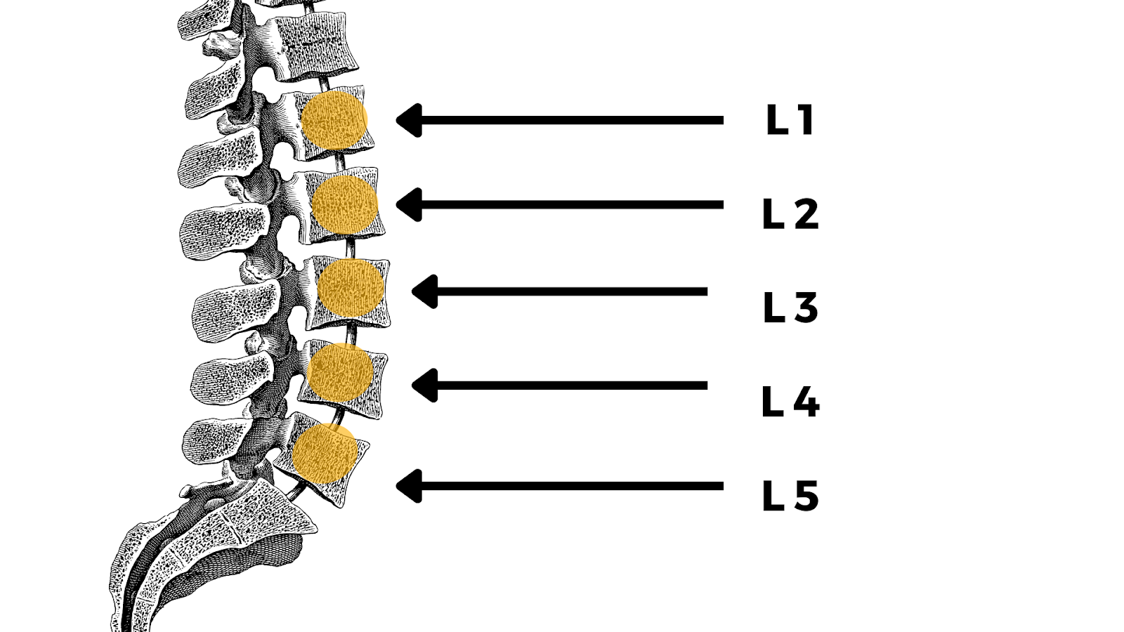
L1: The topmost section of the lumbar spinal column.
L2: Contains the end of the spinal cord proper. All vertebrae beyond this point only have the spinal nerves and not the spinal cord.
L3: The middle vertebra of the lumbar spine
L4: This is the second to last section of the lumbar spinal column
L5: The final section of the lumbar spine.
The lumbar being sprained or strained are the most common forms of back pain. The body can be over-exerted from specific situations such as heavy lifting or traumatic injury. It can also be affected with age, where the spinal discs are more susceptible to cracks and tears over time. The sprain or strain causes pain in the muscles of the back, which may cause a burning or aching sensation.
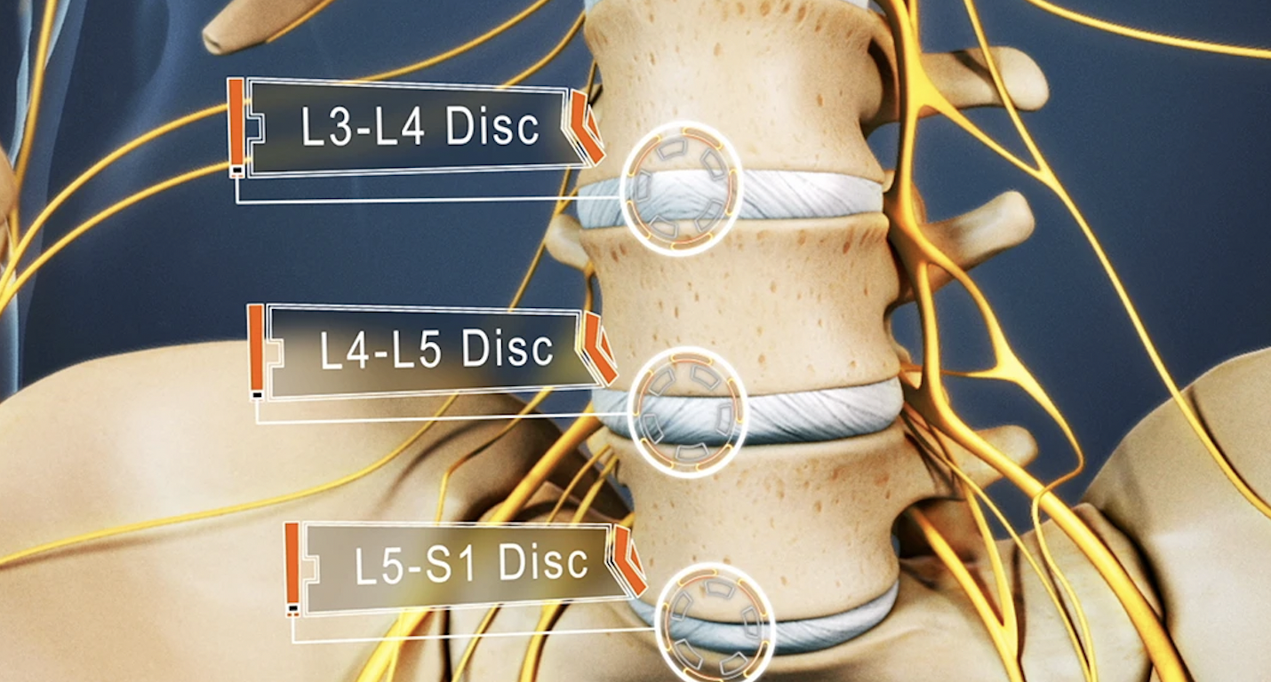
L4 and L5 are the fourth and fifth vertebrae in the lumbar spine, respectively. The lumbar spine is located in the lower back and consists of five vertebrae. The L4 and L5 are the two lowest vertebrae of the lumbar spine.
Together with the intervertebral disc, joints, nerves, and soft tissues, the L4-L5 spinal motion segment provides various functions, including and not limited to supporting the upper body and allowing trunk motion in multiple directions.
Because of its heavy load-bearing function and the wide range of flexibility required, L4-L5 is usually more susceptible to developing degenerative changes and/or pain when compared to other lumbar segments.
The L4-L5 spinal motion segment consists of the following structures:
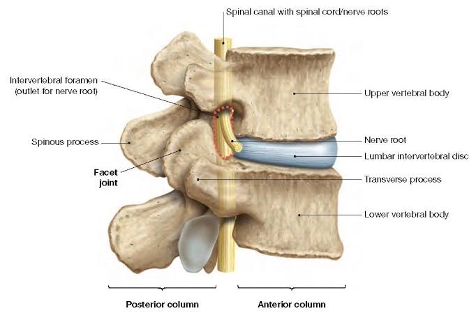
Lamina is the region between the spinous process and the transverse process, and the pedicle is the region between the transverse process and the vertebral body. The vertebrae are joined by facet joints, and these are covered by articulating cartilage to ensure smooth movements amidst the joint surfaces.
L4 and L5 vertebral bodies are taller in front than they are behind. To help resist compressive loads placed on the spine, the upper and lower ends of each vertebral body are covered by bony endplates.
The discs protect the spinal vertebrae and nerves from sudden impact. They also absorb shock from movements of the spine like bending, twisting, and jumping.
The L4 dermatome is an area of skin that receives sensations through the L4 spinal nerve and includes parts of the leg, thigh, knee, and foot. The L4 myotome is a group of muscles controlled by the L4 spinal nerve and consists of several muscles in the pelvis, leg, back, thigh, and foot. The L4-L5 motion segment provides a bony enclosure for the cauda equina (nerves that continue down from the spinal cord) and other delicate structures.
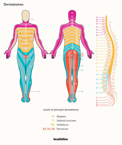
The L5 nerve root is most commonly affected in an L4-L5 disc herniation. The L5 nerve root exits the spine at the level of the L4-L5 vertebrae and innervates (supplies sensation to) the muscles of the thigh and knee, as well as the skin over the front and side of the thigh.
A herniated disc at this level can cause pain, numbness, or weakness in the thigh, knee, and leg.
There are 23 intervertebral discs in the spinal column that protect the spinal vertebrae and nerves from sudden impact. They also absorb shock from movements of the spine like bending, twisting, and jumping.
Unfortunately, the disc's outer wall, the annulus fibrosus, can develop traumatic tears (annular tears), allowing the jelly-like nucleus pulposus to push backward out of the tear into the spinal canal or neural foramen.
The part of the jelly nucleus pulposus that pushes out through the tear is called the herniation. In many cases, this hernia can impinge on a nerve, giving rise to inflammation and irritation of the affected nerve.
Disc herniation is caused by several factors which can affect the joints individually or as a combination. The most prevalent of these factors is wear and tear in the spine. As humans age, the cartilage that connects the discs in the spine to the corresponding vertebrae members can become lax and lose elasticity. Herniated discs can also be caused by sudden impact and trauma from accidents or falls.
In the lumbar spine, a common symptom associated with a herniated disc is sciatica (also known as leg pain). A herniated disc at L4-L5 usually causes L5 nerve impingement. And in addition to the sciatica pain, this type of herniated disc, also leads to weakness when raising the big toe and possibly in the ankle, otherwise known as foot drop. Others include:
Nonsurgical treatments are usually used to treat pain and minor symptoms in the L4-L5 spinal mobility segments. In extreme cases, patients might develop neurological impairments, such as paralysis and bowel/bladder control loss. When such symptoms are observed, surgery becomes a necessary course of action.
One of the most common non-surgical means of herniated disc treatment is lumbar traction. Traction is a pain-relieving technique that involves stretching and realigning the spine. A therapist can do the stretch manually or with the use of spinal traction equipment. This treatment relieves spinal nerve compression by opening up the neuroforamina.
If nonsurgical methods fail to provide relief from the pain, your doctor may recommend surgery. There are different surgery options available for patients who have been diagnosed with herniated discs.
The surgical treatment selection typically depends on two factors; the associated symptoms and severity of the patient’s injury. These options range from minimally invasive surgical procedures to artificial disc replacements or spinal fusion in extreme cases.
When considering surgery, it is important to note that surgery usually involves reducing the compression on the nerve and eliminating the pain caused by the herniated disc. Patients should be aware that some surgical options carry more risk of complications than others. These complications could include infection or excessive blood loss and often tend to compromise the integrity of the spine and general patient wellbeing.
Before a procedure is selected, patients are advised to consult a spine expert to conduct a full MRI scan and review, and allow for a thorough examination and subsequent localized diagnosis.
The most advanced surgery in the world for a herniated disc is the Deuk Laser Disc Repair.
This revolutionary procedure is Deuk Spine Institute’s specialized alternative to dangerous invasive surgeries like spinal fusion or total disc replacement. The laser disc repair does not weaken or compromise the health and integrity of the spine.
Our modernized approach to laser spine surgery has a 95% success rate with no complications in any patient over 15 years of performing this procedure and over 1,300 patients treated.
Deuk Laser Disc Repair surgery is a form of endoscopic spine surgery performed in our state-of-the-art surgery center under sedation while the patient relaxes. Through a ¼-inch incision, the injured disc is visualized using an endoscope and live imaging via a high-definition camera attached to the spinal endoscope.
With this method, Dr. Deukmedjian carefully eliminates only the injured disc tissue causing pain and discomfort and leaves the rest of the patient’s own natural disc in place to preserve spinal motion and function. Fusions and artificial discs are not necessary because the patient's repaired natural disc is left in place.
Deuk Laser Disc Repair uses a precision laser to vaporize the herniated tissue and provide the most effective laser spine surgery available. Bone and surrounding tissues are not damaged or removed during this procedure, unlike traditional microdiscectomy, artificial discs and spinal fusions.
Dr. Ara Deukmedjian uses FDA-approved tools to access the disc through a natural space in the spine where he does not drill through bone as is done with microdiscectomy. Drilling through bone weakens the spine, which leads to future complications that may require fusion surgery.
Once the herniation and annular tear have been gently vaporized, the body can heal naturally. Irritation around the spine decreases, and neurological symptoms from nerve root pressure subside. In time, the disc functions as it did before injury and herniation.
After surgery, patients wake up to immediate relief and a surgical scar so small the surgeon can cover it with a Band-Aid. Only a few drops of blood are lost and no hospitalization is required. All 1,300 Deuk Laser Disc Repair surgeries done to date have been outpatient with a 1-hour recovery.
Physical therapy is one of the most suitable ways to improve the condition of the lumbar spine. Sessions should always be conducted with a qualified physiotherapist to avoid worsening the injury and pain.
However, such a course of treatment should only be undergone when the patient has no underlying medical conditions or dilapidating spinal injury.
When physical therapy is determined to be suitable, methods like ice packs, heat therapy, massage therapy, ultrasound, electrotherapy, and other passive therapeutic modalities are offered.
Passive physical therapy is to aid in the reduction of pain and edema. Active physical therapy can also be incorporated into the treatment regimen to improve the strength of the lumbar spine.
These mainly include:
Lumbar disc conditions can be diagnosed through X-Ray, Magnetic Resonance Imaging (MRI), Myelogram, Computed Tomography Scan (CT or CAT scan), and Electromyography (EMG).
At Deuk Spine Institute, we specialize in minimally invasive surgical techniques and comprehensive spine treatments to cure back and neck pain. Our world-class physicians are personally invested in the well-being of every patient.
Start your treatment with us today by submitting your MRI online for a free remote review to determine your candidacy for surgery.